Article

Aerobic Cytotoxicity of Aromatic N-Oxides: The Role of NAD(P)H:Quinone Oxidoreductase (NQO1)

by
Aušra Nemeikaitė-Čėnienė
1,
Jonas Šarlauskas
2,
Lina Misevičienė
2,
Audronė Marozienė
2,
Violeta Jonušienė
3,
Mindaugas Lesanavičius
2 and
Narimantas Čėnas
2,*
1
State Research Institute Center for Innovative Medicine, Santariškių St. 5, LT-08406 Vilnius, Lithuania
2
Institute of Biochemistry of Vilnius University, Saulėtekio 7, LT-10257 Vilnius, Lithuania
3
Institute of Biosciences of Vilnius University, Saulėtekio 7, LT-10257 Vilnius, Lithuania
*
Author to whom correspondence should be addressed.
Int. J. Mol. Sci. 2020, 21(22), 8754; https://doi.org/10.3390/ijms21228754
Submission received: 20 October 2020 / Revised: 13 November 2020 / Accepted: 18 November 2020 / Published: 19 November 2020
(This article belongs to the Section Molecular Toxicology)

Abstract

:
Derivatives of tirapazamine and other heteroaromatic N-oxides (ArN→O) exhibit tumoricidal, antibacterial, and antiprotozoal activities, which are typically attributed to bioreductive activation and free radical generation. In this work, we aimed to clarify the role of NAD(P)H:quinone oxidoreductase (NQO1) in ArN→O aerobic cytotoxicity. We synthesized 9 representatives of ArN→O with uncharacterized redox properties and examined their single-electron reduction by rat NADPH:cytochrome P-450 reductase (P-450R) and Plasmodium falciparum ferredoxin:NADP+ oxidoreductase (PfFNR), and by rat NQO1. NQO1 catalyzed both redox cycling and the formation of stable reduction products of ArN→O. The reactivity of ArN→O in NQO1-catalyzed reactions did not correlate with the geometric average of their activity towards P-450R- and PfFNR, which was taken for the parameter of their redox cycling efficacy. The cytotoxicity of compounds in murine hepatoma MH22a cells was decreased by antioxidants and the inhibitor of NQO1, dicoumarol. The multiparameter regression analysis of the data of this and a previous study (DOI: 10.3390/ijms20184602) shows that the cytotoxicity of ArN→O (n = 18) in MH22a and human colon carcinoma HCT-116 cells increases with the geometric average of their reactivity towards P-450R and PfFNR, and with their reactivity towards NQO1. These data demonstrate that NQO1 is a potentially important target of action of heteroaromatic N-oxides.

1. Introduction

N-oxides of 1,2,4-benzotriazine, quinoxaline, and phenazine (ArN→O) possess promising antitumor, antiprotozoal, and antibacterial activities, including their potential application as hypoxia-specific antitumor agents ([1,2,3,4,5,6], and references therein). In most cases, their action is attributed to the enzymatic reduction and free radical generation. Among their representatives, the reactions of 3-amino-1,2,4-benzotriazine-1,4-dioxide (tirapazamine, TPZ) and its derivatives have been studied most comprehensively. TPZ (a) is enzymatically reduced in a single-electron way to a free radical (b), which forms DNA-damaging species under hypoxic conditions, namely, an oxidizing hydroxyl radical (OH.) ([2,7,8], and references therein), and/or a highly reactive benzotriazinyl radical (c) that abstracts a hydrogen atom from DNA ([9,10], and references therein (Scheme 1). The nature of DNA-damaging species remains a matter of debate. The final relatively nontoxic metabolites of TPZ are its mono-N-oxide (d), formed possibly via free radical (c) intermediate, and its nor-oxide (f) [2,7,11]. Their formation is strongly inhibited under aerobic conditions.
The aerobic cytotoxicity of ArN→O is typically attributed to their redox cycling (Scheme 1), leading to the formation of superoxide (O2) and subsequent oxidative stress [1,2]. This phenomenon deserves certain interest because some TPZ analogs possess anticancer activity at micromolar concentrations even under oxic conditions [12,13,14], and/or are toxic to normal tissues [15]. Besides, the oxidative stress-type mammalian cell cytotoxicity may be important as a side effect in the antimicrobial and antiparasitic action of ArN→O ([2,6], and references therein). Flavoenzyme NADPH:cytochrome P-450 reductase (P-450R) and/or insufficiently characterized intranuclear NAD(P)H-oxidizing flavoenzymes are assumed to be mainly responsible for the single-electron reduction of ArN→O irrespectively of hypoxic or oxic conditions ([16,17], and references therein). Another enzyme, potentially relevant to the cytotoxicity of ArN→O, is NAD(P)H:quinone oxidoreductase (NQO1, DT-diaphorase). NQO1 is a dimeric enzyme containing one molecule of FAD per subunit, located mainly in the cytosol [18]. It reduces quinones and nitroaromatic compounds in an obligatory two-electron way ([19,20], and references therein), and is considered to be a target of bioreductively-activated quinone antitumor agents [21]. NQO1 reduces TPZ and its derivatives much slower than quinones in a mixed single- and the two-electron way [22]. The data on the impact of NQO1 on cytotoxicity of TPZ are controversial [22,23,24,25]. However, the studies of its reactions with ArN→O deserve certain interest because the activity of NQO1 is frequently elevated in various tumors ([26], and references therein).
The aerobic cytotoxicity of several ArN→O in MH22a mouse hepatoma cells roughly increased with their single-electron reduction midpoint potential (E17) [22], which demonstrates an involvement of P-450R and/or other single-electron transferring flavoenzymes. In parallel, the cytotoxicity was decreased by dicoumarol, which points to the possible participation of NQO1 in cytotoxic events. In this work, we extended these studies using a series of previously unexplored aromatic N-oxides and have shown that NQO1 unequivocally contributes to the cytotoxicity of ArN→O.

2. Results

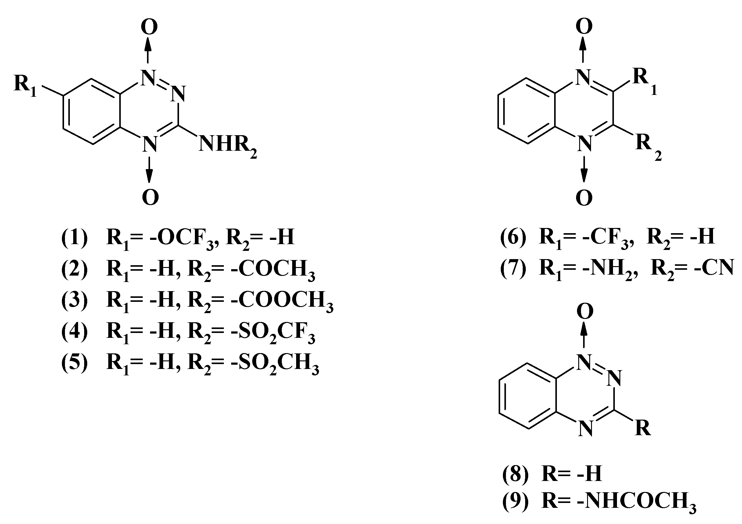
2.1. Reactions of Aromatic N-Oxides with Flavoenzymes Dehydrogenases-Electrontransferases

Previously we studied the enzymatic reactions and cytotoxicity of a number of ArN→O, mostly 7-substituted tirapazamine derivatives, whose E17 varied between −0.318 V and −0.575 V [22]. In this work, we extended these studies using representatives of several groups of ArN→O with uncharacterized values of E17 (Figure 1).
Table 1 contains bimolecular rate constants (kcat/Km) of reduction of examined ArN→O by P-450R and Plasmodium falciparum ferredoxin:NADP+ oxidoreductase (PfFNR), which was used as a model single-electron transferring enzyme [27]. For the most active oxidants of P-450R studied in this work, 7-CF3O-tirapazamine, and 3-CH3CONH- and 3-CH3OCONH-substituted 1,2,4-benzotriazine-1,4-dioxides (Table 1), the kcat at their saturated concentrations were in the range of 16.0–18.0 s−1, i.e., close to 50% of the rate of cytochrome c reduction by P-450R. The above compounds were also the most active oxidants of PfFNR (Table 1), with kcat of their reduction reaching 11.0–13.0 s−1. In other cases, the reaction rates were proportional to the concentration of compounds up to the limits of their solubility, 300–600 µM. Because PfFNR does not reduce cytochrome c directly [27], it was possible to demonstrate the redox cycling of synthesized ArN→O monitoring the reduction of cytochrome c added to the reaction mixture. The rates of this reaction were equal to 175–190% NADPH oxidation rate. The cytochrome c reduction was inhibited by 20–40% by 100 U/mL superoxide dismutase (SOD), which points to the formation of superoxide.
Typically, the logarithms of kcat/Km of single-electron reduction of ArN→O by flavoenzymes dehydrogenases-electrontransferases such as P-450R or PfFNR linearly increase with E17 [22,27]. This is attributed to an “outer-sphere” electron transfer mechanism, where the reactivity of compounds is insignificantly influenced by their structural peculiarities [28]. Therefore, log kcat/Km of homologous oxidants in single-electron enzymatic reduction reaction may serve as the parameter characterizing their E17, i.e., the energetic of single-electron reduction. The use of the geometric average of kcat/Km obtained in several enzymatic systems improves the prediction accuracy [29,30]. Table 1 contains the logarithms of geometric averages of kcat/Km of ArN→O in P-450R- and PfFNR-catalyzed reactions (log kcat/Km (avge) = 0.5 log kcat/Km (P-450R) + 0.5 log kcat/Km (PfFNR)). For comparison, the reduction rate constants of compounds 1018 obtained in our previous studies [22,27] are also presented (Table 1). The log kcat/Km (avge) of these compounds correlates well with their E17 values:
log kcat/Km (avge) = (7.23 ± 0.29) + (6.96 ± 0.65) E17, r2 = 0.9430.
A similar correlation is obtained including the assumed E17 values for compounds 6,8 (Table 1):
log kcat/Km (avge) = (7.24 ± 0.27) + (6.97 ± 0.59) E17, r2 = 0.9400.
For compounds with unavailable E17 values, the introduction of electron-accepting substituents into 3-NH2 group or into 7- or 2-positions of the aromatic system in most cases increased their log kcat/Km (avge) as compared with parent compounds (Table 1). Therefore, in this case, log kcat/Km (avge) appears to be a suitable parameter describing the ease of single-electron reduction of ArN→O.
Table 1. The single-electron reduction midpoint potentials (E17) of aromatic N-oxides, the steady-state bimolecular rate constants (kcat/Km) of their reduction by P-450R and PfFNR, and the logs of the geometric averages of their reactivity (log kcat/Km (avge)).
No.CompoundE17 (V) akcat/Km (M−1·s−1) log kcat/Km (avge)
P-450RPfFNR
17-CF3O-tirapazamine 4.6 ± 0.4 × 1043.8 ± 0.4 × 1044.62
23-CH3CONH-1,2,4-benzotriazine-1,4-dioxide 7.0 ± 0.5 × 1046.2 ± 0.5 × 1044.82
33-CH3OCONH-1,2,4-benzotriazine-1,4-dioxide 8.0 ± 0.9 × 1044.8 ± 0.4 × 1044.78
43-CF3SO2NH-1,2,4-benzotriazine-1,4-dioxide 2.5 ± 0.3 × 1042.6 ± 0.3 × 1044.41
53-CH3SO2NH-1,2,4-benzotriazine-1,4-dioxide 2.7 ± 0.3 × 1037.9 ± 0.5 × 1033.67
62-CF3-quinoxaline-1,4-dioxide(−0.465) b2.7 ± 0.3 × 1048.9 ± 0.7 × 1034.19
72-NH2-3-CN-quinoxaline-1,4-dioxide 4.7 ± 0.4 × 1031.8 ± 0.2 × 1043.96
81,2,4-Benzotriazine-1-oxide(−0.431) b1.7 ± 0.2 × 1044.3 ± 0.3 × 1033.94
93-CH3CONH-1,2,4-benzotriazine-1-oxide 8.7 ± 0.9 × 1031.6 ± 0.1 × 1033.58
ArN→O with available E17 values
101,2,4-Benzotriazine-1,4-dioxide−0.3184.3 ± 0.4 × 10⁵ c2.5 ± 0.3 × 10⁴ 5.00
117-CF3-tirapazamine−0.3458.7 ± 0.7 × 10⁴ c5.2 ± 0.4 × 10⁴ d4.83
127-Cl-tirapazamine−0.4006.9 ± 0.7 × 10⁴ c3.7 ± 0.4 × 10⁴ d4.71
137-F-tirapazamine−0.4003.4 ± 0.3 × 10⁴ c2.7 ± 0.2 × 10⁴ d4.48
14Tirapazamine−0.4551.1 ± 0.1 × 10⁴ c4.4 ± 0.5 × 10³ d3.84
157-CH3-tirapazamine−0.4748.6 ± 0.7 × 10³ c5.0 ± 0.6 × 10³ d3.82
167-C2H5O-tirapazamine−0.4944.5 ± 0.5 × 10³ c4.5 ± 0.5 × 10³ d3.65
173-Amino-1,2,4-benzotriazine-1-oxide−0.5682.8 ± 0.2 × 10³ c3.2 ± 0.2 × 10³ d3.48
18Quinoxaline-1,4-dioxide−0.5753.3 ± 0.2 × 10³ c8.2 ± 0.9 × 10² d3.22
aE17 of compounds taken from Ref. [7,31,32], b Calculated using the E17 differences between compounds 11 and 14, 0.110 V, and compounds 17 and 14, −0.113 V, respectively, c Taken from Ref. [22], d Taken from Ref. [27].

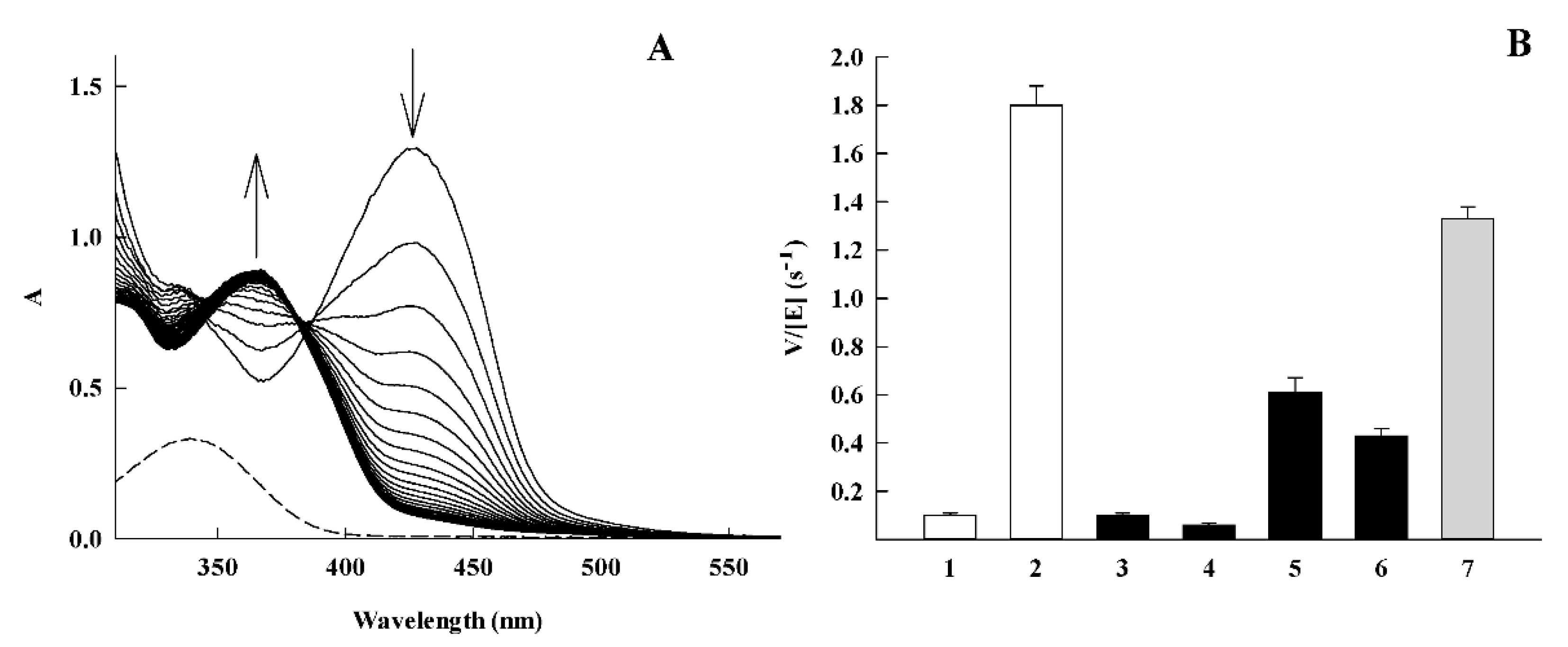
2.2. NQO1-Catalyzed Reduction of Aromatic N-Oxides

NQO1 performs a slow reduction of ArN→O [22,33]. The reactivity of compounds 19 determined in this work was similar to that of compounds 1018 [22], except for its larger variation (Table 2). The log kcat/Km for compounds 118 (Table 2) does not correlate with the energetics of their single-electron reduction, because of its dependence on E17 or log kcat/Km (avge) is described by r2 = 0.0451 and r2 = 0.1441, respectively. Because the reactivity of quinones towards NQO1 depends on their van der Waals volume (VdWvol) [19], we also used this parameter for calculations (Table 2). However, the combined use of log kcat/Km (avge) and VdWvol or VdWvol + (VdWvol)2 as independent variables also did not give satisfactory results, being described by r2 = 0.2302 and r2 = 0.3846, respectively. Thus, the structural requirements for the fast reduction of ArN→O by NQO1 remain unclear at this time.
Under anaerobic conditions, NQO1 reduces TPZ into its 4-monoxide (d) and nor-oxide (f) (Scheme 1) [33]. The reduction is strongly inhibited by O2 [33] and is accompanied by redox cycling process [22]. Because of the low rate of this reaction, we selected more reactive 3-CH3CONH-1,2,4-benzotriazine-1,4-dioxide (2) (Table 2) for subsequent studies. The temperature dependence of kcat/Km of reaction determined according to NADPH oxidation in the absence of activators gives the activation enthalpy (∆H) of 37.76 ± 1.93 kJ mol−1 and the activation entropy (∆S) of −42.97 ± 6.68 J mol−1 K−1. In the presence of the NADPH regeneration system under aerobic conditions, the disappearance of compound absorbance at 427 nm is accompanied by absorbance rise at 375 nm (Figure 2A). This shows that the possible reaction product is CH3CONH-1,2,4-benzotriazine-1-oxide (9) (Figure 1), which possesses absorbance maximum at this wavelength (data not shown). The more detailed characterization of reaction products is beyond the scope of the present work.
The data of Figure 2B show that the rate of 3-CH3CONH-1,2,4-benzotriazine-1,4-dioxide disappearance was equal to 70–75% NADPH oxidation rate determined in the absence of NADPH regeneration system. The rate of compound disappearance was directly proportional to NQO1 concentration in the range of 12.5–100 nM. The reaction was accompanied by the reduction of added cytochrome c with the rate being equal to 30–35% NADPH oxidation rate (Figure 2B). The reduction of cytochrome c was partly inhibited by superoxide dismutase, which demonstrates the formation of O2. During the reduction of (2) by PfFNR, the rate of added cytochrome c reduction was much higher, being equal to 180–190% NADPH oxidation rate. The cytochrome c reduction was inhibited by SOD by 20%.

2.3. Cytotoxicity of Aromatic N-Oxides

In cytotoxicity studies, we determined the concentrations of ArN→O for 50% cell survival (cL50) in murine hepatoma MH22a cells and their concentrations for 50% of maximal inhibition (GI50) of the proliferation of human colon adenocarcinoma HCT-116 cells (Table 3). The data of our previous study [22] are also presented for comparison.
We previously demonstrated the prooxidant character of cytotoxicity of ArN→O in MH22a cells [22]. In line with this, the cytotoxicity of several randomly selected ArN→O explored in this work, was decreased by desferrioxamine (DESF) and the antioxidant N,N′-diphenyl-p-phenylenediamine (DPPD), and enhanced by 1,3-bis(2-chloroethyl)-1-nitrosourea (BCNU), the latter inactivating glutathione reductase and depleting reduced glutathione [35] (Table 4). Besides, their cytotoxicity was decreased by an inhibitor of NQO1, dicoumarol (DIC) (Table 4).
In quantitative analysis of cytotoxicity of ArN→O, we used the ease of their single-electron reduction (log kcat/Km (avge), Table 1), lipophilicity (log D, Table 3)), and reactivity towards NQO1 (log kcat/Km (NQO1), Table 2) as the correlation parameters. The data were analyzed using the linear multiparameter regression:
log cL50 (or log GI50) = a + b log kcat/Km (avge) + c log D + d log kcat/Km (NQO1)
The calculated coefficients in Equation (3) (Table 5) show that the cytotoxicity of compounds in both cell lines roughly increases with their log kcat/Km (avge) when it is used as a single variable. The introduction of log kcat/Km (NQO1) as a second variable improved the correlations, and also unequivocally demonstrated that the ArN→O cytotoxicity increases with their reactivity towards NQO1 (Table 5).
In all cases (Table 5), the coefficient d characterizing an involvement of NQO1 (Equation (3)) is statistically significant (p < 0.05). The use of log D as an additional variable did not improve the correlations and demonstrated a limited impact of lipophilicity on ArN→O cytotoxicity (Table 5), which is similar to the data of a previous study [31].
The role of NQO1 in the cytotoxicity of ArN→O was additionally assessed using bovine leukemia virus-transformed lamb embryo kidney fibroblasts (line FLK [36]), murine embryonic liver cells (line BNL CL.2 [37]), and primary mice splenocytes [38] (Supplemental Information, Table S1). In this case, DIC protected against the cytotoxicity of TPZ and 3-CH3CONH-1,2,4-benzotriazine-1,4-dioxide in cell lines with a relatively high content of NQO1, but did not protect against their cytotoxicity in mice splenocytes with low NQO1 content, 4.0 nmol cytochrome c reduced × min−1 × mg−1 [38] (Table S2).

3. Discussion

In this study, we examined the relationship between enzymatic redox properties and aerobic cytotoxicity of aromatic N-oxides with a particular emphasis on the role of NQO1. The rate constants of single-electron reduction of ArN→O 1–9 by P-450R and PfFNR (Table 1) are in line with previous findings [22,27], showing that the reactivity is mostly determined by the electron-accepting potency of ArN→O, and hardly depend on their structural peculiarities. Their log kcat/Km (avge) proved to be a reasonable substitute for E17 because of their good interrelationship (Equations (1) and (2)). Interestingly, its use for the description of ArN→O cytotoxicity (Equation (3), Table 5) resulted in r2 values close to those in previously obtained log (cytotoxicity) vs. E17 relationships [31]. On the other hand, the use of a large group of ArN→O with highly variable and reduction potential-independent reactivity towards NQO1 (Table 2), enabled us to characterize the role of this enzyme in their cytotoxicity.
In regards to the possible mechanism of reduction of ArN→O by NQO1, one may note their structural similarity with 1,4-naphthoquinones, which are planar bicyclic molecules with similar electron self-exchange rate constants, ~108 M−1 s−1 [22,39]. The available data on the mechanism of quinone reduction are summarized below: (i) a net two-electron reduction of quinones by NQO1 is attributed to low stability of its FAD semiquinone (FAD), 8% under equilibrium [40], and the efficient electronic coupling, which is provided by the sandwiching of quinone ring between isoalloxazine ring of FAD and Phe-178’ [41]; (ii) The instability of FAD does not restrict the reactivity of NQO1 with single-electron acceptors [19]. NQO1 reduces nitroaromatic compound tetryl in a mixed single- and two-electron way [42], and its reactions with quinones may be described by a multistep (e,H+,e) hydride transfer model [19]; (iii) The reactivity of quinones roughly increases with their E17 and sharply decreases when their VdWvol is above 200 Å3 [19]. An increase in VdWvol may increase the distance between N5 of isoalloxazine and quinone ring [43], and (iv) The ΔS values of ‘fast’ quinone oxidants with VdWvol ≤ 200 Å3 are lower than ΔS of ‘slow’ quinones (VdWvol > 200 Å3) by 40–70 J mol−1 K−1 [19]. More negative ΔS values point to the stronger electronic coupling between the molecules of reactants [44].
In our case, NQO1 reduces ArN→O 102–103 times slower than 1,4-naphthoquinones with comparable E17 and VdWvol values, −0.36–−0.46 V, and 180–200 Å3, respectively [19]. The use of a relatively fast NQO1 substrate, 3-CH3CONH-1,2,4-benzotriazine-1,4-dioxide (2) (Table 2, Figure 2A,B), enabled the characterization of this process more thoroughly. The SOD-sensitive reduction of added cytochrome c during the reaction (Figure 2B) points to the formation of a free radical ((b), Scheme 1), which enters the redox equilibrium with O2/O2 couple. In our opinion, the lower rate of ArN→O reduction when compared to quinones and the dissociation of free radicals may be caused by a weak electronic coupling of reactants. The electronic coupling may be weakened by the loss of planarity of (b) due to the adoption of markedly pyramidal geometries of N1 and N4 in a free radical state [45]. This is supported by ∆S value of 3-CH3CONH-1,2,4-benzotriazine-1,4-dioxide, -43 J mol−1 K−1, which is far less negative than ΔS of ‘fast’ quinone oxidants, −85–−75 J mol−1 K−1 [19]. However, the yield of free radicals in NQO1-catalyzed reduction of this compound, expressed as the ratio of cytochrome c reduction and doubled NADPH oxidation rates [27], is close to 30% (Figure 2B). This is much lower than the single-electron flux of 90–95%, observed in the reaction with single-electron transferring PfFNR. In parallel, 3-CH3CONH-1,2,4-benzotriazine-1,4-dioxide disappears in the course of the reaction (Figure 2A,B). Analogous although relatively slower process takes place during the reduction of TPZ by NQO1 [22]. The sum of this reaction rate and the halved rate of cytochrome c reduction is close to the total rate of NADPH oxidation (Figure 2B). The disappearance of oxidant is not caused by free radical dismutation, because its rate exhibits a linear, but not a square dependence on the enzyme concentration that is characteristic for the dismutation-driven process [46]. Thus, the possible explanation is that NQO1 performs mixed single- and two-electron reduction of ArN→O with the initial single-electron transfer step. The elucidation of the mechanism of a second electron transfer is more problematic because of debates whether the 1,4-dioxide (a) reduction product, 1-monoxide (d), is formed from free radical (b) directly, or via aryl radical (c) (Scheme 1) [2,7,8,9,10,11]. This problem is an object of our future studies, together with the detailed characterization of the reaction product(s).
The multiparameter regression analysis (Equation (3), Table 3 and Table 5) provides the first quantitative confirmation of NQO1 contribution to the cytotoxicity of ArN→O in two cell lines. Given the data currently available, the most credible mechanism of their action is NQO1-catalyzed redox cycling. Apart from MH22a cells (Table 4), dicoumarol protected against TPZ aerobic cytotoxicity in A549 cells, besides, the TPZ-resistant A549 subline almost completely lost NQO1 activity [24]. On the other hand, there also exist data on the absence of the relationship between the content of NQO1 and the cytotoxicity of TPZ in several cell lines [23,25]. Because P-450R and/or nuclear reductases typically play the most significant role in the bioactivation of TPZ [16,17], it is possible that in certain cases their action may shield the role of NQO1. One may also expect that the role of NQO1 may be more expressed using its substrates more efficient than TPZ and especially possessing lower E17 values, i.e., lower reactivity towards P-450R (Table 1 and Table 2). In conclusion, our data indicate that NQO1 may be an important target of ArN→O in the cell. The further studies of electron transfer mechanism and substrate specificity of these reactions may increase the variety of efficient therapeutic agents of this group, and contribute to the optimization of their action.

4. Materials and Methods

4.1. Enzymes and Chemicals

Recombinant rat P-450R was a generous gift of Dr. Alexey Yantsevich (Institute of Bioorganic Chemistry, NAS of Belarus, Minsk, Belarus), and was prepared as described in [47]. Recombinant P. falciparum ferredoxin:NADP+ oxidoreductase (PfFNR) was a generous gift of Professor Alessandro Aliverti (Department of Biosciences, Universita degli Studi di Milano, Milano, Italy), and was prepared as previously described in [48]. NQO1 was prepared from rat liver according to Prochaska [49]. The enzyme concentrations were determined spectrophotometrically according to ε456 = 21.4 mM−1·cm−1 (P-450R [47]), ε454 = 10.0 mM−1.cm−1 (PfFNR [48]), and ε460 = 11.0 mM−1·cm−1 (NQO1 [49]). Derivatives of tirapazamine (1–5) (Figure 1) were synthesized as described in [31,50]. Quinoxaline-1,4-dioxide and its derivatives (6,7) (Figure 1) were synthesized as described in [51,52]. 1,2,4-Benzotriazine-1,4-dioxide and derivatives of 1,2,4-benzotriazine-1-oxide (8,9) (Figure 1) were synthesized as described in [53,54]. The compound purity was characterized by IR and NMR spectrometry, melting point, and elemental analysis. NADPH, cytochrome c, superoxide dismutase, and other reagents were obtained from Sigma-Aldrich (St. Louis, MO, USA), and used as received.

4.2. Enzymatic Assays

The kinetic measurements were carried out spectrophotometrically using a PerkinElmer Lambda 25 spectrophotometer (PerkinElmer, Waltham, MA, USA) in 0.1 M K-phosphate buffer (pH 7.0) containing 1 mM EDTA at 25 °C. The enzyme activities determined according to the rate of reduction of 50 µM cytochrome c (∆ε550 = 20 mM−1·cm−1) at substrate concentrations indicated below were close to those reported previously [22]: 39 s−1 (P-450R, [NADPH] = 100 µM), and 1750 s−1 (NQO1, [NADPH] = 150 µM, [menadione] = 10 µM). In this case, 0.01% Tween 20 and 0.25 mg/mL bovine serum albumin were added as NQO1 activators. The activity of PfFNR, determined according to the reduction rate of 1.0 mM ferricyanide (∆ε420 = 1.03 mM−1.cm−1) at [NADPH] = 100 µM was equal to 48 s−1. The initial rates of P-450R- or PfFNR-catalyzed NADPH-dependent N-oxide reduction were determined according to ∆ε340 = 6.2 mM−1·cm−1 after the subtraction of intrinsic NADPH oxidase activities of enzymes, 0.05 s−1 (P-450R), and 0.1 s−1 (PfFNR). The stock solutions of oxidants were prepared in DMSO (dilution factor 100). The initial rates of NQO1-catalyzed ArN→O reduction were determined according to the rate of NADPH oxidation. In this case, the rates were corrected for 340 nm absorbance changes due to ArN→O disappearance. The latter were obtained under the same conditions in the presence of NADPH regeneration system, 10 mM glucose-6-phosphate and 0.3 mg/mL glucose-6-phosphate dehydrogenase. The loss of 3-CH3CONH-1,2,4-benzotriazine-1,4-dioxide during its reduction by NQO1 was monitored according to ∆ε427 = 6.45 mM−1.cm−1, which was calculated according to the absorbance difference of compounds (2) and (9) (Figure 1). The values of turnover rate, kcat, reflecting the maximal number of moles NADPH oxidized or oxidant reduced per mole of the enzyme active center per second, and kcat/Km, the bimolecular rate constant (or catalytic efficiency constant), corresponds to the inverse intercepts and slopes in Lineweaver-Burk coordinates, [E]/v vs. 1/[oxidant]. These rate constants were obtained by fitting the experimental data to the parabolic expression using the SigmaPlot 2000 (version 11.0, Systal Software, San Jose, CA, USA). The temperature dependence of reaction rate was examined at seven fixed temperatures between 15 and 45 °C in the absence of activators, the activation enthalpies and entropies of reaction were calculated from Eyring plots of ln (kcat/Km)/T vs. 1/T.

4.3. Cytotoxicity Assays

Murine hepatoma MH22a cells obtained from the Institute of Cytology of the Russian Academy of Sciences (St. Petersburg, Russia), were grown and maintained at 37 °C in DMEM medium, supplemented with 10% fetal bovine serum, 100 U/mL penicillin, and 0.1 mg/mL streptomycin, as described in [22]. In the cytotoxicity experiments, 3.0 × 10⁴/mL cells were seeded in 5-mL flasks either in the presence or in the absence of compounds and were grown for 24 h. In the absence of compounds, cells reached 40–50% confluence. The adherent cells were counted under a light microscope. Typically, they did not accumulate Trypan blue and their viability was 98.5–99.3%. Human colon adenocarcinoma cells HCT-116 obtained from ATCC (Manassas, VA, USA), were grown and maintained at 37 °C in 5% CO2 in RPMI 1640 DMEM medium, supplemented with 10% fetal bovine serum, 2 mM l-glutamine, and 0.05 mg/mL gentamycin. In the cytotoxicity experiments, 1.0 × 10⁵/mL cells were seeded in the absence or the presence of compounds and were grown for 48 h. In the absence of compounds, cells reached 65–75% confluence. Their viability was determined by staining with crystal violet [55]. Stock solutions of compounds were prepared in DMSO. Its concentration in cultivation media did not exceed 0.2% and did not affect cell viability. The experiments were conducted in triplicate.

4.4. Statistical Analysis and Calculations

The statistical analysis was performed using Statistica (version 4.3, Statsoft, Toronto, ON, Canada). Octanol/water distribution coefficients at pH 7.0 (log D) were calculated using LogD Predictor (https://chemaxon.com).

Supplementary Materials

Supplementary materials can be found at https://www.mdpi.com/1422-0067/21/22/8754/s1.

Author Contributions

A.N.-Č. and V.J. performed the cytotoxicity studies, J.Š. synthesized compounds, L.M., A.M., and M.L. performed kinetic experiments and purified NQO1, N.Č. designed and supervised the experiments and wrote the manuscript. All authors have read and agreed to the published version of the manuscript.

Funding

This work was supported by the European Social Fund (Measure No. 09.33-LMT-K-712, grant No. DOTSUT-34/09.3.3.-LMT-K712-01-0058/LSS-600000-58).

Acknowledgments

We thank Alexey Yantsevich and Alesandro Aliverti for their generous gift of purified P-450R and PfFNR, respectively, and Valė Miliukienė (Institute of Biochemistry) for mice splenocyte cytotoxicity studies.

Conflicts of Interest

The authors declare no conflict of interest.

Abbreviations

ArN→OHeteroaromatic N-oxide
BCNU1,3-bis(2-chloroethyl)-1-nitrosourea
cL50Concentration for 50% cell survival
DESFDesferrioxamine
DICDicoumarol
DPPDN,N′-diphenyl-p-phenylene diamine
E17Single-electron reduction midpoint potential at pH 7.0
GI50Concentration for 50% inhibition of maximal cell proliferation
kcatCatalytic constant
kcat/KmBimolecular rate constant
kcat/Km (avge) Geometric average of kcat/Km in P-450R- and PfFNR-catalyzed reactions
kcat/Km (NQO1)kcat/Km in NQO1-catalyzed reaction
log DOctanol/water distribution coefficient at pH 7.0
NQO1NAD(P)H:quinone oxidoreductase
P-450RNADPH:cytochrome P-450 reductase
PfFNRPlasmodium falciparum ferredoxin:NADP+ oxidoreductase
SODSuperoxide dismutase
TPZTirapazamine

References

  1. Wardman, P.; Dennis, M.F.; Everett, S.A.; Patel, K.B.; Stratford, M.R.L.; Tracy, M. Radicals from one-electron reduction of nitro compounds, aromatic N-oxides and quinones: The kinetic basis for hypoxia-selective, bioreductive drugs. Biochem. Soc. Symp. 1995, 61, 171–194. [Google Scholar] [PubMed] [Green Version]
  2. Shen, X.; Gates, K.S. Enzyme-activated generation of reactive oxygen species from heterocyclic N-oxides under aerobic and anaerobic conditions and its relevance to hypoxia-selective prodrugs. Chem. Res. Toxicol. 2019, 32, 348–361. [Google Scholar] [CrossRef] [PubMed]
  3. Shah, Z.; Mahnuba, R.; Turcotte, B. The anticancer drug tirapazamine has antimicrobial activity against Escherichia coli, Staphylococcus aureus and Clostridium difficile. FEMS Microbiol. Lett. 2013, 347, 61–69. [Google Scholar] [CrossRef] [PubMed] [Green Version]
  4. Gil, A.; Pabon, A.; Galiano, S.; Burguete, A.; Perez-Silanes, S.; Deharo, E.; Monge, A.; Aldana, I. Synthesis, biological evaluation and structure-activity relationships of new quinoxaline derivatives as anti-Plasmodium falciparum agents. Molecules 2014, 19, 2166–2180. [Google Scholar] [CrossRef] [Green Version]
  5. Cheng, G.; Li, B.; Wang, C.; Zhang, H.; Liang, G.; Weng, Z.; Hao, H.; Wang, X.; Liu, Z.; Dai, M.; et al. Systematic and molecular basis of the antibacterial action of quinoxaline 1,4-di-N-oxides against Escherichia coli. PLoS ONE 2015, 10, e0136450. [Google Scholar] [CrossRef] [Green Version]
  6. Perez-Silanes, S.; Torrers, E.; Arbillaga, L.; Varela, J.; Cerecetto, H.; Gonzalez, M.; Azqueta, A.; Moreno-Viguri, E. Synthesis and biological evaluation of quinoxaline di-N-oxide derivatives with in vitro trypanocidal activity. Bioorg. Med. Chem. Lett. 2016, 26, 903–906. [Google Scholar] [CrossRef]
  7. Shen, X.; Rajapakse, A.; Galazzi, F.; Junnotula, V.; Fuchs-Knotts, T.; Glaser, R.; Gates, K.S. Isotopic labeling experiments that elucidate the mechanism of DNA strand cleavage by the hypoxia-selective antitumor agent 1,2,4-benzotriazine 1,4-di-N-oxide. Chem. Res. Toxicol. 2013, 22, 111–118. [Google Scholar] [CrossRef] [Green Version]
  8. Yadav, P.; Marshall, A.J.; Reynisson, J.; Denny, W.A.; Hay, M.P.; Anderson, R.F. Fragmentation of the quinoxaline N-oxide bond to the .OH radical upon one-electron bioreduction. Chem. Commun. 2014, 50, 13729–13731. [Google Scholar] [CrossRef]
  9. Shinde, S.S.; Maroz, A.; Hay, M.P.; Patterson, A.V.; Denny, V.A.; Anderson, R.F. Characterization of radicals formed following enzymatic reduction of 3-substituted analogues of the hypoxia-selective cytotoxin 3-amino-1,2,4-benzotriazine 1,4-dioxide (tirapazamine). J. Am. Chem. Soc. 2010, 132, 2591–2599. [Google Scholar] [CrossRef]
  10. Anderson, R.F.; Yadav, P.; Shinde, S.S.; Hong, C.R.; Pullen, S.M.; Reynisson, J.; Wilson, W.R.; Hay, M.P. Radical chemistry and cytotoxicity of bioreductive 3-substituted quinoxaline di-N-oxides. Chem. Res. Toxicol. 2016, 29, 1310–1324. [Google Scholar] [CrossRef]
  11. Fuchs, T.; Chowdhury, G.; Fuchs, C.L.; Gates, K.S. 3-Amino-1,2,4-benzotriazine 4-oxide: Characterization of a new metabolite arising from bioreductive processing of the antitumour agent 3-amino-1,2,4-benzotriazine 1,4-dioxide (tirapazamine). J. Org. Chem. 2001, 66, 107–114. [Google Scholar] [CrossRef] [PubMed]
  12. Zarranz, B.; Jaso, A.; Aldana, I.; Monge, A. Synthesis and anticancer activity evaluation of new 2-alkylcarbonyl and 2-benzoyl-1-trifluoromethyl-quinoxaline-1,4-di-N-oxide derivatives. Bioorg. Med. Chem. 2004, 12, 3711–3721. [Google Scholar] [CrossRef] [PubMed]
  13. Hu, Y.; Xia, Q.; Shangguan, S.; Liu, X.; Hu, Y.; Sheng, R. Synthesis and biological evaluation of 3-aryl-quinoxaline-2-carbonitrile 1,4-di-N-oxide derivatives as hypoxic selective anti-tumour agents. Molecules 2012, 17, 9683–9696. [Google Scholar] [CrossRef] [PubMed] [Green Version]
  14. Chowdhury, G.; Sarkar, U.; Pullen, S.; Wilson, W.R.; Rajapakse, A.; Fuchs-Knotts, T.; Dates, K.S. DNA strand cleavage by the phenazine di-N-oxide natural product myxin under both aerobic and anaerobic conditions. Chem. Res. Toxicol. 2012, 25, 197–206. [Google Scholar] [CrossRef]
  15. Gu, Y.; Chang, T.T.-A.; Wang, J.; Jaiswal, J.K.; Edwards, D.; Downes, N.J.; Liyanage, H.D.S.; Lynch, C.H.R.; Pruijn, F.B.; Hickey, A.J.R.; et al. Reductive metabolism influences the toxicity and pharmacokinetics of the hypoxia-targeted benzotriazine-dioxide anticancer agent SN30000 in mice. Front. Pharmacol. 2017, 8, 531. [Google Scholar] [CrossRef] [Green Version]
  16. Hunter, F.W.; Young, R.J.; Shalev, Z.; Vellanki, R.N.; Wang, J.; Gu, Y.; Joshi, N.; Sreebhavan, S.; Weinreb, J.; Goldstein, D.P.; et al. Identification of P450 oxidoreductase as a major determinant of sensitivity to hypoxia-activated prodrugs. Cancer Res. 2015, 75, 4211–4223. [Google Scholar] [CrossRef] [Green Version]
  17. Delahussaye, Y.M.; Evans, J.W.; Brown, J.M. Metabolism of tirapazamine by multiple reductases in the nucleus. Biochem. Pharmacol. 2001, 62, 1201–1209. [Google Scholar]
  18. Ross, D.; Siegel, D. NAD(P)H:quinone oxidoreductase I (NQO1, DT-diaphorase), functions and pharmacogenetics. Meth. Enzymol. 2004, 382B, 115–144. [Google Scholar]
  19. Anusevičius, Ž.; Šarlauskas, J.; Čėnas, N. Two-electron reduction of quinones by rat liver NAD(P)H: Quinone oxidoreductase: Quantitative structure-activity relationships. Arch. Biochem. Biophys. 2002, 404, 254–262. [Google Scholar] [CrossRef]
  20. Misevičienė, L.; Anusevičius, Ž.; Šarlauskas, J.; Čėnas, N. Reduction of nitroaromatic compounds by NAD(P)H:quinone oxidoreductase (NQO1): The role of electron-accepting potency and structural parameters in the substrate specificity. Acta Biochim. Pol. 2006, 53, 569–576. [Google Scholar] [CrossRef]
  21. Di Francesco, A.; Ward, T.; Butler, J. Diaziridinylbenzoquinones. Meth. Enzymol. 2004, 382B, 174–193. [Google Scholar]
  22. Nemeikaitė-Čėnienė, A.; Šarlauskas, J.; Jonušienė, V.; Marozienė, A.; Misevičienė, L.; Yantsevich, A.V.; Čėnas, N. Kinetics of flavoenzyme-catalyzed reduction of tirapazamine derivatives: Implications for their prooxidant cytotoxicity. Int. J. Mol. Sci. 2019, 20, 4602. [Google Scholar] [CrossRef] [PubMed] [Green Version]
  23. Plumb, J.A.; Gerritsen, M.; Workman, P. DT-diaphorase protects cells from the hypoxic cytotoxicity of indoloquinone EO9. Br. J. Cancer 1994, 70, 1136–1143. [Google Scholar] [CrossRef] [PubMed] [Green Version]
  24. Elwell, J.H.; Siim, B.G.; Evans, J.W.; Brown, J.M. Adaptation of human tumour cells to tirapazamine under aerobic conditions. Implications of increased antioxidant enzyme activity to mechanism of aerobic toxicity. Biochem. Pharmacol. 1997, 54, 249–257. [Google Scholar] [CrossRef]
  25. Sharp, S.Y.; Kelland, L.R.; Valenti, M.R.; Brunton, L.A.; Hobbs, S.; Workman, P. Establishment of an isogenic human colon tumor model for NQO1 gene expression: Application to investigate the role of DT-diaphorase in bioreductive drug activation in vitro and in vivo. Mol. Pharmacol. 2000, 58, 1146–1155. [Google Scholar] [CrossRef] [Green Version]
  26. Beaver, S.K.; Mesa-Tores, N.; Pey, A.L.; Timson, D.J. NQO1: A target for the treatment of cancer and neurological diseases, and a model to understand loss of function disease mechanism. Biochim. Biophys. Acta Proteins Proteom. 2019, 1867, 663–676. [Google Scholar] [CrossRef]
  27. Lesanavičius, M.; Aliverti, A.; Šarlauskas, J.; Čėnas, N. Reactions of Plasmodium falciparum ferredoxin: NADP+ oxidoreductase with redox cycling xenobiotics: A mechanistic study. Int. J. Mol. Sci. 2020, 21, 3234. [Google Scholar] [CrossRef]
  28. Marcus, R.A.; Sutin, N. Electron transfers in chemistry and biology. Biochim. Biophys. Acta 1985, 811, 265–322. [Google Scholar] [CrossRef]
  29. Čėnas, N.; Nemeikaitė-Čėnienė, A.; Sergedienė, E.; Nivinskas, H.; Anusevičius, Ž.; Šarlauskas, J. Quantitative structure-activity relationships in enzymatic single-electron reduction of nitroaromatic explosives: Implications for their cytotoxicity. Biochim. Biophys. Acta 2001, 1528, 31–38. [Google Scholar] [CrossRef]
  30. Šarlauskas, J.; Nivinskas, H.; Anusevičius, Ž.; Misevičienė, L.; Marozienė, A.; Čėnas, N. Estimation of single-electron reduction potentials (E17) of nitroaromatic compounds according to the kinetics of their single-electron reduction by flavoenzymes. Chemija 2006, 17, 31–37. [Google Scholar]
  31. Hay, M.P.; Gamage, S.A.; Kovacs, M.S.; Pruijn, F.B.; Anderson, R.F.; Patterson, A.V.; Wilson, W.R.; Brown, J.M.; Denny, W.A. Structure-activity relationships of 1,2,4-benzotriazine 1,4-dioxides as hypoxia-selective analogues of tirapazamine. J. Med. Chem. 2003, 46, 169–182. [Google Scholar] [CrossRef] [PubMed]
  32. Anderson, R.F.; Shinde, S.S.; Hay, M.P.; Denny, W.A. Potentiation of the cytotoxicity of the anticancer agent tirapazamine by benzotriazine-N-oxides. The role of redox equilibria. J. Am. Chem. Soc. 2006, 128, 245–249. [Google Scholar] [CrossRef] [PubMed]
  33. Cahill, A.; Jenkins, T.C.; White, I.N.H. Metabolism of 3-amino-1,2,4-benzotriazine-1,4-dioxide (SR 4233) by purified DT-diaphorase under aerobic and anaerobic conditions. Biochem. Pharmacol. 1993, 45, 321–329. [Google Scholar] [CrossRef]
  34. Zhao, Y.H.; Abraham, M.H.; Zissimos, A.M. Fast calculation of van der Waals volume as a sum of atomic and bond contributions and its application to drug compounds. J. Org. Chem. 2003, 68, 7368–7373. [Google Scholar] [CrossRef]
  35. Öllinger, K.; Brunmark, A. Effects of hydroxyl substituent position on 1,4-naphthoquinone toxicity to rat hepatocytes. J. Biol. Chem. 1991, 266, 21496–21503. [Google Scholar]
  36. Nemeikaitė, A.; Čėnas, N. The changes of prooxidant and antioxidant enzyme activities in bovine leukemia virus-transformed cells. Their influence on quinone cytotoxicity. FEBS Lett. 1993, 326, 65–68. [Google Scholar] [CrossRef] [Green Version]
  37. Liu, C.-H.; Chiu, T.-Y.; Hu, M.-L. Fucoxanthin enhances HO-1 and NQO1 expression in murine hepatic BNL CL.2 cells through activation of the Nrf2/ARE system partially by its prooxidant activity. J. Agric. Food Chem. 2011, 59, 11344–11351. [Google Scholar] [CrossRef]
  38. Miliukienė, V.; Nivinskas, H.; Čėnas, N. Cytotoxicity of anticancer aziridinyl-substituted benzoquinones in primary mice splenocytes. Acta Biochim. Pol. 2014, 61, 833–836. [Google Scholar] [CrossRef] [Green Version]
  39. Grampp, G.; Jaenicke, W. ESR-spectroscopic investigation of the parallel electron and proton exchange between quinones and their radicals: Part I. Measurements at 298 K. J. Electroanal. Chem. 1987, 229, 297–303. [Google Scholar] [CrossRef]
  40. Tedeschi, G.; Chen, S.; Massey, V. DT-diaphorase. Redox potential, steady-state, and rapid reaction studies. J. Biol. Chem. 1995, 270, 1198–1204. [Google Scholar] [CrossRef] [Green Version]
  41. Faig, M.; Bianchet, M.A.; Talalay, P.; Chen, S.; Winski, S.; Ross, D.; Amzel, L.M. Structures of recombinant human and mouse NAD(P)H:quinone oxidoreductases: Species comparison and structural changes with substrate binding and release. Proc. Natl. Acad. Sci. USA 2000, 97, 3177–3182. [Google Scholar] [CrossRef] [PubMed]
  42. Anusevičius, Ž.; Šarlauskas, J.; Nivinskas, H.; Segura-Aguilar, J.; Čėnas, N. DT-diaphorase catalyzes N-denitration and redox cycling of tetryl. FEBS Lett. 1998, 436, 144–148. [Google Scholar] [CrossRef] [Green Version]
  43. Mendoza, M.F.; Hollabaugh, N.M.; Hettiarachchi, S.U.; McCarley, R.L. Human NAD(P)H: Quinone oxidoreductase type I (hNQO1) activation of quinone propionic acid trigger groups. Biochemistry 2012, 51, 8014–8026. [Google Scholar] [CrossRef] [PubMed] [Green Version]
  44. Hubig, S.M.; Rathore, R.; Kochi, J.K. Steric control of electron transfer. Changeover from outer-sphere to inner-sphere mechanism in arene/quinone redox pairs. J. Am. Chem. Soc. 1999, 121, 617–626. [Google Scholar] [CrossRef]
  45. Yin, J.; Glaser, R.; Gates, K.S. Electron and spin-density analysis of tirapazamine reduction chemistry. Chem. Res. Toxicol. 2012, 25, 620–633. [Google Scholar] [CrossRef] [PubMed]
  46. Holzman, J.L.; Crankshaw, D.L.; Peterson, F.J.; Polnaszek, C.F. The kinetics of the aerobic reduction of nitrofurantoin by NADPH-cytochrome P450 (c) reductase. Mol. Pharmacol. 1981, 20, 669–673. [Google Scholar]
  47. Pechurskaja, T.A.; Harnastai, I.N.; Grabovec, I.P.; Gilep, A.A.; Usanov, S.A. Adrenodoxin supports reactions catalyzed by microsomal steroidgenetic cytochrome P450s. Biochem. Biophys. Res. Commun. 2007, 353, 598–604. [Google Scholar] [CrossRef]
  48. Balconi, E.; Pennati, A.; Crobu, D.; Pandini, V.; Cerutti, R.; Zanetti, G.; Aliverti, A. The ferredoxin-NADP+ reductase/ferredoxin electron transfer system of Plasmodium falciparum. FEBS J. 2009, 276, 2249–4260. [Google Scholar] [CrossRef]
  49. Prochaska, H.J. Purification and crystallization of rat liver NAD(P)H:quinone-acceptor oxidoreductase by cibacron blue affinity chromatography: Identification of a new and potent inhibitor. Arch. Biochem. Biophys. 1988, 267, 529–538. [Google Scholar] [CrossRef]
  50. Polmickaitė-Smirnova, E.; Šarlauskas, J.; Krikštopaitis, K.; Lukšienė, Ž.; Staniulytė, Z.; Anusevičius, Ž. Preliminary investigation on the antibacterial activity of antitumor drug 3-amino-1,2,4-benzotriazine-1,4-dioxide (tirapazamine) and its derivatives. Appl. Sci. 2020, 10, 4062. [Google Scholar] [CrossRef]
  51. Abushanab, E. Long-range hydrogen-fluorine spin-spin coupling. Further support for the “through-space” (direct) mechanism. J. Am. Chem. Soc. 1971, 93, 6532–6536. [Google Scholar] [CrossRef]
  52. Monge, A.; Palop, J.A.; Del Castillo, J.C.; Caldero, J.M.; Roca, J.; Romero, G.; Del Rio, J.; Lasheras, B. Novel antagonists of 5-HT3 receptors: Synthesis and biological evaluation of piperazinylquinoxaline derivatives. J. Med. Chem. 1993, 36, 2745–2750. [Google Scholar] [CrossRef] [PubMed]
  53. Robbins, R.F.; Schofield, K. Polyazabicyclic compounds. Part II. Further derivatives of benzo-1:2:4-triazine. J. Chem. Soc. 1957, 3186–3194. [Google Scholar] [CrossRef]
  54. Bodzioch, A.; Pomiklo, D.; Celeda, M.; Pietrzak, A.; Kaszynski, P. 3-Substituted benzo[e][1,2,4]triazines: Synthesis and electronic effects of the C(3) substituents. J. Org. Chem. 2019, 84, 6377–6394. [Google Scholar] [CrossRef]
  55. Ito, M. Microassay for studying anticellular effects of human interferons. J. Interferon. Res. 1984, 4, 603–608. [Google Scholar] [CrossRef] [PubMed]
Scheme 1. Pathways of the reduction of tirapazamine in the cell.
Ijms 21 08754 sch001
Figure 1. Formulae of heteroaromatic N-oxides used in this work: derivatives of tirapazamine (15), quinoxaline-1,4-dioxide (6,7), and 1,2,4-benzotriazine-1-oxide (8,9).
Ijms 21 08754 g001
Figure 2. Reduction of 3-CH3CONH-1,2,4-benzotriazine-1,4-dioxide (2) by NAD(P)H:quinone oxidoreductase (NQO1). (A) The spectral changes of 200 µM (2) in the presence of 50 nM NQO1, and 50 µM NADPH and NADPH regeneration system. The absorbance of NADPH is shown by a dashed line. The scans are recorded each 15 min. (B) The rates of NQO1-catalyzed oxidation of NADPH (1,2), reduction of cytochrome c (3–6), and depletion of 200 µM (2) in the presence of 200 µM NADPH (7). Additions: NADPH (1), NADPH + (2) (2,7), NADPH + cytochrome c (3), NADPH + cytochrome c + 100 U/mL SOD (4), NADPH + (2) + cytochrome c (5), NADPH + (2) + cytochrome c + SOD (6), n = 3, p < 0.01 for 3 against 4 and for 5 against 6.
Ijms 21 08754 g002
Table 2. The steady-state rate constants of reduction of aromatic N-oxides by NQO1 and their calculated van der Waals volumes (VdWvol).
No.Compoundkcat (s−1)kcat/Km (M−1.s−1)VdWvol (Å3) a
17-CF3O-tirapazamine 0.11 ± 0.021.6 ± 0.1 × 103175.8
23-CH3CONH-1,2,4-benzotriazine-1,4-dioxide3.40 ± 0.201.3 ± 0.1 × 104172.3
3.00 ± 0.20 b1.0 ± 0.1 × 10⁴ b
33-CH3OCONH-1,2,4-benzotriazine-1,4-dioxide2.40 ± 0.223.0 ± 0.2 × 103180.7
43-CF3SO2NH-1,2,4-benzotriazine-1,4-dioxide0.34 ± 0.061.0 ± 0.2 × 103203.5
53-CH3SO2NH-1,2,4-benzotriazine-1,4-dioxide0.40 ± 0.051.2 ± 0.2 × 103184.9
62-CF3-quinoxaline-1,4-dioxide13.5 ± 1.722.4 ± 0.2 × 104168.2
72-NH2-3-CN-quinoxaline-1,4-dioxide0.11 ± 0.021.4 ± 0.1 × 103146.2
81,2,4-Benzotriazine-1-oxide1.10 ± 0.122.1 ± 0.2 × 103111.7
93-CH3CONH-1,2,4-benzotriazine-1-oxide0.06 ± 0.025.0 ± 1.0 × 102163.5
101,2,4-Benzotriazine-1,4-dioxide c1.00 ± 0.105.1 ± 0.7 × 103120.5
117-CF3-tirapazamine c1.30 ± 0.103.7 ± 0.4 × 103167.0
127-Cl-tirapazamine c1.00 ± 0.104.3 ± 0.4 × 103152.6
137-F-tirapazamine c0.70 ± 0.104.9 ± 0.6 × 103143.5
14Tirapazamine c0.20 ± 0.102.4 ± 0.2 × 103131.5
157-CH3-tirapazamine c0.30 ± 0.051.7 ± 0.1 × 103148.8
167-C2H5O-tirapazamine c0.30 ± 0.041.0 ± 0.1 × 103174.9
173-Amino-1,2,4-benzotriazine-1-oxide c0.10 ± 0.021.6 ± 0.2 × 103122,7
18Quinoxaline-1,4-dioxide c0.60 ± 0.107.9 ± 0.9 × 103126.8
a Calculated according to [34], b Determined in the absence of activators, c Taken from Ref. [22].
Table 3. The logarithms of the geometric averages of ArN→O reactivity in P450R- and PfFNR-catalyzed reactions (log kcat/Km (avge)), their octanol/water distribution coefficients at pH 7.0 (log D), their concentrations for 50% cell survival (cL50) in murine hepatoma MH22a cells, and their concentrations causing 50% maximal proliferation inhibition (GI50) of human colon carcinoma HCT-116 cells.
No. Compoundlog kcat/Km (avge)log DcL50 (µM) MH22aGI50 (µM) HCT-116
17-CF3O-tirapazamine 4.621.323.6 ± 0.717 ± 3.0
23-CH3CONH-1,2,4-benzotriazine-1,4-dioxide4.82−0.821.5 ± 0.32.5 ± 0.5
33-CH3OCONH-1,2,4-benzotriazine-1,4-dioxide4.78−0.089.4 ± 1.46.3 ± 1.0
43-CF3SO2NH-1,2,4-benzotriazine-1,4-dioxide4.41−0.4044 ± 6.5125 ± 19
53-CH3SO2NH-1,2,4-benzotriazine-1,4-dioxide3.67−2.42184 ± 25185 ± 23
62-CF3-quinoxaline-1,4-dioxide4.190.4210 ± 2.012.5 ± 2.0
72-NH2-3-CN-quinoxaline-1,4-dioxide3.96−0.18358 ± 52125 ± 17
81,2,4-Benzotriazine-1-oxide3.940.45168 ± 21225 ± 27
93-CH3CONH-1,2,4-benzotriazine-1-oxide3.580.37≥600≥1000
101,2,4-Benzotriazine-1,4-dioxide5.00−0.7011 ± 1.5 a18 ± 2.0
117-CF3-tirapazamine4.830.763.4 ± 0.4 a6.0 ± 1.0 a
127-Cl-tirapazamine4.710.493.1 ± 0.5 a13 ± 1.5 a
137-F-tirapazamine4.480.037.2 ± 1.0 a25 ± 4.0
14Tirapazamine3.840.1131 ± 5.5 a
28 ± 4.0
75 ± 7.0 a
64 ± 7.0
157-CH3-tirapazamine3.820.4083 ± 10 a50 ± 6.0 a
167-C2H5O-tirapazamine3.650.0864 ± 10 a60 ± 7.0 a
173-Amino-1,2,4-benzotriazine-1-oxide3.480.30≥600 a≥600 a
18Quinoxaline-1,4-dioxide3.22−0.90325 ± 40 a≥800
a Taken from Ref. [22].
Table 4. Modulation of the cytotoxicity of aromatic N-oxides in MH22a cells by desferrioxamine (DESF), N,N′-diphenyl-p-phenylenediamine (DPPD), 1,3-bis(2-chloroethyl)-1-nitrosourea (BCNU), and dicoumarol (DIC). The additions of compounds did not affect the cell viability in control experiments, 98.5–99.3%, n = 3, * p < 0.05, ** p < 0.02, *** p < 0.01.
No. CompoundCell Viability (%)
No AdditionsAdditions:
DESF (1.0 mM)DPPD (2.5 µM)BCNU (20 µM)DIC (20 µM)
13-CH3CONH-1,2,4-benzotriazine-1,4-dioxide, 1.5 µM47.2 ± 4.071.8 ± 4.0 ***64.9 ± 3.8 ***34.0 ± 3.0 **83.7 ± 6.1 ***
22-NH2-3-CN-quinoxaline-1,4-dioxide, 350 µM47.3 ± 4.283.5 ± 7.0 **72.1 ± 5.7 **35.3 ± 3.6 *71.6 ± 5.1 **
31,2,4-Benzotriazine-1,4-dioxide, 10 µM55.6 ± 4.5n.d.n.d.n.d.81.2 ± 7.4 **
43-CF3SO2NH-1,2,4-benzotriazine-1,4-dioxide, 40 µM55.8 ± 4.9n.d. n.d. n.d. 79.0 ± 3.1 **
52-CF3-quinoxaline-1,4-dioxide, 10 µM 57.6 ± 4.5n.d. n.d.n.d.83.1 ± 5.6 **
Table 5. The results of the multiparameter analysis of ArN→O cytotoxicity (Table 3) according to Equation (3).
Cell Lineabcdr2
7.22 ± 0.80−1.36 ± 0.19--0.7638
MH22a7.07 ± 0.84−1.33 ± 0.20−0.09 ± 0.13-0.7704
8.44 ± 0.88−1.20 ± 0.18-−0.55 ± 0.240.8266
8.30 ± 0.88−1.14 ± 0.19−0.13 ± 0.118−0.59 ± 0.240.8405
6.68 ± 0.73−1.19 ± 0.17--0.7440
HCT-1166.63 ± 0.79−1.18 ± 0.19−0.03 ± 0.13-0.7448
7.74 ± 0.82−1.05 ± 0.17-−0.48 ± 0.220.8052
7.67 ± 0.85−1.02 ± 0.18−0.06 ± 0.11−0.50 ± 0.230.8093
Publisher’s Note: MDPI stays neutral with regard to jurisdictional claims in published maps and institutional affiliations.

