Review

The Pathogenic Role of Very Low Density Lipoprotein on Atrial Remodeling in the Metabolic Syndrome

by
Hsiang-Chun Lee
1,2,3,4,5,* and
Yi-Hsiung Lin
2,3
1
Center for Lipid Biosciences, Kaohsiung Medical University Hospital, Kaohsiung 807, Taiwan
2
Lipid Science and Aging Research Center, Kaohsiung Medical University, Kaohsiung 807, Taiwan
3
Division of Cardiology, Department of Internal Medicine, Kaohsiung Medical University Hospital, Kaohsiung Medical University, Kaohsiung 807, Taiwan
4
Department of Internal Medicine, School of Medicine, College of Medicine, Kaohsiung Medical University, Kaohsiung 807, Taiwan
5
Institute/Center of Medical Science and Technology, National Sun Yat-sen University, Kaohsiung 807, Taiwan
*
Author to whom correspondence should be addressed.
Int. J. Mol. Sci. 2020, 21(3), 891; https://doi.org/10.3390/ijms21030891
Submission received: 13 December 2019 / Revised: 24 January 2020 / Accepted: 28 January 2020 / Published: 30 January 2020

Abstract

:
Atrial fibrillation (AF) is the most common persistent arrhythmia, and can lead to systemic thromboembolism and heart failure. Aging and metabolic syndrome (MetS) are major risks for AF. One of the most important manifestations of MetS is dyslipidemia, but its correlation with AF is ambiguous in clinical observational studies. Although there is a paradoxical relationship between fasting cholesterol and AF incidence, the benefit from lipid lowering therapy in reduction of AF is significant. Here, we reviewed the health burden from AF and MetS, the association between two disease entities, and the metabolism of triglyceride, which is elevated in MetS. We also reviewed scientific evidence for the mechanistic links between very low density lipoproteins (VLDL), which primarily carry circulatory triglyceride, to atrial cardiomyopathy and development of AF. The effects of VLDL to atria suggesting pathogenic to atrial cardiomyopathy and AF include excess lipid accumulation, direct cytotoxicity, abbreviated action potentials, disturbed calcium regulation, delayed conduction velocities, modulated gap junctions, and sarcomere protein derangements. The electrical remodeling and structural changes in concert promote development of atrial cardiomyopathy in MetS and ultimately lead to vulnerability to AF. As VLDL plays a major role in lipid metabolism after meals (rather than fasting state), further human studies that focus on the effects/correlation of postprandial lipids to atrial remodeling are required to determine whether VLDL-targeted therapy can reduce MetS-related AF. On the basis of our scientific evidence, we propose a pivotal role of VLDL in MetS-related atrial cardiomyopathy and vulnerability to AF.

1. Introduction

1.1. Atrial Fibrillation (AF) Remains a Major Health Problem, Especially for Aging Populations

AF is the most common sustained cardiac arrhythmia and it causes major morbidities such as systemic thromboembolism and heart failure, and it increases the mortality as well. Although there are many advances on cardiovascular therapies, such as medications and devices, AF-related burden to the healthcare systems and the worldwide AF prevalence are increasing in parallel [1,2,3,4]. For instance, the prevalence of AF in over 60-year-old individuals is estimated to increase from 3.9 million to 9 million by the year 2050 [5]. The scope of AF-related health problems has also been deliberated and pressed by the Heart Rhythm Society [6].
The causes for increasing incidence and thromboembolic complication of AF are suggested as being driven by two factors [3]. First, AF is more prevalent in the elderly population, and the population in most developed countries is aging [7]. On the other hand, age is also a major determinant for risk of stroke. For instance, individuals older than 75 years old carry an approximate fivefold increased risk for stroke than ones younger than 65 years old [8]. Second, the metabolic syndrome (MetS), which is another common health problem, also increases the risk for AF.

1.2. Atrial Remodeling and Atrial Cardiomyopathy Are Long-Term Changes Preceding the Occurrence of AF

With sufficient genetic predisposition, such as rare cases with a gap junction alpha-5 protein (GJA5) loss-of-function mutation, AF onset can occur at a relatively young age without coexisting structural changes [9]. In most common cases, AF develops upon a complex of genetic impacts and additional disease-related remodeling, and can be clinically manifested with electrical and/or structural remodeling, which is defined as atrial cardiomyopathy [10]. The following working definition of atrial cardiomyopathy has been proposed and published, and is based on experts’ consensus: “any complex of structural, architectural, contractile or electrophysiological changes affecting the atria with the potential to produce clinically-relevant manifestations” [10]. With this new definition, atrial cardiomyopathy can precede the onset of AF by a couple of decades [11].
During ventricular diastole, pressure is equalized between left ventricle and left atrial (LA). The LA remodeling with chamber dilatation can therefore develop in a condition of elevated filling pressure of LV, such as hypertension, myocardial infarction, heart failure or cardiomyopathy, and significant aortic valve diseases. LA enlargement has been shown an independent long-term predictor of cardiovascular events (hazard ratio, 2.0, P = 0.04) in the Pressioni Arteriose Monitorate E Loro Associazioni (PAMELA) cohort study [12] and also an incremental predictor of cardiovascular morbidity and mortality in type 2 diabetes mellitus patients [13]. P wave duration on electrocardiogram, which can reflect the electrical remodeling of atria, is shown to provide a simple risk marker for AF development in hypertensive patients [14].
In a clinical study performed on hypertensive patients receiving catheter ablation for arrhythmias, the abbreviated effective refractory periods in pulmonary veins, the prolonged interatrial and intra-atrial conduction times, and inducibility of AF are early electrical remodeling before the onset of AF [15]. Likewise, another study used cardiovascular magnetic resonance (CMR) imaging to investigate the structural and functional parameters of LA that can predict the risk of AF in 427 patients of hypertrophic cardiomyopathy [16]. Unlike AF-induced remodeling, in which the atria are affected selectively, most pathological processes that affect the atria also involve the ventricles to a greater or lesser extent [10].

1.3. MetS Is a Major Risk for Atrial Cardiomyopathy and AF

The MetS is a global phenomenon that may be caused by a complex of genetic predisposition, unhealthy diet, and sedentary lifestyle. The most commonly used definition for MetS is according to the guideline of the National Cholesterol Education Program Third Adult Treatment Panel (NCEP-ATP III) [17]. The diagnosis of MetS is determined on baseline examinations, when at least three of the following criteria are met: (1) central obesity, waist circumference ≥ 90 cm for men and ≥80 cm for women; (2) elevated triglycerides (≥150 mg/dL; 1.7 mmol/L); (3) low high-density lipoprotein cholesterol (HDL-C) level (<40 mg/dL; 1.03 mmol/L in men and <50 mg/dL; 1.29 mmol/L in women); (4) elevated blood pressure (systolic blood pressure ≥ 130 mmHg; diastolic blood pressure ≥ 85 mmHg); and (5) impaired glucose tolerance (fasting glucose ≥ 100 mg/dL; 5.6 mmol/L and/or a history of diabetes).
Previous studies on clinical observation have reported that MetS is associated with AF risk; this association is observed in western populations [18,19,20], and also in Asian countries [21,22,23]. MetS is common in AF patients without overt coronary heart diseases, and is independently correlated with increased risk of cardiac death [24]. In addition, MetS also has an impact on the outcome and recurrence risk of AF after a catheter ablation termination [25,26].
Among the MetS manifestations, hypertension and diabetes are strong independent risk for development of AF [27,28,29,30]. Aging, hypertension, and diabetes are also reported to be related to atrial fibrosis, which is also a risk of developing AF. Hypertension, in particular, is closely related to LA remodeling and AF, wherein approximately 30% of AF patients have hypertension. However, patients with MetS and hyperlipidemia often have co-existing hypertension and diabetes, and thus it is clinically difficult to identify the cause of atrial remodeling or dysfunction. Experimental results from rat hypertensive model showed that hypertension rapidly induces hypertrophy, fibrosis, and inflammation of LA, along with electrophysiological changes including atrial wavelength shortening and changes of Ca2+ current density [31], suggesting the pathogenic role of hypertension on atrial remodeling. In the case of hypertension clinically, the regression of electrocardiographic left ventricular hypertrophy (LVH) during antihypertensive therapy, regardless of the drug target, is associated with a lower likelihood of new-onset AF [32]. Likewise, the relationship between LVH and AF was reported in another study in which electrocardiographic LVH was shown a strong and independent predictor for recurrence of radiofrequency catheter ablation in patients with paroxysmal AF [33].
Diabetic cardiomyopathy or diabetes-induced cardiac dysfunction is a direct consequence of uncontrolled MetS. The clinical relevance of diabetes to progress of pathological cardiac remodeling, including electrical, structural, and autonomic remodeling in the atria, has been well recognized. However, the mechanisms underlying the increased susceptibility to AF in diabetes patients are incompletely understood. Some evidence has shown gap junction dysfunction and action potential alterations that may lead to atrial conduction abnormalities [34], and alteration in outward K+ channel expression [35], as electrical remodeling of the atrium.
Obesity is associated with the activation of the renin-angiotensin system, sympathetic nervous system, the release of adipokines, and ultimately lead to insulin resistance and hypertension. Obesity is associated with a 50% increase in AF risk and has also shown as an independent risk for AF [19,36]. Synergistically with hypertension, body weight status has been shown to be associated with new-onset AF [37].
Watanabe et al. reported that even in the absence of diabetes and hypertension, MetS is an independent risk factor for AF [38]. Zanoli et al. reported significant correlation between high-sensitive C-reactive protein (hsCRP) to atherosclerosis and arterial stiffness reflected by intima-medial thickness and aortic pulse wave velocity, respectively [39]. They suggested that MetS is characterized by an inflammation-dependent acceleration in cardiovascular ageing, which can lead to vascular dysfunction, and would contribute to make patients with MetS more susceptible to atrial remodeling [39].

1.4. The Ambiguous Relationship between Dyslipidemia and AF in Clinical Studies

Among features in the MetS, dyslipidemia including hypertriglyceridemia and a low level of HDL-C, is clearly involved in atherosclerosis. Nevertheless, the real connection between dyslipidemia and AF remains unclear [3]. The relationship between dyslipidemia and AF is ambiguous among clinical studies. A paradox relationship between cholesterol and AF has been shown in the numbers of clinical studies [40,41,42] and also in Asian countries [43,44]. This paradox relationship indicates that low cholesterol levels are associated with higher risks for AF. The inverse correlation between plasma low density lipoprotein cholesterol (LDL-C) and total cholesterol to new onset AF during hospitalization is also observed in patients with acute ST-segment elevation myocardial infarction [45]. Similarly, total cholesterol and LDL-C levels are inversely correlated with AF recurrence after radiofrequency catheter ablation in women [46]. The causes of this paradoxical correlation between dyslipidemia and AF remain undetermined. Likewise, very low density lipoproteins (VLDL), which are derived by the cholesterol-poor small LDL, have been shown to be inversely associated with AF events [30].
Although plenty of clinical evidence exists that supports the paradoxical correlation between hypercholesterolemia and AF risk, on the other hand, the pleiotropic effects of statins have been shown to prevent AF development in numbers of clinical observational studies [47,48,49]. In the Framingham Heart Study, which had enrolled multi-ethnic non-AF men and women and followed up for a mean duration of 9.6 years, high triglyceride was found to be associated with a higher risk of AF [50]. These observations suggest that the inverse correlation between cholesterol and AF cannot exclude the contributing role of lipids on the pathogenesis in atrial remodeling and AF.
Kim et al. reported that serum lipoproteins are different in middle-age, paroxysmal AF patients and ones without any AF. The differences include higher triglyceride content and higher oxidized lipid species in VLDL, lesser cholesterol content in LDL, and less anti-oxidant activities of HDL [51]. Alteration in characteristics of lipoproteins, including VLDL, and LDL from AF patients, have been shown to exhibit an easier uptake into macrophages by phagocytosis [51]. HDL from AF patients, on the other hand, exhibits decreased antioxidant activities and approximate 30% lower apolipoprotein AI (ApoA-I) expression compared to non-AF controls [52], also suggesting that the uptake of lipids may be much more important than the circulatory level for the impact of lipids on atrial remodeling.

1.5. The Metabolism of VLDL

The plasma VLDL-TG concentration is regulated by a complex of multiple metabolic factors, such as insulin, adipose tissue, and intrahepatic triglycerides, and is also affected by race and sex [53,54]. Overproduction and/or delayed clearance of VLDL is considered as causing hypertriglyceridemia. The metabolism of VLDLs involves numbers of apolipoproteins. Apolipoprotein (apo) C-III is a glycoprotein that is synthesized by the liver and the intestine, and it is exchangeable between triglyceride-rich lipoproteins and HDL. VLDL-ApoC-III is shown overproduced with elevated concentration and causes delayed catabolism of triglycerides and ApoB in VLDLs [55]. On the other hand, apolipoprotein AII (Apo-AII), which is mostly associated with HDLs and related to insulin resistance, is a regulator of VLDL metabolism [56].
After meals, VLDL particles have been shown to be strongly bound and internalized into cells expressing the VLDL receptor (VLDLR) [57], such as cardiomyocytes. The VLDLR, a 118 kDa protein, is a member of LDL receptor family and it functions as a route for triglyceride-rich lipoproteins and remnants to be delivered into heart tissue, adipose tissue, endothelial cells, and the brain. In addition to lipoproteins, VLDLR can bind lipoprotein(a) (Lp(a)), receptor-associated protein, reelin, thrombospondin-1, proprotein convertase subtilisin/kexin type 9 (PCSK9), urokinase plasminogen activator inhibitor-1 complex, and tissue factor pathway inhibitor [57]. In endothelial cells, VLDLR interacts with fibrin, and as a result, transcellular leukocyte migration and inflammation are promoted [58].
The VLDL secretion of the liver is a normal chronical response to elevated glucose and insulin after meals. Following secretion from liver into the circulation, the size and contents of VLDLs change by interacting with several enzymes, such as phospholipid transfer protein (PLTP), cholesteryl ester transfer protein (CETP), and lipoprotein lipase (LPL). Hepatic PLTP modulates VLDL secretion from liver. In circulation, PLTP facilitates transfer of phospholipids from VLDLs to HDLs. CETP is expressed predominantly in the liver, spleen, and adipose tissue, and facilitates the exchange of cholesteryl esters from HDLs to TG-rich lipoproteins. Increased risk of AF for post-menopausal women has been shown associated with CETP B2B2/AA genotype [59]. This clinical observation is another piece of evidence suggesting the association of AF with an abnormal VLDL-related lipid metabolism.

2. Cardiac Lipotoxicity

The heart is capable of utilizing a variety of substrates for energy sources, including glucose, fatty acids, lactate, and ketones. Under normal aerobic condition, the majority (approximate 70%) of ATP production is from utilization of lipids [60]. Responding to stress, the ATP production from lipid utilization is reduced. For instance, under pressure overload, the hypertrophic left ventricle utilizes more glucose and less fatty acids for ATP production [61].
The heart expresses LPL gene abundantly and the synthesized LPL from cardiomyocytes can be subsequently transferred to the luminal surface of endothelial cells by binding to the molecule heparan sulphate proteoglycan. On the other hand, LPL located over the cellular surface of cardiomyocytes also mediates cardiac triglyceride uptake [60,62]. Besides LPL-mediated hydrolysis, there is receptor-mediated endocytosis, including at least VLDLR and apolipoprotein B-48 (Apo-B48) receptor [63].
A relationship between myocardial VLDLR expression and circulatory VLDL has been suggested by studies since the VLDLR gene was cloned in 1992 [64,65]. Interestingly, interaction between VLDLR and LPL exists as well. Upon binding to the VLDLR, LPL hydrolysis was facilitated [66]. On the other hand, the receptor-associated protein that inhibits VLDLR also inhibits LPL activity [67]. Although it is well known that lipid-loaded lipoproteins or lipoprotein remnants enter cardiomyocytes via lipoprotein receptors, the physiological role of these receptors is still unclear [61]. For instance, the VLDLR is a member of the LDL receptor superfamily and binds ApoE-TG-rich VLDLs and chylomicrons [64]. VLDLRs have been shown to be abundantly expressed in the heart [61,64] and endothelial cells [68]. In a mouse model of myocardial infarction, the VLDLR was shown to promote lipotoxicity and increase mortality [69]. It is possible that VLDL-induced reduction of glucose transport into cardiomyocytes may impair metabolic stress-stimulated adaptation during ischemic injury [70].
Fine coupling between lipid uptake and its oxidation prevents excess accumulation of lipids in the cardiomyocytes. The excess lipid accumulation can cause structural damage, apoptosis, mitochondrial dysfunction, and increased fibrosis, and can lead to reduced cardiac function [71,72,73]. Several conditions, such as insulin resistance and type 2 diabetes mellitus, cause cardiac lipid accumulation [74,75]. Fast heart rate per se can induce lipid accumulation in LA appendage of a canine AF model, which is created by atrial rapid pacing. Metabolic impairments, such as alterations in the mitochondrial ATP synthase, decreased atrial ATP content, elevation of hypoxia-inducible factor 1α (HIF1α), and reduced LA capillary density, have been shown in the atria of chronic AF [76,77]. Marfella et al. also reported evidence of myocardial lipid accumulation in aortic stenosis patients with MetS and pressure-overloaded hearts [78].
Early in 1978, Nestel and Poyser reported significant, positive correlations between plasma VLDL cholesterol levels and cholesterol content of atrial tissues that are obtained at the time of coronary artery surgery [79]. Although they failed to find any correlation between tissue cholesterol and circulatory levels of LDL or HDL, this study remains an in vivo piece of evidence supporting the attributing role of VLDL to the magnitude of cholesterol influx into atrial tissues.

3. VLDL of MetS Exhibits Cytotoxicity to Atrial Myocytes

From evaluating individuals with and without MetS, our colleagues reported the diversity of VLDLs, in which VLDL of MetS carries a more negative charge and is significantly cytotoxic to human vascular endothelial cells when compared with VLDL of non-MetS subjects [80]. Later we showed that VLDL of MetS patients induced cytosolic oxidative stress and exhibited more cytotoxicity to atrial myocytes (HL-1 cells) compared with VLDL of non-MetS subjects [81]. In vivo, intravenous administration of VLDL of MetS with 6 weeks’ duration induced myocytes apoptosis, which was demonstrated by histological study with in situ terminal deoxynucleotidyl transferase (TUNEL) staining. This finding was accompanied by increased lipid accumulation in atrial tissues (Figure 1) [81]. According to the latest expert consensus, excessive lipid accumulation is classified as EHRAS (for EHRA/HRS/APHRS/SOLAECE) Pathology Class IVf of atrial cardiomyopathy [10].

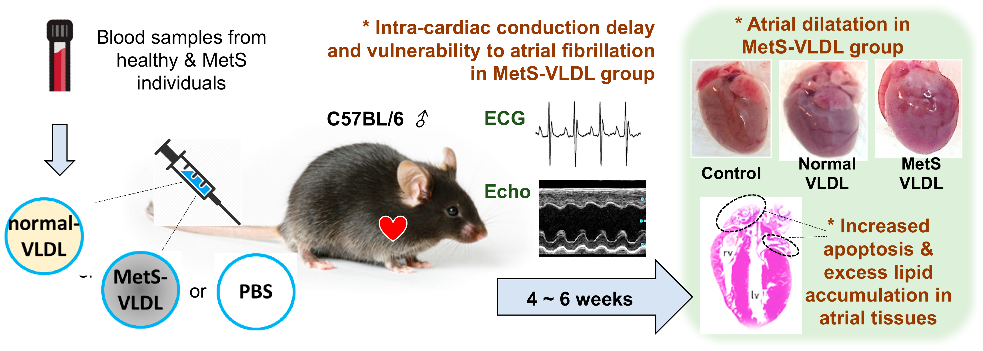
4. VLDL of MetS Induces Atrial Remodeling, Ventricular Hypertrophy, and Vulnerability to AF

In our VLDLs injection mice, LVH developed after 4 to 6 weeks (Figure 1). Nevertheless, only the MetS group had left atrial dilatation. The atrial dilatation takes place before ventricular hypertrophy [81], therefore the VLDL-induced atrial dilatation is considered as a primary change rather than secondary to the ventricular hypertrophy. Most pathological processes contributing to atrial remodeling also affect the ventricles; likewise, VLDL also induces ventricular hypertrophy.
In addition to the cardiac remodeling, VLDL also causes ventricular function impairment. In our first publication on using the VLDL injection mouse model, there was a borderline reduced ejection fraction of the left ventricle in the MetS group of young mice [81]. In our second publication regarding in vivo effects of VLDL on cardiac function, the MetS-VLDL injection caused reduced contractility of ventricle of middle-aged mice [82].
In electrocardiography, young mice of the MetS group were found to have ventricular premature beats at baseline without any stress, and they also had AF after receiving an intraperitoneal injection of isoproterenol, a rapid-onset β-receptor agonist to mimic elevated sympathetic tone under stress [81]. We also observed spontaneous, unprovoked AF among half of the middle-aged mice receiving MetS-VLDL injection [81]. These findings provide direct evidence that VLDL of MetS can induce atrial remodeling and vulnerability to AF. According to the aforementioned results, we suggest that the atrial cardiomyopathy in MetS is related to VLDL-induced lipotoxicity.

5. VLDL of MetS Induces Intra-Cardiac Conduction Delay via Modulation of Cardiac Gap Junction Cx40/43

The intra-cardiac conduction can be reflected by utilizing each component of electrocardiographic waves, including P waves, PR intervals, QRS durations, and QT intervals that indicate the atrial depolarization/repolarization, impulse conduction from the atria to the AV node, ventricular depolarization, and finish of ventricular repolarization, respectively. In the VLDL injection mice, the MetS group had significantly prolonged P waves, PR intervals, QRS durations, and QT intervals, indicating that the delayed cardiac conduction is intra-atrial, between atria-ventricular and intra-ventricular [82]. By using the optical mapping technique on ex vivo hearts, the slowed conduction velocities over bilateral atria and bilateral ventricles were demonstrated in the MetS group compared with the control and non-MetS VLDL group [82]. One of the possible causes of the cardiac conduction delay, intra-cardiac fibrosis, was excluded by our negative finding of the histological study.
Gap junctions facilitate efficient electrical propagation in the heart and determine the intracardiac conduction throughout the atria, ventricles, and conduction system. We checked the expression levels of gap junction proteins Cx40 and Cx43, and their genes GJA5 and GJA1, respectively. Cx43 is expressed throughout the heart, whereas Cx40 is exclusively expressed in the atria, the His bundle, and Purkinje fibers. Our results found that the expression for both Cx40 (GJA5) and Cx43 (GJA1) was significantly reduced in the MetS mice atria. Besides down-regulation of gap junction proteins and gene expression, MetS-VLDL was found to also modulate the Cx40 and Cx43 proteins with enhanced O-GlcNAylation, which is postulated to affect the gap junction assembly and stability by interfering with the electrostatic balance on neighbor molecule interaction [82]. The O-GlcNAylation of Cx40 under high glucose condition has been reported to affect gap junction in endothelial cells [83]. Our report was the first to demonstrate the VLDL-induced O-GlcNAylation of Cx40 and Cx43 in atrial myocyte HL-1 cells. The possibility of gap junction dysfunction or altered gating property upon O-GlcNAcylation requires further investigation.

6. VLDL of MetS Suppresses the Store-Operated Calcium Entry (SOCE) and Downstream Calcineurin-Nuclear Factor of Activated T Cells (NFAT) Pathway

The cardiac hypertrophy is an adaptive capacity to stress and it is regulated by the calcineurin–nuclear factor of activated T cells (NFAT) pathway, which is linked to the stromal interaction molecule 1 (STIM1)/calcium release-activated calcium channel protein 1 (Orai1)-mediated store-operated Ca2+ entry (SOCE) [84,85]. We reported evidence that VLDL of MetS attenuates SOCE and down-regulates the calcineurin-NFAT pathways [86]. The STIM1 is a Ca2+-binding protein and the Ca2+ sensor in the sarcoplasmic reticulum (SR), and it interacts with Ca2+ inward channels on the plasma membrane to replenish the SR calcium storage [87]. On treatment with VLDL of MetS, STIM1 is down-regulated on its protein and mRNA expressions, and it is also modulated by O-GlcNAylation. These findings are associated the inhibition of the calcineurin-NFAT pathway [86], which has been shown to be critical in both pathological hypertrophy and cardiac adaptation to biomechanical stress. We suggest that in MetS, VLDL affects the cardiac adaptation capacity of atrium.

7. VLDL of MetS Disturbs Myofilament Regulation

Sarcomere proteins of cardiomyocytes are regulated by a complex of mechanisms, and the alteration affects the motor function in which the interaction between thin and thick filaments leads to contraction of cardiomyocytes [88]. Genetic and hereditary cardiomyopathies, aging hearts, and heart failure are associated with predominant sarcomere proteins disorders [88,89]. On HL-1 cells treated with VLDL of MetS and on MetS group mice, alteration of sarcomere protein expression has been observed, including cMYBPC, desmin, troponin-T, troponin-I, and MLC2. This finding is associated with ultrastructural changes that are demonstrated by transmission electron microscopy, including disorganized Z-lines and sarcomeres, and morphological changes of mitochondria along the myofilaments [86]. We suggest that in atrial cardiomyopathy of MetS, VLDL causes the sarcomere derangement, which partially contributes to the impaired contractility in our VLDL-injected mice.

8. The Pathogenic Role of VLDL in Atrial Cardiomyopathy and Future Directions

On the basis of the available research findings thus far, as well as existing knowledge, we propose the pivotal and pathogenic role of VLDL in MetS-related atrial cardiomyopathy, manifested with both electrical and structural remodeling. The electrical remodeling includes triggered activities, delayed intra-cardiac conduction, vulnerability to AF upon sympathetic stimulation, and unprovoked AF. The structural remodeling includes atrial dilatation, ventricular dilatation, and hypertrophy associated with impaired contractility, along with increased myocardial apoptosis, excessive lipid accumulation, sarcomere disorders, and myofilament derangements, and also mitochondria morphological changes (Figure 2) [81,82,86].
Hypertriglyceridemia is a feature of MetS, and TG-rich chylomicrons and VLDL are largely produced and secreted after meals. Nakajima et al. reported that postprandial VLDL remnants have a higher affinity to the VLDLR compared to fasting VLDL, and they suggest both remnant lipoprotein-ApoB48 and -ApoB100 are endogenous VLDL remnants produced in the liver postprandially [57,91]. To understand the role of VLDL in atrial cardiomyopathy for human subjects, it is important to clarify postprandial concentrations and characteristics of VLDL and VLDL remnants in terms of the composition of apolipoproteins, lipid cores, charge, affinity to receptors, and favorability of internalization to cardiomyocytes.
We hypothesize that VLDLR plays a determinant role in atrial cardiomyopathy of MetS. We examined the cardiac remodeling on a mouse model that carries VLDLR loss-of-function mutant [92] and will utilize cardiac-specific VLDLR gene knock-out mice to perform more experiments. The results will offer direct evidence for the role of VLDLR in the pathogenesis of MetS-related atrial cardiomyopathy. In addition to intra-cardiac effects, our colleagues have reported that VLDL isolated from subjects with MetS induces microglia activation associated with elevation of tumor necrosis factor (TNF-α) and prostaglandin E2 (PGE2). This finding indicates that VLDL in MetS can contribute to neuroinflammation [93]. The autonomic nervous system plays an important role in arrhythmogenesis, and we hypothesize that VLDL may affect the autonomic innervation or activities on the heart, and therefore result in vulnerability to arrhythmias, especially for individuals with MetS. On the other hand, the intestinal postprandial production of TG-rich chylomicron and the secretion of VLDL from the liver that may be feedbacked by the circulatory TG-rich lipoproteins/remnants, may be intervened by the autonomic function as well [94]. If this is true, the post-prandial VLDL might be the most important factor determining the pathogenesis and progress of MetS-related atrial cardiomyopathy and ultimately vulnerability to the development of AF, as well as AF-related cognitive dysfunction. Further human studies are required in terms of focusing on the effects/correlation of postprandial lipids on atrial remodeling and its progression to determine whether VLDL-targeted therapy can reduce the occurrence of MetS-related AF.

Funding

The authors thank the technical support from colleagues in the College of Medicine, Kaohsiung Medical University. This study was funded by the National Health Research Institutes (NHRI-EX107-10724SC and NHRI-EX108-10724SC to H.C. Lee), by the Taiwan Ministry of Science and Technology (MOST 107-2314-B-037-110, MOST 108-2314-B-037-098, and MOST 108-2622-B-037-003-CC1 to H.C. Lee), by Chi-Mei Medical Center and Kaohsiung Medical University Research Foundation (108CM-KMU-009 to H.C. Lee), and by grants from Kaohsiung Medical University Hospital (KMUH107-7R11 and KMUH108-7R11 to H.C. Lee).

Conflicts of Interest

The authors declare no conflict of interest.

References

  1. Wattigney, W.A.; Mensah, G.A.; Croft, J.B. Increasing Trends in Hospitalization for Atrial Fibrillation in the United States, 1985 Through 1999. Implic. Prim. Prev. 2003, 108, 711–716. [Google Scholar] [CrossRef] [Green Version]
  2. Friberg, J.; Buch, P.; Scharling, H.; Gadsbphioll, N.; Jensen, G.B. Rising rates of hospital admissions for atrial fibrillation. Epidemiology 2003, 14, 666–672. [Google Scholar] [CrossRef]
  3. Hajhosseiny, R.; Matthews, G.K.; Lip, G.Y. Metabolic syndrome, atrial fibrillation, and stroke: Tackling an emerging epidemic. Heart Rhythm 2015, 12, 2332–2343. [Google Scholar] [CrossRef]
  4. Zakeri, R.; Van Wagoner, D.R.; Calkins, H.; Wong, T.; Ross, H.M.; Heist, E.K.; Meyer, T.E.; Kowey, P.R.; Mentz, R.J.; Cleland, J.G.; et al. The burden of proof: The current state of atrial fibrillation prevention and treatment trials. Heart Rhythm 2017, 14, 763–782. [Google Scholar] [CrossRef] [PubMed] [Green Version]
  5. Rahman, F.; Kwan, G.F.; Benjamin, E.J. Global epidemiology of atrial fibrillation. Nat. Rev. Cardiol. 2014, 11, 639–654. [Google Scholar] [CrossRef] [PubMed]
  6. Van Wagoner, D.R.; Piccini, J.P.; Albert, C.M.; Anderson, M.E.; Benjamin, E.J.; Brundel, B.; Califf, R.M.; Calkins, H.; Chen, P.S.; Chiamvimonvat, N.; et al. Progress toward the prevention and treatment of atrial fibrillation: A summary of the Heart Rhythm Society Research Forum on the Treatment and Prevention of Atrial Fibrillation, Washington, DC, December 9-10, 2013. Heart Rhythm 2015, 12, e5–e29. [Google Scholar] [CrossRef] [PubMed] [Green Version]
  7. Go, A.S.; Hylek, E.M.; Phillips, K.A.; Chang, Y.; Henault, L.E.; Selby, J.V.; Singer, D.E. Prevalence of diagnosed atrial fibrillation in adults: national implications for rhythm management and stroke prevention: the AnTicoagulation and Risk Factors in Atrial Fibrillation (ATRIA) Study. JAMA 2001, 285, 2370–2375. [Google Scholar] [CrossRef]
  8. Friberg, L.; Rosenqvist, M.; Lip, G.Y. Evaluation of risk stratification schemes for ischaemic stroke and bleeding in 182 678 patients with atrial fibrillation: the Swedish Atrial Fibrillation cohort study. Eur. Heart J. 2012, 33, 1500–1510. [Google Scholar] [CrossRef]
  9. Bai, D. Atrial fibrillation-linked GJA5/connexin40 mutants impaired gap junctions via different mechanisms. FEBS Lett. 2014, 588, 1238–1243. [Google Scholar] [CrossRef] [Green Version]
  10. Goette, A.; Kalman, J.M.; Aguinaga, L.; Akar, J.; Cabrera, J.A.; Chen, S.A.; Chugh, S.S.; Corradi, D.; D’Avila, A.; Dobrev, D.; et al. EHRA/HRS/APHRS/SOLAECE expert consensus on Atrial cardiomyopathies: definition, characterization, and clinical implication. Europace 2016. [Google Scholar] [CrossRef]
  11. Heijman, J.; Voigt, N.; Nattel, S.; Dobrev, D. Cellular and molecular electrophysiology of atrial fibrillation initiation, maintenance, and progression. Circ. Res. 2014, 114, 1483–1499. [Google Scholar] [CrossRef] [Green Version]
  12. Bombelli, M.; Facchetti, R.; Cuspidi, C.; Villa, P.; Dozio, D.; Brambilla, G.; Grassi, G.; Mancia, G. Prognostic Significance of Left Atrial Enlargement in a General Population: Results of the PAMELA Study. Hypertension 2014, 64, 1205–1211. [Google Scholar] [CrossRef] [Green Version]
  13. Poulsen, M.K.; Dahl, J.S.; Henriksen, J.E.; Hey, T.M.; Høilund-Carlsen, P.F.; Beck-Nielsen, H.; Møller, J.E. Left atrial volume index: relation to long-term clinical outcome in type 2 diabetes. J. Am. Coll. Cardiol. 2013. [Google Scholar] [CrossRef] [Green Version]
  14. Francia, P.; Ricotta, A.; Balla, C.; Adduci, C.; Semprini, L.; Frattari, A.; Modestino, A.; Mercanti, F.; Sensini, I.; Caprinozzi, M.; et al. P-wave duration in lead aVR and the risk of atrial fibrillation in hypertension. Ann. Noninvasive Electrocardiol. 2015, 20, 167–174. [Google Scholar] [CrossRef]
  15. Yin, X.; Zhao, Y.; Xi, Y.; Cheng, N.; Xia, Y.; Zhang, S.; Dong, Y.; Chang, D.; Cheng, J.; Yang, Y.; et al. The Early Stage of the Atrial Electroanatomic Remodeling as Substrates for Atrial Fibrillation in Hypertensive Patients. J. Am. Heart Assoc. 2014, 3. [Google Scholar] [CrossRef] [Green Version]
  16. Maron, B.J.; Haas, T.S.; Maron, M.S.; Lesser, J.R.; Browning, J.A.; Chan, R.H.; Olivotto, I.; Garberich, R.F.; Schwartz, R.S. Left atrial remodeling in hypertrophic cardiomyopathy and susceptibility markers for atrial fibrillation identified by cardiovascular magnetic resonance. Am. J. Cardiol. 2014, 113, 1394–1400. [Google Scholar] [CrossRef]
  17. Executive Summary of The Third Report of The National Cholesterol Education Program (NCEP) Expert Panel on Detection, Evaluation, And Treatment of High Blood Cholesterol In Adults (Adult Treatment Panel III). JAMA 2001, 285, 2486–2497. [CrossRef]
  18. Chamberlain, A.M.; Agarwal, S.K.; Ambrose, M.; Folsom, A.R.; Soliman, E.Z.; Alonso, A. Metabolic syndrome and incidence of atrial fibrillation among blacks and whites in the Atherosclerosis Risk in Communities (ARIC) Study. Am. Heart J. 2010, 159, 850–856. [Google Scholar] [CrossRef] [Green Version]
  19. Nystrom, P.K.; Carlsson, A.C.; Leander, K.; de Faire, U.; Hellenius, M.L.; Gigante, B. Obesity, metabolic syndrome and risk of atrial fibrillation: A Swedish, prospective cohort study. PLoS ONE 2015, 10, e0127111. [Google Scholar] [CrossRef] [Green Version]
  20. Tanner, R.M.; Baber, U.; Carson, A.P.; Voeks, J.; Brown, T.M.; Soliman, E.Z.; Howard, V.J.; Muntner, P. Association of the metabolic syndrome with atrial fibrillation among United States adults (from the REasons for Geographic and Racial Differences in Stroke [REGARDS] Study). Am. J. Cardiol. 2011, 108, 227–232. [Google Scholar] [CrossRef] [Green Version]
  21. Kwon, C.H.; Kim, H.; Kim, S.H.; Kim, B.S.; Kim, H.J.; Kim, D.K.; Han, S.W.; Ryu, K.H.; Dong Sung, J. The Impact of Metabolic Syndrome on the Incidence of Atrial Fibrillation: A Nationwide Longitudinal Cohort Study in South Korea. J. Clin. Med. 2019, 8. [Google Scholar] [CrossRef] [PubMed] [Green Version]
  22. Kim, Y.G.; Choi, K.J.; Han, S.; Hwang, K.W.; Kwon, C.H.; Park, G.M.; Won, K.B.; Ann, S.H.; Kim, J.; Kim, S.J.; et al. Metabolic Syndrome and the Risk of New-Onset Atrial Fibrillation in Middle-Aged East Asian Men. Circ. J. 2018, 82, 1763–1769. [Google Scholar] [CrossRef] [PubMed] [Green Version]
  23. Umetani, K.; Kodama, Y.; Nakamura, T.; Mende, A.; Kitta, Y.; Kawabata, K.; Obata, J.E.; Takano, H.; Kugiyama, K. High prevalence of paroxysmal atrial fibrillation and/or atrial flutter in metabolic syndrome. Circ. J. 2007, 71, 252–255. [Google Scholar] [CrossRef] [PubMed] [Green Version]
  24. Polovina, M.; Hindricks, G.; Maggioni, A.; Piepoli, M.; Vardas, P.; Asanin, M.; Ethikic, D.; Ethuricic, N.; Milinkovic, I.; Seferovic, P.M. Association of metabolic syndrome with non-thromboembolic adverse cardiac outcomes in patients with atrial fibrillation. Eur. Heart J. 2018, 39, 4030–4039. [Google Scholar] [CrossRef]
  25. Lin, K.J.; Cho, S.I.; Tiwari, N.; Bergman, M.; Kizer, J.R.; Palma, E.C.; Taub, C.C. Impact of metabolic syndrome on the risk of atrial fibrillation recurrence after catheter ablation: systematic review and meta-analysis. J. Interv. Card. Electrophysiol. 2014, 39, 211–223. [Google Scholar] [CrossRef] [Green Version]
  26. Wojcik, M.; Berkowitsch, A.; Kuniss, M.; Zaltsberg, S.; Pitschner, H.F.; Hamm, C.W.; Neumann, T. Outcomes of atrial fibrillation ablation in patients with metabolic syndrome. J. Am. Coll Cardiol. 2013, 61, 109–110. [Google Scholar] [CrossRef] [Green Version]
  27. Tsai, C.-T.; Chang, S.-H.; Chang, S.-N.; Hwang, J.-J.; Wu, C.-K.; Wang, Y.-C.; Tseng, C.-D.; Yeh, H.-M.; Lai, L.-P.; Chiang, F.-T.; et al. Additive effect of the metabolic syndrome score to the conventional CHADS2 score for the thromboembolic risk stratification of patients with atrial fibrillation. Heart Rhythm 2014, 11, 352–357. [Google Scholar] [CrossRef]
  28. Movahed, M.-R.; Hashemzadeh, M.; Mazen Jamal, M. Diabetes mellitus is a strong, independent risk for atrial fibrillation and flutter in addition to other cardiovascular disease. Int. J. Cardiol. 2005, 105, 315–318. [Google Scholar] [CrossRef]
  29. Huxley, R.R.; Filion, K.B.; Konety, S.; Alonso, A. Meta-Analysis of Cohort and Case–Control Studies of Type 2 Diabetes Mellitus and Risk of Atrial Fibrillation. Am. J. Cardiol. 2011, 108, 56–62. [Google Scholar] [CrossRef] [Green Version]
  30. Verdecchia, P.; Angeli, F.; Reboldi, G. Hypertension and Atrial Fibrillation: Doubts and Certainties From Basic and Clinical Studies. Circ. Res. 2018, 122, 352–368. [Google Scholar] [CrossRef]
  31. Choisy, S.C.; Arberry, L.A.; Hancox, J.C.; James, A.F. Increased susceptibility to atrial tachyarrhythmia in spontaneously hypertensive rat hearts. Hypertension 2007, 49, 498–505. [Google Scholar] [CrossRef] [PubMed] [Green Version]
  32. Okin, P.M.; Wachtell, K.; Devereux, R.B.; Harris, K.E.; Jern, S.; Kjeldsen, S.E.; Julius, S.; Lindholm, L.H.; Nieminen, M.S.; Edelman, J.M.; et al. Regression of electrocardiographic left ventricular hypertrophy and decreased incidence of new-onset atrial fibrillation in patients with hypertension. JAMA 2006, 296, 1242–1248. [Google Scholar] [CrossRef] [PubMed] [Green Version]
  33. Li, S.N.; Wang, L.; Dong, J.Z.; Yu, R.H.; Long, D.Y.; Tang, R.B.; Sang, C.H.; Jiang, C.X.; Liu, N.; Bai, R.; et al. Electrocardiographic left ventricular hypertrophy predicts recurrence of atrial arrhythmias after catheter ablation of paroxysmal atrial fibrillation. Clin. Cardiol. 2018, 41, 797–802. [Google Scholar] [CrossRef] [PubMed] [Green Version]
  34. Bohne, L.J.; Johnson, D.; Rose, R.A.; Wilton, S.B.; Gillis, A.M. The Association Between Diabetes Mellitus and Atrial Fibrillation: Clinical and Mechanistic Insights. Front. Physiol. 2019, 10, 135. [Google Scholar] [CrossRef] [Green Version]
  35. Lopez-Izquierdo, A.; Pereira, R.O.; Wende, A.R.; Punske, B.B.; Abel, E.D.; Tristani-Firouzi, M. The absence of insulin signaling in the heart induces changes in potassium channel expression and ventricular repolarization. Am. J. Physiol. Heart Circ. Physiol. 2014, 306, H747–H754. [Google Scholar] [CrossRef] [Green Version]
  36. Wang, T.J.; Parise, H.; Levy, D.; D’Agostino, R.B., Sr.; Wolf, P.A.; Vasan, R.S.; Benjamin, E.J. Obesity and the risk of new-onset atrial fibrillation. JAMA 2004, 292, 2471–2477. [Google Scholar] [CrossRef] [Green Version]
  37. Kim, Y.G.; Han, K.D.; Choi, J.I.; Yung Boo, K.; Kim, D.Y.; Oh, S.K.; Lee, K.N.; Shim, J.; Kim, J.S.; Kim, Y.H. Impact of the Duration and Degree of Hypertension and Body Weight on New-Onset Atrial Fibrillation: A Nationwide Population-Based Study. Hypertension 2019. [Google Scholar] [CrossRef]
  38. Watanabe, H.; Tanabe, N.; Watanabe, T.; Darbar, D.; Roden, D.M.; Sasaki, S.; Aizawa, Y. Metabolic syndrome and risk of development of atrial fibrillation: The Niigata preventive medicine study. Circulation 2008, 117, 1255–1260. [Google Scholar] [CrossRef] [Green Version]
  39. Zanoli, L.; Di Pino, A.; Terranova, V.; Di Marca, S.; Pisano, M.; Di Quattro, R.; Ferrara, V.; Scicali, R.; Rabuazzo, A.M.; Fatuzzo, P.; et al. Inflammation and ventricular-vascular coupling in hypertensive patients with metabolic syndrome. Nutr. Metab. Cardiovasc. Dis. 2018, 28, 1222–1229. [Google Scholar] [CrossRef] [Green Version]
  40. Mora, S.; Akinkuolie, A.O.; Sandhu, R.K.; Conen, D.; Albert, C.M. Paradoxical association of lipoprotein measures with incident atrial fibrillation. Circ. Arrhythm. Electrophysiol. 2014, 7, 612–619. [Google Scholar] [CrossRef] [Green Version]
  41. Annoura, M.; Ogawa, M.; Kumagai, K.; Zhang, B.; Saku, K.; Arakawa, K. Cholesterol paradox in patients with paroxysmal atrial fibrillation. Cardiology 1999, 92, 21–27. [Google Scholar] [CrossRef] [PubMed]
  42. Mourtzinis, G.; Kahan, T.; Bengtsson Bostrom, K.; Schioler, L.; Cedstrand Wallin, L.; Hjerpe, P.; Hasselstrom, J.; Manhem, K. Relation Between Lipid Profile and New-Onset Atrial Fibrillation in Patients With Systemic Hypertension (From the Swedish Primary Care Cardiovascular Database [SPCCD]). Am. J. Cardiol. 2018, 122, 102–107. [Google Scholar] [CrossRef] [PubMed]
  43. Liu, C.; Geng, J.; Ye, X.; Yuan, X.; Li, A.; Zhang, Z.; Xu, B.; Wang, Y. Change in lipid profile and risk of new-onset atrial fibrillation in patients with chronic heart failure: A 3-year follow-up observational study in a large Chinese hospital. Medicine 2018, 97, e12485. [Google Scholar] [CrossRef] [PubMed]
  44. Li, X.; Gao, L.; Wang, Z.; Guan, B.; Guan, X.; Wang, B.; Han, X.; Xiao, X.; Waleed, K.B.; Chandran, C.; et al. Lipid profile and incidence of atrial fibrillation: A prospective cohort study in China. Clin. Cardiol. 2018, 41, 314–320. [Google Scholar] [CrossRef] [PubMed] [Green Version]
  45. Xue, Y.; Zhou, Q.; Shen, J.; Liu, G.; Zhou, W.; Wen, Y.; Luo, S. Lipid Profile and New-Onset Atrial Fibrillation in Patients With Acute ST-Segment Elevation Myocardial Infarction (An Observational Study in Southwest of China). Am. J. Cardiol. 2019. [Google Scholar] [CrossRef] [PubMed]
  46. Shang, Y.; Chen, N.; Wang, Q.; Zhuo, C.; Zhao, J.; Lv, N.; Huang, Y. Blood lipid levels and recurrence of atrial fibrillation after radiofrequency catheter ablation: a prospective study. J. Interv. Card. Electrophysiol. 2019. [Google Scholar] [CrossRef] [PubMed]
  47. Veronese, G.; Montomoli, J.; Schmidt, M.; Horvath-Puho, E.; Sorensen, H.T. Statin Use and Risk of Atrial Fibrillation or Flutter: A Population-based Case-Control Study. Am. J. Ther. 2015, 22, 186–194. [Google Scholar] [CrossRef]
  48. Zhou, X.; Du, J.L.; Yuan, J.; Chen, Y.Q. Statin therapy is beneficial for the prevention of atrial fibrillation in patients with coronary artery disease: a meta-analysis. Eur. J. Pharm. 2013, 707, 104–111. [Google Scholar] [CrossRef]
  49. Fauchier, L.; Clementy, N.; Babuty, D. Statin therapy and atrial fibrillation: systematic review and updated meta-analysis of published randomized controlled trials. Curr. Opin. Cardiol. 2013, 28, 7–18. [Google Scholar] [CrossRef]
  50. Alonso, A.; Yin, X.; Roetker, N.S.; Magnani, J.W.; Kronmal, R.A.; Ellinor, P.T.; Chen, L.Y.; Lubitz, S.A.; McClelland, R.L.; McManus, D.D.; et al. Blood lipids and the incidence of atrial fibrillation: the multi-ethnic study of atherosclerosis and the framingham heart study. J. Am. Heart Assoc. 2014, 3. [Google Scholar] [CrossRef] [Green Version]
  51. Kim, S.M.; Kim, J.M.; Shin, D.G.; Kim, J.R.; Cho, K.H. Relation of atrial fibrillation (AF) and change of lipoproteins: male patients with AF exhibited severe pro-inflammatory and pro-atherogenic properties in lipoproteins. Clin. Biochem. 2014, 47, 869–875. [Google Scholar] [CrossRef] [PubMed]
  52. Kim, S.M.; Lee, J.H.; Kim, J.R.; Shin, D.G.; Lee, S.H.; Cho, K.H. Female patients with atrial fibrillation have increased oxidized and glycated lipoprotein properties and lower apolipoprotein A-I expression in HDL. Int. J. Mol. Med. 2011, 27, 841–849. [Google Scholar] [PubMed] [Green Version]
  53. Mittendorfer, B.; Yoshino, M.; Patterson, B.W.; Klein, S. VLDL Triglyceride Kinetics in Lean, Overweight, and Obese Men and Women. J. Clin. Endocrinol. Metab. 2016, 101, 4151–4160. [Google Scholar] [CrossRef] [PubMed]
  54. Miller, B.V., 3rd; Patterson, B.W.; Okunade, A.; Klein, S. Fatty acid and very low density lipoprotein metabolism in obese African American and Caucasian women with type 2 diabetes. J. Lipid Res. 2012, 53, 2767–2772. [Google Scholar] [CrossRef] [Green Version]
  55. Chan, D.C.; Watts, G.F.; Nguyen, M.N.; Barrett, P.H. Apolipoproteins C-III and A-V as predictors of very-low-density lipoprotein triglyceride and apolipoprotein B-100 kinetics. Arter. Thromb. Vasc. Biol. 2006, 26, 590–596. [Google Scholar] [CrossRef] [Green Version]
  56. Castellani, L.W.; Nguyen, C.N.; Charugundla, S.; Weinstein, M.M.; Doan, C.X.; Blaner, W.S.; Wongsiriroj, N.; Lusis, A.J. Apolipoprotein AII is a regulator of very low density lipoprotein metabolism and insulin resistance. J. Biol. Chem. 2008, 283, 11633–11644. [Google Scholar] [CrossRef] [Green Version]
  57. Nakajima, K.; Tokita, Y.; Tanaka, A.; Takahashi, S. The VLDL receptor plays a key role in the metabolism of postprandial remnant lipoproteins. Clin. Chim. Acta 2019, 495, 382–393. [Google Scholar] [CrossRef]
  58. Yakovlev, S.; Belkin, A.M.; Chen, L.; Cao, C.; Zhang, L.; Strickland, D.K.; Medved, L. Anti-VLDL receptor monoclonal antibodies inhibit fibrin-VLDL receptor interaction and reduce fibrin-dependent leukocyte transmigration. Thromb. Haemost. 2016, 116, 1122–1130. [Google Scholar] [CrossRef] [Green Version]
  59. Galati, F.; Colonna, P.; Galati, A.; Ciardiello, C.; Bozzetti, M.P.; Massari, S. CETP TaqIB Polymorphism, Serum Lipid Levels And Risk Of Atrial Fibrillation: A Case-Control Study. J. Atr. Fibrillation 2014, 6, 964. [Google Scholar]
  60. Niu, Y.G.; Evans, R.D. Very-low-density lipoprotein: complex particles in cardiac energy metabolism. J. Lipids 2011, 2011, 189876. [Google Scholar] [CrossRef]
  61. Masuzaki, H.; Jingami, H.; Matsuoka, N.; Nakagawa, O.; Ogawa, Y.; Mizuno, M.; Yoshimasa, Y.; Yamamoto, T.; Nakao, K. Regulation of very-low-density lipoprotein receptor in hypertrophic rat heart. Circ. Res. 1996, 78, 8–14. [Google Scholar] [CrossRef] [PubMed]
  62. Bharadwaj, K.G.; Hiyama, Y.; Hu, Y.; Huggins, L.A.; Ramakrishnan, R.; Abumrad, N.A.; Shulman, G.I.; Blaner, W.S.; Goldberg, I.J. Chylomicron- and VLDL-derived lipids enter the heart through different pathways: in vivo evidence for receptor- and non-receptor-mediated fatty acid uptake. J. Biol. Chem. 2010, 285, 37976–37986. [Google Scholar] [CrossRef] [PubMed] [Green Version]
  63. Whitman, S.C.; Daugherty, A.; Post, S.R. Macrophage colony-stimulating factor rapidly enhances beta-migrating very low density lipoprotein metabolism in macrophages through activation of a Gi/o protein signaling pathway. J. Biol. Chem. 2000, 275, 35807–35813. [Google Scholar] [CrossRef] [PubMed] [Green Version]
  64. Takahashi, S.; Kawarabayasi, Y.; Nakai, T.; Sakai, J.; Yamamoto, T. Rabbit very low density lipoprotein receptor: a low density lipoprotein receptor-like protein with distinct ligand specificity. Proc. Natl. Acad. Sci. USA 1992, 89, 9252–9256. [Google Scholar] [CrossRef] [PubMed] [Green Version]
  65. Kwok, S.; Singh-Bist, A.; Natu, V.; Kraemer, F.B. Dietary regulation of the very low density lipoprotein receptor in mouse heart and fat. Horm. Metab. Res. 1997, 29, 524–529. [Google Scholar] [CrossRef]
  66. Argraves, K.M.; Battey, F.D.; MacCalman, C.D.; McCrae, K.R.; Gafvels, M.; Kozarsky, K.F.; Chappell, D.A.; Strauss, J.F., 3rd; Strickland, D.K. The very low density lipoprotein receptor mediates the cellular catabolism of lipoprotein lipase and urokinase-plasminogen activator inhibitor type I complexes. J. Biol. Chem. 1995, 270, 26550–26557. [Google Scholar] [CrossRef] [PubMed] [Green Version]
  67. Yagyu, H.; Lutz, E.P.; Kako, Y.; Marks, S.; Hu, Y.; Choi, S.Y.; Bensadoun, A.; Goldberg, I.J. Very low density lipoprotein (VLDL) receptor-deficient mice have reduced lipoprotein lipase activity. Possible causes of hypertriglyceridemia and reduced body mass with VLDL receptor deficiency. J. Biol. Chem. 2002, 277, 10037–10043. [Google Scholar] [CrossRef] [Green Version]
  68. Wyne, K.L.; Pathak, K.; Seabra, M.C.; Hobbs, H.H. Expression of the VLDL receptor in endothelial cells. Arter. Thromb. Vasc. Biol. 1996, 16, 407–415. [Google Scholar] [CrossRef]
  69. Perman, J.C.; Bostrom, P.; Lindbom, M.; Lidberg, U.; StAhlman, M.; Hagg, D.; Lindskog, H.; Scharin Tang, M.; Omerovic, E.; Mattsson Hulten, L.; et al. The VLDL receptor promotes lipotoxicity and increases mortality in mice following an acute myocardial infarction. J. Clin. Invest. 2011, 121, 2625–2640. [Google Scholar] [CrossRef] [Green Version]
  70. Papageorgiou, I.; Viglino, C.; Brulhart-Meynet, M.C.; James, R.W.; Lerch, R.; Montessuit, C. Impaired stimulation of glucose transport in cardiac myocytes exposed to very low-density lipoproteins. Nutr. Metab. Cardiovasc. Dis. 2016, 26, 614–622. [Google Scholar] [CrossRef] [Green Version]
  71. Schulze, P.C.; Drosatos, K.; Goldberg, I.J. Lipid Use and Misuse by the Heart. Circ. Res. 2016, 118, 1736–1751. [Google Scholar] [CrossRef] [PubMed] [Green Version]
  72. Elezaby, A.; Sverdlov, A.L.; Tu, V.H.; Soni, K.; Luptak, I.; Qin, F.; Liesa, M.; Shirihai, O.S.; Rimer, J.; Schaffer, J.E.; et al. Mitochondrial remodeling in mice with cardiomyocyte-specific lipid overload. J. Mol. Cell. Cardiol. 2015, 79, 275–283. [Google Scholar] [CrossRef] [PubMed] [Green Version]
  73. Drosatos, K.; Schulze, P.C. Cardiac lipotoxicity: molecular pathways and therapeutic implications. Curr. Heart Fail. Rep. 2013, 10, 109–121. [Google Scholar] [CrossRef] [PubMed] [Green Version]
  74. Van de Weijer, T.; Schrauwen-Hinderling, V.B.; Schrauwen, P. Lipotoxicity in type 2 diabetic cardiomyopathy. Cardiovasc Res. 2011, 92, 10–18. [Google Scholar] [CrossRef] [PubMed] [Green Version]
  75. Schaffer, J.E. Lipotoxicity: when tissues overeat. Curr. Opin. Lipidol. 2003, 14, 281–287. [Google Scholar] [CrossRef] [PubMed]
  76. Bukowska, A.; Schild, L.; Keilhoff, G.; Hirte, D.; Neumann, M.; Gardemann, A.; Neumann, K.H.; Röhl, F.W.; Huth, C.; Goette, A.; et al. Mitochondrial Dysfunction and Redox Signaling in Atrial Tachyarrhythmia. Exp. Biol. Med. 2008, 233, 558–574. [Google Scholar] [CrossRef]
  77. Opacic, D.; van Bragt, K.A.; Nasrallah, H.M.; Schotten, U.; Verheule, S. Atrial metabolism and tissue perfusion as determinants of electrical and structural remodelling in atrial fibrillation. Cardiovasc. Res. 2016, 109, 527–541. [Google Scholar] [CrossRef] [Green Version]
  78. Marfella, R.; Di Filippo, C.; Portoghese, M.; Barbieri, M.; Ferraraccio, F.; Siniscalchi, M.; Cacciapuoti, F.; Rossi, F.; D’Amico, M.; Paolisso, G. Myocardial lipid accumulation in patients with pressure-overloaded heart and metabolic syndrome. J. Lipid Res. 2009, 50, 2314–2323. [Google Scholar] [CrossRef] [Green Version]
  79. Nestel, P.J.; Poyser, A. Cholesterol content of the human atrium is related to plasma lipoprotein levels. Atherosclerosis 1978, 30, 177–183. [Google Scholar] [CrossRef]
  80. Chen, C.H.; Lu, J.; Chen, S.H.; Huang, R.Y.; Yilmaz, H.R.; Dong, J.; Elayda, M.A.; Dixon, R.A.; Yang, C.Y. Effects of electronegative VLDL on endothelium damage in metabolic syndrome. Diabetes Care 2012, 35, 648–653. [Google Scholar] [CrossRef] [Green Version]
  81. Lee, H.C.; Lin, H.T.; Ke, L.Y.; Wei, C.; Hsiao, Y.L.; Chu, C.S.; Lai, W.T.; Shin, S.J.; Chen, C.H.; Sheu, S.H.; et al. VLDL from Metabolic Syndrome Individuals Enhanced Lipid Accumulation in Atria with Association of Susceptibility to Atrial Fibrillation. Int J. Mol. Sci. 2016, 17. [Google Scholar] [CrossRef] [PubMed] [Green Version]
  82. Lee, H.C.; Chen, C.C.; Tsai, W.C.; Lin, H.T.; Shiao, Y.L.; Sheu, S.H.; Wu, B.N.; Chen, C.H.; Lai, W.T. Very-Low-Density Lipoprotein of Metabolic Syndrome Modulates Gap Junctions and Slows Cardiac Conduction. Sci. Rep. 2017, 7, 12050. [Google Scholar] [CrossRef] [PubMed] [Green Version]
  83. Issad, T. O-GlcNAcylation of connexin 40: A sweet connection between diabetes and endothelial cell dysfunction? Focus on “O-GlcNAcase overexpression reverses coronary endothelial cell dysfunction in type 1 diabetic mice”. Am. J. Physiol. 2015, 309, C590–C592. [Google Scholar] [CrossRef] [PubMed] [Green Version]
  84. Luo, X.; Hojayev, B.; Jiang, N.; Wang, Z.V.; Tandan, S.; Rakalin, A.; Rothermel, B.A.; Gillette, T.G.; Hill, J.A. STIM1-dependent store-operated Ca2+ entry is required for pathological cardiac hypertrophy. J. Mol. Cell. Cardiol. 2012, 52, 136–147. [Google Scholar] [CrossRef] [Green Version]
  85. Rosenberg, P. Socking It to cardiac hypertrophy: STIM1-mediated Ca2+ entry in the cardiomyocyte. Circulation 2011, 124, 766–768. [Google Scholar] [CrossRef] [Green Version]
  86. Shiou, Y.L.; Lin, H.T.; Ke, L.Y.; Wu, B.N.; Shin, S.J.; Chen, C.H.; Tsai, W.C.; Chu, C.S.; Lee, H.C. Very Low-Density Lipoproteins of Metabolic Syndrome Modulates STIM1, Suppresses Store-Operated Calcium Entry, and Deranges Myofilament Proteins in Atrial Myocytes. J. Clin. Med. 2019, 8. [Google Scholar] [CrossRef] [Green Version]
  87. Kurosaki, T.; Baba, Y. Ca2+ signaling and STIM1. Prog. Biophys. Mol. Biol. 2010, 103, 51–58. [Google Scholar] [CrossRef]
  88. Frazier, A.H.; Ramirez-Correa, G.A.; Murphy, A.M. Molecular mechanisms of sarcomere dysfunction in dilated and hypertrophic cardiomyopathy. Prog. Pediatr. Cardiol. 2011, 31, 29–33. [Google Scholar] [CrossRef] [Green Version]
  89. Machackova, J.; Barta, J.; Dhalla, N.S. Myofibrillar remodelling in cardiac hypertrophy, heart failure and cardiomyopathies. Can. J. Cardiol. 2006, 22, 953–968. [Google Scholar] [CrossRef] [Green Version]
  90. Lee, H.C.; Wei, C.; Ke, L.Y.; Tsai, P.S.; Lin, H.T.; Shiao, Y.L.; Wu, B.N.; Chen, C.H.; Sheu, S.H. Atherogenic Very-Low-Density Lipoprotein Shortens Atrial Action Potential Duration by Increasing Potassium Currents and Calcium Transient. Biophys. J. 2015, 108, 586a. [Google Scholar]
  91. Nakajima, K.; Tokita, Y.; Tanaka, A. Hypothesis II: The majority of VLDL-apoB48 remnants in postprandial plasma are derived from the liver, not from the intestine. Clin. Chim. Acta 2019, 490, 12–16. [Google Scholar] [CrossRef]
  92. Frykman, P.K.; Brown, M.S.; Yamamoto, T.; Goldstein, J.L.; Herz, J. Normal plasma lipoproteins and fertility in gene-targeted mice homozygous for a disruption in the gene encoding very low density lipoprotein receptor. Proc. Natl. Acad. Sci. USA 1995, 92, 8453–8457. [Google Scholar] [CrossRef] [Green Version]
  93. Li, C.L.; Chu, C.H.; Lee, H.C.; Chou, M.C.; Liu, C.K.; Chen, C.H.; Ke, L.Y.; Chen, S.L. Immunoregulatory effects of very low density lipoprotein from healthy individuals and metabolic syndrome patients on glial cells. Immunobiology 2019, 224, 632–637. [Google Scholar] [CrossRef]
  94. Luchoomun, J.; Zhou, Z.; Bakillah, A.; Jamil, H.; Hussain, M.M. Assembly and secretion of VLDL in nondifferentiated Caco-2 cells stably transfected with human recombinant ApoB48 cDNA. Arter. Thromb. Vasc. Biol. 1997, 17, 2955–2963. [Google Scholar] [CrossRef]
Figure 1. In vivo effects of very low density lipoproteins (VLDLs) on atrial remodeling of C57BL/6 male mice. Blood samples were collected from heathy volunteers and individuals with metabolic syndrome (MetS). Normal-VLDL and MetS-VLDL were isolated respectively. The VLDLs were injected via tail vein of the mouse three times a week for 6 consecutive weeks. The electrocardiography shows intra-cardiac conduction delay with significantly prolonged P waves and PR intervals in the MetS group. Upon isoproterenol challenge, some mice of the MetS group had atrial fibrillation whereas other groups did not. The echocardiography shows dilatation of left atria in the MetS group. In histological studies, MetS group were found to have increased lipid accumulation and increased myocyte apoptosis when compared with the control and normal-VLDL group.
Ijms 21 00891 g001
Figure 2. The pathogenic role of VLDL in MetS-related atrial cardiomyopathy, exhibited with structurally (in pink blocks) and electrical (in blue blocks) remodeling. The structural remodeling includes atrial dilatation, ventricular dilatation, and hypertrophy associated with impaired contractility, along with increased myocardial apoptosis, excessive lipid accumulation (consistent with EHRAS class IVf of atrial cardiomyopathy [10]), sarcomere disorders and myofilament derangements, and mitochondria morphological changes. The electrical remodeling includes shortening of action potentials [90], triggered activities, delayed intra-cardiac conduction, vulnerability to AF upon sympathetic stimulation, and unprovoked occurrence of AF. SOCE: store-operated calcium entry, NFAT: nuclear factor of activated T cells.
Ijms 21 00891 g002

