Article

The 15q11.2 BP1-BP2 Microdeletion (Burnside–Butler) Syndrome: In Silico Analyses of the Four Coding Genes Reveal Functional Associations with Neurodevelopmental Disorders

Departments of Psychiatry & Behavioral Sciences and Pediatrics, University of Kansas Medical Center, Kansas City, KS 66160, USA
*
Authors to whom correspondence should be addressed.
Int. J. Mol. Sci. 2020, 21(9), 3296; https://doi.org/10.3390/ijms21093296
Submission received: 10 February 2020 / Revised: 29 April 2020 / Accepted: 29 April 2020 / Published: 6 May 2020
(This article belongs to the Collection Feature Papers in Molecular Genetics and Genomics)

Abstract

:
The 15q11.2 BP1-BP2 microdeletion (Burnside–Butler) syndrome is emerging as the most frequent pathogenic copy number variation (CNV) in humans associated with neurodevelopmental disorders with changes in brain morphology, behavior, and cognition. In this study, we explored functions and interactions of the four protein-coding genes in this region, namely NIPA1, NIPA2, CYFIP1, and TUBGCP5, and elucidate their role, in solo and in concert, in the causation of neurodevelopmental disorders. First, we investigated the STRING protein-protein interactions encompassing all four genes and ascertained their predicted Gene Ontology (GO) functions, such as biological processes involved in their interactions, pathways and molecular functions. These include magnesium ion transport molecular function, regulation of axonogenesis and axon extension, regulation and production of bone morphogenetic protein and regulation of cellular growth and development. We gathered a list of significantly associated cardinal maladies for each gene from searchable genomic disease websites, namely MalaCards.org: HGMD, OMIM, ClinVar, GTR, Orphanet, DISEASES, Novoseek, and GeneCards.org. Through tabulations of such disease data, we ascertained the cardinal disease association of each gene, as well as their expanded putative disease associations. This enabled further tabulation of disease data to ascertain the role of each gene in the top ten overlapping significant neurodevelopmental disorders among the disease association data sets: (1) Prader–Willi Syndrome (PWS); (2) Angelman Syndrome (AS); (3) 15q11.2 Deletion Syndrome with Attention Deficit Hyperactive Disorder & Learning Disability; (4) Autism Spectrum Disorder (ASD); (5) Schizophrenia; (6) Epilepsy; (7) Down Syndrome; (8) Microcephaly; (9) Developmental Disorder, and (10) Peripheral Nervous System Disease. The cardinal disease associations for each of the four contiguous 15q11.2 BP1-BP2 genes are NIPA1- Spastic Paraplegia 6; NIPA2—Angelman Syndrome and Prader–Willi Syndrome; CYFIP1—Fragile X Syndrome and Autism; TUBGCP5—Prader–Willi Syndrome. The four genes are individually associated with PWS, ASD, schizophrenia, epilepsy, and Down syndrome. Except for TUBGCP5, the other three genes are associated with AS. Unlike the other genes, TUBGCP5 is also not associated with attention deficit hyperactivity disorder and learning disability, developmental disorder, or peripheral nervous system disease. CYFIP1 was the only gene not associated with microcephaly but was the only gene associated with developmental disorders. Collectively, all four genes were associated with up to three-fourths of the ten overlapping neurodevelopmental disorders and are deleted in this most prevalent known pathogenic copy number variation now recognized among humans with these clinical findings.

1. Introduction

The 15q11.2 BP1-BP2 deletion (Burnside–Butler) syndrome is an emerging condition that encompasses four protein-coding genes (NIPA1, NIPA2, CYFIP1, and TUBGCP5) within this chromosome region. When disturbed, these four genes lead to cognitive impairment with speech and/or motor delay along with dyslexia and psychiatric/behavior problems (attention deficit hyperactivity, autism, schizophrenia or psychosis), ataxia or poor coordination, seizures, congenital anomalies and structural brain defects [1]. These genes are associated with neurological, cognitive, or behavior problems as well as playing a role in both Prader–Willi and Angelman syndromes, first examples in humans of genomic imprinting [2].
These imprinting disorders typically are caused by a deletion in the majority of cases involving the 15q11-q13 chromosome region of different parental origin [i.e., paternal in Prader–Willi syndrome (PWS) and maternal in Angelman syndrome (AS)] [1,2]. The typical 15q11-q13 deletion involves proximal chromosome 15 breakpoints BP1 or BP2 and the more distally placed BP3 containing repetitive DNA segments allowing malalignment in meiosis of the homologous chromosome 15s leading to deletions or duplications in the region. The individuals with the larger typical type I deletion (involving BP1 and BP3) are found in both Prader–Willi and Angelman syndromes. They are reported with more severe developmental symptoms and clinical severity than individuals with the smaller typical type II deletion (involving BP2 and BP3) [3]. The 15q11.2 BP1-BP2 microdeletion encompasses a 500kb region located between breakpoints BP1 and BP2 proximal to the centromere of chromosome 15 including the four protein-coding genes (i.e., NIPA1, NIPA2, CYFIP1, TUBGCP5) that become the major focus of this review.
Hundreds of patients have now been reported with the 15q11.2 BP1-BP2 microdeletion often associated with the above listed neurodevelopmental disorders. A summary of the clinical features reported in over 200 individuals were described by Cox and Butler [4] and further reviewed by Butler [1] by grouping into five categories. These categories are (1) growth and development; (2) dysmorphic features; (3) intelligence and academic achievement; (4) behavioral and psychiatric problems, and (5) other related medical concerns. Developmental problems were reported in 73% of cases and speech delay in 67%; dysmorphic ears (46%) and palatal anomalies (46%); writing (60%) and reading (57%) difficulties, memory problems (60%) and verbal IQ scores ≤75 (50%); behavior problems, unspecified (55%); and abnormal brain imaging findings (43%). Additional clinical features were not as common and included motor delay (42%), ADD/attention deficit hyperactivity disorder (35%), autism spectrum disorder (27%), seizures or epilepsy (26%), and schizophrenia/paranoid psychosis (20%).
A recent report on structural brain anomalies using detailed imaging in hundreds of patients with the chromosome 15q11.2 BP1-BP2 microdeletion and controls showed a smaller brain surface area with a thicker cortex. These findings were more common in the frontal, cingulate, and parietal lobes in those with the deletions, supporting clinical observations and evidence as an emerging syndrome. Aberrations, leading to copy number variation (CNV) in these genes are estimated to be present in 0.5% to 1.0% of the population, making this specific site as the most prevalent known pathogenic copy number variation in humans [5]. However, not all individuals with this microdeletion are clinically affected, yet the collection of findings appears to share biological pathways that are hitherto unexplored. Their presumed genetic mechanisms require further analysis, as illustrated and discussed in our report.
Summarized results from chromosomal microarray analysis by a certified commercial clinical testing laboratory of patients presenting with genetic services were reported by Ho et al. [6]. The microarray analysis included 2.8 million probes optimized for the detection of CNVs associated with neurodevelopmental disorders. They reported an overall CNV detection rate of 28.1% in 10,351 consecutive patients which rose to nearly 33% of cases without ASD but with developmental delay/intellectual disability and/or multiple congenital anomalies. The overall rate of detection for those with ASD was also significant at 24.4%. The 15q11.2 BP1-BP2 deletion (Burnside–Butler) syndrome was found to be the most common disturbance (9%) seen in 85 genetic defects associated with neurodevelopmental disorders in this large cohort of consecutive patients and the most common finding in those presenting with ASD.
The larger 15q11-q13 type I deletion is approximately 6.6Mb in size seen in both PWS and AS and includes four genes (NIPA1, NIPA2, CYFIP1, and TUBGCP5) located within the proximal 15q11.2 BP1-BP2 microdeletion region. The smaller typical 15q11-q13 type II deletion has these four genes intact (see Figure 1). These genes are highly conserved and expressed in the brain (GeneCards.org & UniProtKB/Swiss-Prot). The NIPA1 gene causes autosomal dominant hereditary spastic paraplegia and postural disturbances when disturbed (https://www.malacards.org/card/spastic_paraplegia_6_autosomal_dominant) and functions as a magnesium transporter (https://www.genecards.org/cgibin/carddisp.pl?gene=NIPA1&keywords=NIPA1).
Mutations of the NIPA2 gene are reported in patients with childhood absence epilepsy with decreased intracellular magnesium concentration in neurons [7,8,9,10,11]. The CYFIP1 gene encodes a protein product that interacts with FMRP, the protein coded by the FMR1 gene causing fragile X syndrome (Entrez Gene: 23191; https://www.genecards.org/cgi-bin/carddisp.pl?gene=CYFIP1&keywords=CYFIP1). The fourth gene is TUBGCP5 associated with the chromosome 15q11.2 deletion syndrome and obsessive-compulsive disorder when disturbed. It also plays a role in microtubule nucleation at the centrosome in cells (UniProtKB: Q96RT8; https://www.genecards.org/cgi-bin/carddisp.pl?gene=TUBGCP5&keywords=TUBGCP5) [8,9,10,11,12,13,14,15,16,17,18].
Overview of 15q11-q13 BP1-BP3 Region Depicting the Proximal Location of BP1-BP2 Microdeletion (Burnside–Butler) Syndrome Region Within the Broader Type I Deletion Adjacent to Prader–Willi Syndrome (PWS)/Angelman Syndrome (AS) Regions.
The location, as well as the order of genes and transcripts (e.g., snoRNAs), are also shown in Figure 1 above that causes PWS/AS. Those genes that are imprinted and paternally expressed causing PWS (in blue) and those causing AS (in red) are also imprinted and maternally expressed. The location and the relative size of the 15q11.2 BP1–BP2 microdeletion region, the typical larger 15q11–q13 Type I deletion involving breakpoints BP1 and BP3, and the typical smaller 15q11–q13 Type II deletion involving breakpoints BP2 and BP3 in both PWS and AS are illustrated. IC: imprinting center controlling the activity of imprinted genes in the 15q11–q13 region.

2. Results

2.1. Overview of the Four Genes in the 15q11.2 BP1-BP2 Region

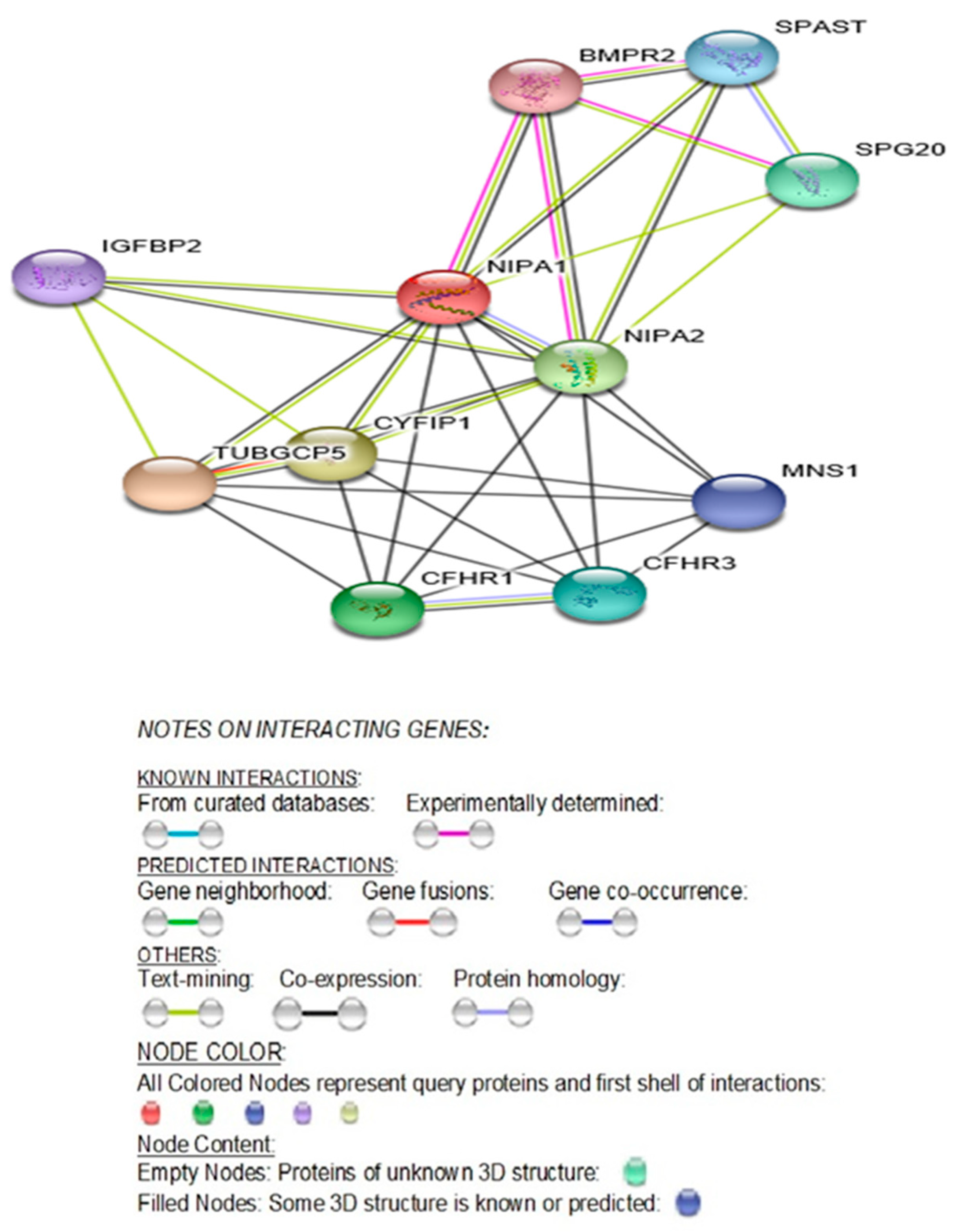
All four syntenic and bi-allelically conserved expressed genes in the 15q11.2 region between breakpoints BP1 and BP2 are functionally predicted to interact with each other along with seven other genes. The predicted STRING functional interactions network encompasses these four genes as illustrated in Figure 2.
The STRING diagram and protein interactions involving all four genes of this microdeletion syndrome are depicted in Figure 2 and their predicted functions are presented in Table 1. The identified biological processes (GO) and molecular functions (GO) are presented in Table 2 (https://version11.stringdb.org/cgi/network.pl?taskId=XJOUKPc8icHM).
Protein Network (STRING) Interactions Encompassing the Four Genes: NIPA1, NIPA2, CYFIP1 and TUBGCP5

2.2. NIPA1 (Non-Imprinted in Prader–Willi/Angelman Syndrome Region Protein 1) Gene

Attributes, Location, Description, Function and Associated Disorders for NIPA1 Gene.
The chromosomal band location for the NIPA1 gene is 15q11.2 with the genomic location (GRCh38/hg38) at 22,773,063–22,829,789 with a size of 56,727 bases in a plus-strand orientation. This gene has the following attributes: Size: 329 amino acids; Molecular mass: 34,562 Da; Quaternary structure:Homodimer (https://www.genecards.org/cgi-bin/carddisp.pl?gene=NIPA1&keywords=NIPA1). NIPA1 has two alternative splice isoforms; isoform 2 differs from the canonical sequence by missing the first 75 amino acids (GeneCards & UniProtKB/Swiss-Prot). It is a multi-pass membrane protein recruited to the cell membrane in response to low extracellular magnesium (GeneCards & UniProtKB/Swiss-Prot). This gene is widely expressed with highest levels in neuronal tissues and overexpressed in the spinal cord (20.8), frontal cortex (17.0), and fetal liver (7.3) (Protein differential expression in normal tissues from Human Integrated Protein Expression Database (HIPED) for NIPA1 Gene: https://www.genecards.org/Guide/GeneCard#protein-differential-expression; https://www.genecards.org/cgi-bin/carddisp.pl?gene=NIPA1&keywords=NIPA1).
NIPA1 protein plays a potential role in nervous system development and maintenance and is most ubiquitously expressed in the brain with a mean RPKM (Reads Per Kilobase per Million reads placed) of 16.56 + 3.055 (https://www.ncbi.nlm.nih.gov/gene/123606/?report=expression; https://www.gtexportal.org/home/gene/NIPA1) [14]. The multiple transmembrane domains of this protein localize to endosomes and plasma membrane. This protein is recruited in response to low extracellular magnesium and functions in Mg2 transport (Q7RTP0-NIPA1_HUMAN) [15]. Subcellular localization with immunofluorescence shows that endogenous NIPA1 protein associates with early endosomes and the cell surface in a variety of neuronal and epithelial cells. As expected of a magnesium-responsive gene, altered magnesium concentration leads to redistribution between the endosomal compartment and the plasma membrane; high magnesium results in diminished cell surface NIPA1 whereas low magnesium leads to accumulation in early endosomes and recruitment to the plasma membrane [15].
An important paralog of this gene, NIPAL1 is also a magnesium ion transmembrane transporter. In addition, there are three other protein-coding transcript variants (paralogs): NIPAL2, NIPAL3, and NIPAL4, which are also magnesium ion transmembrane transporters. Other genes also perform a similar magnesium transport function, namely, MAGT1, MMGT1, and MRS2 (GeneCards.Org). Although NIPA1 protein acts as a Mg (2+) transporter, it can also transport other divalent cations such as Fe(2+), Sr(2+), Ba(2+), Mn(2+), and Co(2+), but to a much less extent than Mg(2+) (UniProtKB/Swiss-Prot Summary). Among its other related pathways are the transport of glucose and other sugars, bile salts and organic acids, metal ions, and amine compounds.
Only mutations (not haploinsufficiency due to 15q11.2 BP1-BP2 microdeletions including the NIPA1 gene) have thus far been associated with autosomal dominant spastic paraplegia 6 / SPG6- linked hereditary spastic paraplegia: HSP (https://www.genecards.org/cgi-bin/carddisp.pl?gene=NIPA1&keywords=NIPA1). Spastic paraplegia 6 is a neurodegenerative disorder characterized by slow, gradual, progressive weakness and spasticity of the lower limbs. Rate of progression and the severity of symptoms are quite variable with initial symptoms of difficulty with balance, weakness, and stiffness in the legs, muscle spasms, and dragging the toes when walking. In some forms of the disorder, bladder symptoms (such as incontinence) may appear, or weakness and stiffness may be present to other parts of the body (UniProtKB/Swiss-Prot) [16,17,18]. Two variants in the NIPA1 gene, namely, p.Thr45Arg (VAR_023440; SNP ID: rs104894496) and p.Gly106Arg (VAR_023441; SNP ID: rs104894490) are disease-causing variations for spastic paraplegia type 6 (SPG6), autosomal dominant form. A rare NIPA1 deletion was also found in a patient with pervasive developmental disorder not otherwise specified and with mild intellectual disability [19]; this deletion is also linked to autism (SFARI.org).
The mouse NIPA1 mutants, p.Thr39Arg and p.Gly100Arg, corresponding to the respective human mutants are associated with hereditary spastic paraplegia (HSP) showing a loss-of-function when expressed in oocytes and altered trafficking in transfected COS7 cells [15]. The NIPA1 gene normally encodes an Mg2+ transporter protein and the loss-of-function of NIPA1, due to abnormal trafficking of the mutated protein provides the basis for the HSP phenotype [15,16,20]. Only abnormal trafficking of the mutated protein is causative for HSP and not from its deficiency as evident by Prader–Willi syndrome (PWS) or Angelman syndrome (AS) with only one copy of the NIPA1 gene in the typical 15q11-q13 type I deletion in both PWS or AS involving breakpoints BP1 and BP3 including the four genes in the 15q11.2 BP1-BP2 region. Other genes or transcripts, both imprinted and biallelic, do not have HSP [16,21]. Key findings in PWS and AS develop from errors of imprinting dependent on the parent of origin [22,23]. Therefore, SPG6 linked HSP is more likely caused by a dominant-negative effect or a toxic gain-of-function mechanism rather than a loss-of-function [16,17,21,24,25].
A perusal of genes involved in numerous other forms of spastic paraplegia indicates causation by genetic defects in various other metabolic pathways unrelated to magnesium transporter protein deficiency (see GeneCards.org). The syntenic NIPA2 gene, which also codes a magnesium ion transmembrane transporter protein and along with NIPA1 in the same region are both deleted in the Type I deletion but not deleted in the smaller typical 15q11-q13 Type II deletion in PWS or AS. However, patients with the larger Type I deletion do not have spastic paraplegia (GeneCards.org). Mutations in the NIPA2 gene have been reported in generalized epilepsy and childhood absence epilepsy [10,11,26]. In addition to the magnesium ion transmembrane transporter activity, NIPA1 protein inhibits Bone Morphogenetic Protein signaling by regulating the endosomal trafficking and degradation of type 2 BMP receptors (BMPR2) in Drosophila and HeLa cells [27,28].
Bone Morphogenetic Proteins (BMPs) are a group of signaling molecules that belong to the Transforming Growth Factor-β (TGFβ) superfamily of proteins. Initially discovered for their ability to induce bone formation, BMPs are now known to play crucial roles in all organ systems. BMPs are important in embryogenesis and development, and also in the maintenance of adult tissue homeostasis. Another relevant role of BMP in the neurological system is neurogenesis. Neural defects are associated with loss of BMP function in mouse models [29]. For instance, BMP11 is involved in spinal cord neurogenesis and secreted from neurons themselves serving as an inhibitory signal in the generation of new neurons from progenitors in the olfactory epithelium [30].
In rodents, BMP signaling is upregulated following lesions of the corticospinal track and suppressing upregulation that promotes regrowth of axons [31]. Targeting specific BMP receptor subunits for therapeutic purposes may provide an approach for manipulating gliosis and enhancing functional outcomes after spinal cord injury [32]. Therefore, it is no surprise that BMP signaling cuts across all hereditary spastic paraplegia (HSP) categories and are widely implicated in neurodegenerative diseases [33]. Among the HSP proteins, NIPA1 is best characterized mechanistically to inhibit BMP signaling [15]. In summary, these studies support abnormal BMP signaling in many cases probably resulting from abnormal BMP receptor trafficking and could be a unifying pathogenic mechanism for some forms of hereditary spastic paraplegia [34].
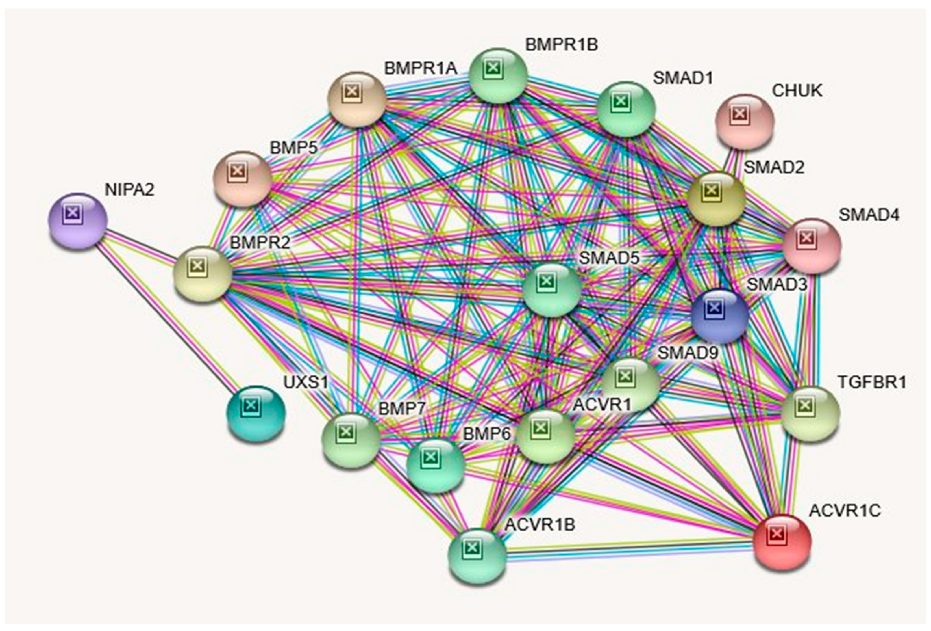
Further highlighting the importance of BMP regulation in neural development is the role of BMP7 in corticogenesis; BMP7 deletions result in reduced cortical thickening and impaired neurogenesis [35]. Interestingly, BMPR type 1A (BMPR1A) is important in the establishment of neurons involved in regulating feeding behavior [32]. Similarly, NIPA1 was shown to inhibit BMP signaling by regulating the endosomal trafficking and degradation of type 2 BMP receptors (BMPR2) in Drosophila and HeLa cells [27,28]. Perhaps, haploinsufficiency of NIPA1 protein in cases of 15q11-q13 BP1 or BP2/BP3 deletions might partially affect its inhibition of BMP signaling by regulating endosomal trafficking and degradation of type 2 BMP receptors, thereby affecting neurons. This might be the causal factor for the reported developmental and language delay, neurobehavioral disturbances, and psychiatric problems, such as autism, seizures, and schizophrenia with occasional mild dysmorphic features seen to a varying degree in patients with the 15q11.2 BP1-BP2 deletion (Burnside–Butler) syndrome [1,36]. Biological processes (GO), molecular functions (GO), cellular components with KEGG, and Reactome pathways are summarized in Table 3. In Table 4 examples of diseases or disorders which have been recognized and associated with the NIPA1 gene when disturbed are collated and ranked by order using MalaCards (MalaCards.org) from GeneCards (GeneCards.org). STRING functional interactions with nodes and edges (see Figure 3). Further illustrations of interactions and functions of NIPA1 with relationship to other proteins can be found in Table Table 5.
Interacting Proteins for NIPA1 Gene: STRING Interaction Network.

2.3. NIPA2 (Non-Imprinted in Prader–Willi/Angelman Syndrome Region Protein 2) Gene

Attributes, Location, Description, Function, and Associated Disorders for NIPA2 Gene.
The chromosomal band location for the NIPA2 gene is 15q11.2 with genomic location (GRCh38/hg38) at 22,838,641- 22,868,384 with a size of 29,744 bases in a plus-strand orientation. This gene is protein-coding specifically for NIPA magnesium transporter 2 (https://www.genecards.org/cgi-bin/carddisp.pl?gene=NIPA2&keywords=nipa2). When disturbed (deleted), this gene is associated with both Angelman and Prader–Willi syndromes due to specific parent of origin deletions of the 15q11-q13 region. The typical 15q11-q13 type I deletion also includes the other three protein-coding genes in the 15q11.2 BP1-BP2 region, namely, NIPA1, CYFIP1, and TUBGCP5. In the case of the smaller typical 15q11-q13 type II deletion, these four genes remain intact in the region (Figure 1) [1,2,3].
The NIPA2 protein has the following attributes: Size: 360 amino acids; Molecular mass:39185 Da. Quaternary structure: No Data Available. This protein has two isoforms; isoform 2 differs from the canonical sequence as follows: 47–66: GQGGHAYLKEWLWWAGLLSM → V. Similar to NIPA1, NIPA2 is also a multi-pass membrane protein found in early endosomes recruited to the cell membrane in response to low extracellular magnesium (UniProtKB/Swiss-Prot for NIPA2 Gene) (Table 6).
Unlike NIPA1, this gene is overexpressed in B-lymphocytes and the placenta ((UniProtKB/Swiss-Prot). In addition to PWS and AS involvement for this gene, it is also associated with childhood absence epilepsy [26], childhood electroclinical syndrome [10,11], and possibly autosomal recessive congenital ichthyosis (MalaCards/GeneCards) and important biological processes and functions (see Table 7).
Among related pathways of this gene are miscellaneous transport and binding events and transport of glucose and other sugars, bile salts and organic acids, metal ions and amine compounds. Gene Ontology (GO) annotations related to this gene also include magnesium ion transmembrane transporter activity. An important paralog of this gene is NIPAL1 (GeneCards). Chai et al. [9] determined that the NIPA2 gene contains 7 exons and spans 29 kb. The coding region extends between exons 3 and 7, and alternative splicing utilizes alternate exons 2 and 2b which results in multiple transcript variants. Pseudogenes of this gene are found on chromosomes 3, 7, and 21 (Entrez Gene: 81614).
Similar to NIPA1, this multi-pass cell membrane protein is localized in the plasma membrane and early endosomes (Gene Ontology (GO)—GO:0005769; GO:0005886) and recruited to the cell membrane in response to low extracellular magnesium (GeneCards; ECO:0000250). Paralogs for the NIPA2 gene are NIPA1, NIPAL1, NIPAL4, NIPAL2, and NIPAL3 (PMID: 28514442; [35]). Although NIPA2 is highly conserved (PMID: 14508708; [9]) and expressed in parts of the brain as well as in most other organ systems, it is significantly overexpressed in B-lymphocytes and the placenta. In addition, five variants have been reported for this gene with either loss or gain of function or both (GeneCards). Table 8 describes the STRING protein interactions and their functions with this gene and description while Table 6 shows biological processes, molecular functions, cellular components, and KEGG and Reactome pathways attributed to the NIPA2 gene. To examine NIPA2 protein interactions and connectivity with other genes and their interrelated proteins, see the STRING interaction network displayed in Figure 4.
Protein–Protein Inter-Relationships Involving the NIPA2 Gene.

2.4. CYFIP1 (Cytoplasmic FMR1 Interacting Protein 1) Gene

Attributes, Location, Description, Function, and Associated Disorders for CYFIP1 Gene.
The chromosomal band location for the CYFIP1 gene is 15q11.2 with the genomic location (GRCh38/hg38) at 22,867,052–22,981,063 with a size of 114,012 bases in a minus strand orientation (Genecards.org). The CYFIP1 protein has the following attributes: Size: 1253 amino acids; Molecular mass: 145,182 Da. Quaternary structure: Component of the WAVE1 complex composed of ABI2, CYFIP1 or CYFIP2, BRK1, NCKAP1, and WASF1/WAVE1. CYFIP1 and CYFIP2 are part of the Wiskott–Aldrich syndrome protein-family verprolin-homologous protein (WAVE) complex that regulates actin polymerization at synapses and CYFIP1 protein has 3 described isoforms produced by alternative splicing (https://www.uniprot.org/uniprot/Q7L576) (see Table 9). This protein is required for neuronal and bristle development in Drosophila [37]. Through the CYFIP1 protein, the fragile X syndrome protein represses activity-dependent translation, and thus implicated in fragile X syndrome [38].
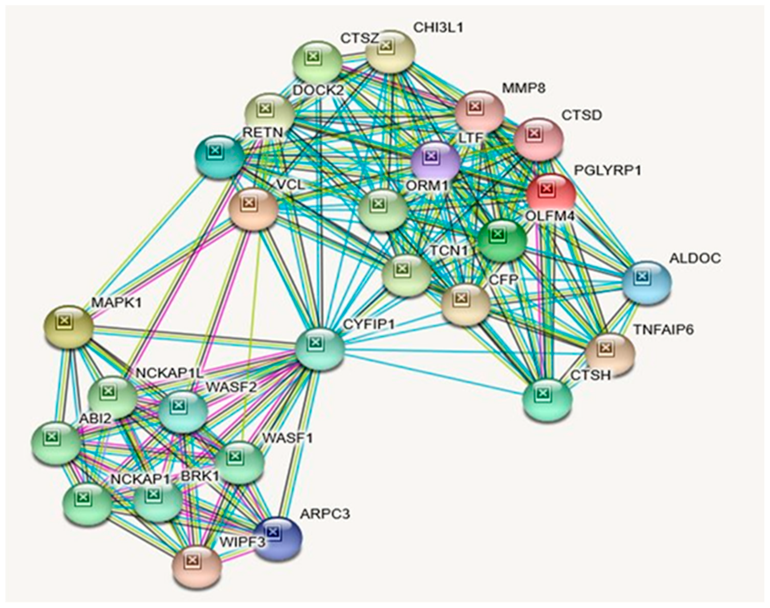
The CYFIP1 gene is highly expressed in the perinuclear region and enriched in synaptosomes. It is also enriched in membrane ruffles and at the tips of lamellipodia with the following subcellular localizations (GeneCards & UniProtKB/Swiss-Prot). The CYFIP1 gene is widely expressed in all tissues (GenecCards). In addition to this gene’s association with fragile X syndrome, it is also associated with autism (see Table 10). A large chromosomal deletion including this gene is associated with increased risk of schizophrenia and epilepsy in human patients and reduced expression of this gene has also been observed in various human cancers, the encoded protein may inhibit tumor invasion (Entrez Gene summary: RefSeq, May 2017). Among this gene’s related pathways are the regulation of actin dynamics for phagocytic cup formation and signaling by Rho GTPases (Table 9). Gene Ontology (GO) annotations related to this gene include Rac GTPase binding. An important paralog of this gene is CYFIP2 (GeneCards). Table 10 lists putative associated diseases for CYFIP1 Gene. STRING protein interactions with their functions and descriptions related to CYFIP1 are presented in Table 11. An illustration of the identified interactions can also be visualized in Figure 5.
Protein–Protein Inter-Relationships Involving CYFIP1 Gene.

2.5. TUBGCP5 (Tubulin Gamma Complex Associated Protein 5) Gene

Attributes, Location, Description, Function, and Associated Disorders for TUBGCP5 Gene.
The chromosomal band location for TUBGCP5 gene is 15q11.2 with the genomic location (GRCh38/hg38) at 22,983,192–23,039,673 with a size of 56,482 bases in a minus strand orientation (Genecards.org). TUBGCP5 protein has the following attributes: Size: 1024 amino acids; Molecular mass:118321 Da; Quaternary structure: Gamma-tubulin complex which is composed of gamma-tubulin, TUBGCP2, TUBGCP3, TUBGCP4, TUBGCP5 and TUBGCP6 proteins (GeneCards & UniProtKB/Swiss-Prot). This protein has 2 isoforms produced by alternative splicing; the sequence of this isoform differs from the canonical sequence as follows: 1010–1024: LESLALSLMAGMEQS → CEYIMLKYFYLCISL. TUBGCP5 gene is widely expressed with the highest levels in the heart and skeletal muscle with moderate levels in the brain (GeneCards & UniProtKB/Swiss-Prot). An important paralog of this gene is TUBGCP6.
Table 12 shows biological processes, molecular functions, cellular components with KEGG and Reactome pathways related to TUBGCP5. Among its related pathways are Nanog in Mammalian ESC Pluripotency and G-Beta Gamma Signaling. Gene Ontology (GO) annotations related to this gene include microtubule binding, and components of the cytoskeleton (Table 12). Table 13 shows diseases associated with TUBGCP5 gene disturbances based on MalaCards and arranged in descending order including Prader–Willi syndrome as the top disorder followed by schizophrenia and then autism. Table 13 shows proteins and their functions that interact with TUBGCP5, as depicted in Figure 6, illustrating the interactions of the TUBGCP5 gene with other related genes.
Protein–Protein Inter-Relationships Involving the TUBGCP5 Gene.

3. Discussion

Features seen in patients with the 15q11.2 BP1-BP2 deletion (Burnside–Butler) syndrome can be quite variable. Different clinical phenotypes are seen in children with this disorder and can depend on the source of the parental deletion [36]. Recent evidence of partial expression or bias of these four genes depending on the parent of origin leads to possible clinical differences in the offspring. For example, when the deletion is paternal, there is a greater risk of having congenital heart defects [36]. If the mother transmits the deletion, then there is a greater risk of intellectual disability and autism in their affected children [36]. A recent review further showed a phenotype with global and regional measures of surface area and cortical thickness in the brain with subcortical volumes and cognition differences compared to controls. This investigation consisted of 203 individuals compared with 4500 controls without the deletion collected from years 2015–2019 and the average age was 56 years [5]. This international study analyzed the largest CNV and neuroimaging results to date in those with the 15q11.2 BP1-BP2 deletions and non-deletion controls. They reported reduced brain surface area, a thicker cortex, and a smaller nucleus accumbens in the 15q11.2 BP1-BP2 deletion subjects. The significant difference in cortical thickness was more evident in the frontal, cingulate and parietal lobes. Furthermore, cognitive ability was lower for those with the deletion compared with non-deletion individuals suggesting involvement in neural plasticity and development leading to functional brain differences identified clinically [5].
As noted in Table 15, all four genes are protein-coding genes and neither were maternally nor paternally imprinted. Their encoded proteins interact with each other in crucial biological processes and molecular pathways (Figure 2, Table 1 and Table 2). The predicted functional interactions encompassing all four genes included 11 nodes with 34 edges that pertain to biological processes and molecular functions utilizing searchable genomic databases involving the four genes in this 15q11.2 BP1-BP2 microdeletion syndrome (see Table 1). Among them, in addition to magnesium ion transport molecular function, significant other biological processes, such as regulation of axonogenesis and axon extension, regulation and production of bone morphogenetic protein (BMP), regulation of cellular growth and development were observed. These biological processes are relevant to variable clinical phenotypes, such as, autism, seizures, and schizophrenia which are also seen in this microdeletion syndrome. These clinical phenotypes often affect neurological development and are occasionally accompanied by mild dysmorphic features [1,9,10,11,12,13].
Although NIPA1 and NIPA2 share related KEGG and Reactome pathways (Table 3 and Table 6), their cardinal disease associations are different (Table 15): Spastic Paraplegia 6, Autosomal Dominant and Spastic Paraplegia 6 for NIPA1 and Angelman Syndrome and Prader–Willi Syndrome for NIPA2. However, as shown in Table 16, they both are also associated with PWS and AS. Unlike NIPA1 and NIPA2, CYFIP1, and TUBGCP5 genes do not share any KEGG or Reactome pathways (Table 9 and Table 12). As shown in Table 15, cardinal diseases that are associated with the CYFIP1 gene are Fragile X Syndrome and Autism. The cardinal disease that is associated with the TUBGCP5 gene is Prader–Willi syndrome. However, as depicted in Table 16, they both are additionally associated with PWS, while the CYFIP1 gene alone is additionally associated with AS. Most notably, all four syntenic genes in this region are associated with Autism Spectrum Disorder; Schizophrenia; Epilepsy and Down Syndrome (Table 16). Except for TUBGC5, all three genes are associated with the 15q11.2 BP1-BP2 Deletion Syndrome with Attention Deficit Hyperactive Disorder & Learning Disability (Table 16). It should be noted that interacting proteins for CYFIP1 and TUBGCP5 proteins are quite different (Figure 5, Table 11; Figure 6, Table 14, respectively), which is unlike the case of NIPA1 and NIPA2 (Figure 3, Table 5; Figure 4, Table 8, respectively).
It is interesting to note that none of the protein-protein interacting genes for the four genes, which total 70, are the same (Table 5, Table 8, Table 11, and Table 14). However, NIPA1 and NIPA2 proteins, given their involvement with common biological processes (GO) and molecular functions (GO) along with BMP6, BMP7, BMPR1A, BMPR1B, and BMPR2-proteins interact with both. In contrast, none of the protein-protein interactants are similar between CYFIP1 and TUBGCP5 proteins (Table 11 and Table 14), thus affirming their functional dissimilarities.
It should be noted, as illustrated in STRING Figure 2 and Table 1, NIPA1, NIPA2, CYFIP1, and TUBGCP5 proteins show protein–protein interactions among themselves with the 11 nodes and 34 edges addressed earlier. As shown in Table 1, NIPA1 protein interacts with TUBGCP5, CYFIP1, and NIPA2 proteins (scores: 0.995, 0.967, and 0.941, respectively) along with seven other protein interactants, and thus predicted to cause significant biological processes (Table 2), and a significant molecular function is magnesium ion transmembrane transporter activity with a 0.0042 false discovery rate (Table 2).
The varied biological processes and predicted functions of these four genes, as noted above, through their protein–protein interactions with seven other proteins (see Figure 2) collectively could play a role in the neurodevelopmental disorders. They complement regulation associated with lipoproteins and lipid metabolism and adipogenesis, encompassing CFHR1, CFHR3, and BMPR2 protein–protein interactions, given marked obesity is a common finding in PWS.
Examining the interaction of NIPA1 protein with its 11 other interacting proteins indicates that three-fourths of the interactions are important for developmental bone morphogenesis or multifunctional proteins that control proliferation, differentiation, and other functions in many cell types (see Figure 3; Table 5). The interactions of NIPA2 protein with 19 other proteins indicate their collective role as intracellular signal transducers and transcriptional modulators that are activated by TGFbeta, and thereby impacting bone morphogenesis (see Figure 4; Table 8). CYFIP1 protein interacts with 25 other proteins. These proteins have a wide range of activity with functions including cytoskeleton organization and actin filament binding with cell-matrix adhesion, MAP kinase signal transduction impacts cell growth, survival and differentiation, stimulation of glucose uptake in cells, intracellular protein breakdown and tissue remodeling (see Figure 5; Table 11). TUBGCP5 protein interacts with 25 other proteins. These proteins are focused on mitotic spindle formation and assembly, microtubule organization, and production of centrosomal proteins involved in the regulation of centriole duplication during cell division (see Figure 6; Table 14).

3.1. Overlapping Association of PWS and AS Specific Genes with Both Syndromes at Varying Degrees

Although UBE3A is a classical AS gene (9.31 score), it is also associated with PWS to a lesser degree (4.106 score) (https://www.malacards.org/search/results/UBE3A). Further, it should be noted that the 15q11-q13 region includes the imprinted gene cluster with two maternally expressed protein-coding genes UBE3A and ATP10A (https://www.malacards.org/search/results?query=ATP10A). Similarly, all of the classical PWS genes overlap with both PWS and AS, albeit to varying degrees of significance: MAGEL2: PWS/AS: SCORE: 7.035/3.937; NDN: PWS/AS: SCORE: 7.435/4.528; NDAP1: PWS/AS: SCORE: 6.321/4.816; SNRPN: PWS/AS: SCORE: 6.563/5.584; SNORD116: PWS/AS: SCORE: 0.426/0.121; and even the IPW region: PWS/AS: SCORE: 7.761/0.100 (https://www.malacards.org). Therefore, it may not be appropriate to assume that PWS/AS are exclusively caused only by the designated PWS and AS regional genes per se, as shown in Figure 1, since most of these genes are also overlappingly associated with both PWS and AS, although to varying degrees. Moreover, PWS/AS phenotype is also overlappingly associated with three proximally located contiguous genes within the 15q11.2 BP1-BP2 region: NIPA1, NIPA2, and CYFIP1, as depicted in Table 16.
PWS is caused by loss of expression of the paternally inherited genes on chromosome 15q11.2-q13, and the cardinal features of PWS are attributable to the critical interval within the 15q11.2-q13 imprinted gene cluster, containing the small nucleolar RNA (snoRNA) SNORD116 and non-coding RNA IPW (Imprinted in Prader–Willi) exons [39,40,41]. Similarly, the cardinal features of AS are attributable to the loss of expression of maternally inherited genes, and UBE3A in particular, which causes a distinct AS neurodevelopmental disorder [42].
Although the PWS core features are attributable to the critical interval within the 15q11.2-q13 imprinted gene cluster and the AS core features are attributable to the loss of expression of maternally inherited gene UBE3A, these core features might be influenced by several other genes that lie within the broader BP1-BP3 region. The functions of all of these genes are in-turn likely modulated by hundreds of their interacting genes that lie throughout the genome, as shown in the STRING protein interactions for NAPI1, NAPI2, CYFIP1, and TUBGCP5: Figure 2, Table 1; Figure 3, Table 5; Figure 4, Table 8; Figure 5, Table 11; Figure 6, Table 14; Table 15 and Table 16. One could speculate that PWS/AS, similar to that of ASD, depends on either the involvement of the PWS critical region in the imprinted segment within the 15q11.2-q13 BP2-BP3 region or the involvement of a critical gene, namely, UBE3A in the maternally imprinted segment [43] and in concert with other genes that lie within chromosome 15, not excluding the 15q11.2 BP1-BP2 region genes.
Every one of these genes along with their hundreds of interacting genes within the genome as a whole, as depicted and tabulated in this study pertaining to NIPA1, NIPA2, CYFIP1, and TUBGCP5 genes all in concert could impact on the PWS/AS signature phenotype in any given proband. Thus, each PWS/AS proband’s clinical presentation is likely to be a unique genomic signature, within the broader spectrum of PWS/AS clinical presentations, which is akin to Autism Spectrum Disorder.

3.2. 15q11.2 B-P1-BP2 Microdeletion (Burnside–Butler) Syndrome: Characterization of Genes Within the BP1-BP2 Region, Meta-Analysis, and Parent-of-origin Effects Reveal Neurodevelopmental Associated Phenotypes

Each of the four evolutionarily conserved genes that escape imprinting and contiguously lie within the BP1-BP2 region are associated with distinct cardinal diseases: NIPA1: Spastic Paraplegia 6, Autosomal Dominant and Spastic Paraplegia 6; NIPA2: Angelman Syndrome and Prader–Willi Syndrome; CYFIP1: Fragile X Syndrome and Autism; and TUBGCP5: Prader–Willi Syndrome (see Table 15). Based on reports of their mutations or disturbed expression patterns, as reported in authentic web resources, such as, GeneCards.org, SAFARI.org, Gene Ontology (GO), OMIM.org, Entrez and UniProtKB/Swiss-Prot, their “collective loss or dosage duplication” is due to the BP1-BP2 microdeletion or microduplication that appears to impact only behavioral and neurological functions. These disorders include speech and motor delays, behavioral problems, seizures, and autism in affected individuals [44]. A recent review of the 15q11.2 BP1–BP2 microdeletion syndrome [1] found common phenotypic features, which included autism, developmental delay, motor and language delays, and behavioral problems.
Parental studies among these subjects demonstrated phenotypically normal carriers in several instances, and mildly affected carriers in others, complicating phenotypic association and/or causality. This could be due to either reduced penetrance or altered gene dosage on a particular genetic background [1,4]. The four non-imprinted and biallelically expressed genes, NIPA1, NIPA2, CFYIP1 and TUBGCP5, in this microdeletion were initially noted to impact the severity of clinical presentation and neurological impairment in these two classical genomic imprinting disorders, Prader–Willi and Angelman syndromes with typical 15q11–q13 deletions depending on the absence or presence of the genomic area between breakpoints BP1 and BP2 containing these four genes, which led to the recognition of this microdeletion (Burnside–Butler) syndrome [4].
Therefore, it is intriguing to note that the review [4] of over 200 15q11.2 BP1–BP2 microdeletion syndrome individuals, as meta-analysis, did not find either the NIPA1 associated cardinal disease: Spastic Paraplegia, the NIPA2 associated cardinal diseases: Angelman Syndrome and Prader–Willi Syndrome, the CYFIP1 associated cardinal disease: Fragile X Syndrome, or the TUBGCP5 associated cardinal disease: Prader–Willi Syndrome, except for the CYFIP1 associated cardinal disease: Autism, as presented in Table 15.
The clinical features as noted in the review [4] included developmental (73%) and speech (67%) delays; dysmorphic ears (46%) and palatal anomalies (46%); writing (60%) and reading (57%) difficulties, memory problems (60%) and verbal IQ scores ≤75 (50%); general behavioral, unspecified (55%) and abnormal brain imaging (43%). Other clinical features noted in this review [4], but not considered as common were seizures/epilepsy (26%), autism spectrum disorder (27%), attention deficit disorder (ADD)/attention deficit hyperactivity disorder (ADHD) (35%), schizophrenia/paranoid psychosis (20%), and motor delay (42%).
The review [4] also found that not all individuals with this deletion were clinically affected, but neuropsychiatric and behavior disturbances and mild dysmorphic features were associated with genomic imbalances of the 15q11.2 BP1–BP2 region, including microdeletions, but with an apparent incomplete penetrance and variable expressivity.

3.3. All Four BP1-BP2 Region Genes Are Significantly Associated with Autism Spectrum Disorder

From reported patient cohorts presenting for genetic services and microarray analysis, this microdeletion syndrome can now be recognized as the most common cytogenetic abnormality found in ASD instead [6], which is not only a cardinal CYFIP1 associated disease (see Table 15), but ASD is also consistently associated with TUBGCP5, NIPA1, and NIPA2 genes (see Table 4, Table 7, Table 10, Table 13, and Table 16).
Thus, all four genes in this narrow segment between BP1 and BP2 are significantly associated with autism spectrum disorder, with an average of >68 MalaCards InFormaTion Score (MIFTS) with annotation strength (max 100) and with Solr (an open-source enterprise-search platform) relevance scores 0.171, 0.088, 4.578/0.377, and 0.21/0.094 for NIPA1, NIPA2, CYFIP1, and TUBGCP5, respectively (Table 4, Table 7, Table 10, Table 13, and Table 16). Moreover, even PWS and AS specific genes that lie in the BP2-BP3 PWS/AS region, such as, MAGEL2, SNRPN, UBE3A, and ATP10A are also significantly co-associated with autism (Malacards.org; SFARI.org) [45]. Therefore, it seems fair to conclude that except for PWS specific genes or transcripts, such as, snoRNAs, most other genes that flank on either side in broadly designated PWS and AS regions are all co-associated with autism. Most of the genes within the broader BP1-BP3 region are also co-associated with not only PWS and AS, but also with autism (Malacards.org; SFARI.org) [45]. Even the GABRB3 gene that is distal to the ATP10A gene is a SFARI (SFARI.org) recognized autism gene (Malacards.org; SFARI.org) [45].

3.4. 15q11.2 B–P1–BP2 Microdeletion (Burnside–Butler) Syndrome: Frequency

The 15q11.2 BP1–BP2 microdeletion syndrome has a reported de novo frequency between 5%–22%, with 51% having inherited the microdeletion from an apparently unaffected parent and 35% having inherited the microdeletion from an affected parent [4,6]. These low penetrance estimates may relate to subclinical manifestations of neuropsychiatric/behavioral problems, incomplete information, or lack of detailed clinical or psychiatric studies in the parents of individuals with 15q11.2 BP1–BP2 microdeletion or members of control cohorts.

3.5. Summary

The four genes within the narrower proximal BP1-BP2 region, NIPA1, NIPA2, CYFIP1, and TUBGCP5, as well as those that lie within the broader BP2-BP3 region, such as MAGEL2, SNRPN, UBE3A, ATP10A, as well as GABRB3 gene that are farther away from the distal ATP10A gene- are all recognized ASD genes. Furthermore, the 15q11.2 region collectively represents PWS, AS, and autism phenotypes (Malacards.org; SFARI.org) [45].
In addition to the above consistent correlation of each of the four genes that lie within the narrow proximal BP1-BP2 region with autism, in a more recent study [36] parent-of-origin effects (POE) of the 15q11.2 BP1-BP2 microdeletion were found to be associated with differences in clinical features in individuals inheriting the deletion. Among all probands studied, maternal deletions were found to be associated with epilepsy, autism spectrum disorder (p = 0.02) and macrocephaly (p = 0.016), while paternal deletions were associated with congenital heart disease (CHD) (p = 0.004) and abnormal muscular phenotypes (p < 0.05), while CHD and abnormal muscular phenotypes were seen in paternal deletions. This study not only supported POEs of this deletion, but notably included ASD, macrocephaly, and epilepsy.
The above key processes and summary of the four genes within the narrower proximal 15q11.2 BP1-BP2 region are collectively critical for normal neuronal development, plasticity, and function [1,5]. Their loss or gain (CNV) are associated with neurodevelopmental disorders, spastic paraplegia, seizures, learning, and gait disturbances with motor delay and neuro-behavioral psychiatric problems including autism, dyslexia, and schizophrenia/paranoid psychosis [1].
The most common maladies found to be individually associated with each of the four genes are: spastic paraplegia found in the top 12 of 15 disorders associated with NIPA1; PWS/AS, epilepsy and psychiatric/behavioral problems (autism or schizophrenia) in the top 10 disorders for NIPA2; fragile X syndrome, autism, schizophrenia, PWS and pervasive developmental disorder in the top 10 disorders for CYFIP1; and PWS, schizophrenia, autism, microcephaly, essential hypertension, body mass index (quantitative trait), epilepsy, and Down syndrome in the top 10 disorders associated with TUBGCP5 (see Table 16). As noted in Table 15, all four genes are neither maternally nor paternally imprinted, but their proteins interact with each other as described and illustrated (see Figure 2, Table 1 and Table 2) in crucial biological processes (GO) and molecular pathways (GO). Collectively, either due to the microdeletion or microduplication, these genes predominantly affect brain morphology [1,4].
Our detailed exploration of the various aspects of each of these four genes, in solo and in concert is meant to enable an in-depth understanding of their individual and collective contribution in the causation of different neurodevelopmental phenotypes that have been variably reported in the literature. All four genes are individually associated with Prader–Willi syndrome, autism spectrum disorder, schizophrenia, epilepsy, and Down syndrome (Maladies.org; GeneCards.org). Except for the TUBGCP5 gene, all three remaining genes are associated with Angelman syndrome, although the TUBGCP5 gene is located most proximal to the Angelman syndrome gene, UBE3A. TUBGCP5 gene is also not associated with attention deficit hyperactivity disorder and learning disability, developmental disorder, and peripheral nervous system disease. CYFIP1 is the only gene that is not associated with microcephaly, but it is the only gene that is associated with a developmental disorder (see Table 16).
Thus, collectively, all four genes are associated with ten overlapping neurodevelopmental maladies up to 77.5% of the time.

4. Materials and Methods

We characterized the four genes (NIPA1, NIPA2, CYFIP1, and TUBGCP5) found within the 15q11.2 BP1-BP2 chromosome region of about 500 bp in size. When this region is deleted, an emerging disorder (i.e., Burnside–Butler syndrome) is being characterized leading to the importance of our description of these four genes. We undertook a literature review of hundreds of patients reported and summarized clinically with this microdeletion reported as the most common genetic defect found in consecutive patients presenting for genetic testing with high-resolution microarray analysis and autism spectrum disorder (e.g., [6]). Genome databases, search platforms for gene interaction, gene ontology (GO) biological process, molecular function, cellular components, along with KEGG and Reactome pathways using STRING network data along with sources, GeneCards.org, SAFARI.org, KEGG, PubMed, Gene Ontology (GO), OMIM.org, Entrez and UniProtKB/Swiss-Prot were summarized in tabular form and STRING Network results illustrated in figures and discussed in the text involving all four genes. An overview of each gene was also described throughout the summary process utilizing an approach reported previously [46].
The Gene Ontology Resource (GO; http://geneontology.org) provides structured, computable knowledge regarding the functions of genes and gene products. Founded in 1998, GO has become widely adopted in the life sciences, and its contents are under continual improvement, both in quantity and in quality. The Gene Ontology resource (GO; http://geneontology.org) is the most comprehensive and widely used knowledge base concerning the functions of genes. In GO, all functional knowledge is structured and represented in a form amenable to computational analysis, which is essential to support modern biological research. The GO knowledge base is structured using a formal ontology, by defining classes of gene functions (GO terms) developed over the past 20 years that have specified relations to each other [47]. GO terms include definitions, or equivalence axioms, that define the term relative to other terms in the GO or other ontologies. Their relationships can be computationally inferred using logical reasoning. The GO structure is constantly evolving in response to new scientific discoveries and are continuously refined using current biological information. The GO knowledgebase consists of ontology and annotations. As of the 5 September 2018 release (doi:10.5281/zenodo.1410625), there were ∼45000 terms in GO: 29698 biological processes, 11147 molecular functions, and 4201 cellular components, linked by almost 134000 relationships [47].
MalaCards Relevance Scores provide information used for the construction of our tables of Putative Associated Diseases (see Table 4, Table 7, Table 10 and Table 13) for the readership. MalaCards is an integrated database of human maladies and their annotations modeled from the architecture and information from the popular GeneCards database of human genes. The search platform used by MalaCards to obtain these data [48] is SOLR, an open-source full-text search platform widely used for analytics based on Apache’s Lucene text search API. When a term is searched by Lucene then it returns a set of scored hits. A “hit” represents a document (a MalaCard), whose fields (actual annotations) were previously indexed by Lucene.
The scoring is calculated by a Lucene defined algorithm:
score(q,d) = coord(q,d) · queryNorm(q) · ∑ tf(t in d) · idf(t) · t.getBoost() · norm(t,d)
 t in q
The factors in this formula are:
  • tf stands for term frequency—the more times a search term appears in a document, the higher the score
  • idf stands for inverse document frequency—matches on rarer terms count more than matches on common terms
  • coord is the coordination factor—if there are multiple terms in a query, the more terms that match, the higher the score
  • lengthNorm—matches on a smaller field score higher than matches on a larger field
  • index-time boost—if a boost was specified for a document at index time, scores for searches that match that document will be boosted.
  • query clause boost—a user may explicitly boost the contribution of one part of a query over another.
To ensure showing the best precision, MalaCards displays the score as (base 2 log of the score) + 10
For more about Lucene’s scoring mechanism see Apache Lucene—Scoring [48].
GeneCards.org—the human gene database [49] is from the GeneCards Suite: From Gene Data Mining to Disease Genome Sequence Analysis.
STRING protein network was utilized to generate Figure 2, Figure 3, Figure 4, Figure 5 and Figure 6 and their accompanying data on the protein–protein interactions as well as their Biological Processes (GO), Molecular Functions (GO), Cellular Component (GO) and KEGG and Reactome Pathways. The data are presented in Table 1, Table 2 and Table 3, Table 5, Table 6, Table 8, Table 9, Table 11, Table 12, and Table 14 and are adopted from STRING CONSORTIUM 2019. Data in Table 15 were derived from the GeneCards website (GeneCards.org) for respective genes, while the data for Table 16 were derived from MalaCards diseases (Malacards.org) as seen in Table 4, Table 7, Table 10, and Table 13 in this study.

Author Contributions

Based on M.G.B.’s suggestion of this research topic, S.K.R. independently researched and originally drafted the entire content of this manuscript for which M.G.B. contributed to Figure 1, contributed towards drafting the introduction and critically reviewed and edited the entire manuscript. Title, abstract, all figure legends, Figure 2, Figure 3, Figure 4, Figure 5 and Figure 6, Tabulations 1 through 16, results, interpretations, discussions, and conclusions were exclusively by S.K.R. Table 15 and Table 16 were originally synthesized by S.K.R. from various reputable databases, such as GeneCards.org, MalaCards.org, SAFARI.Org, KEGG, PubMed, Gene Ontology (GO), OMIM.org, Entrez, UniProtKB/Swiss-Prot and from https://compartments.jensenlab.org. All authors have read and agree to the published version of the manuscript.

Funding

We acknowledge the National Institute of Child Health and Human Development (NICHD) grants number HD02528 and KUMC Research Institute and the KUMC Research Institute, Clinical Pilot Research Grant Program.

Acknowledgments

We thank the reviewers for their constructive and valid suggestions and comments. We thank Waheeda Hossain at KUMC for her help in formatting this manuscript and amending Figure 1. We also thank Grace Graham at KUMC for her technical help. Syed Rafi obtained permissions via emails from the following organizations for the reproduction of their website figures and data pertaining to the four genes studied and we are grateful: 1. STRING Corporation 2019: On Nov. 11, 2019 under STRING’s CC BY 4.0′ license. 2. GeneCards.org, on Nov. 11, 2019, Marilyn Safran, Head of GeneCard’s Development. 3. MalaCards.org, on Dec. 23, 2019, Marilyn Safran, Head of GeneCard’s Development, and 4. UniProtKB/Swiss-Prot; Uniprot.Org, on Dec. 23, 2019.

Conflicts of Interest

There are no conflicts of interest noted by either author.

References

  1. Butler, M.G. Clinical and genetic aspects of the 15q11.2 BP1-BP2 microdeletion disorder. J. Intellect. Disabil. Res. 2017, 61, 568–579. [Google Scholar] [CrossRef] [PubMed]
  2. Bittel, D.C.; Butler, M.G. Prader-Willi syndrome: Clinical genetics, cytogenetics and molecular biology. Expert Rev. Mol. Med. 2005, 7, 1–20. [Google Scholar] [CrossRef] [PubMed]
  3. Butler, M.G.; Lee, P.D.K.; Whitman, B.Y. Management of Prader-Willi Syndrome, 3rd ed.; Springer: New York, NY, USA, 2006. [Google Scholar]
  4. Cox, D.M.; Butler, M.G. The 15q11.2 BP1-BP2 microdeletion syndrome: A review. Int. J. Mol. Sci. 2015, 16, 4068–4082. [Google Scholar] [PubMed]
  5. Writing Committee for the ENIGMA-CNV Working Group; Van Der Meer, D.; Sønderby, I.E.; Kaufmann, T.; Walters, G.B.; Abdellaoui, A.; Ames, D.; Amunts, K.; Andersson, M.; Armstrong, N.J.; et al. Association of copy number variation of the 15q11.2 BP1-BP2 region with cortical and subcortical morphology and cognition. JAMA Psychiatry 2019, 30, 1–11. [Google Scholar]
  6. Ho, K.S.; Wassman, E.R.; Baxter, A.L.; Hensel, C.H.; Martin, M.M.; Prasad, A.; Twede, H.; Vanzo, R.J.; Butler, M.G. Chromosomal microarray analysis of consecutive individuals with autism spectrum disorders using an ultra-high-resolution chromosomal microarray optimized for neurodevelopmental disorders. Int. J. Mol. Sci. 2016, 17, 2070. [Google Scholar] [CrossRef] [Green Version]
  7. Butler, M.G. Magnesium supplement and the 15q11.2 BP1-BP2 microdeletion (Burnside-Butler) syndrome: A potential treatment? Int. J. Mol. Sci. 2019, 20, 2914. [Google Scholar] [CrossRef] [Green Version]
  8. Das, D.K.; Tapias, V.; D’’Aiuto, L.; Chowdari, K.V.; Francis, L.; Zhi, Y.; Ghosh, A.; Surti, U.; Tischfield, J.; Sheldon, M.; et al. Genetic and morphological features of human iPSC-derived neurons with chromosome 15q11.2 (BP1-BP2) deletions. Mol. Neuropsychiatry 2015, 1, 116–123. [Google Scholar] [CrossRef] [Green Version]
  9. Chai, J.-H.; Locke, D.; Greally, J.M.; Knoll, J.H.M.; Ohta, T.; Dunai, J.; Yavor, A.; Eichler, E.E.; Nicholls, R.D. Identification of four highly conserved genes between breakpoint hotspots BP1 and BP2 of the Prader-Willi/Angelman syndromes deletion region that have undergone evolutionary transposition mediated by flanking duplicons. Am. J. Hum. Genet. 2003, 73, 898–925. [Google Scholar] [CrossRef] [Green Version]
  10. Jiang, Y.; Zhang, Y.; Zhang, P.; Sang, T.; Zhang, F.; Ji, T.; Huang, Q.; Xie, H.; Du, R.; Cai, B.; et al. NIPA2 located in 15q11.2 is mutated in patients with childhood absence epilepsy. Hum. Genet. 2012, 131, 1217–1224. [Google Scholar]
  11. Jiang, Y.; Zhang, Y.; Zhang, P.; Zhang, F.; Xie, H.; Chan, P.; Wu, X. NIPA2 mutations are correlative with childhood absence epilepsy in the Han Chinese population. Hum. Genet. 2014, 133, 675–676. [Google Scholar] [CrossRef]
  12. Jerkovich, A.M.; Butler, M.G. Further phenotypic expansion of 15q11.2 BP1-BP2 microdeletion (Burnside-Butler) syndrome. J. Pediatric Genet. 2014, 3, 41–44. [Google Scholar] [CrossRef] [PubMed]
  13. Jerkovich, A.M.; Butler, M.G. 15q11.2 Microdeletion (BP1-BP2) and developmental delay, behaviour issues, epilepsy and congenital heart disease: A series of 52 patients. Eur. J. Med. Genet. 2015, 58, 140–147. [Google Scholar]
  14. Fagerberg, L.; Hallström, B.M.; Oksvold, P.; Kampf, C.; Djureinovic, D.; Odeberg, J.; Habuka, M.; Tahmasebpoor, S.; Danielsson, A.; Edlund, K.; et al. Analysis of the human tissue-specific expression by genome-wide integration of transcriptomics and antibody-based proteomics. Mol. Cell Proteomics 2014, 13, 397–406. [Google Scholar] [CrossRef] [PubMed] [Green Version]
  15. Goytain, A.; Hines, R.; El-Husseini, A.; Quamme, G.A. NIPA1(SPG6), the basis for autosomal dominant form of hereditary spastic paraplegia, encodes a functional Mg2+ transporter. J. Biol. Chem. 2007, 282, 8060–8068. [Google Scholar] [CrossRef] [Green Version]
  16. Rainier, S.; Chai, J.-H.; Tokarz, D.; Nicholls, R.D.; Fink, J.K. NIPA1 gene mutations cause autosomal dominant hereditary spastic paraplegia (SPG6). Am. J. Hum. Genet. 2003, 73, 967–971. [Google Scholar] [CrossRef] [Green Version]
  17. Zhao, J.; Matthies, D.S.; Botzolakis, E.J.; Macdonald, R.L.; Blakely, R.D.; Hedera, P. Hereditary spastic paraplegia-associated mutations in the NIPA1 gene and its Caenorhabditis elegans homolog trigger neural degeneration in vitro and in vivo through a gain-of-function mechanism. J. Neurosci. 2008, 28, 13938–13951. [Google Scholar] [CrossRef] [Green Version]
  18. Chen, C.-P.; Lin, S.-P.; Lee, C.; Chern, S.-R.; Wu, P.-S.; Chen, Y.-N.; Chen, S.-W.; Wang, W. Familial transmission of recurrent 15q11.2 (BP1-BP2) microdeletion encompassing NIPA1, NIPA2, CYFIP1, and TUBGCP5 associated with phenotypic variability in developmental, speech, and motor delay. Taiwan J. Obstet. Gynecol. 2017, 56, 93–97. [Google Scholar] [CrossRef]
  19. Leblond, C.S.; Heinrich, J.; Delorme, R.; Proepper, C.; Betancur, C.; Huguet, G.; Konyukh, M.; Chaste, P.; Ey, E.; Råstam, M.; et al. Genetic and functional analyses of SHANK2 mutations suggest a multiple hit model of autism spectrum disorders. PLoS Genet. 2012, 8, E1002521. [Google Scholar] [CrossRef] [Green Version]
  20. Klebe, S.; Durr, A.; Bouslam, N.; Grid, D.; Paternotte, C.; Depienne, C.; Hanein, S.; Bouhouche, A.; Elleuch, N.; Azzedine, H.; et al. Spastic Paraplegia 5: Locus refinement, candidate gene analysis and clinical description. Am. J. Med. Genet. B Neuropsychiatr. Genet. 2007, 144B, 854–861. [Google Scholar] [CrossRef]
  21. Botzolakis, E.J.; Zhao, J.; Gurba, K.N.; Macdonald, R.L.; Hedera, P. The effect of HSP-causing mutations in SPG3A and NIPA1 on the assembly, trafficking, and interaction between atlastin-1 and NIPA1. Mol. Cell Neurosci. 2011, 46, 122–135. [Google Scholar] [CrossRef] [Green Version]
  22. Butler, M.G. Single gene and syndromic causes of obesity: Illustrative examples. In Progress in Molecular Biology and Translational Science; Tao, Y., Ed.; Elsevier Inc: Chennai, India, 2016; Volume 140, pp. 1–45. [Google Scholar]
  23. Butler, M.G.; Hartin, S.N.; Hossain, W.A.; Manzardo, A.M.; Kimonis, V.E.; Dykens, E.; Gold, J.A.; Kim, S.-J.; Weisensel, N.; Tamura, R.; et al. Molecular genetic classification in Prader-Willi syndrome: A multisite cohort study. J. Med. Genet. 2019, 56, 149–153. [Google Scholar] [CrossRef] [PubMed]
  24. Chen, S.; Song, C.; Guo, H.; Xu, P.; Huang, W.; Zhou, Y.; Sun, J.; Li, C.-X.; Du, Y.; Li, X.; et al. Distinct novel mutations affecting the same base in the NIPA1 gene cause autosomal dominant hereditary spastic paraplegia in two Chinese families. Hum. Mutat. 2005, 25, 135–141. [Google Scholar] [CrossRef] [PubMed]
  25. Zhao, M.; Chen, Y.-J.; Wang, M.-W.; Lin, X.-H.; Dong, E.-L.; Chen, W.-J.; Wang, N.; Lin, X. Genetic and clinical profile of Chinese patients with autosomal dominant spastic paraplegia. Mol. Diagn. Ther. 2019, 23, 781–789. [Google Scholar] [CrossRef] [PubMed]
  26. Hildebrand, M.S.; Damiano, J.A.; Mullen, S.A.; Bellows, S.; Scheffer, I.E.; Berkovic, S.F. Does variation in NIPA2 contribute to genetic generalized epilepsy? Hum. Genet. 2014, 133, 673–674. [Google Scholar] [CrossRef] [Green Version]
  27. Cowan, C.M.; Jiang, X.; Hsu, T.; Soo, C.; Zhang, B.; Wang, J.Z.; Kuroda, S.; Wu, B.; Zhang, Z.; Zhang, X.; et al. Synergistic effects of Nell-1 and BMP-2 on the osteogenic differentiation of myoblasts. J. Bone Miner. Res. 2007, 22, 918–930. [Google Scholar] [CrossRef] [Green Version]
  28. Wang, X.; Shaw, W.R.; Tsang, H.T.H.; Reid, E.; O’Kane, C.J. Drosophila spichthyin inhibits BMP signaling and regulates synaptic growth and axonal microtubules. Nat. Neursci. 2007, 10, 177–185. [Google Scholar] [CrossRef] [Green Version]
  29. Lee, K.J.; Mendelsohn, M.; Jessel, T.M. Neuronal patterning by BMPs: A requirement for GDF7 in the generation of a discrete class of commissural interneurons in the mouse spinal cord. Genes Dev. 1998, 12, 3394–3407. [Google Scholar] [CrossRef] [Green Version]
  30. Wu, H.-H.; Ivkovic, S.; Murray, R.C.; Jaramillo, S.; Lyons, K.S.; Johnson, J.E.; Calof, A.L. Autoregulation of neurogenesis by GDF11. Neuron 2003, 37, 197–207. [Google Scholar] [CrossRef] [Green Version]
  31. Matsuura, I.; Taniguchi, J.; Hata, K.; Saeki, N.; Yamashita, T. BMP inhibition enhances axonal growth and functional recovery after spinal cord injury. J. Neurochem. 2008, 105, 1471–1479. [Google Scholar] [CrossRef]
  32. Sahni, V.; Mukhopadhyay, A.; Tysseling, V.; Hebert, A.; Birch, D.; Mcguire, T.L.; Kessler, J.A. BMPR1a and BMPR1b signaling exert opposing effects on gliosis after spinal cord injury. J. Neurosci. 2010, 30, 1839–1855. [Google Scholar] [CrossRef] [Green Version]
  33. Bayat, V.; Jaiswal, M.; Bellen, H.J. The BMP signaling pathway at the Drosophila neuromuscular junction and its links to neurodegenerative diseases. Curr. Opin. Neurobiol. 2011, 21, 182–188. [Google Scholar] [CrossRef] [PubMed] [Green Version]
  34. Blackstone, C. Cellular pathways of hereditary spastic paraplegia. Annu. Rev. Neurosci. 2012, 35, 25–47. [Google Scholar] [CrossRef] [PubMed] [Green Version]
  35. Huttlin, E.L.; Bruckner, R.J.; Paulo, J.A.; Cannon, J.R.; Ting, L.; Baltier, K.; Colby, G.; Gebreab, F.; Gygi, M.P.; Parzen, H.; et al. Architecture of the human interactome defines protein commmunities and disease networks. Nature 2017, 25, 505–509. [Google Scholar] [CrossRef] [PubMed]
  36. Davis, K.W.; Serrano, M.A.; Loddo, S.; Robinson, C.; Alesi, V.; Dallapiccola, B.; Novelli, A.; Butler, M.G. Parent-of-origin effects in 15q11.2 BP1-BP2 microdeletion (Burnside-Butler) syndrome. Int. J. Mol. Sci. 2019, 20, 1459. [Google Scholar] [CrossRef] [Green Version]
  37. Bogdan, S.; Grewe, O.; Strunk, M.; Mertens, A.; Klämbt, C. Sra-1 interacts with Kette and Wasp and is required for neuronal and bristle development in Drosophila. Development 2004, 131, 3981–3989. [Google Scholar] [CrossRef] [Green Version]
  38. Napoli, I.; Mercaldo, V.; Boyl, P.P.; Eleuteri, B.; Zalfa, F.; De Rubeis, S.; Di Marino, D.; Mohr, E.; Massimi, M.; Falconi, M.; et al. The fragile X syndrome protein represses activity-dependent translation through CYFIP1, a new 4E-BP. Cell 2008, 134, 1042–1054. [Google Scholar] [CrossRef] [Green Version]
  39. Doe, C.M.; Relkovic, D.; Garfield, A.S.; Dalley, J.W.; Theobald, D.E.; Humby, T.; Wilkinson, L.S.; Isles, A.R. Loss of the imprinted snoRNA mbii-52 leads to increased 5htr2c pre-RNA editing and altered 5HT2CR-mediated behaviour. Hum. Mol. Genet. 2009, 18, 2140–2148. [Google Scholar] [CrossRef]
  40. Stelzer, Y.; Sagi, I.; Yanuka, O.; Eiges, R.; Benvenisty, N. The noncoding RNA IPW regulates the imprinted DLK1-DIO3 locus in an induced pluripotent stem cell model of Prader-Willi syndrome. Nat. Genet. 2014, 46, 551–557. [Google Scholar] [CrossRef]
  41. Garfield, A.; Davies, J.; Burke, L.; Furby, H.; Wilkinson, L.; Heisler, L.; Isles, A. Increased alternate splicing of Htr2c in a mouse model for Prader-Willi syndrome leads disruption of 5HT2C receptor mediated appetite. Mol. Brain 2016, 9, 95. [Google Scholar] [CrossRef] [Green Version]
  42. Clayton-Smith, J.; Laan, L. Angelman syndrome: A review of the clinical and genetic aspects. J. Med. Genet. 2003, 40, 87–95. [Google Scholar] [CrossRef]
  43. Galiveti, C.R.; Raabe, C.A.; Konthur, Z.; Rozhdestvensky, T.S. Differential regulation of non-protein coding RNAs from Prader-Willi syndrome locus. Sci. Rep. 2014, 4, 6445. [Google Scholar] [CrossRef] [PubMed] [Green Version]
  44. Burnside, R.D.; Pasion, R.; Mikhail, F.M. Microdeletion/microduplication of proximal 15q11.2 between BP1 and BP2: A susceptibility region for neurological dysfunction including developmental and language delay. Hum. Genet. 2011, 130, 517–528. [Google Scholar] [CrossRef] [PubMed]
  45. Butler, M.G.; Rafi, S.K.; Manzardo, A.M. High-resolution chromosome ideogram representation of currently recognized genes for autism spectrum disorders. Int. J. Mol. Sci. 2015, 16, 6464–6495. [Google Scholar] [CrossRef] [PubMed] [Green Version]
  46. Rafi, S.; Fernández-Jaén, A.; Alvarez, S.; Nadeau, O.W.; Butler, M.G. High functioning autism with missense mutations in Synaptotagmin-like protein 4 (SYTL4) and Transmembrane protein 187 (TMEM187) genes: SYTL4- protein modeling, protein-protein interaction, expression profiling and microRNA studies. Int. J. Mol. Sci. 2019, 20, 3358. [Google Scholar] [CrossRef] [Green Version]
  47. Ontology, G.; Genet, N. The Gene Ontology Consortium. Nucleic Acids Res. 2019, 47, D330–D338. [Google Scholar]
  48. Rappaport, N.; Twik, M.; Plaschkes, I.; Nudel, R.; Iny Stein, T.; Levitt, J.; Lancet, D. MalaCards: An amalgamated human disease compendium with diverse clinical and genetic annotation and structured search. Nucleic Acids Res. 2017, 45, D877–D887. [Google Scholar] [CrossRef] [Green Version]
  49. Stelzer, G.; Rosen, N.; Plaschkes, I.; Zimmerman, S.; Twik, M.; Fishilevich, S.; Kaplan, S. The GeneCards suite: From gene data mining to disease genome sequence analyses. Curr. Protoc. Bioinform. 2016, 54, 1–30. [Google Scholar] [CrossRef]
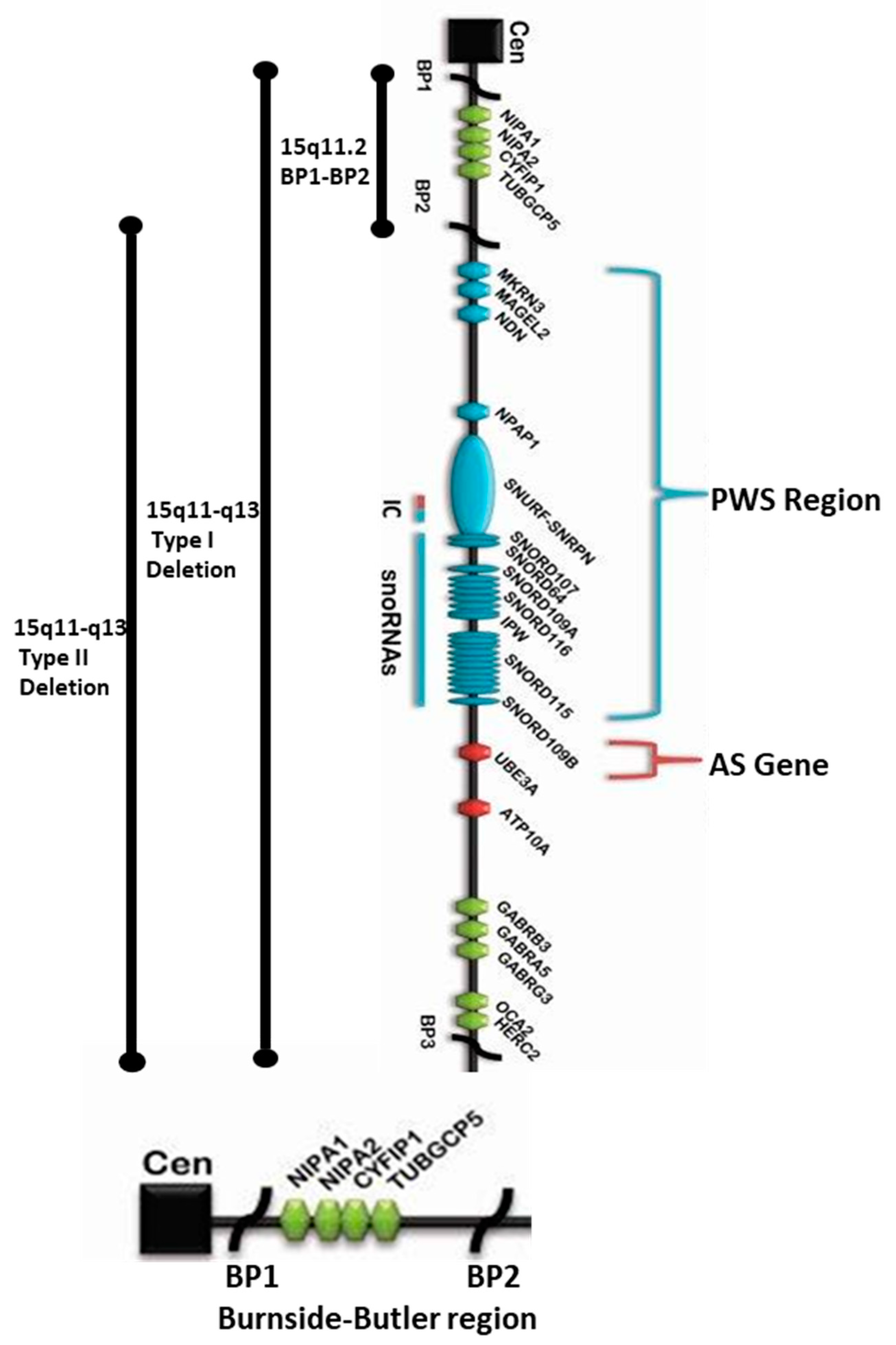
Figure 1. 15q11.2 BP1-BP2 microdeletion (Burnside–Butler) syndrome region found at the proximal end of Prader–Willi syndrome (PWS) / Angelman syndrome (AS) regions within the 15q11-q13 Type I deletion depicting the location and order of the four protein-coding genes therein: NIPA1, NIPA2, CYFIP1, and TUBGCP5 within the 15q11.2 region distal to the centromere and proximal to the imprinted PWS/AS genes. The bottom enlarged horizontal chromatin figure exclusively depicts NIPA1, NIPA2, CYFIP1, and TUBGCP5 genes in the BP1-BP2 region.
Ijms 21 03296 g001
Figure 2. STRING Protein-Protein Interaction network involving NIPA1, NIPA2, CYFIP1, and TUBGCP5 genes with functional interactions showing 11 nodes (Table 1) and 34 edges and predicted functional interactions, such as, biological process (GO) and molecular function (GO) (Table 2), as designated in STRING: 9606.ENSP00000337452 (STRING Consortium 2019). Network nodes represent proteins (Table 1) with splice isoforms or post-translational modifications collapsed into each node for all proteins produced by a single protein-coding gene. Edges represent protein-protein associations that are considered specific and meaningful, i.e., proteins jointly contribute to a shared function, such as, Biological Process (GO) and Molecular Function (GO) (Table 2).
Ijms 21 03296 g002
Figure 3. STRING Protein–Protein Interaction Network for NIPA1 gene with functional interactions with 12 nodes (Source: STRING.org). Network nodes represent proteins with splice isoforms or post-translational modifications collapsed into each node for all proteins produced by a single protein-coding gene. Edges represent protein-protein associations that are considered specific and meaningful (i.e., proteins jointly contribute to a shared function, such as, Biological Process (GO) and Molecular Function (GO) (Table 3). STRING interactants and their functions related to other proteins associated with NIPA1 gene are listed in Table 5. (See Figure 2 for legend with a description of symbols and notes on interpreting the interaction of genes and their encoded proteins).
Ijms 21 03296 g003
Figure 4. STRING Protein–Protein Interaction Network for NIPA2 gene with functional interactions with 17 nodes. (Source: STRING.org). Network nodes represent proteins with splice isoforms or post-translational modifications collapsed into each node for all proteins produced by a single protein-coding gene. Edges represent protein-protein associations that are considered specific and meaningful (i.e., proteins jointly contribute to a shared function, such as, Biological Process (GO) and Molecular Function (GO) (Table 6). NIPA2 gene protein Interactants and their functions are listed in Table 8. (See Figure 2 for legend with a description of symbols and notes on interpreting the interaction of genes and their encoded proteins). STRING Protein Interactants and Their Functions for NIPA2 gene are listed in Table 8.
Ijms 21 03296 g004
Figure 5. STRING Protein–Protein Interaction Network for CYFIP1 gene with functional interactions with 26 nodes. (Source: STRING.org). Network nodes represent proteins with splice isoforms or post-translational modifications collapsed into each node for all proteins produced by a single protein-coding gene. Edges represent protein-protein associations that are considered specific and meaningful, i.e., proteins jointly contribute to a shared function, such as, Biological Process (GO) and Molecular Function (GO) (Table 9). CYFIP1 gene protein–protein interactants and their functions are listed in Table 11. (See Figure 2 for legend with a description of symbols and notes on interpreting the interaction of genes and their encoded proteins).
Ijms 21 03296 g005
Figure 6. STRING Protein–Protein Interaction Network for TUBGCP5 gene with functional interactions with 26 nodes (Source: STRING.org). Network nodes represent proteins with splice isoforms or post-translational modifications collapsed into each node for all proteins produced by a single protein-coding gene. Edges represent protein-protein associations that are considered specific and meaningful i.e., proteins jointly contribute to a shared function, such as, Biological Process (GO) and Molecular Function (GO) (Table 12). TUBGCP5 gene interactants and their functions are listed in Table 14. (See Figure 2 for legend with a description of symbols and notes on interpreting the interaction of genes and their encoded proteins).
Ijms 21 03296 g006
Table 1. STRING: Predicted Functions for NIPA1, NIPA2, CYFIP1, and TUBGCP5 Genes.
GENEDescriptionCategoriesScore
NIPA1Magnesium transporter NIPA1; Acts as a Mg (2+) transporter. Can also transport other divalent cations such as Fe(2+), Sr(2+), Ba(2+), Mn(2+) and Co(2+) but to a much less extent than Mg(2+) (By similarity); Belongs to the NIPA family.Gene FusionCo-occurrenceCo-expressionExperimentsDatabasesText mining[Homology]
TUBGCP5Gamma-tubulin complex component 5; Gamma-tubulin complex is necessary for microtubule nucleation at the centrosome.YESYES0.995
CYFIP1Cytoplasmic FMR1-interacting protein 1; Component of the CYFIP1-EIF4E-FMR1 complex which binds to the mRNA cap and mediates translational repression. In the CYFIP1-EIF4E-FMR1 complex this subunit is an adapter between EIF4E and FMR1. Promotes the translation repression activity of FMR1 in brain probably by mediating its association with EIF4E and mRNA (By similarity). Regulates formation of membrane ruffles and lamellipodia. Plays a role in axon outgrowth. Binds to F-actin but not to RNA. Part of the WAVE complex that regulates actin filament reorganization.YESYES0.967
NIPA2Magnesium transporter NIPA2; Acts as a selective Mg (2+) transporter; Belongs to the NIPA family.YESYES0.941
CFHR1Complement factor H-related protein 1; Involved in complement regulation.YES0.937
SPG20Spartin; May be implicated in endosomal trafficking, or microtubule dynamics, or both. Participates in cytokinesis.YES0.87
CFHR3Complement factor H-related protein 3; Might be involved in complement regulation.YES0.832
SPASTSpastin; ATP-dependent microtubule severing protein that recognizes and cuts polyglutamylated microtubules.YES0.808
MNS1Meiosis-specific nuclear structural protein 1; May play a role in the control of meiotic division and germ cell differentiation through regulation of pairing and recombination during meiosis.YES0.769
IGFBP2Insulin-like growth factor-binding protein 2; Inhibits IGF-mediated growth and developmental rates.YES0.746
BMPR2Bone morphogenetic protein receptor type 2YES0.736
Source: STRING.org.: NIPA1, NIPA2, CYFIP1, and TUBGCP5: STRING: 9606.ENSP00000337452.
Table 2. Predicted STRING Protein–Protein Functional Interactions for NIPA1, NIPA2, CYFIP1, and TUBGCP5 Genes.
GO-termDescriptionCount in Gene SetFalse Discovery Rate
Biological Process (GO)
GO:0061387Regulation of extent of cell growth3 of 960.0147
GO:1903830Magnesium ion transmembrane transport2 of 170.0179
GO:0050770Regulation of axonogenesis3 of 1620.0179
GO:0001558Regulation of cell growth4 of 4020.0179
GO:0120035Regulation of plasma membrane-bounded cell projection organization4 of 6000.0243
GO:0045773Positive regulation of axon extension2 of 390.0243
GO:0090287Regulation of cellular response to growth factor stimulus3 of 2540.025
GO:0048638Regulation of developmental growth3 of 3020.0317
GO:0031346Positive regulation of cell projection organization3 of 3430.0375
GO:0007052Mitotic spindle organization2 of 700.0375
GO:0030510Regulation of BMP signaling pathway2 of 860.0412
GO:0120034Positive regulation of plasma membrane-bounded cell projection assembly2 of 930.0456
Molecular Function (GO)
GO:0015095Magnesium ion transmembrane transporter activity2 of 170.0042
Source: STRING.org.: NIPA1, NIPA2, CYFIP1, and TUBGCP5: STRING: 9606.ENSP00000337452.
Table 3. STRING: Biological Processes (GO), Molecular Function (GO), Cellular Components with KEGG, and Reactome Pathway for the NIPA1 Gene.
GO-termDescriptionCount in Gene SetFalse Discovery Rate
Biological Process (GO)
GO:0030509BMP signaling pathway10 of 923.30 × 10⁻¹⁵
GO:0010862Positive regulation of pathway restricted SMAD protein phosphorylation9 of 493.30 × 10⁻¹⁵
GO:0061448Connective tissue development11 of 1947.18 × 10−15
GO:0030501Positive regulation of bone mineralization8 of 351.05 × 10⁻¹⁴
GO:0051216Cartilage development10 of 1473.31 × 10−15
Molecular Function (GO)
GO:0005160Transforming growth factor beta receptor binding7 of 501.37 × 10⁻¹¹
GO:0070700BMP receptor binding5 of 99.71 × 10⁻¹¹
GO:0019199Transmembrane receptor protein kinase activity6 of 785.79 × 10⁻⁹
GO:0008083Growth factor activity7 of 1605.79 × 10−9
GO:0005125Cytokine activity7 of 2163.33 × 10−8
Cellular Component (GO)
GO:0043025Neuronal cell body6 of 4600.00089
GO:1990712HFE-transferrin receptor complex2 of 80.0026
GO:0005615Extracellular space7 of 11340.0031
GO:0030425Dendrite5 of 5310.0048
GO:0043235Receptor complex4 of 3050.0052
KEGG Pathways
HSA04350TGF-beta signaling pathway9 of 835.28 × 10⁻¹⁵
HSA04390Hippo signaling pathway9 of 1524.68 × 10−13
HSA04060Cytokine–cytokine receptor interaction6 of 2633.07 × 10−6
HSA04360Axon guidance5 of 1738.95 × 10−6
Reactome Pathways
HSA201451Signaling by BMP5 of 279.97 × 10⁻⁹
HAS9006936 Signaling by TGF-beta family members6 of 1003.10 × 10−8
HSA2129379Molecules associated with elastic fibers4 of 372.03 × 10⁻⁶
HSA1474244Extracellular matrix organization5 of 2980.00017
HSA8866652Synthesis of active ubiquitin: roles of E1 and E2 enzymes2 of 300.0055
Source: STRING.org.
Table 4. Putative Diseases or Disorders Identifiable Using GeneCards.org, HGMD, and DISEASES as Referenced Sources for the NIPA1 Gene.
NumberMalaCards IDName of Associated DiseaseMIFTSSolr Relevance Score
1SPS127Spastic Paraplegia 6, Autosomal Dominant407.269
2SPS041Spastic Paraplegia 6266.526
3PRP016Paraplegia545.9
4HRD010Hereditary Spastic Paraplegia675.563
5ANG001Angelman Syndrome653.787
6ATS013Autosomal Recessive Congenital Ichthyosis653.647
7SPS215Spastic Paraplegia 3, Autosomal Dominant563.104
8SPS147Spastic Paraplegia 4, Autosomal Dominant503.027
9SPS148Spastic Paraplegia 31, Autosomal Dominant413.027
10CMP101Complex Hereditary Spastic Paraplegia233.027
11PRH002Pure Hereditary Spastic Paraplegia223.027
12PRD006Prader–Willi Syndrome602.978
13SPS107Spastic Paraplegia 18, Autosomal Recessive342.978
14SPS099Spastic Paraplegia 42, Autosomal Dominant342.978
15SPS021Spastic Paraplegia 10351.081
16SCH015Schizophrenia760.187
17ATS364Autism680.171
18SPS057Spasticity380.171
19AMY091Amyotrophic Lateral Sclerosis 1880.132
20LTR001Lateral Sclerosis560.132
21SPS012Spastic Paraplegia 3a260.132
22EPL164Epilepsy730.108
23HYP595Hypertension- Essential870.076
24BDY004Body Mass Index Quantitative Trait Locus 11780.076
25DWN001Down Syndrome700.076
26ATS007Autism Spectrum Disorder690.076
27THR014Thrombocytopenia670.076
28DYS154Dystonia650.076
29PRP019Peripheral Nervous System Disease640.076
30PRC016Pre-Eclampsia630.076
MalaCards InFormaTion Score (MIFTS)—annotation strength (max 100). Solr Relevance Score is generated from the Apache Lucene project by integrating, indexing, and clustering data. Source: MalaCards.org, HGMD, OMIM, ClinVar, GTR, Orphanet, DISEASES, Novoseek, and GeneCards.org.
Table 5. STRING Interactants and Their Functions Related to Other Proteins Associated with NIPA1 Gene.
GENEDescription
NIPA1Non-Imprinted in Prader-Willi/Angelman Syndrome Region Protein 1; Acts as a Mg (2+) transporter. Can also transport other divalent cations such as Fe (2+), Sr (2+), Ba (2+), Mn (2+) and Co (2+) but to a much less extent than Mg (2+) Belongs to the NIPA family.
TGFB1Transforming growth factor beta 1; Multifunctional protein that controls proliferation, differentiation and other functions in many cell types.
BMP4Bone morphogenetic protein- 4; Induces cartilage and bone formation. Also act in mesoderm induction, tooth development, limb formation and fracture repair.
BMP6Bone morphogenetic protein 6; Induces cartilage and bone formation.
BMP8BBone morphogenetic protein 8B; Induces cartilage and bone formation. Plays a role in calcium regulation and bone homeostasis; Belongs to the TGF-beta family.
MUSKMuscle, skeletal receptor tyrosine-protein kinase; Receptor tyrosine kinase which plays a central role in the formation and the maintenance of the neuromuscular junction (NMJ), the synapse between the motor neuron and the skeletal muscle.
BMPR2Bone morphogenetic protein receptor type 2; On ligand binding, forms a receptor complex consisting of two type II and two type I transmembrane serine/threonine kinases.,
BMP2Bone morphogenetic protein 2; Induces cartilage and bone formation. Stimulates the differentiation of myoblasts into osteoblasts via the EIF2AK3-EIF2A- ATF4 pathway
BMP7Bone morphogenetic protein 7; Induces cartilage and bone formation. May be the osteoinductive factor responsible for the phenomenon of epithelial osteogenesis. Plays a role in calcium regulation and bone homeostasis.
BMPR1BBone morphogenetic protein receptor type-1B; On ligand binding, forms a receptor complex consisting of two type II and two type I transmembrane serine/threonine kinases.
GDF2Growth/differentiation factor 2; Potent circulating inhibitor of angiogenesis. Signals through the type I activin receptor ACVRL1. Signaling through SMAD1 in endothelial cells requires TGF-beta coreceptor endoglin.
Source: STRING.org.
Table 6. Biological Process (GO), Molecular Function (GO), Cellular Component (GO) with KEGG, and Reactome Pathways for the NIPA2 Gene.
GO-termDescriptionCount in Gene SetFalse Discovery Rate
Biological Process (GO)
GO:0007178Transmembrane receptor protein serine/threonine kinase signaling pathway16 of 1891.49 × 10−23
GO:0030509BMP signaling pathway11 of 927.85 × 10⁻¹⁷
GO:0007167Enzyme-linked receptor protein signaling pathway17 of 6987.85 × 10−17
GO:0010862Positive regulation of pathway restricted SMAD protein phosphorylation9 of 493.68 × 10⁻¹⁵
GO:0090100Positive regulation of transmembrane receptor protein serine/threonine kinase signaling pathway10 of 1021.24 × 10−14
Molecular Function (GO)
GO:0046332SMAD binding9 of 736.30 × 10⁻¹⁴
GO:0004675Transmembrane receptor protein serine/threonine kinase activity7 of 176.30 × 10⁻¹⁴
GO:0005072Transforming growth factor beta receptor, cytoplasmic mediator activity6 of 109.18 × 10⁻¹³
GO:0019199Transmembrane receptor protein kinase activity8 of 783.47 × 10⁻¹²
GO:0030618Transforming growth factor beta receptor, pathway-specific cytoplasmic mediator activity5 of 51.79 × 10⁻¹¹
Cellular Component (GO)
GO:0071141SMAD protein complex6 of 75.77 × 10⁻¹³
GO:0098802Plasma membrane receptor complex7 of 1588.00 × 10−8
GO:0043235Receptor complex8 of 3051.50 × 10−7
GO:1902554Serine/threonine protein kinase complex5 of 691.33 × 10⁻⁶
GO:0048179Activin receptor complex3 of 31.33 × 10⁻⁶
KEGG Pathways
hsa04350TGF-beta signaling pathway16 of 832.41 × 10⁻³⁰
hsa04550Signaling pathways regulating pluripotency of stem cells12 of 1382.17 × 10−18
hsa04390Hippo signaling pathway11 of 1524.78 × 10−16
hsa04060Cytokine-cytokine receptor interaction7 of 2634.15E × 10−7
Reactome Pathways
HSA9006936Signaling by TGF-beta family members12 of 1002.36 × 10−19
HSA201451Signaling by BMP7 of 275.78 × 10⁻¹³
HSA1502540Signaling by Activin5 of 136.95 × 10⁻¹⁰
HSA181150Signaling by NODAL5 of 203.21 × 10⁻⁹
HSA3315487SMAD2/3 MH2 Domain Mutants in Cancer4 of 61.01 × 10⁻⁸
Source: STRING.org.
Table 7. Putative Associated Diseases for the NIPA2 Gene.
NumberMalaCards IDName of Associated DiseasesMIFTSSolr Relevance Score
1PD006Prader–Willi Syndrome605.045
2CHL002Childhood Absence Epilepsy604.53
3ANG001Angelman Syndrome654.495
4CHL058Childhood Electroclinical Syndrome213.568
5ATS013Autosomal Recessive Congenital Ichthyosis652.523
6SCH015Schizophrenia760.215
7ATS364Autism680.196
8EPL164Epilepsy730.124
9HYP595Hypertension, Essential870.088
10OST002Osteoporosis790.088
11BDY004Body Mass Index Quantitative Trait Locus 11780.088
12DWN001Down Syndrome700.088
13ATS007Autism Spectrum Disorder690.088
14THR014Thrombocytopenia670.088
15MCR010Microcephaly560.088
16BNM029Bone Mineral Density Quantitative Trait Locus 15510.088
17HYD064Hydrocephalus, Congenital, 1470.088
18BNM022Bone Mineral Density Quantitative Trait Locus 8430.088
19CHR523Chromosome 15q11.2 Deletion Syndrome310.088
20IMM162Immunoglobulin E Concentration, Serum290.088
MalaCards InFormaTion Score (MIFTS)-annotation strength (max 100). Source: MalaCards: HGMD, OMIM, ClinVar, GTR, Orphanet, DISEASES, Novoseek, GeneCards, and MalaCards.
Table 8. STRING Protein Interactants and Their Functions for NIPA2.
GENEDescription
NIPA2Magnesium transporter NIPA2; Acts as a selective Mg(2+) transporter; Belongs to the NIPA family.
ACVR1CActivin receptor type-1C; Serine/threonine protein kinase which forms a receptor complex on ligand binding.
SMAD2Mothers against decapentaplegic homolog 2; also known as SMAD family member 2; Receptor-regulated SMAD (R-SMAD) that is an intracellular signal transducer and transcriptional modulator activated by TGF-beta (transforming growth factor) and activin type 1 receptor kinases.
ACVR1Activin receptor type 1; On ligand binding, forms a receptor complex consisting of two type II and two type I transmembrane serine/threonine kinases.
BMP6Bone morphogenetic protein 6; Induces cartilage and bone formation; Bone morphogenetic proteins.
UXS1UDP-glucuronic acid decarboxylase 1; Catalyzes the NAD-dependent decarboxylation of UDP- glucuronic acid to UDP-xylose. Necessary for the biosynthesis of the core tetrasaccharide in glycosaminoglycan biosynthesis.
SMAD3Mothers against decapentaplegic homolog 3; also known as SMAD family member 3; Receptor-regulated SMAD (R-SMAD) that is an intracellular signal transducer and transcriptional modulator activated by TGF-beta (transforming growth factor) and activin type 1 receptor kinases.
SMAD4Mothers against decapentaplegic homolog 4; also known as SMAD family member 4; In muscle physiology, plays a central role in the balance between atrophy and hypertrophy.
CHUKInhibitor of nuclear factor kappa-B kinase subunit alpha; Serine kinase that plays an essential role in the NF- kappa-B signaling pathway.
BMP5Bone morphogenetic protein 5; Induces cartilage and bone formation; Bone morphogenetic proteins.
BMPR1ABone morphogenetic protein receptor type-1A; On ligand binding, forms a receptor complex consisting of two type II and two type I transmembrane serine/threonine kinases.
BMPR2Bone morphogenetic protein receptor type- 2; On ligand binding, forms a receptor complex consisting of two type II and two type I transmembrane serine/threonine kinases.
TGFBR1TGF-beta receptor type-1; Transmembrane serine/threonine kinase forming with the TGF-beta type II serine/threonine kinase receptor. Regulating a plethora of physiological and pathological processes including cell cycle arrest in epithelial and hematopoietic cells, control of mesenchymal cell proliferation and differentiation, wound healing, extracellular matrix production, and immunosuppression.
SMAD9Mothers against decapentaplegic homolog 9; also known as SMAD family member 9; Transcriptional modulator activated by BMP (bone morphogenetic proteins) type 1 receptor kinase.
BMP7Bone morphogenetic protein 7; Induces cartilage and bone formation. May be the osteoinductive factor responsible for the phenomenon of epithelial osteogenesis. Plays a role in calcium regulation and bone homeostasis; Bone morphogenetic proteins.
BMPR1BBone morphogenetic protein receptor type-1B; On ligand binding, forms a receptor complex consisting of two type II and two type I transmembrane serine/threonine kinases.
SMAD1Mothers against decapentaplegic homolog 1; also known as SMAD family member 1; Transcriptional modulator activated by BMP (bone morphogenetic proteins) type 1 receptor kinase.
SMAD5Mothers against decapentaplegic homolog 5; also known as SMAD family member 5; Transcriptional modulator activated by BMP (bone morphogenetic proteins) type 1 receptor kinase. SMAD5 is a receptor-regulated SMAD (R-SMAD).
ACVR1BActivin receptor type-1B; Transmembrane serine/threonine kinase activin type-1 receptor forming an activin receptor complex with activin receptor type-2 (ACVR2A or ACVR2B). Transduces the activin signal from the cell surface to the cytoplasm and is thus regulating many physiological and pathological processes including neuronal differentiation and neuronal survival, hair follicle development and cycling.
Source: STRING.org.
Table 9. STRING: Biological Process (GO), Molecular Function (GO), Cellular Component (GO) with KEGG, and Reactome Pathways for the CYFIP1 Gene.
GO-termDescriptionCount in Gene SetFalse Discovery Rate
Biological Process (GO)
GO:0002252Immune effector process24 of 9279.03 × 10−27
GO:0016192Vesicle-mediated transport25 of 16995.71 × 10−23
GO:0043312Neutrophil degranulation19 of 4851.14 × 10−22
GO:0051179Localization26 of 52337.42 × 10−14
GO:0038096Fc-gamma receptor signaling pathway involved in phagocytosis8 of 734.66 × 10⁻¹²
Molecular Function (GO)
GO:0044877Protein-containing complex binding8 of 9680.00062
GO:0008092Cytoskeletal protein binding8 of 8820.00062
GO:0004252Serine-type endopeptidase activity5 of 1800.00062
GO:0004175Endopeptidase activity6 of 3990.00062
GO:0003779Actin binding6 of 4130.00062
Cellular Component (GO)
GO:0035580Specific granule lumen14 of 629.87 × 10⁻²⁶
GO:0034774Secretory granule lumen17 of 3232.31 × 10−22
GO:1904724Tertiary granule lumen12 of 553.63 × 10⁻²²
GO:0030141Secretory granule19 of 8281.51 × 10−19
GO:0070820Tertiary granule13 of 1644.33 × 10−19
KEGG Pathways
hsa04810Regulation of actin cytoskeleton10 of 2051.08 × 10−11
hsa05131Shigellosis5 of 631.55 × 10⁻⁶
hsa04666Fc gamma R-mediated phagocytosis5 of 895.32 × 10⁻⁶
hsa04520Adherens junction4 of 717.99 × 10⁻⁵
Reactome Pathways
HSA168249Innate Immune System26 of 10129.01 × 10−32
HSA6798695Neutrophil degranulation19 of 4718.77 × 10−24
HSA5663213RHO GTPases Activate WASPs and WAVEs10 of 353.26 × 10⁻¹⁹
HSA2029482Regulation of actin dynamics for phagocytic cup formation10 of 603.18 × 10⁻¹⁷
HSA4420097VEGFA-VEGFR2 Pathway7 of 951.15 × 10⁻⁹
Source: STRING.org.
Table 10. Putative Associated Diseases for CYFIP1 Gene.
NumberMalaCards IDName of Associated DiseasesMIFTSSolr
Relevance
Score
1FRG001Fragile X Syndrome695.035
2ATS364Autism684.578
3SCH015Schizophrenia764.481
4PRD006Prader–Willi Syndrome603.561
5CHR523Chromosome 15q11.2 Deletion Syndrome313.561
6PRV006Pervasive Developmental Disorder582.518
7ATS007Autism Spectrum Disorder690.377
8ALC028Alacrima, Achalasia, and Mental Retardation Syndrome650.188
9HYP595Hypertension, Essential870.084
10BDY004Body Mass Index Quantitative Trait Locus 11780.084
11EPL164Epilepsy730.084
12DWN001Down Syndrome700.084
13LKM002Leukemia690.084
14LKM062Leukemia, Acute Lymphoblastic680.084
15NSP012Nasopharyngeal Carcinoma670.084
16THR014Thrombocytopenia670.084
17TRN020Turner Syndrome660.084
18ANG001Angelman Syndrome650.084
19ETN001Eating Disorder610.084
20SQM006Squamous Cell Carcinoma600.084
MalaCards InFormaTion Score (MIFTS)-annotation strength (max 100). Source: MalaCards: HGMD, OMIM, ClinVar, GTR, Orphanet, DISEASES, Novoseek, and GeneCards.
Table 11. STRING Protein–Protein Interactants and Their Functions for CYFIP1.
GENEDescription
CYFIP1Cytoplasmic FMR1-interacting protein 1; Component of the CYFIP1-EIF4E-FMR1 complex which binds to the mRNA cap and mediates translational repression. In the CYFIP1-EIF4E-FMR1 complex this subunit is an adapter between EIF4E and FMR1. Promotes the translation repression activity of FMR1 in brain probably by mediating its association with EIF4E and mRNA (By similarity). Regulates formation of membrane ruffles and lamellipodia. Plays a role in axon outgrowth. Binds to F-actin but not to RNA. Part of the WAVE complex that regulates actin filament reorganization.
PGLYRP1Peptidoglycan recognition protein 1; Pattern receptor that binds to murein peptidoglycans (PGN) of Gram-positive bacteria. Has bactericidal activity towards Gram-positive bacteria.
VCLVinculin; Actin filament (F-actin)-binding protein involved in cell-matrix adhesion and cell-cell adhesion. Regulates cell- surface E-cadherin expression and potentiates mechanosensing by the E-cadherin complex. May also play important roles in cell morphology and locomotion; Belongs to the vinculin/alpha-catenin family.
MAPK1Mitogen-activated protein kinase 1; Serine/threonine kinase which acts as an essential component of the MAP kinase signal transduction pathway. MAPK1/ERK2 and MAPK3/ERK1 are the 2 MAPKs which play an important role in the MAPK/ERK cascade. Depending on the cellular context, the MAPK/ERK cascade mediates diverse biological functions such as cell growth, adhesion, survival and differentiation through the regulation of transcription, translation, cytoskeletal rearrangements.
CTSZCathepsin Z; Exhibits carboxy-monopeptidase as well as carboxy- dipeptidase activity.
OLFM4Olfactomedin-4; May promote proliferation of pancreatic cancer cells by favoring the transition from the S to G2/M phase. In myeloid leukemic cell lines, inhibits cell growth and induces cell differentiation and apoptosis.
CTSHPro-cathepsin H; Important for the overall degradation of proteins in lysosomes; Belongs to the peptidase C1 family.
RETNResistin; Hormone that seems to suppress insulin ability to stimulate glucose uptake into adipose cells (By similarity). Potentially links obesity to diabetes (By similarity). Promotes chemotaxis in myeloid cells.
ALDOCAldolase, fructose-bisphosphate C.
ARPC3Actin-related protein 2/3 complex subunit 3; Functions as component of the Arp2/3 complex which is involved in regulation of actin polymerization and together with an activating nucleation-promoting factor (NPF) mediates the formation of branched actin networks.
LTFLactotransferrin; Lactoferroxins A, B and C have opioid antagonist activity.
CTSDCathepsin D; Acid protease active in intracellular protein breakdown. Involved in the pathogenesis of several diseases such as breast cancer and possibly Alzheimer disease; Cathepsins.
MMP8Neutrophil collagenase; Can degrade fibrillar type I, II, and III collagens; Belongs to the peptidase M10A family.
WIPF3WAS/WASL-interacting protein family member 3; May be a regulator of cytoskeletal organization. May have a role in spermatogenesis (By similarity); Belongs to the verprolin family.
TNFAIP6Tumor necrosis factor-inducible gene 6 protein; Possibly involved in cell-cell and cell-matrix interactions during inflammation and tumorigenesis.
CFPProperdin; A positive regulator of the alternate pathway of complement. It binds to and stabilizes the C3- and C5-convertase enzyme complexes.
CHI3L1Chitinase-3-like protein 1; Carbohydrate-binding lectin with a preference for chitin. Has no chitinase activity. May play a role in tissue remodeling and in the capacity of cells to respond to and cope with changes in their environment.
DOCK2Dedicator of cytokinesis protein 2; Involved in cytoskeletal rearrangements required for lymphocyte migration in response of chemokines.
TCN1Transcobalamin-1; Binds vitamin B12 with femtomolar affinity and protects it from the acidic environment of the stomach; Belongs to the eukaryotic cobalamin transport proteins family.
ORM1Alpha-1-acid glycoprotein 1; Functions as transport protein in the blood stream. Binds various ligands in the interior of its beta-barrel domain. Appears to function in modulating the activity of the immune system during the acute-phase reaction.
NCKAP1LNck-associated protein 1-like; Essential hematopoietic-specific regulator of the actin cytoskeleton (Probable). Controls lymphocyte development, activation, proliferation and homeostasis, erythrocyte membrane stability, as well as phagocytosis and migration by neutrophils and macrophages. Component of the WAVE2 complex which signals downstream of RAC to stimulate F- actin polymerization.
ABI2Abl interactor 2; May act in regulation of cell growth and transformation by interacting with nonreceptor tyrosine kinases ABL1 and/or ABL2. Part of the WAVE complex that regulates lamellipodia formation. The WAVE complex regulates actin filament reorganization via its interaction with the Arp2/3 complex.
NCKAP1Nck-associated protein 1; Part of the WAVE complex that regulates lamellipodia formation. The WAVE complex regulates actin filament reorganization via its interaction with the Arp2/3 complex. As component of the WAVE1 complex, required for BDNF-NTRK2 endocytic trafficking and signaling from early endosomes.
WASF1Wiskott-Aldrich syndrome protein family member 1; Downstream effector molecule involved in the transmission of signals from tyrosine kinase receptors and small GTPases to the actin cytoskeleton. Promotes formation of actin filaments. Part of the WAVE complex that regulates lamellipodia formation.
BRK1Protein BRICK1; Involved in regulation of actin and microtubule organization. Part of a WAVE complex that activates the Arp2/3 complex. As component of the WAVE1 complex, required for BDNF- NTRK2 endocytic trafficking and signaling from early endosomes.
WASF2Wiskott-Aldrich syndrome protein family member 2; Downstream effector molecule involved in the transmission of signals from tyrosine kinase receptors and small GTPases to the actin cytoskeleton. Promotes formation of actin filaments. Part of the WAVE complex that regulates lamellipodia formation. The WAVE complex regulates actin filament reorganization via its interaction with the Arp2/3 complex; Wiskott-Aldrich Syndrome protein family.
Source: STRING.org.
Table 12. STRING: Biological Process (GO), Molecular Functions (GO), Cellular Components (GO) with KEGG, and Reactome Pathways for the TUBGCP5 Gene.
GO-termDescriptionCount in Gene SetFalse Discovery Rate
Biological Process (GO)
GO:0000086G2/M transition of mitotic cell cycle20 of 1234.48 × 10−36
GO:1903047Mitotic cell cycle process25 of 5642.99 × 10−35
GO:0010389Regulation of G2/M transition of mitotic cell cycle20 of 1496.29 × 10−35
GO:0070925Organelle assembly25 of 6666.97 × 10−34
Molecular Function (GO)
GO:0043015Gamma-tubulin binding6 of 283.66 × 10⁻¹⁰
GO:0015631Tubulin binding8 of 3445.61 × 10−7
GO:0005200Structural constituent of cytoskeleton4 of 1060.0004
GO:0008017Microtubule binding4 of 2530.0065
GO:0030291Protein serine/threonine kinase inhibitor activity2 of 330.0155
Cellular Component (GO)
GO:0005813Centrosome23 of 4683.98 × 10−32
GO:0005815Microtubule organizing center24 of 6834.49 × 10−31
GO:0015630Microtubule cytoskeleton25 of 11181.64 × 10−28
GO:0044430Cytoskeletal part25 of 15473.73 × 10−25
GO:0044450Microtubule organizing center part14 of 1675.27 × 10−21
KEGG Pathways
hsa04114Oocyte meiosis4 of 1160.0013
hsa04110Cell cycle3 of 1230.0218
hsa05203Viral carcinogenesis3 of 1830.0447
hsa05169Epstein–Barr virus infection3 of 1940.0447
Reactome Pathways
HSA380270Recruitment of mitotic centrosome proteins and complexes26 of 795.07 × 10−59
HSA380320Recruitment of NuMA to mitotic centrosomes26 of 914.11 × 10−58
HSA380259Loss of Nlp from mitotic centrosomes20 of 684.97 × 10−42
HSA8854518AURKA Activation by TPX220 of 718.45 × 10−42
HSA2565942Regulation of PLK1 Activity at G2/M Transition20 of 851.69 × 10−40
Source: STRING.org.
Table 13. Putative Associated Diseases for the TUBGCP5 Gene.
NumberMalaCards IDName of Associated DiseasesMIFTSSolr
Relevance
Score
1PRD006Prader–Willi Syndrome604.293
2SCH015Schizophrenia760.23
3ATS364Autism680.21
4MCR010Microcephaly560.188
5HYP595Hypertension, Essential870.094
6BDY004Body Mass Index Quantitative Trait Locus 11780.094
7EPL164Epilepsy730.094
8DWN001Down Syndrome700.094
9ATS007Autism Spectrum Disorder690.094
10THR014Thrombocytopenia670.094
11HYD064Hydrocephalus, Congenital, 1470.094
12PRM031Primary Autosomal Recessive Microcephaly470.094
13PRM212Primary Microcephaly420.094
MalaCards InFormaTion Score (MIFTS)-annotation strength (max 100). Source: MalaCards.Org: HGMD, OMIM, ClinVar, GTR, Orphanet, DISEASES, Novoseek, and GeneCards.
Table 14. STRING Interactants and Their Functions for the TUBGCP5 Gene.
GENEDescription
TUBGCP5Gamma-tubulin complex component 5; Gamma-tubulin complex is necessary for microtubule nucleation at the centrosome.
HAUS4HAUS augmin-like complex subunit 4; Contributes to mitotic spindle assembly, maintenance of centrosome integrity and completion of cytokinesis.
CEP41Centrosomal protein of 41 kDa; Required during ciliogenesis for tubulin glutamylation in cilium.
HAUS3HAUS augmin-like complex subunit 3; Contributes to mitotic spindle assembly, maintenance of centrosome integrity and completion of cytokinesis.
TUBGCP6Gamma-tubulin complex component 6; Gamma-tubulin complex is necessary for microtubule nucleation at the centrosome.
HAUS8HAUS augmin-like complex subunit 8; Contributes to mitotic spindle assembly, maintenance of centrosome integrity and completion of cytokinesis.
CEP135Centrosomal protein of 135 kDa; Centrosomal protein involved in centriole biogenesis. Acts as a scaffolding protein during early centriole biogenesis. Required for the targeting of centriole satellite proteins to centrosomes such as of PCM1, SSX2IP and CEP290 and recruitment of WRAP73 to centrioles. Also required for centriole-centriole cohesion during interphase.
DCTN3Dynactin subunit 3; Together with dynein may be involved in spindle assembly and cytokinesis.
HAUS2HAUS augmin-like complex subunit 2; Contributes to mitotic spindle assembly, maintenance of centrosome integrity and completion of cytokinesis.
TUBGCP4Gamma-tubulin complex component 4; Gamma-tubulin complex is necessary for microtubule nucleation at the centrosome.
TUBGCP3Gamma-tubulin complex component 3; Gamma-tubulin complex is necessary for microtubule nucleation at the centrosome.
CEP76Centrosomal protein of 76 kDa; Centrosomal protein involved in regulation of centriole duplication. Required to limit centriole duplication to once per cell cycle by preventing centriole reduplication;
CLASP1CLIP-associating protein 1; Microtubule plus-end tracking protein that promotes the stabilization of dynamic microtubules. Involved in the nucleation of noncentrosomal microtubules originating from the trans-Golgi network (TGN). Required for the polarization of the cytoplasmic microtubule arrays in migrating cells towards the leading edge of the cell.
YWHAE14-3-3 protein epsilon; Adapter protein implicated in the regulation of a large spectrum of both general and specialized signaling pathways.
CEP72Centrosomal protein of 72 kDa; Involved in the recruitment of key centrosomal proteins to the centrosome. Provides centrosomal microtubule-nucleation activity on the gamma-tubulin ring complexes (gamma-TuRCs) and has critical roles in forming a focused bipolar spindle, which is needed for proper tension generation between sister chromatids. Involved in centriole duplication.
CEP70Centrosomal protein of 70 kDa; Plays a role in the organization of both preexisting and nascent microtubules in interphase cells. During mitosis, required for the organization and orientation of the mitotic spindle.
PRKAR2BcAMP-dependent protein kinase type II-beta regulatory subunit; Regulatory subunit of the cAMP-dependent protein kinases involved in cAMP signaling in cells.
PLK4Serine/threonine-protein kinase PLK4; Serine/threonine-protein kinase that plays a central role in centriole duplication. Able to trigger procentriole formation on the surface of the parental centriole cylinder.
NINLNinein-like protein; Involved in the microtubule organization in interphase cells. Overexpression induces the fragmentation of the Golgi and causes lysosomes to disperse toward the cell periphery; it also interferes with mitotic spindle assembly.
CEP164Centrosomal protein of 164 kDa; Plays a role in microtubule organization and/or maintenance for the formation of primary cilia (PC), a microtubule-based structure that protrudes from the surface of epithelial cells. Plays a critical role in G2/M checkpoint and nuclear divisions. A key player in the DNA damage-activated ATR/ATM signaling cascade.
MZT2BMitotic spindle organizing protein 2B.
HAUS1HAUS augmin-like complex subunit 1; Contributes to mitotic spindle assembly, maintenance of centrosome integrity and completion of cytokinesis.
PLK1Serine/threonine-protein kinase PLK1; Serine/threonine-protein kinase that performs several important functions throughout M phase of the cell cycle, including the regulation of centrosome maturation and spindle assembly, the removal of cohesins from chromosome arms, the inactivation of anaphase-promoting complex/cyclosome (APC/C) inhibitors, and the regulation of mitotic exit and cytokinesis.
YWHAG14-3-3 protein gamma; Adapter protein implicated in the regulation of a large spectrum of both general and specialized signaling pathways.
PRKACAcAMP-dependent protein kinase catalytic subunit alpha; Phosphorylates a large number of substrates in the cytoplasm and the nucleus. Regulates the abundance of compartmentalized pools of its regulatory subunits through phosphorylation of PJA2 which binds and ubiquitinates these subunits, leading to their subsequent proteolysis. Required for glucose- mediated adipogenic differentiation increase and osteogenic differentiation inhibition from osteoblasts.
MZT1Mitotic-spindle organizing protein 1; Required for gamma-tubulin complex recruitment to the centrosome.
Source: STRING.org.
Table 15. Summary of Functions, Nature, Compartmentalization, Related Pathways, and Cardinal Diseases Associated with NIPA1, NIPA2, CYFIP1, and TUBGCP5 Genes in the 15q11.2 BP1-BP2 Region.
NIPA1NIPA2CYFIP1TUBGCP5
NIPA Magnesium Transporter 1NIPA Magnesium Transporter 2Cytoplasmic FMR1 Interacting Protein 1Tubulin Gamma Complex Associated Protein 5
Protein Coding GeneProtein Coding GeneProtein Coding GeneProtein Coding Gene
Cellular Compartmental Distribution with confidence number:Cellular Compartmental Distribution with confidence number:Cellular Compartmental Distribution with confidence number:Cellular Compartmental Distribution with confidence number:
plasma membrane (5) *plasma membrane (4) *extracellular (4) *cytoskeleton (5) *
endosome (4) *endosome (3) *cytosol (4) *cytosol (5) *
Golgi apparatus (1) *cytoskeleton (1) *nucleus (2) *
mitochondrion (1) *
nucleus (1) *
Among its Related Pathways are:Among its Related Pathways are:Among its Related Pathways are:Among its Related Pathways are:
miscellaneous transport and binding events and transport of glucose and other sugars, bile salts and organic acids, metal ions and amine compounds.miscellaneous transport and binding events and transport of glucose and other sugars, bile salts and organic acids, metal ions and amine compounds.Regulation of actin dynamics for phagocytic cup formation and signaling by Rho GTPases.Nanog in Mammalian ESC Pluripotency and G-Beta Gamma Signaling.
Cardinal Diseases Associated with NIPA1:Cardinal Diseases Associated with NIPA2:Cardinal Diseases Associated with CYFIP1:Cardinal Disease Associated with TUBGCP5:
Spastic Paraplegia 6, Autosomal Dominant & Spastic Paraplegia 6.Angelman Syndrome and Prader–Willi Syndrome.Fragile X Syndrome and Autism.Prader–Willi Syndrome.
Source: GeneCards.org, MalaCards.org, SAFARI.Org, KEGG, PubMed, Gene Ontology (GO), OMIM.org, Entrez, and UniProtKB/Swiss-Prot. * Subcellular Locations Confidence Levels: #5 denotes highest confidence level; #1 denotes lowest confidence level; Confidence Levels obtained from https://compartments.jensenlab.org.
Table 16. Summary of Associated Neurodevelopmental Maladies Across All Four Genes.
NumberNeurodevelopmental Disorders/DiseasesNIPA1NIPA2CYFIP1TUBGCP5
1Prader–Willi SyndromeYESYESYESYES
2Angelman SyndromeYESYESYESNO
315q11.2 Deletion Syndrome with Attention Deficit Hyperactive Disorder & Learning DisabilityYESYESYESNO
4Autism Spectrum DisorderYESYESYESYES
5SchizophreniaYESYESYESYES
6EpilepsyYESYESYESYES
7Down SyndromeYESYESYESYES
8MicrocephalyYESYESNOYES
9Developmental DisorderNONOYESNO
10Peripheral Nervous System DiseaseYESNONONO
Source: GeneCards.org, MalaCards.org, SAFARI.Org, KEGG, PubMed, Gene Ontology (GO), OMIM.org, Entrez. and UniProtKB/Swiss-Prot.