Article

Cyclic Adenosine Monophosphate Eliminates Sex Differences in Estradiol-Induced Elastin Production from Engineered Dermal Substitutes

by
Andreja Moset Zupan
1,
Carolyn Nietupski
1 and
Stacey C. Schutte
1,2,*
1
Department of Biomedical Engineering, University of Cincinnati, Cincinnati, OH 45221, USA
2
Department of Research, Shriners Hospitals for Children-Cincinnati, Cincinnati, OH 45221, USA
*
Author to whom correspondence should be addressed.
Int. J. Mol. Sci. 2021, 22(12), 6358; https://doi.org/10.3390/ijms22126358
Submission received: 18 May 2021 / Revised: 9 June 2021 / Accepted: 10 June 2021 / Published: 14 June 2021
(This article belongs to the Special Issue Molecular Mechanism and Function of Progesterone Receptor)

Abstract

:
Lack of adult cells’ ability to produce sufficient amounts of elastin and assemble functional elastic fibers is an issue for creating skin substitutes that closely match native skin properties. The effects of female sex hormones, primarily estrogen, have been studied due to the known effects on elastin post-menopause, thus have primarily included older mostly female populations. In this study, we examined the effects of female sex hormones on the synthesis of elastin by female and male human dermal fibroblasts in engineered dermal substitutes. Differences between the sexes were observed with 17β-estradiol treatment alone stimulating elastin synthesis in female substitutes but not male. TGF-β levels were significantly higher in male dermal substitutes than female dermal substitutes and the levels did not change with 17β-estradiol treatment. The male dermal substitutes had a 1.5-fold increase in cAMP concentration in the presence of 17β-estradiol compared to no hormone controls, while cAMP concentrations remained constant in the female substitutes. When cAMP was added in addition to 17β-estradiol and progesterone in the culture medium, the sex differences were eliminated, and elastin synthesis was upregulated by 2-fold in both male and female dermal substitutes. These conditions alone did not result in functionally significant amounts of elastin or complete elastic fibers. The findings presented provide insights into differences between male and female cells in response to female sex steroid hormones and the involvement of the cAMP pathway in elastin synthesis. Further explorations into the signaling pathways may identify better targets to promote elastic fiber synthesis in skin substitutes.

1. Introduction

Mature elastic fibers are a key component of the dermis that is lacking in most skin substitutes and is reduced in aging skin. Elastin is essential to elastic fibers; it is an insoluble protein that makes up approximately 2–4% of the human dermal extracellular matrix [1,2]. It confers both mechanical and cell signaling properties upon tissues in which it is incorporated. As elastin has a half-life of approximately 70 years [3], cells past the early neonatal period generally do not produce elastin [4]. In the cases of injury when elastin is produced, the small amounts synthesized are insufficient to replace what was lost and the fibers are generally disorganized [5]. Loss of elasticity as part of the healthy aging process, [6,7] and disorders such as cutis laxa that occur due to defects in mature elastin [8,9,10,11], demonstrate the need for elastin in engineered skin substitutes to fully recapitulate normal healthy skin. While different methods have been attempted to stimulate elastin synthesis in the skin during wound healing or to incorporate elastin into scaffolds for engineered skin substitutes, none to date have been able to form a fully functional network of mature elastic fibers. This structural omission can lead to mobility and other quality-of-life issues following treatment; therefore, a better understanding of elastin synthesis is necessary to facilitate more favorable outcomes post-injury.
Estrogen is a regulator of elastin synthesis in the skin and other tissues as demonstrated by changes that occur post-menopause when estrogen levels drop [12,13,14,15]. The literature has already established that 17β-estradiol (E2), the most potent of the estrogens, can stimulate elastin synthesis in the skin by increasing transforming growth factor-β (TGF-β) [16,17,18]. Similarly, increases in elastin due to culture with E2 have been found in aortic smooth muscle [19] and adipose-derived mesenchymal stem cells [20]. Application of E2 also results in increased elastin content in the carotid artery [21], vagina [22], vocal fold [23,24], and skin [25]. Most of these studies utilized animal studies or human cells from aged participants. The conditions were often driven by trying to restore normal physiologic levels. While TGF-β has been identified as one mechanism that can lead to increased elastin synthesis, other mechanisms are likely. We hypothesized that cyclic adenosine monophosphate (cAMP) may also play a role. Studies have shown that cAMP does influence elastin synthesis; however, the results are limited and inconsistent [26,27] as elastin synthesis due to the addition of cAMP can vary based on concentration [28].
The current study was undertaken to investigate methods for increasing elastin synthesis in dermal substitutes, which may improve functional outcomes after transplantation to burn wounds or other wounds requiring skin grafts. Our goal was to have more generalizable data by studying the effects on both sexes in a large range of ages. The long-term goal is to identify methods to stimulate elastin synthesis in any adult dermal fibroblasts for skin graft and wound healing applications.

2. Results

2.1. Patient Demographics

Equal numbers of male and female patient samples were used in this study. There were no significant differences between the sexes in terms of age, race, ethnicity, or biopsy location. The patient demographics for the cells that were used are summarized in Table 1. The data below represents the demographics of the samples used for this study; where replicates were used, the samples were averaged and used as a single value for analysis.

2.2. Elastin Contents in Engineered Dermal Substitutes due to Steroid Hormone Culture

After culture, the elastin content of the tissues was measured and the results were normalized to DNA content. The fold changes in elastin content for treatment versus vehicle control were compared (Figure 1A). The elastin content was significantly increased after culture with E2 in the female engineered dermal substitutes (EDS) but not in the male EDS though the amounts of elastin in the EDS were still relatively low, 14.13 ± 2.88 mg/µg DNA in male EDS and 24.11 ± 4.91 mg/µg DNA in female EDS cultured with E2. To determine which receptors were involved in the increased elastin content after culture with E2, agonists specific to ER-α, ER-β, and GPER-1 were used in the culture medium of the EDS. Agonists for ER-α and ER-β led to significant increases in elastin in the female EDS only (Figure 1B). The amount of elastin after culture with the agonists was similar to that found after treatment with E2. This also confirms that the increase in elastin synthesis observed with the addition of E2 was indeed a function mediated via activation of the estrogen receptors.

2.3. Receptor Densities in the Human Dermal Fibroblasts

The initial receptor densities were determined on isolated cells alone prior to being incorporated into the EDS to determine if differences in receptor number may be involved. No significant difference in receptor densities was observed between male and female human dermal fibroblasts (hDF) for any of the receptors (Figure 2). Final receptor densities were also quantified to determine if there were any changes to the receptor densities after culture with E2 or P4. Again, there were no significant differences in the quantity of ER-α after culture with E2 nor changes in the PR after culture with P4 in either the male or female EDS. In both cultures, the ER-β receptor densities significantly decreased in both male and female EDS after culture with E2 alone. This reduction was compared to vehicle controls which contained 6.20 ± 3.07 ng ER-β/µg DNA for male EDS and 6.09 ± 2.96 ng ER-β/µg DNA for female EDS.

2.4. Cellularity of the EDS

E2 is known to be mitogenic and as such could be a potential confounding factor when studying elastin content [29,30]. To ensure that the differences in the elastin content were due to elevated expression of elastin rather than increased cell number, the DNA content was determined for all EDS. The elastin content was normalized to DNA content before determining fold change. To look at overall cellularity, the DNA contents were normalized to protein content to account for any differences in tissue size. There were no significant changes in DNA content for any conditions when evaluated as content alone or as fold change compared to vehicle control. The female EDS and male EDS cultured with the vehicle control contained 3.51 ± 1.67 and 3.86 ± 1.31 µg DNA/mg protein content, respectively. When cultured with E2, the contents were 3.56 ± 1.18 and 3.72 ± 0.74 µg DNA/mg protein content, which represents a change of 1.01 ± 0.11-fold and 1.03 ± 0.25-fold compared to the vehicle for female and male EDS, respectively.

2.5. TGF-β1 and cAMP Concentrations in the Culture Medium after Culture with E2

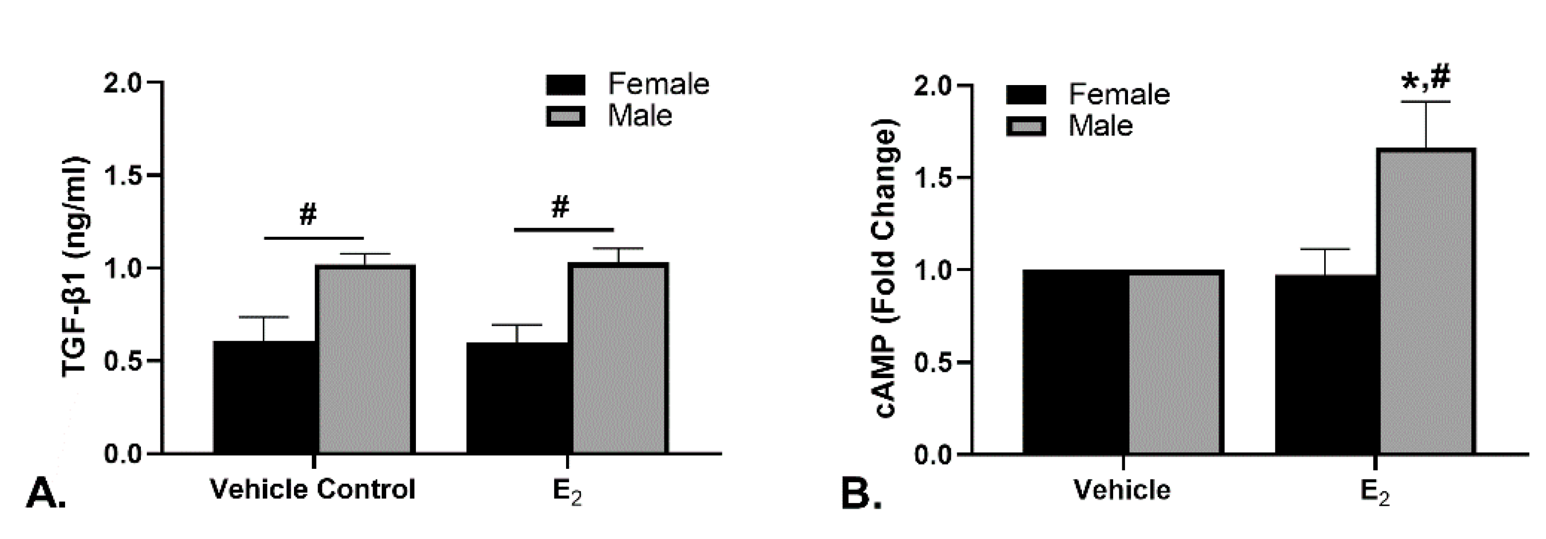
The TGF-β1 concentration in the spent medium was investigated as a possible mechanism for the differences in elastin synthesis (Figure 3A). There were no significant changes in the TGF-β1 concentration due to E2 treatment when compared to control for either the female or male EDS. There was a significant difference between the sexes with the male EDS producing significantly more TGF-β1 than the females which does not correspond with the changes in elastin synthesis.
The cAMP levels in the EDS homogenates were determined (Figure 3B). These data are presented as the fold change of the cAMP concentration normalized to DNA content. The male EDS homogenates had significantly increased cAMP levels in response to E2 compared to vehicle and the female EDS. The average concentration of the vehicle control for male EDS was 2.66 ± 0.54 fmol/µg DNA and the average for the female EDS was 2.41 ± 0.26 fmol/µg DNA.

2.6. Elastin Content When Exposed to Steroid Hormones and cAMP

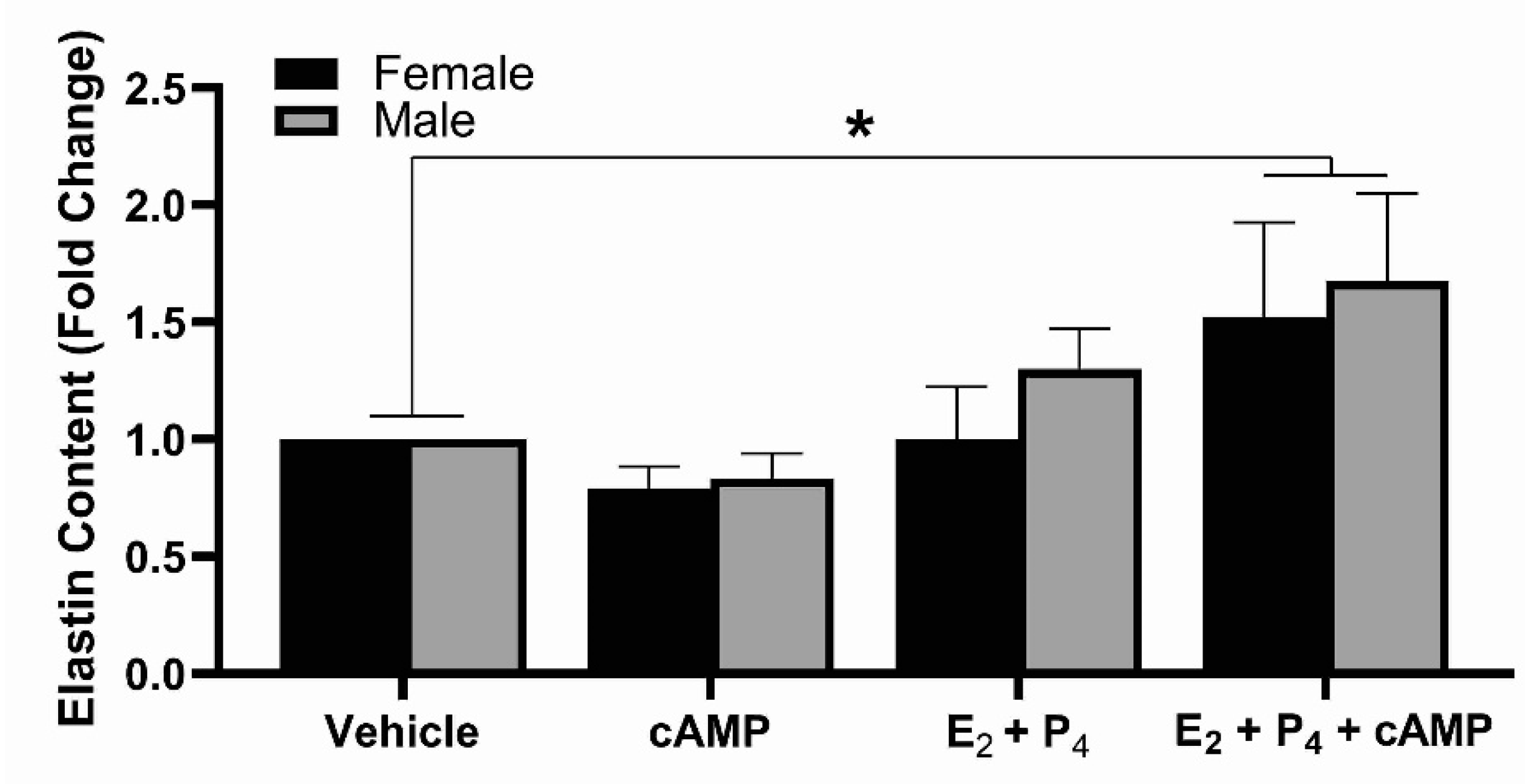
The addition of cAMP alone did not lead to an increase in elastin synthesis; however, when combined with E2 and P4 elastin synthesis significantly increased in both male and female EDS (Figure 4).

3. Discussion

This study has confirmed that E2 has a stimulatory effect on elastin production in EDS containing female hDF, and further shows this is via activation of the nuclear ERs, primarily ER-β. The elastogenic effect of E2 treatment was absent in male EDS and cAMP levels in the EDS in response to E2 were significantly higher in the male EDS than in the female or vehicle. The sex differences in elastin synthesis could be eliminated when the EDS were cultured with cAMP, E2, and P4. The elastogenic effects of E2 on hDF have been previously demonstrated in vivo and in vitro [16,18,31]. Several of these studies have also found correlations between TGF-β1 levels and the increased elastin synthesis in response to E2 [16,32]. Unlike these studies, TGF-β1 levels were not altered in our EDS cultures in response to E2, suggesting other pathways are involved.
The effects of sex hormones on gene expression are dependent on the presence of sex steroid hormone receptors within the tissue. It is now well recognized that the actions of estrogen are mediated via interaction with the two nuclear estrogen receptors (ERα and ERβ) which function as transcription factors within the nucleus, and/or the membrane estrogen receptor (GPER-1), which activates specific second messenger signaling pathways. Due to the different molecular mechanisms by which estrogen action is modulated upon activation of these receptors, estrogen signaling can be classified as genomic (ER-α/β) or non-genomic (GPER-1). Similar to the ERs, the progesterone receptor functions as a classic nuclear receptor and mediates gene expression via genomic signaling when bound to progesterone.
The male and female EDS were shown to express the same initial density of hormone receptors; however, their level of activity in response to the same treatment conditions with respective hormones/agonists was dramatically different between male and female EDS. Our findings indicate that treatment with E2 increases elastin content selectively in females. A similar effect could be achieved with ER selective agonists, confirming that the increase in elastin synthesis was a result of increased activation of the ERs in response to E2 in females. When the GPER-1 selective agonist was used, no change in elastin for either female or male EDS was observed. Because the primary effect of GPER activation is stimulation of adenylyl cyclase, which subsequently catalyzes the production of cAMP, we next sought to determine if cAMP levels were altered in EDS in the presence of E2. In contrast to the ERs, we found that cAMP levels were significantly increased in response to E2 in the male EDS, but unaltered in the female compared to vehicle. This indicates that although E2 treatment did not lead to increased elastin production, E2 did induce signaling via nongenomic mechanisms in males.
Collectively, these findings demonstrate sex-specific differences in signaling mechanisms in which E2 signals via the nuclear ERs selectively in females, and we hypothesize that it signals via GPER-1 selectively in males. The theory that estrogen can function as a sex-specific biased agonist presents a novel and sophisticated mechanism by which hormones may regulate gene expression, and specifically in the current study, underly the difference in elastin expression observed between the female and male EDS. Furthermore, the differential outcome in elastin synthesis between male and female EDS following culture with E2 could be attributed to the relative density levels of the GPER and the ERs. While the density of hormone receptors did not differ between sexes, there were significant differences in the density levels among the receptor types. If E2 selectively signals through ERs in females and GPER-1 in males, then the higher receptor density, and thus signal response, would support why E2 alone could increase elastin in females only.
While culture with E2 or P4 alone did not alter elastin content in male EDS, there was a non-significant, but observable increase in elastin when cultured in presence of both E2 and P4 in the male cells. One explanation for this could be an increase in GPER density, thus signaling, in response to P4. We did not measure receptor levels following coculture with E2 and P4, but this notion is supported by results from previous studies that demonstrate progestin up-regulates GPER-1 expression [33]. This P4-induced increase in GPER-1 expression paralleled an increase in receptor-bound estrogen, which also correlated to increased cAMP production [34]. When exogenous cAMP was also added to culture treatment with E2 and P4, a further increase in elastin was seen in males, resulting in a significant increase in elastin content compared to vehicle in both male and female EDS. Co-expression of ER-α and/or ER-β with GPER-1 suggests the possibility of interactions between these receptors and likely involves cross-talk between their signaling pathways. The observation that the addition of P4 with E2 treatment increased elastin while also eliminating the E2-induced increase elastin via ER activation in females raises the possibility of coordinated hormonal control of GPER-1 and the ERs by P4 in hDF. We believe that this increase in elastin synthesis is likely due to upregulation of GPER-1 in males, and that this response will also be present in other cells expressing these receptors. By estrogen acting as a GPER-1-biased ligand in males, a trend of increasing elastin content could be proposed from the collective findings presented here.
While there was an increase in elastin content in our EDS, no elastic fibers were found and the overall production of elastin was still low. Because this was a relatively short-term study, a longer culture may lead to different results, as elastic fibers are not seen in wound repair until weeks to months after injury [35,36,37]. We believe that these results are still significant, as tropoelastin synthesis, the pro-form of elastin, is a limiting factor in elastic fiber production in engineered skin [38]. Additionally, we have discovered a proposed mechanism for sex-specific signaling and a method by which these sex differences could be eliminated. By further investigating the pathways involved, new methods to stimulate the production of elastin by hDF in skin substitutes may be found. Mechanical forces have also been shown to induce elastin synthesis and result in elastic fiber formation in blood vessel substitutes. The combination of mechanical forces and hormones may result in elastic fiber formation and a better understanding of the specific mechanisms involved.
A major limitation of this study is the sample size along with the presence of confounding factors. The confounding factors of sex, age, and race in combination with the sample size increased the sample variability. Age is known to have an impact on elastin synthesis, in response to E2 treatment older hDF synthesize more elastin than younger hDF, especially in females [16,37]. Race is also known to influence elastin synthesis; Black subjects were found to produce more TGF-β and, thus, elastin than their White counterparts [39]. This study aimed to find more generalizable data; however, these factors need to be considered. A larger sample size may be needed or, if it can be justified, a smaller range of ages in subjects should be considered.
This study demonstrated that there are culture conditions by which we can induce elastin synthesis in both male and female hDF. This is a first step towards determining methods for stimulating elastin synthesis and assembly by hDF used in fabrication of engineered tissue substitutes. By identifying pathways, additional targets can also be identified for wound healing applications, where drugs can be developed and applied topically or through drug eluting scaffolds to promote proper elastin synthesis and assembly.

4. Materials and Methods

4.1. Tissue Sources

De-identified, discarded human skin was obtained from elective plastic and reconstructive surgery procedures at the University of Cincinnati Medical Center and Shriners Hospitals for Children—Cincinnati.

4.2. Isolation and Culture of Primary Dermal Fibroblasts

Isolation of hDF was performed following the method described in McFarland et al. [40]. Briefly, the full-thickness skin was incubated overnight at 4 °C in Dispase II (Sigma-Aldrich, St. Louis, MO, USA), after which the epidermis was mechanically separated from the dermis. The dermis was minced and digested with type II collagenase (Worthington, Lakewood, NJ, USA), with periodic agitation. Cells were cultured with medium consisting of Dulbecco’s modified eagle medium (DMEM, Gibco, Grand Island, NY, USA) supplemented with 5% v/v fetal bovine serum (FBS, Gibco), 10 ng/mL epidermal growth factor (Peprotech, Rocky Hill, NJ, USA), 0.5 μg/mL hydrocortisone (Sigma), 5 μg/mL insulin (Sigma), 0.1 M L-ascorbic acid 2-phosphate sesquimagnesium salt hydrate (AA2P, Sigma), and 1% v/v antibiotic-antimycotic (Gibco). The hDF were expanded and utilized between passages 1 and 3. The methods described for isolation have been used extensively and results in >98% purity of the dermal fibroblasts; identification for this study was primarily based on morphology. Cells from each batch are archived.

4.3. Fabrication and Culture of Engineered Dermal Substitutes

EDS were fabricated by mixing hDF with fibrinogen (Sigma), 25 U/mL thrombin (Sigma), and tissue culture medium for a final concentration of 2 × 10⁶ cells/mL and 2 mg/mL fibrin. After fibrillogenesis, additional tissue culture medium consisting of DMEM supplemented with 10% FBS, 1% antibiotic-antimycotic, 2 mg/mL ε-amino-η-caproic acid (ACA, EMD Millipore, Burlington, MA, USA), and 50 μg/mL AA2P was added. ACA inhibits plasminogen activation and thereby inhibits fibrin degradation. Over the first 2 weeks, the ACA content of the medium was gradually reduced to a final concentration of 1.25 mg/mL at which time the EDS were rinsed in phosphate-buffered saline (Sigma) and placed in hormone-containing medium for an additional 2 weeks. The hormone medium consisted of phenol red-free DMEM (Gibco) supplemented with 10% charcoal stripped-FBS (Innovative Research, Novi, MI, USA), 1% antibiotic-antimycotic, 1.25 mg/mL ACA, 50 μg/mL AA2P, and the desired hormones, agonists, or appropriate vehicle control. The steroid hormones E2 (Sigma) and P4 (Sigma) were solubilized in 10% ethanol (Sigma) and used alone or in combination at concentrations of 10 nM and 100 nM, respectively. Dibutyryl cAMP (Sigma) was used at a concentration of 500 μM. To determine which estrogen receptors were involved, 10 μM propyl pyrazole triol (PPT, Tocris Bioscience, Minneapolis, MN) and 10 μM diaproprionitrile (DPN, Tocris), or 1 µM G1 (Cayman Chemicals, Ann Arbor, MI, USA) were used. These are agonists for ER-α, ER-β, and GPER-1 respectively. The ER agonists were solubilized in dimethyl sulfoxide (Sigma) which was used as the vehicle control for the agonist studies. The EDS were cultured for an additional 2 weeks with hormones or agonists.

4.4. Protein and DNA Quantification

ELISAs were used to quantify the concentrations of ER- α, ER-β, GPER-1, and the progesterone receptors (all from Aviva Systems Biology, San Diego, CA, USA) and cAMP (Enzo Life Sciences, Inc., Farmingdale, NY, USA) in the tissue homogenates. The spent medium was assayed using an ELISA for TGF-β1 (R&D Systems, Minneapolis, MN, USA). The Fastin™ Elastin assay kit (Accurate Chemical, Westbury, NY, USA) was used for quantitation of elastin in the tissue per manufacturer instructions. The tissue samples for the elastin assay were frozen and stored at −80 °C prior to analysis. For total protein quantification and ELISAs, the samples were placed in RIPA buffer (Sigma) with proteinase inhibitor (Sigma) and homogenized. Protein quantification was performed via the Pierce BCA protein assay kit (ThermoFisher Scientific, Rockford, IL, USA). DNA content was determined for cell lysates and tissue homogenates using a Hoescht 33258 dye (Sigma) assay described previously [41]; calf thymus DNA (Sigma) was used for a standard curve. This was used for direct comparisons as well as for normalization.

4.5. Statistical Analysis

Data are presented as mean ± standard error of the mean. Statistical analysis was performed using JMP Pro 15 (SAS Institute, Inc., Cory, NC, USA). Student’s t-tests were used for comparison of age and initial receptor densities. Chi-squared analysis was used to compare race. A two-way ANOVA with a post hoc Tukey analysis was performed for the analysis of elastin, collagen, receptor density, and DNA contents. Statistical significance was determined using a 95% confidence interval.

Author Contributions

Conceptualization, S.C.S.; methodology, S.C.S. and A.M.Z.; formal analysis, S.C.S., C.N. and A.M.Z.; writing—original draft preparation, S.C.S.; writing—review and editing, C.N. and A.M.Z. All authors have read and agreed to the published version of the manuscript.

Funding

This research received no external funding.

Institutional Review Board Statement

The University of Cincinnati Institutional Review Board determined that this did not constitute human subjects research and was exempt from requirements for informed consent according to 45CFR46.101(b)(4).

Informed Consent Statement

Not applicable for reasons stated above.

Data Availability Statement

The data presented in this study are available on request from the corresponding author.

Conflicts of Interest

The authors declare no conflict of interest.

References

  1. Hult, A.M.; Goltz, R.W. The Measurement of Elastin in Human Skin and Its Quantity in Relation to Age. J. Invest. Dermatol. 1965, 44, 408–412. [Google Scholar] [CrossRef] [Green Version]
  2. Uitto, J.; Paul, J.L.; Brockley, K.; Pearce, R.H.; Clark, J.G. Elastic fibers in human skin: Quantitation of elastic fibers by computerized digital image analyses and determination of elastin by radioimmunoassay of desmosine. Lab. Invest. 1983, 49, 499–505. [Google Scholar]
  3. Shapiro, S.D.; Endicott, S.K.; Province, M.A.; Pierce, J.A.; Campbell, E.J. Marked longevity of human lung parenchymal elastic fibers deduced from prevalence of D-aspartate and nuclear weapons-related radiocarbon. J. Clin. Invest. 1991, 87, 1828–1834. [Google Scholar] [CrossRef] [PubMed] [Green Version]
  4. Parks, W.C.; Secrist, H.; Wu, L.C.; Mecham, R.P. Developmental regulation of tropoelastin isoforms. J. Biol. Chem. 1988, 263, 4416–4423. [Google Scholar] [CrossRef]
  5. Almine, J.F.; Wise, S.G.; Weiss, A.S. Elastin signaling in wound repair. Birth Defects Res. C Embryo Today 2012, 96, 248–257. [Google Scholar] [CrossRef] [PubMed]
  6. Weihermann, A.C.; Lorencini, M.; Brohem, C.A.; de Carvalho, C.M. Elastin structure and its involvement in skin photoageing. Int. J. Cosmet. Sci. 2017, 39, 241–247. [Google Scholar] [CrossRef] [PubMed]
  7. Huertas, A.C.M.; Schmelzer, C.E.; Hoehenwarter, W.; Heyroth, F.; Heinz, A. Molecular-level insights into aging processes of skin elastin. Biochimie 2016, 128–129, 163–173. [Google Scholar] [CrossRef] [PubMed]
  8. Olsen, D.R.; Fazio, M.J.; Shamban, A.T.; Rosenbloom, J.; Uitto, J. Cutis laxa: Reduced elastin gene expression in skin fibroblast cultures as determined by hybridizations with a homologous cDNA and an exon 1-specific oligonucleotide. J. Biol. Chem. 1988, 263, 6465–6467. [Google Scholar] [CrossRef]
  9. Zhang, M.C.; He, L.; Giro, M.; Yong, S.L.; Tiller, G.E.; Davidson, J.M. Cutis laxa arising from frameshift mutations in exon 30 of the elastin gene (ELN). J. Biol. Chem. 1999, 274, 981–986. [Google Scholar] [CrossRef] [Green Version]
  10. Hu, Q.; Loeys, B.L.; Coucke, P.J.; De Paepe, A.; Mecham, R.P.; Choi, J.; Davis, E.C.; Urban, Z. Fibulin-5 mutations: Mechanisms of impaired elastic fiber formation in recessive cutis laxa. Hum. Mol. Genet. 2006, 15, 3379–3386. [Google Scholar] [CrossRef] [Green Version]
  11. Markova, D.; Zou, Y.; Ringpfeil, F.; Sasaki, T.; Kostka, G.; Timpl, R.; Uitto, J.; Chu, M.L. Genetic heterogeneity of cutis laxa: A heterozygous tandem duplication within the fibulin-5 (FBLN5) gene. Am. J. Hum. Genet. 2003, 72, 998–1004. [Google Scholar] [CrossRef] [Green Version]
  12. Boss, G.R.; Seegmiller, J.E. Age-related physiological changes and their clinical significance. West. J. Med. 1981, 135, 434–440. [Google Scholar] [PubMed]
  13. Calleja-Agius, J.; Brincat, M. The effect of menopause on the skin and other connective tissues. Gynecol. Endocrinol. 2012, 28, 273–277. [Google Scholar] [CrossRef]
  14. Wilkinson, H.N.; Hardman, M.J. The role of estrogen in cutaneous ageing and repair. Maturitas 2017, 103, 60–64. [Google Scholar] [CrossRef]
  15. Rzepecki, A.K.; Murase, J.E.; Juran, R.; Fabi, S.G.; McLellan, B.N. Estrogen-deficient skin: The role of topical therapy. Int. J. Womens Dermatol. 2019, 5, 85–90. [Google Scholar] [CrossRef] [PubMed]
  16. Son, E.D.; Lee, J.Y.; Lee, S.; Kim, M.S.; Lee, B.G.; Chang, I.S.; Chung, J.H. Topical application of 17beta-estradiol increases extracellular matrix protein synthesis by stimulating tgf-Beta signaling in aged human skin in vivo. J. Invest. Dermatol. 2005, 124, 1149–1161. [Google Scholar] [CrossRef] [Green Version]
  17. Zong, W.; Jiang, Y.; Zhao, J.; Zhang, J.; Gao, J.G. Estradiol plays a role in regulating the expression of lysyl oxidase family genes in mouse urogenital tissues and human Ishikawa cells. J. Zhejiang Univ. Sci. B 2015, 16, 857–864. [Google Scholar] [CrossRef] [Green Version]
  18. Punnonen, R.; Vaajalahti, P.; Teisala, K. Local oestriol treatment improves the structure of elastic fibers in the skin of postmenopausal women. Ann. Chir. Gynaecol. Suppl. 1987, 202, 39–41. [Google Scholar]
  19. Natoli, A.K.; Medley, T.L.; Ahimastos, A.A.; Drew, B.G.; Thearle, D.J.; Dilley, R.J.; Kingwell, B.A. Sex steroids modulate human aortic smooth muscle cell matrix protein deposition and matrix metalloproteinase expression. Hypertension 2005, 46, 1129–1134. [Google Scholar] [CrossRef] [Green Version]
  20. Mangir, N.; Hillary, C.J.; Chapple, C.R.; MacNeil, S. Oestradiol-releasing Biodegradable Mesh Stimulates Collagen Production and Angiogenesis: An Approach to Improving Biomaterial Integration in Pelvic Floor Repair. Eur. Urol. Focus 2019, 5, 280–289. [Google Scholar] [CrossRef] [Green Version]
  21. Baron, Y.M.; Galea, R.; Brincat, M. Carotid artery wall changes in estrogen-treated and -untreated postmenopausal women. Obstet. Gynecol. 1998, 91, 982–986. [Google Scholar] [PubMed]
  22. Tyagi, T.; Alarab, M.; Leong, Y.; Lye, S.; Shynlova, O. Local oestrogen therapy modulates extracellular matrix and immune response in the vaginal tissue of post-menopausal women with severe pelvic organ prolapse. J. Cell Mol. Med. 2019, 23, 2907–2919. [Google Scholar] [CrossRef] [PubMed]
  23. Kim, J.M.; Shin, S.C.; Park, G.C.; Lee, J.C.; Jeon, Y.K.; Ahn, S.J.; Thibeault, S.; Lee, B.J. Effect of sex hormones on extracellular matrix of lamina propria in rat vocal fold. Laryngoscope 2020, 130, 732–740. [Google Scholar] [CrossRef] [PubMed]
  24. Yildiz, M.; Yigit, O.; Sunter, A.V.; Edizer, D.T.; Dursun, N.; Okcu, O. Effects of Intracordal Estradiol and Dexamethasone Injection on Wound Healing in Vocal Fold Injuries. J. Voice 2019, 33, 759–766. [Google Scholar] [CrossRef] [PubMed]
  25. Nallasamy, S.; Yoshida, K.; Akins, M.; Myers, K.; Iozzo, R.; Mahendroo, M. Steroid Hormones Are Key Modulators of Tissue Mechanical Function via Regulation of Collagen and Elastic Fibers. Endocrinology 2017, 158, 950–962. [Google Scholar] [CrossRef] [PubMed] [Green Version]
  26. Rich, C.B.; Fontanilla, M.R.; Nugent, M.; Foster, J.A. Basic fibroblast growth factor decreases elastin gene transcription through an AP1/cAMP-response element hybrid site in the distal promoter. J. Biol. Chem. 1999, 274, 33433–33439. [Google Scholar] [CrossRef] [Green Version]
  27. Sakamoto, A.; Weinstein, L.S.; Plagge, A.; Eckhaus, M.; Kelsey, G. GNAS haploinsufficiency leads to subcutaneous tumor formation with collagen and elastin deposition and calcification. Endocr. Res. 2009, 34, 1–9. [Google Scholar] [CrossRef] [Green Version]
  28. Mecham, R.P.; Levy, B.D.; Morris, S.L.; Madaras, J.G.; Wrenn, D.S. Increased cyclic GMP levels lead to a stimulation of elastin production in ligament fibroblasts that is reversed by cyclic AMP. J. Biol. Chem. 1985, 260, 3255–3258. [Google Scholar] [CrossRef]
  29. Stevenson, S.; Nelson, L.D.; Sharpe, D.T.; Thornton, M.J. 17beta-estradiol regulates the secretion of TGF-beta by cultured human dermal fibroblasts. J. Biomater. Sci. Polym. Ed. 2008, 19, 1097–1109. [Google Scholar] [CrossRef]
  30. Bullough, W.S. The mitogenic actions of androgenic and oestrogenic hormones. Acta Physiol. Pharmacol. Neerl. 1950, 1, 357–358. [Google Scholar]
  31. Varila, E.; Rantala, I.; Oikarinen, A.; Risteli, J.; Reunala, T.; Oksanen, H.; Punnonen, R. The effect of topical oestradiol on skin collagen of postmenopausal women. Br. J. Obstet. Gynaecol. 1995, 102, 985–989. [Google Scholar] [CrossRef]
  32. Ashcroft, G.S.; Dodsworth, J.; van Boxtel, E.; Tarnuzzer, R.W.; Horan, M.A.; Schultz, G.S.; Ferguson, M.W. Estrogen accelerates cutaneous wound healing associated with an increase in TGF-beta1 levels. Nat. Med. 1997, 3, 1209–1215. [Google Scholar] [CrossRef] [PubMed]
  33. Ahola, T.M.; Purmonen, S.; Pennanen, P.; Zhuang, Y.H.; Tuohimaa, P.; Ylikomi, T. Progestin upregulates G-protein-coupled receptor 30 in breast cancer cells. Eur. J. Biochem. 2002, 269, 2485–2490. [Google Scholar] [CrossRef] [PubMed]
  34. Thomas, P.; Pang, Y.; Filardo, E.J.; Dong, J. Identity of an estrogen membrane receptor coupled to a G protein in human breast cancer cells. Endocrinology 2005, 146, 624–632. [Google Scholar] [CrossRef] [PubMed] [Green Version]
  35. Raghunath, M.; Bachi, T.; Meuli, M.; Altermatt, S.; Gobet, R.; Bruckner-Tuderman, L.; Steinmann, B. Fibrillin and elastin expression in skin regenerating from cultured keratinocyte autografts: Morphogenesis of microfibrils begins at the dermo-epidermal junction and precedes elastic fiber formation. J. Invest. Dermatol. 1996, 106, 1090–1095. [Google Scholar] [CrossRef] [PubMed] [Green Version]
  36. Amadeu, T.P.; Braune, A.S.; Porto, L.C.; Desmouliere, A.; Costa, A.M. Fibrillin-1 and elastin are differentially expressed in hypertrophic scars and keloids. Wound Repair Regen 2004, 12, 169–174. [Google Scholar] [CrossRef] [PubMed]
  37. Ashcroft, G.S.; Kielty, C.M.; Horan, M.A.; Ferguson, M.W. Age-related changes in the temporal and spatial distributions of fibrillin and elastin mRNAs and proteins in acute cutaneous wounds of healthy humans. J. Pathol. 1997, 183, 80–89. [Google Scholar] [CrossRef]
  38. Mithieux, S.M.; Weiss, A.S. Design of an elastin-layered dermal regeneration template. Acta Biomater. 2017, 52, 33–40. [Google Scholar] [CrossRef] [PubMed]
  39. Fantasia, J.; Lin, C.B.; Wiwi, C.; Kaur, S.; Hu, Y.P.; Zhang, J.; Southall, M.D. Differential levels of elastin fibers and TGF-beta signaling in the skin of Caucasians and African Americans. J. Dermatol. Sci. 2013, 70, 159–165. [Google Scholar] [CrossRef]
  40. McFarland, K.L.; Glaser, K.; Hahn, J.M.; Boyce, S.T.; Supp, D.M. Culture medium and cell density impact gene expression in normal skin and abnormal scar-derived fibroblasts. J. Burn Care Res. 2011, 32, 498–508. [Google Scholar] [CrossRef] [Green Version]
  41. Schutte, S.C.; Taylor, R.N. A tissue-engineered human endometrial stroma that responds to cues for secretory differentiation, decidualization, and menstruation. Fertil. Steril. 2012, 97, 997–1003. [Google Scholar] [CrossRef] [PubMed] [Green Version]
Figure 1. Elastin content of EDS. (A) Culture with E2 alone significantly increased elastin content in the female, but not the male EDS. (B) Culture with agonists specific for either ER-α (PPT) or ER-β (DPN) significantly increased elastin content in female EDS. Agonists to GPER-1 (G1) did not have a significant effect. n = 5, * indicates p < 0.05.
Ijms 22 06358 g001
Figure 2. Receptor densities were similar in male and female cells but differ between receptor types. (A) initial receptor densities of the three estrogen receptors and the progesterone receptors. (B) After culture, the receptor densities were determined in the EDS homogenates and the fold change between vehicle control and either E2 treated (ERs) or P4 treated (PRs). n = 5; * indicates p < 0.05 compared to vehicle control.
Ijms 22 06358 g002
Figure 3. TGF-β1 and cAMP concentrations after E2 culture. (A) The concentration of TGF-β1 was higher in male cultures than in female cultures but was not affected by treatment. (B) The presence of cAMP in the male tissues treated with E2 was significantly higher than that of females and the vehicle control. n = 5; * indicates p < 0.05 compared to vehicle control; # indicates p < 0.05 compared to the opposite sex.
Ijms 22 06358 g003
Figure 4. Effects of cAMP on elastin synthesis. The addition of cAMP alone does not affect elastin synthesis; however, when added with E2 and P4 an increase in elastin synthesis in both male and female EDS occurs. n = 5; * indicates p < 0.05.
Ijms 22 06358 g004
Table 1. Sample source demographics.
FemalesMalesp-Value
Age (yrs)28.4 ± 17.422.1 ± 1.90.26
Race 0.58
White23
Black32
Ethnicity 0.93
Hispanic10
Non-Hispanic/Not disclosed45
Biopsy Location 0.17
Abdomen13
Breast30
Foot01
Thigh11
Publisher’s Note: MDPI stays neutral with regard to jurisdictional claims in published maps and institutional affiliations.