Article

Prenatal Molecular Hydrogen Administration Ameliorates Several Findings in Nitrofen-Induced Congenital Diaphragmatic Hernia

1
Department of Obstetrics and Gynecology, Nagoya University Graduate School of Medicine, 65 Tsurumai-Cho, Showa-Ku, Nagoya 466-8550, Japan
2
Department of Obstetrics and Gynecology, Japanese Red Cross Nagoya First Hospital, 3-35 Michisita-Cho, Nakamura-Ku, Nagoya 453-8511, Japan
3
Laboratory of Bell Research Center, Department of Obstetrics and Gynecology Collaborative Research, Nagoya University Graduate School of Medicine, 65 Tsurumai-Cho, Showa-Ku, Nagoya 466-8550, Japan
4
Department of Obstetrics and Gynecology, Kasugai Municipal Hospital, Kasugai 486-8510, Japan
5
Department of Obstetrics and Gynecology, Fujita Health University Graduate School of Medicine, Toyoake 470-1192, Japan
6
Department of Pathology and Biological Responses, Nagoya University Graduate School of Medicine, 65 Tsurumai-Cho, Showa-Ku, Nagoya 466-8550, Japan
7
Center for Maternal-Neonatal Care, Division of Perinatology, Nagoya University Hospital, 65 Tsurumai-Cho, Showa-Ku, Nagoya 466-8560, Japan
*
Author to whom correspondence should be addressed.
Int. J. Mol. Sci. 2021, 22(17), 9500; https://doi.org/10.3390/ijms22179500
Submission received: 30 June 2021 / Revised: 27 August 2021 / Accepted: 27 August 2021 / Published: 31 August 2021
(This article belongs to the Section Biochemistry)

Abstract

:
Oxidative stress plays a pathological role in pulmonary hypoplasia and pulmonary hypertension in congenital diaphragmatic hernia (CDH). This study investigated the effect of molecular hydrogen (H2), an antioxidant, on CDH pathology induced by nitrofen. Sprague-Dawley rats were divided into three groups: control, CDH, and CDH + hydrogen-rich water (HW). Pregnant dams of CDH + HW pups were orally administered HW from embryonic day 10 until parturition. Gasometric evaluation and histological, immunohistochemical, and real-time polymerase chain reaction analyses were performed. Gasometric results (pH, pO2, and pCO2 levels) were better in the CDH + HW group than in the CDH group. The CDH + HW group showed amelioration of alveolarization and pulmonary artery remodeling compared with the CDH group. Oxidative stress (8-hydroxy-2′-deoxyguanosine-positive-cell score) in the pulmonary arteries and mRNA levels of protein-containing pulmonary surfactant that protects against pulmonary collapse (surfactant protein A) were significantly attenuated in the CDH + HW group compared with the CDH group. Overall, prenatal H2 administration improved respiratory function by attenuating lung morphology and pulmonary artery thickening in CDH rat models. Thus, H2 administration in pregnant women with diagnosed fetal CDH might be a novel antenatal intervention strategy to reduce newborn mortality due to CDH.

1. Introduction

Congenital diaphragmatic hernia (CDH) is a life-threatening anomaly with a mortality rate as high as 20–40% [1,2]. Its prevalence is known to be 1:4000 live births [3]. CDH presents a defect in the diaphragm through which the abdominal organs migrate into the thoracic cavity. Therefore, pulmonary growth is restricted by the compression of the migrated organs into the thoracic cavity during the fetal period. Pulmonary hypoplasia is associated with pulmonary hypertension (PH) after birth. PH seen in CDH is categorized as PH due to developmental lung disorders [4], PH has been increasingly recognized as a significant factor for mortality and morbidity in newborns with CDH [5]. Extracorporeal membrane oxygenation (ECMO) has been used in patients with CDH who are unresponsive to optimal medication and ventilation. However, this therapeutic approach can only be used after birth. The pathology of PH in patients with CDH is characterized by an extensive muscularization of vessels and pulmonary vascular remodeling, which starts early during gestation [6]. Currently, a clinical trial of fetoscopic endoluminal tracheal occlusion (FETO) as a prenatal intervention for CDH is ongoing [7]. FETO has been shown to improve survival rate by increasing lung volume but has the risk of premature delivery due to fetoscopy [5,8]. Therefore, prenatal medical therapies should be explored as alternatives [9]. We and others have reported the pathological role of oxidative stress in pulmonary hypoplasia and PH in CDH [10,11,12].
Molecular hydrogen (H2) was first reported as an antioxidant that reduces reactive oxygen species (ROS; OH· and peroxynitrite) [13]. H2 has several benefits in clinical applications, including rapid diffusion and lack of toxicity. H2 has been reported to exert preventive and therapeutic effects on various pathologies, such as ischemia, injury, metabolic syndrome, and inflammation in different organs [14]. We have reported that maternal administration of H2 increases its concentration in the organs of pups and improves brain or lung damage in pups [15,16,17,18]. Another study reported that H2 ameliorates pulmonary artery hypertension (PH) with decreased smooth muscle cell (SMC) proliferation [19]. However, the mechanisms underlying the effects of H2 on the pulmonary artery remain unknown. Receptor tyrosine kinases, including platelet-derived growth factor (PDGF), have been used as therapeutic targets for PH [20] and several studies have indicated an essential role for PDGF, especially the PDGF-B chain, in PH [21,22]. PDGF-B and PDGF receptor-beta (PDGFRβ) signaling contributes to the excessive proliferation and migration of pulmonary SMCs [20,23]. PDGF-BB has a more substantial mitogenic effect on human vascular SMCs than PDGF-AA [24]. PDGF levels are reported to increase in the amniotic fluid of human CDH [25] and the lung tissues of animal CDH models [11,21]. Furthermore, PDGF-B signaling plays a pathological role in CDH and is associated with oxidative stress [11]. Although H2 indirectly inhibits signal transduction, the modulation of PDGF-B signaling by H2 remains unknown.
Based on these findings, we aimed to investigate whether prenatal H2 treatment could improve pulmonary development and pulmonary artery remodeling in CDH. We also investigated the effect of H2 on oxidative stress in pulmonary arteries.

2. Results

2.1. Prenatal H2 Administration Did Not Change Lung Weight but Improved Blood Gas Results in CDH Rats

CDH occurrence was not significantly different between the Nitrofen (n = 14) and Nitrofen + hydrogen-rich water (HW, n = 17) groups (37.9 ± 22.4% vs. 39.4 ± 17.9%, p = 0.829). The lung weight to body weight (LW/BW) ratio of the pups in the CDH group was significantly lower than that in the control group (p < 0.01, Figure 1a), and the LW/BW ratio did not improve in the CDH + HW group (Figure 1a). The neonatal blood gasometric results are shown in Figure 1b–d. The pH and pO2 values of the CDH group were significantly lower than those of the control and CDH + HW groups (Figure 1b,d, p < 0.01). Furthermore, the pCO2 values in the CDH group were significantly higher than those in the control and CDH + HW groups (Figure 1c, p < 0.01, and p < 0.05, respectively). The pH values in the CDH + HW group were improved to a level similar to that in the control group (Figure 1b), but the pCO2 and pO2 values were significantly different between the CDH + HW and control groups (Figure 1c,d, p < 0.05, and p < 0.01, respectively).

2.2. The Effects of Prenatal H2 Administration on Alveolarization and Pulmonary Artery Remodeling in CDH Rats

Reduced alveolarization and thickened pulmonary artery walls were observed in the CDH group compared to the control and CDH + HW groups (Figure 2a,c). More airspaces were observed in the CDH + HW group than in the CDH group (Figure 2a). The mean linear intercept (MLI) was used as a quantitative parameter for alveolarization. The MLI in the CDH group was significantly lower than that in the control and CDH + HW groups (Figure 2b, p < 0.01, and p < 0.05, respectively), and the MLI in the CDH + HW group was similar to that in the control group (Figure 2b).
Thickening of the pulmonary artery walls was observed in the CDH group, which was significantly improved by H2 treatment (Figure 2c, shown with two-way arrows). To determine the effect of H2 on pulmonary artery remodeling, the medial wall thickness (MWT) was compared among the control, CDH, and CDH + HW groups. The average MWT (%) in the CDH + HW group was significantly lower than that in the CDH group (Figure 2d, p < 0.01) and was similar to that in the control group (Figure 2d).

2.3. Prenatal H2 Administration Reduced Oxidative Stress but Did Not Affect PDGFRβ Expression in the Fetal Pulmonary Arterial Wall in CDH

Oxidative stress was examined by 8-hydroxy-2′-deoxyguanosine (8-OHdG) positive cell scores in the wall of the pulmonary arteries among the control, CDH, and CDH + HW groups (Figure 3a, arrowheads). The scores of 8-OHdG positive cells in the CDH group were significantly higher than those in the control and CDH + HW groups (Figure 3b, p < 0.01, and p < 0.05, respectively). However, the score in the CDH + HW group was significantly higher than that in the control group (Figure 3b, p < 0.01).
PDGFRβ expression in the wall of the pulmonary arteries was investigated among the control, CDH, and CDH + HW groups (Figure 3c) by grading the intensity of immunohistochemical staining (Figure 3e). PDGFRβ expression in the CDH group was significantly higher than that in the control group (Figure 3d, p < 0.05). The CDH + HW group showed a decreasing trend compared with that in the CDH group, but no significant difference was detected between the CDH and CDH + HW groups (Figure 3d).
Furthermore, a positive relationship was detected between the 8-OHdG positive cell score and MWT (Figure 3f, p < 0.01, ρ = 0.441).
To examine the mechanisms underlying the effect of H2 on the MWT, the expression of vascular endothelial growth factor receptor (VEGFR) mRNA, Flt1 (Fms related receptor tyrosine kinase 1, Vegfr-1) and Kdr (kinase insert domain receptor, Vegfr-2), was examined among the three groups. Flt1 and Kdr mRNA levels were significantly lower in the CDH group than in the control group (Figure 4a,b, p < 0.01 and p < 0.05, respectively). Flt1 and Kdr expression in the CDH + HW group was significantly higher than that in the CDH group (Figure 4a,b, p < 0.05), and was the same as that in the control group (Figure 4a,b).

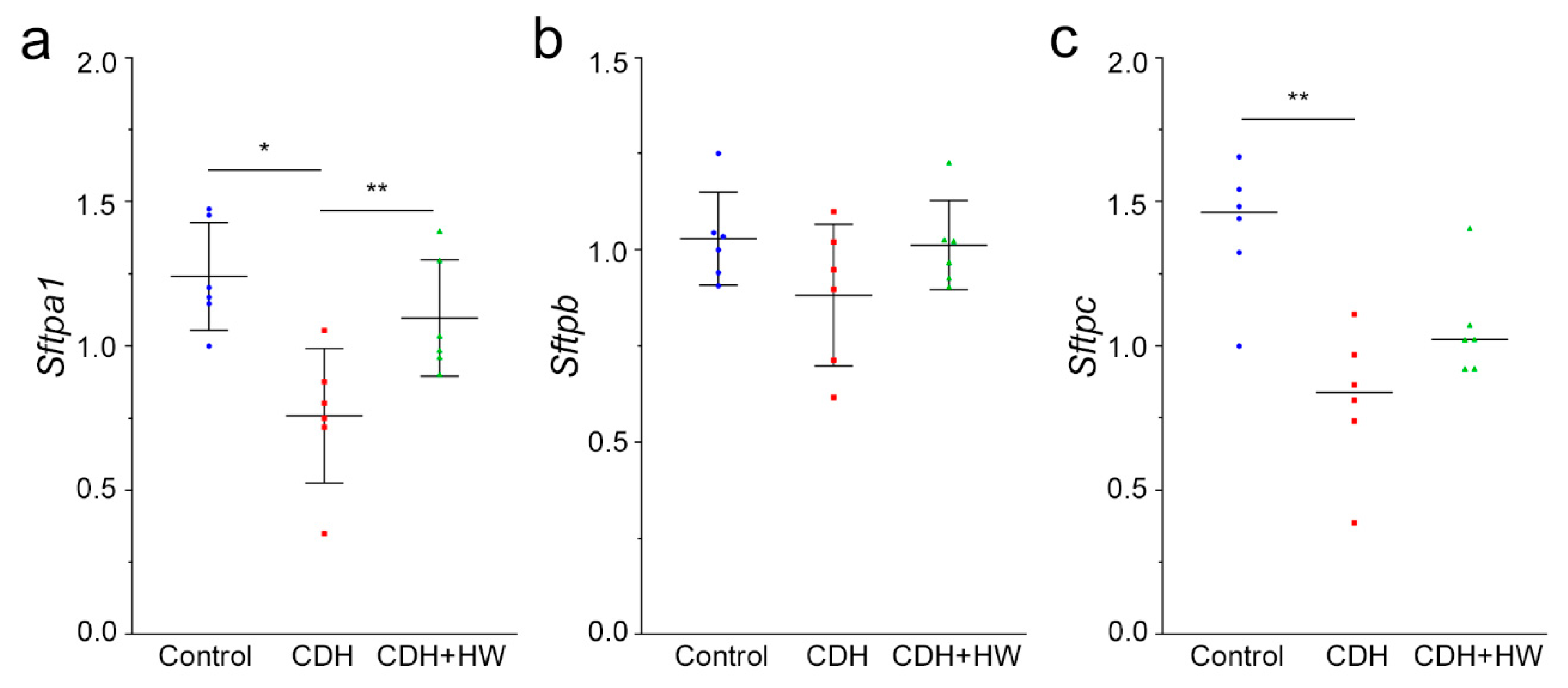
2.4. Prenatal H2 Administration Ameliorated Surfactant Protein A Production Insufficiency in Lungs of Pups with CDH

To examine the mechanisms underlying the effect of H2 on the MLI, the mRNA expression of surfactant protein A (Sftpa1), B (Sftpb), and C (Sftpc) was compared among the three groups. The expression of Sftpa1 and Sftpc was significantly lower in the CDH group than in the control group (Figure 5a and c, p < 0.01, and p < 0.05, respectively). Sftpa1 expression in the CDH + HW group was significantly increased compared to that in the CDH group (p < 0.05), to the same level as that in the control group (Figure 5a). However, Sftpc mRNA levels in the CDH + HW group were unchanged compared to those in the CDH group (Figure 5c). There was no significant difference in Sftpb mRNA levels among the control, CDH, and CDH + HW groups (Figure 5b).

3. Discussion

In this study, we demonstrated the positive effect of prenatal H2 administration on pulmonary function in rats with CDH. Prenatal H2 administration improved blood gas parameters in CDH, although it did not affect CDH occurrence and pulmonary hypoplasia. Morphological analysis revealed that prenatal H2 administration ameliorated alveolarization and pulmonary artery remodeling in CDH rats. Immunohistochemical analyses showed that prenatal H2 administration reduced oxidative stress in pulmonary arterial wall cells. Moreover, prenatal H2 administration increased Sftpa1, Flt1 and Kdr levels in the lungs of CDH rats. These results suggest that although prenatal H2 administration could not improve pulmonary hypoplasia, it might improve pulmonary function by enhancing alveolarization and ameliorating pulmonary remodeling. Furthermore, the reduction in oxidative stress by prenatal H2 administration improved pulmonary artery remodeling.
PH is the cornerstone of the CDH pathophysiology as well as pulmonary hypoplasia and is thought to be associated with SMC proliferation and pulmonary arterial remodeling [26]. Oxidative stress is a well-known cause of SMC proliferation in adult PH [27]. Recently, increased ROS have also been shown to contribute to pulmonary vascular remodeling by stimulating SMC proliferation in PH in newborns with or without CDH [28]. In the present study, oxidative damage to the pulmonary arterial wall was increased in rats with CDH. However, prenatal H2 administration reduces oxidative damage and attenuates pulmonary arterial remodeling in CDH. Furthermore, a positive correlation was detected between the 8-OHdG score and MWT in the pulmonary arteries. These findings suggest that oxidative damage to the pulmonary artery wall is associated with pulmonary artery remodeling in CDH. Furthermore, H2 has been reported to reduce the oxidative stress of pulmonary vessels and ameliorate PH in an adult mouse model of monocrotaline-induced PH [19]. H2 was found to suppress 8-OHdG-positive cells in the pulmonary arterial walls, which is consistent with the results of the present study. These findings suggest that a reduction of oxidative stress could also improve PH by attenuating pulmonary remodeling in CDH.
The present study demonstrated that PDGFRβ was significantly increased in the lungs of rats with CDH, which is consistent with previous reports [21,22]. ROS from mitochondria have been reported to positively regulate PDGF signaling [29]. Furthermore, H2O2 increases PDGFRβ phosphorylation in primary neuronal cultures [30]. N-acetylcysteine, an antioxidant, has been reported to decrease PDGFRβ protein and ROS production in vascular SMCs [31]. However, others have reported that N-acetylcysteine reduces PDGF signaling in vascular SMCs without altering PDGFRβ expression [32]. These findings suggest that reducing oxidative stress results in the suppression of PDGF signaling. Prenatal H2 administration did not affect PDGFRβ expression in the pulmonary artery but attenuated pulmonary artery remodeling in the lungs of pups with CDH. Thus, H2 might attenuate pulmonary artery remodeling without affecting PDGFRβ expression. Additionally, the expression of Flt1 and Kdr, as VEGFRs, was significantly decreased in CDH rats, which is consistent with previous studies [33,34,35]. These previous studies also reported that prenatal treatment with all trans retinoic acid, corticosteroids, or tracheal occlusion attenuates Flt1 and Kdr expression in nitrofen-induced rat CDH, suggesting an association between increased VEGFR expression and decreased MWT. Prenatal H2 administration significantly increased Flt1 and Kdr expression, which might attenuate the MWT in CDH + HW. H2 has been reported to increase Kdr expression in rat lung, which is consistent with the present result [36]. Melatonin, an antioxidant, increases VEGFR-1 levels [37], which suggests that antioxidants improve VEGFR-1 expression.
In the present study, the MLI was lower in the CDH group than in the control group, which is consistent with a previous report [38]. The reduced MLI was improved by prenatal H2 administration. The MLI is thought to be reduced in CDH owing to lung compression by prolapsed abdominal organs, as well as by impaired differentiation of the lung. Surfactant proteins A, B, and C, which are synthesized by alveolar epithelial cells, constitute pulmonary surfactants with surfactant phospholipids [39]. Pulmonary surfactant increases airspace by reducing the surface tension in the alveoli and plays a role in the prevention of pulmonary collapse and maintenance of a stable ventilatory capacity. Sftpa [40] and Sftpc [41] expression is reduced in the lungs of CDH rat models. A lower level of surfactant protein A from tracheal aspirates has also been reported in human CDH [42]. In the present study, insufficient Sftpa1 expression in CDH lungs was attenuated by prenatal H2 administration. Thus, the recovery of Sftpa1 expression could improve the MLI in CDH + HW. A previous report suggested that surfactant protein A mRNA expression is reduced by oxidative stress and might be reversed by antioxidants in the human pulmonary epithelial cell line [43], which is consistent with the present results.
The strengths of this study are as follows: First, this is the first study to examine the effects of H2 on CDH. Second, we demonstrated oxidative stress in the pulmonary arterial wall of the CDH rat model. Third, H2 is considered to be a safe agent for human applications [44]. Many clinical trials of HW administration in humans have been conducted previously [45,46,47]. The long-term safety of inhaled hydrogen gas in mice has already been reported [48]. Although several therapeutic and preventive effects for various obstetric diseases such as preterm labor [49], preeclampsia [50], fetal brain injury [16,51], and fetal lung injury [15] have been reported, these studies did not report short-term adverse effects. Fourth, it is already known that this method of prenatal H2 administration increases the hydrogen concentration in fetal lungs [15]. Fifth, prenatal H2 administration had an effect similar to that of vitamin D [52]. Taken together, these results suggest that prenatal H2 administration can be adapted to clinical settings. However, more convincing data including the duration and timing of H2 administration, are required.
Our study had several limitations. First, due to high lethality, we could not confirm pulmonary arterial hypertension in the nitrofen-induced CDH model. Second, we studied only CDH rats. As shown in some papers, lung hypoplasia is also observed in non-CDH pups prenatally exposed to nitrofen. Therefore, it remains unclear whether pulmonary hypoplasia can be attributed to CDH pathology or nitrofen toxicity. Third, lung hypoplasia could not be improved by prenatal H2 treatment, even though FETO enhanced lung volume. Thus, prenatal H2 treatment could not be an alternative to FETO but would be effective as a combined therapy as H2 would mainly act on the pulmonary arteries. However, the effect of H2 on pulmonary artery remodeling was only shown by histological analysis in this study. Sildenafil has been reported to attenuate right ventricular hypertrophy and angiogenesis in the same CDH model [53]. Further study is required to establish the effect of H2 on pulmonary artery remodeling.

4. Materials and Methods

4.1. Reagents

Nitrofen (2,4-dichlorophenyl-p-nitrophenyl ether; product no. 33374, Sigma Aldrich, Saint Louis, USA) (100 mg) was dissolved in 1 mL of olive oil and administered to animals via a gastric tube. HW was prepared using a hydrogen machine (H-DX1, Arega Co., Komaki, Japan). Its concentration was approximately 0.6 ppm. HW had a content of >0.4 mM H2, as reported previously [15,16,17,18,49,50,51]. Anti-PDGFRβ antibody [Y92] (ab32570, lot no. GR212663-9) was purchased from Abcam (Cambridge, UK). The anti-8-OHdG monoclonal antibody N45.1 (lot no.017 MOG-020P, Nikken Zeil, Fukuroi, Japan) was used to quantitate oxidative DNA damage in the pulmonary artery.

4.2. Animals

The experimental procedures in this study were approved by the Animal Experiment Committee of the Nagoya University Graduate School of Medicine (approval number: 28241 at 11 March 2016, 29306 at 10 March 2017, 31380 at 19 March 2018, 30282 at 22 March 2019, 20285 at 13 March 2020, M210726-001 at 23 March 2021), which complied with the ARRIVE guidelines and were carried out according to the National Institutes of Health Guide for the Care and Use of Laboratory Animals. All pregnant Sprague-Dawley rats were purchased from Japan SLC Inc. (Hamamatsu, Japan). All rats were maintained on a 12 h light/12 h dark lighting schedule (lights on at 9:00 a.m., off at 9:00 p.m.), and allowed free access to water and standard chow.

4.3. Experimental Design

Pregnant rats were randomly divided into three groups (Figure 6): control, Nitrofen, and Nitrofen + HW. The nitrofen-induced rat CDH models were prepared as previously reported [54,55]. One hundred milligrams of nitrofen (2,4-dichlorophenyl-p-nitrophenylether, Sigma-Aldrich, Inc., St. Louis, MO, USA) dissolved in 1 mL of olive oil was administered using a gastric tube on embryonic day 9 (E9) in the Nitrofen and Nitrofen + HW groups. In the control group, 1 mL of olive oil was administered on E9. In the Nitrofen + HW group, the rats were orally administered HW from E10 until parturition. Subsequently, HW was provided ad libitum, and the rats drank approximately 120 mL/kg of HW per day, as previously described [15]. HW was aliquoted into glass drinking bottles, which were changed every 24 h to prevent H2 degassing and air refilling, as previously reported [15,16,17,18,49,50,51].

4.4. Tissue Collection

Lung tissues were collected as described previously [54]. On E21, rats were subjected to cesarean section under anesthesia. All fetuses were investigated for the lack of a diaphragm by thoracolaparotomy. The incidence of CDH was compared between the Nitrofen and Nitrofen + HW groups. Lung samples with a diagnosis of CDH were used for subsequent analyses in both the Nitrofen and Nitrofen + HW groups. The three groups were reconstructed as follows: control; CDH, CDH pups in the Nitrofen group; and CDH + HW, CDH pups in the Nitrofen + HW group (Figure 6).
Fetal lungs from each group were weighed to calculate the LW/BW ratio. To minimize variability, we excluded the top and bottom two values for each group. The sample size of each group is as follows: control, n = 63; CDH, n = 40; and CDH + HW, n = 76. The lungs were fixed by tracheal instillation of 4% paraformaldehyde, immersed overnight, and paraffin-embedded for immunohistochemical analysis. After slicing the paraffin-embedded lungs, paraffin sections were stretched, and three different groups of lungs, one from each of the three groups, were mounted on the same glass slide.

4.5. Gasometric Evaluation

The measurements were performed as previously reported [54,56]. Pups were delivered on E21 via cesarean section. Blood was collected from the neck 5 min after birth. The pups were dissected to investigate the lack of a diaphragm. The pH, pCO2, and pO2 values were measured using an i-STAT analyzer (Fuso, Japan). The sample size of each group is as follows: control, n = 25; CDH, n = 26; and CDH + HW, n = 43.

4.6. Histological Analysis

Serial 4 µm sections were stained with hematoxylin and eosin (HE) and Elastica Van Gieson (EVG). The stained sections were analyzed using an Axiocam 506 color system (Carl Zeiss, Oberkochen, Germany). In HE-stained lung sections, lung morphology was assessed at 200× magnification (control, n = 12; CDH, n = 13, CDH + HW, n = 11). MLI was measured using ImageJ [57]. In EVG-stained lung sections, MWT was measured in the pulmonary arteries to quantify pulmonary artery remodeling, as previously described [53,54]. Only arteries with an external diameter ranging from 25 to 150 µm were assessed at 400× magnification (13 fetuses per group; median ten arteries [range: 3–16] per fetus). The thickness was shown as a percentage of the MWT, calculated as (2 × wall thickness/external diameter) × 100%.

4.7. Immunohistochemistry

The sections were also stained with primary antibodies against 8-OHdG (1:50) and PDGFRβ (1:50) overnight at 4 °C. Eight representative fields (median: 2–13) were photographed per pup and the average value was calculated for each pup (8-OHdG: control, n = 12; CDH, n = 12, CDH + HW, n = 10; PDGFRβ: control, n = 15; CDH, n = 14, CDH + HW, n = 13). To quantify 8-OHdG, the percentage of positive nuclei stained with 8-OHdG were scored among the arterial blood vessel cells as follows: 1, 0–5%; 2, 5–25%; 3, 25–50%; 4, 50–75%; 5, >75%. To quantify PDGFRβ, the staining intensity was classified as grade 0 to grade 3, as shown in Figure 3e. Two examiners independently performed the quantification, and the average was used for the analysis.

4.8. Real-Time Polymerase Chain Reaction (PCR)

Total RNA from the left lung of the pups was extracted using an RNeasy Mini kit and treated with DNase (Qiagen, Hilden, Germany). For first-strand complementary DNA (cDNA) synthesis, reverse transcription reaction with 1 μg of total RNA was carried out using a first-strand cDNA synthesis kit ReverTra Ace qPCR Master Mix (Toyobo Co., Ltd., Osaka, Japan). Real-time PCR was performed with Step One Plus (Applied Biosystems) using the Fast SYBR Green Reaction Mix (Applied Biosystems, Thermo Fisher Scientific Inc., Waltham, MA, USA). Quantification was performed by calculating the ratio to β-actin mRNA, the endogenous control, using the standard method [58].
The following primers were used: Sftpa1 (NM_001270645.1): forward 5′-CAAACAATGGGAGTCCTCAGC-3′ and reverse 5′-CGCAATACTTGCAATGGCCT-3′; Sftpb (NM_138842.2): forward 5′-AGCCTGGAGCAAGCGATAC-3′ and reverse 5′-AACTTCCGGATCGTGTCCTG-3′, Sftpc (NM_017342.2): forward 5′-GATGGAGAGCCCACCGGATT-3′ and reverse 5′-CTGACTCATGTGAAGGCCCA-3′, Actb (NM_031144.3): forward 5′-CGAGTACAACCTTCTTGCAGC-3′ and reverse 5′-ATACCCACCATCACACCCTGG-3′, Flt1 (NM_001309381.1): forward 5′-ACCCATCGGCAGACCAATAC-3′ and reverse 5′-GGTCAATCCGCTGCCTGATA-3′, Kdr (NM_013062.2): forward 5′-ACAGCATCACCAGCAGTCAG-3′ and reverse 5′-CAGGTCCCTGTGGATACACTTC-3′.

4.9. Statistical Analysis

Statistical analysis was performed using JMP Pro 15 software (Japan). The Steel Dwass test and Tukey’s post hoc test were used for multiple comparisons with non-normal and normal distributions, respectively. The relationship between 8-OHdG and MWT was estimated using Spearman’s correlation analysis for non-normal distributions. Statistical significance was set at p < 0.05.

5. Conclusions

To our knowledge, this is the first study to show the effects of prenatal H2 administration on CDH. Although H2 could not prevent the occurrence of CDH and increase lung weight, it improved pulmonary function with attenuated lung morphology and the pulmonary artery thickness in the CDH rat models. Thus, H2 administration in pregnant women with a diagnosis of CDH in the fetus might present a new antenatal intervention strategy to reduce neonatal mortality; however, further research is required to determine the exact molecular mechanisms underlying these effects.

Author Contributions

Conceptualization, M.M., K.I., H.T. and T.K.; Formal analysis, M.M. and T.K.; Funding acquisition, H.T. and T.K.; Investigation, M.M., K.I., H.T., R.M., S.T. (Sho Tano), Y.I. (Yumiko Ito), S.H.-T. and Y.I. (Yukako Iitani); Methodology, K.I., H.T., R.M., Y.M., T.U., Y.I. (Yukako Iitani) and T.N.-K. Data curation, M.M. and T.K.; Writing—original draft preparation, M.M. and T.K.; Writing—review and editing, T.K.; Supervision, S.T. (Shinya Toyokuni) and H.K.; Project administration, T.K. All authors have read and agreed to the published version of the manuscript.

Funding

This research was funded by a grant from the Japan Society for the Promotion of Science (JSPS) KAKENHI, grant numbers 17K11227 and 17K11230.

Informed Consent Statement

Not applicable.

Data Availability Statement

The datasets used and/or analyzed during the current study are available from the corresponding author upon reasonable request.

Acknowledgments

The authors thank Sachiko Morisaki (Department of Obstetrics and Gynecology) and Nobuaki Misawa (Department of Pathology and Biological Responses) for their valuable technical assistance.

Conflicts of Interest

The authors declare no potential conflict of interest. The funders had no role in the study design; in the collection, analyses, or interpretation of data; in the writing of the manuscript; or in the decision to publish the results.

References

  1. Montalva, L.; Antounians, L.; Zani, A. Pulmonary hypertension secondary to congenital diaphragmatic hernia: Factors and pathways involved in pulmonary vascular remodeling. Pediatr. Res. 2019, 85, 754–768. [Google Scholar] [CrossRef]
  2. Lally, K.P. Congenital diaphragmatic hernia—The past 25 (or so) years. J. Pediatr. Surg. 2016, 51, 695–698. [Google Scholar] [CrossRef] [PubMed]
  3. Garne, E.; Haeusler, M.; Barisic, I.; Gjergja, R.; Stoll, C.; Clementi, M.; Euroscan Study, G. Congenital diaphragmatic hernia: Evaluation of prenatal diagnosis in 20 European regions. Ultrasound. Obstet. Gynecol. 2002, 19, 329–333. [Google Scholar] [CrossRef]
  4. Rosenzweig, E.B.; Abman, S.H.; Adatia, I.; Beghetti, M.; Bonnet, D.; Haworth, S.; Ivy, D.D.; Berger, R.M.F. Paediatric pulmonary arterial hypertension: Updates on definition, classification, diagnostics and management. Eur. Respir. J. 2019, 53, 1801916. [Google Scholar] [CrossRef]
  5. Mous, D.S.; Kool, H.M.; Wijnen, R.; Tibboel, D.; Rottier, R.J. Pulmonary vascular development in congenital diaphragmatic hernia. Eur. Respir. Rev. 2018, 27, 170104. [Google Scholar] [CrossRef]
  6. Sluiter, I.; van der Horst, I.; van der Voorn, P.; Boerema-de Munck, A.; Buscop-van Kempen, M.; de Krijger, R.; Tibboel, D.; Reiss, I.; Rottier, R.J. Premature differentiation of vascular smooth muscle cells in human congenital diaphragmatic hernia. Exp. Mol. Pathol 2013, 94, 195–202. [Google Scholar] [CrossRef] [PubMed]
  7. Deprest, J.; Brady, P.; Nicolaides, K.; Benachi, A.; Berg, C.; Vermeesch, J.; Gardener, G.; Gratacos, E. Prenatal management of the fetus with isolated congenital diaphragmatic hernia in the era of the TOTAL trial. Semin. Fetal. Neonatal. Med. 2014, 19, 338–348. [Google Scholar] [CrossRef]
  8. Deprest, J.A.; Nicolaides, K.H.; Benachi, A.; Gratacos, E.; Ryan, G.; Persico, N.; Sago, H.; Johnson, A.; Wielgoś, M.; Berg, C.; et al. Randomized Trial of Fetal Surgery for Severe Left Diaphragmatic Hernia. N. Engl. J. Med. 2021, 385, 107–118. [Google Scholar] [CrossRef]
  9. Kashyap, A.; DeKoninck, P.; Crossley, K.; Thio, M.; Polglase, G.; Russo, F.M.; Deprest, J.; Hooper, S.; Hodges, R. Antenatal Medical Therapies to Improve Lung Development in Congenital Diaphragmatic Hernia. Am. J. Perinatol. 2018, 35, 823–836. [Google Scholar] [CrossRef]
  10. Aras-López, R.; Tovar, J.A.; Martínez, L. Possible role of increased oxidative stress in pulmonary hypertension in experimental diaphragmatic hernia. Pediatr. Surg. Int. 2016, 32, 141–145. [Google Scholar] [CrossRef]
  11. Dingemann, J.; Doi, T.; Ruttenstock, E.; Puri, P. Abnormal platelet-derived growth factor signaling accounting for lung hypoplasia in experimental congenital diaphragmatic hernia. J. Pediatr. Surg. 2010, 45, 1989–1994. [Google Scholar] [CrossRef]
  12. Hirako, S.; Tsuda, H.; Ito, F.; Okazaki, Y.; Hirayama, T.; Nagasawa, H.; Nakano, T.; Imai, K.; Kotani, T.; Kikkawa, F.; et al. Role of catalytic iron and oxidative stress in nitrofen-induced congenital diaphragmatic hernia and its amelioration by Saireito (TJ-114). J. Clin. Biochem. Nutr. 2017, 61, 176–182. [Google Scholar] [CrossRef] [Green Version]
  13. Ohsawa, I.; Ishikawa, M.; Takahashi, K.; Watanabe, M.; Nishimaki, K.; Yamagata, K.; Katsura, K.; Katayama, Y.; Asoh, S.; Ohta, S. Hydrogen acts as a therapeutic antioxidant by selectively reducing cytotoxic oxygen radicals. Nat. Med. 2007, 13, 688–694. [Google Scholar] [CrossRef] [PubMed]
  14. Ohta, S. Direct Targets and Subsequent Pathways for Molecular Hydrogen to Exert Multiple Functions: Focusing on Interventions in Radical Reactions. Curr. Pharm. Des. 2021, 27, 595–609. [Google Scholar] [CrossRef]
  15. Hattori, Y.; Kotani, T.; Tsuda, H.; Mano, Y.; Tu, L.; Li, H.; Hirako, S.; Ushida, T.; Imai, K.; Nakano, T.; et al. Maternal molecular hydrogen treatment attenuates lipopolysaccharide-induced rat fetal lung injury. Free Radic. Res. 2015, 49, 1026–1037. [Google Scholar] [CrossRef]
  16. Imai, K.; Kotani, T.; Tsuda, H.; Mano, Y.; Nakano, T.; Ushida, T.; Li, H.; Miki, R.; Sumigama, S.; Iwase, A.; et al. Neuroprotective potential of molecular hydrogen against perinatal brain injury via suppression of activated microglia. Free Radic Biol. Med. 2016, 91, 154–163. [Google Scholar] [CrossRef]
  17. Mano, Y.; Kotani, T.; Ito, M.; Nagai, T.; Ichinohashi, Y.; Yamada, K.; Ohno, K.; Kikkawa, F.; Toyokuni, S. Maternal molecular hydrogen administration ameliorates rat fetal hippocampal damage caused by in utero ischemia-reperfusion. Free Radic. Biol. Med. 2014, 69, 324–330. [Google Scholar] [CrossRef]
  18. Nakano, T.; Kotani, T.; Mano, Y.; Tsuda, H.; Imai, K.; Ushida, T.; Li, H.; Miki, R.; Sumigama, S.; Sato, Y.; et al. Maternal molecular hydrogen administration on lipopolysaccharide-induced mouse fetal brain injury. J. Clin. Biochem. Nutr. 2015, 57, 178–182. [Google Scholar] [CrossRef] [PubMed] [Green Version]
  19. Kishimoto, Y.; Kato, T.; Ito, M.; Azuma, Y.; Fukasawa, Y.; Ohno, K.; Kojima, S. Hydrogen ameliorates pulmonary hypertension in rats by anti-inflammatory and antioxidant effects. J. Thorac. Cardiovasc. Surg. 2015, 150, 645–654.e3. [Google Scholar] [CrossRef] [Green Version]
  20. Montani, D.; Chaumais, M.C.; Guignabert, C.; Gunther, S.; Girerd, B.; Jais, X.; Algalarrondo, V.; Price, L.C.; Savale, L.; Sitbon, O.; et al. Targeted therapies in pulmonary arterial hypertension. Pharmacol. Ther. 2014, 141, 172–191. [Google Scholar] [CrossRef]
  21. Chang, Y.T.; Ringman Uggla, A.; Osterholm, C.; Tran, P.K.; Eklöf, A.C.; Lengquist, M.; Hedin, U.; Tran-Lundmark, K.; Frenckner, B. Antenatal imatinib treatment reduces pulmonary vascular remodeling in a rat model of congenital diaphragmatic hernia. Am. J. Physiol.-Lung. Cell. Mol. Physiol. 2012, 302, L1159–L1166. [Google Scholar] [CrossRef] [PubMed]
  22. Okolo, F.C.; Zhang, G.; Rhodes, J.; Potoka, D.A. Intra-amniotic Sildenafil Treatment Modulates Vascular Smooth Muscle Cell Phenotype in the Nitrofen Model of Congenital Diaphragmatic Hernia. Sci. Rep. 2018, 8, 17668. [Google Scholar] [CrossRef]
  23. Dalvi, P.N.; Gupta, V.G.; Griffin, B.R.; O’Brien-Ladner, A.; Dhillon, N.K. Ligand-Independent Activation of Platelet-Derived Growth Factor Receptor β during Human Immunodeficiency Virus-Transactivator of Transcription and Cocaine-Mediated Smooth Muscle Hyperplasia. Am. J. Respir. Cell Mol. Biol. 2015, 53, 336–345. [Google Scholar] [CrossRef] [Green Version]
  24. Jiang, B.; Yamamura, S.; Nelson, P.R.; Mureebe, L.; Kent, K.C. Differential effects of platelet-derived growth factor isotypes on human smooth muscle cell proliferation and migration are mediated by distinct signaling pathways. Surgery 1996, 120, 427–431. [Google Scholar] [CrossRef]
  25. Candilera, V.; Bouchè, C.; Schleef, J.; Pederiva, F. Lung growth factors in the amniotic fluid of normal pregnancies and with congenital diaphragmatic hernia. J. Matern.-Fetal Neonatal Med. 2016, 29, 2104–2108. [Google Scholar] [CrossRef] [Green Version]
  26. Gupta, V.S.; Harting, M.T. Congenital diaphragmatic hernia-associated pulmonary hypertension. Semin. Perinatol 2020, 44, 151167. [Google Scholar] [CrossRef]
  27. Ranchoux, B.; Meloche, J.; Paulin, R.; Boucherat, O.; Provencher, S.; Bonnet, S. DNA Damage and Pulmonary Hypertension. Int. J. Mol. Sci. 2016, 17, 990. [Google Scholar] [CrossRef] [Green Version]
  28. Wedgwood, S.; Steinhorn, R.H.; Lakshminrusimha, S. Optimal oxygenation and role of free radicals in PPHN. Free Radic. Biol. Med. 2019, 142, 97–106. [Google Scholar] [CrossRef] [PubMed]
  29. Frijhoff, J.; Dagnell, M.; Augsten, M.; Beltrami, E.; Giorgio, M.; Ostman, A. The mitochondrial reactive oxygen species regulator p66Shc controls PDGF-induced signaling and migration through protein tyrosine phosphatase oxidation. Free Radic. Biol. Med. 2014, 68, 268–277. [Google Scholar] [CrossRef]
  30. Kruk, J.S.; Vasefi, M.S.; Heikkila, J.J.; Beazely, M.A. Reactive oxygen species are required for 5-HT-induced transactivation of neuronal platelet-derived growth factor and TrkB receptors, but not for ERK1/2 activation. PLoS ONE 2013, 8, e77027. [Google Scholar] [CrossRef]
  31. Shimizu, H.; Hirose, Y.; Nishijima, F.; Tsubakihara, Y.; Miyazaki, H. ROS and PDGF-beta [corrected] receptors are critically involved in indoxyl sulfate actions that promote vascular smooth muscle cell proliferation and migration. Am. J. Physiol. Cell Physiol. 2009, 297, C389–C396. [Google Scholar] [CrossRef]
  32. Kappert, K.; Sparwel, J.; Sandin, A.; Seiler, A.; Siebolts, U.; Leppänen, O.; Rosenkranz, S.; Ostman, A. Antioxidants relieve phosphatase inhibition and reduce PDGF signaling in cultured VSMCs and in restenosis. Arterioscler. Thromb. Vasc. Biol. 2006, 26, 2644–2651. [Google Scholar] [CrossRef] [Green Version]
  33. Schmidt, A.F.; Gonçalves, F.L.; Figueira, R.L.; Scorletti, F.; Peiró, J.L.; Sbragia, L. Combined antenatal therapy with retinoic acid and tracheal occlusion in a rat model of congenital diaphragmatic hernia. Pediatr. Surg. Int. 2016, 32, 591–598. [Google Scholar] [CrossRef]
  34. Schmidt, A.F.; Gonçalves, F.L.; Nassr, A.C.; Pereira, L.A.; Farmer, D.; Sbragia, L. Antenatal steroid and tracheal occlusion restore vascular endothelial growth factor receptors in congenital diaphragmatic hernia rat model. Am. J. Obstet. Gynecol. 2010, 203, 184.e13–184.e20. [Google Scholar] [CrossRef] [PubMed]
  35. Schmidt, A.F.; Goncalves, F.L.; Regis, A.C.; Gallindo, R.M.; Sbragia, L. Prenatal retinoic acid improves lung vascularization and VEGF expression in CDH rat. Am. J. Obstet Gynecol. 2012, 207, 76.e25–76.e32. [Google Scholar] [CrossRef] [PubMed]
  36. Muramatsu, Y.; Ito, M.; Oshima, T.; Kojima, S.; Ohno, K. Hydrogen-rich water ameliorates bronchopulmonary dysplasia (BPD) in newborn rats. Pediatr. Pulmonol. 2016, 51, 928–935. [Google Scholar] [CrossRef]
  37. Ivanov, D.; Mazzoccoli, G.; Anderson, G.; Linkova, N.; Dyatlova, A.; Mironova, E.; Polyakova, V.; Kvetnoy, I.; Evsyukova, I.; Carbone, A.; et al. Melatonin, Its Beneficial Effects on Embryogenesis from Mitigating Oxidative Stress to Regulating Gene Expression. Int. J. Mol. Sci. 2021, 22, 5885. [Google Scholar] [CrossRef]
  38. Liu, H.; Li, X.; Yu, W.Q.; Liu, C.X. Upregulated EFNB2 and EPHB4 promotes lung development in a nitrofen-induced congenital diaphragmatic hernia rat model. Int. J. Mol. Med. 2018, 42, 2373–2382. [Google Scholar] [CrossRef] [PubMed] [Green Version]
  39. Whitsett, J.A.; Weaver, T.E. Alveolar development and disease. Am. J. Respir. Cell Mol. Biol. 2015, 53, 1–7. [Google Scholar] [CrossRef] [Green Version]
  40. Mysore, M.R.; Margraf, L.R.; Jaramillo, M.A.; Breed, D.R.; Chau, V.L.; Arévalo, M.; Moya, F.R. Surfactant protein A is decreased in a rat model of congenital diaphragmatic hernia. Am. J. Respir. Crit. Care Med. 1998, 157, 654–657. [Google Scholar] [CrossRef]
  41. Fox, Z.D.; Jiang, G.; Ho, K.K.Y.; Walker, K.A.; Liu, A.P.; Kunisaki, S.M. Fetal lung transcriptome patterns in an ex vivo compression model of diaphragmatic hernia. J. Surg. Res. 2018, 231, 411–420. [Google Scholar] [CrossRef] [PubMed] [Green Version]
  42. Cogo, P.E.; Zimmermann, L.J.; Rosso, F.; Tormena, F.; Gamba, P.; Verlato, G.; Baritussio, A.; Carnielli, V.P. Surfactant synthesis and kinetics in infants with congenital diaphragmatic hernia. Am. J. Respir. Crit. Care Med. 2002, 166, 154–158. [Google Scholar] [CrossRef]
  43. Khalak, R.; Huyck, H.L.; Pryhuber, G.S. Antagonistic effects of pyrrolidine dithiocarbamate and N-acetyl-L-cysteine on surfactant protein A and B mRNAs. Exp. Lung Res. 1999, 25, 479–493. [Google Scholar] [CrossRef] [PubMed]
  44. Ostojic, S.M. Molecular hydrogen in sports medicine: New therapeutic perspectives. Int. J. Sports Med. 2015, 36, 273–279. [Google Scholar] [CrossRef] [Green Version]
  45. Mikami, T.; Tano, K.; Lee, H.; Lee, H.; Park, J.; Ohta, F.; LeBaron, T.W.; Ohta, S. Drinking hydrogen water enhances endurance and relieves psychometric fatigue: A randomized, double-blind, placebo-controlled study (1). Can. J. Physiol. Pharmacol. 2019, 97, 857–862. [Google Scholar] [CrossRef] [PubMed]
  46. Sim, M.; Kim, C.S.; Shon, W.J.; Lee, Y.K.; Choi, E.Y.; Shin, D.M. Hydrogen-rich water reduces inflammatory responses and prevents apoptosis of peripheral blood cells in healthy adults: A randomized, double-blind, controlled trial. Sci. Rep. 2020, 10, 12130. [Google Scholar] [CrossRef]
  47. Yoritaka, A.; Takanashi, M.; Hirayama, M.; Nakahara, T.; Ohta, S.; Hattori, N. Pilot study of H₂ therapy in Parkinson’s disease: A randomized double-blind placebo-controlled trial. Mov. Disord. 2013, 28, 836–839. [Google Scholar] [CrossRef]
  48. Cole, A.R.; Raza, A.; Ahmed, H.; Polizzotti, B.D.; Padera, R.F.; Andrews, N.; Kheir, J.N. Safety of inhaled hydrogen gas in healthy mice. Med. Gas. Res. 2019, 9, 133–138. [Google Scholar] [CrossRef]
  49. Nakano, T.; Kotani, T.; Imai, K.; Iitani, Y.; Ushida, T.; Tsuda, H.; Li, H.; Iwase, A.; Toyokuni, S.; Kikkawa, F. Effect of molecular hydrogen on uterine inflammation during preterm labour. Biomed. Rep. 2018, 8, 454–460. [Google Scholar] [CrossRef]
  50. Ushida, T.; Kotani, T.; Tsuda, H.; Imai, K.; Nakano, T.; Hirako, S.; Ito, Y.; Li, H.; Mano, Y.; Wang, J.; et al. Molecular hydrogen ameliorates several characteristics of preeclampsia in the Reduced Uterine Perfusion Pressure (RUPP) rat model. Free Radic. Biol. Med. 2016, 101, 524–533. [Google Scholar] [CrossRef]
  51. Imai, K.; Kotani, T.; Tsuda, H.; Nakano, T.; Ushida, T.; Iwase, A.; Nagai, T.; Toyokuni, S.; Suzumura, A.; Kikkawa, F. Administration of molecular hydrogen during pregnancy improves behavioral abnormalities of offspring in a maternal immune activation model. Sci. Rep. 2018, 8, 9221. [Google Scholar] [CrossRef]
  52. Ito, Y.; Tsuda, H.; Imai, K.; Miki, R.; Miura, M.; Tachi, A.; Tano, S.; Hirako-Takamura, S.; Moriyama, Y.; Ushida, T.; et al. Vitamin D improves pulmonary function in a rat model for congenital diaphragmatic hernia. Arch. Biochem. Biophys. 2021, 700, 108769. [Google Scholar] [CrossRef]
  53. Luong, C.; Rey-Perra, J.; Vadivel, A.; Gilmour, G.; Sauve, Y.; Koonen, D.; Walker, D.; Todd, K.G.; Gressens, P.; Kassiri, Z.; et al. Antenatal sildenafil treatment attenuates pulmonary hypertension in experimental congenital diaphragmatic hernia. Circulation 2011, 123, 2120–2131. [Google Scholar] [CrossRef] [Green Version]
  54. Hirako, S.; Tsuda, H.; Kotani, T.; Sumigama, S.; Mano, Y.; Nakano, T.; Imai, K.; Li, H.; Toyokuni, S.; Kikkawa, F. Antenatal Saireito (TJ-114) Can Improve Pulmonary Hypoplasia and Pulmonary Vascular Remodeling in Nitrofen-Induced Congenital Diaphragmatic Hernia. Phytother. Res. 2016, 30, 1474–1480. [Google Scholar] [CrossRef]
  55. Watanabe, Y.; Tsuda, H.; Kotani, T.; Sumigama, S.; Mano, Y.; Hayakawa, M.; Sato, Y.; Kikkawa, F. Amniotic lamellar body count and congenital diaphragmatic hernia in humans and in a rat model. Pediatr. Res 2013, 73, 344–348. [Google Scholar] [CrossRef]
  56. Nogueira-Silva, C.; Carvalho-Dias, E.; Piairo, P.; Nunes, S.; Baptista, M.J.; Moura, R.S.; Correia-Pinto, J. Local fetal lung renin-angiotensin system as a target to treat congenital diaphragmatic hernia. Mol. Med. 2012, 18, 231–243. [Google Scholar] [CrossRef] [Green Version]
  57. Crowley, G.; Kwon, S.; Caraher, E.J.; Haider, S.H.; Lam, R.; Batra, P.; Melles, D.; Liu, M.; Nolan, A. Quantitative lung morphology: Semi-automated measurement of mean linear intercept. BMC Pulm. Med. 2019, 19, 206. [Google Scholar] [CrossRef]
  58. Morrison, T.B.; Weis, J.J.; Wittwer, C.T. Quantification of low-copy transcripts by continuous SYBR Green I monitoring during amplification. Biotechniques 1998, 24, 954–958, 960, 962. [Google Scholar]
Figure 1. (a) The lung weight-to-body weight (LW/BW) ratio of control, congenital diaphragmatic hernia (CDH), and CDH + hydrogen-rich water (HW) pups. The LW/BW ratio in the CDH group was significantly lower than that in the control group, and was not improved by prenatal treatment with HW; (bd) Neonatal blood pH (b), pCO2 (c), and pO2 (d) were examined. Neonatal blood was harvested 5 min after birth. Prenatal HW treatment significantly ameliorated the results of blood gas tests compared with those in the CDH group. The bars represent the mean ± standard deviation (ac) and median (d) for normal and non-normal distributions, respectively. * p < 0.01, ** p < 0.05.
Ijms 22 09500 g001
Figure 2. Alveolarization and pulmonary artery remodeling. (a) Representative images of the lungs stained with hematoxylin and eosin (HE). Original magnification, 200×. Scale bars = 100 μm; (b) The mean linear intercept (MLI) is shown for the control (n = 12), CDH (n = 13), and CDH + hydrogen-rich water (HW, n = 11) groups. Data are shown as mean ± standard deviation (SD).; (c) Representative images for pulmonary arteries stained with HE (upper panels) and Elastica Van Gieson (EVG, lower panels). Long and short arrows show measurements of the external diameter and wall thickness, respectively. Original magnification, 400×. Scale bars = 50 μm; (d) Medial wall thickness (MWT, %) was calculated as (2 × wall thickness/external diameter) × 100. MWT is shown for the control, CDH, and CDH + HW groups (each group, n = 13). Data are shown as mean ± SD. * p < 0.01, ** p < 0.05.
Ijms 22 09500 g002
Figure 3. Expression of 8-hydroxy-2′-deoxyguanosine (8-OHdG) and platelet-derived growth factor receptor-beta (PDGFRβ) in pulmonary arteries. (a) Representative immunohistochemistry images of the pulmonary artery for 8-OHdG. The 8-OHdG positive cells are indicated by arrowheads. Original magnification, 400×. Scale bars = 100 μm; (b) The ratio of 8-OHdG positive cells to arterial wall cells was scored as follows, 1, 0–5%; 2, 5–25%; 3, 25–50%; 4, 50–75%; 5, 75–100%. The bars indicate the mean ± standard deviation. Pulmonary arterial walls in CDH showed significantly more 8-OHdG positive cells than in the control (n = 12); 8-OHdG positive cells were less in CDH+ hydrogen-rich water (HW, n = 10) than in CDH (n = 12); (c) Representative immunohistochemistry images of the pulmonary artery for PDGFRβ. Original magnification, 400X. Scale bars = 100 μm; (d) The grades of pulmonary artery immunohistochemical staining intensity for PDGFRβ are shown in the control (n = 15), CDH (n = 14), and CDH + HW (n = 13) groups. The bars indicate the median; (e) The grades 0–3 were defined as shown. Original magnification, 400X. Scale bars = 100 μm; (f) MWT showed a significantly positive correlation with 8-OHdG positive cells score (p < 0.01, ρ = 0.441). Values are expressed as means (solid lines), and 95% confidence bands (dotted lines). * p < 0.01, ** p < 0.05.
Ijms 22 09500 g003
Figure 4. Vascular endothelial growth factor receptors (VEGFRs) mRNA expression. (a) Flt1 (Fms related receptor tyrosine kinase 1, Vegfr-1) and (b) Kdr (kinase insert domain receptor, Vegfr-2) mRNA expression in the lungs derived from pups in the control, CDH, and CDH + hydrogen-rich water (HW) groups (each group, n = 6). The bars indicate the mean ± standard deviation (SD) (a) and the median (b) for normal and non-normal distributions, respectively. * p < 0.01, ** p < 0.05.
Ijms 22 09500 g004
Figure 5. Surfactant protein A, B, C production. (a) Surfactant protein A1 mRNA (Sftpa1) expression in the lungs derived from pups in the control, CDH, and CDH + hydrogen-rich water (HW) groups (each group, n = 6). The bars indicate the mean ± standard deviation (SD). * p < 0.01, ** p < 0.05; (b) Surfactant protein B mRNA (Sftpb) expression in the lungs derived from pups in the control, CDH, and CDH + HW groups (each group, n = 6). The bars indicate the mean ± SD; (c) Surfactant protein C mRNA (Sftpc) expression in the lungs derived from pups in the control, CDH, and CDH + HW groups (each group, n = 6). The bars represent the median. ** p < 0.05.
Ijms 22 09500 g005
Figure 6. Schema of the design and protocols. Three groups were reconstructed as control, CDH, and CDH + HW. All analyses were compared among these groups. Blood samples were collected before CDH was diagnosed, but the data were only used in these specific groups. CDH, congenital diaphragmatic hernia. HW, hydrogen-rich water.
Ijms 22 09500 g006
Publisher’s Note: MDPI stays neutral with regard to jurisdictional claims in published maps and institutional affiliations.

