Review

Using the Zebrafish as a Genetic Model to Study Erythropoiesis

MOE Key Laboratory of Biosystems Homeostasis & Protection, College of Life Sciences, Zhejiang University, Hangzhou 310058, China
*
Author to whom correspondence should be addressed.
Int. J. Mol. Sci. 2021, 22(19), 10475; https://doi.org/10.3390/ijms221910475
Submission received: 13 August 2021 / Revised: 18 September 2021 / Accepted: 25 September 2021 / Published: 28 September 2021
(This article belongs to the Special Issue Zebrafish: A Powerful Model for Genetics and Genomics)

Abstract

:
Vertebrates generate mature red blood cells (RBCs) via a highly regulated, multistep process called erythropoiesis. Erythropoiesis involves synthesis of heme and hemoglobin, clearance of the nuclei and other organelles, and remodeling of the plasma membrane, and these processes are exquisitely coordinated by specific regulatory factors including transcriptional factors and signaling molecules. Defects in erythropoiesis can lead to blood disorders such as congenital dyserythropoietic anemias, Diamond–Blackfan anemias, sideroblastic anemias, myelodysplastic syndrome, and porphyria. The molecular mechanisms of erythropoiesis are highly conserved between fish and mammals, and the zebrafish (Danio rerio) has provided a powerful genetic model for studying erythropoiesis. Studies in zebrafish have yielded important insights into RBC development and established a number of models for human blood diseases. Here, we focus on latest discoveries of the molecular processes and mechanisms regulating zebrafish erythropoiesis and summarize newly established zebrafish models of human anemias.

1. Introduction

Erythropoiesis is a complex process in which the erythroid progenitors undergo multiple differentiation events to become mature red blood cells (RBCs). The common ancestors of erythroid cells, the megakaryocyte–erythroid progenitors (MEPs), are derived from hematopoietic stem cells (HSCs). Early stages of erythropoiesis involve the proliferation and differentiation of erythroid progenitors including the burst forming unit-erythroid (BFU-E) and the colony forming unit-erythroid (CFU-E), in an erythropoietin (EPO)-dependent manner. CFU-Es mature through several morphologically distinguishable stages, including proerythroblasts and basophilic, polychromatic, and orthochromatic erythroblasts, and develop into biconcave-shaped reticulocytes [1,2]. The differentiation process between CFU-Es and reticulocytes, called terminal erythropoiesis, involves production of vast amounts of hemoglobin, nuclear condensation and extrusion, membrane remodeling, cell cycle exit, and clearance of all cellular organelles [3,4,5]. During terminal differentiation, the intermediate erythroid precursors become less responsive to EPO, and iron instead plays an important regulatory role [6,7]. Each step of erythropoiesis is exquisitely regulated by specific factors especially transcription factors and signaling molecules. For example, the transcription factors TAL1, LMO2, and GATA2 are necessary for the establishment of erythroid lineage, while JAK-STAT and BMP-SMAD signaling pathways regulate the expansion and differentiation of erythroid progenitors [8,9]. Defects in erythropoiesis may lead to many types of anemias, such as β-thalassemia, microcytic hypochromic anemia, congenital dyserythropoietic anemias (CDAs), Diamond–Blackfan anemias (DBAs), and myelodysplastic syndrome (MDS).
Studies on genetic models have provided many important insights into the fundamental understanding of erythropoiesis. The model organisms Saccharomyces cerevisiae and Caenorhabditis elegans are widely used to investigate iron and heme trafficking pathways. The mouse as a mammal has well-defined hematopoietic cell lineages in the fetal liver and bone marrow, and it provides an excellent model for tracing the lineages of progenitor cells and for analyzing the cell–cell interaction within the complex hematopoietic niche. For example, the multipotent nature of HSCs was first revealed by studies in mice.
The zebrafish has emerged as an important genetic model for studying erythropoiesis. It has several advantages over other model organisms, including rapid development, external fertilization, optically transparent embryos, large number of progeny, similar erythropoiesis process to mammals, and well-established genetic techniques including morpholino-mediated knockdown, CRISPR-mediated knockout, knock-in, and mutagenesis screening [10,11]. Gene silencing in zebrafish embryos with morpholinos enables rapid gene function analysis in vivo, and the large progeny size facilitates forward genetic screens and chemical screens. In addition, zebrafish embryos can tolerate extreme anemia, making it feasible to analyze the physiological function of essential erythroid genes in vivo. Thus, the zebrafish has been used not only to study the cellular and molecular mechanisms of red cell development but also as a powerful tool for large-scale compound screens in order to identify new therapeutic drugs for blood diseases [12,13,14].

2. Zebrafish Erythropoiesis

Zebrafish erythropoiesis comprises two successive waves, the primitive wave and the definitive wave. The primitive wave generates erythrocytes and macrophages for the development of early embryos, while the definitive wave generates definitive HSCs to maintain blood cell production throughout the zebrafish’s lifetime [15,16]. During primitive and definitive erythropoiesis, HSCs are derived from hemangioblast and hemogenic endothelium, respectively. HSCs are at the top of the hematopoietic hierarchy and may differentiate to several blood lineages including RBCs, megakaryocytes, myeloid cells (monocyte/macrophage and neutrophil), and lymphocytes [2]. In zebrafish, HSCs reside in a highly specialized anatomic location, known as the hematopoietic niche, which is composed of endothelial cells, stromal cells, primitive myeloid cells, and melanocytes. The cell-to-cell signaling in the hematopoietic niche is crucial for regulating the self-renewal and differentiation of HSCs. For more details on zebrafish HSCs and the hematopoietic niche, please refer to the previous reviews [1,17,18]. In this part, we mainly focus on the production of erythrocytes, including both primitive erythropoiesis and definitive erythropoiesis (Figure 1).

2.1. Primitive Erythropoiesis in Zebrafish

In zebrafish, hematopoietic cells, as well as vascular endothelial cells, pronephros, and kidney, are generated from the ventral mesoderm. When zebrafish embryos develop to ~11 hours post fertilization (hpf), the primitive erythropoiesis starts at the anterior lateral mesoderm (ALM) and the posterior lateral mesoderm (PLM) where hemangioblasts give birth to HSCs. The hemangioblasts act as bi-potential progenitors of both erythroid cells and endothelial cells [19,20,21]. The intermediate cell mass (ICM), the functional equivalent of the extraembryonic blood islands of mammals, is derived from later-stage PLM. In PLM/ICM, HSCs differentiate into erythroid progenitors, which subsequently generates erythrocytes.
A number of transcription factors, including tal1, gata2a, and lmo2, are expressed in the ALM and PLM for the specification of HSCs into erythroid progenitors during primitive erythropoiesis. Tal1, also known as stem cell leukemia (scl), is a transcription factor with a helix–loop–helix structure required for both primitive and definitive erythropoiesis [22]. GATA2 has a key role in the maintenance of HSCs in mammals [23]. Zebrafish has two orthologs of mammalian GATA2, gata2a and gata2b, and they play distinct roles in erythropoiesis [24,25]. Lmo2 may interact with scl to control the transcription of erythropoiesis-related genes [26]. Another transcription factor crucial to primitive erythropoiesis is gata1, which contains two zinc finger domains [27]. GATA1 activates the expression of many erythroid genes including those encoding hemoglobin subunits, heme synthesis enzymes, and iron uptake proteins. Additionally, GATA1 may transcriptionally regulate genes involved in autophagy and exosomes to coordinate these processes with erythropoiesis [28,29,30].
Figure 1. Primitive and definitive erythropoiesis in zebrafish. Primitive erythropoiesis is shown on the top. The transient wave and definitive erythropoiesis are shown at the bottom. Transcription factors critical to erythropoiesis are shown in red. Dotted arrows represent migration, while solid arrows stand for differentiation or self-renewal. HSC, hematopoietic stem cell; ALM, anterior lateral mesoderm; PLM; the posterior lateral mesoderm; ICM, intermediate cell mass; EMPs, erythro-myeloid progenitors; CMP, common myeloid progenitors; PBI, posterior blood island; AGM, aorta–gonad–mesonephros; CHT, caudal hematopoietic tissues.
Ijms 22 10475 g001

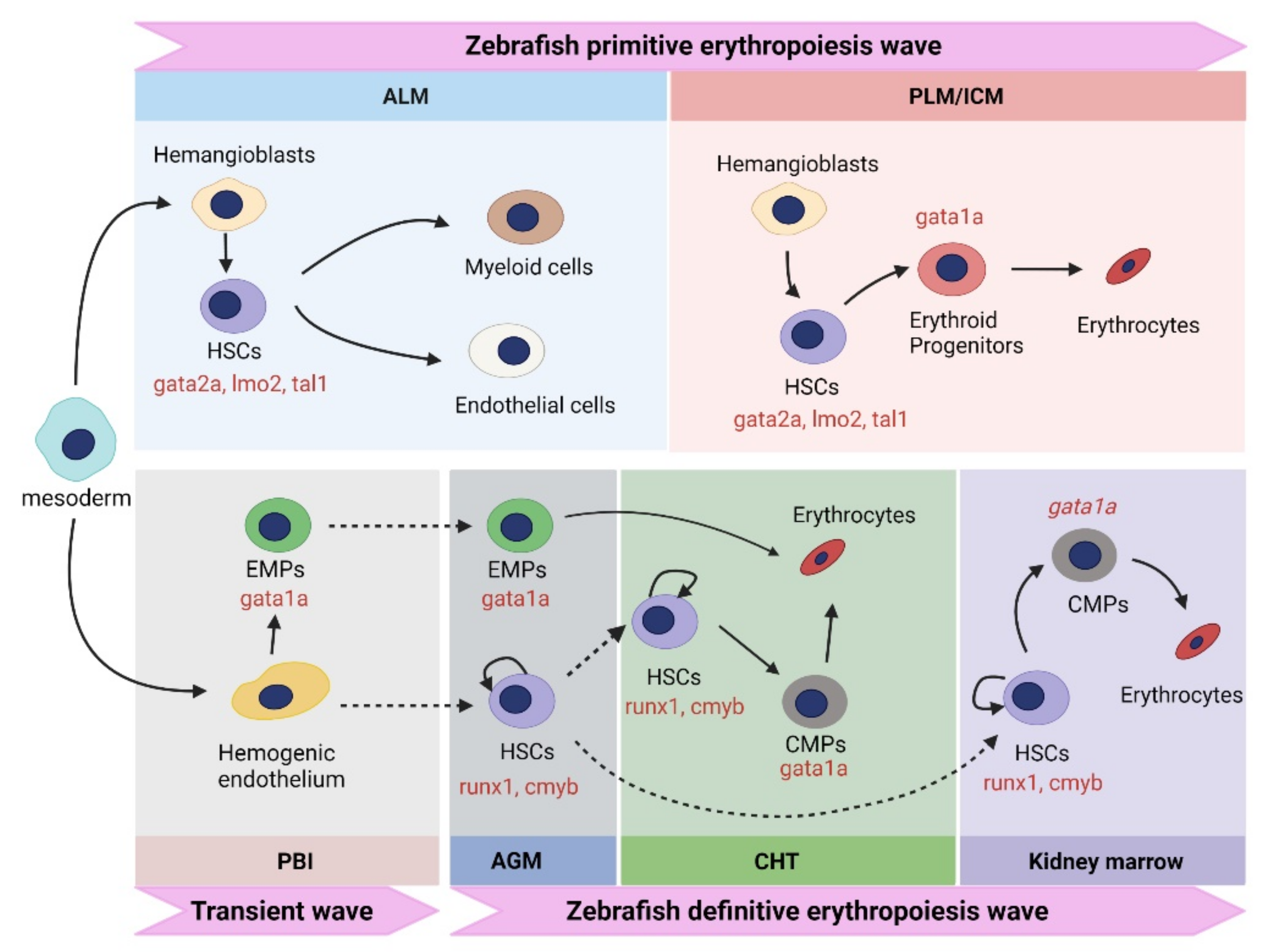
2.2. Definitive Erythropoiesis in Zebrafish

The aorta–gonad–mesonephros (AGM), caudal hematopoietic tissue (CHT), and kidney marrow are definitive hematopoiesis sites in zebrafish. The definitive erythropoiesis in zebrafish may be subdivided into two waves. A transient wave takes place at the posterior blood island (PBI) where hemogenic endothelium differentiates into erythro-myeloid progenitors (EMPs). EMPs then migrate into the dorsal aorta at ~26 hpf and differentiate into erythrocytes in CHT at ~48 hpf.
The second definitive wave starts when the hemogenic endothelium gives birth to HSCs in dorsal aorta. At ~26 hpf, the definitive HSCs first appear in the AGM, which is located between the dorsal aorta and the axial vein [31]. HSCs then migrate from the AGM to the pronephros via two discrete routes. The first route is through CHT and circulation (Figure 1). The endothelial-to-hematopoietic transition (EHT) takes place at the ventral wall of the dorsal aorta (VDA) where endothelial cells change their shape, egress into the subaortic space, and enter circulation via the axial vein [32]. HSCs express cluster of differentiation 41 (cd41), enter the blood circulation, and colonize the CHT at ~48 hpf [33]. From ~48 to 96 hpf, the nascent HSCs migrate to the CHT, followed by expansion and differentiation [33,34]. The expanded HSCs then migrate to the pronephros at ~4 days post fertilization (dpf), where they differentiate into common myeloid progenitors (CMPs) [33]. CMPs differentiate into MEPs and subsequently into erythroblasts in a gata1-dependent manner. Definitive erythrocytes populate the circulation at ~5 dpf [35]. The second migration route does not involve circulation. The HSCs derived from the AGM enter the pronephros via pronephric tubules [33].
In zebrafish, definitive HSCs express a number of marker genes, including runt-related transcription factor 1 (runx1), cmyb, and cluster of differentiation 41 (cd41) [34,36,37]. Runx1 is a transcription factor essential for the generation of HSCs from endothelial cells, as its deficiency impairs the initiation of EHT in zebrafish embryos [32]. HSCs expressing runx1 first appear at ~24 hpf in the VDA, validating it as the initiation site of definitive erythropoiesis [38]. At ~26 hpf, runx1+ HSCs originating from the dorsa aorta start to express cmyb, which is essential for the migration of HSCs [39]. Similar to that in the mouse, zebrafish cd41 serves as an early surface marker of HSCs in the dorsal aorta [33].

2.3. Comparison of Zebrafish Erythropoiesis with Mammalian Erythropoiesis

The molecular and cellular pathways, as well as the regulatory mechanisms, of erythropoiesis are highly conserved between teleosts and mammals. Erythropoiesis in both zebrafish and humans involves two successive waves, the primitive wave and definitive wave. Additionally, the regulatory network of erythropoiesis is highly conserved between zebrafish and humans [40]. For instance, homologs of almost all transcription factors with important roles in mammalian erythropoiesis are present in zebrafish [41]. Due to the high conservation of erythropoiesis in vertebrates, mutations in erythropoietic genes commonly lead to similar phenotypes in zebrafish and humans [42].
Although zebrafish erythropoiesis closely resembles that of mammals, it is distinct from mammalian erythropoiesis in a few aspects. First, the anatomical sites for generating erythrocytes are different. In zebrafish, primitive erythropoiesis takes place in the ICM, whereas the extra-embryonic yolk sac is the location of primitive erythropoiesis in mammals. Similarly, definitive erythropoiesis takes place in the PBI, dorsal aorta, CHT, and the kidney marrow in zebrafish, whereas the mammalian definitive erythropoietic sites are the AGM, placenta, fetal liver, and the bone marrow. Second, unlike mammalian RBCs, mature erythrocytes in zebrafish have nuclei. While zebrafish erythroblasts also undergo substantial nuclear condensation, they do not extrude the nuclei.

3. Using Zebrafish to Study the Regulation of Erythropoiesis

Vertebrate erythropoiesis is controlled by complex regulatory networks and feedback mechanisms in a spatially and temporally coordinated manner. First, each step of erythroid differentiation is controlled by specific transcription factors such as TAL1, GATA2, LMO2, RUNX1, CMYB, and GATA1. Second, two classical signal transduction pathways, the EPO–JAK–STAT pathway and the BMP–SMAD pathway, are known to play essential roles in regulating erythropoiesis in vertebrates. These signaling pathways are strictly controlled during erythropoiesis. Erythroferrone (ERFE), a hormone discovered recently, coordinates erythropoiesis with iron metabolism via regulating the BMP–SMAD pathway [43,44]. Third, erythropoiesis is regulated by microRNAs and long noncoding RNAs (lncRNAs). A number of microRNAs (miRNAs), including miR-126, miR-144, miR-451, miR-26a, and miR-23a, have been reported to regulate gene expression during red cell development [45,46,47,48,49,50]. In addition, erythropoiesis is regulated by recruiter lncRNAs, including LncHSC-2, lincRNA-EPS, and alncRNA-EC7/Bloodlinc, and decoy lncRNAs, including lnc-DC and lnc-MC [51]. For more detailed pathways and mechanisms regulating vertebrate erythropoiesis, please refer to the previous reviews [1,41,52,53]. In this section, we summarize recent progress on the regulation of erythropoiesis with emphasis on the studies in zebrafish.

3.1. Erythroid Transcription Factors and Their Regulation

GATA1 is a transcription factor critical for erythropoiesis. Both loss and overexpression of GATA1 impair red cell development, demonstrating the importance of GATA1 homeostasis during erythropoiesis [54,55]. Recent studies in zebrafish have gained new insights into the regulation of gata1 and demonstrated that gata1 can be regulated at transcriptional and post-transcriptional levels. The ten–eleven translocation (TET) is a protein family responsible for converting methylcytosine (5mC) into hydroxymethylcytosine (5hmC) which may lead to DNA demethylation [56,57]. Zebrafish has three TET family genes, tet1, tet2, and tet3, and all of them can catalyze the formation of 5hmC in zebrafish embryos [58]. tet2 can activate the expression of lineage-specific genes such as gata1, scl, and cmyb, and thus plays a key role in erythropoiesis [58]. Knockdown of tet2 in zebrafish embryos resulted in decreased 5hmC levels in the intermediate CpG promoters of gata1, scl, and cmyb, leading to reduced expression of these transcription factors and impaired erythropoiesis [58]. Hypoxia-inducible factors Hif1α and Hif3α play important roles in upregulating gata1 expression to promote erythropoiesis [59,60]. Hif1α and Hif3α bind to the hypoxia response elements (HREs) in the 3’ flanking region and promoter region of gata1, respectively [59,60]. Loss of these hif genes in zebrafish impaired erythropoiesis, as the consequence of inadequate activation of gata1 expression [59,60]. In addition to transcription activation, inhibitory mechanisms exist to prevent excessive erythropoiesis. For example, deficiency of p2y12, an ADP receptor crucial for purinergic signaling [61], resulted in excessive primitive erythropoiesis in zebrafish embryos characterized by increased expression of α-globin, β-globin, and gata1 [62]. This phenotype was mainly attributed to enhanced expression of gata1 as it was rescued by injection of a gata1-specific morpholino [62].
The expression of gata1 is also controlled at post-transcriptional levels during erythropoiesis. Elavl1 is an RNA-binding protein that binds AU-rich elements in mRNAs. In zebrafish embryos, elavl1a was found to bind the 3’ UTR of gata1 mRNA [63]. Silencing of elavl1a caused a dramatic reduction in gata1 expression, leading to impaired erythropoiesis [63]. Further studies reveal that the activity of ELAVL1 is controlled by protein kinase C (PKC), which phosphorylates ELAVL1 on Ser219 and Ser316 and promotes the translocation of ELAVL1 from the nucleus to cytoplasm to stabilize mRNA targets [64]. Similar to the knockdown of elavl1a, PKC inhibitors impaired erythropoiesis in zebrafish embryos [64]. In addition, the gata1 protein may be degraded through a caspase-dependent mechanism. In zebrafish, infection-induced inflammasome can activate caspase 1, which leads to cleavage and degradation of gata1 and dyserythropoiesis [65]. Pharmacological inhibition of caspase 1 can rescue the anemic phenotype in gata1-deficient zebrafish, providing a potential therapeutic strategy to treat this erythropoietic disorder [56].
Recent studies also revealed the critical roles of several other transcription factors in zebrafish erythropoiesis. Functional analyses of Krüppel-like transcription factor (klf) genes in zebrafish embryos revealed important roles of klf3 and klf6a in regulating erythroid differentiation [66]. Klf3 promoted erythropoiesis through inhibiting the expression of ferric-chelate reductase 1b (frrs1b), while klf6a acted in a distinct manner by negatively regulating the expression of cdkn1a to control the cell cycle [66]. Forkhead box O3 (FOXO3), a member of the forkhead family, is also found to play an important role in vertebrate erythropoiesis. Morpholino knockdown of foxo3b resulted in decreased expression of gata1 as well as globin genes and defective erythropoiesis in zebrafish embryos [67]. Another transcription factor, etv7, regulates the expression of the zebrafish lss gene, which encodes lanosterol synthase, an enzyme in the cholesterol synthesis pathway [68]. Knockdown of zebrafish etv7 led to reduced hemoglobin production in erythroblasts, and this phenotype could be rescued by injection of exogenous cholesterol [68]. Importantly, zebrafish has a unique advantage in the study of the function of etv7 in erythropoiesis because mice, unlike humans, lack this gene [68].

3.2. Signaling Pathways

The role of the EPO–JAK–STAT pathway in erythropoiesis is highly conserved in vertebrates. In zebrafish, both primitive and definitive erythropoiesis require epo [69]. Three splice variants of the epo gene, epo-L1, epo-L2, and epo-S, have been detected in zebrafish [70]. epo-S is mainly expressed in the kidney marrow, whereas epo-L1 and epo-L2 are expressed in the liver and heart [70]. It has been well established that epo signaling is regulated by hypoxia-induced factors [71,72]. KIT ligand (KITLG) is another cytokine that regulates proliferation and differentiation of erythroid cells by binding to its receptors [73]. Recent gain-of-function studies in zebrafish showed that expression of kitlga and kitlgb both enhanced erythroid cell expansion by cooperating with epo [74]. The function of Kitlga in erythropoiesis was also verified in ex vivo suspension cultures of zebrafish hematopoietic progenitor cells [74]. Additionally, lysophosphatidic acid (LPA) was shown to cooperate with EPO signaling to regulate erythropoiesis, and its receptors, LPA2 and LPA3, play opposing roles in this process [75]. In human K562 cells, knockdown of LPA2 enhanced erythropoiesis, whereas knockdown of LPA3 inhibited RBC differentiation [75]. Consistently, hemoglobin expression in zebrafish embryos was significantly increased by treatment with lpa3 agonist but was inhibited by lpa2 agonist [75]. Accordingly, pharmacological activation of these LPA receptor subtypes may be a new strategy to enhance or inhibit erythropoiesis.
Once EPOR binds EPO, it recruits JAK, a tyrosine kinase that can phosphorylate several tyrosine residues in EPOR. Zebrafish has three homologs of JAK: jak1, jak2a, and jak2b [76]. Overexpression of jak2a in zebrafish embryos increased the expression of gata1 and hemoglobin, indicating a crucial role of jak2a in zebrafish erythropoiesis [77]. STAT proteins are transcription factors regulated by protein kinases and phosphatases. Protein tyrosine phosphatase ptpn9 was considered to regulate erythroid cell differentiation by disrupting the inhibitory complex of phosphorylated stat3, gata1, and zbp-89 in zebrafish [78]. Additionally, cytokine-inducible SH2 domain-containing protein (CISH) may suppress the STAT signaling induced by cytokines. Knockdown of cish.a, an ortholog of human CISH, led to enhanced erythropoiesis and stat5.1 activation in zebrafish embryos, providing in vivo evidence to support the role of CISH in regulating STAT5 and erythropoiesis [79]. Additionally, a lipid kinase named PI4KA was shown to regulate JAK–STAT signaling and erythropoiesis [80]. Knockdown of PI4KA in mouse hematopoietic stem progenitor cells (HSPCs) impaired erythropoiesis in vitro. Loss of pi4kaa, the zebrafish homolog of PI4KA, led to impaired erythroid differentiation [80].
Apart from the EPO–JAK–STAT pathway, regulation of the mTORC1, Rho, BMP, and TGF-β signaling pathways is also essential for zebrafish erythropoiesis. Analysis of the amino acid transporter LAT3, a gene upregulated during erythroblast maturation, showed that L-leucine availability controls erythropoiesis via regulating the mTORC1/4E-BP pathway [81]. Inadequate NEAA uptake induced by lat3 deficiency and pharmacologic inhibitors of mTORC1/4E-BP pathway both led to reduced hemoglobin production in zebrafish [81]. A genome-wide association study combined with functional analyses in zebrafish revealed the critical role of the ARHGEF12–RhoA–p38 signaling pathway in erythroid regeneration [82]. During stress erythropoiesis, GATA1 upregulates the expression of ARHGEF12, which is a RhoA guanine nucleotide exchange factor that converts RhoA-GDP to RhoA-GTP [82]. The active RhoA promotes erythropoiesis through the p38–MAPK pathway [82]. Both knockout and knockdown of arhgef12 in the zebrafish impair the erythroid differentiation, and the anemic phenotype can be rescued by treatment with p38 activator [82]. A recent study identified a new gene named BMP inhibitory factor 1 (bif1), which inhibits BMP signaling and is required for both primitive and definitive erythropoiesis in zebrafish [83]. Given that the BMP is known to regulate mesodermal lineage determination [84,85], it is likely that inhibition of the BMP–SMAD pathway by Bif1 promotes the expansion of hematopoietic progenitors. Research in zebrafish also uncovered a role of splicing factor 3B subunit 1 (sf3b1) in regulating TGF-β signaling during erythropoiesis [84]. SF3B1 is a core component of the spliceosome, and its mutations can cause human erythropoietic diseases due to aberrant splicing of pre-mRNAs [86,87,88,89]. Deficiency of sf3b1 in zebrafish embryos caused abnormal activation of the TGF-β signaling as well as cell cycle arrest and macrocytic anemia [90]. The phenotype of increased G0/G1-stage erythroid progenitors could be rescued by the inhibition of TGF-β signaling in the morphant zebrafish embryos [90].

3.3. MicroRNAs

A number of miRNAs, such as miR-126, miR-144, and miR-451, have been demonstrated to regulate gene expression during erythropoiesis. The miR-144/451 genomic region was first identified as a GATA-1-regulated locus essential for erythropoiesis in humans [50,91]. Zebrafish miR-451 is also clustered with miR-144, and they are in the same primary transcript, but miR-451 is about 7.5-fold more abundant than miR-144 in zebrafish erythroblasts [92]. This paradox is due to a negative-feedback loop in which miR-144 represses Dicer, which is an RNase required for the production of many miRNAs, during erythropoiesis in zebrafish [92]. Interestingly, miR-451 is refractory to the loss of Dicer because it is processed in an Ago2-dependent manner [92]. Thus, miR-451 can by-pass the global miRNA turnover during erythropoiesis to become the most abundant miRNA in erythroblasts [92]. miR-451 represses the expression of gata2 by binding to its 3′UTR in developing erythroblasts [93]. Diminished expression of miR-451, as observed in the meunier mutant, impairs erythrocyte maturation in zebrafish [93]. Several other miRNAs also play a regulatory role in erythroblasts. For example, miR-26a was found to regulate erythroid proliferation and differentiation via targeting the 3’UTR of the Nemo-like kinase (NLK) in human and zebrafish models of DBA [45]. miR-200a may inhibit erythropoiesis via targeting the 3’UTR of programmed cell death 4 (PDCD4) and thyroid hormone receptor beta (THRB) [46]. miR-23a is critical for maintaining the morphology of erythrocytes, likely by targeting the protein tyrosine phosphatase SHP2 [49].

3.4. E3 Ubiquitin Ligases

The ubiquitin–proteasome system was first discovered in the circulating reticulocytes [94,95,96]. However, the function of ubiquitinating factors in erythropoiesis remains largely unexplored. In 2017, the E2 ubiquitin-conjugating enzyme Ube2o was reported to promote ubiquitination and degradation of ribosomal proteins in developing erythroblasts [97]. Recent studies in zebrafish led to the identification of several E3 ubiquitin ligases with critical roles in erythropoiesis. Cullin–RING E3 ligase complexes are well-known ubiquitin ligases with important roles in many biological processes [98]. Loss of cul4a in zebrafish embryos resulted in severely reduced erythrocyte production due to decreased expression of transcription factors gata1, scl, and lmo2 [99]. The level of H3K4me3, a histone marker of transcription activation, in the promoter region of gata1 was decreased after cul4a depletion, indicating that cul4a can activate the transcription of gata1 by promoting H3K4 trimethylation [99].
Fish erythrocytes have nuclei, however, their nuclei are much smaller than that of the erythroid progenitors [100]. A study in zebrafish and mammal hematopoietic cell models demonstrated that Wdr26 functions as a core subunit of an E3 ubiquitin ligase complex to promote the ubiquitination and degradation of nuclear proteins during erythropoiesis [101]. Loss of wdr26 resulted in severe anemia and enlarged nuclei in mature erythrocytes in zebrafish [101]. In developing erythroblasts, Wdr26 regulates the ubiquitination of nuclear proteins including lamins and histones [101]. Degradation of lamin B further promotes the formation of large nuclear openings, which are transiently formed on the nuclear envelope to expedite the export and degradation of nuclear proteins during erythroblast differentiation [101,102].
During the recent studies of zebrafish erythropoiesis, a number of knockout mutants have been generated (Table 1). These mutants serve as valuable genetic models for further exploring the mechanisms of erythropoiesis as well the related blood disorders.

4. Using Zebrafish to Study Iron and Heme Homeostasis during Erythropoiesis

Terminal erythropoiesis involves several characteristic processes including synthesis of hemoglobin, condensation and extrusion of nuclei, clearance of organelles, and remodeling of the membrane and proteome. Hemoglobin, the most abundant protein in erythrocytes, is responsible for binding and transporting oxygen in vertebrate circulation. Each hemoglobin molecule is composed of four globin chains and four heme moieties, which are iron-containing porphyrins. Accordingly, developing erythroid cells absorb massive amounts iron for the synthesis of heme and hemoglobin. A number of regulatory mechanisms for erythroid iron uptake and heme synthesis were made by studies in zebrafish, and therefore, we focus on iron and heme metabolism in this section.

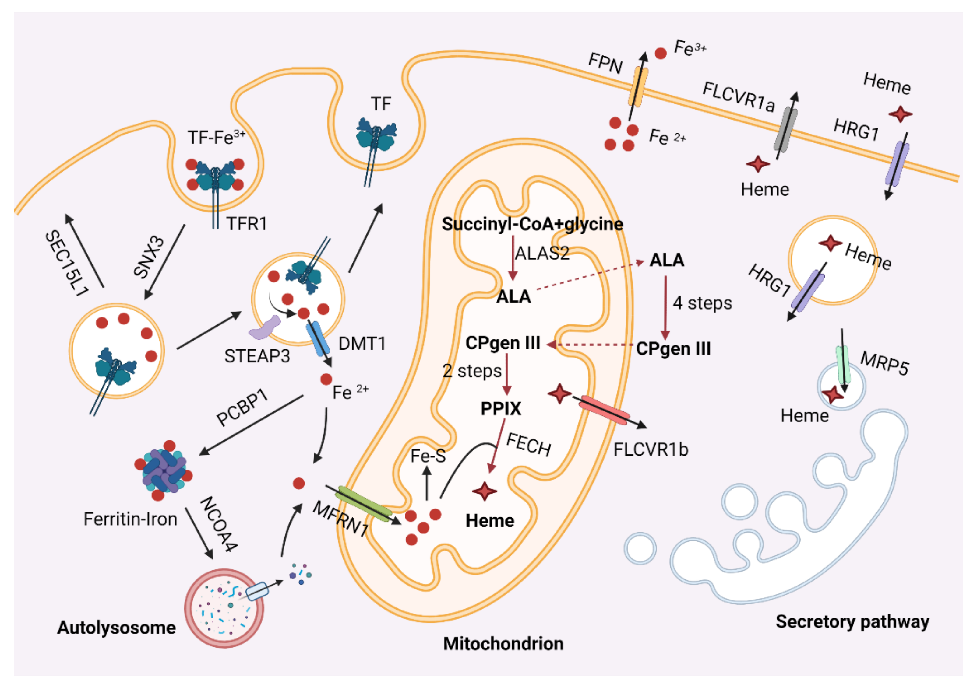
4.1. Iron and Heme Metabolism during Erythropoiesis

In vertebrates, developing erythroblasts assimilate iron primarily through the transferrin receptor (Tfr1), which binds diferric transferrin (Tf). The Tf–Tfr1 complex is internalized into endosomes through clathrin-mediated endocytosis [106,107] (Figure 2). The Fe3+ in maturing endosomes is reduced to Fe2+ by STEAP3 [108] and exported out of endosomes by divalent metal transporter 1 (DMT1) [109]. Once inside the cytosol, iron either enters mitochondria via mitoferrin 1 (MFRN1) [110,111] for the biosynthesis of heme and iron–sulfur clusters (Fe–S) or is sequestered in the ferritin complex [112]. The rest of the Tf–Tfr1 complex undergoes sorting process in endosomes and returns to the cell membrane for the next iron uptake cycle. Given that the Tf cycle involves vesicular transport, trafficking proteins play crucial roles in this process. For example, the retromer protein SNX3 is essential for sorting the Tf–Tfr1 complex into recycling endosomes, while the exocyst component, SEC15L1, regulates the trafficking of this protein complex from recycling endosomes to the cell surface [113,114].
In addition to importing iron, animals may mobilize and utilize iron stored in ferritin for heme synthesis. Poly r(C)-binding protein (PCBP1) is a cytosolic iron chaperone responsible for transporting iron to ferritin [115]. The ferritin complex may undergo autophagic degradation, a process called ferritinophagy, to release the stored iron [116,117]. Ferritinophagy is specifically controlled by nuclear receptor coactivator 4 (NCOA4), which has been shown to be an autophagy receptor [116]. Silencing of NCOA4 led to impaired erythropoiesis in both human erythroblast cells and zebrafish embryos [118]. Ncoa4-mediated ferritin degradation is critical for macrophage iron release and erythropoiesis in mice [119]. In addition, HERC2, an E3 ubiquitin ligase, may regulate the protein level of NCOA4 in response to cellular iron levels [118].
Heme is synthesized via eight enzymatic reactions that take place in both the cytosol and mitochondria [3,120] (Figure 2). The first step, generation of δ-aminolevulinic acid (ALA) catalyzed by ALAS2, and the terminal step, formation of heme catalyzed by ferrochelatase (FECH), are two rate-limiting steps in heme synthesis. Following its synthesis, heme must be translocated from the mitochondria to the cytosol where hemoglobin resides [121,122,123] (Figure 2). In non-erythroid cells, heme also needs to be transported to other cellular compartments to be incorporated into hemoproteins [124]. Since free heme is cytotoxic, cellular heme homeostasis is strictly controlled by specific transporters and chaperones. The feline leukemia virus subgroup C receptor-related protein 1a (FLVCR1a) and its splicing variant FLVCR1b are reported to mediate the transport of heme out of the cell and the mitochondria, respectively [122,123,125]. The multidrug resistance protein 5 (MRP5) transports cytosolic heme into the secretory pathway [126], while heme responsive gene 1 (HRG1) is a heme importer localized in the plasma and endosomal membranes [127].

4.2. Recent Progress Studying Erythroid Iron and Heme Metabolism Using Zebrafish

4.2.1. Iron Metabolism

Zebrafish erythroblasts rely on the Tf cycle to assimilate iron. Zebrafish has two homologs of TFR1, tfr1a and tfr1b. tfr1a is mainly expressed in erythroid cells, whereas tfr1b is ubiquitously expressed without a specific lineage preference [128]. In consistency with their expression patterns, mutations in tfr1a led to anemia, while morpholino knockdown of tfr1b did not induce defects in hemoglobin synthesis [128]. tfr1b may play an important role in iron uptake in neuronal systems as its silencing resulted in neurologic phenotypes [129]. In addition to Tfr1, zebrafish expresses snx3, a sorting nexin, in the erythroid tissues [113]. Knockdown of snx3 by morpholinos induced profound iron-deficiency anemia during early development, and this defect can be complemented by supplementing non-Tf bound iron [113].
Work on the frascati (frs) zebrafish mutant has provided key insights into mitochondrial iron import [111]. The frs mutant developed hypochromic anemia, which was found to be caused by a missense mutation in a gene that encodes a solute carrier family protein (SLC25A37) [111]. This gene was shown to be a mitochondrial iron importer and named mitoferrin (mfrn1) [111]. Another mitochondrial inner-membrane protein, FAM210B, may also regulate mitochondrial iron homeostasis and heme biosynthesis in developing erythroblasts [130]. Fam210b is highly expressed in vertebrate erythroid tissues, and its silencing induced profound defects in heme production in both erythroblast cell models and zebrafish embryos [130]. Currently, the mechanistic role of Fam210b still remains elusive. Interestingly, the model organism C. elegans has a Fam210 homolog, but it does not synthesize heme, excluding the direct role of Fam210 family proteins in regulating heme synthesis [131].

4.2.2. Heme Synthesis and Transport

Recent work in zebrafish has uncovered several new mechanisms regulating the heme synthesis genes alas2 and fech. The expression of alas2 was shown to be controlled by the hif1α/vgll4/irf2bp2 oxygen sensing pathway [103]. Under hypoxic conditions, hif1α upregulates vgll4 expression through notch1 [103]. Vgll4 sequesters irf2bp2, a transcriptional repressor of alas2, and induces alas2 expression and heme synthesis in erythroid tissues [103]. The vgll4b knockout fish displayed reduced erythroid heme production, leading to mitochondriopathy, increased immature erythrocytes, and reduced animal survival rate [103]. Additionally, the mitochondrial matrix peptidase, CLPX, plays dual roles in controlling the activity of ALAS. It catalyzes the incorporation of the cofactor pyridoxal phosphate into ALAS, and low-activity CLPX increases the stability of ALAS protein [132,133]. High activity of CLPX may accelerate the cofactor incorporation and ALAS degradation [133]. Morpholino knockdown of zebrafish Clpx homologs, clpxa and clpxb, results in reduced hemoglobin production and impaired erythropoiesis [132]. In addition, the activity of FECH, the final heme synthesis enzyme, may be controlled at the post-translational level. For instance, phosphorylation of FECH by the mitochondrial outer-membrane PKA is critical for FECH activity [134]. Analysis of pinotage, an anemic zebrafish mutant identified through a mutagenesis screen, uncovered a critical role of mitochondrial pH in controlling the activity of FECH [135]. Loss of atpif1, the gene responsible for the phenotype in pinotage, results in increased mitochondrial pH, which inhibits FECH activity [135].
The physiological roles of major heme transporters, including HRG1, MRP5, and FLVCR, have been examined in zebrafish. Transient knockdown of zebrafish hrg1 and mrp5 both induced defective hemoglobinization [126,127]. Hrg1 plays an essential role in recycling heme released by damaged or senescent RBCs in kidney macrophages in zebrafish [136]. Double knockout of zebrafish hrg1a and hrg1b caused heme accumulation in kidney macrophages [136]. Zebrafish has two isoforms of flvcr1, i.e., flvcr1a and flvcr1b. flvcr1a is required for the expansion of committed erythroid progenitors but cannot drive their terminal differentiation [121]. flvcr1b contributes to the expansion phase and is required for erythroblast differentiation [121]. The coordinated expression of flvcr1a and flvcr1b controls the cytosolic heme pool, which is critical for the regulation of erythroid progenitors and hemoglobin synthesis during terminal erythropoiesis [121].

5. Zebrafish Models of Erythropoietic Disorders

In humans, defects in erythropoiesis can cause various types of blood disorders such as β-thalassemia, sickle cell anemia, DBA, CDA, congenital sideroblastic anemia, microcytic hypochromic anemia, MDS, and erythropoietic protoporphyria [137,138,139]. For example, mutations in human heme synthesis genes ALAS2 and FECH lead to congenital sideroblastic anemia and erythropoietic protoporphyria, respectively. Reduced globin expression and iron deficiency result in smaller erythrocytes, defined as hypochromic anemia, while iron overload can induce hemochromatosis. Additionally, abnormal structures of spectrin proteins and the erythrocyte membrane are common causes of hemolytic anemia and hereditary spherocytosis in humans. Many of these blood disorders have been characterized in zebrafish. For instance, the sauternes, quem, and dracula zebrafish mutants carry mutations in different heme synthesis genes, and they have been used as genetic models to study microcytic hypochromic anemia, hepatoerythropoietic porphyria, and erythropoietic protoporphyria, respectively [140,141,142]. Here, we focus on recently generated zebrafish models for CDAs, DBAs, and MDS, and these models may serve as valuable tools to develop new therapeutics for these diseases.

5.1. CDAs

CDAs are characterized by reduced erythropoiesis efficiency and can be divided into several groups including CDA type I, II, III, CDA variants, and transcription-factor-related CDAs. Clinically, CDAI presents with moderate to severe giant cell anemia and abnormal erythrocyte precursors, as well as a spongy heterochromatin phenotype. This disease is caused by mutations in CDAN1 gene. In zebrafish embryos, knockdown of cdan1 resulted in reduced gata1a expression, increased gata2a expression, and impaired primitive erythropoiesis [143]. These results complement the data obtained from mice and suggest that cdan1 deficiency may cause CDAI via misregulation of the erythroid transcription factors gata1 and gata2 [143].
Human CDAII is an erythroid-related disease caused by abnormal cell division, which leads to cell apoptosis, multinuclear erythroblasts, and anemias. Mutations in the anion exchanger protein BAND 3 can cause CDAII-related dyserythropoiesis [144]. The zebrafish retsina mutant, which harbors a mutation in band 3, exhibited erythroid-specific defects in cell division and dyserythropoiesis, which resemble the symptoms of human CDAII diseases [145]. The CDAII can also be caused by bi-allelic mutations in the SEC23B gene that encodes a component of the COPII coatomer. Despite the fact that SEC23B is ubiquitous expressed in many tissues, mutations of this gene cause an erythroid-lineage-specific phenotype [146,147]. Knockdown of sec23b in zebrafish embryos impaired erythroid development, while knockout of sec23b induced more a severe phenotype and was lethal to fish within 3 weeks [148]. Expression of sec23a could fully rescue the phenotypes in the sec23b mutant fish [148], providing a potential way of gene therapy for treating CDAII. In addition, studies in zebrafish have provided important insights into the physiological function of heat shock cognate B (HSCB) in erythropoiesis and CDAs [149].

5.2. DBAs

DBAs are congenital bone marrow failure syndromes characterized by the defective development of erythroblasts. This disease has been associated with mutations and large deletions in ribosomal protein (RP) genes including RPS7, RPS10, RPS17, RPS19, RPS24, RPS26, RPS29, RPL5, RPL11, RPL26, and RPL35A as well as GATA1 [138,150]. Elevated activity of the tumor suppressor p53 was also observed in DBA patients [151]. Zebrafish has been used for pathogenesis studies and drug screens related to DBAs because it has advantages in high-throughput genetic screens and chemical biological studies. Chemical screens in zebrafish have identified a number of compounds such as L-leucine, sotatercept, and trifluoperazine as potentially effective drugs for DBAs [152].
In recent years, the physiological roles of many ribosomal genes in zebrafish erythropoiesis have been characterized. Transient knockdown of these genes with morpholinos often leads to defective erythropoiesis in zebrafish. For instance, rpl5 is required for both primitive and definitive erythropoiesis processes in zebrafish embryos [153]. rpl18-deficient zebrafish embryos developed anemia with reduced gata1 and globin expression [154]. In addition, silencing of rpl27 and rps27 both resulted in reduced hemoglobin expression and impaired erythrocyte production [155].
Increased activity of the tumor suppressor p53 is observed in DBA patients [151]. In zebrafish, loss of rps9 and rpl10a both led to impairment of erythroblast maturation and anemia in a p53-dependent manner [156,157]. Similarly, rpl18 deficiency increased the activities of both p53 and jak2–stat3 pathways, which could be rescued by inhibitors of jak2 or stat3 phosphorylation [154]. rps29 mutant zebrafish embryos, another model of DBAs, showed a p53-dependent anemia, and this phenotype could be rescued by calmodulin inhibitors [14]. In addition, ribosomal dysfunction in DBA patients can activate Nemo-like kinase (NLK), which is hyperactivated in committed erythroid progenitors. Knockdown of rps19 in zebrafish embryos induced anemia, which could be rescued by metformin injection [45]. Further studies demonstrated that metformin induced the upregulation of miR-26a, which targets NLK for degradation, to improve erythropoiesis in human cells [45].

5.3. MDSs

MDSs are a group of malignant bone marrow disorders, which are associated with ineffective erythropoiesis. The current standard of the classification of MDS is based on patients of refractory anemia (RA) with or without ringed sideroblasts. Recent studies in zebrafish mainly focus on two subtypes of MDSs, including refractory anemia with ring sideroblasts (RARS) and MDS associated with isolated del(5q). Splicing factor 3B subunit 1 (SF3B1) is one of the most prevalently mutated factors in RARS subtype of MDS [90]. Loss-of-function mutations in zebrafish sf3b1 induce macrocytic anemias with enhanced TGF-β signaling, and inhibition of TGF-β signaling pathway can release the G0/G1 block of erythroid progenitors in the mutants [90]. Thus, combined medication with TGFβ superfamily inhibitors and known SF3B1-modulating drugs may be a more effective method to treat MDS patients [90]. The other major subtype of MDS, del(5q) MDS, includes deletion of the RPS14 gene, which results in macrocytic anemia [158]. The rps14-knockout zebrafish has been used to screen for new drugs for the treatment of del(5q) MDS [159]. This screen identified inhibitors of matrix metalloproteinase 9 (MMP9) that significantly rescued the erythroid defects in rps14-deficient zebrafish [159]. Further study demonstrated that mmp9 was upregulated in rps14-deficient cells to inhibit erythropoiesis via enhanced TGF-β signaling [159]. This work suggests that MMP9 inhibitors may serve as therapeutic agents for patients with del(5q) MDS. It was reported that mutations in DEAD-box helicase 41 (DDX41) was also associated with blood disorders including MDS [160]. Functional analysis of ddx41 in zebrafish embryos revealed that it was required for the expansion and differentiation of erythroid progenitors [161], and these results promoted the understanding of hematologic malignancies induced by mutations in DDX41.

6. Conclusions

The zebrafish is an excellent vertebrate model to study erythropoiesis, because it has many advantages for genetic studies and the processes of erythropoiesis are highly conserved between fish and mammals. This model animal permits forward genetic screens, reverse genetic studies, large-scale chemical screens, and targeted gene function studies. Studies on zebrafish have provided important insights into the fundamental understanding of erythropoiesis, especially in iron and heme metabolism and regulation of erythropoiesis. The establishment of zebrafish models of blood-related diseases provides valuable resources for developing new therapeutic treatments of these diseases.

Author Contributions

Conceptualization, Y.Z. and C.C.; writing—original draft preparation, Y.Z.; writing—review and editing, M.C. and C.C. All authors have read and agreed to the published version of the manuscript.

Funding

This research was funded by the National Natural Science Foundation of China (31871200 and 32071155) and the National Key Research and Development Program of China (2018YFA0507802).

Institutional Review Board Statement

Not applicable.

Informed Consent Statement

Not applicable.

Data Availability Statement

Not applicable.

Conflicts of Interest

The authors declare no conflict of interest.

References

  1. Orkin, S.H.; Zon, L.I. Hematopoiesis: An evolving paradigm for stem cell biology. Cell 2008, 132, 631–644. [Google Scholar] [CrossRef] [Green Version]
  2. Dzierzak, E.; Philipsen, S. Erythropoiesis: Development and differentiation. Cold Spring Harb. Perspect. Med. 2013, 3, a011601. [Google Scholar] [CrossRef]
  3. Chung, J.; Chen, C.; Paw, B.H. Heme metabolism and erythropoiesis. Curr. Opin. Hematol. 2012, 19, 156–162. [Google Scholar] [CrossRef]
  4. Mei, Y.; Liu, Y.; Ji, P. Understanding terminal erythropoiesis: An update on chromatin condensation, enucleation, and reticulocyte maturation. Blood Rev. 2021, 46, 100740. [Google Scholar] [CrossRef]
  5. Liu, J.; Mohandas, N.; An, X.L. Membrane assembly during erythropoiesis. Curr. Opin. Hematol. 2011, 18, 133–138. [Google Scholar] [CrossRef]
  6. Muckenthaler, M.U.; Rivella, S.; Hentze, M.W.; Galy, B. A Red Carpet for Iron Metabolism. Cell 2017, 168, 344–361. [Google Scholar] [CrossRef] [Green Version]
  7. Schultz, I.J.; Chen, C.; Paw, B.H.; Hamza, I. Iron and porphyrin trafficking in heme biogenesis. J. Biol. Chem. 2010, 285, 26753–26759. [Google Scholar] [CrossRef] [Green Version]
  8. Hattangadi, S.M.; Wong, P.; Zhang, L.; Flygare, J.; Lodish, H.F. From stem cell to red cell: Regulation of erythropoiesis at multiple levels by multiple proteins, RNAs, and chromatin modifications. Blood 2011, 118, 6258–6268. [Google Scholar] [CrossRef] [PubMed] [Green Version]
  9. Kuhrt, D.; Wojchowski, D.M. Emerging EPO and EPO receptor regulators and signal transducers. Blood 2015, 125, 3536–3541. [Google Scholar] [CrossRef] [PubMed] [Green Version]
  10. Lin, C.-Y.; Chiang, C.-Y.; Tsai, H.-J. Zebrafish and Medaka: New model organisms for modern biomedical research. J. Biomed. Sci. 2016, 23, 19. [Google Scholar] [CrossRef] [PubMed] [Green Version]
  11. Kafina, M.D.; Paw, B.H. Using the Zebrafish as an Approach to Examine the Mechanisms of Vertebrate Erythropoiesis. Methods Mol. Biol. 2018, 1698, 11–36. [Google Scholar] [CrossRef]
  12. Ridges, S.; Heaton, W.L.; Joshi, D.; Choi, H.; Eiring, A.; Batchelor, L.; Choudhry, P.; Manos, E.J.; Sofla, H.; Sanati, A.; et al. Zebrafish screen identifies novel compound with selective toxicity against leukemia. Blood 2012, 119, 5621–5631. [Google Scholar] [CrossRef] [PubMed] [Green Version]
  13. Leet, J.K.; Lindberg, C.D.; Bassett, L.A.; Isales, G.M.; Yozzo, K.L.; Raftery, T.D.; Volz, D.C. High-content screening in zebrafish embryos identifies butafenacil as a potent inducer of anemia. PLoS ONE 2014, 9, e104190. [Google Scholar] [CrossRef] [Green Version]
  14. Taylor, A.M.; Macari, E.R.; Chan, I.T.; Blair, M.C.; Doulatov, S.; Vo, L.T.; Raiser, D.M.; Siva, K.; Basak, A.; Pirouz, M.; et al. Calmodulin inhibitors improve erythropoiesis in Diamond-Blackfan anemia. Sci. Transl. Med. 2020, 12, eabb5831. [Google Scholar] [CrossRef]
  15. Kulkeaw, K.; Sugiyama, D. Zebrafish erythropoiesis and the utility of fish as models of anemia. Stem Cell Res. 2012, 3, 55. [Google Scholar] [CrossRef] [PubMed] [Green Version]
  16. Gore, A.V.; Pillay, L.M.; Venero Galanternik, M.; Weinstein, B.M. The zebrafish: A fintastic model for hematopoietic development and disease. Wiley Interdiscip. Rev. Dev. Biol. 2018, 7, e312. [Google Scholar] [CrossRef] [PubMed]
  17. Zhang, C.; Patient, R.; Liu, F. Hematopoietic stem cell development and regulatory signaling in zebrafish. Biochim. Biophys. Acta 2013, 1830, 2370–2374. [Google Scholar] [CrossRef]
  18. Wattrus, S.J.; Zon, L.I. Stem cell safe harbor: The hematopoietic stem cell niche in zebrafish. Blood Adv. 2018, 2, 3063–3069. [Google Scholar] [CrossRef] [Green Version]
  19. Dooley, K.A.; Davidson, A.J.; Zon, L.I. Zebrafish scl functions independently in hematopoietic and endothelial development. Dev. Biol. 2005, 277, 522–536. [Google Scholar] [CrossRef] [Green Version]
  20. Liu, F.; Walmsley, M.; Rodaway, A.; Patient, R. Fli1 acts at the top of the transcriptional network driving blood and endothelial development. Curr. Biol. 2008, 18, 1234–1240. [Google Scholar] [CrossRef] [Green Version]
  21. Liu, F.; Patient, R. Genome-wide analysis of the zebrafish ETS family identifies three genes required for hemangioblast differentiation or angiogenesis. Circ. Res. 2008, 103, 1147–1154. [Google Scholar] [CrossRef] [Green Version]
  22. Gering, M.; Rodaway, A.R.F.; Gottgens, B.; Patient, R.K.; Green, A.R. The SCL gene specifies haemangioblast development from early mesoderm. EMBO J. 1998, 17, 4029–4045. [Google Scholar] [CrossRef] [Green Version]
  23. De Pater, E.; Kaimakis, P.; Vink, C.S.; Yokomizo, T.; Yamada-Inagawa, T.; van der Linden, R.; Kartalaei, P.S.; Camper, S.A.; Speck, N.; Dzierzak, E. Gata2 is required for HSC generation and survival. J. Exp. Med. 2013, 210, 2843–2850. [Google Scholar] [CrossRef] [PubMed] [Green Version]
  24. Dobrzycki, T.; Mahony, C.B.; Krecsmarik, M.; Koyunlar, C.; Rispoli, R.; Peulen-Zink, J.; Gussinklo, K.; Fedlaoui, B.; de Pater, E.; Patient, R.; et al. Deletion of a conserved Gata2 enhancer impairs haemogenic endothelium programming and adult Zebrafish haematopoiesis. Commun. Biol. 2020, 3, 71. [Google Scholar] [CrossRef] [PubMed]
  25. Gioacchino, E.; Koyunlar, C.; Zink, J.; de Looper, H.; de Jong, M.; Dobrzycki, T.; Mahony, C.B.; Hoogenboezem, R.; Bosch, D.; van Strien, P.M.H.; et al. Essential role for Gata2 in modulating lineage output from hematopoietic stem cells in zebrafish. Blood Adv. 2021, 5, 2687–2700. [Google Scholar] [CrossRef]
  26. Patterson, L.J.; Gering, M.; Eckfeldt, C.E.; Green, A.R.; Verfaillie, C.M.; Ekker, S.C.; Patient, R. The transcription factors Scl and Lmo2 act together during development of the hemangioblast in zebrafish. Blood 2007, 109, 2389–2398. [Google Scholar] [CrossRef] [PubMed] [Green Version]
  27. Lyons, S.E.; Lawson, N.D.; Lei, L.; Bennett, P.E.; Weinstein, B.M.; Liu, P.P. A nonsense mutation in zebrafish gata1 causes the bloodless phenotype in vlad tepes. Proc. Natl. Acad. Sci. USA 2002, 99, 5454–5459. [Google Scholar] [CrossRef] [PubMed] [Green Version]
  28. Kang, Y.A.; Sanalkumar, R.; O’Geen, H.; Linnemann, A.K.; Chang, C.J.; Bouhassira, E.E.; Farnham, P.J.; Keles, S.; Bresnick, E.H. Autophagy driven by a master regulator of hematopoiesis. Mol. Cell Biol. 2012, 32, 226–239. [Google Scholar] [CrossRef] [PubMed] [Green Version]
  29. McIver, S.C.; Kang, Y.A.; DeVilbiss, A.W.; O’Driscoll, C.A.; Ouellette, J.N.; Pope, N.J.; Camprecios, G.; Chang, C.J.; Yang, D.; Bouhassira, E.E.; et al. The exosome complex establishes a barricade to erythroid maturation. Blood 2014, 124, 2285–2297. [Google Scholar] [CrossRef] [PubMed] [Green Version]
  30. McIver, S.C.; Katsumura, K.R.; Davids, E.; Liu, P.; Kang, Y.A.; Yang, D.; Bresnick, E.H. Exosome complex orchestrates developmental signaling to balance proliferation and differentiation during erythropoiesis. Elife 2016, 5. [Google Scholar] [CrossRef]
  31. Bertrand, J.Y.; Chi, N.C.; Santoso, B.; Teng, S.; Stainier, D.Y.; Traver, D. Haematopoietic stem cells derive directly from aortic endothelium during development. Nature 2010, 464, 108–111. [Google Scholar] [CrossRef] [Green Version]
  32. Kissa, K.; Herbomel, P. Blood stem cells emerge from aortic endothelium by a novel type of cell transition. Nature 2010, 464, 112–115. [Google Scholar] [CrossRef]
  33. Bertrand, J.Y.; Kim, A.D.; Teng, S.; Traver, D. CD41+ cmyb+ precursors colonize the zebrafish pronephros by a novel migration route to initiate adult hematopoiesis. Development 2008, 135, 1853–1862. [Google Scholar] [CrossRef] [Green Version]
  34. Tamplin, O.J.; Durand, E.M.; Carr, L.A.; Childs, S.J.; Hagedorn, E.J.; Li, P.; Yzaguirre, A.D.; Speck, N.A.; Zon, L.I. Hematopoietic stem cell arrival triggers dynamic remodeling of the perivascular niche. Cell 2015, 160, 241–252. [Google Scholar] [CrossRef] [Green Version]
  35. Liao, E.C.; Trede, N.S.; Ransom, D.; Zapata, A.; Kieran, M.; Zon, L.I. Non-cell autonomous requirement for the bloodless gene in primitive hematopoiesis of zebrafish. Development 2002, 129, 649–659. [Google Scholar] [CrossRef] [PubMed]
  36. North, T.E.; Goessling, W.; Walkley, C.R.; Lengerke, C.; Kopani, K.R.; Lord, A.M.; Weber, G.J.; Bowman, T.V.; Jang, I.H.; Grosser, T.; et al. Prostaglandin E2 regulates vertebrate haematopoietic stem cell homeostasis. Nature 2007, 447, 1007–1011. [Google Scholar] [CrossRef]
  37. Lin, H.F.; Traver, D.; Zhu, H.; Dooley, K.; Paw, B.H.; Zon, L.I.; Handin, R.I. Analysis of thrombocyte development in CD41-GFP transgenic zebrafish. Blood 2005, 106, 3803–3810. [Google Scholar] [CrossRef] [Green Version]
  38. Kalev-Zylinska, M.L.; Horsfield, J.A.; Flores, M.V.C.; Postlethwait, J.H.; Vitas, M.R.; Baas, A.M.; Crosier, P.S.; Crosier, K.E. Runx1 is required for zebrafish blood and vessel development and expression of a human RUNX1-CBF2T1 transgene advances a model for studies of leukemogenesis. Development 2002, 129, 2015–2030. [Google Scholar] [CrossRef]
  39. Zhang, Y.; Jin, H.; Li, L.; Qin, F.X.; Wen, Z. cMyb regulates hematopoietic stem/progenitor cell mobilization during zebrafish hematopoiesis. Blood 2011, 118, 4093–4101. [Google Scholar] [CrossRef] [PubMed] [Green Version]
  40. Howe, K.; Clark, M.D.; Torroja, C.F.; Torrance, J.; Berthelot, C.; Muffato, M.; Collins, J.E.; Humphray, S.; McLaren, K.; Matthews, L.; et al. The zebrafish reference genome sequence and its relationship to the human genome. Nature 2013, 496, 498–503. [Google Scholar] [CrossRef] [Green Version]
  41. Gautam, D.K.; Chimata, A.V.; Gutti, R.K.; Paddibhatla, I. Comparative hematopoiesis and signal transduction in model organisms. J. Cell. Physiol. 2021, 236, 5592–5619. [Google Scholar] [CrossRef] [PubMed]
  42. Shin, J.T.; Fishman, M.C. From Zebrafish to human: Modular medical models. Annu. Rev. Genom. Hum. Genet. 2002, 3, 311–340. [Google Scholar] [CrossRef] [PubMed]
  43. Kautz, L.; Jung, G.; Valore, E.V.; Rivella, S.; Nemeth, E.; Ganz, T. Identification of erythroferrone as an erythroid regulator of iron metabolism. Nat. Genet. 2014, 46, 678–684. [Google Scholar] [CrossRef] [Green Version]
  44. Latour, C.; Wlodarczyk, M.F.; Jung, G.; Gineste, A.; Blanchard, N.; Ganz, T.; Roth, M.P.; Coppin, H.; Kautz, L. Erythroferrone contributes to hepcidin repression in a mouse model of malarial anemia. Haematologica 2017, 102, 60–68. [Google Scholar] [CrossRef] [Green Version]
  45. Wilkes, M.C.; Siva, K.; Varetti, G.; Mercado, J.; Wentworth, E.P.; Perez, C.A.; Saxena, M.; Kam, S.; Kapur, S.; Chen, J.; et al. Metformin-induced suppression of Nemo-like kinase improves erythropoiesis in preclinical models of Diamond-Blackfan anemia through induction of miR-26a. Exp. Hematol. 2020, 91, 65–77. [Google Scholar] [CrossRef]
  46. Li, Y.; Zhang, Q.; Du, Z.; Lu, Z.; Liu, S.; Zhang, L.; Ding, N.; Bao, B.; Yang, Y.; Xiong, Q.; et al. MicroRNA 200a inhibits erythroid differentiation by targetingPDCD4 andTHRB. Br. J. Haematol. 2017, 176, 50–64. [Google Scholar] [CrossRef] [PubMed]
  47. Chan, J.; Hu, X.; Wang, C.; Xu, Q. miRNA-152 targets GATA1 to regulate erythropoiesis in Chionodraco hamatus. Biochem Biophys. Res. Commun. 2018, 501, 711–717. [Google Scholar] [CrossRef]
  48. Grabher, C.; Payne, E.M.; Johnston, A.B.; Bolli, N.; Lechman, E.; Dick, J.E.; Kanki, J.P.; Look, A.T. Zebrafish microRNA-126 determines hematopoietic cell fate through c-Myb. Leukemia 2011, 25, 506–514. [Google Scholar] [CrossRef] [Green Version]
  49. Zhu, Y.; Wang, D.; Wang, F.; Li, T.; Dong, L.; Liu, H.; Ma, Y.; Jiang, F.; Yin, H.; Yan, W.; et al. A comprehensive analysis of GATA-1-regulated miRNAs reveals miR-23a to be a positive modulator of erythropoiesis. Nucleic Acids Res. 2013, 41, 4129–4143. [Google Scholar] [CrossRef]
  50. Rasmussen, K.D.; Simmini, S.; Abreu-Goodger, C.; Bartonicek, N.; Di Giacomo, M.; Bilbao-Cortes, D.; Horos, R.; Von Lindern, M.; Enright, A.J.; O’Carroll, D. The miR-144/451 locus is required for erythroid homeostasis. J. Exp. Med. 2010, 207, 1351–1358. [Google Scholar] [CrossRef]
  51. Alvarez-Dominguez, J.R.; Lodish, H.F. Emerging mechanisms of long noncoding RNA function during normal and malignant hematopoiesis. Blood 2017, 130, 1965–1975. [Google Scholar] [CrossRef]
  52. Tothova, Z.; Tomc, J.; Debeljak, N.; Solar, P. STAT5 as a Key Protein of Erythropoietin Signalization. Int. J. Mol. Sci. 2021, 22, 7109. [Google Scholar] [CrossRef]
  53. Tomc, J.; Debeljak, N. Molecular Insights into the Oxygen-Sensing Pathway and Erythropoietin Expression Regulation in Erythropoiesis. Int. J. Mol. Sci. 2021, 22, 7074. [Google Scholar] [CrossRef] [PubMed]
  54. Ferreira, R.; Ohneda, K.; Yamamoto, M.; Philipsen, S. GATA1 function, a paradigm for transcription factors in hematopoiesis. Mol. Cell Biol. 2005, 25, 1215–1227. [Google Scholar] [CrossRef] [Green Version]
  55. Whyatt, D.; Lindeboom, F.; Karis, A.; Ferreira, R.; Milot, E.; Hendriks, R.; de Bruijn, M.; Langeveld, A.; Gribnau, J.; Grosveld, F.; et al. An intrinsic but cell-nonautonomous defect in GATA-1-overexpressing mouse erythroid cells. Nature 2000, 406, 519–524. [Google Scholar] [CrossRef]
  56. Tahiliani, M.; Koh, K.P.; Shen, Y.; Pastor, W.A.; Bandukwala, H.; Brudno, Y.; Agarwal, S.; Iyer, L.M.; Liu, D.R.; Aravind, L.; et al. Conversion of 5-methylcytosine to 5-hydroxymethylcytosine in mammalian DNA by MLL partner TET1. Science 2009, 324, 930–935. [Google Scholar] [CrossRef] [Green Version]
  57. Ito, S.; D’Alessio, A.C.; Taranova, O.V.; Hong, K.; Sowers, L.C.; Zhang, Y. Role of Tet proteins in 5mC to 5hmC conversion, ES-cell self-renewal and inner cell mass specification. Nature 2010, 466, 1129–1133. [Google Scholar] [CrossRef] [Green Version]
  58. Ge, L.; Zhang, R.P.; Wan, F.; Guo, D.Y.; Wang, P.; Xiang, L.X.; Shao, J.Z. TET2 plays an essential role in erythropoiesis by regulating lineage-specific genes via DNA oxidative demethylation in a zebrafish model. Mol. Cell Biol. 2014, 34, 989–1002. [Google Scholar] [CrossRef] [PubMed] [Green Version]
  59. Cai, X.; Zhou, Z.; Zhu, J.; Liao, Q.; Zhang, D.; Liu, X.; Wang, J.; Ouyang, G.; Xiao, W. Zebrafish Hif3alpha modulates erythropoiesis via regulation of gata1 to facilitate hypoxia tolerance. Development 2020, 147, dev185116. [Google Scholar] [CrossRef] [PubMed]
  60. Chung, H.Y.; Lin, B.A.; Lin, Y.X.; Chang, C.W.; Tzou, W.S.; Pei, T.W.; Hu, C.H. Meis1, Hi1alpha, and GATA1 are integrated into a hierarchical regulatory network to mediate primitive erythropoiesis. FASEB J. 2021, 35, e21915. [Google Scholar] [CrossRef] [PubMed]
  61. Burnstock, G. Blood cells: An historical account of the roles of purinergic signalling. Purinergic Signal. 2015, 11, 411–434. [Google Scholar] [CrossRef] [Green Version]
  62. Li, F.-F.; Liang, Y.-L.; Han, X.-S.; Guan, Y.-N.; Chen, J.; Wu, P.; Zhao, X.-X.; Jing, Q. ADP receptor P2y12 prevents excessive primitive hematopoiesis in zebrafish by inhibiting Gata1. Acta Pharmacol. Sin. 2021, 42, 414–421. [Google Scholar] [CrossRef]
  63. Li, X.; Lu, Y.-C.; Dai, K.; Torregroza, I.; Hla, T.; Evans, T. Elavl1a regulates zebrafish erythropoiesis via posttranscriptional control of gata1. BLOOD 2014, 123, 1384–1392. [Google Scholar] [CrossRef] [Green Version]
  64. Zhou, X.Y.; Wang, S.; Zheng, M.M.; Kuver, A.; Wan, X.Y.; Dai, K.Z.; Li, X. Phosphorylation of ELAVL1 (Ser219/Ser316) mediated by PKC is required for erythropoiesis. Biochim. Biophys. Acta Mol. Cell Res. 2019, 1866, 214–224. [Google Scholar] [CrossRef]
  65. Tyrkalska, S.D.; Pérez-Oliva, A.B.; Rodríguez-Ruiz, L.; Martínez-Morcillo, F.J.; Alcaraz-Pérez, F.; Martínez-Navarro, F.J.; Lachaud, C.; Ahmed, N.; Schroeder, T.; Pardo-Sánchez, I.; et al. Inflammasome Regulates Hematopoiesis through Cleavage of the Master Erythroid Transcription Factor GATA1. Immunity 2019, 51, 50–63.e55. [Google Scholar] [CrossRef]
  66. Xue, Y.; Gao, S.; Liu, F. Genome-wide analysis of the zebrafish Klf family identifies two genes important for erythroid maturation. Dev. Biol. 2015, 403, 115–127. [Google Scholar] [CrossRef] [Green Version]
  67. Wang, H.; Li, Y.; Wang, S.; Zhang, Q.; Zheng, J.; Yang, Y.; Qi, H.; Qu, H.; Zhang, Z.; Liu, F.; et al. Knockdown of transcription factor forkhead box O3 (FOXO3) suppresses erythroid differentiation in human cells and zebrafish. Biochem. Biophys. Res. Commun. 2015, 460, 923–930. [Google Scholar] [CrossRef]
  68. Quintana, A.M.; Picchione, F.; Klein Geltink, R.I.; Taylor, M.R.; Grosveld, G.C. Zebrafishetv7 regulates red blood cell development through the cholesterol synthesis pathway. Dis. Models Mech. 2013, 7, 265–270. [Google Scholar] [CrossRef] [Green Version]
  69. Paffett-Lugassy, N.; Hsia, N.; Fraenkel, P.G.; Paw, B.; Leshinsky, I.; Barut, B.; Bahary, N.; Caro, J.; Handin, R.; Zon, L.I. Functional conservation of erythropoietin signaling in zebrafish. Blood 2007, 110, 2718–2726. [Google Scholar] [CrossRef] [PubMed] [Green Version]
  70. Chu, C.Y.; Cheng, C.H.; Yang, C.H.; Huang, C.J. Erythropoietins from teleosts. Cell Mol. Life Sci. 2008, 65, 3545–3552. [Google Scholar] [CrossRef] [PubMed]
  71. Haase, V.H. Regulation of erythropoiesis by hypoxia-inducible factors. Blood Rev. 2013, 27, 41–53. [Google Scholar] [CrossRef] [PubMed] [Green Version]
  72. Elks, P.M.; Renshaw, S.A.; Meijer, A.H.; Walmsley, S.R.; van Eeden, F.J. Exploring the HIFs, buts and maybes of hypoxia signalling in disease: Lessons from zebrafish models. Dis. Model. Mech. 2015, 8, 1349–1360. [Google Scholar] [CrossRef] [Green Version]
  73. Broudy, V.C. Stem cell factor and hematopoiesis. Blood 1997, 90, 1345–1364. [Google Scholar] [CrossRef] [PubMed] [Green Version]
  74. Oltova, J.; Svoboda, O.; Machonova, O.; Svatonova, P.; Traver, D.; Kolar, M.; Bartunek, P. Zebrafish Kit ligands cooperate with erythropoietin to promote erythroid cell expansion. Blood Adv. 2020, 4, 5915–5924. [Google Scholar] [CrossRef]
  75. Lin, K.-H.; Ho, Y.-H.; Chiang, J.-C.; Li, M.-W.; Lin, S.-H.; Chen, W.-M.; Chiang, C.-L.; Lin, Y.-N.; Yang, Y.-J.; Chen, C.-N.; et al. Pharmacological activation of lysophosphatidic acid receptors regulates erythropoiesis. Sci. Rep. 2016, 6, 27050. [Google Scholar] [CrossRef]
  76. Hou, S.X.; Zheng, Z.; Chen, X.; Perrimon, N. The Jak/STAT pathway in model organisms: Emerging roles in cell movement. Dev. Cell 2002, 3, 765–778. [Google Scholar] [CrossRef] [Green Version]
  77. Zhu, X.; Liu, R.; Guan, J.; Zeng, W.; Yin, J.; Zhang, Y. Jak2a regulates erythroid and myeloid hematopoiesis during zebrafish embryogenesis. Int. J. Med. Sci. 2017, 14, 758–763. [Google Scholar] [CrossRef] [Green Version]
  78. Bu, Y.; Su, F.; Wang, X.; Gao, H.; Lei, L.; Chang, N.; Wu, Q.; Hu, K.; Zhu, X.; Chang, Z.; et al. Protein tyrosine phosphatase PTPN9 regulates erythroid cell development through STAT3 dephosphorylation in zebrafish. J. Cell Sci. 2014, 127, 2761–2770. [Google Scholar] [CrossRef] [PubMed] [Green Version]
  79. Lewis, R.S.; Noor, S.M.; Fraser, F.W.; Sertori, R.; Liongue, C.; Ward, A.C. Regulation of embryonic hematopoiesis by a cytokine-inducible SH2 domain homolog in zebrafish. J. Immunol. 2014, 192, 5739–5748. [Google Scholar] [CrossRef] [Green Version]
  80. Ziyad, S.; Riordan, J.D.; Cavanaugh, A.M.; Su, T.; Hernandez, G.E.; Hilfenhaus, G.; Morselli, M.; Huynh, K.; Wang, K.; Chen, J.-N.; et al. A Forward Genetic Screen Targeting the Endothelium Reveals a Regulatory Role for the Lipid Kinase Pi4ka in Myelo- and Erythropoiesis. Cell Rep. 2018, 22, 1211–1224. [Google Scholar] [CrossRef] [PubMed] [Green Version]
  81. Chung, J.; Bauer, D.E.; Ghamari, A.; Nizzi, C.P.; Deck, K.M.; Kingsley, P.D.; Yien, Y.Y.; Huston, N.C.; Chen, C.; Schultz, I.J.; et al. The mTORC1/4E-BP pathway coordinates hemoglobin production with L-leucine availability. Sci. Signal. 2015, 8, ra34. [Google Scholar] [CrossRef] [Green Version]
  82. Xie, Y.; Gao, L.; Xu, C.; Chu, L.; Gao, L.; Wu, R.; Liu, Y.; Liu, T.; Sun, X.J.; Ren, R.; et al. ARHGEF12 regulates erythropoiesis and is involved in erythroid regeneration after chemotherapy in acute lymphoblastic leukemia patients. Haematologica 2020, 105, 925–936. [Google Scholar] [CrossRef]
  83. Ghersi, J.J.; Mahony, C.B.; Bertrand, J.Y. bif1, a new BMP signaling inhibitor, regulates embryonic hematopoiesis in the zebrafish. Development 2019, 146, dev164103. [Google Scholar] [CrossRef] [PubMed] [Green Version]
  84. Winnier, G.; Blessing, M.; Labosky, P.A.; Hogan, B.L. Bone morphogenetic protein-4 is required for mesoderm formation and patterning in the mouse. Genes Dev. 1995, 9, 2105–2116. [Google Scholar] [CrossRef] [PubMed] [Green Version]
  85. Liu, B.; Sun, Y.; Jiang, F.; Zhang, S.; Wu, Y.; Lan, Y.; Yang, X.; Mao, N. Disruption of Smad5 gene leads to enhanced proliferation of high-proliferative potential precursors during embryonic hematopoiesis. Blood 2003, 101, 124–133. [Google Scholar] [CrossRef] [PubMed] [Green Version]
  86. Cazzola, M.; Rossi, M.; Malcovati, L.; Associazione Italiana per la Ricerca sul Cancro Gruppo Italiano Malattie Mieloproliferative. Biologic and clinical significance of somatic mutations of SF3B1 in myeloid and lymphoid neoplasms. Blood 2013, 121, 260–269. [Google Scholar] [CrossRef] [Green Version]
  87. Papaemmanuil, E.; Cazzola, M.; Boultwood, J.; Malcovati, L.; Vyas, P.; Bowen, D.; Pellagatti, A.; Wainscoat, J.S.; Hellstrom-Lindberg, E.; Gambacorti-Passerini, C.; et al. Somatic SF3B1 mutation in myelodysplasia with ring sideroblasts. N. Engl. J. Med. 2011, 365, 1384–1395. [Google Scholar] [CrossRef] [PubMed] [Green Version]
  88. Malcovati, L.; Karimi, M.; Papaemmanuil, E.; Ambaglio, I.; Jadersten, M.; Jansson, M.; Elena, C.; Galli, A.; Walldin, G.; Della Porta, M.G.; et al. SF3B1 mutation identifies a distinct subset of myelodysplastic syndrome with ring sideroblasts. Blood 2015, 126, 233–241. [Google Scholar] [CrossRef] [Green Version]
  89. Pimentel, H.; Parra, M.; Gee, S.L.; Mohandas, N.; Pachter, L.; Conboy, J.G. A dynamic intron retention program enriched in RNA processing genes regulates gene expression during terminal erythropoiesis. Nucleic Acids Res. 2016, 44, 838–851. [Google Scholar] [CrossRef] [PubMed] [Green Version]
  90. De La Garza, A.; Cameron, R.C.; Gupta, V.; Fraint, E.; Nik, S.; Bowman, T.V. The splicing factor Sf3b1 regulates erythroid maturation and proliferation via TGF beta signaling in zebrafish. Blood Adv. 2019, 3, 2093–2104. [Google Scholar] [CrossRef] [PubMed] [Green Version]
  91. Dore, L.C.; Amigo, J.D.; Dos Santos, C.O.; Zhang, Z.; Gai, X.; Tobias, J.W.; Yu, D.; Klein, A.M.; Dorman, C.; Wu, W.; et al. A GATA-1-regulated microRNA locus essential for erythropoiesis. Proc. Natl. Acad. Sci. USA 2008, 105, 3333–3338. [Google Scholar] [CrossRef] [Green Version]
  92. Kretov, D.A.; Walawalkar, I.A.; Mora-Martin, A.; Shafik, A.M.; Moxon, S.; Cifuentes, D. Ago2-Dependent Processing Allows miR-451 to Evade the Global MicroRNA Turnover Elicited during Erythropoiesis. Mol. Cell 2020, 78, 317–328.e6. [Google Scholar] [CrossRef]
  93. Pase, L.; Layton, J.E.; Kloosterman, W.P.; Carradice, D.; Waterhouse, P.M.; Lieschke, G.J. miR-451 regulates zebrafish erythroid maturation in vivo via its target gata2. Blood 2009, 113, 1794–1804. [Google Scholar] [CrossRef] [Green Version]
  94. Etlinger, J.D.; Goldberg, A.L. A soluble ATP-dependent proteolytic system responsible for the degradation of abnormal proteins in reticulocytes. Proc. Natl. Acad. Sci. USA 1977, 74, 54–58. [Google Scholar] [CrossRef] [Green Version]
  95. Hershko, A.; Ciechanover, A.; Rose, I.A. Resolution of the ATP-dependent proteolytic system from reticulocytes: A component that interacts with ATP. Proc. Natl. Acad. Sci. USA 1979, 76, 3107–3110. [Google Scholar] [CrossRef] [Green Version]
  96. Ciechanover, A.; Heller, H.; Elias, S.; Haas, A.L.; Hershko, A. ATP-dependent conjugation of reticulocyte proteins with the polypeptide required for protein degradation. Proc. Natl. Acad. Sci. USA 1980, 77, 1365–1368. [Google Scholar] [CrossRef] [Green Version]
  97. Nguyen, A.T.; Prado, M.A.; Schmidt, P.J.; Sendamarai, A.K.; Wilson-Grady, J.T.; Min, M.; Campagna, D.R.; Tian, G.; Shi, Y.; Dederer, V.; et al. UBE2O remodels the proteome during terminal erythroid differentiation. Science 2017, 357, eaan0218. [Google Scholar] [CrossRef] [Green Version]
  98. Petroski, M.D.; Deshaies, R.J. Function and regulation of cullin-RING ubiquitin ligases. Nat. Rev. Mol. Cell Biol. 2005, 6, 9–20. [Google Scholar] [CrossRef] [PubMed] [Green Version]
  99. Yang, F.; Hu, H.; Liu, Y.; Shao, M.; Shao, C.; Gong, Y. Cul4a promotes zebrafish primitive erythropoiesis via upregulating scl and gata1 expression. Cell Death Dis. 2019, 10, 388. [Google Scholar] [CrossRef] [PubMed]
  100. Carradice, D.; Lieschke, G.J. Zebrafish in hematology: Sushi or science? Blood 2008, 111, 3331–3342. [Google Scholar] [CrossRef] [PubMed] [Green Version]
  101. Zhen, R.; Moo, C.; Zhao, Z.; Chen, M.; Feng, H.; Zheng, X.; Zhang, L.; Shi, J.; Chen, C. Wdr26 regulates nuclear condensation in developing erythroblasts. Blood 2020, 135, 208–219. [Google Scholar] [CrossRef] [PubMed]
  102. Zhao, B.; Mei, Y.; Schipma, M.J.; Roth, E.W.; Bleher, R.; Rappoport, J.Z.; Wickrema, A.; Yang, J.; Ji, P. Nuclear Condensation during Mouse Erythropoiesis Requires Caspase-3-Mediated Nuclear Opening. Dev. Cell 2016, 36, 498–510. [Google Scholar] [CrossRef] [PubMed] [Green Version]
  103. Wang, Y.; Liu, X.; Xie, B.; Yuan, H.; Zhang, Y.; Zhu, J. The NOTCH1-dependent HIF1α/VGLL4/IRF2BP2 oxygen sensing pathway triggers erythropoiesis terminal differentiation. Redox Biol. 2020, 28, 101313. [Google Scholar] [CrossRef] [PubMed]
  104. Hernandez, J.A.; Castro, V.L.; Reyes-Nava, N.; Montes, L.P.; Quintana, A.M. Mutations in the zebrafish hmgcs1 gene reveal a novel function for isoprenoids during red blood cell development. Blood Adv. 2019, 3, 1244–1254. [Google Scholar] [CrossRef] [Green Version]
  105. Kim, M.; Tan, Y.S.; Cheng, W.-C.; Kingsbury, T.J.; Heimfeld, S.; Civin, C.I. MIR144 and MIR451 regulate human erythropoiesis via RAB14. Br. J. Haematol. 2015, 168, 583–597. [Google Scholar] [CrossRef] [PubMed] [Green Version]
  106. Philpott, C.; Ryu, M.S. Emerging Mechanisms of Cellular Iron Transport and Trafficking. Blood 2018, 132 (Suppl. 1), SCI-2. [Google Scholar] [CrossRef]
  107. Kaplan, J. Mechanisms of cellular iron acquisition: Another iron in the fire. Cell 2002, 111, 603–606. [Google Scholar] [CrossRef] [Green Version]
  108. Ohgami, R.S.; Campagna, D.R.; Greer, E.L.; Antiochos, B.; McDonald, A.; Chen, J.; Sharp, J.J.; Fujiwara, Y.; Barker, J.E.; Fleming, M.D. Identification of a ferrireductase required for efficient transferrin-dependent iron uptake in erythroid cells. Nat. Genet. 2005, 37, 1264–1269. [Google Scholar] [CrossRef] [Green Version]
  109. Gunshin, H.; Mackenzie, B.; Berger, U.V.; Gunshin, Y.; Romero, M.F.; Boron, W.F.; Nussberger, S.; Gollan, J.L.; Hediger, M.A. Cloning and characterization of a mammalian proton-coupled metal-ion transporter. Nature 1997, 388, 482–488. [Google Scholar] [CrossRef]
  110. Troadec, M.B.; Warner, D.; Wallace, J.; Thomas, K.; Spangrude, G.J.; Phillips, J.; Khalimonchuk, O.; Paw, B.H.; Ward, D.M.; Kaplan, J. Targeted deletion of the mouse Mitoferrin1 gene: From anemia to protoporphyria. Blood 2011, 117, 5494–5502. [Google Scholar] [CrossRef]
  111. Shaw, G.C.; Cope, J.J.; Li, L.; Corson, K.; Hersey, C.; Ackermann, G.E.; Gwynn, B.; Lambert, A.J.; Wingert, R.A.; Traver, D.; et al. Mitoferrin is essential for erythroid iron assimilation. Nature 2006, 440, 96–100. [Google Scholar] [CrossRef]
  112. Leidgens, S.; Bullough, K.Z.; Shi, H.; Li, F.; Shakoury-Elizeh, M.; Yabe, T.; Subramanian, P.; Hsu, E.; Natarajan, N.; Nandal, A.; et al. Each member of the poly-r(C)-binding protein 1 (PCBP) family exhibits iron chaperone activity toward ferritin. J. Biol. Chem. 2013, 288, 17791–17802. [Google Scholar] [CrossRef] [PubMed] [Green Version]
  113. Chen, C.; Garcia-Santos, D.; Ishikawa, Y.; Seguin, A.; Li, L.; Fegan, K.H.; Hildick-Smith, G.J.; Shah, D.I.; Cooney, J.D.; Chen, W.; et al. Snx3 Regulates Recycling of the Transferrin Receptor and Iron Assimilation. Cell Metab. 2013, 17, 343–352. [Google Scholar] [CrossRef] [PubMed] [Green Version]
  114. Lim, J.E.; Jin, O.; Bennett, C.; Morgan, K.; Wang, F.; Trenor, C.C.; Fleming, M.D.; Andrews, N.C. A mutation in Sec15l1 causes anemia in hemoglobin deficit (hbd) mice. Nat. Genet. 2005, 37, 1270–1273. [Google Scholar] [CrossRef]
  115. Shi, H.F.; Bencze, K.Z.; Stemmler, T.L.; Philpott, C.C. A cytosolic iron chaperone that delivers iron to ferritin. Science 2008, 320, 1207–1210. [Google Scholar] [CrossRef] [PubMed] [Green Version]
  116. Mancias, J.D.; Wang, X.; Gygi, S.P.; Harper, J.W.; Kimmelman, A.C. Quantitative proteomics identifies NCOA4 as the cargo receptor mediating ferritinophagy. Nature 2014, 509, 105–109. [Google Scholar] [CrossRef]
  117. Asano, T.; Komatsu, M.; Yamaguchi-Iwai, Y.; Ishikawa, F.; Mizushima, N.; Iwai, K. Distinct mechanisms of ferritin delivery to lysosomes in iron-depleted and iron-replete cells. Mol. Cell Biol. 2011, 31, 2040–2052. [Google Scholar] [CrossRef] [Green Version]
  118. Mancias, J.D.; Pontano Vaites, L.; Nissim, S.; Biancur, D.E.; Kim, A.J.; Wang, X.; Liu, Y.; Goessling, W.; Kimmelman, A.C.; Harper, J.W. Ferritinophagy via NCOA4 is required for erythropoiesis and is regulated by iron dependent HERC2-mediated proteolysis. eLife 2015, 4, e10308. [Google Scholar] [CrossRef]
  119. Nai, A.; Lidonnici, M.R.; Federico, G.; Pettinato, M.; Olivari, V.; Carrillo, F.; Crich, S.G.; Ferrari, G.; Camaschella, C.; Silvestri, L.; et al. NCOA4-mediated ferritinophagy in macrophages is crucial to sustain erythropoiesis in mice. Haematologica 2021, 106, 795–805. [Google Scholar] [CrossRef] [Green Version]
  120. Yien, Y.Y.; Robledo, R.F.; Schultz, I.J.; Takahashi-Makise, N.; Gwynn, B.; Bauer, D.E.; Dass, A.; Yi, G.; Li, L.; Hildick-Smith, G.J.; et al. TMEM14C is required for erythroid mitochondrial heme metabolism. J. Clin. Investig. 2014, 124, 4294–4304. [Google Scholar] [CrossRef] [PubMed] [Green Version]
  121. Mercurio, S.; Petrillo, S.; Chiabrando, D.; Bassi, Z.I.; Gays, D.; Camporeale, A.; Vacaru, A.; Miniscalco, B.; Valperga, G.; Silengo, L.; et al. The heme exporter Flvcr1 regulates expansion and differentiation of committed erythroid progenitors by controlling intracellular heme accumulation. Haematologica 2015, 100, 720–729. [Google Scholar] [CrossRef] [Green Version]
  122. Quigley, J.G.; Yang, Z.; Worthington, M.T.; Phillips, J.D.; Sabo, K.M.; Sabath, D.E.; Berg, C.L.; Sassa, S.; Wood, B.L.; Abkowitz, J.L. Identification of a human heme exporter that is essential for erythropoiesis. Cell 2004, 118, 757–766. [Google Scholar] [CrossRef] [PubMed] [Green Version]
  123. Chiabrando, D.; Marro, S.; Mercurio, S.; Giorgi, C.; Petrillo, S.; Vinchi, F.; Fiorito, V.; Fagoonee, S.; Camporeale, A.; Turco, E.; et al. The mitochondrial heme exporter FLVCR1b mediates erythroid differentiation. J. Clin. Investig. 2012, 122, 4569–4579. [Google Scholar] [CrossRef] [PubMed] [Green Version]
  124. Galmozzi, A.; Kok, B.P.; Kim, A.S.; Montenegro-Burke, J.R.; Lee, J.Y.; Spreafico, R.; Mosure, S.; Albert, V.; Cintron-Colon, R.; Godio, C.; et al. PGRMC2 is an intracellular haem chaperone critical for adipocyte function. Nature 2019, 576, 138–142. [Google Scholar] [CrossRef] [PubMed]
  125. Keel, S.B.; Doty, R.T.; Yang, Z.; Quigley, J.G.; Chen, J.; Knoblaugh, S.; Kingsley, P.D.; De Domenico, I.; Vaughn, M.B.; Kaplan, J.; et al. A heme export protein is required for red blood cell differentiation and iron homeostasis. Science 2008, 319, 825–828. [Google Scholar] [CrossRef] [PubMed] [Green Version]
  126. Korolnek, T.; Zhang, J.; Beardsley, S.; Scheffer, G.L.; Hamza, I. Control of Metazoan Heme Homeostasis by a Conserved Multidrug Resistance Protein. Cell Metab. 2014, 19, 1008–1019. [Google Scholar] [CrossRef] [Green Version]
  127. Rajagopal, A.; Rao, A.U.; Amigo, J.; Tian, M.; Upadhyay, S.K.; Hall, C.; Uhm, S.; Mathew, M.K.; Fleming, M.D.; Paw, B.H.; et al. Haem homeostasis is regulated by the conserved and concerted functions of HRG-1 proteins. Nature 2008, 453, 1127–1131. [Google Scholar] [CrossRef] [Green Version]
  128. Wingert, R.A.; Brownlie, A.; Galloway, J.L.; Dooley, K.; Fraenkel, P.; Axe, J.L.; Davidson, A.J.; Barut, B.; Noriega, L.; Sheng, X.; et al. The chianti zebrafish mutant provides a model for erythroid-specific disruption of transferrin receptor 1. Development 2004, 131, 6225–6235. [Google Scholar] [CrossRef] [Green Version]
  129. Fraenkel, P.G.; Gibert, Y.; Holzheimer, J.L.; Lattanzi, V.J.; Burnett, S.F.; Dooley, K.A.; Wingert, R.A.; Zon, L.I. Transferrin-a modulates hepcidin expression in zebrafish embryos. Blood 2009, 113, 2843–2850. [Google Scholar] [CrossRef] [Green Version]
  130. Yien, Y.Y.; Shi, J.; Chen, C.; Cheung, J.T.M.; Grillo, A.S.; Shrestha, R.; Li, L.; Zhang, X.; Kafina, M.D.; Kingsley, P.D.; et al. FAM210B is an erythropoietin target and regulates erythroid heme synthesis by controlling mitochondrial iron import and ferrochelatase activity. J. Biol. Chem. 2018, 293, 19797–19811. [Google Scholar] [CrossRef] [Green Version]
  131. Kang, J.; Zhou, H.D.; Sun, F.X.; Chen, Y.T.; Zhao, J.Z.; Yang, W.J.; Xu, S.H.; Chen, C.Y. Caenorhabditis elegans homologue of Fam210 is required for oogenesis and reproduction. J. Genet. Genom. 2020, 47, 694–704. [Google Scholar] [CrossRef]
  132. Kardon, J.R.; Yien, Y.Y.; Huston, N.C.; Branco, D.S.; Hildick-Smith, G.J.; Rhee, K.Y.; Paw, B.H.; Baker, T.A. Mitochondrial ClpX Activates a Key Enzyme for Heme Biosynthesis and Erythropoiesis. Cell 2015, 161, 858–867. [Google Scholar] [CrossRef] [Green Version]
  133. Yien, Y.Y.; Ducamp, S.; van der Vorm, L.N.; Kardon, J.R.; Manceau, H.; Kannengiesser, C.; Bergonia, H.A.; Kafina, M.D.; Karim, Z.; Gouya, L.; et al. Mutation in human CLPX elevates levels of delta-aminolevulinate synthase and protoporphyrin IX to promote erythropoietic protoporphyria. Proc. Natl. Acad. Sci. USA 2017, 114, E8045–E8052. [Google Scholar] [CrossRef] [Green Version]
  134. Chung, J.; Wittig, J.G.; Ghamari, A.; Maeda, M.; Dailey, T.A.; Bergonia, H.; Kafina, M.D.; Coughlin, E.E.; Minogue, C.E.; Hebert, A.S.; et al. Erythropoietin signaling regulates heme biosynthesis. eLife 2017, 6, e24767. [Google Scholar] [CrossRef] [Green Version]
  135. Shah, D.I.; Takahashi-Makise, N.; Cooney, J.D.; Li, L.; Schultz, I.J.; Pierce, E.L.; Narla, A.; Seguin, A.; Hattangadi, S.M.; Medlock, A.E.; et al. Mitochondrial Atpif1 regulates haem synthesis in developing erythroblasts. Nature 2012, 491, 608–612. [Google Scholar] [CrossRef] [PubMed] [Green Version]
  136. Zhang, J.; Chambers, I.; Yun, S.; Phillips, J.; Krause, M.; Hamza, I. Hrg1 promotes heme-iron recycling during hemolysis in the zebrafish kidney. PLoS Genet. 2018, 14, e1007665. [Google Scholar] [CrossRef] [PubMed]
  137. Balwani, M.; Desnick, R.J. The porphyrias: Advances in diagnosis and treatment. Blood 2012, 120, 4496–4504. [Google Scholar] [CrossRef] [PubMed]
  138. Da Costa, L.; Leblanc, T.; Mohandas, N. Diamond-Blackfan anemia. Blood 2020, 136, 1262–1273. [Google Scholar] [CrossRef] [PubMed]
  139. Taher, A.T.; Weatherall, D.J.; Cappellini, M.D. Thalassaemia. Lancet 2018, 391, 155–167. [Google Scholar] [CrossRef]
  140. Brownlie, A.; Donovan, A.; Pratt, S.J.; Paw, B.H.; Oates, A.C.; Brugnara, C.; Witkowska, H.E.; Sassa, S.; Zon, L.I. Positional cloning of the zebrafish sauternes gene: A model for congenital sideroblastic anaemia. Nat. Genet. 1998, 20, 244–250. [Google Scholar] [CrossRef] [PubMed]
  141. Wang, H.; Long, Q.; Marty, S.D.; Sassa, S.; Lin, S. A zebrafish model for hepatoerythropoietic porphyria. Nat. Genet. 1998, 20, 239–243. [Google Scholar] [CrossRef]
  142. Childs, S.; Weinstein, B.M.; Mohideen, M.A.; Donohue, S.; Bonkovsky, H.; Fishman, M.C. Zebrafish dracula encodes ferrochelatase and its mutation provides a model for erythropoietic protoporphyria. Curr. Biol. 2000, 10, 1001–1004. [Google Scholar] [CrossRef] [Green Version]
  143. Noy-Lotan, S.; Dgany, O.; Marcoux, N.; Atkins, A.; Kupfer, G.M.; Bosques, L.; Gottschalk, C.; Steinberg-Shemer, O.; Motro, B.; Tamary, H. Cdan1 Is Essential for Primitive Erythropoiesis. Front. Physiol. 2021, 12, 685242. [Google Scholar] [CrossRef] [PubMed]
  144. Denecke, J.; Marquardt, T. Congenital dyserythropoietic anemia type II (CDAII/HEMPAS): Where are we now? BBA Mol. Basis Dis. 2009, 1792, 915–920. [Google Scholar] [CrossRef] [PubMed] [Green Version]
  145. Paw, B.H.; Davidson, A.J.; Zhou, Y.; Li, R.; Pratt, S.J.; Lee, C.; Trede, N.S.; Brownlie, A.; Donovan, A.; Liao, E.C.; et al. Cell-specific mitotic defect and dyserythropoiesis associated with erythroid band 3 deficiency. Nat. Genet. 2003, 34, 59–64. [Google Scholar] [CrossRef] [PubMed]
  146. Schwarz, K.; Iolascon, A.; Verissimo, F.; Trede, N.S.; Horsley, W.; Chen, W.; Paw, B.H.; Hopfner, K.P.; Holzmann, K.; Russo, R.; et al. Mutations affecting the secretory COPII coat component SEC23B cause congenital dyserythropoietic anemia type II. Nat. Genet. 2009, 41, 936–940. [Google Scholar] [CrossRef] [PubMed]
  147. Satchwell, T.J.; Pellegrin, S.; Bianchi, P.; Hawley, B.R.; Gampel, A.; Mordue, K.E.; Budnik, A.; Fermo, E.; Barcellini, W.; Stephens, D.J.; et al. Characteristic phenotypes associated with congenital dyserythropoietic anemia (type II) manifest at different stages of erythropoiesis. Haematologica 2013, 98, 1788–1796. [Google Scholar] [CrossRef] [PubMed]
  148. Iolascon, A.; Andolfo, I.; Russo, R. Congenital dyserythropoietic anemias. Blood 2020, 136, 1274–1283. [Google Scholar] [CrossRef]
  149. Crispin, A.; Guo, C.; Chen, C.; Campagna, D.R.; Schmidt, P.J.; Lichtenstein, D.; Cao, C.; Sendamarai, A.K.; Hildick-Smith, G.J.; Huston, N.C.; et al. Mutations in the iron-sulfur cluster biogenesis protein HSCB cause congenital sideroblastic anemia. J. Clin. Investig. 2020, 130, 5245–5256. [Google Scholar] [CrossRef] [PubMed]
  150. Horos, R.; von Lindern, M. Molecular mechanisms of pathology and treatment in Diamond Blackfan Anaemia. Br. J. Haematol. 2012, 159, 514–527. [Google Scholar] [CrossRef]
  151. Dutt, S.; Narla, A.; Lin, K.; Mullally, A.; Abayasekara, N.; Megerdichian, C.; Wilson, F.H.; Currie, T.; Khanna-Gupta, A.; Berliner, N.; et al. Haploinsufficiency for ribosomal protein genes causes selective activation of p53 in human erythroid progenitor cells. Blood 2011, 117, 2567–2576. [Google Scholar] [CrossRef] [Green Version]
  152. Uechi, T.; Kenmochi, N. Zebrafish Models of Diamond-Blackfan Anemia: A Tool for Understanding the Disease Pathogenesis and Drug Discovery. Pharmaceuticals 2019, 12, 151. [Google Scholar] [CrossRef] [Green Version]
  153. Wan, Y.; Zhang, Q.; Zhang, Z.; Song, B.; Wang, X.; Zhang, Y.; Jia, Q.; Cheng, T.; Zhu, X.; Leung, A.Y.-H.; et al. Transcriptome analysis reveals a ribosome constituents disorder involved in the RPL5 downregulated zebrafish model of Diamond-Blackfan anemia. BMC Med. Genom. 2016, 9, 13. [Google Scholar] [CrossRef] [PubMed] [Green Version]
  154. Chen, C.; Lu, M.; Lin, S.; Qin, W. The nuclear gene rpl18 regulates erythroid maturation via JAK2-STAT3 signaling in zebrafish model of Diamond–Blackfan anemia. Cell Death Dis. 2020, 11, 135. [Google Scholar] [CrossRef] [Green Version]
  155. Wang, R.; Yoshida, K.; Toki, T.; Sawada, T.; Uechi, T.; Okuno, Y.; Sato-Otsubo, A.; Kudo, K.; Kamimaki, I.; Kanezaki, R.; et al. Loss of function mutations inRPL27 andRPS27 identified by whole-exome sequencing in Diamond-Blackfan anaemia. Br. J. Haematol. 2015, 168, 854–864. [Google Scholar] [CrossRef] [PubMed]
  156. Chen, C.; Huang, H.; Yan, R.; Lin, S.; Qin, W. Loss of rps9 in Zebrafish Leads to p53 -Dependent Anemia. G3-Genes Genom. Genet. 2019, 9, 4149–4157. [Google Scholar] [CrossRef] [PubMed] [Green Version]
  157. Palasin, K.; Uechi, T.; Yoshihama, M.; Srisowanna, N.; Choijookhuu, N.; Hishikawa, Y.; Kenmochi, N.; Chotigeat, W. Abnormal development of zebrafish after knockout and knockdown of ribosomal protein L10a. Sci. Rep. 2019, 9, 18130. [Google Scholar] [CrossRef] [PubMed] [Green Version]
  158. Ebert, B.L.; Pretz, J.; Bosco, J.; Chang, C.Y.; Tamayo, P.; Galili, N.; Raza, A.; Root, D.E.; Attar, E.; Ellis, S.R.; et al. Identification of RPS14 as a 5q- syndrome gene by RNA interference screen. Nature 2008, 451, 335–339. [Google Scholar] [CrossRef] [PubMed]
  159. Youn, M.; Huang, H.; Chen, C.; Kam, S.; Wilkes, M.C.; Chae, H.-D.; Sridhar, K.J.; Greenberg, P.L.; Glader, B.; Narla, A.; et al. MMP9 inhibition increases erythropoiesis in RPS14-deficient del(5q) MDS models through suppression of TGF-beta pathways. Blood Adv. 2019, 3, 2751–2763. [Google Scholar] [CrossRef] [PubMed]
  160. Kettleborough, R.N.; Busch-Nentwich, E.M.; Harvey, S.A.; Dooley, C.M.; de Bruijn, E.; van Eeden, F.; Sealy, I.; White, R.J.; Herd, C.; Nijman, I.J.; et al. A systematic genome-wide analysis of zebrafish protein-coding gene function. Nature 2013, 496, 494–497. [Google Scholar] [CrossRef] [PubMed] [Green Version]
  161. Weinreb, J.T.; Gupta, V.; Sharvit, E.; Weil, R.; Bowman, T.V. Ddx41 inhibition of DNA damage signaling permits erythroid progenitor expansion in zebrafish. Haematologica 2021. [Google Scholar] [CrossRef] [PubMed]
Figure 2. Iron and heme metabolism during erythropoiesis. Iron metabolism is shown on the left. Heme biosynthesis is shown in the middle. Heme trafficking is shown on the right. TF, transferrin; MFRN1, mitoferrin 1; DMT1, divalent metal transporter 1; PCBP1, poly r(C)-binding protein; NCOA4, nuclear receptor coactivator 4; FPN, ferroportin; FLVCR1, feline leukemia virus subgroup C receptor-related protein 1; HRG1, heme responsive gene 1; MRP5, multidrug resistance protein 5; FECH, ferrochelatase; ALA, δ-aminolevulinic acid; CPgen III, coproporphyrinogen III; PPIX, protoporphyrin IX.
Ijms 22 10475 g002
Table 1. Recently generated zebrafish mutants with defective erythropoiesis.
Mammalian GenesZebrafish MutantsPhenotypes in ErythropoiesisReferences
VGLL4Bvgll4b−/−Abnormal erythrocytes[103]
WDR26Bwdr26b−/−Anemia, impaired nuclear condensation, susceptible to hypoxia[101]
CUL4Acul4a−/−Impaired erythroid differentiation, reduced gata1 level[99]
P2Y12p2y12−/−Excessive erythropoiesis, increased globin expression[62]
HMGCS1hmgcs1−/−Decreased number of mature RBCs, reduced gata1 expression [104]
MIR-144miR-144−/−Enlarged nuclei, impaired chromatin condensation, impaired erythrocyte maturation[92,105]
Publisher’s Note: MDPI stays neutral with regard to jurisdictional claims in published maps and institutional affiliations.

