Article

Transcriptome Profiling of Cu Stressed Petunia Petals Reveals Candidate Genes Involved in Fe and Cu Crosstalk

1
School of Agriculture and Biology, Shanghai Jiao Tong University, Shanghai 200240, China
2
Institute of Plant Protection, Shandong Academy of Agricultural Sciences, Jinan 250100, China
3
Laboratory of Genetics, Wageningen University and Research, Droevendaalsesteeg 1, 6708 PB Wageningen, The Netherlands
*
Author to whom correspondence should be addressed.
Int. J. Mol. Sci. 2021, 22(21), 11604; https://doi.org/10.3390/ijms222111604
Submission received: 19 September 2021 / Revised: 21 October 2021 / Accepted: 23 October 2021 / Published: 27 October 2021
(This article belongs to the Section Molecular Plant Sciences)

Abstract

:
Copper (Cu) is an essential element for most living plants, but it is toxic for plants when present in excess. To better understand the response mechanism under excess Cu in plants, especially in flowers, transcriptome sequencing on petunia buds and opened flowers under excess Cu was performed. Interestingly, the transcript level of FIT-independent Fe deficiency response genes was significantly affected in Cu stressed petals, probably regulated by basic-helix-loop-helix 121 (bHLH121), while no difference was found in Fe content. Notably, the expression level of bHLH121 was significantly down-regulated in petals under excess Cu. In addition, the expression level of genes related to photosystem II (PSII), photosystem I (PSI), cytochrome b6/f complex, the light-harvesting chlorophyll II complex and electron carriers showed disordered expression profiles in petals under excess Cu, thus photosynthesis parameters, including the maximum PSII efficiency (FV/FM), nonphotochemical quenching (NPQ), quantum yield of the PSII (ΦPS(II)) and photochemical quenching coefficient (qP), were reduced in Cu stressed petals. Moreover, the chlorophyll a content was significantly reduced, while the chlorophyll b content was not affected, probably caused by the increased expression of chlorophyllide a oxygenase (CAO). Together, we provide new insight into excess Cu response and the Cu–Fe crosstalk in flowers.

1. Introduction

Copper (Cu) is an essential micronutrient for most living plants as a cofactor in metalloproteins involved in many biological processes, such as ethylene signaling, reactive oxygen species (ROS) response, photosynthesis and respiration [1]. About 30% of leaf Cu is found in chloroplasts [2,3]. The most abundant Cu-containing protein in high plants is plastocyanin (PC), which localizes in chloroplast thylakoid lumen and functions as the electron carrier transporting electrons from the cytochrome b6/f complex (Cyt b6/f) to photosystem I (PSI) [4,5]. Under Cu limitation, chlorophyll biosynthesis and photosynthesis are inhibited causing chlorosis of young leaves and reproductive organs [6,7]. As a redox metal, Cu also can be toxic to plants by generating reactive oxygen species (ROS) under excess conditions. Therefore, the content of Cu in plant cells must be tightly regulated. Several proteins have been reported involved in Cu detoxification, such as metallothioneins (MTs), heavy metal P-type ATPases (HMA5I and HMA5II) and Copper chaperons (CCH) [8,9,10]. However, the transcription factors (TFs) involved in Cu detoxification have not been identified yet. The Cu deficiency response in Arabidopsis roots is controlled by squamosa promoter binding protein-Like7 (SPL7), which is constitutively expressed in roots [11]. Under Cu deficiency, SPL7 activates expression of copper transporter2 (COPT2), ferric reductase oxidase 4 (FRO4) and FRO5 which promotes Cu uptake into the roots [11,12].
In addition to Cu, iron (Fe) is also an essential micronutrient for all living plants. Up to 80% of leaf Fe is found in chloroplasts [2]. Fe serves as a cofactor in metalloproteins that participate in photosynthesis, such as Psb proteins in PSII, Pet proteins in cyt b6/f, Psa proteins in PSI and ferredoxin (FD) [13]. Thus, Fe deficiency causes severe damage to photosynthesis. Fe homeostasis is tightly regulated by numerous basic-helix-loop-helix (bHLH) TFs, including the bHLH Ib subgroup TFs (bHLH38, bHLH39, bHLH100 and bHLH101), the bHLH IVc subgroup TFs (bHLH034, bHLH104, bHLH105/ILR3 and bHLH115), the bHLH IVb subgroup TFs (PYE, bHLH121 and bHLH11) and Fer-like iron deficiency-induced transcription factor (FIT). Under Fe deficiency, FIT interacts with bHLH Ib TFs to activate the transcription of Iron-regulated transporter1 (ITR1) and Ferric reductase oxidase2 (FRO2) to enhance the uptake of Fe into the roots [14,15]. FIT is especially expressed in roots, whereas the Fe homeostasis in shoots is controlled by FIT-independent genes, including bHLH Ib TFs, Brutus (BTS), BTS-like(BTSL), PYE, FRO3, Oligopeptide transporter3 (OPT3), Nicotianamine synthases4 (NAS4) and Zinc-induced facilitator (ZIF1) [16], and the expression of these genes is regulated by bHLH121 [17].
Crosstalk between Fe and Cu has been reported [18,19,20,21]. Under Fe deficiency, FIT and bHLH Ib TFs complex directly bind to the promoter of COPT2, FRO4 and FRO5 to activate their expression for the uptake of Cu to alleviate the Fe deficiency stress [18]. In addition, under Fe deficiency, Fe responsive genes, such as Iron mans (IMAs) and OPT3, were activated by SPL7 in Arabidopsis roots [21]. Besides this, genes involved in both Fe homeostasis and Cu homeostasis also have been found in some species by transcriptome sequencing [19,20,21]. However, most of these studies were focused on the response in roots under metal deficiency conditions, and little is yet known about the response mechanism in the above-ground tissues, especially under excess Cu condition. It has been reported that excess Cu has serious impact on the flower pigmentation, petal senescence, flowering period and flower yield [22,23,24,25]. However, the response mechanism in petals under excess Cu was largely unknown. Moreover, several Cu homeostasis related genes, such as HMA5, COPT1, COPT6, Natural resistance-associated macrophage proteins1 (NRAMP1) and NRAMP3 show different expression patterns in petals and leaves, indicating that Cu response mechanism might be different in petals and leaves [26,27].
Here, we investigated the response genes in petunia petals under excess Cu by transcriptome sequencing. The main effects of excess Cu on petunia petals were Fe–S cluster assembling and photosynthesis-related, with most of the genes affected also being responsive to Fe deficiency. The expression level of petunia bHLH121-like was down-regulated in petals under excess Cu, and unaffected by Fe deficiency, suggesting that bHLH121-like protein might be involved in Cu homeostasis in petunia petals.

2. Results

2.1. Dry Weight and Element Content in Petunia Petals under Excess Cu Treatment

Upon exposure to excess Cu, the dry weight (DW) of opened flowers (stage 7) was significantly lower than the control, while there was no significant difference between flower buds (stage 4) (Figure 1a). The Cu concentration in stage 4 and stage 7 petals was significantly increased under excess Cu treatment (Figure 1b), whereas the Fe, Zn and Mn concentrations were not significantly affected by the presence of 40 μM Cu (Figure 1c–e).

2.2. Transcriptome Profiling Analysis

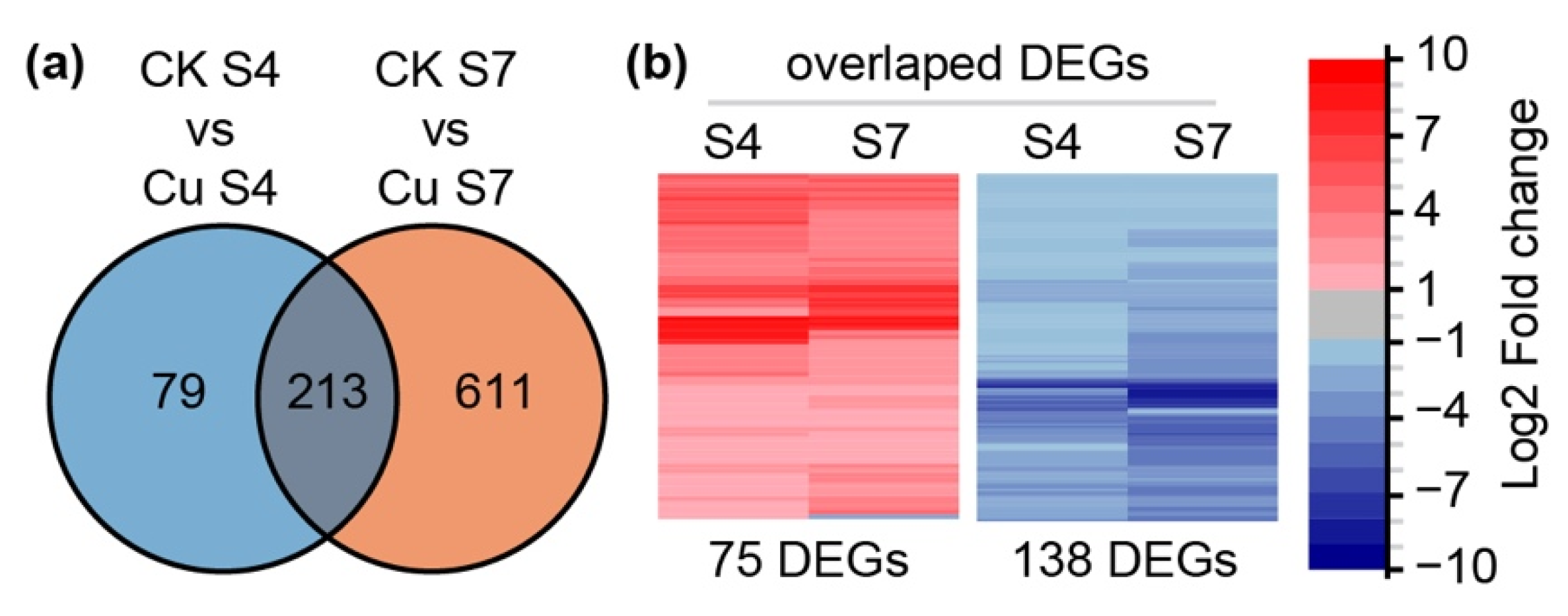
In order to study the regulatory mechanism in petunia petal under excess Cu, RNA-seq was performed using stage 4 and stage 7 petals from petunia treated with 40 μM Cu. The comparison of these transcriptomes identified 292 genes (106 up-regulated and 186 down-regulated) and 824 genes (303 up-regulated and 521 down-regulated) with significant differential expression in Cu treated stage 4 petals and stage 7 petals, respectively, using fold-change > 2 and false discovery rate correction adjusted (FDR) <0.05 as a cutoff (Figure 2a). Among those differentially expressed genes (DEGs), 213 genes were found in both stage 4 and stage 7 transcriptomes. Except for one gene, encoding a Glycine-rich protein, all of them had the same expression profile under excess Cu treatment, of which 74 were up-regulated and 138 were down-regulated (Figure 2b).
To verify the RNAseq results, petunia genes were selected from stage 4 and stage 7 transcriptomes for real-time RT-PCR (qRT-PCR) analysis. The expression patterns of these selected genes were in line with the RNA-seq results, and there was a strong positive correlation between RNAseq and qRT-PCR results in both stage 4 and stage 7 (Figures S1 and S2), suggesting that the RNA-seq data were reliable.

2.3. KEGG and GO Enrichment Analysis

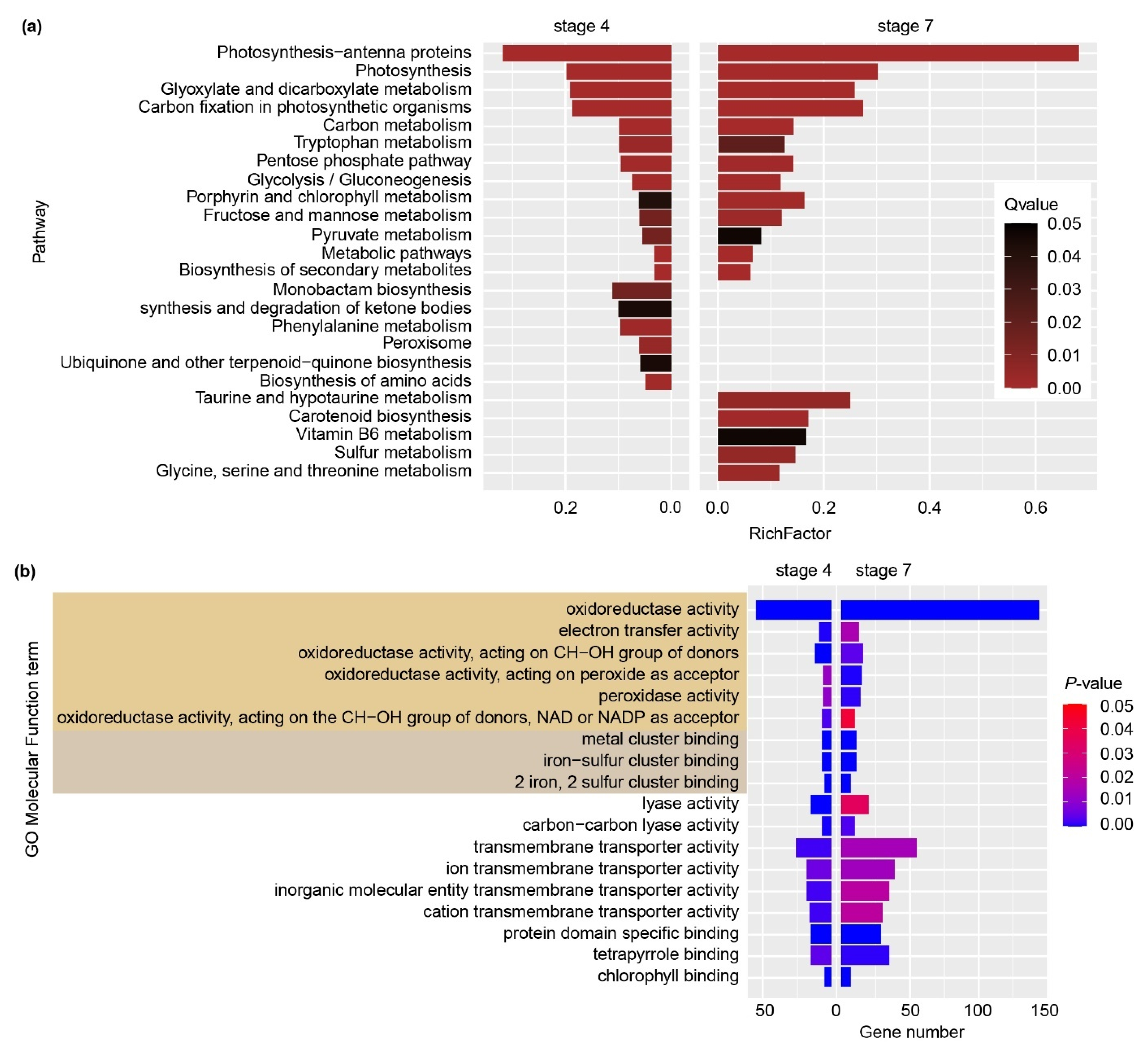
To gain more information about the response mechanism in petunia petals under excess Cu treatment, Kyoto Encyclopedia of Genes and Genomes (KEGG) pathway enrichment analysis was performed. “Photosynthesis-antenna proteins” and “Photosynthesis” were the top2 enriched KEGG pathways in both stage 4 and stage 7 petals. Beside these two pathways, “carbon fixation in photosynthetic organisms”, “carbon metabolism”, “porphyrin and chlorophyll metabolism”, all of them being photosynthesis-related, were all shared by stage 4 and stage 7 petals (Figure 3a).
Gene ontology (GO) annotation enrichment was also performed with p-value < 0.05 as a cut-off (Figure 3b). Both stage 4 and stage 7 petals appeared to be enriched in “oxidoreductase activity”, “electron transfer activity”, “metal cluster binding”, “transmembrane transporter activity”, “chlorophyll binding” and “lyase activity”, within in the molecular function category. Notably, most of these categories are closely related to photosynthesis and heavy metal homeostasis.

2.4. Expression Pattern of Cu Homeostasis Genes

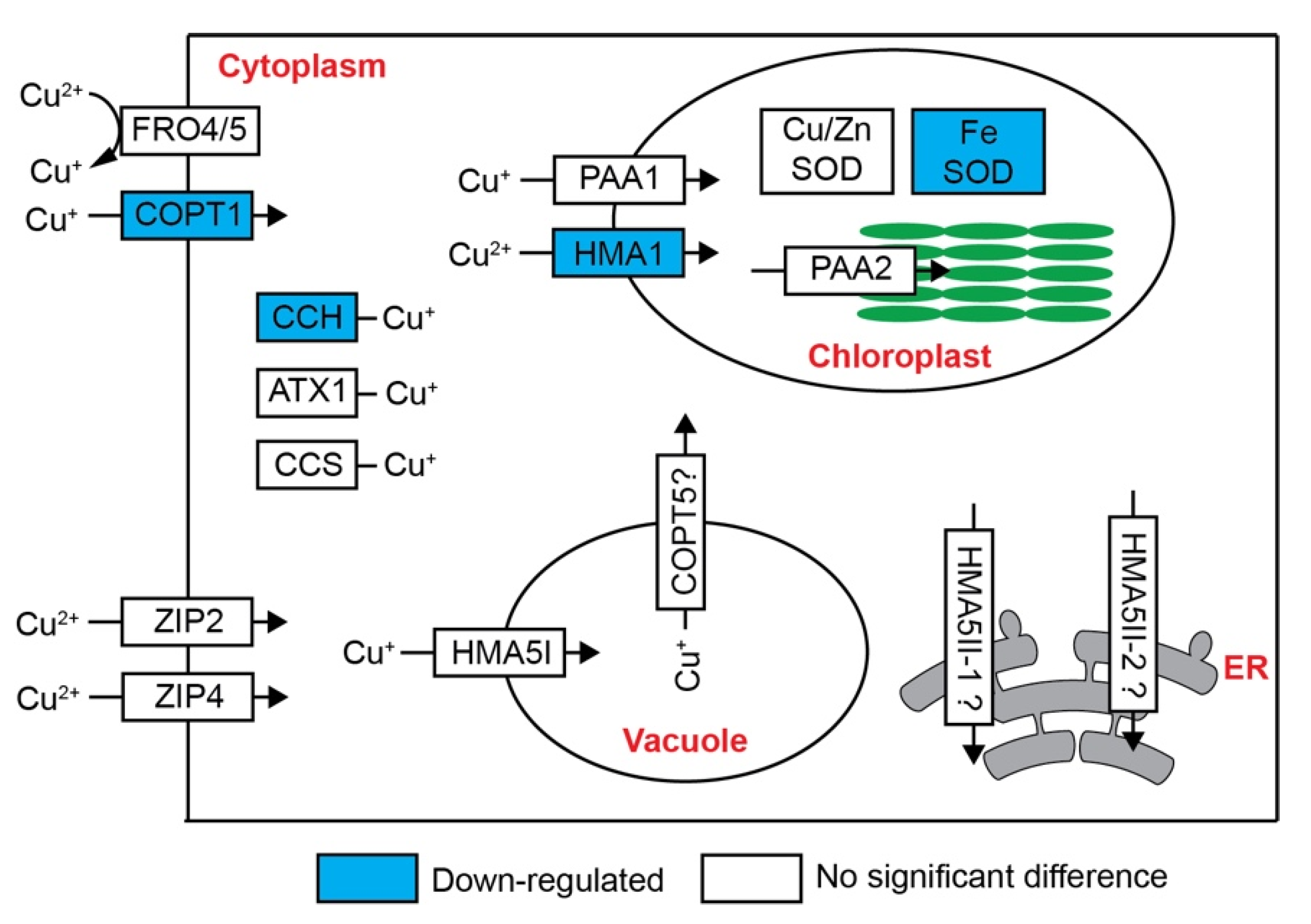
Cu detoxication genes in roots and shoots have been studied in Arabidopsis, rice and Silene vulgaris [9,10,28]. We investigated the expression of known Cu homeostasis genes in petunia petal under excess Cu treatment. Surprisingly, only COPT1, encoding COPT-family transporter, CCH, one Cu chaperone, and HMA1, encoding chloroplast copper transporter, were differently expressed in petunia under excess Cu (Figure 4, Table 1).

2.5. Expression Profile of Fe Homeostasis Genes

In contrast to Cu homeostasis related genes, many more genes involved in Fe deficiency response were differently expressed in petunia petal transcriptomes (Table 1, Figure 5).
Unlike that of Arabidopsis, the Petunia axillaris genome encodes four proteins belonging to the bHLH IVb subfamily, including two Pypeye (PYE) orthologous proteins and two proteins closed related to bHLH121 (Figure S3). The expression levels of these two PYEs, named PYE1 and PYE2, were both elevated under excess Cu in petunia petals (Table 1). In contrast with these PYEs, the ortholog of bHLH121 was repressed under excess Cu treatment in both stage 4 and stage 7 petals (Table 1). Two bHLH38/39/100/101 orthologs were found in the DEG lists, and both were more highly expressed in petals after excess Cu treatment (Table 1, Figure S4).
Members of the Brutus (BTS) hemerythrin E3 ligase family, e.g., Brutus-like1 (BTL1) and BTL2 in Arabidopsis thaliana, also play an important role in the Fe deficiency response [29,30]. Unlike in Arabidopsis, three BTS and one BTL homologs were identified in the Petunia axillaris genome (Figure S5). Two, one BTS and one BTL, were overexpressed in stage 4 and stage 7 petunia petals under Cu treatment (Table 1, Figure 5).
Two myeloblastosis (MYB) subgroup 3 proteins, MYB10 and MYB72, are involved in Fe homeostasis through the driving the expression of nicotinamine synthase 4 (NAS4) [31,32]. Only one MYB protein belonging to MYB subgroup 3, including MYB10, MYB72, MYB63 and MYB58, was found in the Petunia axillaris genome, and it was up-regulated in Cu-treated stage 7 petals (Table 1 and Figure S6).
The ferrous transporter IRT1 and ferric chelate reductase2 (FRO2) are two major Fe acquisition genes, which are strongly upregulated under Fe deficiency in Arabidopsis [33,34,35]. Petunia axillaris encodes three AtIRT orthologous proteins, all of them not expressed in flowers (Table 1 and Figure S7). Three genes encoding FRO proteins were found in stage 4 and stage 7 transcriptomes, two belonging to the AtFRO1/2/3 subgroup and one to the FRO8 subgroup (Figure S8). The expression of all of these three FROs was induced by excess Cu in petals (Table 1). One oligopeptide transporter OPT3 is also involved in Fe transport [36]. Petunia OPT3 was more expressed upon exposure to Cu excess in petals (Table 1, Figure 5 and Figure S9). Yellow stripe-like proteins (YSLs) mediate metal-nicotinamine (NA) transport [37,38,39,40]. Two YSL genes, all encoding proteins belonging to YSL1/2/3 subfamily, were up-regulated in petals under excess Cu treatment (Table 1, Figure 5 and Figure S10). The Petunia axillaris genome encodes two NAS proteins, one of them showing lower expression under excess Cu treatment, while the other was barely expressed, in S4 and S7 petals (Table 1, Figure 5).
Two genes encoding Natural resistance-associated macrophage proteins (NRAMPs) were found in our transcriptomes. According to the phylogenetic analysis, they were related to NRAMP2/5 and NRAMP3/4 in Arabidopsis, respectively. NRAMP2, belonging to AtNRAMP2/5 subgroup, only showed lower expression in stage 7 petunia petals under Cu stress, while no difference was found in stage 4 petals. NRAMP3 belonging to AtNRAMP3/4 subgroup showed enhanced expression in petunia petals under Cu stress. (Table 1, Figure 5 and Figure S11).
Ferritins are the major proteins involved in Fe storage. The Petunia axillaris genome encodes three Ferritin (Fer) proteins; notably, the expression of all of these three genes was decreased in stage 4 and stage 7 upon exposure to Cu excess (Table 1, Figure 5 and Figure S12).
Together, all the results indicated that the network controlling the Fe deficiency response was significantly affected in petunia petals under excess Cu.

2.6. Excess Cu Affects Photosynthesis and Fe-Dependent Proteins in Petal

The KEGG and GO enrichment results led us to investigate the effects of excess Cu on photosynthesis-related symptoms. The chlorophyll a content, chlorophyll a+b content and chl a/b ratio was significantly decreased, while the chlorophyll b content was not affected by excess Cu in petals (Figure 6a–d). Accordingly, the expression level of HEMA2, encoding glutamyl-tRNA reductase, was down-regulated in petals under excess Cu, while chlorophyllide a oxygenase (CAO), encoding enzyme for chlorophyll b biosynthesis, was induced (Figure 6e and Table S1).
In addition, the FV/FM, nonphotochemical quenching (NPQ), ΦPS(II) and qP values were significantly decreased in the petals under excess Cu (Figure 6f–i). All these results agreed with the RNA-seq result that genes involved in electron transport, including plastocyanin (PC), ferredoxin-NADP reductase (FNR) and two ferredoxin (FD) genes, were significantly down-regulated in petals under excess Cu (Figure 6e). Moreover, the photosynthesis changes also were in agreement with the KEGG pathway enrichment results that the expression of genes related with photosystem II (such as PsbO, PsbP, PsbQ, PsbW and PsbY), photosystem I (such as PsaD, PsaF, PsaH, PsaK, PsaI and PsaN), the cytochrome b6/f complex (PetC) and the light-harvesting chlorophyll II complex were also markedly down-regulated by excess Cu treatment in petunia petals (Figure S13).
Iron–sulfur (Fe–S) clusters are the most abundant iron cofactors in the plant, and the Fe–S cluster biosynthesis and mobilization inside the cell is tightly controlled, to avoid toxicity of free or loosely bound iron [41]. According to the GO enrichment analysis, Fe–S cluster binding is significantly enriched in stage 4 and stage 7 petals (Figure 3b). The expression profile of genes involved in Fe–S cluster metabolism was investigated in more detail (Figure 6e). Except for CAO, all the genes involved in Fe–S cluster binding were down-regulated in Cu-stressed petals, including those encoding two subunits of the Fe–S scaffold complex (SurB and SurD) and NEET (Figure 6e).
Fe metabolism is tightly linked with ROS homeostasis in plants, especially with regard to Fe-containing proteins, such as catalase (CAT), ascorbate peroxidase (APX) and Fe superoxide dismutase (FeSOD). CAT, APX and FeSOD were all down-regulated in petunia petals under Cu excess (Figure 6e). Notably, the Petunia axillaris genome encodes four CAT proteins and all the four CAT genes were significant down-regulated in petunia petals under excess Cu (Figure 6e). Accordingly, the CAT activity was significantly lower in petals under excess Cu (Figure 6j).

2.7. Expression Levels of DEGs in Petunia Petals under Fe-Deficiency Treatment

To gain more information about the underlying mechanisms in petunia petal response to Cu stress, the expression pattern of Fe homeostasis related genes in petunia petals under Fe deficiency was studied using real-time PCR (qRT-PCR). Petunias were grown in -Fe half Hoagland’s nutrient solution and the total RNA was extracted from stage 7 petals for the qRT-PCR analysis (Figure 7).
The expression level of BTSL, NAS1, OPT3, FER2, FER3 and NRAMP3 was affected by Fe deficiency (fold change > 2), and the fold change was not different from that in excess Cu treated petals. The expression level of bHLH121-like, PYE1, BTS1, YSL1, YSL2, NRAMP2, FRO8 and CCH was not significantly affected by Fe deficiency treatment (fold change < 2), indicating these genes are responsive to excess Cu, rather than Fe deficiency. The expression level of COPT1, PYE2, bHLH038, bHLH039, FRO1 and NEET was differently expressed in both Fe deficiency treated and excess Cu treated petals, while the fold change in petals under excess Cu treatment was higher than that under Fe deficiency, indicating these genes might be involved in both Fe deficiency and Cu stress response pathways (Figure 7).
Heavy metal-induced Fe deficiency responses in above-ground tissues have also been observed in cadmium-stressed Noccaea caerulescens shoots [42,43]. The expression profiles of Fe homeostasis-related genes in both transcriptomes were also compared (Figure 8). The transcript levels of bHLH039, BTSL, OPT3, NEET, FER2 and FER3 were significantly affected by excess Cd treatment in N. caerulescens shoots and Fe deficiency treatment in Arabidopsis shoots (fold change > 2 and FDR < 0.05). The transcript levels of bHLH121-like, PYE1, FRO8 and NRAMP2 were not affected by excess Cd treatment in N. caerulescens shoots and by Fe deficiency treatment in Arabidopsis shoots, confirming that these genes may specifically respond to Cu stress in petunia petals. On the other hand, the transcript fold-change of NEET in petunia petals under Fe deficiency is similar to that in excess Cd-treated N. caerulescens shoots and Fe deficiency-treated Arabidopsis shoots, and markedly lower than that in excess Cu-treated petunia petals.

3. Discussion

In this study, we investigated the transcriptional response to excess Cu in petunia petals, and provided new insights into the crosstalk between iron and copper homeostasis. Interestingly, the primary effects of excess Cu in petunia petals are related to Fe deficiency, such as disordered expression profiles of Fe deficiency response genes, the blocking of Fe-S cluster metabolism, and a dysfunctioning photosynthesis.

3.1. Fe Deficiency Response in Petunia Petals under Excess Cu

Two sets of Fe-regulated genes have been described in Arabidopsis, i.e., FIT-dependent and FIT-independent [16]. The FIT-dependent pathway is controlled by the FIT and bHLH Ib subfamilies of TFs, including bHLH38/39/100/101. FIT interacts with bHLH Ib TFs to activate the expression of IRT1 and FRO2, which are involved in Fe uptake into the root [45]. Certainly, this is not the case in petunia petals, because the orthologs of FIT and IRT are not expressed in petals (Table 1). Thus, the Fe deficiency response in petunia petals is FIT-independent pathway, like in Arabidopsis shoots [16]. Among the known Fe deficiency response genes, the expression levels of bHLH39, BTSL, OPT3, FRO1, NRAMP3, NEET, FER2, FER3 and NAS1 were significant affected by Fe deficiency or excess Cu in petals. Interestingly, except for PYE2, all the other known Fe-responsive genes that responded to Cu stress in petunia petals, were also significantly affected in Cd-stressed N. caerulescens shoots and/or Arabidopsis shoots upon Fe deficiency, indicating that the Fe deficiency response in the shoot, triggered either directly by Fe deficiency, or by Cd or Cu excess, is conserved among different species.
FER2 and FER3 encode ferritin proteins, which are an iron source for maintaining the function of iron-containing proteins in leaves and seeds [46]. In plants, ferritin proteins are mainly located in the plastids and barely in the cytoplasm [47]. In Arabidopsis seeds, ferritins are proposed to release Fe for the use of Fe-containing proteins after germination [46]. Beside ferritins, the vacuole is another compartment for iron storage, and NRAMP3 and NRAMP4 transport Fe from vacuole to the cytoplasm when needed. NRAMP3 and NRAMP4 are induced under Fe deficiency and functionally redundant in Arabidopsis [48,49]. The increased expression level of NRAMP3 and decreased expression level of FER2 and FER3 in petunia petals under excess Cu or Fe deficiency indicate the redistribution of Fe from the storage compartment to the cytoplasm. Chloroplasts are the largest Fe storage compartment in the plant cell, containing up to 80% of total leaf Fe [2]. A NEET protein recently has been reported to participate in Fe distribution in the plant cell. The Fe content in the chloroplasts of the neet mutant is higher than in wild-type, and the mutant accumulates more Fe in the seedlings compared to the wild-type; however, the neet mutant exhibits an Fe deficiency response in its shoot. In addition, the Fe–S cluster biogenesis and chlorophyll content were also affected by the disruption of NEET function [50,51].
All these results suggest that the redistribution of Fe inside the cell is a primary response to Fe deficiency.

3.2. Cu Detoxification and Crosstalk with Fe in Petunia Petal under Excess Cu

Copper is translocated as Cu(II)-NA in the xylem [52]. Cu(II) needs to be reduced to Cu(I) before being taken up by shoot cells. FRO4/5 are the key proteins involved in Cu(II) reduction in Arabidopsis roots [12,33]. However, the orthologs of FRO4/5 in petunia, are barely expressed and also not induced by excess Cu in petals (Table 1), suggesting there might be another FRO protein(s) involved in Cu(II) reduction in petals, if there is any. FRO2 is the key protein involved in the Fe(III) reduction in Arabidopsis roots, which takes place at the root surface, prior to Fe uptake (as Fe(II)), and is strongly induced under Fe deficiency [33]. Petunia FRO1, an ortholog of Arabidopsis FRO1/2/3, was up-regulated in petals under excess Cu or Fe deficiency, but the fold change in Fe deficiency is much lower than that under excess Cu treatment (Figure 7). This result indicates that petunia FRO1 maybe involved in the response to excess Cu, rather than to Fe deficiency, and might primarily reduce Cu(II), rather than Fe(III).
Its reduction to Cu(I), possibly through FRO1, is expected to increase the availability of Cu for transmembrane (intracellular or cell-to-cell) transport, either as Cu+ or Cu(I)-NA. The cuprous ion is mainly taken up by high-affinity copper transport proteins (COPTs), and the Petunia axillaris genome encodes six COPT proteins, the same number as Arabidopsis, but only one, named COPT1, has been found in the DEG lists. The expression level of COPT1 was down-regulated in petunia petals under excess Cu or Fe deficiency (Table 1 and Figure 7), indicating that it may be involved in Cu and/or Fe homeostasis. Similarly, Arabidopsis COPT2 has been reported to be involved in iron and copper crosstalk [18,53]. AtCOPT2 encodes a plasma membrane-located Cu(I) transporter in yeast [54]. The transcripts level of AtCOPT2 is down-regulated by excess Cu [55]. However, unlike petunia COPT1, AtCOPT2 is up-regulated in the roots upon exposure to Fe deficiency [18]. In any case, the downregulation of COPT1 in response to excess Cu in petunia petals may be taken to suggest that it is primarily a Cu transporter, possibly involved in the reloading of Cu, as Cu+ from the vascularity.
Beside COPTs, ZIP2 and ZIP4 are also involved in the Cu uptake in Arabidopsis [56,57], but their expression level is not effected by excess Cu in petunia petals.
The subgroup I of YSL proteins, including AtYSL1/2/3 and OsYSL16, have been reported to be involved in metal-NA transport [58]. Arabidopsis YSL1 and YSL3 both encode plasma membrane-located proteins and are abundantly expressed in shoots, roots and flowers. Their expression levels are down-regulated both under Fe deficiency and excess Cu [37,55]. The single mutants ysl1 and ysl3 have no apparent phenotypes, while the ysl1ysl3 double mutant accumulates less Fe in the shoot when grown on MS medium [37]. Recently, it has been reported that YSL1 and YSL3 are the down-stream genes of the SUMO E3 ligase, SIZ1. The siz1 mutant is more sensitive to excess Cu than wild-type. After excess Cu treatment, siz1 mutants accumulate more Cu in the leaves, because the siz1 mutant is unable to repress the expression of YSL1 and YSL3 [55]. YSL2 encodes a plasma membrane-located protein, which is expressed in both shoots and roots, and its expression in the shoots is repressed by excess Cu and Fe deficiency [40]. YSL2 transports both Fe(II)-NA and Cu(I)-NA in yeast, but the ysl2 mutant doesn’t show any phenotype. The Fe and Cu concentrations are not significantly different between wild type and the ysl2 mutant, irrespective of Fe and Cu treatment levels [40]. Rice YSL16 is also involved in Cu homeostasis. YSL16 encodes a plasma membrane-located protein, which transports Cu-NA complex in yeast. The rice ysl16 mutant accumulates less Cu in shoots [59]. The ortholog of OsYSL16 in Brachypodium distachyon, BbYSL3, recently has been reported to be localized at the plasma membrane, and to mediate copper transport. The expression of BbYSL3 was induced by Cu deficiency and repressed by Fe deficiency [60,61]. The Petunia axillaris genome encodes three proteins belonging to YSL1/2/3 subgroup. Petunia YSL1 and YSL2 were both down-regulated in Cu-stressed petals, but not affected by Fe deficiency, indicating that these two genes are involved in the homeostasis of Cu, rather than Fe.
Once inside the cell, Cu(I) interacts with Cu chaperones to avoid Cu+ toxicity. CCH is the only chaperone affected by excess Cu in petunia petals, in agreement with the observations in Arabidopsis shoots [62]. Heavy metal ATPase 5 (HMA5) plays an important role in Cu detoxification [8,9,10]. The Petunia axillaris genome encodes three HMA5 orthologs, one of HMA5I and two of HMA5II [10]. The HMA5II genes are barely expressed in petunia petals, and the expression of HMA5I is not affected by excess Cu (Table 1), which is not surprising, because its ortholog in rice, OsHMA4, is also not affected by excess Cu in shoots [8]. Another HMA superfamily protein HMA1 is involved in the delivery of Cu to the chloroplast. Under excess Cu, petunia HMA1 was down-regulated in petals (Table 1), probably to reduce the copper uptake into the chloroplast.
Recently, it has been reported that the crosstalk between Cu and Fe in Arabidopsis root under Cu and Fe deficiency is regulated by SPL7, FIT and bHLH Ib proteins. FIT interacts with bHLH Ib proteins to activate the expression of COPT2, FRO4 and FRO5 [12,18]. Again, this is apparently not the regular network underlying the Fe and Cu crosstalk in petunia petals, because FIT is barely expressed in petals. There were several TFs showing significantly altered expression levels in petals under excess Cu and Fe deficiency, such as PYE2, bHLH38 and bHLH39. Although the expression level of these genes was induced by both Cu excess and Fe deficiency, the fold change in petals under excess Cu is higher than that under Fe deficiency. Besides these three TFs, one bHLH121-like protein was down-regulated in petals under excess Cu, but not affected by Fe deficiency. Arabidopsis bHLH121/URI and bHLH11 both belong to the bHLH IVb subfamily, and play important roles in the Fe deficiency response [17,63,64]. Arabidopsis bHLH11 is mainly expressed in roots, and induced under Fe sufficient conditions. It acts as a transcriptional repressor. The expression level of FRO2 and FER1 was down-regulated in bHLH11 overexpression lines, while NRAMP4 and NAS4 were induced in the roots of bHLH11-overexpressing lines under Fe deficiency [63]. In contrast to bHLH11, bHLH121 functions as a direct transcriptional activator of genes involved in the Fe deficiency response. Around 50% of the Fe homeostasis-related genes are regulated by bHLH121, including bHLH39, BTS, BTSL1, BTSL2, YSL3, OPT3, MYB10, FER1, FER3, FER4, NEET, NAS4 and more [17]. Notably, the expression of these genes’ orthologs is affected by excess Cu in petunia with the same expression trends, and the expression profile in excess Cu and Fe deficiency treated petunia petals showed a strong correlation (Figure S14). The expression of bHLH121 is not affected by the Fe status in Arabidopsis. In contrast, Fe availability affects the cellular localization of bHL121 protein in roots. However, the expression of bHLH121 orthologs are down-regulated (about 40-fold) in petunia petals under excess Cu, indicating that it might be involved in Cu homeostasis. Whether bHLH121 plays an important role in Cu and Fe crosstalk still needs to be confirmed by detailed genetic analysis.
In conclusion, excess Cu treatment induced a FIT-independent Fe deficiency response in petunia petals, with strong effects on Fe-S cluster assembling and photosynthesis. In addition, the Fe deficiency response network that controlled by bHLH121 was markedly disordered. The expression level of petinia bHLH121-like protein is significantly reduced under excess Cu, indicating it might also be involved in Cu homeostasis in petals. Further genetic studies are needed to evaluate this possibility.

4. Materials and Methods

4.1. Plant Material and Growth Conditions

Petunia seeds (cv Mitchell; W115) were sterilized with 1:10 (v/v) diluted household bleach for 10 min. Sterile seeds were stored in water in the fridge for 48 h, then seeds were sown on solid Murashige & Skoog (MS) medium. Two weeks later, the seedlings were transferred to modified half-strength Hoagland’s nutrient solution, renewed once per week [65]: 3 mM KNO3, 2 mM Ca(NO3)2, 1 mM K2HPO4, 0.5 mM MgSO4, 20 μM FeEDDHA, 50 μM KCl, 25 μM H3BO3, 2 μM ZnSO4, 2 μM MnSO4, 1 μM CuSO4, 0.5 μM (NH4)6Mo7O24. The pH was buffered at 5.5, using KOH and 2-(N-morpholino) ethane-sulfonic acid (MES, 2 mM). After four weeks, plants were transferred to nutrient solution supplied with 40 μM CuSO4 (+Cu) or without Fe (-Fe) until flowering. Then stage 4 (flower bud size 40–50 mm) and stage 7 (open flower open anthers) flowers were collected for RNA-seq and mineral analyses. Petunia was grown in a climate room (temperature around 25 °C, photon flux density at plant level 120 μmoles m−2s−1, 14 h per day, air humidity (RH) around 75%).

4.2. RNA-Seq

The experiments were performed with three independent biological replicates, and each replicate was a pooled sample of three individual petals. Total RNA was isolated using RNAprep pure Plant plus kit (Tiangen Biotech, China) and 1 μg total RNA from those samples was used for RNA sequencing library constructed. RNA sequencing was performed using an Illumina Novaseq 6000 instrument at GENEWIZ (Suzhou, China). In order to remove technical sequences, including adapters, polymerase chain reaction (PCR) primers, or fragments thereof, and quality of bases lower than 20, pass filter data of fastq format were processed by Cutadapt V1.9.1 (phred cutoff: 20, error rate: 0.1, adapter overlap: 1 bp, minimum length: 75, proportion of N: 0.1) to be high quality clean data. Reads were aligned to the Petunia axillaris genome sequence [66] using Hista2 v2.0.1. The Hisat2 parameters set were: -x Ref --dta-cufflinks -1 clean_R1.fq -2 clean_R2.fq --known-splicesite-infile splicesites.txt -p threads -S sam. Gene expression level were estimated as fragments per kilobase of transcript per million fragments (FPKM). Differential expression analysis was performed using EdgeR with adjusted p-value < 0.05 as a cut-off setting.

4.3. GO and KEGG Enrichment

The Petunia Gene Ontology (GO) annotation file was download from Plaza 4.0 [67] (http://bioinformatics.psb.ugent.be, accessed on 11 July 2021). GO and KEGG enrichment was performed using TBtools v1.098661 with p < 0.05 as a significance threshold [68]. Heatmaps were created using “ggplot2” package in R v4.1.0.

4.4. Real-Time RT-PCR

First strand cDNA was synthesized from 2 μg total RNA using PrimeScriptTM RT reagent Kit using gDNA Eraser (Perfect Real Time) (Takara). Real-time RT-PCR was performed with Green® Premix Ex Taq™ II (Tli RNaseH Plus) (TaKaRa) on a Bio-Rad CFX Connect. The relative expression level was normalized to EF1α by the 2−ΔΔCT method [69,70]. Primers are listed in Table S2.

4.5. Element Analysis

The experiments were performed with four independent biological replicates (3 individual petals per replicate). Samples were digested in H2O2/HNO3 (1:1, v/v) after drying at 80 °C for 48 h before measuring dry weight [71]. Minerals were analyzed using inductively coupled plasma optical emission spectrometry (ICP-OES; Optima 8000, PerkinElmer, Waltham, MA, USA).

4.6. Chlorophyll Content and Photochemical Activityp Measurement

Fresh petals were collected from each treatment, and chlorophyll were extracted with Acetone: Ethanol (1:1, v/v) at room temperature in the dark for 24 h. Chlorophyll content was determined photometrically by measuring absorption at 663 and 645 nm using a microplate reader (Synergy2, BioTek, Winooski, VT, USA), and was then calculated using the following formulae.
Chla (mg/g Fw) = (12.7 × A663 − 2.69 × A645) × V/1000/Fw (g)
Chlb (mg/g Fw) = (22.9 × A645 − 4.68 × A663) × V/1000/Fw (g)
Total Chl (mg/g Fw) = (20.21 × A645 + 8.02 × A663) × V/1000/Fw (g)
where V refers to the volume (mL) of extracting solution.
Dual portable fluorescence (Dual-PAM-100, WALZ, Effeltrich, Germany) was used to determine the chlorophyll fluorescence. Before measurements, all plants were kept in the dark for 30 min. A saturating pulse (20,000 μmol photons m−2 s−1, 300 ms) was applied to measure the maximum fluorescence, and then petals were illuminated at a saturating light of 358 μmol photons m−2 s−1 for 5 min to activate the electron sink in photosynthesis, and saturation pulses were generated every 20 s. PSII parameters were calculated as follows [72]: the maximum quantum yield of PSII, Fv/Fm = (Fm − Fo)/Fm; the effective quantum yield of PSII, Y(II) = (Fm’ − Fs)/Fm’; qP = F’m − F/F’m − F’o; non-photochemical quenching (NPQ) in PSII, NPQ = (Fm − Fm’)/Fm’.

4.7. Malondialdehyde (MDA) and Catalase (CAT) Measurement

MDA was measured using the Malondialdehyde (MDA) Content Assay Kit (Solarbio, Beijing, China), based on the thio-barbituric acid assay [73]. MDA was analyzed spectrophotometrically at 450, 532 and 600 nm. The experiments were performed with three independent biological repetitions, one petunia petal used for each measurement. CAT activity was measured using a Catalase (CAT) Activity Assay Kit (Solarbio, Beijing, China), based on the protocol of Aebi [74]. One stage 4 petal was used for each measurement, with three independent biological repetitions. The absorbance was determined at 240 nm.

4.8. Phylogenetic Analysis

The putative orthologs search of interesting proteins was carried out by blast search in BLAST+ v2.2.31, through TBtools v1.098661 software. Full length protein sequences were aligned using MAFFT v7.475 with default setting [75]. The alignment curation was performed using BMGE, with settings as described in Morel et al. [76]. The substitution model for the tree building was calculated with ModelFinder using the Akaike information criterion in IQ-tree v1.6.8 [77], through PhyloSuite v1.2.2 [78]. Then the phylogeny was generated using IQ-tree v1.6.8, with the parameter settings included in the descriptions of individual trees.

4.9. Statistical Analysis

Tukey HSD analysis was performed using the “agricolae” package in R v4.1.0. Correlation analysis between RNA-seq and qRT-PCR was also performed using R v4.1.0.

Supplementary Materials

The following are available online at https://www.mdpi.com/article/10.3390/ijms222111604/s1.

Author Contributions

Conceptualization, Y.L. and H.S.; methodology, J.W. and K.L.; Formal analysis, J.W., K.L. and Y.L.; writing—original draft preparation, Y.L.; writing—review and editing, Y.L., H.S. and J.L.; supervision, Y.L.; project administration, Y.L.; funding acquisition, Y.L. All authors have read and agreed to the published version of the manuscript.

Funding

This research was funded by Shanghai Pujiang Program (Grant: 19PJ1406700) and Shanghai Jiao Tong University (WF220515003).

Data Availability Statement

All RNA-seq raw reads have been deposited in the NCBI Sequence Read Archive (SRA) under BioProject PRJNA774370.

Acknowledgments

We thank Biao Lai (School of Advanced Agriculture and Bioengineering, Yangtze Normal University) for technical assistance.

Conflicts of Interest

The authors declare no conflict of interest.

References

  1. Hirayama, T.; Kieber, J.J.; Hirayama, N.; Kogan, M.; Guzman, P.; Nourizadeh, S.; Alonso, J.M.; Dailey, W.P.; Dancis, A.; Ecker, J.R. RESPONSIVE-TO-ANTAGONIST1, a Menkes/Wilson disease-related copper transporter, is required for ethylene signaling in Arabidopsis. Cell 1999, 97, 383–393. [Google Scholar] [CrossRef] [Green Version]
  2. Terry, N.; Low, G. Leaf Chlorophyll Content and Its Relation to the Intracellular Localization of Iron. J. Plant Nutr. 1982, 5, 301–310. [Google Scholar] [CrossRef]
  3. Shikanai, T. PAA1, a P-Type ATPase of Arabidopsis, Functions in Copper Transport in Chloroplasts. Plant Cell 2003, 15, 1333–1346. [Google Scholar] [CrossRef] [Green Version]
  4. Katoh, S. A new copper protein from Chlorella ellisoidea. Nature 1960, 186, 533–534. [Google Scholar] [CrossRef] [PubMed]
  5. Gorman, D.S.; Levine, R.P. Cytochrome f and plastocyanin: Their sequence in the photosynthetic electron transport chain of Chlamydomonas reinhardi. Proc. Natl. Acad. Sci. USA 1965, 54, 1665–1669. [Google Scholar] [CrossRef] [Green Version]
  6. Hänsch, R.; Mendel, R.R. Physiological functions of mineral micronutrients (Cu, Zn, Mn, Fe, Ni, Mo, B, Cl). Curr. Opin. Plant Biol. 2009, 12, 259–266. [Google Scholar] [CrossRef]
  7. Castruita, M.; Casero, D.; Karpowicz, S.J.; Kropat, J.; Vieler, A.; Hsieh, S.I.; Yan, W.; Cokus, S.; Loo, J.A.; Benning, C.; et al. Systems biology approach in Chlamydomonas reveals connections between copper nutrition and multiple metabolic steps. Plant Cell 2011, 23, 1273–1292. [Google Scholar] [CrossRef] [Green Version]
  8. Huang, X.Y.; Deng, F.; Yamaji, N.; Pinson, S.R.M.; Fujii-Kashino, M.; Danku, J.; Douglas, A.; Guerinot, M.L.; Salt, D.E.; Ma, J.F. A heavy metal P-type ATPase OsHMA4 prevents copper accumulation in rice grain. Nat. Commun. 2016, 7, 12138. [Google Scholar] [CrossRef] [PubMed] [Green Version]
  9. Andrés-Colás, N.; Sancenón, V.; Rodríguez-Navarro, S.; Mayo, S.; Thiele, D.J.; Ecker, J.R.; Puig, S.; Peñarrubia, L. The Arabidopsis heavy metal P-type ATPase HMA5 interacts with metallochaperones and functions in copper detoxification of roots. Plant J. 2006, 45, 225–236. [Google Scholar] [CrossRef]
  10. Li, Y.; Iqbal, M.; Zhang, Q.; Spelt, C.; Bliek, M.; Hakvoort, H.W.J.; Quattrocchio, F.M.; Koes, R.; Schat, H. Two Silene vulgaris copper transporters residing in different cellular compartments confer copper hypertolerance by distinct mechanisms when expressed in Arabidopsis thaliana. New Phytol. 2017, 215, 1102–1114. [Google Scholar] [CrossRef] [Green Version]
  11. Yamasaki, H.; Hayashi, M.; Fukazawa, M.; Kobayashi, Y.; Shikanai, T. SQUAMOSA promoter binding protein-like7 is a central regulator for copper homeostasis in Arabidopsis. Plant Cell 2009, 21, 347–361. [Google Scholar] [CrossRef] [Green Version]
  12. Bernal, M.; Casero, D.; Singh, V.; Wilson, G.T.; Grande, A.; Yang, H.; Dodani, S.C.; Pellegrini, M.; Huijser, P.; Connolly, E.L.; et al. Transcriptome sequencing identifies SPL7-regulated copper acquisition genes FRO4/FRO5 and the copper dependence of iron homeostasis in Arabidopsis. Plant Cell 2012, 24, 738–761. [Google Scholar] [CrossRef] [Green Version]
  13. Schmidt, S.B.; Eisenhut, M.; Schneider, A. Chloroplast Transition Metal Regulation for Efficient Photosynthesis. Trends Plant Sci. 2020, 25, 817–828. [Google Scholar] [CrossRef] [PubMed]
  14. Yuan, Y.; Wu, H.; Wang, N.; Li, J.; Zhao, W.; Du, J.; Wang, D.; Ling, H.Q. FIT interacts with AtbHLH38 and AtbHLH39 in regulating iron uptake gene expression for iron homeostasis in Arabidopsis. Cell Res. 2008, 18, 385–397. [Google Scholar] [CrossRef]
  15. Wang, N.; Cui, Y.; Liu, Y.; Fan, H.; Du, J.; Huang, Z.; Yuan, Y.; Wu, H.; Ling, H.Q. Requirement and functional redundancy of Ib subgroup bHLH proteins for iron deficiency responses and uptake in Arabidopsis thaliana. Mol. Plant 2013, 6, 503–513. [Google Scholar] [CrossRef] [Green Version]
  16. Schwarz, B.; Bauer, P. FIT, a regulatory hub for iron deficiency and stress signaling in roots, and FIT-dependent and -independent gene signatures. J. Exp. Bot. 2020, 71, 1694–1705. [Google Scholar] [CrossRef] [Green Version]
  17. Kim, S.A.; LaCroix, I.S.; Gerber, S.A.; Guerinot, M. Lou The iron deficiency response in Arabidopsis thaliana requires the phosphorylated transcription factor URI. Proc. Natl. Acad. Sci. USA 2019, 116, 24933–24942. [Google Scholar] [CrossRef] [PubMed] [Green Version]
  18. Cai, Y.; Li, Y.; Liang, G. FIT and bHLH Ib transcription factors modulate iron and copper crosstalk in Arabidopsis. Plant Cell Environ. 2021, 44, 1679–1691. [Google Scholar] [CrossRef] [PubMed]
  19. Waters, B.M.; McInturf, S.A.; Amundsen, K. Transcriptomic and physiological characterization of the fefe mutant of melon (Cucumis melo) reveals new aspects of iron-copper crosstalk. New Phytol. 2014, 203, 1128–1145. [Google Scholar] [CrossRef] [Green Version]
  20. Garcia-Molina, A.; Marino, G.; Lehmann, M.; Leister, D. Systems biology of responses to simultaneous copper and iron deficiency in Arabidopsis. Plant J. 2020, 103, 2119–2138. [Google Scholar] [CrossRef]
  21. Kastoori Ramamurthy, R.; Xiang, Q.; Hsieh, E.J.; Liu, K.; Zhang, C.; Waters, B.M. New aspects of iron-copper crosstalk uncovered by transcriptomic characterization of Col-0 and the copper uptake mutant: Spl7 in Arabidopsis thaliana. Metallomics 2018, 10, 1824–1840. [Google Scholar] [CrossRef] [PubMed]
  22. Lee, C.W.; Pak, C.H.; Choi, J.M.; Self, J.R. Induced micronutrient toxicity in Petunia hybrida. J. Plant Nutr. 1992, 15, 327–339. [Google Scholar] [CrossRef]
  23. Jones, M.L. Mineral nutrient remobilization during corolla senescence in ethylene-sensitive and -insensitive flowers. AoB Plants 2013, 5, plt023. [Google Scholar] [CrossRef] [PubMed] [Green Version]
  24. Khaodee, W.; Aeungmaitrepirom, W.; Tuntulani, T. Effectively simultaneous naked-eye detection of Cu(II), Pb(II), Al(III) and Fe(III) using cyanidin extracted from red cabbage as chelating agent. Spectrochim. Acta Part A Mol. Biomol. Spectrosc. 2014, 126, 98–104. [Google Scholar] [CrossRef]
  25. Brun, L.A.; Le Corff, J.; Maillet, J. Effects of elevated soil copper on phenology, growth and reproduction of five ruderal plant species. Environ. Pollut. 2003, 122, 361–368. [Google Scholar] [CrossRef]
  26. Puig, S.; Andrés-Colás, N.; García-Molina, A.; Peñarrubia, L. Copper and iron homeostasis in Arabidopsis: Responses to metal deficiencies, interactions and biotechnological applications. Plant Cell Environ. 2007, 30, 271–290. [Google Scholar] [CrossRef]
  27. Andrés-Colás, N.; Perea-García, A.; Puig, S.; Peñarrubia, L. Deregulated copper transport affects Arabidopsis development especially in the absence of environmental cycles. Plant Physiol. 2010, 153, 170–184. [Google Scholar] [CrossRef] [Green Version]
  28. Deng, F.; Yamaji, N.; Xia, J.; Ma, J.F. A Member of the Heavy Metal P-Type ATPase OsHMA5 Is Involved in Xylem Loading of Copper in Rice. Plant Physiol. 2013, 163, 1353–1362. [Google Scholar] [CrossRef] [Green Version]
  29. Rodríguez-Celma, J.; Connorton, J.M.; Kruse, I.; Green, R.T.; Franceschetti, M.; Chen, Y.T.; Cui, Y.; Ling, H.Q.; Yeh, K.C.; Balk, J. Arabidopsis BRUTUS-LIKE E3 ligases negatively regulate iron uptake by targeting transcription factor FIT for recycling. Proc. Natl. Acad. Sci. USA 2019, 116, 17584–17591. [Google Scholar] [CrossRef] [Green Version]
  30. Selote, D.; Samira, R.; Matthiadis, A.; Gillikin, J.W.; Long, T.A. Iron-binding e3 ligase mediates iron response in plants by targeting basic helix-loop-helix transcription factors. Plant Physiol. 2015, 167, 273–286. [Google Scholar] [CrossRef] [Green Version]
  31. Palmer, C.M.; Hindt, M.N.; Schmidt, H.; Clemens, S.; Guerinot, M. Lou MYB10 and MYB72 Are Required for Growth under Iron-Limiting Conditions. PLoS Genet. 2013, 9. [Google Scholar] [CrossRef] [Green Version]
  32. Dubos, C.; Stracke, R.; Grotewold, E.; Weisshaar, B.; Martin, C.; Lepiniec, L. MYB transcription factors in Arabidopsis. Trends Plant Sci. 2010, 15, 573–581. [Google Scholar] [CrossRef] [PubMed]
  33. Robinson, N.J.; Procter, C.M.; Connolly, E.L.; Guerinot, M. Lou A ferric-chelate reductase for iron uptake from soils. Nature 1999, 397, 694–697. [Google Scholar] [CrossRef] [PubMed]
  34. Connolly, E.L.; Fett, J.P.; Guerinot, M. Lou Expression of the IRT1 metal transporter is controlled by metals at the levels of transcript and protein accumulation. Plant Cell 2002, 14, 1347–1357. [Google Scholar] [CrossRef] [Green Version]
  35. Vert, G.; Grotz, N.; Dédaldéchamp, F.; Gaymard, F.; Guerinot, M.L.; Briat, J.-F.; Curie, C. IRT1, an Arabidopsis transporter essential for iron uptake from the soil and for plant growth. Plant Cell 2002, 14, 1223–1233. [Google Scholar] [CrossRef] [PubMed] [Green Version]
  36. Zhai, Z.; Gayomba, S.R.; Jung, H.I.; Vimalakumari, N.K.; Piñeros, M.; Craft, E.; Rutzke, M.A.; Danku, J.; Lahner, B.; Punshon, T.; et al. OPT3 is a phloem-specific iron transporter that is essential for systemic iron signaling and redistribution of iron and cadmium in Arabidopsis. Plant Cell 2014, 26, 2249–2264. [Google Scholar] [CrossRef] [PubMed] [Green Version]
  37. Waters, B.M.; Chu, H.H.; DiDonato, R.J.; Roberts, L.A.; Eisley, R.B.; Lahner, B.; Salt, D.E.; Walker, E.L. Mutations in Arabidopsis Yellow Stripe-Like1 and Yellow Stripe-Like3 reveal their roles in metal ion homeostasis and loading of metal ions in seeds. Plant Physiol. 2006, 141, 1446–1458. [Google Scholar] [CrossRef] [PubMed] [Green Version]
  38. Zhang, C.; Shinwari, K.I.; Luo, L.; Zheng, L. OsYSL13 is involved in iron distribution in rice. Int. J. Mol. Sci. 2018, 19, 3537. [Google Scholar] [CrossRef] [Green Version]
  39. Jean, M.L.; Schikora, A.; Mari, S.; Briat, J.F.; Curie, C. A loss-of-function mutation in AtYSL1 reveals its role in iron and nicotianamine seed loading. Plant J. 2005, 44, 769–782. [Google Scholar] [CrossRef]
  40. DiDonato, R.J.; Roberts, L.A.; Sanderson, T.; Eisley, R.B.; Walker, E.L. Arabidopsis Yellow Stripe-Like2 (YSL2): A metal-regulated gene encoding a plasma membrane transporter of nicotianamine-metal complexes. Plant J. 2004, 39, 403–414. [Google Scholar] [CrossRef]
  41. Balk, J.; Schaedler, T.A. Iron Cofactor Assembly in Plants. Annu. Rev. Plant Biol. 2014, 65, 125–153. [Google Scholar] [CrossRef]
  42. Halimaa, P.; Blande, D.; Baltzi, E.; Aarts, M.G.M.; Granlund, L.; Keinänen, M.; Kärenlampi, S.O.; Kozhevnikova, A.D.; Peräniemi, S.; Schat, H.; et al. Transcriptional effects of cadmium on iron homeostasis differ in calamine accessions of Noccaea caerulescens. Plant J. 2019, 97, 306–320. [Google Scholar] [CrossRef] [PubMed] [Green Version]
  43. Roosens, N.; Verbruggen, N.; Meerts, P.; Ximénez-Embún, P.; Smith, J.A.C. Natural variation in cadmium tolerance and its relationship to metal hyperaccumulation for seven populations of Thlaspi caerulescens from western Europe. Plant Cell Environ. 2003, 26, 1657–1672. [Google Scholar] [CrossRef] [Green Version]
  44. Schuler, M.; Keller, A.; Backes, C.; Philippar, K.; Lenhof, H.P.; Bauer, P. Transcriptome analysis by GeneTrail revealed regulation of functional categories in response to alterations of iron homeostasis in Arabidopsis thaliana. BMC Plant Biol. 2011, 11, 87. [Google Scholar] [CrossRef] [Green Version]
  45. Colangelo, E.P.; Guerinot, M. Lou The essential basic helix-loop-helix protein FIT1 is required for the iron deficiency response. Plant Cell 2004, 16, 3400–3412. [Google Scholar] [CrossRef] [Green Version]
  46. Ravet, K.; Touraine, B.; Boucherez, J. Ferritins control interaction between iron homeostasis and oxidative stress in Arabidopsis. Plant J. 2009, 57, 400–412. [Google Scholar] [CrossRef] [PubMed]
  47. Briat, J.F.; Ravet, K.; Arnaud, N.; Duc, C.; Boucherez, J.; Touraine, B.; Cellier, F.; Gaymard, F. New insights into ferritin synthesis and function highlight a link between iron homeostasis and oxidative stress in plants. Ann. Bot. 2010, 105, 811–822. [Google Scholar] [CrossRef] [Green Version]
  48. Lanquar, V.; Lelièvre, F.; Bolte, S.; Hamès, C.; Alcon, C.; Neumann, D.; Vansuyt, G.; Curie, C.; Schröder, A.; Krämer, U.; et al. Mobilization of vacuolar iron by AtNRAMP3 and AtNRAMP4 is essential for seed germination on low iron. EMBO J. 2005, 24, 4041–4051. [Google Scholar] [CrossRef] [PubMed]
  49. Lanquar, V.; Ramos, M.S.; Lelièvre, F.; Barbier-Brygoo, H.; Krieger-Liszkay, A.; Krämer, U.; Thomine, S. Export of vacuolar manganese by AtNRAMP3 and AtNRAMP4 is required for optimal photosynthesis and growth under manganese deficiency. Plant Physiol. 2010, 152, 1986–1999. [Google Scholar] [CrossRef] [Green Version]
  50. Zandalinas, S.I.; Song, L.; Sengupta, S.; McInturf, S.A.; Grant, D.A.G.; Marjault, H.B.; Castro-Guerrero, N.A.; Burks, D.; Azad, R.K.; Mendoza-Cozatl, D.G.; et al. Expression of a dominant-negative AtNEET-H89C protein disrupts iron–sulfur metabolism and iron homeostasis in Arabidopsis. Plant J. 2020, 101, 1152–1169. [Google Scholar] [CrossRef]
  51. Nechushtai, R.; Conlan, A.R.; Harir, Y.; Song, L.; Yogev, O.; Eisenberg-Domovich, Y.; Livnah, O.; Michaeli, D.; Rosen, R.; Ma, V.; et al. Characterization of Arabidopsis NEET reveals an ancient role for NEET proteins in iron metabolism. Plant Cell 2012, 24, 2139–2154. [Google Scholar] [CrossRef] [PubMed] [Green Version]
  52. Rellán-Alvarez, R.; Giner-Martínez-Sierra, J.; Orduna, J.; Orera, I.; Rodríguez-Castrillón, J.A.; García-Alonso, J.I.; Abadía, J.; Alvarez-Fernández, A. Identification of a tri-iron(III), tri-citrate complex in the xylem sap of iron-deficient tomato resupplied with iron: New insights into plant iron long-distance transport. Plant Cell Physiol. 2010, 51, 91–102. [Google Scholar] [CrossRef] [Green Version]
  53. Perea-Garcia, A.; Garcia-Molina, A.; Andres-Colas, N.; Vera-Sirera, F.; Perez-Amador, M.A.; Puig, S.; Penarrubia, L. Arabidopsis Copper Transport Protein COPT2 Participates in the Cross Talk between Iron Deficiency Responses and Low-Phosphate Signaling. Plant Physiol. 2013, 162, 180–194. [Google Scholar] [CrossRef] [PubMed] [Green Version]
  54. Sancenón, V.; Puig, S.; Mateu-Andrés, I.; Dorcey, E.; Thiele, D.J.; Peñarrubia, L. The Arabidopsis Copper Transporter COPT1 Functions in Root Elongation and Pollen Development. J. Biol. Chem. 2004, 279, 15348–15355. [Google Scholar] [CrossRef] [Green Version]
  55. Chen, C.C.; Chen, Y.Y.; Tang, I.C.; Liang, H.M.; Lai, C.C.; Chiou, J.M.; Yeh, K.C. Arabidopsis SUMO E3 ligase SIZ1 is involved in excess copper tolerance. Plant Physiol. 2011, 156, 2225–2234. [Google Scholar] [CrossRef] [Green Version]
  56. Wintz, H.; Fox, T.; Wu, Y.Y.; Feng, V.; Chen, W.; Chang, H.S.; Zhu, T.; Vulpe, C. Expression Profiles of Arabidopsis thaliana in Mineral Deficiencies Reveal Novel Transporters Involved in Metal Homeostasis. J. Biol. Chem. 2003, 278, 47644–47653. [Google Scholar] [CrossRef] [PubMed] [Green Version]
  57. Grotz, N.; Fox, T.; Connolly, E.; Park, W.; Guerinot, M.L.; Eide, D. Identification of a family of zinc transporter genes from Arabidopsis that respond to zinc deficiency. Proc. Natl. Acad. Sci. USA 1998, 95, 7220–7224. [Google Scholar] [CrossRef] [Green Version]
  58. Curie, C.; Cassin, G.; Couch, D.; Divol, F.; Higuchi, K.; Le Jean, M.; Misson, J.; Schikora, A.; Czernic, P.; Mari, S. Metal movement within the plant: Contribution of nicotianamine and yellow stripe 1-like transporters. Ann. Bot. 2009, 103, 1–11. [Google Scholar] [CrossRef] [PubMed] [Green Version]
  59. Zheng, L.; Yamaji, N.; Yokosho, K.; Ma, J.F. YSL16 Is a Phloem-Localized Transporter of the Copper-Nicotianamine Complex That Is Responsible for Copper Distribution in Rice. Plant Cell 2012, 24, 3767–3782. [Google Scholar] [CrossRef] [PubMed] [Green Version]
  60. Sheng, H.; Jiang, Y.; Rahmati, M.; Chia, J.C.; Dokuchayeva, T.; Kavulych, Y.; Zavodna, T.O.; Mendoza, P.N.; Huang, R.; Smieshka, L.M.; et al. YSL3-mediated copper distribution is required for fertility, seed size and protein accumulation in Brachypodium. Plant Physiol. 2021, 186, 655–676. [Google Scholar] [CrossRef] [PubMed]
  61. Yordem, B.K.; Conte, S.S.; Ma, J.F.; Yokosho, K.; Vasques, K.A.; Gopalsamy, S.N.; Walker, E.L. Brachypodium distachyon as a new model system for understanding iron homeostasis in grasses: Phylogenetic and expression analysis of Yellow Stripe-Like (YSL) transporters. Ann. Bot. 2011, 108, 821–833. [Google Scholar] [CrossRef]
  62. Mira, H.; Martínez-García, F.; Peñarrubia, L. Evidence for the plant-specific intercellular transport of the Arabidopsis copper chaperone CCH. Plant J. 2001, 25, 521–528. [Google Scholar] [CrossRef] [PubMed]
  63. Tanabe, N.; Noshi, M.; Mori, D.; Nozawa, K.; Tamoi, M.; Shigeoka, S. The basic helix-loop-helix transcription factor, bHLH11 functions in the iron-uptake system in Arabidopsis thaliana. J. Plant Res. 2019, 132, 93–105. [Google Scholar] [CrossRef]
  64. Gao, F.; Robe, K.; Dubos, C. Further insights into the role of bHLH121 in the regulation of iron homeostasis in Arabidopsis thaliana. Plant Signal. Behav. 2020. [Google Scholar] [CrossRef]
  65. Schat, H.; Vooijs, R.; Kuiper, E. Identical major gene loci for heavy metal tolerances that have independently evolved in different local populations and subspecies of Silene vulgaris. Evolution (N. Y.) 1996, 50, 1888–1895. [Google Scholar] [CrossRef]
  66. Richert-Pöggeler, K.R.; Davies, K.; Franken, P.; Bao, M.; Fernandez-Pozo, N.; Bombarely, A.; Tang, H.; Sims, T.L.; Kuhlemeier, C.; Moser, M.; et al. Insight into the evolution of the Solanaceae from the parental genomes of Petunia hybrida. Nat. Plants 2016, 2, 16074. [Google Scholar] [CrossRef] [Green Version]
  67. Van Bel, M.; Diels, T.; Vancaester, E.; Kreft, L.; Botzki, A.; Van de Peer, Y.; Coppens, F.; Vandepoele, K. PLAZA 4.0: An integrative resource for functional, evolutionary and comparative plant genomics. Nucleic Acids Res. 2018, 46, D1190–D1196. [Google Scholar] [CrossRef]
  68. Chen, C.; Chen, H.; Zhang, Y.; Thomas, H.R.; Frank, M.H.; He, Y.; Xia, R. TBtools: An Integrative Toolkit Developed for Interactive Analyses of Big Biological Data. Mol. Plant 2020, 13, 1194–1202. [Google Scholar] [CrossRef] [PubMed]
  69. Lischewski, S.; Mallona, I.; Hause, B.; Egea-Cortines, M.; Weiss, J. Validation of reference genes for quantitative real-time PCR during leaf and flower development in Petunia hybrida. BMC Plant Biol. 2010, 10, 4. [Google Scholar] [CrossRef] [Green Version]
  70. Livak, K.J.; Schmittgen, T.D. Analysis of relative gene expression data using real-time quantitative PCR and the 2−ΔΔCT method. Methods 2001, 25, 402–408. [Google Scholar] [CrossRef] [PubMed]
  71. Dong, Q.; Hu, S.; Fei, L.; Liu, L.; Wang, Z. Interaction between cd and zn on metal accumulation, translocation and mineral nutrition in tall fescue (Festuca arundinacea). Int. J. Mol. Sci. 2019, 20, 3332. [Google Scholar] [CrossRef] [PubMed] [Green Version]
  72. Su, L.; Lv, A.; Wen, W.; Zhou, P.; An, Y. Auxin Is Involved in Magnesium-Mediated Photoprotection in Photosystems of Alfalfa Seedlings Under Aluminum Stress. Front. Plant Sci. 2020, 11, 746. [Google Scholar] [CrossRef]
  73. Buege, J.A.; Aust, S.D. Microsomal lipid peroxidation. Methods Enzymol. 1978, 52, 302–310. [Google Scholar]
  74. Aebi, H. Catalase in vitro. Methods Enzymol. 1984, 105, 121–126. [Google Scholar] [CrossRef] [PubMed]
  75. Katoh, K.; Standley, D.M. MAFFT multiple sequence alignment software version 7: Improvements in performance and usability. Mol. Biol. Evol. 2013, 30, 772–780. [Google Scholar] [CrossRef] [PubMed] [Green Version]
  76. Morel, P.; Chambrier, P.; Boltz, V.; Chamot, S.; Rozier, F.; Rodrigues Bento, S.; Trehin, C.; Monniaux, M.; Zethof, J.; Vandenbussche, M. Divergent Functional Diversification Patterns in the SEP/AGL6/AP1 MADS-box Transcription Factor Superclade. Plant Cell 2019, 31, 3033–3056. [Google Scholar] [CrossRef] [PubMed] [Green Version]
  77. Nguyen, L.-T.; Schmidt, H.A.; von Haeseler, A.; Minh, B.Q. IQ-TREE: A fast and effective stochastic algorithm for estimating maximum-likelihood phylogenies. Mol. Biol. Evol. 2015, 32, 268–274. [Google Scholar] [CrossRef] [PubMed]
  78. Zhang, D.; Gao, F.; Jakovlić, I.; Zou, H.; Zhang, J.; Li, W.X.; Wang, G.T. PhyloSuite: An integrated and scalable desktop platform for streamlined molecular sequence data management and evolutionary phylogenetics studies. Mol. Ecol. Resour. 2020, 20, 348–355. [Google Scholar] [CrossRef]
Figure 1. Dry weight and element concentrations in stage 4 and stage 7 petunia petals in plants that were unexposed (Control), or exposed to 40 μM Cu (+Cu). (a) Dry weight of stage 4 and stage 7 petal upon exposure to excess Cu. Bars are mean ± SD (n = 6). (b) Cu, (c) Fe, (d) Zn and (e) Mn content in petunia petals under excess Cu. Bars represent mean ± SD (n = 4 × 3 petals). * indicates significant differences by Tukey’s HSD test (p < 0.05).
Ijms 22 11604 g001
Figure 2. Differentially expressed genes (DEGs) in stage 4 and stage 7 petals after exposure to 40 μM Cu. (a) Venn diagram showing the number of DEGs, fold change > 2 and FDR < 0.05, in stage 4 and stage 7 petals. (b) Heatmap showing the log2-fold change of DEGs shared by stage 4 and stage 7 petals.
Ijms 22 11604 g002
Figure 3. KEGG and Gene ontology (GO) enrichment analysis of transcripts significantly differently expressed in stage 4 and stage 7 petunia petal after exposure to excess Cu. (a) Enriched KEGG pathway in petunia petal upon excess Cu exposure by using p-value < 0.05 as a cut-off. (b) Enriched GO terms in molecular function ontology on level 3 to level 6 shared by stage 4 and stage 7 petal transcriptomes. Terms related to “oxidoreductase activity” and “metal cluster binding” are marked with colored backgrounds.
Ijms 22 11604 g003
Figure 4. Expression pattern of genes relate to Cu transport and distribution in petunia petal under excess Cu. Blue box indicates down-regulated genes, whiter box indicates no significant difference after excess Cu treatment. FRO, ferric reductase oxidase; COPT, copper transporter; ZIP, zinc and iron regulated transporter proteins; HMA, heavy metal P-ATPase; CCH, copper chaperone; CCS, copper chaperone for superoxide dismutase; ATX1, antioxidant 1; PAA1, HMA6; PAA2, HMA8; ER, endoplasmic reticulum.
Ijms 22 11604 g004
Figure 5. Schematic diagram of genes involved in Cu–Fe crosstalk in petunia petal: (a) expression profile of transcription factors involved in Fe homeostasis, (b) expression pattern of genes involved in Fe transport and distribution in petunia petals. bHLH, basic-helix-loop-helix; PYE, popeye; BTS, Brutus; BTSL, Brutus-like; NRAMP, Natural resistance-associated macrophage proteins; NAS, Nicotianamine synthases; FRO, ferric reductase oxidase; YSL, Yellow stripe-like proteins; OPT3, Oligopeptide transporter3; COPT1, copper transporter1.
Ijms 22 11604 g005
Figure 6. Effects of excess Cu treatment on photosynthesis in petunia petals. (ad) Content of chlorophyll a (a), chlorophyll b (b), chlorophyll a+b (c) and chl a/b ratio (d) in stage 4 petunia petals. Bars are means ± SD (n = 5 petals). (e) Expression profiles of Fe–S metabolism related genes under the GO:0051536 item, which were enriched in stage 4 and stage 7 petals. (f) FV/FM, maximum potential PS II efficiency; (g) NPQ, non-photochemical quenching value; (h) ΦPS(II), actual PS II efficiency; and (i) qP, photochemical quenching value was measured in stage 4 petals under excess Cu. Bars are means ± SD (n = 6 petals). (j) CAT activity in petunia stage 4 petals upon Cu excess. Bars are mean ± SD (n = 3). * indicates significant differences (p < 0.05) using Tukey’s HSD test.
Ijms 22 11604 g006
Figure 7. The relative expression level of genes involved in Fe and Cu homeostasis in petunia petals under Fe deficiency or excess Cu. Bars indicate the relative expression level determined by qRT-PCR in petunia petals exposed to 40 μM Cu (+Cu) or 0 μM Fe (-Fe). EF1α as the reference gene. Values (mean ± SD, n = 3) are normalized to the gene expression level in untreated petals (set as 1). Since bHLH038 was barely expressed in untreated petals (Cq value > 35), we used the relative expression value of -Fe as 1. The green dash line indicates the fold change value of genes in petunia stage 7 petal transcriptome.
Ijms 22 11604 g007
Figure 8. Heatmap showing the expression profile of genes related to Fe homeostasis in petunia petals under Cu excess, petunia petals under Fe depletion, N. caerulescens shoots under Cd excess and Arabidopsis shoots under Fe deficiency. The value of petunia is log2 fold change of qRT-PCR. The log2 fold change of genes with fold change < 2 or FDR (p-value for Col) >0.05, all been marked as 0. * data was download from Halimaa et al. [42]. ** data was download from Schuler et al. [44]. *** data was download from Kastoori Ramamurthy et al. [21].
Ijms 22 11604 g008
Table 1. Expression profiles of genes involved in Cu and Fe homeostasis.
Gene DescriptionPaxiN NumberFPKM (Stage 4 CK)FPKM (Stage 4 + Cu)Log2 FC (Stage 4 CK versus + Cu)FPKM (Stage 7, CK)FPKM (Stage 7, + Cu)Log2 FC (S7 CK versus S7 + Cu)
COPT1Peaxi162Scf00179g00097109.49114.58ND32.2510.52−1.65
COPT proteinPeaxi162Scf00664g0003200ND0.050ND
COPT proteinPeaxi162Scf00765g0001200ND00ND
COPT proteinPeaxi162Scf00009g003370.220.09ND00ND
COPT proteinPeaxi162Scf00132g008190.750.24ND6.542.57ND
COPT5Peaxi162Scf00486g00012157.85160.02ND369.44351.72ND
ZIP2Peaxi162Scf00299g0053200ND0.020ND
ZIP4Peaxi162Scf00158g00077101.633.84ND93.4556.48ND
FRO4/5Peaxi162Scf00444g0001800ND00ND
FRO4/5Peaxi162Scf00444g0013000ND00ND
CCHPeaxi162Scf00173g01010759.64406.96ND826.08262.30−1.71
ATX1Peaxi162Scf01633g00011445.43368.77ND477.01357.93ND
CCSPeaxi162Scf00620g0024110.0514.77ND22.8034.69ND
COX17-1Peaxi162Scf00069g014306.686.39ND17.1114.96ND
COX17-2Peaxi162Scf00027g002256.756.44ND9.426.30ND
MTPeaxi162Scf00362g0013300ND6.3313.93ND
MT2b-1Peaxi162Scf00774g00011160.82200.47ND1180.83793.71ND
MT2b-2Peaxi162Scf00946g0002813.0510.60ND8.474.63ND
HMA5IPeaxi162Scf00029g0281247.2237.56ND31.2530.65ND
HMA5II-1Peaxi162Scf00119g005100.110.12ND0.010ND
HMA5II-2Peaxi162Scf00119g005190.620.62ND0.560.62ND
HMA1Peaxi162Scf00945g0021663.8020.52−1.6430.5618.21ND
PAA1Peaxi162Scf00001g010393.172.90ND0.820.67ND
PAA2Peaxi162Scf00486g0009623.1517.08ND19.6217.38ND
RAN1Peaxi162Scf00199g008109.609.46ND21.0620.17ND
SPL7Peaxi162Scf00095g0000129.7026.56ND28.1023.88ND
FIT-likePeaxi162Scf00490g0001800ND00ND
FIT-likePeaxi162Scf00530g0003100ND00.09ND
FIT-likePeaxi162Scf00530g0006300.06ND0.060ND
FITPeaxi162Scf00530g000660.070ND00.09ND
bHLH121-likePeaxi162Scf00684g000196.890.75−3.1613.630.38−5.19
bHLH11-likePeaxi162Scf00362g010438.418.81ND7.588.57ND
PYE1Peaxi162Scf00274g008345.6819.071.7517.0841.431.2
PYE2Peaxi162Scf00712g000291.7767.705.245.9290.733.87
bHLH38Peaxi162Scf00772g0007604.039.050.1264.049.0
bHLH39Peaxi162Scf00772g000480.0824.888.200.3692.367.91
BTS1Peaxi162Scf00753g001198.3931.411.929.9459.192.50
BTS2Peaxi162Scf00021g005270.027.348.100.315.874.17
BTSLPeaxi162Scf00009g005296.4721.581.757.0075.483.37
MYB10-likePeaxi162Scf00452g004120.040.4ND1.065.952.44
FRO2Peaxi162Scf00585g001150.001.358.630.020.373.74
FRO1Peaxi162Scf00585g000141.14543.088.881.68736.188.71
FRO8Peaxi162Scf00089g014336.7510.38ND10.7923.351.04
AHA2Peaxi162Scf01054g00032212.50189.98ND249.42205.23ND
IRT-likePeaxi162Scf00623g0021300ND00ND
IRT-likePeaxi162Scf00623g0011400ND00ND
IRT-likePeaxi162Scf00149g0012800ND00.12ND
OPT3Peaxi162Scf00442g0071075.43947.233.6538.56667.364.03
YSL1Peaxi162Scf00074g002326.290.27−4.506.882.04−1.85
YSL2Peaxi162Scf00542g0062215.253.34−2.1716.232.62−2.72
NRAMP3Peaxi162Scf00264g0051268.84293.862.09151.88460.161.55
NRAMP2Peaxi162Scf00081g0061839.3021.30ND28.108.25−1.83
NEETPeaxi162Scf00192g0082839.980.15−7.94110.740.27−8.62
MFL1Peaxi162Scf00263g0092735.0338.27ND15.6715.52ND
PIC1Peaxi162Scf00016g0232452.4849.68ND44.7447.49ND
MIT1/2Peaxi162Scf00390g0072227.0425.67ND26.4228.44ND
Ferritin3Peaxi162Scf00284g0052137.810.33−6.7944.997.04−2.76
Ferritin2Peaxi162Scf01149g0031853.4911.83−2.16104.0816.55−2.74
Ferritin1Peaxi162Scf01132g003150.690.05−3.550.340.00−5.55
NAS1Peaxi162Scf00673g0004289.2012.80−2.8238.627.3−2.45
NAS2Peaxi162Scf00750g0001300ND00ND
ND: no significant difference, fold change < 2 or FDR > 0.05.
Publisher’s Note: MDPI stays neutral with regard to jurisdictional claims in published maps and institutional affiliations.