Review

Reductive Stress of Sulfur-Containing Amino Acids within Proteins and Implication of Tandem Protein–Lipid Damage

by
Chryssostomos Chatgilialoglu
1,2,* and
Carla Ferreri
1,*
1
ISOF, Consiglio Nazionale delle Ricerche, 40129 Bologna, Italy
2
Center of Advanced Technologies, Adam Mickiewicz University, 61-712 Poznań, Poland
*
Authors to whom correspondence should be addressed.
Int. J. Mol. Sci. 2021, 22(23), 12863; https://doi.org/10.3390/ijms222312863
Submission received: 23 October 2021 / Revised: 25 November 2021 / Accepted: 26 November 2021 / Published: 28 November 2021

Abstract

:
Reductive radical stress represents the other side of the redox spectrum, less studied but equally important compared to oxidative stress. The reactivity of hydrogen atoms (H) and hydrated electrons (eaq) connected with peptides/proteins is summarized, focusing on the chemical transformations of methionine (Met) and cystine (CysS–SCys) residues into α-aminobutyric acid and alanine, respectively. Chemical and mechanistic aspects of desulfurization processes with formation of diffusible sulfur-centered radicals, such as methanethiyl (CH3S) and sulfhydryl (HS) radicals, are discussed. These findings are further applied to biomimetic radical chemistry, modeling the occurrence of tandem protein–lipid damages in proteo-liposomes and demonstrating that generation of sulfur-centered radicals from a variety of proteins is coupled with the cis–trans isomerization of unsaturated lipids in membranes. Recent applications to pharmaceutical and pharmacological contexts are described, evidencing novel perspectives in the stability of formulations and mode of action of drugs, respectively.

1. Reductive Stress

Redox homeostasis in biology represents the variety of endogenous adaptative mechanisms to preserve physiological processes in a cell/organism and balances two sides: oxidative stress and reductive stress [1]. Attention has been largely given to the former starting from the fact that most organisms live under aerobic conditions, and oxidative challenges can often occur and affect/modify biomolecules. The state-of-art in this field is regularly reviewed and, also recently, reviews have summarized the role of reactive oxygen species (ROS) to be themselves or to generate signaling molecules, involved in fundamental cellular functions [2,3].
On the other side of the redox spectrum, reductive stress has been less widely investigated [4,5], although this process is not less important than the oxidative one. Indeed, reductive stresses may affect cellular redox homeostasis, alter biomolecules and, eventually, determine specific pathological states [6]. In particular, under hypoxia conditions, cancer biology evidenced the key role of reductive stress [7]. Generally, reductive stress is associated with redox couples of NADH/NAD+, NADPH/NADP+ and GSH/GSSG, exploring the consequences due to exceeding reduced species, which are as harmful as oxidation products [8,9,10].
Nowadays research interest on various cellular and organ systems are directed toward the study of the delicate balance between reductive and oxidative signaling, and how it can become, for example, a vital factor in cancer cell homeostasis associated with cell proliferation, progression, and drug resistance [1]. Interests include the study of the balance created by enzymatic systems [5], those involved in the oxidative stress for generation of ROS or reactive nitrogen species (RNS), such as NAD (P)H oxidases (NOX) for the generation of superoxide radical anion (O2•−) and uncoupled nitric oxidases for the generation of nitric oxide (NO), as well as those involved in the reductive environment created by electron exchange, as in the case of the mitochondrial electron transport chain.
In the frame of redox balance, the reactivity of free radicals, such as, for example, in the above-mentioned ROS/RNS production, is connected with the control given by antioxidant systems. Free radical species generated in the biological environment display a wide range of partition coefficients and rate constants, for example, going from the hydrophilic fast reactive hydroxyl radical (HO) to the diffusible and slow reactive O2•−, and both factors have an influence on their effects [11]. In particular, electrons (e) and hydrogen atoms (H), which can derive from the interaction of water in the biological environment with ionizing radiations, have been the subject of research activity within various groups since the seventies [12,13,14]. In those times, the diffusible H was found to react selectively with thiols and sulfur-containing residues in peptides and proteins. The generation of solvated electrons without radiations was also connected with the reactivity of vitamins (E, C, carotenoids) [15]. Our group brought the reductive stress reactivity in models of biomimetic chemistry with generation of thiyl radical species [16,17], which are known to have a rich redox chemistry in nature [18].
In this review, we wish to give an overview of the redox balance from the reductive stress side, treating the chemistry associated with the reactions of H and eaq with sulfur-containing amino acids within peptides and proteins, and then delineating the information obtained from biomimetic models, including the interesting consequence of the tandem protein–lipid damage.

2. Transient Reductive Species Generated by γ-Radiolysis

Gamma-radiolysis of neutral water is the most direct method of generation of the primary reactive species eaq, HO, and H, together with H+ and H2O2, as shown in Reaction 1, without using additives. The values in brackets represent the radiation chemical yields (G) in units of μmol J−1 [19]. The first three reactive species are expected to react very fast with peptides and proteins. The rate constants of the three reactive species with a few biomolecules of interest in this article are reported in Table 1.
H2O Ijms 22 12863 i001 eaq (0.27), HO (0.28), H (0.06), H+ (0.27), H2O2 (0.07)
To investigate the reactivity of reductive species (eaq and H) and their consequences on protein structures, HO have to be scavenged. To achieve this, the most efficient procedure is based on the addition of tert-butanol (t-BuOH), which efficiently scavenges HO radicals (Reaction 2, k2 = 6.0 × 10⁸ M−1 s−1), whereas it reacts slowly with H atoms (Reaction 3, k3 = 1.7 × 10⁵ M−1 s−1). For example, by using an Ar-flushed aqueous solution containing 0.2 M t-BuOH and 1 × 10⁻⁴ M peptide/protein and assuming that HO reacts with peptide/protein with a rate constant of 2.4 × 10¹⁰ M−1 s−1, HO will be scavenged by t-BuOH 50 times faster than by peptide/protein.
HO + t-BuOH → (CH3)2C(OH)CH2 + H2O
H + t-BuOH → (CH3)2C(OH)CH2 + H2
It is also possible to investigate the reactivity of H alone. In N2O-saturated solutions (~0.02 M of N2O), eaq are efficiently transformed into HO via Reaction 4 (k4 = 9.1 × 10⁹ M−1 s−1). Under these conditions, H and HO account for 10 and 90% of the reactive species, respectively. Therefore, by using N2O-saturated solutions in combination with t-BuOH as additive, the reactivity of H atoms can be investigated. Alternatively, electrons are known to be efficiently converted to H when they react with protons (H+) or dihydrogen phosphate anions (H2PO4). In Ar-deoxygenated H2PO4 (10 mM) aqueous solutions, eaq may be converted into H atoms (Reaction 5, k5 = 1.5 × 10⁷ M−1 s−1), depending on the pH and other additives.
eaq + N2O + H2O → HO + N2 + HO
eaq + H2PO4 → H + HPO42−

3. The Reductive Desulfurization of Methionine (Met) and Cystine (CysS-SCys) Residues in Peptides/Proteins

A variety of sulfur-containing amino acids within peptides and proteins have been studied. The reactions of H with Met [21] and with various peptides containing a single Met residue, like Gly-Met-Gly [22], enkephalins [20] and amyloid-β [23] have been reported. The selective attack of H on sulfur atom of Met residues results in the formation of α-aminobutyric acid (Aba). Scheme 1a summarizes the proposed reaction mechanisms for these transformations. The desulfurization process of Met residue generates Aba and the diffusible methanethiyl radical (CH3S) via the sulfuranyl adduct 1. The reaction 1 → Aba may be single or two-steps process with formation of CH3S and/or CH3SH. Indeed, in the γ-irradiation of the oxygenated aqueous solution of Met [21] or Gly-Met-Gly [22], Aba is replaced with homoserine which derives from the trapping of primary alkyl radical by oxygen, as shown in Scheme 1a. Pulse radiolysis studies on enkephalins indicated that Met residue is the main target of H-atoms [20]. In Met-enkephalin (Tyr-Gly-Gly-Phe-Met) the attack of H occurs about 50% on Met with formation of CH3S radical, and the remaining 50% is partitioned roughly evenly between Tyr and Phe. Experiments on amyloid-β peptide (Aβ) also showed the selective attack of H on the Met35 residue, with formation of a modified peptide containing an Aba residue [23].
Bovine pancreatic ribonuclease A (RNase A) and lysozyme (Lyso) are two medium-sized proteins that contain both Met residues and disulfide bridges with no free thiol group present. Early work on the inactivation of ribonuclease and ribonucleoside S-peptide by primary reactive species was reported [24,25,26]. In particular, the selective attack of H on CysS–SCys and Met residues resulted in the formation of alanine (Ala) and Aba (see Scheme 1). More recently, damage to bovine pancreatic RNase A, due to H and/or eaq attack at the protein sulfur-containing residues, was investigated by Raman spectroscopy and mass spectrometry techniques [27,28]. RNase A is a single polypeptide chain cross-linked by four disulfides bridges and contains also four Met. Indeed, reductive desulfurization process affects both Met and CysS–SCys residues. Mapping experiments demonstrated that desulfurization selectively affected Met79, Cys110, Cys58 and Cys72 during the first stages of reaction [28]. The concomitant occurrence of Met → Aba and CysS–SCys → Ala chemical mutations cannot be ascribed to modification processes in aqueous media other than radical-based desulfurization reactions, thus they are specifically attributed to reductive radical stress conditions. In the case of Lyso, a single polypeptide chain cross-linked by four disulfides bridges and contains two Met, the changes observed in the Raman spectra indicated that the degradation of sulfur moieties does not occur immediately, but as irradiation progresses [29]. Electron paramagnetic resonance (EPR) studies in the vacuum solid-state radiolysis of Lyso demonstrated that perthiyl radical (RSS) residue is formed as the major product. In analogy with thiourea clathrate, where a head-to-head arrangement is imposed and there is no possibility of diffusible thiyl radicals, the equilibrium RS + RSH ⇆ RSS + (H)R can be followed by an irreversible conversion of sulfuranyl radical to perthiyl radical [30].
Scheme 1b summarizes the proposed reaction mechanisms for the reactions of H and eaq with CysS–SCys residues in proteins. The H attack gives rise to the sulfuranyl adduct 2, which is in equilibrium with its deprotonated form, namely the disulfide radical anion 3. The latter species may also be derived from the direct eaq attachment to the disulfide moiety. Both intermediates 2 and 3 dissociate reversibly into two entities, RS/RSH and RS/RSH respectively, but do not diffuse freely owing to the tertiary structure of the protein. Under these conditions, radical 2 undergoes irreversible decay with formation of Ala and perthiyl radical 4. Radical anion 3 is also an excellent one-electron reducing agent [31].
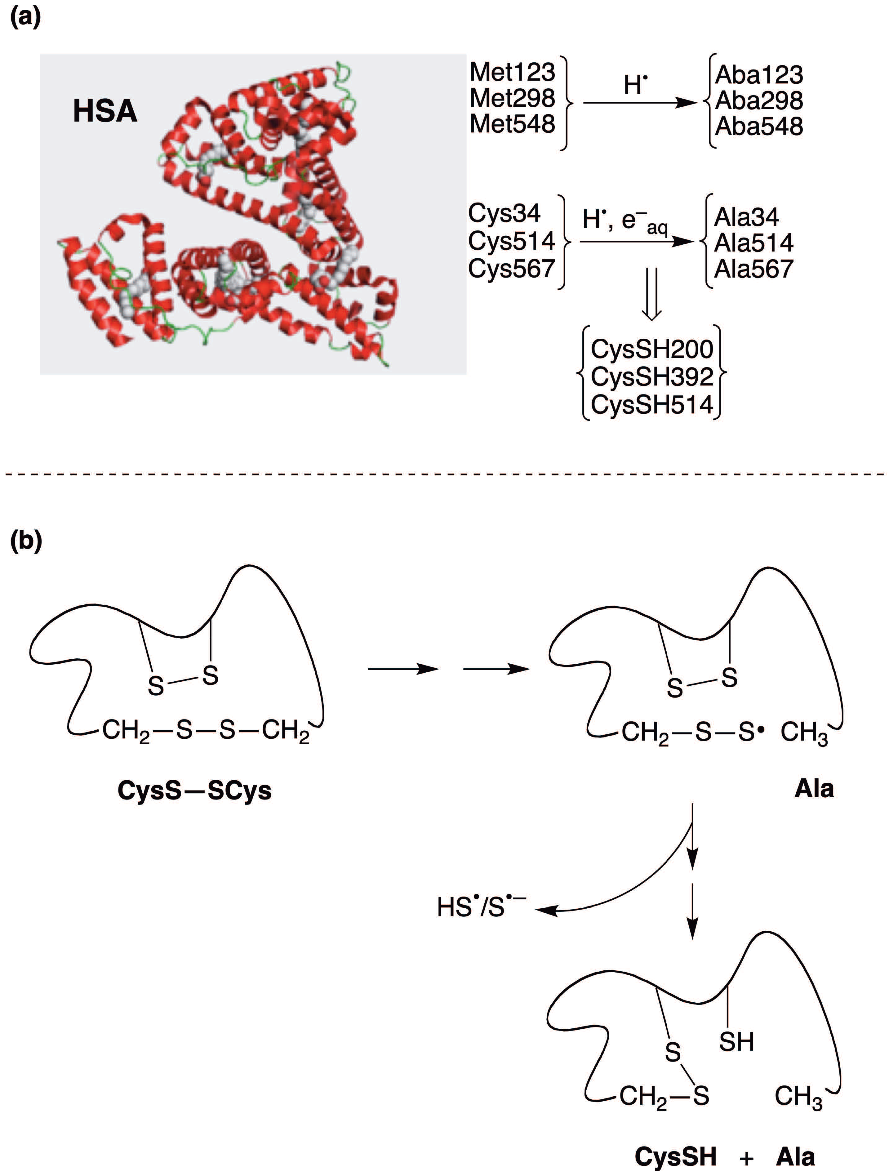
Human serum albumin (HSA) is a large protein and accounts for over 50% of the total plasma protein content. HSA contains 6 Met and 35 Cys residues that form 17 CysS–SCys bridges and leave a unique thiol functionality that is involved in various in vivo HSA actions [32]. More than 70% of the total free radical-trapping activity of serum is due to HSA [33]. The reactivity of sulfur-containing amino acids in HSA under reductive radical stress conditions was also investigated by γ-radiolysis coupled with Raman spectroscopy and mass spectrometry techniques [34,35]. On the basis of the observed S–S stretching band changes, it was suggested that albumin disulfide bridges should eventually scramble upon reductive species attack, which can be correlated with secondary structure changes. MALDI-TOF and semiquantitative LC-ESI-IT-MS/MS peptide mapping experiments on different enzymatic digests demonstrated the following chemical mutations since the early stages of reaction: (i) Met123, Met298 and Met548 conversions into the corresponding Aba; (ii) the modification of Cys34, Cys514, and Cys567 in the cystine moieties into the corresponding Ala; and (iii) the formation of Cys200, Cys392 and Cys514 as free thiols of CysSH residues (Figure 1a). Based on the known cystine pairing in native albumin, no relationships were observed between desulfurization (formation of Ala residues) and the liberated cysteines (CysSH200, CysSH392, CysSH514). Figure 1b illustrates the suggested pathway where the CysS-S intermediate in the presence of another disulfide bridge of the molecule undergoes a quick disulfide scrambling event, generating the new free cysteine residues, the new disulfide bridge in a more stable secondary structure and the diffusible reactive sulfhydryl radical (HS/S•−) responsible of the liposome interaction (see below) [34].
Metallothioneins (MTs) are sulfur-rich proteins capable of binding metal ions to give metal clusters. Metal–MT complexes are characterized by a high number of cysteine residues, the absence of disulfide bridges and the presence of labile sulfide anions (S2−) as non-protein (metal) ligands [36]. Reactions of reactive reductive species (H atoms and eaq), produced by γ-irradiation of water, with ZnII–MT and CdII–MT were carried out in aqueous solutions and monitored by various spectroscopic and spectrometric techniques (Raman, CD, and ESI-MS) [37,38,39]. The preferential attack site in both MTs is the Cys residues, as evidenced by measuring the intensity decrease in the Raman spectrum of some metal-S stretching bands. The modification of the metal–MT aggregates can induce significant structural changes, such as partial deconstruction and/or rearrangement of the metal clusters and breaking of the protein backbone. Substantial differences were observed in the behavior of the ZnII–MT and CdII–MT aggregates towards the reactive species, depending on the different folding of the polypeptide in these two cases.

4. Biomimetic Radical Chemistry

4.1. Cis–Trans Isomerization of Fatty Acid Residues in Liposomes by Sulfur-Centered Radicals

Free-radical-catalyzed cis−trans isomerization of unsaturated lipids has been a subject of interest in our laboratory for the past two decades [40,41,42,43]. We demonstrated the reaction mechanism for this isomerization in various contexts, from simple reactions in various solvents to complex biomimetic models. We provided indications for an endogenous origin of trans unsaturated fatty acids in cells [44], animals [45], and humans [46,47] based on a mono-trans isomer of arachidonic acid as biomarkers [48,49].
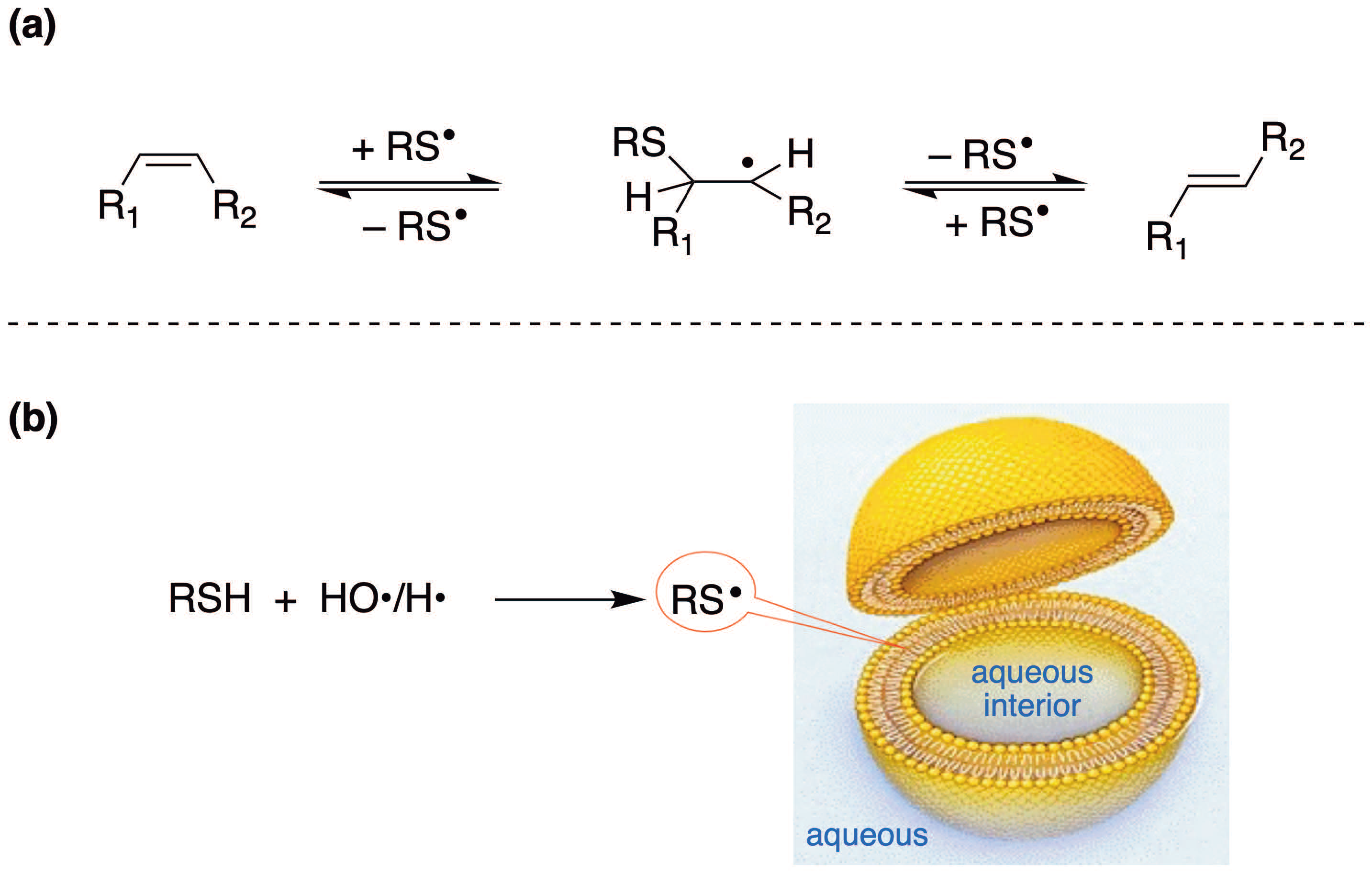
Τhe reaction mechanism of cis-trans isomerization is reported in Figure 2a and consists of a reversible addition of thiyl radical (RS) to the double bond [43,50,51]. Indeed, the reconstitution of the double bond is obtained by β-elimination of RS and favors the trans geometry, which is the most thermodynamically favorable conformation. In this context, the radical RS acts as a catalyst for cis-trans isomerization, excluding the formation of positional isomers as reaction products because the mechanism does not allow a double-bond shift [43,51].
A simple biomimetic model of vesicle suspension demonstrated the potential efficiency of thiyl radicals (RS) and their candidacy as culprits for the cis–trans isomerization in vivo. Liposomes can be represented as shown in Figure 2b, that is, a double layer formed by the spontaneous organization of phospholipid components in water, which delimits an aqueous cavity. Fatty acid tails can be saturated or unsaturated, and the disposition of the double bonds in the vesicle depends on the supramolecular arrangement of the bilayer. Unilamellar vesicles are the closest models of biological membranes and can be formed by different techniques, such as the extrusion and the injection methodologies. Studies of cis–trans isomerization of unsaturated moieties of DOPC (dioleoyl phosphatidyl choline) showed that the amphiphilic HOCH2CH2SH in LUVET (large unilamellar vesicle by extrusion technique) affords an efficient geometric isomerization, indicating that thiyl radicals generated in the aqueous compartment enter the hydrophobic region of the bilayer and isomerize the 9,10-double bond of oleic acid moieties.
The use of the biomimetic model of vesicle suspension demonstrated the potential of sulfhydryl radicals (HS/S•−, pKa~4) derived from H2S to diffuse and reach the hydrophobic fatty acid chains, thus reacting with double bonds and producing trans-fatty acid containing phospholipids in high yield [52].
Experimental results supported the correlation of the origin of an endogenous formation of trans lipids with radical stress conditions that generate isomerizing reactive species [53,54,55,56,57]. However, the most feasible culprits for the lipid cis–trans isomerization in vivo are still a matter of discussion. Thiyl radicals RS are favored over other radicals [43,46], e.g., such as nitrogen centered radicals [53,54], on the basis of the efficiency of the thiyl radical-catalyzed cis−trans isomerization, demonstrated in vitro, and the presence of several sulfur-containing compounds in the cell, like peptides/proteins that contribute to the biological production of diffusible thiyl radicals.

4.2. Tandem Protein–Lipid Damage and Build-Up of Molecular Libraries

Tandem lesions are caused by a single radical event that leads to initial damage and, at the same time, produces reactive species able to damage another type of molecule. This tandem process involving lipids and proteins can involve two different cell compartments. Tandem damages are considered to be more harmful than single events since they can impair, or render more complicated, the cellular repair systems [58].
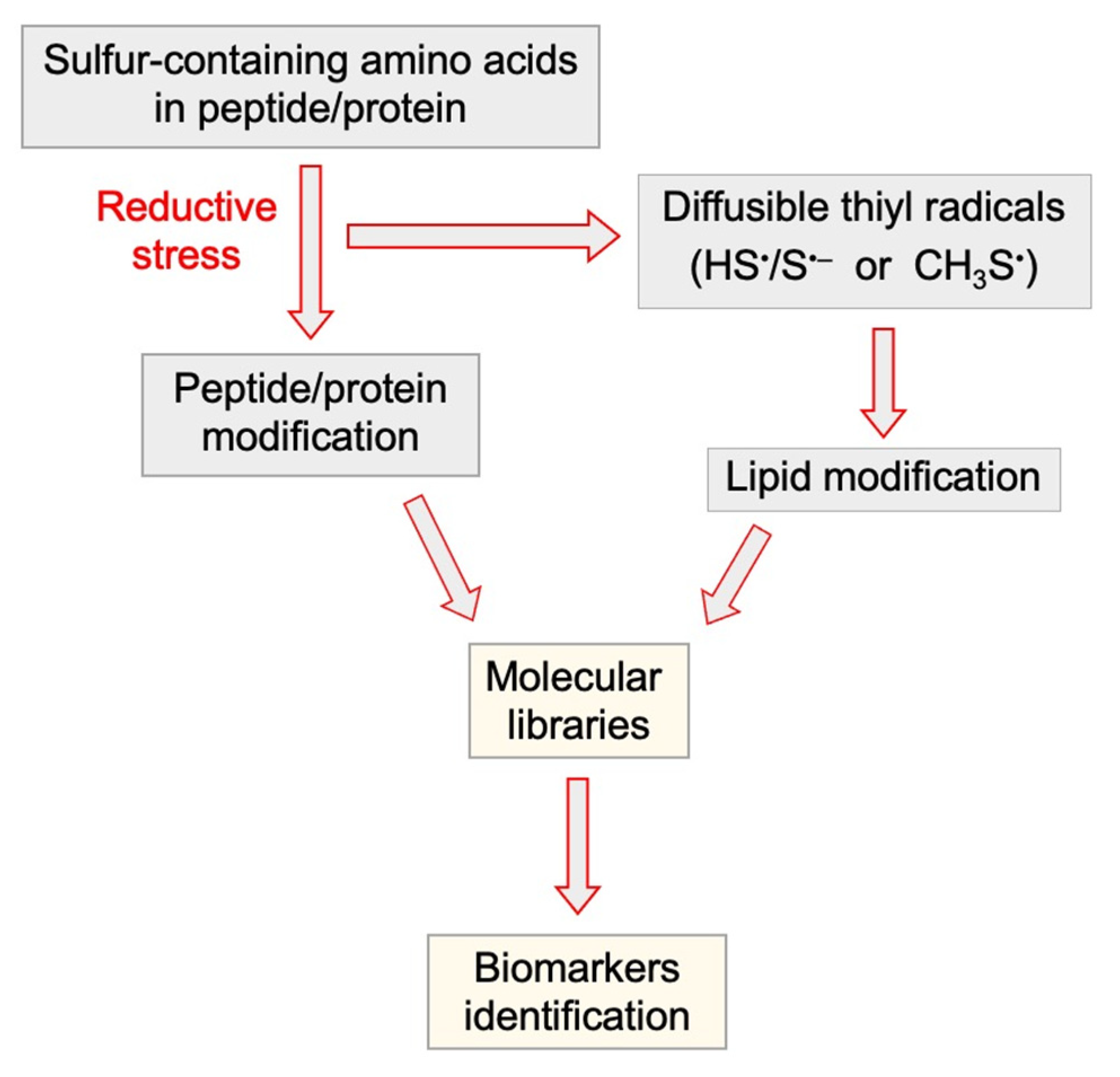
Diffusible thiyl radicals generated by reductive stress of peptides or proteins are the best candidates to cause the isomerization of membrane unsaturated lipids. A radical reaction with the sulfur-containing amino acid as primary target will lead to modified proteins or peptides on one hand, and on the other hand to secondary diffusible radicals that can cause isomerization of membrane lipids. The modification of peptides/proteins and lipids will create molecular libraries to be implemented as potential biomarkers for reductive radical stress (Figure 3).
Biomimetic models were developed to study the process of lipid–protein damage. As described previously, ionizing radiation allows specific damage to be examined as a consequences of the reactivity of H-atoms and hydrated electrons, and this methodology was used to establish the first molecular basis of reductive radical stress. Based on the reductive desulfurization of Met and Cys residues described in Section 3, experiments carried out on proteo-liposome biomimetic models clearly showed that radical degradation of sulfur-containing residues produces sulfur-centered radical able to migrate into the hydrophobic part of the liposome and cause the radical-catalyzed isomerization of lipid double bonds.
The model system of DOPC vesicles containing RNase A was the first example of tandem radical damage transferred from the protein to the lipid domain [17]. The proteo-liposome suspension (lipid:protein ratio ≥ 50:1) was exposed to the radical stress condition. To examine the fatty acid residues it is necessary to isolate phospholipids and perform the transesterification reaction in known conditions to obtain the corresponding fatty acid methyl esters (FAME). The FAME extract can be analyzed by gas chromatography (GC) which is the gold standard for the separation of geometrical and positional cis and trans isomers [43]. Figure 4 shows the various sulfur residues in proteins and the integrated vision of reductive radical stress which involves lipid modification and protein modification. The thiyl radical species formed from protein degradation can diffuse into the lipid bilayer and cause isomerization of the naturally occurring cis double bonds, using the mechanism shown in Figure 2a. The trans unsaturated fatty acids formed in vesicles result to be very sensitive markers of this protein damage, since the isomerization process is catalytic, therefore it can detect very low quantities of radicals generated in the environment. Protein damages were also determined by using Raman spectroscopy, to evidence changes in the secondary structures, such as, for example, in the case of RNase by detecting the 1230–1280 cm−1 region (amide III), which is conformationally susceptible, and indicated an increase in the percentage of β-sheet conformation of the protein [17]. Other analyses such as SDS-page demonstrated the formation of aggregates thus involving the tertiary structures of proteins, however, specific information on such changes were not achieved.
Based on the radical chemistry and modifications herein described, the development of trans fatty acid isomers as biomarkers of radical stress in vivo can be sustainable, with libraries developed for the recognition in biological samples. In particular, trans isomers not deriving from the diet, such as the trans isomer of sapienic acid (6trans-C16:1) and the 5trans and 8trans isomers of arachidonic acid (5cis,8cis,11cis,14cis-C20:4), are the best candidates to follow-up complex radical and redox balance in health and diseases [49,59,60].
As mentioned above and shown in Figure 1 and Figure 4, the model of palmitoyl oleoyl phosphatidyl choline (POPC) vesicles containing HSA showed the potential of proteins not containing free Cys moieties to produce diffusible sulfur radicals able to induce a cis–trans isomerization of lipids at the onset of irradiation [34]. This phenomenon led to focus our attention on the mechanism of the chemical modification of the sulfur-containing moieties (in particular, cystine) by reductive radical stress, as explained in Section 3. The modes of formation of S•− species needs further comment, also in consideration that hydrogen sulfide (H2S) is a naturally occurring gas with roles in nervous and cardiovascular systems, and in pathological conditions, such as inflammation [61,62] and that perthiyl radicals are assuming more relevance in the frame of the thiyl radical reactivity and signaling events in biology [61].
From H2S the corresponding sulfhydryl radical (HS) is generated, that has a pKa value of ca. 4, therefore its deprotonated form S•− dominates at neutral pH in an aqueous environment [52]. Indeed, using H2S generated in situ by its salt Na2S in the biomimetic model of vesicle suspension, we demonstrated the potential of sulfhydryl radicals (HS/S•−) to induce efficiently the lipid cis–trans isomerization (cf. Figure 2b). When reductive radical stress conditions are used, the cystine residues can react, as summarized in Scheme 1b, to generate disulfide radical anions, derived from the direct electron attachment, that is generally in equilibrium with the sulfuranyl radical, also obtained by the direct H-atom attack on the sulfide moiety. The evolution of these intermediates is crucial since, as shown in Scheme 1b, the formation of alanine residue can be associated with the concomitant formation of perthiyl radical (RSS). At this point, the question is: is the RSS radical an isomerizing species of unsaturated fatty acid residues? To our knowledge, no information exist on this reaction. The answer is probably negative based on the stability of this radical. Indeed, the bond dissociation energy of RSS–H is ca. 17 kcal/moL lower than the corresponding RS–H value [63]. There is evidence that t-BuSS adds reversibly to styrene where the benzyl-type adduct radical is stabilized by the phenyl ring [63].
Another question is: what is the fate of RSS radical? As shown in Figure 1a, the formation of Ala is associated with formation of CysSH. Based on the known cystine pairing in native albumin, no relationships were observed between desulfurization and formation of cystines, suggesting that the initially formed CysSS (see Figure 1b) undergoes a quick disulfide scrambling events toward more stable disulfide/thiol populations [34]. Based on the known data, we can propose that such scrambling events produce the observed CysSH (see Figure 1b) and HS/S species that catalyze the isomerization of double bonds.
It is worth also recalling the above reported proteo-liposome experiments on the reactivities of metallothioneins (MTs) under reductive stress. These proteins have different arrangements of metal–thiolate clusters formed by cysteine or histidine with endogenous sulfur content, and regulate the exogenous concentration of sulfide anions S2− [36,37]. As mentioned above, substantial differences were observed in the behavior of the ZnII–MT and CdII–MT aggregates towards the reductive reactive species (H and eaq), due to different folding of the polypeptide in these two cases, using a biomimetic model based on unsaturated lipid vesicle suspensions [37,38,39,64]. Interestingly, CdII-MT have a higher aptitude to induce cis–trans isomerization than ZnII–MT and this behavior is parallel with the number of acid-labile S2− ligands, being higher in the former aggregate [37,43]. This suggests a major role of the S2− present in the metal clusters as a precursor of diffusible isomerizing radicals like HS/S•− [52], although a different behavior due to the metal properties cannot be excluded.
To bring the tandem protein-lipid damage under radical stress conditions in an even larger context of molecular interactions, some interesting observations are reported for the reactivity of sulfur-containing drugs entrapped into liposome formulations. Besides being phospholipids the main components of cellular membranes, they have been developed as drug carriers. On the basis of the reactivity explained so far, and depending on the presence of the sulfur moieties on the molecular structures of the drug, it is interesting to evaluate the lipid cis-trans isomerization process and the outcome of radical stress conditions in two new perspectives, that are starting to be explored recently: (i) as a pharmaceutically relevant degradative process of the liposome formulation; (ii) as a pharmacologically relevant synergic process coupling the effect of the drug with the biological effect of the carrier containing trans lipids which are non-natural counterparts of the cis-lipids. It is worth mentioning that trans lipids have several adverse effects in biological health-related processes [65]. The entrapment of somatostatin, a natural peptide hormone that controls the cell growth, in a lecithin-based multilayer vesicle was obtained [66]. Somatostatin is a 14-amino acid containing molecule with two Cys residues linked by a disulfide bridge. The liposome formulation was seen to prolong its lifetime from 3 min to more than 24 h in human plasma. At the same time, when exposed to radical stress conditions, the formation of trans lipids in the liposome carrier was detected, clearly attributable to the formation of diffusible thiyl radicals from the reactivity of the disulfide bridge, as previously described [66]. In the frame of the drug mode of action, it is worth mentioning that, when drug activity involves complexes with metals and thiols in the biological environment reductive radical stress conditions can be created, as previously described for metallothioneins (see Section 3). This is the case of the anticancer drug bleomycin (BLM) and in vitro, it has been demonstrated that BLM-FeIII complex with thiols is able to follow a reductive path of FeIII to FeII with generation of RS and subsequent involvement of the membrane lipids in the cis-trans isomerization process [44]. A recent study of protein-lipid tandem damage showed that other amino acids of the molecular structure can trigger the formation of thiyl radicals by reductive stress and induce tandem damages. This is the case of a monoclonal antibody containing tryptophan moieties that, exposed to photoirradiation at 300 nm wavelength in the presence of Polysorbate-80, produces a tryptophan radical cation and a hydrated electron, which in its turn reduces a protein disulfide bond thus forming thiyl radicals and creating the lipid isomerization catalyst [67].

5. Conclusions

The evaluation of reductive radical stress in redox biology is still in its infancy compared with oxidative stress, despite the importance of the biomolecular transformations that such conditions can induce. The chemical basis of processes sustained by reductive species has been assessed, however, more investigation is needed especially concerning the monitoring of this reactivity under conditions of hypoxia, inflammation, or exposure to environmental, microbial and pharmacological agents, taking inspiration from the examples described in this review. Expanding such knowledge will also clarify aspects of the ongoing debate on the real advantages of antioxidant strategies, with the aim of an integrated vision of redox balance in various biological and pathological conditions.

Author Contributions

Conceptualization, C.C., C.F.; data curation, C.C., C.F.; writing first draft and revisions, C.C., C.F. All authors have read and agreed to the published version of the manuscript.

Funding

This research received no external funding.

Institutional Review Board Statement

Not applicable.

Informed Consent Statement

Not applicable.

Acknowledgments

We acknowledge the coauthors of our papers cited in the references for the fruitful exchange of ideas and collaborations.

Conflicts of Interest

The authors declare no conflict of interest.

References

  1. Rajasekaran, N.S. Reductive stress—Neglected science. Antioxid. Redox Signal. 2020, 8114. [Google Scholar] [CrossRef]
  2. Hawkins, C.L.; Davies, M.J. Detection, identification, and quantification of oxidative protein modifications. J. Biol. Chem. 2019, 294, 19683–19708. [Google Scholar] [CrossRef] [Green Version]
  3. Davies, M.J.; Hawkins, C.L. The role of myeloperoxidase in biomolecule modification, chronic inflammation, and disease. Antioxid. Redox Signal. 2020, 32, 957–981. [Google Scholar] [CrossRef] [PubMed] [Green Version]
  4. Lipinski, B. Evidence in support of a concept of reductive stress. Br. J. Nutr. 2002, 87, 93–94. [Google Scholar] [CrossRef] [PubMed]
  5. Xiao, W.; Loscalzo, J. Metabolic responses to reductive stress. Antioxid. Redox Signal. 2020, 32, 1330–1347. [Google Scholar] [CrossRef] [Green Version]
  6. Rajasekaran, N.S.; Connell, P.; Christians, E.S.; Yan, L.J.; Taylor, R.P.; Orosz, A.; Zhang, X.Q.; Stevenson, T.J.; Peshock, R.M.; Leopold, J.A.; et al. Human αB-crystallin mutation causes oxido-reductive stress and protein aggregation cardiomyopathy in mice. Cell 2007, 130, 427–439. [Google Scholar] [CrossRef] [PubMed] [Green Version]
  7. Mavi, P.S.; Singh, S.; Kumar, A. Reductive stress: New insights in physiology and drug tolerance of mycobacterium. Antioxid. Redox Signal. 2020, 32, 1348–1366. [Google Scholar] [CrossRef] [PubMed]
  8. Xiao, W.; Wang, R.S.; Handy, D.E.; Loscalzo, J. NAD(H) and NADP(H) redox couples and cellular energy metabolism. Antioxid. Redox Signal. 2018, 28, 251–272. [Google Scholar] [CrossRef]
  9. Schafer, F.Q.; Buettner, G.R. Redox environment of the cell as viewed through the redox state of the couple. Free Radic. Biol. Med. 2001, 30, 1191–1212. [Google Scholar] [CrossRef]
  10. Korge, P.; Calmettes, G.; Weiss, N.J. Increased reactive oxygen species production during reductive stress: The roles of mitochondrial glutathione and thioredoxin reductases. Biochim. Biophys. Acta Bioenerg. 2015, 1847, 514–525. [Google Scholar] [CrossRef] [Green Version]
  11. Winterbourn, C.C. Reconciling the chemistry and biology of reactive oxygen species. Nat. Chem. Biol. 2008, 4, 278–286. [Google Scholar] [CrossRef] [PubMed]
  12. Neta, P.; Schuler, R.H. Rate constants for reaction of hydrogen atoms with compounds of biochemical interest. Radiat. Res. 1971, 47, 612–627. [Google Scholar] [CrossRef]
  13. Tung, T.L.; Kuntz, R.R. The reactions of hydrogen atoms in aqueous solutions: Thiols. Radiat. Res. 1973, 55, 10–17. [Google Scholar] [CrossRef] [PubMed]
  14. Pryor, W.A.; Stanley, J.P.; Griffith, M.G. The hydrogen atom and its reactions in solution. Science 1970, 169, 181–183. [Google Scholar] [CrossRef] [PubMed]
  15. Getoff, N. Vitamin-induced intracellular electrons are the mechanism for their well-known beneficial effects: A review. Nutrition 2013, 29, 597–604. [Google Scholar] [CrossRef]
  16. Ferreri, C.; Manco, I.; Faraone-Mennella, M.R.; Torreggiani, A.; Tamba, M.; Chatgilialoglu, C. A biomimetic model of tandem radical damage involving sulfur-containing protein and unsaturated lipids. ChemBioChem 2004, 5, 1710–1712. [Google Scholar] [CrossRef] [PubMed]
  17. Ferreri, C.; Manco, I.; Faraone-Mennella, M.R.; Torreggiani, A.; Tamba, M.; Manara, S.; Chatgilialoglu, C. The reaction of hydrogen atoms with methionine residues: A model of reductive radical stress causing tandem protein-lipid damage. ChemBioChem 2006, 7, 1738–1744. [Google Scholar] [CrossRef] [PubMed]
  18. McLean, J.T.; Benny, A.; Nolan, M.D.; Swinand, G.; Scanlan, E.M. Cysteinyl radicals in chemical synthesis and in nature. Chem. Soc. Rev. 2021, 50, 10857–10894. [Google Scholar] [CrossRef] [PubMed]
  19. Buxton, G.V.; Greenstock, C.L.; Helman, W.P.; Ross, A.B. Critical review of rate constants for reactions of hydrated electrons, hydrogen atoms and hydroxyl radicals (OH/O) in aqueous solution. J. Phys. Chem. Ref. Data 1988, 17, 513–886. [Google Scholar] [CrossRef] [Green Version]
  20. Mozziconacci, O.; Bobrowski, K.; Ferreri, C.; Chatgilialoglu, C. Reaction of hydrogen atom with Met-enkephalin and related peptides. Chem. Eur. J. 2007, 13, 2029–2033. [Google Scholar] [CrossRef] [PubMed]
  21. Barata-Vallejo, S.; Ferreri, C.; Postigo, A.; Chatgilialoglu, C. Radiation chemical studies of methionine in aqueous solution: Understanding the role of molecular oxygen. Chem. Res. Toxicol. 2010, 23, 258–263. [Google Scholar] [CrossRef] [PubMed]
  22. Barata-Vallejo, S.; Ferreri, C.; Zhang, T.; Permentier, H.; Bischoff, R.; Bobrowski, K.; Chatgilialoglu, C. Radiation chemical studies of Gly-Met-Gly in aqueous solution. Free Radic. Res. 2016, 50, S24–S39. [Google Scholar] [CrossRef] [PubMed] [Green Version]
  23. Kadlcik, V.; Sicard-Roselli, C.; Houée-Levin, C.; Kodicek, M.; Ferreri, C.; Chatgilialoglu, C. Reductive modification of methionine residue in amyloid-β peptide. Angew. Chem. Int. Ed. 2006, 45, 2595–2598. [Google Scholar] [CrossRef] [PubMed]
  24. Holmes, B.E.; Navon, G.; Stein, G. Action of atomic hydrogen on ribonuclease in aqueous solution. Nature 1967, 213, 1087–1091. [Google Scholar] [CrossRef] [PubMed]
  25. Shapira, R.; Stein, G. Reactions of aromatic and sulfur amino acids in ribonuclease with hydrogen atoms in water solution. Science 1968, 162, 1489–1491. [Google Scholar] [CrossRef]
  26. Mee, L.K.; Adelstein, S.J. Inactivation of ribonuclease S-peptide by the primary aqueous radicals. Radiat. Res. 1974, 60, 422–431. [Google Scholar] [CrossRef]
  27. Torreggiani, A.; Tamba, M.; Manco, I.; Faraone-Mennella, M.R.; Ferreri, C.; Chatgilialoglu, C. Investigation of radical-based damage of RNase A in aqueous solutions and lipid vesicles. Biopolymers 2006, 81, 39–50. [Google Scholar] [CrossRef] [PubMed]
  28. Ferreri, C.; Chatgilialoglu, C.; Torreggiani, A.; Salzano, A.M.; Renzone, G.; Scaloni, A. The reductive desulfurization of Met and Cys residues in bovine RNAse A associated with the trans lipid formation in a mimetic model of biological membranes. J. Proteome Res. 2008, 7, 2007–2015. [Google Scholar] [CrossRef] [PubMed]
  29. Torreggiani, A.; Tamba, M.; Manco, I.; Faraone-Mennella, M.R.; Ferreri, C.; Chatgilialoglu, C. Radiation damage of lysozyme in a biomimetic model: Some insights by Raman spectroscopy. J. Mol. Struct. 2005, 744-747, 767–773. [Google Scholar] [CrossRef] [Green Version]
  30. Faucitano, A.; Buttafava, A.; Mariani, A.; Chatgilialoglu, C. The Influence of solid–state molecular organization on the reaction paths of thiyl radicals: EPR experimental evidence for radiation induced reactions in clathrates, polymer matrices and lysozyme. ChemPhysChem 2005, 6, 1100–1107. [Google Scholar] [CrossRef] [PubMed]
  31. Barata-Vallejo, S.; Skotnicki, K.; Ferreri, C.; Marciniak, B.; Bobrowski, K.; Chatgilialoglu, C. Biomimetic ketone reduction by disulfide radical anion. Molecules 2021, 26, 5429. [Google Scholar] [CrossRef] [PubMed]
  32. Carter, D.C.; Ho, J.X. Structure of serum albumin. Adv. Protein Chem. 1994, 45, 153–203. [Google Scholar]
  33. Bourdon, E.; Blache, D. The importance of proteins in defense against oxidation. Antioxid. Redox Signal. 2001, 3, 293–311. [Google Scholar] [CrossRef] [PubMed]
  34. Salzano, A.M.; Renzone, G.; Scaloni, A.; Torreggiani, A.; Ferreri, C.; Chatgilialoglu, C. Human serum albumin modifications associated with reducing radical stress. Mol. BioSyst. 2011, 7, 889–898. [Google Scholar] [CrossRef] [PubMed]
  35. Chatgilialoglu, C.; Ferreri, C.; Torreggiani, A.; Renzone, G.; Salzano, A.M.; Scaloni, A. Radiation-induced reductive modifications of sulfur-containing amino acids within peptides and proteins. J. Proteomics 2011, 74, 2264–2273. [Google Scholar] [CrossRef]
  36. Capdevila, M.; Domènech, J.; Pagani, A.; Tío, L.; Villarreal, L.; Atrian, S. Zn- and Cd-metallothionein recombinant species from the most diverse phyla may contain sulfide (S2−) ligands. Angew. Chem. Int. Ed. 2005, 44, 4618–4622. [Google Scholar] [CrossRef] [PubMed]
  37. Torreggiani, A.; Domènech, J.; Ferreri, C.; Orihuela, R.; Atrian, S.; Capdevila, M.; Chatgilialoglu, C. Zinc and cadmium complexes of a plant metallothionein under radical stress: Desulfurisation reactions associated with the formation of trans lipids in model membranes. Chem. Eur. J. 2009, 15, 6015–6024. [Google Scholar] [CrossRef] [PubMed]
  38. Torreggiani, A.; Chatgilialoglu, C.; Ferreri, C.; Melchiorre, M.; Atrian, S.; Capdevila, M. Non-enzymatic modifications in metallothioneins connected to lipid membrane damages: Structural and biomimetic studies under reductive radical stress. J. Proteom. 2013, 92, 204–215. [Google Scholar] [CrossRef]
  39. Tomàs Giner, M.; Jiménez-Marti, E.; Bofill Arasa, R.; Tinti, A.; Di Foggia, M.; Chatgilialoglu, C.; Torreggiani, A. Analysis of the soybean metallothionein system under radical stress: Protein modification connected to lipid membrane damages. Metallomics 2018, 10, 1792–1804. [Google Scholar] [CrossRef] [PubMed]
  40. Chatgilialoglu, C.; Ferreri, C. Trans lipids: The free radical path. Acc. Chem. Res. 2005, 38, 441–448. [Google Scholar] [CrossRef] [PubMed]
  41. Ferreri, C.; Chatgilialoglu, C. Geometrical trans lipid isomers: A new target for lipidomics. ChemBioChem 2005, 6, 1722–1734. [Google Scholar] [CrossRef] [PubMed]
  42. Ferreri, C.; Chatgilialoglu, C. Lipid isomerization. In Encyclopedia of Radicals in Chemistry, Biology and Materials; Chatgilialoglu, C., Studer, A., Eds.; Wiley: Chichester, UK, 2012; Volume 3, pp. 1599–1622. [Google Scholar]
  43. Chatgilialoglu, C.; Ferreri, C.; Melchiorre, M.; Sansone, A.; Torreggiani, A. Lipid geometrical isomerism: From chemistry to biology and diagnostics. Chem. Rev. 2014, 114, 255–284. [Google Scholar] [CrossRef] [PubMed]
  44. Cort, A.; Melchiore, M.; Chatgilialoglu, C.; Ferreri, C.; Sansone, A.; Ozben, T. Effects of bleomycin and antioxidants on the fatty acid profile of testicular cancer cell membranes. Biochim. Biophys. Acta Biomembr. 2016, 1858, 434–441. [Google Scholar] [CrossRef]
  45. Zambonin, L.; Ferreri, C.; Cabrini, L.; Prata, C.; Chatgilialoglu, C.; Landi, L. Occurrence of trans fatty acids in rats fed a trans-free diet: A free radical-mediated formation? Free Radic. Biol. Med. 2006, 40, 1549–1556. [Google Scholar] [CrossRef] [PubMed]
  46. Ferreri, C.; Chatgilialoglu, C. Role of fatty acid-based functional lipidomics in the development of molecular diagnostic tools. Expert Rev. Mol. Diagn. 2012, 12, 767–780. [Google Scholar] [CrossRef] [PubMed]
  47. Ferreri, C.; Chatgilialoglu, C. Membrane Lipidomics for Personalized Health; Wiley: Chichester, UK, 2015. [Google Scholar]
  48. Ferreri, C.; Samadi, A.; Sassatelli, F.; Landi, L.; Chatgilialoglu, C. Regioselective cis-trans isomerization of arachidonic double bonds by thiyl radicals: The influence of phospholipid supramolecular organization. J. Am. Chem. Soc. 2004, 126, 1063–1072. [Google Scholar] [CrossRef] [PubMed]
  49. Ferreri, C.; Faraone Mennella, M.R.; Formisano, C.; Landi, L.; Chatgilialoglu, C. Arachidonate geometrical isomers generated by thiyl radicals: The relationship with trans lipids detected in biological samples. Free Radic. Biol. Med. 2002, 33, 1516–1526. [Google Scholar] [CrossRef]
  50. Chatgilialoglu, C.; Altieri, A.; Fischer, H. The Kinetics of thiyl radical induced reactions of monounsaturated fatty acid esters. J. Am. Chem. Soc. 2002, 124, 12816–12823. [Google Scholar] [CrossRef]
  51. Chatgilialoglu, C.; Bowry, V.W. Why not trans? Inhibited radical isomerization cycles and coupling chains of lipids and alkenes with alkane-thiols. J. Org. Chem. 2018, 83, 9178–9189. [Google Scholar] [CrossRef] [PubMed]
  52. Lykakis, I.N.; Ferreri, C.; Chatgilialoglu, C. Sulfhydryl radical (HS/S•–): A contender for the isomerization of double bonds in membrane lipids. Angew. Chem. Int. Ed. 2007, 46, 1914–1916. [Google Scholar] [CrossRef] [PubMed]
  53. Jiang, H.; Kruger, N.; Lahiri, D.R.; Wang, D.; Vatele, J.M.; Balazy, M. Nitrogen dioxide induces cis-trans-isomerisation of arachidonic acid within cellular phospholipids. Detection of trans-arachidonic acids in vivo. J. Biol. Chem. 1999, 274, 16235–16241. [Google Scholar] [CrossRef] [PubMed] [Green Version]
  54. Balazy, M. Trans-arachidonic acids: New mediators of inflammation. J. Physiol. Pharmacol. 2000, 51, 597–607. [Google Scholar] [PubMed]
  55. Hung, W.-L.; Ho, C.-T.; Hwang, L.S. Inhibitory activity of natural occurring antioxidants on thiyl radical-induced trans-arachidonic acid formation. J. Agric. Food Chem. 2011, 59, 1968–1973. [Google Scholar] [CrossRef] [PubMed]
  56. Hsu, B.-Y.; Hung, W.-L.; Ho, C.-T.; Cheng, I.H.; Hwang, L.S. Protective effects of sesamol and ferulic acid on the formation of endogenous trans-arachidonic acid in hAPP J20 mice. J. Funct. Foods 2015, 16, 378–385. [Google Scholar] [CrossRef]
  57. Hung, W.-L.; Hsu, B.-Y.; Tung, Y.-C.; Ho, C.-T.; Hwang, L.S. Inhibitory effects of antioxidant vitamins against thiyl radical-induced trans fatty acid formation in PC-12 cells. J. Funct. Foods 2016, 21, 212–222. [Google Scholar] [CrossRef]
  58. Esterbauer, H.; Schaur, R.J.; Zollner, H. Chemistry and biochemistry of 4-hydroxynonenal, malonaldehyde and related aldehydes. Free Radic. Biol. Med. 1991, 11, 81–128. [Google Scholar] [CrossRef]
  59. Ferreri, C.; Angelini, F.; Chatgilialoglu, C.; Dellonte, S.; Moschese, V.; Rossi, P.; Chini, L. Trans fatty acids and atopic eczema/dermatitis syndrome: The relationship with a free radical cis-trans isomerization of membrane lipids. Lipids 2005, 40, 661–667. [Google Scholar] [CrossRef]
  60. Sansone, A.; Melchiorre, M.; Chatgilialoglu, C.; Ferreri, C. Hexadecenoic fatty acid isomers: A chemical biology approach for human plasma biomarker development. Chem. Res. Toxicol. 2013, 26, 1703–1709. [Google Scholar] [CrossRef] [PubMed]
  61. Filipovic, M.R.; Zivanovic, J.; Alvarez, B.; Benerjee, R. Chemical biology of H2S signaling throughpersulfidation. Chem. Rev. 2018, 118, 1253–1337. [Google Scholar] [CrossRef] [PubMed]
  62. Ni, X.; Kelly, S.S.; Xu, S.; Xian, M. The path to controlled delivery of reactive sulfur species. Acc. Chem. Res. 2021, 54, 3968–3978. [Google Scholar] [CrossRef] [PubMed]
  63. Chauvin, J.-P.R.; Griesser, M.; Pratt, D.A. Hydropersulfides: H-atom transfer agents par excellence. J. Am. Chem. Soc. 2017, 139, 6484–6493. [Google Scholar] [CrossRef] [PubMed]
  64. Torreggiani, A.; Tinti, A.; Jurasekova, Z.; Capdevila, M.; Saracino, M.; Foggia, M.D. Structural lesions of proteins connected to lipid membrane damages caused by radical stress: Assessment by biomimetic systems and Raman spectroscopy. Biomolecules 2019, 9, 794. [Google Scholar] [CrossRef] [Green Version]
  65. Hung, W.-L.; Hwang, L.S.; Shahidi, F.; Pan, M.H.; Wang, Y.; Ho, C.-T. Endogenous formation of trans fatty acids: Health implications and potential dietary intervention. J. Funct. Foods 2016, 25, 14–24. [Google Scholar] [CrossRef]
  66. Larocca, A.V.; Toniolo, G.; Tortorella, S.; Krokidis, M.G.; Menounou, G.; Chatgilialoglu, C.; Ferreri, C. The entrapment of somatostatin in a lipid formulation: Retarded release and free radical reactivity. Molecules 2019, 24, 3085. [Google Scholar] [CrossRef] [PubMed] [Green Version]
  67. Prajapati, I.; Peters, B.-H.; Larson, N.R.; Wei, Y.; Choudhary, S.; Kalonia, C.; Hudak, S.; Esfandiary, R.; Middaugh, C.R.; Schöneich, C. Cis/trans isomerization of unsaturated fatty acids in polysorbate 80 during light exposure of a monoclonal antibody-containing formulation. J. Pharm. Sci. 2020, 109, 603–613. [Google Scholar] [CrossRef] [Green Version]
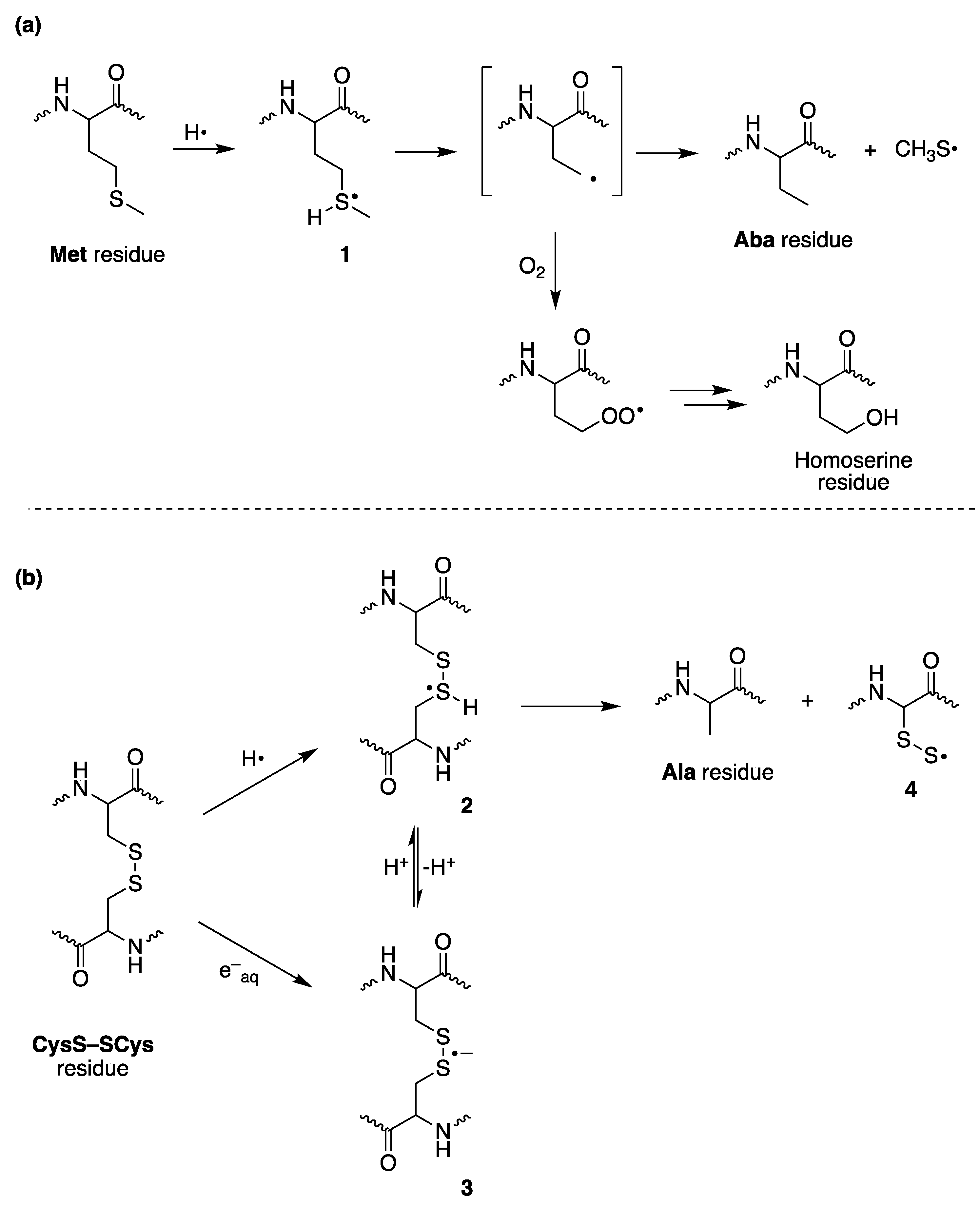
Scheme 1. (a) Mechanism of reaction of H with Met residues in aqueous solution and the formation of α-aminobutyric acid (Aba) residue and diffusible CH3S radical. (b) Mechanism of reaction of H and eaq with CysS–SCys residues in aqueous solution and the formation of alanine (Ala) residue and perthiyl radical 4.
Ijms 22 12863 sch001
Figure 1. Human serum albumin (HSA): (a) Methionine (Met) and Cysteine (CysS–SCys) residues are modified by attack of reductive transient species (H-atoms or hydrated electrons), yielding α-aminobutyric acid (Aba) and alanine (Ala)/CysSH, respectively; (b) Proposed mechanism for the formation of alanine residues (cf. Scheme 1b) and CysSH derived from a scrambling event with other CysS–SCys.
Ijms 22 12863 g001
Figure 2. (a) Mechanism of thiyl radical catalyzed cis-trans double bond isomerization of unsaturated fatty acid moieties in phospholipids. (b) Thiyl radical generation in aqueous compartment and diffusion in the lipid compartment of large unilamellar vesicle (LUV) made of L-α-phosphatidylcholines (PC).
Ijms 22 12863 g002
Figure 3. Concept of reductive stress and steps of the tandem protein–lipid damage leading to molecular libraries for biomarker identification.
Ijms 22 12863 g003
Figure 4. Tandem protein–lipid damage under reductive radical stress. Methionine and cystine residues are modified to α-aminobutyric acid and alanine/cysteine, respectively, by the attack of H and/or eaq. Concomitant formation of diffusible sulfur-centered radicals (CH3S or S•−) able to migrate into the lipid bilayer induces cis−trans isomerization of unsaturated fatty acid residues. Taken from reference [43].
Ijms 22 12863 g004
Table 1. Rate constant (M−1 s−1) of transient species derived from γ-radiolysis of water reacting with peptides/proteins [19].
Substratek(eaq )k(H)k(HO)
Methionine4.0 × 1073.5 × 1088.3 × 109
Glutathione disulfide3.7 × 1091.0 × 10109.6 × 109
Met-enkephalin 13.8 × 10¹⁰
Ribonuclease2.1 × 10101.5 × 10102.4 × 1010
Lysozyme3.1 × 1010 4.9 × 10¹⁰
Serum albumin8.2 × 109 7.8 × 10¹⁰
1 From reference [20].
Publisher’s Note: MDPI stays neutral with regard to jurisdictional claims in published maps and institutional affiliations.