Article

Genetic Elements Orchestrating Lactobacillus crispatus Glycogen Metabolism in the Vagina

1
Department of Molecular Cell Biology, Vrije Universiteit Amsterdam, De Boelelaan 1108, 1081 HZ Amsterdam, The Netherlands
2
ARTIS-Micropia, Plantage Kerklaan 38-40, 1018 CZ Amsterdam, The Netherlands
3
Biodata & Translation Innovation, Royal DSM, Alexander Fleminglaan 1, 2613 AX Delft, The Netherlands
4
Laboratory for Mass Spectrometry of Biomolecules, University of Amsterdam, Science Park 904, 1098 XH Amsterdam, The Netherlands
*
Author to whom correspondence should be addressed.
Int. J. Mol. Sci. 2022, 23(10), 5590; https://doi.org/10.3390/ijms23105590
Submission received: 22 April 2022 / Revised: 10 May 2022 / Accepted: 11 May 2022 / Published: 17 May 2022
(This article belongs to the Special Issue The Role of Commensal Microbiota in Human Health)

Abstract

:
Glycogen in the female lower reproductive tract is a major carbon source for colonization and acidification by common vaginal Lactobacillus species, such as Lactobacillus crispatus. Previously, we identified the amylopullulanase encoding gene pulA of Lactobacillus crispatus to correlate with the ability to autonomously utilize glycogen for growth. Here, we further characterize genetic variation and differential regulation of pulA affecting the presence of its gene product on the outer surface layer. We show that alpha-glucan degrading activity dissipates when Lactobacillus crispatus is grown on glucose, maltose and maltotriose, in agreement with carbon catabolite repression elements flanking the pulA gene. Proteome analysis of the S-layer confirmed that the amylopullulanase protein is highly abundant in an S-layer enriched fraction, but not in a strain with a defective amylopullulanase variant or in an amylopullulanase-sufficient strain grown on glucose. In addition, we provide evidence that Lactobacillus crispatus pulA mutants are relevant in vivo, as they are commonly observed in metagenome datasets of human vaginal microbial communities. Analysis of the largest publicly available dataset of 1507 human vaginal metagenomes indicates that among the 270 samples that contain a Lactobacillus crispatus pulA gene, 62 samples (23%) had a defective variant of this gene. Taken together, these results demonstrate that both environmental, as well as genetic factors explain the variation of Lactobacillus crispatus alpha-glucosidases in the vaginal environment.

1. Introduction

The vaginal environment of reproductive-age women is unique amongst vertebrates in several aspects: it has a low pH due to high levels of lactate (~100–150 mM) [1], it has abundant glycogen levels [2], and its microbial community is dominated by Lactobacillus species [3,4]. In recent years, more evidence has emerged that the vaginal microbiome is associated with reproductive and sexual health. Women with a Lactobacillus crispatus microbiome are at lower risk of vaginal infections and sexually transmitted diseases [5,6,7]. In addition, vaginal L. crispatus is inversely associated with more serious health outcomes, such as subfertility [8,9], preterm birth [10] and persistent HPV infection [11,12].
We previously demonstrated that most vaginal L. crispatus isolates are capable of autonomous growth on glycogen. A disrupted N-terminal signal peptide sequence in the amylopullulanase encoding pulA gene, either by small mutations or structural variants, coincided with the inability of growing on glycogen [13].
A recent key study [14] confirmed alpha-glucosidase activity of glycogen-degrading enzymes from different vaginal bacteria by heterologous expression. It showed that the L. crispatus amylopullulanase degrades glycogen, pullulan and amylose retaining activity at pH values that are common in a Lactobacillus-dominated vaginal environment. L. crispatus strains isolated from low-glycogen environments, such as the human or poultry gut, more often lacked the pulA gene indicating niche-specificity [15].
An open question in the field is the role of vaginal glycogen stores in bacterial acidification and the origin of vaginal glycogen degrading enzymes. Vaginal samples show alpha-glucosidase activity, which was postulated to be mostly of human origin on the basis of antibody [16,17] and proteomic analysis [18]. At the same time, results from two studies have indicated that alpha-glucosidases in a subset of vaginal samples may be of bacterial origin, on the basis of optimal pH and substrate and product profiling [14,19]. However, a metaproteomic study of vaginal lavages from women with a L. crispatus-dominated microbiome failed to detect any L. crispatus amylopullulanase protein, which poses questions about host-related or genetic factors influencing its presence [18].
Here we present results showing suppression of PulA activity in glucose-, maltose- and maltotriose-grown cultures by enzymatic assays for alpha-glucosidase. We use a mass spectrometric approach to demonstrate the presence of the L. crispatus amylopullulanase enzyme in an S-layer enriched fraction and its loss in the presence of glucose or in a strain with a defective pulA gene. In addition, the analysis of a large vaginal metagenomic dataset demonstrates the presence of a significant number of mutations and structural variants, which are expected to fully disrupt amylopullulanase functionality in vivo, indicating that both environmental, as well as genetic factors, may restrainvaginal L. crispatus from metabolizing glycogen.

2. Results

2.1. Substrate Utilization of PulA-Sufficient and Deficient Lactobacillus crispatus Strains

Previously, we reported a collection of L. crispatus strains with variable growth on glycogen associated with the presence of an intact amino-terminal signal peptide for protein secretion in the pulA gene product (PulA) [13]. Here we further explore the substrate range of a L. crispatus deficient (RL09, hereafter referred to as pulA) and sufficient (RL10, hereafter referred to as pulA+) strain.
To verify the identified deletion of two nucleotides, we used a PCR-amplified 300 bp DNA fragment encoding the pulA N-terminal region (including 42 nucleotides upstream the start site) of strains RL09 and RL10 and resequenced with Sanger dideoxy sequencing. This confirmed the presence of the deletion of two nucleotides in the signal peptide sequence of RL09 compared to RL10 (Appendix A Figure A1).
Previously, we found the pulA strain RL09 to be unable to grow on glycogen. We reassessed the growth and acidification of this strain and the pulA+ strain RL10 on NYCIII medium without glucose supplemented with 11 different carbohydrates to analyze other differences between the strains in their metabolic profiles. Optical density and medium pH after 24 h of anaerobic growth are shown in Figure 1A,B, respectively.
Both L. crispatus strains were equally capable of growth on all carbon sources, except for lactose, raffinose, cellobiose and, in disagreement with previous findings, lactulose [20]. Growth was accompanied by acidification of the medium to a pH of 3.9, corresponding to the in vivo vaginal pH [1] and the pKa of lactic acid. Glycogen, both of oyster and bovine origin, was the only carbohydrate resulting in a clear difference between the strains: the pulA deficient strain grew to an optical density of ~0.5 and acidified to a pH of 6.0 (Oyster glycogen) and 5.5 (Bovine glycogen) possibly due to the presence of smaller maltodextrins and glucose after autoclaving the glycogen. The growth of the pulA sufficient strain on glycogen was comparable to that on glucose, acidifying the medium to a pH of 3.9. Regarding growth on the 11 carbohydrates tested, we conclude that pulA+ and pulA strains do not substantially differ in their metabolic capacities apart from the ability to grow on glycogen.

2.2. Starch Degradation Activity of PulA Sufficient and Deficient L. crispatus Strains on Various Carbon Sources

Next, we measured the enzymatic activity of the amylopullulanase enzyme to understand its regulation and to find amylopullulanase-expressing conditions that would allow for a direct proteomic comparison between the two strains. Genetic elements in the locus of the pulA gene are strongly indicative of carbon catabolite repression, similar to the regulation of amylopullulanases in other Lactobacilli [21]. The promoter region of the gene has a cre (catabolite responsive element) like palindromic sequence (TGTTATCGATAACA). The catabolite responsive element contains a well-known binding site for the global regulator CcpA (carbon catabolite protein A), which is known for suppressing metabolic pathways for alternative carbohydrates in the presence of glucose in various Lactobacillus species [22,23,24]. Downstream of the pulA gene, we identified two open reading frames with homology to the LacI family of repressors, containing a DNA binding domain in one open reading frame (1–110) and an effector domain (91–219) in the other, possibly involved in catabolite repression through binding of the breakdown products of glycogen (Figure 2).
To verify the predicted regulation by carbon sources, we grew the pulA sufficient strain L. crispatus to stationary phase on NYCIII medium supplemented with glycogen, maltotriose, maltose, glucose, or galactose, and measured alpha-glucosidase activity in the pellet and supernatant. We included galactose since (i) lactic acid bacteria generally show a preference for glucose over galactose [25,26], and (ii) we hypothesize that galactose does not repress pulA expression to the same extent as glucose.
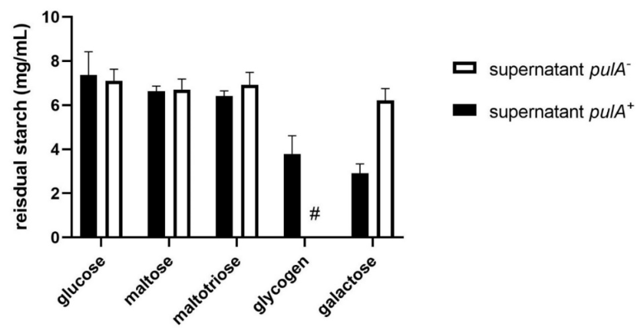
After 48 h of growth, spent medium and bacterial cells were mixed with a starch solution to detect starch degradation in a standard starch-iodine assay. Starch was used as a substitute for glycogen since it can be easily detected using iodine and has an amylopectin moiety with alpha1–4 and alpha1–6 linkages comparable to glycogen. Total starch degradation for a period of 24 h at 37 °C was used as a measure of alpha-glucosidase activity in the sample. Starch degradation was observed in the pellet (Figure 3) as well as the spent growth media (Appendix A Figure A2) of L. crispatus pulA+ strain RL10 after growth on glycogen and galactose, but not after growth on glucose, maltose and maltotriose, confirming the repression of this gene under these conditions.
Alpha-glucosidase activity in cells and spent medium of the pulA+ strain was comparable to that of the pulA strain on all substrates except for glycogen since the pulA strain did not sufficiently grow on glycogen to measure starch degradation. Bacterial cells or supernatants of the pulA strain grown on glucose, maltose, maltotriose, or galactose did not degrade any starch confirming the link between the defective amylopullulanase enzyme and glycogen metabolism.

2.3. S-Layer Enrichment and Proteome Analysis

In our previous study, we identified a surface layer associated protein (SLAP) domain in the L. crispatus amylopullulanase protein sequence [13], which prompted us to investigate its localization in the surface layer. We hypothesized that the natural variation in the amino-terminal signal peptide for secretion of amylopullulanase would result in the presence or absence of an active amylopullulanase in the S-layer. Furthermore, we wanted to confirm the catabolite repression of amylopullulanase by glucose, as observed in this study by use of the starch degradation assay (Figure 3), would be reflected by the levels of amylopullulanase in the S-layer. We utilized galactose as a carbon source since it (i) allows for the growth of both pulA and pulA+ strains and (ii) is a condition under which we observed cell-associated starch degradation activity (Figure 3). We compared cells of the pulA+ RL10 strain grown on galactose, glucose, and glycogen, in order to verify the expression of amylopullulanase in an S-layer enriched fraction under these conditions.
The S-layers form the outermost structure of the cell envelope and represent up to 15% of the total protein content [27]. As the S-layer protein subunits (SLPs) and S-layer associated proteins (SLAPs) are non-covalently linked to the cell wall, we released the S-layer fraction from the cells and let it disintegrate into monomeric polypeptides by exposure to high concentrations of the denaturing agent LiCl. Mass-spectrometric analysis of this isolated S-layer fraction led to the identification of peptides of at least 28 SLAP-domain containing proteins, including a number of abundant SlpA-domain containing proteins (Table 1). The most prominent distinction in the amount of all proteins identified in the RL09 and RL10 S-layers is a 90-fold difference for amylopullulanase The minor background of amylopullulanase peptides in RL09 may result from lysed cells releasing intracellular amylopullulanase. S-layer fractions of the pulA+ strain RL10 grown on glucose showed a 29-fold lower amylopullulanase presence compared to fractions from glycogen grown cells. Two uncharacterized surface layer proteins (A0A7T1TNA6 and A0A125P6L8) are strongly upregulated in the presence of glucose by a factor of over 200; all other proteins show minor differences in levels with 2log ratios between ranging from −2.2 to 1.4 The overall fraction of S-layer proteins appear to have been reduced in the presence of glucose (0.2 versus 0.4–0.5 in the other three fractions).

2.4. Metagenome Analysis

Our previous comparative genomics analysis of a collection of L. crispatus strains isolated from the outpatient clinic in Amsterdam revealed variations in the N-terminal signal peptide region of the pulA gene that correlated with the ability of a strain to grow on glycogen [13]. In this strain collection, we found seven amylopullulanase-deficient strains sharing four different types of disruptions of the N-terminal sequence (Table 2). Although not all published L. crispatus genomes show the presence of a pulA gene [15], the N-terminal disruptions of the pulA sequence observed in our collection were not found in other L. crispatus genomes reported thus far.
We hypothesize that the isolation of these pulA variants is not a result of selective enrichment during the isolation procedure, but that L. crispatus strains with defective pulA genes are common in the vaginal microbiota. To test this hypothesis, we assessed the prevalence and frequency of disruptive pulA gene sequence variants, such as those observed in our strain collection (Table 2) in a vaginal metagenome dataset of 1507 samples [28]. To our knowledge, this is the largest publicly available vaginal microbiome data collection, which includes samples from several different studies of North American, African, and Chinese women. In total, we analyzed 28.6 billion sequencing reads that made up 3.9 tera base pairs (Tbp) of metagenome sequencing data (on average, 19 million reads and 2.6 Gbp data per sample).
We first determined the prevalence of small but disruptive variations across the pulA gene sequence, similar to those observed in pulA strains RL02 and RL09 (Table 2). Out of 1507 samples we aligned to the L. crispatus RL10 pulA gene sequence, 270 resulted in alignments with a minimum of 10 times coverage. Variant calling on these 270 samples resulted in a total of 3202 unique variants. After low-frequency (allele frequency lower than 0.05) and low prevalence (observed in less than three samples) variants were filtered out, 54 variants remained, of which 19, 28 and 6 were low (e.g., synonymous mutations), moderate (e.g., mutations that cause an amino acid change), and high impact (stop gain or frameshift mutations), respectively (Figure 4).
In 15 out of 270 samples (5.5%), at least one of the high-impact mutations was observed at a high allele frequency (>0.7), where we expect an amylopullulanase deficient phenotype (Supplementary File S1 pulA_small_variants.xlsx). Three of these high-impact mutations were near the N-terminus of the pulA sequence at positions 156, 207, and 225, impacting 11 samples in total.
Subsequently, we analyzed the same set of 270 samples for larger structural variants compared to the pulA+ RL10 pulA gene sequence. In 56 out of 270 samples (20%), we identified large structural variants which had a high frequency (>0.7) (Supplementary File S2 pulA_structural_variants.xlsx). The disruptive structural variation in 17 out of 56 of these samples was found to be due to the insertion of transposase sequences into the amylopullulanase gene sequence, as in the case of the pulA locus from strains RL06, RL07, RL19, and RL26 in our own strain collection. Taken together, we identified high-frequency mutations in 62 of 270 L. crispatus pulA samples, which is indicative of a dysfunctional amylopullulanase of the dominant strain and the inability of L. crispatus to contribute to the acidification of the vaginal environment with glycogen as a carbon and energy source.

3. Discussion

In this study, we focus on genetic variation and disruption of the Lactobacillus crispatus pulA gene. Amylopullulanase is thought to play a central role in vaginal metabolomics. It catalyzes the first step of a metabolic pathway turning the most abundant vaginal carbohydrate (glycogen) into the most abundant metabolite (lactate) and is produced by one of the most abundant vaginal species L. crispatus. In a previous study, we published 33 genomes of vaginal L. crispatus isolates that displayed remarkable variation in the gene locus. Strains with a disrupted pulA gene, through small indels or structural variants, were incapable of growth on glycogen.
The wealth of publicly available metagenome datasets, repositories, and gene catalogs allows for a more hypothesis-driven research approach concentrating on specific genes that are associated with in vivo functionality. In the largest available metagenome database, a substantial number of samples had similar genetic variations compared with our previously reported whole-genome sequences of L. crispatus isolates. These variations consisted of both small deletions as well as relatively large structural variations disrupting the functionality of this gene.
Disruptions in the secretome of catalytically active enzymes are a well-known phenomenon in the microbial world. Bacteria may take advantage of cells in close proximity that cleave ’public goods’, such as proteins or glycans into smaller peptides or maltodextrins. For instance, a protease-positive Lactococcus lactis population gradually lost protease activity in the entire population in a spatial-dependent manner. In this case, the ‘cheating’ proteolytic negative strains can utilize peptides supplied by the proteolytic positive strains without having the cost of protease production [29]. These disruptions may provide a fitness advantage to isolates, especially when, as we show in our mass spectrometric analysis, the enzyme forms a substantial fraction of the outer surface layer.
In the vaginal metagenome dataset we analyzed here, we did not find evidence of an extensive variety of amylopullulanase sequences within a single vaginal microbiome: most samples only showed the presence of one variant, which we assume coincides with the dominant presence of one strain. We hypothesize that the metabolic advantage that a cheating strain in complex ecological samples may experience, is not so much a matter of intraspecies competition, but rather due to interspecies cross-feeding of metabolites. The presence of other glycogen degrading bacteria or human amylase may create an environment in which a L. crispatus strain has energetic advantages from shutting down the expression and secretion of S-layer associated enzymes while benefiting from the breakdown products of extracellular glycogen hydrolysis by other species.
A recent study did not find such evolutionary pressure to mutate the amylopullulanase: even after cultivation of up to 1000 generations by sequential sub-culturing in synthetic vaginal fluid, no genetic variations in the pulA locus of strains of L. crispatus were detected [30]. We hypothesize that this may be due to the available carbohydrates in this simulated vaginal fluid, which contains both glucose and glycogen and is further supplemented with amylase which degrades the glycogen into smaller maltodextrins. The results of our alpha-glucosidase assays and proteome analysis imply that L. crispatus grown in such an environment will first grow on glucose, maltose and maltotriose and will suppress amylopullulanase expression until these carbohydrates are depleted. This will diminish the advantage of cheating or any other evolutionary pressure on the production of alpha-glucosidase.
The genetic disruption affected a minority of vaginal samples in the metagenomic dataset we analyzed (23%) and a minority of isolates in our strain collection. In most environments, L. crispatus maintains an intact amylopullulanase gene, even though disruption of the pulA gene occurs at relatively high frequencies. A recent metagenomics study and targeted PCR by Lithgow et al. revealed that 36% of L. crispatus-dominated metagenomes from their African cohort lacked a functional L. crispatus pulA gene, and reported a three-fold higher frequency of gene loss than that seen in metagenomes from European and North American women [31]. However, this is not reflected by the prevalence of disrupted pulA genes among the known ethnic groups in the metagenome data set of 1507 vaginal samples we have analyzed (Appendix A Table A1). Our study shows that from 781 black or African American women, 102 contained a pulA gene, including 12 disrupted genes (12%), while from 163 white or Caucasian women, 55 contained a pulA gene, including 19 disrupted genes (34%).
Our study suggests that carbon catabolite repression may further reduce the activity of L. crispatus amylopullulanase activity in vivo. Bacteria generally regulate metabolic pathways, optimizing their growth strategy by prioritizing carbon sources based on nutritional quality. The presence of glucose, maltose and maltotriose in the vagina has been well-documented, especially in Lactobacillus-dominated environments [32,33].
However, given the positive relationship between amylopullulanase activity and smaller maltodextrins in the study by Lithgow et al. [31], we think further studies should include analyses of metatranscriptomes and carbohydrate concentration, to clarify whether the observed in vitro pulA regulation, depending on carbohydrate availability in L. crispatus, corresponds to its in vivo regulation. Based on the prevalence of these carbon sources in vaginal metabolomics studies, as well as human amylase, we expect that L. crispatus in the vaginal environment preferentially relies on glycogen cleavage by other bacteria or host amylase and will only express amylopullulanase when other carbon sources are depleted.

4. Materials and Methods

4.1. Standard Cultivation Conditions

Lactobacillus crispatus amylopullulanase deficient strain (L. crispatus RL09) and an amylopullulanase sufficient strain (L. crispatus RL10) were routinely cultivated on NYCIII medium with HEPES (2.4 g/L), proteose peptone #3 (Becton Dickinson Franklin Lakes, NJ, USA, 211693) (15 g/L), yeast extract (ThermoFisher Scientific Waltham, MA USA, LP0021) (3.8 g/L), NaCl (5 g/L), glucose monohydrate (Santa Cruz Biotechnology, Dallas, TX, USA 14431-43-7) (5.5 g/L g), heat inactivated horse serum (10%) and for agar plates 1,5% (w/v) agarose. Plates, precultures and subcultures were grown under anaerobic conditions (gas mixture of 10% CO2 and 90% N2 at 37 °C.

4.2. PCR and Sequencing of Signal Peptide Region L. crispatus Amylopullulanase

Previously, Illumina-whole genome sequencing showed a two nucleotide deletion in the N-terminal pulA signal peptide sequence of L. crispatus RL09. In order to confirm this frameshift mutation, we performed a genetic analysis of the area with deletion by PCR amplification and Sanger sequencing. Genomic DNA was isolated from a 24 h liquid culture using the GenEluteTM Bacterial Genomic DNA Kit (Merck, Darmstadt, Germany). A 300 base pair sequence at the start site of the amylopullulanase was amplified with a forward primer GCAAATGAAAGCGCATACGTTT (annealing 42 base pairs upstream the start site) and a reverse primer TGTTGACGCTGCTTTGCTT (annealing 257 base pairs downstream the start site) using a High Fidelity Phusion® DNA polymerase (ThermoFisher, Waltham, MA, USA). The size of the PCR product was verified with ethidium bromide on a gel to be the predicted size of 299 bp. The PCR product was purified using the GeneJET PCR Purification Kit (ThermoFisher, Waltham, MA, USA) and sequenced by Sanger Dideoxy sequencing (Eurofins, Luxembourg City, Luxembourg).

4.3. Serial Propagation on Various Carbohydrates

Growth on various carbohydrate sources was tested with NYCIII without glucose supplemented with D-lactose monohydrate (Sigma-Aldrich, St. Louis, MO, USA), D-raffinose pentahydrate (Alfa Aesar Haverhill, MA, USA), D-cellobiose (Sigma-Aldrich, St. Louis, MO, USA), D-melibiose (Sigma-Aldrich, St. Louis, MO, USA), D-maltose monohydrate (Honeywell, Charlotte, NC, USA), D-trehalose dihydrate (Sigma-Aldrich, St. Louis, MO, USA), D-galactose (Sigma-Aldrich, St. Louis, MO, USA), lactulose (Sigma-Aldrich, St. Louis, MO, USA), glucose monohydrate (Merck, Darmstadt, Germany), bovine glycogen (Sigma-Aldrich, St. Louis, MO, USA) and oyster glycogen (Alfa Aesar Haverhill, MA, USA). The growth of L. crispatus RL09 and L. crispatus RL10 on these carbohydrate sources was examined by serial propagation on a glucose-free NYCIII medium supplemented with 5% (w/v) of the different carbohydrates. The serial propagation was executed using two passages of twentyfold sub-culturing in a 96-well plate. Between each passage, the cells were anaerobically grown for 24h in jars (ThermoFisher, Waltham, MA, USA). To monitor growth, optical density was measured at 600 nm in a sterile 96-well flat bottom plate in a spectrophotometer (Spectramax Plus 384; Molecular Devices, San Jose, CA, USA) after ten times dilution in sterile phosphate-buffered saline solution. Acidification of the medium by L. crispatus was assessed by pH measurements (HI 2210 pH meter, Hanna Instruments, Woonsocket, RI, USA) after every passage after 24 h of anaerobic growth. Statistical testing of optical densities and pH values was conducted with t-tests: two samples assuming unequal variations (α = 0.05, two-sided).

4.4. The Starch Degradation Assay

Glycogen is a polymer of α−1,4-linked and α−1,6-linked glucose residues. Starch degradation, measured in a basic starch-iodine assay, can be used as a proxy since the amylopectin moiety of starch has a similar branched structure with α−1,4-linked and α−1,6-linked glucose residues. To test α-glucosidase activity strains were grown for 24 h. Cells were spun down and resuspended in amylase buffer (100 mM Na-Acetate with 5 mM CaCl2, pH 5,3). 50 uL supernatant or resuspended pellet was added to 150 uL of 10 g/L starch solution (Sigma-Aldrich, St. Louis, MO, USA, S9765) in amylase buffer. Chloramphenicol (Sigma-Aldrich, St. Louis, MO, USA, C0378-25G) from a stock solution in ethanol was added to a final concentration of 10 ng/mL to prevent growth; 50 µL of the resuspended pellet of culture spent medium was mixed with 150 µL of this starch working solution and incubated at 37 °C for 24 h. To visualize residual starch, 10 uL was added to 290 uL of an iodine stock solution consisting of 0.2 g I2 and 2 g Kl in 100 mL 50 mM HCl. Absorption was measured at 600 nm in a 96-well plate reader. Experiments were carried out with a minimum of (two) biological replicates for RL09 on maltose and maltotriose; all other conditions had between three and nine replicates carried out on separate days in a total of four independent experiments.

4.5. Enrichment for Surface Layer (Associated) Proteins

The enrichment for SLPs and SLAPs was modified from a standard LiCl S-layer extraction protocol for L. acidophilus (Goh et al., 2009; Lortal et al., 1992). Bacterial cells of L. crispatus were grown to stationary phase (24 h), centrifuged at 2236 g for 10 min (4 °C), and washed twice with 25 mL cold PBS (Gibco), pH 7.4. Cells were agitated for 15 min at 4 °C following the addition of 5 M LiCl (ThermoFisher, Waltham, MA, USA). Supernatants, containing SLPs and SLAPs, were harvested via centrifugation at 8994 g for 10 min (4 °C) and transferred to a 6000–8000 kDa Spectra/Por molecular porous membrane (Spectrum Laboratories Rancho Dominguez, CA, USA) and dialyzed against cold distilled water for 24 h, changing the water every 2 h for the first 8 h. The dialyzed precipitate was harvested via centrifugation at 20,000× g for 30 min (4 °C).

4.6. Sample Preparation for Proteomics Analysis

Protein pellets were resuspended in 200 µL of digestion buffer (5 mM DTT, 50 mM NH4HCO3, pH 8.0) and incubated at 55 °C for one hour to reduce disulfide bridges. Then samples were treated with 15 mM iodoacetamide (IAA) to alkylate free cysteines in the dark for 45 min. Subsequently, samples were digested with trypsin (1:100 w/w protease:protein ratio) at 37 °C for 18 h. The mixture of peptides was desalted using C18 solid phase extraction columns (Agilent Technologies, Santa Clara, CA, USA) following the manufacturer’s protocol. The final peptide mixture was dried in a vacuum centrifuge and suspended in 0.1% formic acid in water (ULC-MS grade, Biosolve, Valkenswaard, The Netherlands) for MS analysis.

4.7. LC-MS/MS Analysis

Mass spectrometric analysis of 200 ng peptides was carried out on a timsTOF Pro (Bruker, Bremen, Germany) equipped with an Ultimate 3000 nanoRSLC UHPLC system (Thermo Scientific, Germeringen, Germany). Samples were injected onto a C18 column (75 µm, 250 mm, 1.6 µm particle size, Aurora, Ionopticks, Fitzroy, Australia) kept at 50 °C. Peptides were loaded at 400 nL/min for 1 min in 3% solvent B and separated by a multi-step gradient: 5% solvent B at 2 min 17%, solvent B at 24 min, 25% solvent B at 29 min, 34% solvent B at 32 min, 99% solvent B at 33 min held until 40 min, returning to 3% solvent B at 40.1 min and held until 58 min to recondition the column (Solvent A: 0.1% formic acid in water, Solvent B: 0.1% formic acid in acetonitrile). MS analysis of eluting peptides was performed by a time-of-flight mass spectrometer. The precursor scan ranged from 100 to 1700 m/z and a time range of 0.6–1.6 V.s/cm2 in PASEF mode. A total of 10 PASEF MS/MS scans following collision induced dissociation (collision energy from 20–59 eV) were collected with a total cycle time of 1.16 s.
Raw MS/MS data were processed using Maxquant software, Dr. Jürgen Cox, Max Planck Institute of Biochemistry, Martinsried, Germany (version 1.6.14.0) [34] searching a pan proteome database of L. crispatus (Uniprot, downloaded 1 November 2021). To estimate the false spectrum assignment rate, a reverse version of the same database was also searched. The settings were as follows: Enzyme Trypsin/P allowing for a maximum of two missed cleavages, variable modifications: Oxidation (M), fixed modifications: Carbamidomethyl (C). Settings were default for timsDDA, and match between runs was enabled with a matching time window of 0.2 min and a matching ion mobility window of 0.05 indices. For label free quantification, both iBAQ and LFQ were enabled [34].

4.8. Analysis of the PulA Gene Region

The contig of L. crispatus RL10 was analyzed with SnapGene version 5.3.2. (GSL Biotech LLC, Chicago, IL, USA). The promoter sequences (−35 and −10 elements) were predicted by the use of BPROM (Prediction of Bacterial Promoters) and FindTerm (Finding Terminators in bacterial genomes) [35]. The catabolite responsive element (cre site) was identified by matching the palindromic nucleotide motif of L. plantarum (TGWAANCGNTNWCA) [36].

4.9. Analysis of Publicly Available Vaginal Metagenomes

The raw shotgun metagenome sequencing data of 1507 publicly available vaginal microbiome samples were downloaded from the VIRGO database [28]. In addition to the 211 samples which were sequenced for the construction of VIRGO, this large data collection contains samples from other studies of North American [37], African [5], and Chinese women [38]. The Illumina reads were processed using FastQC to determine data statistics, such as data yield per sample. The samples were taxonomically characterized using Kraken 2 [39] and Bracken [40]. The human read counts in the species-level results from Bracken were removed and the data were normalized to obtain relative abundances of non-human taxa, such as bacteria, archaea, viruses, and fungi. Taxa that had less than 0.5% relative abundance and which were observed in less than 5% of the samples were filtered out. The downstream taxonomic analyses and visualization were performed using R v3.6.3 (R Core Team, 2020, Robert Gentleman and Ross Ihaka, the ‘R & R’ of the Statistics Department of the University of Auckland, New Zealand with R packages vegan, phyloseq [41], ComplexHeatmap [42], and ggplot2 v3.3.3.

4.10. Amylopullulanase Sequence Variant Detection

The raw reads were aligned to the RL10 pulA gene sequence using bowtie2 [43] with default parameters except for “--very-sensitive-local --no-unal”. Alignment statistics for each sample, such as coverage were calculated using samtools (John Marshall and Petr Danecek et al Initial release: 2009) v1.13 [44]. Single-nucleotide polymorphisms (SNPs) and short indels in samples with a minimum 10× coverage were determined using FreeBayes v1.2 [45] in a multi-sample variant calling approach using default parameters except for “-min-alternate-count 2 -min-coverage 10 -min-alternate-fraction 0.01 -use-duplicate-reads -min-mapping-quality 0 -ploidy 1 -pooled-continuous”. The variants were annotated in terms of their impact on the RL10 pulA protein sequence using SnpEff v4.3t [46]: high (e.g., mutations that result in frameshifts or stop gain), moderate (e.g., mutations that result in an amino acid change), and low (synonymous mutations). The variant allele frequency heatmap for variants that had at least 0.05 alternate allele frequency (i.e., supported by at least 5% of the reads at that position) and were observed in at least three samples was plotted using R packages VariantAnnotation [47], and ggplot2. SNPs and short indels with a minimum frequency of 0.7 were classified as high-frequency short variants.
The same sample set used in small variant detection which had a minimum coverage of 10 times along the RL10 pulA sequence was used for detecting larger and structural variations, such as insertions, deletions, inversions, translocations, and duplications in read mappings. To this end, the indels and Structural Variants tool in CLC Genomics v.12.0.3 (Health Sciences Library System, Pittsburgh, PA, USA) was used with default parameters (p-value threshold = 0.0001) except for “minimum number of reads = 10”. Structural variants with a minimum frequency of 0.7 were classified as high-frequency large variants.

4.11. Identification of PulA Genes with Transposase Insertions

To create a database of L. crispatus transposase gene sequences, we first downloaded 147 annotated L. crispatus genomes from NCBI (National Center for Biotechnology Information, Bethesda, MD, USA) (assembly level: scaffold, chromosome, and complete). We then created a FASTA file of 19.551 redundant transposase gene sequences by combining the coding sequences which had a ‘transposase’ annotation in the 147 L. crispatus genomes. This database was clustered at 95% sequence identity using CD-HIT using default parameters [48] to obtain a non-redundant transposase database of 508 gene sequences.
To identify pulA rich samples that had transposase insertions, we mapped the reads which were mapped to the L. crispatus RL10 pulA sequence to the above-mentioned transposase database using bowite2 as described above. The list of samples where more than 20 transposase reads were found was compared to the list of samples with high-frequency structural variants to determine the impact of transposase insertions in causing disruptive structural variations.

Supplementary Materials

The following supporting information can be downloaded at: https://www.mdpi.com/article/10.3390/ijms23105590/s1.

Author Contributions

Conceptualization, R.H. and R.K.; Methodology and data analysis, R.H., A.M., G.K. and R.K.; Experiments: R.H., G.K., I.v.V., D.M. and R.K.; data curation: A.M. and R.H., Writing—original draft preparation, R.H., A.M., G.K. and R.K.; Writing review and editing R.H. and R.K.; Supervision, R.H., D.M. and R.K. All authors have read and agreed to the published version of the manuscript.

Funding

This research received no external funding.

Institutional Review Board Statement

Not applicable.

Informed Consent Statement

Not applicable.

Data Availability Statement

All data presented here are included in the manuscript, Appendix A or supplemental file. The mass-spectrometry data have been deposited in Proteome Exchange.

Acknowledgments

We would like to thank the students Omar Beganović, Deborah Jekel, Yumiko van Diest, Leon Steenbergen, and Ritesh Panchoe for their contributions to the research presented in this publication and Jurgen Haanstra for his contribution to student supervision. We thank Herwig Bachmann (Vrije Universiteit Amsterdam) for critically reading the manuscript and we acknowledge Jacques Ravel and Michael France (University of Maryland School of Medicine) for kindly sharing metadata of the VIRGO metagenome dataset.

Conflicts of Interest

RYH and RK are unpaid board members of the crispatus foundation: www.crispatus.org (accessed on 10 May 2022). The other authors declare no conflict of interest.

Appendix A

Figure A1. The sequence of the L. crispatus pulA signal peptide region of a pulA+ strain RL10 and pulA strain RL09.
Ijms 23 05590 g0a1
Figure A2. Starch degrading activity in spent medium of L. crispatus amylopullulanase sufficient and deficient strains grown on NYC-, glucose, maltose, maltotriose, glycogen (only pulA+) and galactose. Cells were cultured in NYCIII medium supplemented with these carbohydrates and after centrifugation and resuspension of the pellet incubated in a starch solution (7.5 g/L). Experiments were carried out with a minimum of (two) biological replicates; averages and standard deviations are shown. Hashtag (#) indicates no data due to the absence of growth of this strain on glycogen.
Ijms 23 05590 g0a2
Table A1. Number of metagenomes (total 1507), with a pulA gene (intact and defective) and a defective pulA gene per ethnic group of women.
Ethnic GroupNumber of MetagenomesNumber of Metagenomes with PulANumber of Metagenomes with Defective PulA
Total150727062
Black or African American78110212
White or Caucasian1635519
Chinese7642
Unknown47210828
Hispanic, mixed or other1511

References

  1. O’Hanlon, D.E.; Moench, T.R.; Cone, R.A. Vaginal pH and Microbicidal Lactic Acid When Lactobacilli Dominate the Microbiota. PLoS ONE 2013, 8, e80074. [Google Scholar] [CrossRef] [PubMed]
  2. Mirmonsef, P.; Hotton, A.L.; Gilbert, D.; Burgad, D.; Landay, A.; Weber, K.M.; Cohen, M.; Ravel, J.; Spear, G.T. Free Glycogen in Vaginal Fluids is Associated with Lactobacillus Colonization and Low Vaginal pH. PLoS ONE 2014, 9, e102467. [Google Scholar] [CrossRef] [PubMed] [Green Version]
  3. Ravel, J.; Gajer, P.; Abdo, Z.; Schneider, G.M.; Koenig, S.S.; McCulle, S.L.; Karlebach, S.; Gorle, R.; Russell, J.; Tacket, C.O.; et al. Vaginal Microbiome of Reproductive-Age Women. Proc. Natl. Acad. Sci. USA 2011, 108 (Suppl. S1), 4680–4687. [Google Scholar] [CrossRef] [Green Version]
  4. Dols, J.A.M.; Molenaar, D.; van der Helm, J.J.; Caspers, M.P.M.; de Kat Angelino-Bart, A.; Schuren, F.H.J.; Speksnijder, A.G.C.L.; Westerhoff, H.V.; Richardus, J.H.; Boon, M.E.; et al. Molecular Assessment of Bacterial Vaginosis by Lactobacillus Abundance and Species Diversity. BMC Infect. Dis. 2016, 16, 180. [Google Scholar] [CrossRef] [Green Version]
  5. Gosmann, C.; Anahtar, M.N.; Handley, S.A.; Farcasanu, M.; Abu-Ali, G.; Bowman, B.A.; Padavattan, N.; Desai, C.; Droit, L.; Moodley, A.; et al. Lactobacillus-Deficient Cervicovaginal Bacterial Communities Are Associated with Increased HIV Acquisition in Young South African Women. Immunity 2017, 46, 29–37. [Google Scholar] [CrossRef] [PubMed] [Green Version]
  6. De Seta, F.; Lonnee-Hoffmann, R.; Campisciano, G.; Comar, M.; Verstraelen, H.; Vieira-Baptista, P.; Ventolini, G.; Lev-Sagie, A. The Vaginal Microbiome: III. The Vaginal Microbiome in Various Urogenital Disorders. J. Low. Genit. Tract Dis. 2022, 26, 85–92. [Google Scholar] [CrossRef]
  7. Ceccarani, C.; Foschi, C.; Parolin, C.; D’Antuono, A.; Gaspari, V.; Consolandi, C.; Laghi, L.; Camboni, T.; Vitali, B.; Severgnini, M.; et al. Diversity of Vaginal Microbiome and Metabolome during Genital Infections. Sci. Rep. 2019, 9, 14095. [Google Scholar] [CrossRef] [Green Version]
  8. Koedooder, R.; Singer, M.; Schoenmakers, S.; Savelkoul, P.H.M.; Morré, S.A.; de Jonge, J.D.; Poort, L.; Cuypers, W.; Beckers, N.G.M.; Broekmans, F.J.M.; et al. The Vaginal Microbiome as a Predictor for Outcome of in Vitro Fertilization with or without Intracytoplasmic Sperm Injection: A Prospective Study. Hum. Reprod. 2019, 34, 1042–1054. [Google Scholar] [CrossRef]
  9. Vergaro, P.; Tiscornia, G.; Barragán, M.; García, D.; Rodriguez, A.; Santaló, J.; Vassena, R. Vaginal Microbiota Profile at the Time of Embryo Transfer Does Not Affect Live Birth Rate in IVF Cycles with Donated Oocytes. Reprod. Biomed. Online 2019, 38, 883–891. [Google Scholar] [CrossRef]
  10. Fettweis, J.M.; Serrano, M.G.; Brooks, J.P.; Edwards, D.J.; Girerd, P.H.; Parikh, H.I.; Huang, B.; Arodz, T.J.; Edupuganti, L.; Glascock, A.L.; et al. The Vaginal Microbiome and Preterm Birth. Nat. Med. 2019, 25, 1012–1021. [Google Scholar] [CrossRef] [Green Version]
  11. Usyk, M.; Schlecht, N.F.; Pickering, S.; Williams, L.; Sollecito, C.C.; Gradissimo, A.; Porras, C.; Safaeian, M.; Pinto, L.; Herrero, R.; et al. MolBV Reveals Immune Landscape of Bacterial Vaginosis and Predicts Human Papillomavirus Infection Natural History. Nat. Commun. 2022, 13, 233. [Google Scholar] [CrossRef] [PubMed]
  12. Dols, J.A.M.; Reid, G.; Kort, R.; Schuren, F.H.J.; Tempelman, H.; Bontekoe, T.R.; Korporaal, H.; Van der Veer, E.M.; Smit, P.W.; Boon, M.E. PCR-Based Identification of Eight Lactobacillus Species and 18 Hr-HPV Genotypes in Fixed Cervical Samples of South African Women at Risk of HIV and BV. Diagn. Cytopathol. 2012, 40, 472–477. [Google Scholar] [CrossRef] [PubMed]
  13. van der Veer, C.; Hertzberger, R.Y.; Bruisten, S.M.; Tytgat, H.L.P.; Swanenburg, J.; de Kat Angelino-Bart, A.; Schuren, F.; Molenaar, D.; Reid, G.; de Vries, H.; et al. Comparative Genomics of Human Lactobacillus crispatus Isolates Reveals Genes for Glycosylation and Glycogen Degradation: Implications for in Vivo Dominance of the Vaginal Microbiota. Microbiome 2019, 7, 49. [Google Scholar] [CrossRef] [PubMed] [Green Version]
  14. Woolston, B.M.; Jenkins, D.J.; Hood-Pishchany, M.I.; Nahoum, S.R.; Balskus, E.P. Characterization of Vaginal Microbial Enzymes Identifies Amylopullulanases That Support Growth of Lactobacillus crispatus on Glycogen. bioRxiv 2021, 2021.07.19.452977. [Google Scholar] [CrossRef]
  15. Pan, M.; Hidalgo-Cantabrana, C.; Barrangou, R. Host and Body Site-Specific Adaptation of Lactobacillus crispatus Genomes. NAR Genom. Bioinform. 2020, 2, lqaa001. [Google Scholar] [CrossRef] [Green Version]
  16. Nasioudis, D.; Beghini, J.; Bongiovanni, A.M.; Giraldo, P.C.; Linhares, I.M.; Witkin, S.S. Alpha-Amylase in Vaginal Fluid: Association With Conditions Favorable to Dominance of Lactobacillus. Reprod. Sci. 2015, 22, 1393–1398. [Google Scholar] [CrossRef]
  17. Spear, G.T.; French, A.L.; Gilbert, D.; Zariffard, M.R.; Mirmonsef, P.; Sullivan, T.H.; Spear, W.W.; Landay, A.; Micci, S.; Lee, B.H.; et al. Human Alpha-Amylase Present in Lower-Genital-Tract Mucosal Fluid Processes Glycogen to Support Vaginal Colonization by Lactobacillus. J. Infect. Dis. 2014, 210, 1019–1028. [Google Scholar] [CrossRef] [Green Version]
  18. Kenetta, L.N.; Geremy, C.C.; Joshua, N.A.; Engbrecht, K.; Fillmore, T.; Forney, L.J.; Vincent, B.Y. Amylases in the Human Vagina. mSphere 2020, 5, e00943-20. [Google Scholar] [CrossRef]
  19. Spear, G.T.; McKenna, M.; Landay, A.L.; Makinde, H.; Hamaker, B.; French, A.L.; Lee, B.H. Effect of PH on Cleavage of Glycogen by Vaginal Enzymes. PLoS ONE 2015, 10, e0132646. [Google Scholar] [CrossRef] [Green Version]
  20. Collins, S.L.; McMillan, A.; Seney, S.; van der Veer, C.; Kort, R.; Sumarah, M.W.; Reid, G. Promising Prebiotic Candidate Established by Evaluation of Lactitol, Lactulose, Raffinose, and Oligofructose for Maintenance of a Lactobacillus-Dominated Vaginal Microbiota. Appl. Environ. Microbiol. 2018, 84, e02200-17. [Google Scholar] [CrossRef] [Green Version]
  21. Møller, M.S.; Goh, Y.J.; Rasmussen, K.B.; Cypryk, W.; Celebioglu, H.U.; Klaenhammer, T.R.; Svensson, B.; Hachem, M.A. An Extracellular Cell-Attached Pullulanase Confers Branched α-Glucan Utilization in Human Gut Lactobacillus Acidophilus. Appl. Environ. Microbiol. 2017, 83, e00402-17. [Google Scholar] [CrossRef] [PubMed] [Green Version]
  22. Muscariello, L.; Marasco, R.; De Felice, M.; Sacco, M. The Functional CcpA Gene Is Required for Carbon Catabolite Repression in Lactobacillus Plantarum. Appl. Environ. Microbiol. 2001, 67, 2903–2907. [Google Scholar] [CrossRef] [PubMed] [Green Version]
  23. Mahr, K.; Hillen, W.; Titgemeyer, F. Carbon Catabolite Repression in Lactobacillus Pentosus: Analysis of the CcpA Region. Appl. Environ. Microbiol. 2000, 66, 277–283. [Google Scholar] [CrossRef] [Green Version]
  24. Barrangou, R.; Azcarate-Peril, M.A.; Duong, T.; Conners, S.B.; Kelly, R.M.; Klaenhammer, T.R. Global Analysis of Carbohydrate Utilization by Lactobacillus Acidophilus Using CDNA Microarrays. Proc. Natl. Acad. Sci. USA 2006, 103, 3816–3821. [Google Scholar] [CrossRef] [Green Version]
  25. Thompson, J.; Turner, K.W.; Thomas, T.D. Catabolite Inhibition and Sequential Metabolism of Sugars by Streptococcus Lactis. J. Bacteriol. 1978, 133, 1163–1174. [Google Scholar] [CrossRef] [PubMed] [Green Version]
  26. Qian, N.; Stanley, G.A.; Bunte, A.; Rdstrm, P. Product Formation and Phosphoglucomutase Activities in Lactococcus Lactis: Cloning and Characterization of a Novel Phosphoglucomutase Gene. Microbiology 1997, 143 Pt 3, 855–865. [Google Scholar] [CrossRef] [Green Version]
  27. Åvall-Jääskeläinen, S.; Palva, A. Lactobacillus Surface Layers and Their Applications. FEMS Microbiol. Rev. 2005, 29, 511–529. [Google Scholar] [CrossRef] [Green Version]
  28. Ma, B.; France, M.T.; Crabtree, J.; Holm, J.B.; Humphrys, M.S.; Brotman, R.M.; Ravel, J. A Comprehensive Non-Redundant Gene Catalog Reveals Extensive within-Community Intraspecies Diversity in the Human Vagina. Nat. Commun. 2020, 11, 940. [Google Scholar] [CrossRef] [Green Version]
  29. Bachmann, H.; Molenaar, D.; Kleerebezem, M.; van Hylckama Vlieg, J.E. High Local Substrate Availability Stabilizes a Cooperative Trait. ISME J. 2011, 5, 929–932. [Google Scholar] [CrossRef]
  30. Brandt, K.; Barrangou, R. Adaptive Response to Iterative Passages of Five Lactobacillus Species in Simulated Vaginal Fluid. BMC Microbiol. 2020, 20, 339. [Google Scholar] [CrossRef]
  31. Lithgow, K.V.; Cochinamogulos, A.; Muirhead, K.; Konschuh, S.; Oluoch, L.; Mugo, N.R.; Roxby, A.C.; Sycuro, L.K. Resolution of glycogen and glycogen-degrading activities reveals correlates of Lactobacillus crispatus dominance in a cohort of young African women. bioRxiv 2022, 2022.03.29.486257. [Google Scholar] [CrossRef]
  32. Srinivasan, S.; Morgan, M.T.; Fiedler, T.L.; Djukovic, D.; Hoffman, N.G.; Raftery, D.; Marrazzo, J.M.; Fredricks, D.N. Metabolic Signatures of Bacterial Vaginosis. mBio 2015, 6, e00204-15. [Google Scholar] [CrossRef] [PubMed] [Green Version]
  33. Gregoire, A.T. Carbohydrates of Human Vaginal Tissue. Nature 1963, 198, 996. [Google Scholar] [CrossRef] [PubMed]
  34. MaxQuant Enables High Peptide Identification Rates, Individualized p.p.b.-Range Mass Accuracies and Proteome-Wide Protein Quantification|Nature Biotechnology. Available online: https://www.nature.com/articles/nbt.1511 (accessed on 15 April 2022).
  35. Solovyev, V.; Salamov, A. Automatic Annotation of Microbial Genomes and Metagenomic Sequences 3 MATERIAL AND METHODS Learning Parameters and Prediction of Protein-Coding Genes. Available online: https://www.semanticscholar.org/paper/Automatic-Annotation-of-Microbial-Genomes-and-3-AND-Solovyev-Salamov/88cd5fdbfb2dba16a6b23987d30ad9437ff0c805 (accessed on 15 April 2022).
  36. Lu, Y.; Song, S.; Tian, H.; Yu, H.; Zhao, J.; Chen, C. Functional Analysis of the Role of CcpA in Lactobacillus Plantarum Grown on Fructooligosaccharides or Glucose: A Transcriptomic Perspective. Microb. Cell Factories 2018, 17, 201. [Google Scholar] [CrossRef] [PubMed]
  37. Human Microbiome Project Consortium. A Framework for Human Microbiome Research. Nature 2012, 486, 215–221. [Google Scholar] [CrossRef] [Green Version]
  38. Li, F.; Chen, C.; Wei, W.; Wang, Z.; Dai, J.; Hao, L.; Song, L.; Zhang, X.; Zeng, L.; Du, H.; et al. The Metagenome of the Female Upper Reproductive Tract. GigaScience 2018, 7, giy107. [Google Scholar] [CrossRef] [PubMed]
  39. Wood, D.E.; Lu, J.; Langmead, B. Improved Metagenomic Analysis with Kraken 2. Genome Biol. 2019, 20, 257. [Google Scholar] [CrossRef] [Green Version]
  40. Lu, J.; Breitwieser, F.P.; Thielen, P.; Salzberg, S.L. Bracken: Estimating Species Abundance in Metagenomics Data. PeerJ Comput. Sci. 2017, 3, e104. [Google Scholar] [CrossRef]
  41. McMurdie, P.J.; Holmes, S. Phyloseq: A Bioconductor Package for Handling and Analysis of High-Throughput Phylogenetic Sequence Data. In Pacific Symposium on Biocomputing; World Scientific: Singapore, 2012; pp. 235–246. [Google Scholar]
  42. Gu, Z.; Eils, R.; Schlesner, M. Complex Heatmaps Reveal Patterns and Correlations in Multidimensional Genomic Data. Bioinformatics 2016, 32, 2847–2849. [Google Scholar] [CrossRef] [Green Version]
  43. Langmead, B.; Salzberg, S.L. Fast Gapped-Read Alignment with Bowtie 2. Nat. Methods 2012, 9, 357–359. [Google Scholar] [CrossRef] [Green Version]
  44. Li, H.; Handsaker, B.; Wysoker, A.; Fennell, T.; Ruan, J.; Homer, N.; Marth, G.; Abecasis, G.; Durbin, R.; 1000 Genome Project Data Processing Subgroup. The Sequence Alignment/Map Format and SAMtools. Bioinformatics 2009, 25, 2078–2079. [Google Scholar] [CrossRef] [PubMed] [Green Version]
  45. Garrison, E.; Marth, G. Haplotype-Based Variant Detection from Short-Read Sequencing. arXiv 2012, arXiv:1207.3907. [Google Scholar]
  46. Cingolani, P.; Platts, A.; Wang, L.L.; Coon, M.; Nguyen, T.; Wang, L.; Land, S.J.; Lu, X.; Ruden, D.M. A Program for Annotating and Predicting the Effects of Single Nucleotide Polymorphisms, SnpEff: SNPs in the Genome of Drosophila Melanogaster Strain W1118; Iso-2; Iso-3. Fly 2012, 6, 80–92. [Google Scholar] [CrossRef] [PubMed] [Green Version]
  47. Obenchain, V.; Lawrence, M.; Carey, V.; Gogarten, S.; Shannon, P.; Morgan, M. VariantAnnotation: A Bioconductor Package for Exploration and Annotation of Genetic Variants. Bioinformatics 2014, 30, 2076–2078. [Google Scholar] [CrossRef] [PubMed] [Green Version]
  48. Fu, L.; Niu, B.; Zhu, Z.; Wu, S.; Li, W. CD-HIT: Accelerated for Clustering the next-Generation Sequencing Data. Bioinformatics 2012, 28, 3150–3152. [Google Scholar] [CrossRef] [PubMed]
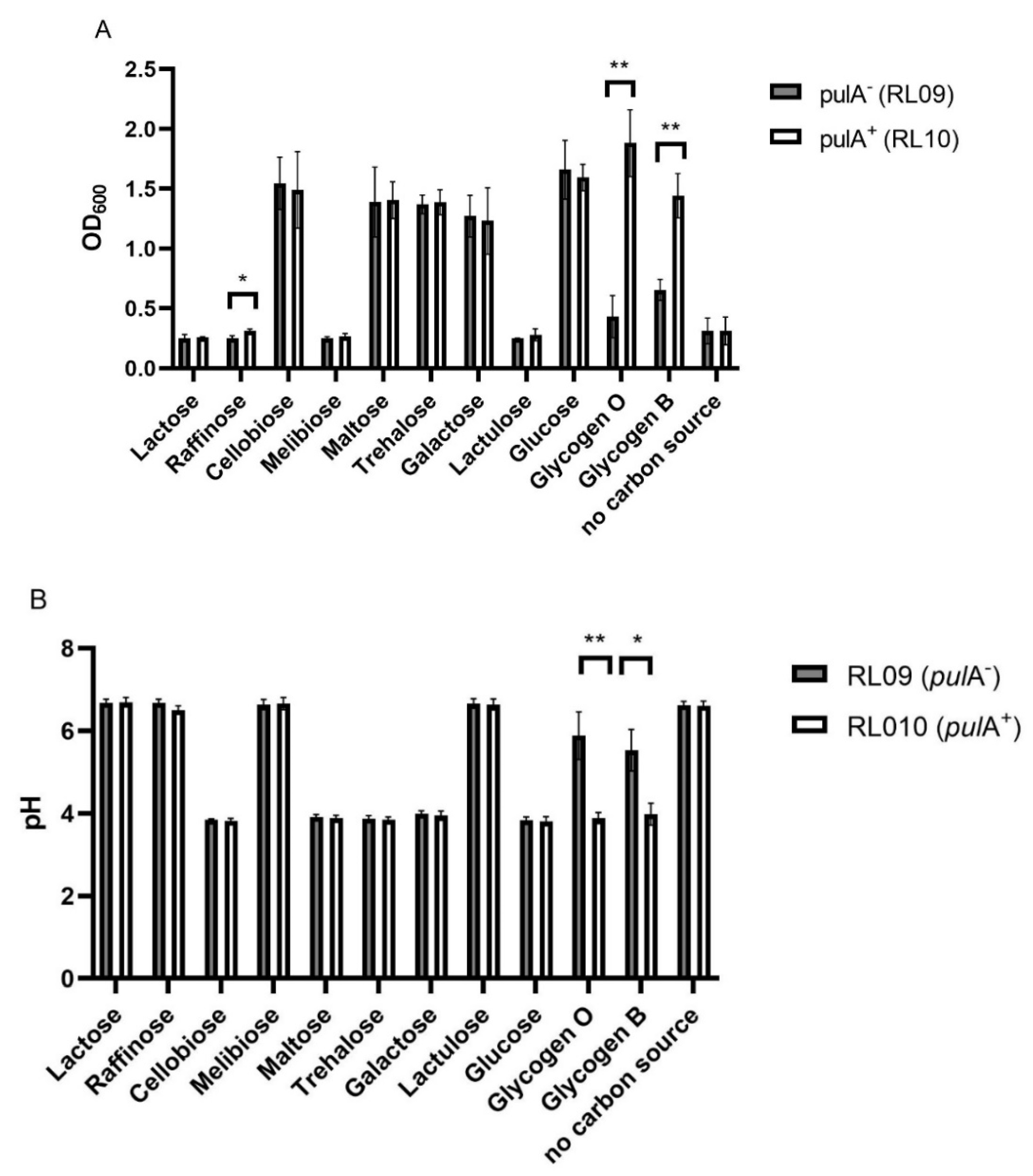
Figure 1. Carbohydrate utilization by L. crispatus strains pulA (RL09) and pulA+ (RL10). (A) Optical density (600 nm), and (B) pH after serial propagation in NYCIII medium supplemented with different carbohydrates. The bars represent the mean of three biological replicates, the standard deviations are shown as error bars. * p < 0.05, ** p < 0.005 (multiple t-test with false discovery rate).
Ijms 23 05590 g001
Figure 2. The pulA locus (4842 bp) in the chromosome of L. crispatus RL10.
Ijms 23 05590 g002
Figure 3. Cell-bound starch degrading activity of L. crispatus pulA sufficient and deficient strains grown on NYC-, glucose, maltose, maltotriose, glycogen (only pulA+) and galactose. Cells were cultured in NYCIII medium supplemented with these carbohydrates and after centrifugation and resuspension of the pellet incubated in a starch solution (7.5 g/L). Asterisks indicate p-values calculated by Mann–Whitney of * p < 0.05, ** p < 0.005, *** p < 0.0005. Hashtag (#) indicates no data due to the absence of growth of this strain on glycogen.
Ijms 23 05590 g003
Figure 4. Small variants in the L. crispatus amylopullulanase gene in the metagenome of vaginal microbial communities. The list and frequency of SNPs and short indels (small variants) identified in the 270 vaginal microbiome samples that showed at least ten times full length coverage for the pulA gene sequence. The variants were classified in terms of their impact on the protein sequence.
Ijms 23 05590 g004
Table 1. Levels of SLAP domain containing proteins in the S-layer enriched fraction of L. crispatus. Columns in the table include ACC NO, UniProt Accession Number; PEP, number of detected peptides; RL09 GAL, expression levels (iBAQ) in L. crispatus pulA strain RL09 cultivated on galactose as only carbon source, RL10 GAL, expression levels in pulA+ strain RL09 cultivated on galactose; RL10 GLU, expression levels in pulA+ strain RL10 cultivated on glucose; RL10 GLY, expression levels in pulA+ strain RL10 cultivated on glycogen; 2LogR1, 2log ratio of expression levels of strains RL10 and RL09 both cultivated on galactose (fold change of expression calculated from LFQ-values from maxquant); 2logR2, 2log ratio of expression levels of pulA+ strain RL10 cultivated on glycogen and glucose; ND, not detected (below the detection limit).
ACC NODescriptionPEPRL09 GALRL10 GALRL10 GLURL10 GLY2logR12logR2
A0A4Q0LMV5Amylopullulanase642.8 × 1052.5 × 1077.5 × 1051.8 × 1076.54.6
A0A135Z5X6Cell surface protein111.5 × 1063.2 ×1061.2 × 1063.2 × 1061.11.4
D5GXS8Lysin (Glycoside Hydrolase Family) 102.9 × 1053.6 × 1054.0 × 1059.7 × 1050.31.3
D5H1Q3Aggregation promoting factor 4NDND1.5 × 1052.2 × 105ND0.6
A0A135ZFI7N-acetylmuramidase171.3 × 1062.0 × 1064.0 × 1064.1 × 1060.60.04
K1MET7Uncharacterized protein 296.4 × 1061.0 × 1077.6 × 1062.3 × 1060.70.04
A0A6M1GK10S-layer protein (Fragment)324.2 × 1056.9 × 1053.0 × 1053.0 × 1050.7−0.02
C7XI68SlpA domain-containing protein143.2 × 1057.3 × 1056.4 × 1056.1 × 1051.2−0.07
A0A6B8SPR2SlpA domain-containing protein153.1 × 1072.2 × 1071.3 × 1071.1 × 107−0.5−0.2
A0A135ZF64Amidase105.7 × 1051.7 × 1061.7 × 1061.4 × 1061.6−0.2
A0A135ZH73Lysin114.5 × 1068.8 × 1066.6 × 1065.6 × 1061.0−0.2
A0A7V8KTK5Putative surface layer protein481.6 × 1072.8 × 1062.7 × 1072.2 × 1070.8−0.3
E3R4X6Uncharacterized protein 92.6 × 1062.8 × 1062.7 × 1062.3 × 1060.1−0.3
K1MJV3Uncharacterized protein 221.6 × 1072.9 × 1072.0 × 1071.6 × 1070.8−0.3
V5EI62Lysin (Glycoside Hydrolase Family)152.4 × 1064.6 × 1061.1 × 1077.6 × 1060.9−0.5
V5EF53SlpA domain-containing protein122.1 × 1073.0 × 1076.1 × 1074.3 × 1070.5−0.5
A0A4Q0LUE2Uncharacterized protein242.7 × 1071.8 × 1073.3 × 1072.3 × 107−0.6−0.5
A0A4R6CSS1Cell surface protein (SlpA)212.4 × 1065.8 × 1066.6 × 1064.6 × 1061.3−0.5
D0DJ09Bacterial surface layer protein453.6 × 1084.8 × 1085.1 × 1083.5 × 1080.4−0.5
K1M186Uncharacterized protein128.0 × 106 9.7 × 1069.5 × 1066.1 × 1060.3−0.6
A0A135YZ62Bacterial surface layer protein (SlpA)322.0 × 1073.3 × 1078.7 × 1075.5 × 1070.8−0.6
D0DIV9Uncharacterized protein365.3 × 1077.6 × 1071.3 × 1087.9 × 1070.5−0.7
V5GA31Lactocepin s-layer protein101.8 × 1062.7 × 1068.4 × 1063.2 × 1060.6−1.4
A0A120DHV3Uncharacterized protein166.0 × 1061.1 × 1072.1 × 1077.7 × 1060.8−1.4
K1N369Uncharacterized protein89.8 × 1043.7 × 1041.4 × 1053.0 × 104−1.4−2.2
A0A125P6L8S-layer protein (SlpA)131.1 × 1041.3 × 1058.7 × 1054.2 × 1033.5−7.7
A0A7T1TNA6Bacterial surface layer protein304.7 × 1065.7 × 1066.1 × 1072.5 × 1050.3−7.9
A0A1C2D5C0Cell surface protein5ND1.5 × 1068.1 × 105NDNDND
Fraction of total detected proteins (iBAQ)0.470.460.220.40
Table 2. Overview of amylopullulanase variants in our strain collection of human vaginal L. crispatus isolates [13].
StrainPulA Variant
RL03, RL08, RL10, RL11, RL13, RL14, RL15, RL16, RL17, RL20, RL21, RL22, RL23, RL24, RL25, RL27, RL28, RL29, RL30, RL33Wild type
RL02 and RL09Two nucleotide deletion
RL06 and RL07N-terminal insertion of mobile element (variant 1)
RL19 and RL26N-terminal insertion of mobile element (variant 2)
RL05Large deletion
Publisher’s Note: MDPI stays neutral with regard to jurisdictional claims in published maps and institutional affiliations.