
Advantages and Limitations of Animal Schizophrenia Models


Abstract
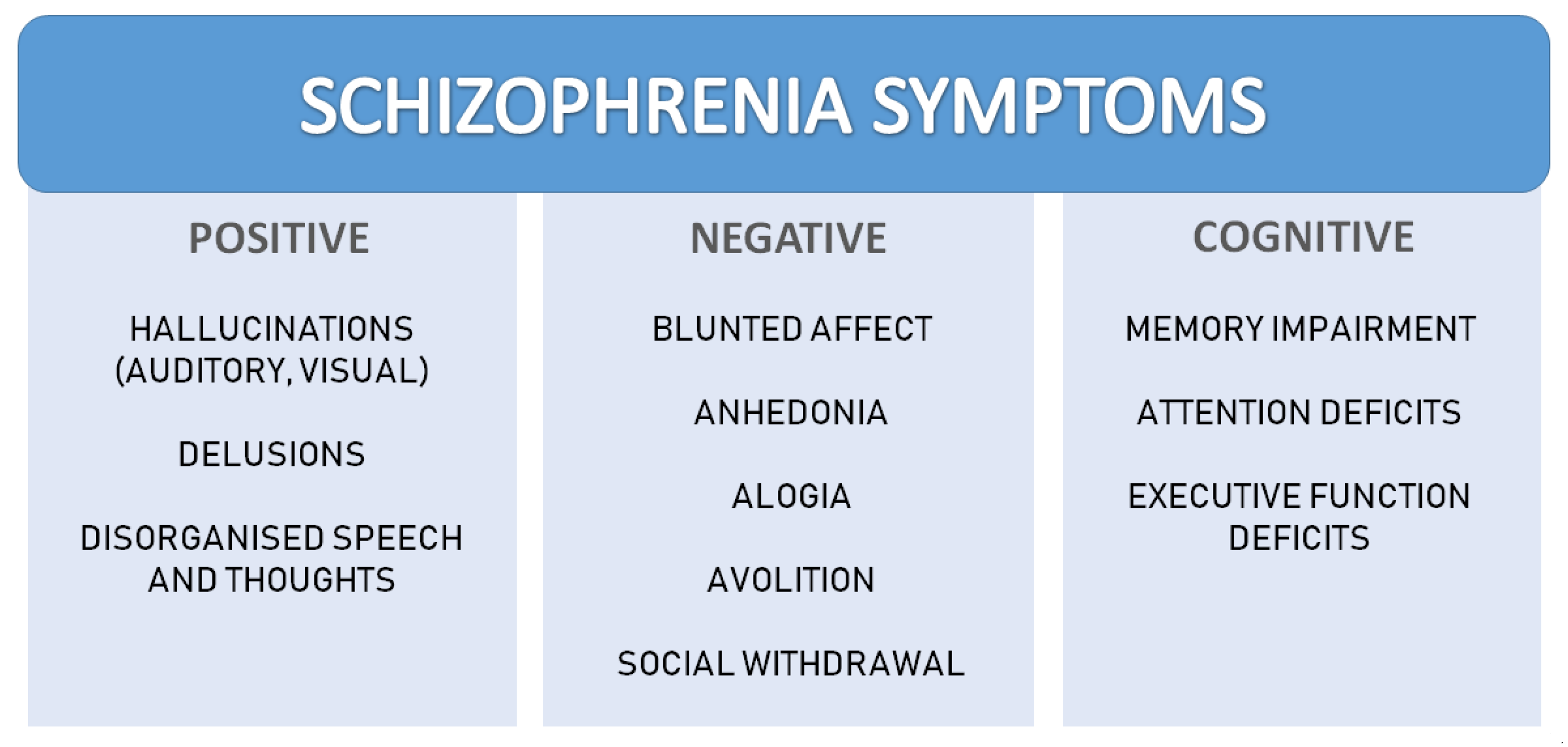
1. Introduction
2. Pharmacological Models
2.1. Dopaminergic Hyperfunction Model
2.2. Dopaminergic Hypofunction Model
2.3. Serotonergic Model
2.4. Glutamatergic Hypofunction Models
3. Genetic Models
3.1. The Disrupted-in-Schizophrenia 1 (DISC1)
3.2. Deletion in the 22q11.2 Region Model
3.3. Dysbindin-1 Model
3.4. Neurotrophic Factor Neuregulin 1 (NRG1) Model
4. Neurodevelopmental Models
4.1. Neonatal Ventral Hippocampal Lesion (NVHL)
4.2. Maternal Immune Activation (MIA)
4.3. Methylazoxymethanol Acetate (MAM) Model
5. Translational Values of the Animal Models
6. Summary
Author Contributions
Funding
Institutional Review Board Statement
Informed Consent Statement
Data Availability Statement
Conflicts of Interest
References
- Tomasik, J.; Rahmoune, H.; Guest, P.C.; Bahn, S. Neuroimmune biomarkers in schizophrenia. Schizophr. Res. 2016, 176, 3–13. [Google Scholar] [CrossRef] [PubMed]
- Keshavan, M.S.; Collin, G.; Guimond, S.; Kelly, S.; Prasad, K.M.; Lizano, P. Neuroimaging in Schizophrenia. Neuroimaging Clin. N. Am. 2020, 30, 73–83. [Google Scholar] [CrossRef] [PubMed]
- Van Os, J.; Kapur, S. Schizophrenia. Lancet 2009, 374, 635–645. [Google Scholar] [CrossRef]
- Queirós, T.; Coelho, F.; Linhares, L.; Telles-Correia, D. Schizophrenia: What Non-Psychiatrist Physicians Need to Know. Acta Med. Port. 2019, 32, 70–77. [Google Scholar] [CrossRef]
- Tandon, R.; Gaebel, W.; Barch, D.M.; Bustillo, J.; Gur, R.E.; Heckers, S.; Malaspina, D.; Owen, M.J.; Schultz, S.; Tsuang, M.; et al. Definition and description of schizophrenia in the DSM-5. Schizophr. Res. 2013, 150, 3–10. [Google Scholar] [CrossRef] [PubMed]
- Lowe, P.; Krivoy, A.; Porffy, L.; Henriksdottir, E.; Eromona, W.; Shergill, S.S. When the drugs don’t work: Treatment-resistant schizophrenia, serotonin and serendipity. Ther. Adv. Psychopharmacol. 2018, 8, 63–70. [Google Scholar] [CrossRef] [PubMed]
- Ali, T.; Sisay, M.; Tariku, M.; Mekuria, A.N.; Desalew, A. Antipsychotic-induced extrapyramidal side effects: A systematic review and meta-analysis of observational studies. PLoS ONE. 2021, 16, e0257129. [Google Scholar] [CrossRef]
- Meltzer, H.Y. Update on typical and atypical antipsychotic drugs. Annu. Rev. Med. 2013, 64, 393–406. [Google Scholar] [CrossRef]
- McCutcheon, R.A.; Abi-Dargham, A.; Howes, O.D. Schizophrenia, Dopamine and the Striatum: From Biology to Symptoms. Trends Neurosci. 2019, 42, 205–220. [Google Scholar] [CrossRef]
- Yang, A.C.; Tsai, S.J. New Targets for Schizophrenia Treatment beyond the Dopamine Hypothesis. Int. J. Mol. Sci. 2017, 18, 1689. [Google Scholar] [CrossRef]
- Nakazawa, K.; Jeevakumar, V.; Nakao, K. Spatial and temporal boundaries of NMDA receptor hypofunction leading to schizophrenia. NPJ Schizophr. 2017, 3, 7. [Google Scholar] [CrossRef] [PubMed]
- Bramnes, J.G.; Rognli, E.B. Psychosis induced by amphetamines. Curr. Opin. Psychiatry 2016, 29, 236–241. [Google Scholar] [CrossRef] [PubMed]
- Featherstone, R.E.; Rizos, Z.; Kapur, S.; Fletcher, P.J. A sensitizing regimen of amphetamine that disrupts attentional set-shifting does not disrupt working or long-term memory. Behav. Brain Res. 2008, 189, 170–179. [Google Scholar] [CrossRef] [PubMed]
- Post, R.; Contel, R. Human and animal studies of cocaine: Implications for development of behavioral pathology. In Stimulants: Neurochemical, Behavioral and Clinical Perspectives; Raven Press: New York, NY, USA, 1983; pp. 169–293. [Google Scholar]
- Ceretta, A.P.C.; Schaffer, L.F.; de Freitas, C.M.; Reinheimer, J.B.; Dotto, M.M.; Fachinetto, R. Gabapentin prevents behavioral changes on the amphetamine-induced animal model of schizophrenia. Schizophr. Res. 2016, 175, 230–231. [Google Scholar] [CrossRef]
- El-Sayed El-Sisi, A.; Sokkar, S.S.; El-Sayed El-Sayad, M.; Sayed Ramadan, E.; Osman, E.Y. Celecoxib and omega-3 fatty acids alone and in combination with risperidone affect the behavior and brain biochemistry in amphetamine-induced model of schizophrenia. Biomed. Pharmacother. 2016, 82, 425–431. [Google Scholar] [CrossRef]
- Tenn, C.C.; Fletcher, P.J.; Kapur, S. Amphetamine-sensitized animals show a sensorimotor gating and neurochemical abnormality similar to that of schizophrenia. Schizophr. Res. 2003, 64, 103–114. [Google Scholar] [CrossRef]
- Tenn, C.C.; Kapur, S.; Fletcher, P.J. Sensitization to amphetamine, but not phencyclidine, disrupts prepulse inhibition and latent inhibition. Psychopharmacology 2005, 180, 366–376. [Google Scholar] [CrossRef]
- Robinson, T.E.; Kolb, B. Persistent structural modifications in nucleus accumbens and prefrontal cortex neurons produced by previous experience with amphetamine. J. Neurosci. 1997, 17, 8491–8497. [Google Scholar] [CrossRef]
- Wolf, M.E. LTP may trigger addiction. Mol. Interv. 2003, 3, 248–252. [Google Scholar] [CrossRef]
- Martinez, V.; Parikh, V.; Sarter, M. Sensitized attentional performance and Fos-immunoreactive cholinergic neurons in the basal forebrain of amphetamine-pretreated rats. Biol. Psychiatry 2005, 57, 1138–1146. [Google Scholar] [CrossRef]
- Featherstone, R.E.; Kapur, S.; Fletcher, P.J. The amphetamine-induced sensitized state as a model of schizophrenia. Prog. Neuropsychopharmacol. Biol. Psychiatry 2007, 31, 1556–1571. [Google Scholar] [CrossRef] [PubMed]
- Banvenutti, R.; Gallas-Lopes, M.; Sachett, A.; Marcon, M.; Strogulski, N.R.; Reis, C.G.; Chitolina, R.; Piato, A.; Herrmann, A.P. How do zebrafish (Danio rerio) respond to MK-801 and amphetamine? Relevance for assessing schizophrenia-related endophenotypes in alternative model organisms. J. Neurosci. Res. 2021, 99, 2844–2859. [Google Scholar] [CrossRef] [PubMed]
- Wearne, T.A.; Mirzaei, M.; Franklin, J.L.; Goodchild, A.K.; Haynes, P.A.; Cornish, J.L. Methamphetamine-induced sensitization is associated with alterations to the proteome of the prefrontal cortex: Implications for the maintenance of psychotic disorders. J. Proteome Res. 2015, 14, 397–410. [Google Scholar] [CrossRef] [PubMed]
- Collo, G.; Mucci, A.; Giordano, G.M.; Merlo Pich, E.; Galderisi, S. Negative Symptoms of Schizophrenia and Dopaminergic Transmission: Translational Models and Perspectives Opened by iPSC Techniques. Front. Neurosci. 2020, 14, 632. [Google Scholar] [CrossRef] [PubMed]
- Salamone, J.D.; Yohn, S.E.; López-Cruz, L.; San Miguel, N.; Correa, M. Activational and effort-related aspects of motivation: Neural mechanisms and implications for psychopathology. Brain 2016, 139, 1325–1347. [Google Scholar] [CrossRef] [PubMed]
- Pycock, C.J.; Kerwin, R.W.; Carter, C.J. Effect of lesion of cortical dopamine terminals on subcortical dopamine receptors in rats. Nature 1980, 286, 74–76. [Google Scholar] [CrossRef]
- Howes, O.D.; Kapur, S. The dopamine hypothesis of schizophrenia: Version III--the final common pathway. Schizophr. Bull. 2009, 35, 549–562. [Google Scholar] [CrossRef]
- Archer, T.; Danysz, W.; Fredriksson, A.; Jonsson, G.; Luthman, J.; Sundström, E.; Teiling, A. Neonatal 6-hydroxydopamine-induced dopamine depletions: Motor activity and performance in maze learning. Pharmacol. Biochem. Behav. 1988, 31, 357–364. [Google Scholar] [CrossRef]
- Fernandez Espejo, E. Prefrontocortical dopamine loss in rats delays long-term extinction of contextual conditioned fear, and reduces social interaction without affecting short-term social interaction memory. Neuropsychopharmacology 2003, 28, 490–498. [Google Scholar] [CrossRef]
- Li, C.R.; Huang, G.B.; Sui, Z.Y.; Han, E.H.; Chung, Y.C. Effects of 6-hydroxydopamine lesioning of the medial prefrontal cortex on social interactions in adolescent and adult rats. Brain Res. 2010, 1346, 183–189. [Google Scholar] [CrossRef]
- Rezvani, A.H.; Eddins, D.; Slade, S.; Hampton, D.S.; Christopher, N.C.; Petro, A.; Horton, K.; Johnson, M.; Levin, E.D. Neonatal 6-hydroxydopamine lesions of the frontal cortex in rats: Persisting effects on locomotor activity, learning and nicotine self-administration. Neuroscience 2008, 154, 885–897. [Google Scholar] [CrossRef] [PubMed]
- Price, A.E.; Sholler, D.J.; Stutz, S.J.; Anastasio, N.C.; Cunningham, K.A. Endogenous Serotonin 5-HT2A and 5-HT2C Receptors Associate in the Medial Prefrontal Cortex. ACS Chem. Neurosci. 2019, 10, 3241–3248. [Google Scholar] [CrossRef] [PubMed]
- Canal, C.E.; Morgan, D. Head-twitch response in rodents induced by the hallucinogen 2,5-dimethoxy-4-iodoamphetamine: A comprehensive history, a re-evaluation of mechanisms, and its utility as a model. Drug Test. Anal. 2012, 4, 556–576. [Google Scholar] [CrossRef] [PubMed]
- Sipes, T.E.; Geyer, M.A. DOI disrupts prepulse inhibition of startle in rats via 5-HT2A receptors in the ventral pallidum. Brain Res. 1997, 761, 97–104. [Google Scholar] [CrossRef]
- Betti, A.H.; Antonio, C.B.; Pompeu, T.E.; Martins, T.S.; Herzfeldt, V.; Stolz, E.D.; Fraga, C.A.; Barreiro, E.; Noël, F.; Rates, S.M. LASSBio-1422: A new molecular scaffold with efficacy in animal models of schizophrenia and disorders of attention and cognition. Behav. Pharmacol. 2017, 28, 48–62. [Google Scholar] [CrossRef] [PubMed]
- Wischhof, L.; Irrsack, E.; Dietz, F.; Koch, M. Maternal lipopolysaccharide treatment differentially affects 5-HT(2A) and mGlu2/3 receptor function in the adult male and female rat offspring. Neuropharmacology 2015, 97, 275–288. [Google Scholar] [CrossRef]
- Sławińska, A.; Wierońska, J.M.; Stachowicz, K.; Marciniakl, M.; Lasoń-Tyburkiewicz, M.; Gruca, P.; Papp, M.; Kusek, M.; Tokarski, K.; Doller, D.; et al. The antipsychotic-like effects of positive allosteric modulators of metabotropic glutamate mGlu4 receptors in rodents. Br. J. Pharmacol. 2013, 169, 1824–1839. [Google Scholar] [CrossRef]
- Cieślik, P.; Woźniak, M.; Tokarski, K.; Kusek, M.; Pilc, A.; Płoska, A.; Radulska, A.; Pelikant-Małecka, I.; Żołnowska, B.; Sławiński, J.; et al. Simultaneous activation of muscarinic and GABAB receptors as a bidirectional target for novel antipsychotics. Behav. Brain Res. 2019, 359, 671–685. [Google Scholar] [CrossRef]
- Griebel, G.; Pichat, P.; Boulay, D.; Naimoli, V.; Potestio, L.; Featherstone, R.; Sahni, S.; Defex, H.; Desvignes, C.; Slowinski, F.; et al. The mGluR2 positive allosteric modulator, SAR218645, improves memory and attention deficits in translational models of cognitive symptoms associated with schizophrenia. Sci. Rep. 2016, 6, 35320. [Google Scholar] [CrossRef]
- Zhang, C.; Marek, G.J. AMPA receptor involvement in 5-hydroxytryptamine2A receptor-mediated pre-frontal cortical excitatory synaptic currents and DOI-induced head shakes. Prog. Neuropsychopharmacol. Biol. Psychiatry. 2008, 32, 62–71. [Google Scholar] [CrossRef]
- Adler, C.M.; Malhotra, A.K.; Elman, I.; Goldberg, T.; Egan, M.; Pickar, D.; Breier, A. Comparison of ketamine-induced thought disorder in healthy volunteers and thought disorder in schizophrenia. Am. J. Pschiatry 1999, 156, 1646–1649. [Google Scholar] [CrossRef] [PubMed]
- Malhotra, A.K.; Pinals, D.A.; Adler, C.M.; Elman, I.; Clifton, A.; Pickar, D.; Breier, A. Ketamine-induced exacerbation of psychotic symptoms and cognitive impairment in neuroleptic-free schizophrenics. Neuropsychopharmacology 1997, 17, 141–150. [Google Scholar] [CrossRef]
- Yavas, E.; Young, A.M.J. Repeated phencyclidine disrupts nicotinic acetylcholine regulation of dopamine release in nucleus accumbens: Implications for models of schizophrenia. Neurochem. Int. 2020, 140, 104836. [Google Scholar] [CrossRef] [PubMed]
- Huang, H.; Zheng, S.; Chen, M.; Xie, L.; Li, Z.; Guo, M.; Wang, J.; Lu, M.; Zhu, X. The potential of the P2X7 receptor as a therapeutic target in a sub-chronic PCP-induced rodent model of schizophrenia. J. Chem. Neuroanatom. 2021, 116, 101993. [Google Scholar] [CrossRef]
- Gigg, J.; McEwan, F.; Smausz, R.; Neill, J.; Harte, M.K. Synaptic biomarker reduction and impaired cognition in the sub-chronic PCP mouse model for schizophrenia. J. Psychopharmacol. 2020, 34, 115–124. [Google Scholar] [CrossRef] [PubMed]
- Seillier, A.; Martinez, A.A.; Giuffrida, A. Differential effects of Δ9-tetrahydrocannabinol dosing on correlates of schizophrenia in the sub-chronic PCP rat model. PLoS ONE 2020, 15, e0230238. [Google Scholar] [CrossRef]
- Rajagopal, L.; Ryan, C.; Elzokaky, A.; Burstein, E.S.; Meltzer, H.Y. Pimavanserin augments the efficacy of atypical antipsychotic drugs in a mouse model of treatment-refractory negative symptoms of schizophrenia. Behav. Brain Res. 2022, 422, 113710. [Google Scholar] [CrossRef]
- Dutra-Tavares, A.C.; Manhães, A.C.; Semeão, K.A.; Maia, J.G.; Couto, L.A.; Filgueiras, C.C.; Ribeiro-Carvalho, A.; Abreu-Villaça, Y. Does nicotine exposure during adolescence modify the course of schizophrenia-like symptoms? Behavioral analysis in a phencyclidine-induced mice model. PLoS ONE 2021, 16, e0257986. [Google Scholar] [CrossRef]
- Tsivion-Visbord, H.; Perets, N.; Sofer, T.; Bikovski, L.; Goldshmit, Y.; Ruban, A.; Offen, D. Mesenchymal stem cells derived extracellular vesicles improve behavioral and biochemical deficits in a phencyclidine model of schizophrenia. Transl. Psychiatry 2020, 10, 327. [Google Scholar] [CrossRef]
- Castañé, A.; Santana, N.; Artigas, F. PCP-based mice models of schizophrenia: Differential behavioral, neurochemical and cellular effects of acute and subchronic treatments. Psychopharmacology 2015, 232, 4085–4097. [Google Scholar] [CrossRef]
- Thomson, D.M.; McVie, A.; Morris, B.J.; Pratt, J.A. Dissociation of acute and chronic intermittent phencyclidine-induced performance deficits in the 5-choice serial reaction time task: Influence of clozapine. Psychopharmacology 2011, 213, 681–695. [Google Scholar] [CrossRef] [PubMed]
- Huang, M.; He, W.; Rajagopal, L.; Kudwa, A.; Grigoriadis, D.E.; Meltzer, H.Y. Effects of NBI-98782, a selective vesicular monoamine transporter 2 (VMAT2) inhibitor, on neurotransmitter efflux and phencyclidine-induced locomotor activity: Relevance to tardive dyskinesia and antipsychotic action. Pharmacol. Biochem. Behav. 2020, 190, 172872. [Google Scholar] [CrossRef] [PubMed]
- Amitai, N.; Kuczenski, R.; Behrens, M.M.; Markou, A. Repeated phencyclidine administration alters glutamate release and decreases GABA markers in the prefrontal cortex of rats. Neuropharmacology 2012, 62, 1422–1431. [Google Scholar] [CrossRef] [PubMed]
- Moghaddam, B.; Adams, B.W. Reversal of phencyclidine effects by a group II metabotropic glutamate receptor agonist in rats. Science 1998, 281, 1349–1352. [Google Scholar] [CrossRef]
- Tanquiero, S.R.; Mouro, F.M.; Ferreira, C.B.; Freitas, C.F.; Fonseca-Gomes, J.; Simões do Couto, F.; Sebastião, A.M.; Dawson, N.; Diógenes, M.J. Sustained NMDA receptor hypofunction impairs brain-derived neurotrophic factor signalling in the PFC, but not in the hippocampus, and disturbs PFC-dependent cognition in mice. J. Psychopharmacol. 2021, 35, 730–743. [Google Scholar] [CrossRef]
- Snighda, S.; Neill, J.C.; McLean, S.L.; Shemar, G.K.; Cruise, L.; Shahid, M.; Henry, B. Phencyclidine (PCP)-induced disruption in cognitive performance is gender-specific and associated with a reduction in brain-derived neurotrophic factor (BDNF) in specific regions of the female rat brain. J. Mol. Neurosci. 2011, 43, 337–345. [Google Scholar] [CrossRef]
- Li, Y.X.; Ye, Z.H.; Chen, T.; Jia, X.F.; He, L. The effects of donepezil on phencyclidine-induced cognitive deficits in a mouse model of schizophrenia. Pharmacol. Biochem. Behav. 2018, 175, 69–76. [Google Scholar] [CrossRef]
- Moghadam, A.A.; Vose, L.R.; Miry, O.; Zhang, X.L.; Stanton, P.K. Pairing of neonatal phencyclidine exposure and acute adolescent stress in male rats as a novel developmental model of schizophrenia. Behav. Brain Res. 2021, 409, 113308. [Google Scholar] [CrossRef]
- Wang, X.; Liu, J.; Dai, Z.; Sui, Y. Andrographolide improves PCP-induced schizophrenia-like behaviors through blocking interaction between NRF2 and KEAP1. J. Pharmacol. Sci. 2021, 147, 9–17. [Google Scholar] [CrossRef]
- Kaiser, S.; Foltz, L.A.; George, C.A.; Kirkwood, S.C.; Bemis, K.G.; Lin, X.; Gelbert, L.M.; Nisenbaum, L.K. Phencyclidine-induced changes in rat cortical gene expression identified by microarray analysis: Implications for schizophrenia. Neurobiol. Dis. 2004, 16, 220–235. [Google Scholar] [CrossRef]
- Cox, D.A.; Gottschalk, M.G.; Wesseling, H.; Ernst, A.; Cooper, J.D.; Bahn, S. Proteomic systems evaluation of the molecular validity of preclinical psychosis models compared to schizophrenia brain pathology. Schizophr. Res. 2016, 177, 98–107. [Google Scholar] [CrossRef] [PubMed]
- Rowland, L.M. Subanesthetic ketamine: How it alters physiology and behavior in humans. Aviat. Space Environ. Med. 2005, 76, C52–C58. [Google Scholar] [PubMed]
- Lahti, A.C.; Weiler, M.; Tamara, M.B.A.; Parwani, A.; Tamminga, C.A. Effects of ketamine in normal and schizophrenic volunteers. Neuropsychopharmacology 2001, 25, 455–467. [Google Scholar] [CrossRef]
- Beck, K.; Hindley, G.; Borgan, F.; Ginestet, C.; McCutcheon, R.; Brugger, S.; Driesen, N.; Ranganathan, M.; D’Souza, D.C.; Taylor, M.; et al. Association of Ketamine with Psychiatric Symptoms and Implications for Its Therapeutic Use and for Understanding Schizophrenia: A Systematic Review and Meta-analysis. JAMA Netw. Open 2020, 3, e204693. [Google Scholar] [CrossRef] [PubMed]
- Riehl, R.; Kyzar, E.; Allain, A.; Green, J.; Hook, M.; Monnig, L.; Rhymes, K.; Roth, A.; Pham, M.; Razavi, R.; et al. Behavioral and physiological effects of acute ketamine exposure in adult zebrafish. Neurotoxicol. Teratol. 2011, 33, 658–667. [Google Scholar] [CrossRef] [PubMed]
- Blackman, R.; MacDonald, A.; Chafee, M. Effects of Ketamine on Context-Processing Performance in Monkeys: A New Animal Model of Cognitive Deficits in Schizophrenia. Neuropsychopharmacology 2013, 38, 2090–2100. [Google Scholar] [CrossRef] [PubMed]
- Frohlich, J.; Van Horn, J.D. Reviewing the ketamine model for schizophrenia. J. Psychopharmacol. 2014, 28, 287–302. [Google Scholar] [CrossRef]
- Ranganathan, M.; DeMartinis, N.; Huguenel, B.; Gaudreault, F.; Bednar, M.M.; Schaffer, C.L.; Gupta, S.; Cahill, J.; Sherif, M.A.; Mancuso, J.; et al. Attenuation of ketamine-induced impairment in verbal learning and memory in healthy volunteers by the AMPA receptor potentiator PF-04958242. Mol. Psychiatry 2017, 22, 1633–1640. [Google Scholar] [CrossRef]
- Roussy, M.; Luna, R.; Duong, L.; Corrigan, B.; Gulli, R.A.; Nogueira, R.; Moreno-Bote, R.; Sachs, A.J.; Palaniyappan, L.; Martinez-Trujillo, J.C. Ketamine disrupts naturalistic coding of working memory in primate lateral prefrontal cortex networks. Mol. Psychiatry 2021, 26, 6688–6703. [Google Scholar] [CrossRef]
- Kozela, E.; Krawczyk, M.; Kos, T.; Juknat, A.; Vogel, Z.; Popik, P. Cannabidiol Improves Cognitive Impairment and Reverses Cortical Transcriptional Changes Induced by Ketamine, in Schizophrenia-Like Model in Rats. Mol. Neurobiol. 2020, 57, 1733–1747. [Google Scholar] [CrossRef]
- Białoń, M.; Żarnowska, M.; Antkiewicz-Michaluk, L.; Wąsik, A. Pro-cognitive effect of 1MeTIQ on recognition memory in the ketamine model of schizophrenia in rats: The behavioural and neurochemical effects. Psychopharmacology 2020, 237, 1577–1593. [Google Scholar] [CrossRef] [PubMed]
- Cao, T.; Tang, M.; Jiang, P.; Zhang, B.; Wu, X.; Chen, Q.; Zeng, C.; Li, N.; Zhang, S.; Cai, H. A Potential Mechanism Underlying the Therapeutic Effects of Progesterone and Allopregnanolone on Ketamine-Induced Cognitive Deficits. Front. Pharmacol. 2021, 12, 612083. [Google Scholar] [CrossRef] [PubMed]
- Ben-Azu, B.; Omogbiya, I.A.; Aderibigbe, A.O.; Umukoro, S.; Ajayi, A.M.; Iwalewa, E.O. Doxycycline prevents and reverses schizophrenic-like behaviors induced by ketamine in mice via modulation of oxidative, nitrergic and cholinergic pathways. Brain Res. Bull. 2018, 139, 114–124. [Google Scholar] [CrossRef] [PubMed]
- Oshodi, T.O.; Ben-Azu, B.; Ishola, I.O.; Ajayi, A.M.; Emokpae, O.; Umukoro, S. Molecular mechanisms involved in the prevention and reversal of ketamine-induced schizophrenia-like behavior by rutin: The role of glutamic acid decarboxylase isoform-67, cholinergic, Nox-2-oxidative stress pathways in mice. Mol. Biol. Rep. 2021, 48, 2335–2350. [Google Scholar] [CrossRef]
- Wąsik, A.; Białoń, M.; Żarnowska, M.; Antkiewicz-Michaluk, L. Comparison of the effects of 1MeTIQ and olanzapine on performance in the elevated plus maze test and monoamine metabolism in the brain after ketamine treatment. Pharmacol. Biochem. Behav. 2019, 181, 17–27. [Google Scholar] [CrossRef]
- Azimi Sanavi, M.; Ghazvini, H.; Zargari, M.; Ghalehnoei, H.; Hosseini-Khah, Z. Effects of clozapine and risperidone antipsychotic drugs on the expression of CACNA1C and behavioral changes in rat ’Ketamine model of schizophrenia. Neurosci. Lett. 2022, 770, 136354. [Google Scholar] [CrossRef]
- Xu, Y.; Deng, C.; Zheng, Y.; Liu, N.; Fu, B. Applying vinpocetine to reverse synaptic ultrastructure by regulating BDNF-related PSD-95 in alleviating schizophrenia-like deficits in rat. Compr. Psychiatry 2019, 94, 152122. [Google Scholar] [CrossRef]
- Zoupa, E.; Gravanis, A.; Pitsikas, N. The novel dehydroepiandrosterone (DHEA) derivative BNN27 counteracts behavioural deficits induced by the NMDA receptor antagonist ketamine in rats. Neuropharmacology 2019, 151, 74–83. [Google Scholar] [CrossRef]
- Sedky, A.A.; Magdy, Y. Reduction in TNF alpha and oxidative stress by liraglutide: Impact on ketamine-induced cognitive dysfunction and hyperlocomotion in rats. Life Sci. 2021, 278, 119523. [Google Scholar] [CrossRef]
- Damazio Pacheco, F.; Canever, L.; Antunes Mastella, G.; Gomes Wessler, P.; Kunz Godoi, A.; Hubbe, I.; da Costa Afonso, A.; Celso, D.; Quevedo, J.; Ioppi Zugno, A. Effects of ketamine on prepubertal Wistar rats: Implications on behavioral parameters for Childhood-Onset Schizophrenia. Int. J. Dev. Neurosci. 2019, 79, 49–53. [Google Scholar] [CrossRef]
- Fujikawa, R.; Yamada, J.; Jinno, S. Subclass imbalance of parvalbumin-expressing GABAergic neurons in the hippocampus of a mouse ketamine model for schizophrenia, with reference to perineuronal nets. Schizophr. Res. 2021, 229, 80–93. [Google Scholar] [CrossRef] [PubMed]
- Becker, A.; Grecksch, G.; Schwegler, H.; Roskoden, T. Expression of mRNA of neurotrophic factors and their receptors are significantly altered after subchronic ketamine treatment. Med. Chem. 2008, 4, 256–263. [Google Scholar] [CrossRef] [PubMed]
- Ben-Azu, B.; Aderibigbe, A.O.; Ajayi, A.M.; Eneni, A.O.; Umukoro, S.; Iwalewa, E.O. Involvement of GABAergic, BDNF and Nox-2 mechanisms in the prevention and reversal of ketamine-induced schizophrenia-like behavior by morin in mice. Brain Res. Bull. 2018, 139, 292–306. [Google Scholar] [CrossRef] [PubMed]
- Canever, L.; Freire, T.G.; Mastella, G.A.; Damázio, L.; Gomes, S.; Fachim, I.; Michels, C.; Carvalho, G.; Godói, A.K.; Peterle, B.R.; et al. Changes in behavioural parameters, oxidative stress and neurotrophins in the brain of adult offspring induced to an animal model of schizophrenia: The effects of FA deficient or FA supplemented diet during the neurodevelopmental phase. Prog. Neuropsychopharmacol. Biol. Psychiatry 2018, 86, 52–64. [Google Scholar] [CrossRef]
- de Araújo, F.Y.R.; Chaves Filho, A.J.M.; Nunes, A.M.; de Oliveira, G.V.; Gomes, P.X.L.; Vasconcelos, G.S.; Carletti, J.; de Moraes, M.O.; de Moraes, M.E.; Vasconcelos, S.M.M.; et al. Involvement of anti-inflammatory, antioxidant, and BDNF up-regulating properties in the antipsychotic-like effect of the essential oil of Alpinia zerumbet in mice: A comparative study with olanzapine. Metab. Brain Dis. 2021, 36, 2283–2297. [Google Scholar] [CrossRef]
- da Silva Araújo, T.; Maia Chaves Filho, A.J.; Monte, A.S.; Isabelle de Góis Queiroz, A.; Cordeiro, R.C.; de Jesus Souza Machado, M.; de Freitas Lima, R.; Freitas de Lucena, D.; Maes, M.; Macêdo, D. Reversal of schizophrenia-like symptoms and immune alterations in mice by immunomodulatory drugs. J. Psychiatr. Res. 2017, 84, 49–58. [Google Scholar] [CrossRef]
- Li, Y.; Shen, R.; Wen, G.; Ding, R.; Du, A.; Zhou, J.; Dong, Z.; Ren, X.; Yao, H.; Zhao, R.; et al. Effects of Ketamine on Levels of Inflammatory Cytokines IL-6, IL-1β, and TNF-α in the Hippocampus of Mice Following Acute or Chronic Administration. Front. Pharmacol. 2017, 8, 139. [Google Scholar] [CrossRef]
- Réus, G.Z.; Simões, L.R.; Colpo, G.D.; Scaini, G.; Oses, J.P.; Generoso, J.S.; Prossin, A.R.; Kaddurah-Daouk, R.; Quevedo, J.; Barichello, T. Ketamine potentiates oxidative stress and influences behavior and inflammation in response to lipolysaccharide (LPS) exposure in early life. Neuroscience 2017, 353, 17–25. [Google Scholar] [CrossRef]
- George, M.Y.; Menze, E.T.; Esmat, A.; Tadros, M.G.; El-Demerdash, E. Potential therapeutic antipsychotic effects of Naringin against ketamine-induced deficits in rats: Involvement of Akt/GSK-3β and Wnt/β-catenin signaling pathways. Life Sci. 2020, 249, 117535. [Google Scholar] [CrossRef]
- Ishola, I.O.; Ben-Azu, B.; Adebayo, O.A.; Ajayi, A.M.; Omorodion, I.L.; Edje, K.E.; Adeyemi, O.O. Prevention and reversal of ketamine-induced experimental psychosis in mice by the neuroactive flavonoid, hesperidin: The role of oxidative and cholinergic mechanisms. Brain Res. Bull. 2021, 177, 239–251. [Google Scholar] [CrossRef]
- Onaolapo, O.J.; Ademakinwa, O.Q.; Olalekan, T.O.; Onaolapo, A.Y. Ketamine-induced behavioural and brain oxidative changes in mice: An assessment of possible beneficial effects of zinc as mono- or adjunct therapy. Psychopharmacology 2017, 234, 2707–2725. [Google Scholar] [CrossRef] [PubMed]
- Keilhoff, G.; Becker, A.; Grecksch, G.; Wolf, G.; Bernstein, H.G. Repeated application of ketamine to rats induces changes in the hippocampal expression of parvalbumin, neuronal nitric oxide synthase and cFOS similar to those found in human schizophrenia. Neuroscience 2004, 126, 591–598. [Google Scholar] [CrossRef] [PubMed]
- Honeycutt, J.A.; Keary Iii, K.M.; Kania, V.M.; Chrobak, J.J. Developmental Age Differentially Mediates the Calcium-Binding Protein Parvalbumin in the Rat: Evidence for a Selective Decrease in Hippocampal Parvalbumin Cell Counts. Dev. Neurosci. 2016, 38, 105–114. [Google Scholar] [CrossRef] [PubMed]
- Pérez, M.Á.; Morales, C.; Santander, O.; García, F.; Gómez, I.; Peñaloza-Sancho, V.; Fuentealba, P.; Dagnino-Subiabre, A.; Moya, P.R.; Fuenzalida, M. Ketamine-Treatment During Late Adolescence Impairs Inhibitory Synaptic Transmission in the Prefrontal Cortex and Working Memory in Adult Rats. Front. Cell. Neurosci. 2019, 13, 372. [Google Scholar] [CrossRef] [PubMed]
- Wallach, J.; Kang, H.; Colestock, T.; Morris, H.; Bortolotto, Z.A.; Collingridge, G.L.; Lodge, D.; Halberstadt, A.L.; Brandt, S.D.; Adejare, A. Pharmacological Investigations of the Dissociative ‘Legal Highs’ Diphenidine, Methoxphenidine and Analogues. PLoS ONE 2016, 11, e0157021. [Google Scholar] [CrossRef] [PubMed]
- Vales, K.; Holubova, K. Minireview: Animal model of schizophrenia from the perspective of behavioral pharmacology: Effect of treatment on cognitive functions. Neurosci. Lett. 2021, 761, 136098. [Google Scholar] [CrossRef] [PubMed]
- Chen, G.; Lin, X.; Li, G.; Jiang, D.; Lib, Z.; Jiang, R.; Zhuo, C. Risperidone reverses the spatial object recognition impairment and hippocampal BDNF-TrkB signalling system alterations induced by acute MK-801 treatment. Biomed. Rep. 2017, 6, 285–290. [Google Scholar] [CrossRef]
- Langova, V.; Vales, K.; Horka, P.; Horacek, J. The Role of Zebrafish and Laboratory Rodents in Schizophrenia Research. Front. Psychiatry 2020, 11, 703. [Google Scholar] [CrossRef]
- Swain, H.A.; Sigstad, C.; Scalzo, F.M. Effects of dizocilpine (MK-801) on circling behavior, swimming activity, and place preference in zebrafish (Danio rerio). Neurotoxicol. Teratol. 2004, 26, 725–729. [Google Scholar] [CrossRef] [PubMed]
- Oliveira, A.W.C.; Pacheco, J.V.N.; Costa, C.S.; Aquino, J.; Maior, R.S.; Barros, M. Scopolamine and MK-801 impair recognition memory in a new spontaneous object exploration task in monkeys. Pharmacol. Biochem. Behav. 2021, 211, 173300. [Google Scholar] [CrossRef]
- Sawahata, M.; Asano, H.; Nagai, T.; Ito, N.; Kohno, T.; Nabeshima, T.; Hattori, M.; Yamada, K. Microinjection of Reelin into the mPFC prevents MK-801-induced recognition memory impairment in mice. Pharmacol. Res. 2021, 173, 105832. [Google Scholar] [CrossRef] [PubMed]
- Xiao, X.; Xu, X.; Li, F.; Xie, G.; Zhang, T. Anti-inflammatory treatment with β-asarone improves impairments in social interaction and cognition in MK-801 treated mice. Brain Res. Bull. 2019, 150, 150–159. [Google Scholar] [CrossRef] [PubMed]
- Kraeuter, A.K.; Mashavave, T.; Suvarna, A.; van den Buuse, M.; Sarnyai, Z. Effects of beta-hydroxybutyrate administration on MK-801-induced schizophrenia-like behaviour in mice. Psychopharmacology 2020, 237, 1397–1405. [Google Scholar] [CrossRef] [PubMed]
- Perdikaris, P.; Dermon, C.R. Behavioral and neurochemical profile of MK-801 adult zebrafish model: Forebrain β2-adrenoceptors contribute to social withdrawal and anxiety-like behavior. Prog. Neuropsychopharmacol. Biol. Psychiatry 2022, 115, 110494. [Google Scholar] [CrossRef]
- Białoń, M.; Chocyk, A.; Majcher-Maślanka, I.; Żarnowska, M.; Michalski, K.; Antkiewicz-Michaluk, L.; Wąsik, A. 1MeTIQ and olanzapine, despite their neurochemical impact, did not ameliorate performance in fear conditioning and social interaction tests in an MK-801 rat model of schizophrenia. Pharmacol. Rep. 2021, 73, 490–505. [Google Scholar] [CrossRef]
- Zhan, J.Q.; Chen, C.N.; Wu, S.X.; Wu, H.J.; Zou, K.; Xiong, J.W.; Wei, B.; Yang, Y.J. Flavonoid fisetin reverses impaired hippocampal synaptic plasticity and cognitive function by regulating the function of AMPARs in a male rat model of schizophrenia. J. Neurochem. 2021, 158, 413–428. [Google Scholar] [CrossRef]
- Akosman, M.S.; Türkmen, R.; Demirel, H.H. Investigation of the protective effect of resveratrol in an MK-801-induced mouse model of schizophrenia. Environ. Sci. Pollut. Res. Int. 2021, 28, 65872–65884. [Google Scholar] [CrossRef]
- Pınar, N.; Akillioglu, K.; Sefil, F.; Alp, H.; Sagir, M.; Acet, A. Effect of clozapine on locomotor activity and anxiety-related behavior in the neonatal mice administered MK-801. Bosn. J. Basic Med. Sci. 2015, 15, 74–79. [Google Scholar] [CrossRef]
- Güneri, S.; Scheel-Krüger, J.; Luo, F. Evaluation of acute and chronic nociception in subchronically administered MK-801-induced rat model of schizophrenia. Behav. Pharmacol. 2021, 32, 571–580. [Google Scholar] [CrossRef]
- Stubbs, B.; Thompson, T.; Acaster, S.; Vancampfort, D.; Gaughran, F.; Correll, C.U. Decreased pain sensitivity among people with schizophrenia: A meta-analysis of experimental pain induction studies. Pain 2015, 156, 2121–2131. [Google Scholar] [CrossRef]
- Yan, Q.S.; Reith, M.E.; Jobe, P.C.; Dailey, J.W. Dizocilpine (MK-801) increases not only dopamine but also serotonin and norepinephrine transmissions in the nucleus accumbens as measured by microdialysis in freely moving rats. Brain Res. 1997, 765, 149–158. [Google Scholar] [CrossRef]
- Sitges, M.; Nekrassov, V.; Guarneros, A. Simultaneous action of MK-801 (dizclopine) on dopamine, glutamate, aspartate and GABA release from striatum isolated nerve endings. Brain Res. 2000, 854, 48–56. [Google Scholar] [CrossRef]
- Wedzony, K.; Gołembiowska, K.; Zazula, M. Differential effects of CGP 37849 and MK-801, competitive and noncompetitive NMDA antagonists, with respect to the modulation of sensorimotor gating and dopamine outflow in the prefrontal cortex of rats. Naunyn Schmiedebergs Arch. Pharmacol. 1994, 350, 555–562. [Google Scholar] [CrossRef] [PubMed]
- Schmidt, C.J.; Fadayel, G.M. Regional effects of MK-801 on dopamine release: Effects of competitive NMDA or 5-HT2A receptor blockade. J. Pharmacol. Exp. Ther. 1996, 277, 1541–1549. [Google Scholar]
- Brisch, R.; Saniotis, A.; Wolf, R.; Bielau, H.; Bernstein, H.G.; Steiner, J.; Bogerts, B.; Braun, K.; Jankowski, Z.; Kumaratilake, J.; et al. The role of dopamine in schizophrenia from a neurobiological and evolutionary perspective: Old fashioned, but still in vogue. Front. Psychiatry 2014, 19, 47. [Google Scholar] [CrossRef]
- Pietraszek, M.; Michaluk, J.; Romańska, I.; Wasik, A.; Gołembiowska, K.; Antkiewicz-Michaluk, L. 1-Methyl-1,2,3,4-tetrahydroisoquinoline antagonizes a rise in brain dopamine metabolism, glutamate release in frontal cortex and locomotor hyperactivity produced by MK-801 but not the disruptions of prepulse inhibition, and impairment of working memory in rat. Neurotox. Res. 2009, 16, 390–407. [Google Scholar] [CrossRef]
- López-Gil, X.; Artigas, F.; Adell, A. Role of different monoamine receptors controlling MK-801-induced release of serotonin and glutamate in the medial prefrontal cortex: Relevance for antipsychotic action. Int. J. Neuropsychopharmacol. 2009, 12, 487–499. [Google Scholar] [CrossRef]
- Wąsik, A.; Białoń, M.; Jantas, D.; Żarnowska, M. The Impact of the Combined Administration of 1MeTIQ and MK-801 on Cell Viability, Oxidative Stress Markers, and Glutamate Release in the Rat Hippocampus. Neurotox. Res. 2021, 39, 1747–1761. [Google Scholar] [CrossRef]
- Moghaddam, B.; Javitt, D. From revolution to evolution: The glutamate hypothesis of schizophrenia and its implication for treatment. Neuropsychopharmacology 2012, 37, 4–15. [Google Scholar] [CrossRef]
- Wang, X.; Hu, Y.; Liu, W.; Ma, Y.; Chen, X.; Xue, T.; Cui, D. Molecular Basis of GABA Hypofunction in Adolescent Schizophrenia-Like Animals. Neural. Plast. 2021, 2021, 9983438. [Google Scholar] [CrossRef]
- Woźniak, M.; Cieślik, P.; Marciniak, M.; Lenda, T.; Pilc, A.; Wieronska, J.M. Neurochemical changes underlying schizophrenia-related behavior in a modified forced swim test in mice. Pharmacol. Biochem. Behav. 2018, 172, 50–58. [Google Scholar] [CrossRef] [PubMed]
- Flores-Barrera, E.; Thomases, D.R.; Tseng, K.Y. MK-801 Exposure during Adolescence Elicits Enduring Disruption of Prefrontal E-I Balance and Its Control of Fear Extinction Behavior. J. Neurosci. 2020, 40, 4881–4887. [Google Scholar] [CrossRef] [PubMed]
- Ma, Y.N.; Sun, Y.X.; Wang, T.; Wang, H.; Zhang, Y.; Su, Y.A.; Li, J.T.; Si, T.M. Subchronic MK-801 treatment during adolescence induces long-term, not permanent, excitatory-inhibitory imbalance in the rat hippocampus. Eur. J. Pharmacol. 2020, 15, 172807. [Google Scholar] [CrossRef] [PubMed]
- Gottschalk, W.A.; Jiang, H.; Tartaglia, N.; Feng, L.; Figurov, A.; Lu, B. Signaling mechanisms mediating BDNF modulation of synaptic plasticity in the hippocampus. Learn. Mem. 1999, 6, 243–256. [Google Scholar] [CrossRef] [PubMed]
- Lu, B.; Martinowich, K. Cell biology of BDNF and its relevance to schizophrenia. Novartis Found. Symp. 2008, 289, 119–129. [Google Scholar] [CrossRef]
- Kim, T.; Kang, H.; Park, J.; Lee, S.; Baek, S.; Kim, C. Voluntary wheel running ameliorates symptoms of MK-801-induced schizophrenia in mice. Mol. Med. Rep. 2014, 10, 2924–2930. [Google Scholar] [CrossRef] [PubMed][Green Version]
- Yu, W.; Zhu, M.; Fang, H.; Zhou, J.; Ye, L.; Bian, W.; Wang, Y.; Zhu, H.; Xiao, J.; Zhu, H.; et al. Risperidone Reverses the Downregulation of BDNF in Hippocampal Neurons and MK801-Induced Cognitive Impairment in Rats. Front. Behav.Neurosci. 2019, 23, 163. [Google Scholar] [CrossRef]
- Yu, W.; Fang, H.; Zhang, L.; Hu, M.; He, S.; Li, H.; Zhu, H. Reversible Changes in BDNF Expression in MK-801-Induced Hippocampal Astrocytes Through NMDAR/PI3K/ERK Signaling. Front. Cell. Neurosci. 2021, 15, 672136. [Google Scholar] [CrossRef]
- Linden, A.M.; Vaisanen, J.; Lakso, M.; Nawa, H.; Wong, G.; Castren, E. Expression of neurotrophins BDNF and NT-3, and their receptors, in rat brain after administration of antipsychotic and psychotrophic agents. J. Mol. Neurosci. 2000, 14, 27–37. [Google Scholar] [CrossRef]
- Lau, A.; Tymianski, M. Glutamate receptors, neurotoxicity and neurodegeneration. Pflugers Arch. 2010, 460, 525–542. [Google Scholar] [CrossRef]
- Moghaddam, B.; Adams, B.; Verma, A.; Daly, D. Activation of glutamatergic neurotransmission by ketamine. A novel step in the pathway from NMDA receptor blockade to dopaminergic and cognitive disruptions associated with the prefrontal cortex. J. Neurosci. 1997, 17, 2921–2927. [Google Scholar] [CrossRef] [PubMed]
- Pose-Utrilla, J.; García-Guerra, L.; Del Puerto, A.; Martín, A.; Jurado-Arjona, J.; De León-Reyes, N.S.; Gamir-Morralla, A.; Sebastián-Serrano, Á.; García-Gallo, M.; Kremer, L. Excitotoxic inactivation of constitutive oxidative stress detoxification pathway in neurons can be rescued by PKD1. Nat. Commun. 2017, 8, 2275. [Google Scholar] [CrossRef] [PubMed]
- Flatow, J.; Buckley, P.; Miller, B.J. Meta-analysis of oxidative stress in schizophrenia. Biol. Psychiatry 2013, 74, 400–409. [Google Scholar] [CrossRef] [PubMed]
- Murray, A.J.; Rogers, J.C.; Katshu, M.Z.U.H.; Liddle, P.F.; Upthegrove, R. Oxidative Stress and the Pathophysiology and Symptom Profile of Schizophrenia Spectrum Disorders. Front. Psychiatry 2021, 22, 703452. [Google Scholar] [CrossRef] [PubMed]
- Ozyurt, B.; Ozyurt, H.; Akpolat, N.; Erdogan, H.; Sarsilmaz, M. Oxidative stress in prefrontal cortex of rat exposed to MK-801 and protective effects of CAPE. Prog. Neuropsychopharmacol. Biol. Psychiatry 2007, 31, 832–838. [Google Scholar] [CrossRef]
- Niu, J.; Cao, Y.; Ji, Y. Resveratrol, a SIRT1 Activator, Ameliorates MK-801-Induced Cognitive and Motor Impairments in a Neonatal Rat Model of Schizophrenia. Front. Psychiatry 2020, 11, 716. [Google Scholar] [CrossRef]
- Genius, J.; Geiger, J.; Dölzer, A.L.; Benninghoff, J.; Giegling, I.; Hartmann, A.M.; Möller, H.J.; Rujescu, D. Glutamatergic dysbalance and oxidative stress in in vivo and in vitro models of psychosis based on chronic NMDA receptor antagonism. PLoS ONE 2013, 15, e59395. [Google Scholar] [CrossRef]
- Cassoli, J.S.; Iwata, K.; Steiner, J.; Guest, P.C.; Turck, C.W.; Nascimento, J.M.; Martins-de-Souza, D. Effect of MK-801 and Clozapine on the Proteome of Cultured Human Oligodendrocytes. Front. Cell. Neurosci. 2016, 3, 52. [Google Scholar] [CrossRef]
- Osorio-Rico, L.; Villeda-Hernández, J.; Santamaría, A.; Königsberg, M.; Galván-Arzate, S. The N-Methyl-d-Aspartate Receptor Antagonist MK-801 Prevents Thallium-Induced Behavioral and Biochemical Alterations in the Rat Brain. Int. J. Toxicol. 2015, 34, 505–513. [Google Scholar] [CrossRef]
- Baxter, P.; Bell, K.; Hasel, P.; Kaindl, A.M.; Fricker, M.; Thomson, D.; Cregan, S.P.; Gillingwater, T.H.; Hardingham, G.E. Synaptic NMDA receptor activity is coupled to the transcriptional control of the glutathione system. Nat. Commun. 2015, 6, 6761. [Google Scholar] [CrossRef]
- Rung, J.P.; Carlsson, A.; Rydén Markinhuhta, K.; Carlsson, M.L. (+)-MK-801 induced social withdrawal in rats; a model for negative symptoms of schizophrenia. Prog. Neuropsychopharmacol. Biol. Psychiatry 2005, 29, 827–832. [Google Scholar] [CrossRef] [PubMed]
- Hafner, H.; Maurer, K.; Loffler, W.; Riecher, R.A. The influence of age and sex on the onset and early course of schizophrenia. Br. J. Psychiatry 1993, 162, 80–86. [Google Scholar] [CrossRef] [PubMed]
- Ellenbroek, B.A.; Cools, A.R. Animal models for the negative symptoms of schizophrenia. Behav. Pharmacol. 2000, 11, 223–233. [Google Scholar] [CrossRef]
- Kovacic, P.; Somanathan, R. Clinical physiology and mechanism of dizocilpine (MK-801): Electron transfer, radicals, redox metabolites and bioactivity. Oxid. Med. Cell. Longev. 2010, 3, 13–22. [Google Scholar] [CrossRef] [PubMed]
- Eyjolfsson, E.M.; Brenner, E.; Kondziella, D.; Sonnewald, U. Repeated injection of MK801: An animal model of schizophrenia? Neurochem. Int. 2006, 48, 541–546. [Google Scholar] [CrossRef] [PubMed]
- Wang, J.; Li, S.; Li, X.; Liu, J.; Yang, J.; Li, Y.; Li, W.; Yang, Y.; Li, J.; Chen, R.; et al. Functional variant rs2270363 on 16p13.3 confers schizophrenia risk by regulating NMRAL1. Brain 2022, awac020. [Google Scholar] [CrossRef] [PubMed]
- Tripathi, A.; Kar, S.K.; Shukla, R. Cognitive Deficits in Schizophrenia: Understanding the Biological Correlates and Remediation Strategies. Clin. Psychopharmacol. Neurosci. 2018, 16, 7–17. [Google Scholar] [CrossRef]
- Faulkner, R.L.; Jang, M.H.; Liu, X.B.; Duan, X.; Sailor, K.A.; Kim, J.Y.; Ge, S.; Jones, E.G.; Ming, G.L.; Song, H.; et al. Development of hippocampal mossy fiber synaptic outputs by new neurons in the adult brain. Proc. Natl. Acad. Sci. USA 2008, 105, 14157–14162. [Google Scholar] [CrossRef]
- Morosawa, S.; Iritani, S.; Fujishiro, H.; Sekiguchi, H.; Torii, Y.; Habuchi, C.; Kuroda, K.; Kaibuchi, K.; Ozaki, N. Neuropeptide Y neuronal network dysfunction in the frontal lobe of a genetic mouse model of schizophrenia. Neuropeptides 2017, 62, 27–35. [Google Scholar] [CrossRef]
- Lee, F.H.; Fadel, M.P.; Preston-Maher, K.; Cordes, S.P.; Clapcote, S.J.; Price, D.J.; Roder, J.C.; Wong, A.H. Disc1 point mutations in mice affect development of the cerebral cortex. J. Neurosci. 2011, 31, 3197–3206. [Google Scholar] [CrossRef]
- Liu, C.M.; Liu, Y.L.; Hwu, H.G.; Fann, C.S.; Yang, U.C.; Hsu, P.C.; Chang, C.C.; Chen, W.J.; Hwang, T.J.; Hsieh, M.H.; et al. Genetic associations and expression of extra-short isoforms of disrupted-in-schizophrenia 1 in a neurocognitive subgroup of schizophrenia. J. Hum. Genet. 2019, 64, 653–663. [Google Scholar] [CrossRef] [PubMed]
- Sullivan, P.F. Questions about DISC1 as a genetic risk factor for schizophrenia. Mol. Psychiatry 2013, 18, 1050–1052. [Google Scholar] [CrossRef] [PubMed]
- Segal-Gavish, H.; Gazit, N.; Barhum, Y.; Ben-Zur, T.; Taler, M.; Hornfeld, S.H.; Gil-Ad, I.; Weizman, A.; Slutsky, I.; Niwa, M.; et al. BDNF overexpression prevents cognitive deficit elicited by adolescent cannabis exposure and host susceptibility interaction. Hum. Mol. Genet. 2017, 26, 2462–2471. [Google Scholar] [CrossRef] [PubMed]
- Kaminitz, A.; Barzilay, R.; Segal, H.; Taler, M.; Offen, D.; Gil-Ad, I.; Mechoulam, R.; Weizman, A. Dominant negative DISC1 mutant mice display specific social behaviour deficits and aberration in BDNF and cannabinoid receptor expression. World, J. Biol. Psychiatry 2014, 15, 76–82. [Google Scholar] [CrossRef]
- Umeda, K.; Iritani, S.; Fujishiro, H.; Sekiguchi, H.; Torii, Y.; Habuchi, C.; Kuroda, K.; Kaibuchi, K.; Ozaki, N. Immunohistochemical evaluation of the GABAergic neuronal system in the prefrontal cortex of a DISC1 knockout mouse model of schizophrenia. Synapse 2016, 70, 508–518. [Google Scholar] [CrossRef]
- Niwa, M.; Kamiya, A.; Murai, R.; Kubo, K.; Gruber, A.J.; Tomita, K.; Lu, L.; Tomisato, S.; Jaaro-Peled, H.; Seshadri, S. Knockdown of DISC1 by in utero gene transfer disturbs postnatal dopaminergic maturation in the frontal cortex and leads to adult behavioral deficits. Neuron 2010, 65, 480–489. [Google Scholar] [CrossRef]
- Karayiorgou, M.; Simon, T.J.; Gogos, J.A. 22q11.2 microdeletions: Linking DNA structural variation to brain dysfunction and schizophrenia. Nat. Rev. Neurosci. 2010, 11, 402–416. [Google Scholar] [CrossRef]
- Ellegood, J.; Markx, S.; Lerch, J.P.; Steadman, P.E.; Genç, C.; Provenzano, F.; Kushner, S.A.; Henkelman, R.M.; Karayiorgou, M.; Gogos, J.A. Neuroanatomical phenotypes in a mouse model of the 22q11.2 microdeletion. Mol. Psychiatry 2014, 19, 99–107. [Google Scholar] [CrossRef]
- Tang, S.X.; Yi, J.J.; Calkins, M.E.; Whinna, D.A.; Kohler, C.G.; Souders, M.C.; McDonald-McGinn, D.M.; Zackai, E.H.; Emanuel, B.S.; Gur, R.C.; et al. Psychiatric disorders in 22q11.2 deletion syndrome are prevalent but undertreated. Psychol. Med. 2014, 44, 1267–1277. [Google Scholar] [CrossRef]
- Qin, X.; Chen, J.; Zhou, T. 22q11.2 deletion syndrome and schizophrenia. Acta. Biochim. Biophys. Sin. 2020, 52, 1181–1190. [Google Scholar] [CrossRef]
- Sun, Z.; Williams, D.J.; Xu, B.; Gogos, J.A. Altered function and maturation of primary cortical neurons from a 22q11.2 deletion mouse model of schizophrenia. Transl. Psychiatry 2018, 8, 85. [Google Scholar] [CrossRef] [PubMed]
- Mukai, J.; Tamura, M.; Fénelon, K.; Rosen, A.M.; Spellman, T.J.; Kang, R.; MacDermott, A.B.; Karayiorgou, M.; Gordon, J.A.; Gogos, J.A. Molecular substrates of altered axonal growth and brain connectivity in a mouse model of schizophrenia. Neuron 2015, 86, 680–695. [Google Scholar] [CrossRef] [PubMed]
- Piskorowski, R.A.; Nasrallah, K.; Diamantopoulou, A.; Mukai, J.; Hassan, S.I.; Siegelbaum, S.A.; Gogos, J.A. Chevaleyre VAge-Dependent Specific Changes in Area CA2 of the Hippocampus and Social Memory Deficit in a Mouse Model of the 22q11.2 Deletion Syndrome. Neuron 2016, 89, 163–176. [Google Scholar] [CrossRef] [PubMed]
- Diamantopoulou, A.; Sun, Z.; Mukai, J.; Xu, B.; Fenelon, K.; Karayiorgou, M.; Gogos, J.A. Loss-of-function mutation in Mirta22/Emc10 rescues specific schizophrenia-related phenotypes in a mouse model of the 22q11.2 deletion. Proc. Natl. Acad. Sci. USA 2017, 114, E6127–E6136. [Google Scholar] [CrossRef] [PubMed]
- Meechan, D.W.; Maynard, T.M.; Tucker, E.S.; Fernandez, A.; Karpinski, B.A.; Rothblat, L.A.; LaMantia, A.S. Modeling a model: Mouse genetics, 22q11.2 Deletion Syndrome, and disorders of cortical circuit development. Prog. Neurobiol. 2015, 130, 1–28. [Google Scholar] [CrossRef]
- Jiang, Y.; Patton, M.H.; Zakharenko, S.S. A Case for Thalamic Mechanisms of Schizophrenia: Perspective from Modeling 22q11.2 Deletion Syndrome. Front. Neural. Circuits 2021, 15, 769969. [Google Scholar] [CrossRef]
- Didriksen, M.; Fejgin, K.; Nilsson, S.R.; Birknow, M.R.; Grayton, H.M.; Larsen, P.H.; Lauridsen, J.B.; Nielsen, V.; Celada, P.; Santana, N.; et al. Persistent gating deficit and increased sensitivity to NMDA receptor antagonism after puberty in a new mouse model of the human 22q11.2 microdeletion syndrome: A study in male mice. J. Psychiatry Neurosci. 2017, 42, 48–58. [Google Scholar] [CrossRef]
- Forsingdal, A.; Jørgensen, T.N.; Olsen, L.; Werge, T.; Didriksen, M.; Nielsen, J. Can Animal Models of Copy Number Variants That Predispose to Schizophrenia Elucidate Underlying Biology? Biol. Psychiatry 2019, 85, 13–24. [Google Scholar] [CrossRef]
- Ma, X.; Fei, E.; Fu, C.; Ren, H.; Wang, G. Dysbindin-1, a schizophrenia-related protein, facilitates neurite outgrowth by promoting the transcriptional activity of p53. Mol. Psychiatry 2011, 16, 1105–1116. [Google Scholar] [CrossRef]
- Mullin, A.P.; Sadanandappa, M.K.; Ma, W.; Dickman, D.K.; VijayRaghavan, K.; Ramaswami, M.; Sanyal, S.; Faundez, V. Gene dosage in the dysbindin schizophrenia susceptibility network differentially affect synaptic function and plasticity. J. Neurosci. 2015, 35, 325–338. [Google Scholar] [CrossRef]
- Trantham-Davidson, H.; Lavin, A. Loss of dysbindin-1 affects GABAergic transmission in the PFC. Psychopharmacology 2019, 236, 3291–3300. [Google Scholar] [CrossRef] [PubMed]
- Wolf, C.; Jackson, M.C.; Kissling, C.; Thome, J.; Linden, D.E. Dysbindin-1 genotype effects on emotional working memory. Mol. Psychiatry 2011, 16, 145–155. [Google Scholar] [CrossRef] [PubMed]
- Scheggia, D.; Mastrogiacomo, R.; Mereu, M.; Sannino, S.; Straub, R.E.; Armando, M.; Managò, F.; Guadagna, S.; Piras, F.; Zhang, F.; et al. Variations in Dysbindin-1 are associated with cognitive response to antipsychotic drug treatment. Nat. Commun. 2018, 9, 2265. [Google Scholar] [CrossRef] [PubMed]
- Cox, M.M.; Tucker, A.M.; Tang, J.; Talbot, K.; Richer, D.C.; Yeh, L.; Arnold, S.E. Neurobehavioral abnormalities in the dysbindin-1 mutant, sandy, on a C57BL/6J genetic background. Genes Brain Behav. 2009, 8, 390–397. [Google Scholar] [CrossRef]
- Papaleo, F.; Yang, F.; Garcia, S.; Chen, J.; Lu, B.; Crawley, J.N.; Weinberger, D.R. Dysbindin-1 modulates prefrontal cortical activity and schizophrenia-like behaviors via dopamine/D2 pathways. Mol. Psychiatry 2012, 17, 85–98. [Google Scholar] [CrossRef] [PubMed]
- Karlsgodt, K.H.; Robleto, K.; Trantham-Davidson, H.; Jairl, C.; Cannon, T.D.; Lavin, A.; Jentsch, J.D. Reduced dysbindin expression mediates N-methyl-D-aspartate receptor hypofunction and impaired working memory performance. Biol. Psychiatry 2011, 69, 28–34. [Google Scholar] [CrossRef]
- Ji, Y.; Yang, F.; Papaleo, F.; Gao, W.J.; Weinberger, D.R.; Lu, B. Role of dysbindin in dopamine receptor trafficking and cortical GABA function. Proc. Natl. Acad. Sci. USA 2009, 106, 19593–19598. [Google Scholar] [CrossRef]
- Murotani, T.; Ishizuka, T.; Hattori, S.; Hashimoto, R.; Matsuzaki, S.; Yamatodani, A. High dopamine turnover in the brains of Sandy mice. Neurosci. Lett. 2007, 421, 47–51. [Google Scholar] [CrossRef]
- Iizuka, Y.; Sei, Y.; Weinberger, D.R.; Straub, R.E. Evidence that the BLOC-1 protein dysbindin modulates dopamine D2 receptor internalization and signaling but not D1 internalization. J. Neurosci. 2007, 27, 12390–12395. [Google Scholar] [CrossRef]
- Talbot, K. The sandy (sdy) mouse: A dysbindin-1 mutant relevant to schizophrenia research. Prog. Brain Res. 2009, 179, 87–94. [Google Scholar] [CrossRef]
- Bhardwaj, S.K.; Baharnoori, M.; Sharif-Askari, B.; Kamath, A.; Williams, S.; Srivastava, L.K. Behavioral characterization of dysbindin-1 deficient sandy mice. Behav. Brain Res. 2009, 197, 435–441. [Google Scholar] [CrossRef] [PubMed]
- Hattori, S.; Murotani, T.; Matsuzaki, S.; Ishizuka, T.; Kumamoto, N.; Takeda, M.; Tohyama, M.; Yamatodani, A.; Kunugi, H.; Hashimoto, R. Behavioral abnormalities and dopamine reductions in sdy mutant mice with a deletion in Dtnbp1, a susceptibility gene for schizophrenia. Biochem. Biophys. Res. Commun. 2008, 373, 298–302. [Google Scholar] [CrossRef] [PubMed]
- Feng, Y.Q.; Zhou, Z.Y.; He, X.; Wang, H.; Guo, X.L.; Hao, C.J.; Guo, Y.; Zhen, X.C.; Li, W. Dysbindin deficiency in sandy mice causes reduction of snapin and displays behaviors related to schizophrenia. Schizophr. Res. 2008, 106, 218–228. [Google Scholar] [CrossRef]
- Weickert, C.S.; Rothmond, D.A.; Hyde, T.M.; Kleinman, J.E.; Straub, R.E. Reduced DTNBP1 (dysbindin-1) mRNA in the hippocampal formation of schizophrenia patients. Schizophr. Res. 2008, 98, 105–110. [Google Scholar] [CrossRef] [PubMed][Green Version]
- Talbot, K.; Eidem, W.L.; Tinsley, C.L.; Benson, M.A.; Thompson, E.W.; Smith, R.J.; Hahn, C.G.; Siegel, S.J.; Trojanowski, J.Q.; Gur, R.E.; et al. Dysbindin-1 is reduced in intrinsic, glutamatergic terminals of the hippocampal formation in schizophrenia. J. Clin. Invest. 2004, 113, 1353–1363. [Google Scholar] [CrossRef] [PubMed]
- Yang, Y.; Zhang, L.; Guo, D.; Zhang, L.; Yu, H.; Liu, Q.; Su, X.; Shao, M.; Song, M.; Zhang, Y.; et al. Association of DTNBP1 With Schizophrenia: Findings from Two Independent Samples of Han Chinese Population. Front. Psychiatry 2020, 11, 446. [Google Scholar] [CrossRef] [PubMed]
- Arguello, P.A.; Gogos, J.A. Modeling madness in mice: One piece at a time. Neuron 2006, 5, 179–196. [Google Scholar] [CrossRef]
- Suárez-Pinilla, P.; Roíz-Santiañez, R.; Mata, I.; Ortiz-García de la Foz, V.; Brambilla, P.; Fañanas, L.; Valle-San Román, N.; Crespo-Facorro, B. Progressive Structural Brain Changes and NRG1 Gene Variants in First-Episode Nonaffective Psychosis. Neuropsychobiology 2015, 71, 103–111. [Google Scholar] [CrossRef]
- Gerlai, R.; Pisacane, P.; Erickson, S. Heregulin, but not ErbB2 or ErbB3, heterozygous mutant mice exhibit hyperactivity in multiple behavioral tasks. Behav. Brain Res. 2000, 109, 219–227. [Google Scholar] [CrossRef]
- Stefansson, H.; Sigurdsson, E.; Steinthorsdottir, V.; Bjornsdottir, S.; Sigmundsson, T.; Ghosh, S.; Brynjolfsson, J.; Gunnarsdottir, S.; Ivarsson, O.; Chou, T.T.; et al. Neuregulin 1 and susceptibility to schizophrenia. Am. J. Hum. Genet. 2002, 71, 877–892. [Google Scholar] [CrossRef]
- Navarro-Gonzalez, C.; Carceller, H.; Benito Vicente, M.; Serra, I.; Navarrete, M.; Domínguez-Canterla, Y.; Rodríguez-Prieto, Á.; González-Manteiga, A.; Fazzari, P. Nrg1 haploinsufficiency alters inhibitory cortical circuits. Neurobiol. Dis. 2021, 157, 105442. [Google Scholar] [CrossRef] [PubMed]
- Wang, H.; Liu, F.; Chen, W.; Sun, X.; Cui, W.; Dong, Z.; Zhao, K.; Zhang, H.; Li, H.; Xing, G.; et al. Genetic recovery of ErbB4 in adulthood partially restores brain functions in null mice. Proc. Natl. Acad. Sci. USA 2018, 115, 13105–13110. [Google Scholar] [CrossRef] [PubMed]
- Neddens, J.; Buonanno, A. Selective populations of hippocampal interneurons express ErbB4 and their number and distribution is altered in ErbB4 knockout mice. Hippocampus 2010, 20, 724–744. [Google Scholar] [CrossRef] [PubMed]
- Exposito-Alonso, D.; Osório, C.; Bernard, C.; Pascual-García, S.; Del Pino, I.; Marín, O.; Rico, B. Subcellular sorting of neuregulins controls the assembly of excitatory-inhibitory cortical circuits. Elife 2020, 9, e57000. [Google Scholar] [CrossRef]
- Marballi, K.; Cruz, D.; Thompson, P.; Walss-Bass, C. Differential neuregulin 1 cleavage in the prefrontal cortex and hippocampus in schizophrenia and bipolar disorder: Preliminary findings. PLoS ONE 2012, 7, e36431. [Google Scholar] [CrossRef]
- Joshi, D.; Fullerton, J.M.; Weickert, C.S. Elevated ErbB4 mRNA is related to interneuron deficit in prefrontal cortex in schizophrenia. J. Psychiatr. Res. 2014, 53, 125–132. [Google Scholar] [CrossRef]
- Chong, V.Z.; Thompson, M.; Beltaifa, S.; Webster, M.J.; Law, A.J.; Weickert, C.S. Elevated neuregulin-1 and ErbB4 protein in the prefrontal cortex of schizophrenic patients. Schizophr. Res. 2008, 100, 270–280. [Google Scholar] [CrossRef]
- Mitazaki, S.; Nakagawasai, O.; Onogi, H.; Watanabe, K.; Takahashi, K.; Tan-No, K.; Quirion, R.; Srivastava, L.K.; Tadano, T. Role of prefrontal cortical 5-HT2A receptors and serotonin transporter in the behavioral deficits in post-pubertal rats following neonatal lesion of the ventral hippocampus. Behav. Brain Res. 2020, 377, 112226. [Google Scholar] [CrossRef]
- Brady, A.M. The Neonatal Ventral Hippocampal Lesion (NVHL) Rodent Model of Schizophrenia. Curr. Protoc. Neurosci. 2016, 77, 9.55.1–9.55.17. [Google Scholar] [CrossRef]
- Morales-Medina, J.C.; Aguilar-Alonso, P.; Di Cerbo, A.; Iannitti, T.; Flores, G. New insights on nitric oxide: Focus on animal models of schizophrenia. Behav. Brain Res. 2021, 409, 113304. [Google Scholar] [CrossRef]
- Swerdlow, N.R.; Powell, S.B.; Breier, M.R.; Hines, S.R.; Light, G.A. Coupling of gene expression in medial prefrontal cortex and nucleus accumbens after neonatal ventral hippocampal lesions accompanies deficits in sensorimotor gating and auditory processing in rats. Neuropharmacology 2013, 75, 38–46. [Google Scholar] [CrossRef] [PubMed]
- Joseph, A.T.; Bhardwaj, S.K.; Srivastava, L.K. Role of Prefrontal Cortex Anti- and Pro-inflammatory Cytokines in the Development of Abnormal Behaviors Induced by Disconnection of the Ventral Hippocampus in Neonate Rats. Front. Behav. Neurosci. 2018, 12, 244. [Google Scholar] [CrossRef] [PubMed]
- Vázquez-Roque, R.A.; Ramos, B.; Tecuatl, C.; Juárez, I.; Adame, A.; de la Cruz, F.; Zamudio, S.; Mena, R.; Rockenstein, E.; Masliah, E. Chronic administration of the neurotrophic agent cerebrolysin ameliorates the behavioral and morphological changes induced by neonatal ventral hippocampus lesion in a rat model of schizophrenia. J. Neurosci. Res. 2012, 90, 288–306. [Google Scholar] [CrossRef] [PubMed]
- Le Pen, G.; Grottick, A.J.; Higgins, G.A.; Moreau, J.L. Phencyclidine exacerbates attentional deficits in a neurodevelopmental rat model of schizophrenia. Neuropsychopharmacology 2003, 28, 1799–1809. [Google Scholar] [CrossRef] [PubMed]
- Lipska, B.K.; Aultman, J.M.; Verma, A.; Weinberger, D.R.; Moghaddam, B. Neonatal damage of the ventral hippocampus impairs working memory in the rat. Neuropsychopharmacology 2002, 27, 47–54. [Google Scholar] [CrossRef]
- Lyon, L.; Saksida, L.M.; Bussey, T.J. Spontaneous object recognition and its relevance to schizophrenia: A review of findings from pharmacological, genetic, lesion and developmental rodent models. Psychopharmacology 2012, 220, 647–672. [Google Scholar] [CrossRef]
- Brady, A.M.; Saul, R.D.; Wiest, M.K. Selective deficits in spatial working memory in the neonatal ventral hippocampal lesion rat model of schizophrenia. Neuropharmacology 2010, 59, 605–611. [Google Scholar] [CrossRef]
- Silva-Gomez, A.B.; Bermudez, M.; Quirion, R.; Srivastava, L.K.; Picazo, O.; Flores, G. Comparative behavioral changes between male and female postpubertal rats following neonatal excitotoxic lesions of the ventral hippocampus. Brain Res. 2003, 973, 285–292. [Google Scholar] [CrossRef]
- Tendilla-Beltran, H.; Meneses-Prado, S.; Vazquez-Roque, R.A.; Tapia-Rodriguez, M.; Vazquez-Hernandez, A.J.; Coatl-Cuaya, H.; Martin-Hernandez, D.; MacDowell, K.S.; Garces-Ramirez, L.; Leza, J.C.; et al. Risperidone ameliorates prefrontal cortex neural atrophy and oxidative/nitrosative stress in brain and peripheral blood of rats with neonatal ventral hippocampus lesion. J. Neurosci. 2019, 39, 8584–8599. [Google Scholar] [CrossRef]
- Camacho-Abrego, I.; González-Cano, S.I.; Aguilar-Alonso, P.; Brambila, E.; la Cruz, F.; Flores, G. Changes in nitric oxide, zinc and metallothionein levels in limbic regions at pre-pubertal and post-pubertal ages presented in an animal model of schizophrenia. J. Chem. Neuroanat. 2021, 111, 101889. [Google Scholar] [CrossRef]
- Gallo, A.; Bouchard, C.; Rompré, P.P. Animals with a schizophrenia-like phenotype are differentially sensitive to the motivational effects of cannabinoid agonists in conditioned place preference. Behav. Brain Res. 2014, 68, 202–212. [Google Scholar] [CrossRef] [PubMed]
- Palacorolla, H.L.; Gyawali, U.; Jarrin, S.E.; Smith, N.K.; Brady, A.M. Compulsive cocaine-seeking in the neonatal ventral hippocampal lesion model of schizophrenia. Soc. Neurosci. 2013, 151, 103. [Google Scholar]
- Berg, S.A.; Sentir, A.M.; Cooley, B.S.; Engleman, E.A.; Chambers, R.A. Nicotine is more addictive, not more cognitively therapeutic in a neurodevelopmental model of schizophrenia produced by neonatal ventral hippocampal lesions. Addict. Biol. 2014, 19, 1020–1031. [Google Scholar] [CrossRef] [PubMed]
- Sentir, A.M.; Bell, R.L.; Engleman, E.A.; Chambers, R.A. Polysubstance addiction vulnerability in mental illness: Concurrent alcohol and nicotine self-administration in the neurodevelopmental hippocampal lesion rat model of schizophrenia. Addict. Biol. 2020, 25, e12704. [Google Scholar] [CrossRef] [PubMed]
- Brady, A.M.; McCallum, S.E.; Glick, S.D.; O’Donnell, P. Enhanced methamphetamine self-administration in a neurodevelopmental rat model of schizophrenia. Psychopharmacology 2008, 200, 205–215. [Google Scholar] [CrossRef] [PubMed][Green Version]
- Flores, G.; Alquicer, G.; Silva-Gómez, A.B.; Zaldivar, G.; Stewart, J.; Quirion, R.; Srivastava, L.K. Alterations in dendritic morphology of prefrontal cortical and nucleus accumbens neurons in post-pubertal rats after neonatal excitotoxic lesions of the ventral hippocampus. Neuroscience 2005, 133, 463–470. [Google Scholar] [CrossRef] [PubMed]
- Tendilla-Beltrán, H.; Vázquez-Roque, R.A.; Vázquez-Hernández, A.J.; Garcés-Ramírez, L.; Flores, G. Exploring the Dendritic Spine Pathology in a Schizophrenia-related Neurodevelopmental Animal Model. Neuroscience 2019, 396, 36–45. [Google Scholar] [CrossRef]
- Zagrebelsky, M.; Tacke, C.; Korte, M. BDNF signaling during the lifetime of dendritic spines. Cell. Tissue. Res. 2020, 382, 185–199. [Google Scholar] [CrossRef]
- Lipska, B.K.; Khaing, Z.Z.; Weickert, C.S.; Weinberger, D.R. BDNF mRNA expression in rat hippocampus and prefrontal cortex: Effects of neonatal ventral hippocampal damage and antipsychotic drugs. Eur. J. Neurosci. 2001, 14, 135–144. [Google Scholar] [CrossRef]
- Ashe, P.C.; Chlan-Fourney, J.; Juorio, A.V.; Li, X.M. Brain-derived neurotrophic factor (BDNF) mRNA in rats with neonatal ibotenic acid lesions of the ventral hippocampus. Brain Res. 2002, 956, 126–135. [Google Scholar] [CrossRef]
- Bringas, M.E.; Morales-Medina, J.C.; Flores-Vivaldo, U.; Negrete-Diaz, J.V.; Aguilar-Alonso, P.; Leon-Chavez, B.A.; Lazcano-Ortiz, Z.; Monroy, E.; Rodriguez-Moreno, A.; Quirion, R.; et al. Clozapine administration reverses behavioral, neuronal, and nitric oxide disturbances in the neonatal ventral hippocampus rat. Neuropharmacology 2012, 62, 1848–1857. [Google Scholar] [CrossRef]
- Cabungcal, J.H.; Counotte, D.S.; Lewis, E.; Tejeda, H.A.; Piantadosi, P.; Pollock, C.; Calhoon, G.G.; Sullivan, E.; Presgraves, E.; Kil, J.; et al. Juvenile antioxidant treatment prevents adult deficits in a developmental model of schizophrenia. Neuron 2014, 83, 1073–1084. [Google Scholar] [CrossRef]
- Feleder, C.; Tseng, K.Y.; Calhoon, G.G.; O’Donnell, P. Neonatal intrahippocampal immune challenge alters dopamine modulation of prefrontal cortical interneurons in adult rats. Biol. Psychiatry 2010, 67, 386–392. [Google Scholar] [CrossRef]
- Stollenwerk, T.M.; Hillard, C.J. Adolescent THC Treatment Does Not Potentiate the Behavioral Effects in Adulthood of Maternal Immune Activation. Cells 2021, 10, 3503. [Google Scholar] [CrossRef]
- Adams, W.; Kendell, R.E.; Hare, E.H.; Munk-Jørgensen, P. Epidemiological evidence that maternal influenza contributes to the aetiology of schizophrenia. An analysis of Scottish, English, and Danish data. Br. J. Psychiatry 1993, 163, 522–534. [Google Scholar] [CrossRef]
- Kępińska, A.P.; Iyegbe, C.O.; Vernon, A.C.; Yolkenm, R.; Murray, R.M.; Pollak, T.A. Schizophrenia and Influenza at the Centenary of the 1918-1919 Spanish Influenza Pandemic: Mechanisms of Psychosis Risk. Front. Psychiatry 2020, 11, 72. [Google Scholar] [CrossRef]
- Estes, M.L.; McAllister, A.K. Maternal immune activation: Implications for neuropsychiatric disorders. Science. 2016, 353, 772–777. [Google Scholar] [CrossRef]
- Haddad, F.L.; Patel, S.V.; Schmid, S. Maternal Immune Activation by Poly I:C as a preclinical Model for Neurodevelopmental Disorders: A focus on Autism and Schizophrenia. Neurosci. Biobehav. Rev. 2020, 113, 546–567. [Google Scholar] [CrossRef]
- Vuillermot, S.; Weber, L.; Feldon, J.; Meyer, U. A longitudinal examination of the neurodevelopmental impact of prenatal immune activation in mice reveals primary defects in dopaminergic development relevant to schizophrenia. J. Neurosci. 2010, 30, 1270–1287. [Google Scholar] [CrossRef]
- Meyer, U.; Engler, A.; Weber, L.; Schedlowski, M.; Feldon, J. Preliminary evidence for a modulation of fetal dopaminergic development by maternal immune activation during pregnancy. Neuroscience 2008, 154, 701–709. [Google Scholar] [CrossRef]
- Zuckerman, L.; Rehavi, M.; Nachman, R.; Weiner, I. Immune Activation During Pregnancy in Rats Leads to a PostPubertal Emergence of Disrupted Latent Inhibition, Dopaminergic Hyperfunction, and Altered Limbic Morphology in the Offspring: A Novel Neurodevelopmental Model of Schizophrenia. Neuropsychopharmacol. 2003, 28, 1778–1789. [Google Scholar] [CrossRef] [PubMed]
- Winter, C.; Djodari-Irani, A.; Sohr, R.; Morgenstern, R.; Feldon, J.; Juckel, G.; Meyer, U. Prenatal immune activation leads to multiple changes in basal neurotransmitter levels in the adult brain: Implications for brain disorders of neurodevelopmental origin such as schizophrenia. Int. J. Neuropsychopharmacol. 2009, 12, 513–524. [Google Scholar] [CrossRef] [PubMed]
- Giovanoli, S.; Engler, H.; Engler, A.; Richetto, J.; Voget, M.; Willi, R.; Winter, C.; Riva, M.A.; Mortensen, P.B.; Feldon, J.; et al. Stress in puberty unmasks latent neuropathological consequences of prenatal immune activation in mice. Science 2013, 339, 1095–1099. [Google Scholar] [CrossRef]
- Ozawa, K.; Hashimoto, K.; Kishimoto, T.; Shimizu, E.; Ishikura, H.; Iyo, M. Immune activation during pregnancy in mice leads to dopaminergic hyperfunction and cognitive impairment in the offspring: A neurodevelopmental animal model of schizophrenia. Biol. Psychiatry 2006, 59, 546–554. [Google Scholar] [CrossRef]
- Mundorf, A.; Kubitza, N.; Hünten, K.; Matsui, H.; Juckel, G.; Ocklenburg, S.; Freund, N. Maternal immune activation leads to atypical turning asymmetry and reduced DRD2 mRNA expression in a rat model of schizophrenia. Behav. Brain Res. 2021, 414, 113504. [Google Scholar] [CrossRef]
- Goeden, N.; Velasquez, J.; Arnold, K.A.; Chan, Y.; Lund, B.T.; Anderson, G.M.; Bonnin, A. Maternal Inflammation Disrupts Fetal Neurodevelopment via Increased Placental Output of Serotonin to the Fetal Brain. J. Neurosci. 2016, 36, 6041–6049. [Google Scholar] [CrossRef]
- MacDowell, K.S.; Munarriz-Cuezva, E.; Meana, J.J.; Leza, J.C.; Ortega, J.E. Paliperidone Reversion of Maternal Immune Activation-Induced Changes on Brain Serotonin and Kynurenine Pathways. Front. Pharmacol. 2021, 12, 682602. [Google Scholar] [CrossRef]
- Roenker, N.L.; Gudelsky, G.; Ahlbrand, R.; Bronson, S.L.; Kern, J.R.; Waterman, H.; Richtand, N.M. Effect of paliperidone and risperidone on extracellular glutamate in the prefrontal cortex of rats exposed to prenatal immune activation or MK-801. Neurosci. Lett. 2011, 500, 167–171. [Google Scholar] [CrossRef][Green Version]
- Rahman, T.; Zavitsanou, K.; Purves-Tyson, T.; Harms, L.R.; Meehan, C.; Schall, U.; Todd, J.; Hodgson, D.M.; Michie, P.T.; Weickert, C.S. Effects of Immune Activation during Early or Late Gestation on N-Methyl-d-Aspartate Receptor Measures in Adult Rat Offspring. Front. Psychiatry 2017, 8, 77. [Google Scholar] [CrossRef]
- Li, Y.; Missig, G.; Finger, B.C.; Landino, S.M.; Alexander, A.J.; Mokler, E.L.; Robbins, J.O.; Manasian, Y.; Kim, W.; Kim, K.S. Maternal and Early Postnatal Immune Activation Produce Dissociable Effects on Neurotransmission in mPFC-Amygdala Circuits. J. Neurosci. 2018, 38, 3358–3372. [Google Scholar] [CrossRef]
- Guerrin, C.G.J.; Doorduin, J.; Sommer, I.E.; de Vries, E.F.J. The dual hit hypothesis of schizophrenia: Evidence from animal models. Neurosci. Biobehav. Rev. 2021, 131, 1150–1168. [Google Scholar] [CrossRef] [PubMed]
- Labouesse, M.A.; Dong, E.; Grayson, D.R.; Guidott, A.; Meyer, U. Maternal immune activation induces GAD1 and GAD2 promoter remodeling in the offspring prefrontal cortex. Epigenetics 2015, 10, 1143–1155. [Google Scholar] [CrossRef] [PubMed]
- Richetto, J.; Calabrese, F.; Meyer, U.; Riva, M.A. Prenatal versus postnatal maternal factors in the development of infection-induced working memory impairments in mice. Brain Behav. Immun. 2013, 33, 190–200. [Google Scholar] [CrossRef] [PubMed]
- Dickerson, D.; Overeem, K.; Wolff, A.; Williams, J.M.; Abraham, W.C.; Bilkey, D.K. Association of aberrant neural synchrony and altered GAD67 expression following exposure to maternal immune activation, a risk factor for schizophrenia. Transl. Psychiatry 2014, 4, e418. [Google Scholar] [CrossRef]
- Osborne, A.L.; Solowij, N.; Babic, I.; Lum, J.S.; Newell, K.A.; Huang, X.F.; Weston-Green, K. Effect of cannabidiol on endocannabinoid, glutamatergic and GABAergic signalling markers in male offspring of a maternal immune activation (poly I:C) model relevant to schizophrenia. Prog. Neuropsychopharmacol. Biol. Psychiatry 2019, 95, 109666. [Google Scholar] [CrossRef]
- Canetta, S.; Bolkan, S.; Padilla-Coreano, N.; Song, L.J.; Sahn, R.; Harrison, N.L.; Gordon, J.A.; Brown, A.; Kellendonk, C. Maternal immune activation leads to selective functional deficits in offspring parvalbumin interneurons. Mol. Psychiatry 2016, 21, 956–968. [Google Scholar] [CrossRef]
- Yang, Y.; Wang, B.; Zhong, Z.; Chen, H.; Ding, W.; Hoi, M.P.M. Clonazepam attenuates neurobehavioral abnormalities in offspring exposed to maternal immune activation by enhancing GABAergic neurotransmission. Biochem. Pharmacol. 2021, 192, 114711. [Google Scholar] [CrossRef]
- Yim, Y.S.; Park, A.; Berrios, J.; Lafourcade, M.; Pascual, L.M.; Soares, N.; Yeon Kim, J.; Kim, S.; Kim, H.; Waisman, A.; et al. Reversing behavioural abnormalities in mice exposed to maternal inflammation. Nature 2017, 549, 482–487. [Google Scholar] [CrossRef]
- Ferguson, B.R.; Gao, W.J. PV Interneurons: Critical Regulators of E/I Balance for Prefrontal Cortex-Dependent Behavior and Psychiatric Disorders. Front. Neural. Circuits 2018, 12, 37. [Google Scholar] [CrossRef]
- Nahar, L.; Delacroix, B.M.; Nam, H.W. The Role of Parvalbumin Interneurons in Neurotransmitter Balance and Neurological Disease. Front. Psychiatry 2021, 18, 679960. [Google Scholar] [CrossRef]
- Luan, W.; Hammond, L.A.; Vuillermot, S.; Meyer, U.; Eyles, D.W. Maternal Vitamin D Prevents Abnormal Dopaminergic Development and Function in a Mouse Model of Prenatal Immune Activation. Sci. Rep. 2018, 8, 9741. [Google Scholar] [CrossRef] [PubMed]
- Howland, J.G.; Cazakoff, B.N.; Zhang, Y. Altered object-in-place recognition memory, prepulse inhibition, and locomotor activity in the offspring of rats exposed to a viral mimetic during pregnancy. Neuroscience 2012, 201, 184–198. [Google Scholar] [CrossRef] [PubMed]
- Rittweger, N.; Ishorst, T.; Barmashenko, G.; Aliane, V.; Winter, C.; Funke, K. Effects of iTBS-rTMS on the Behavioral Phenotype of a Rat Model of Maternal Immune Activation. Front. Behav. Neurosci. 2021, 15, 670699. [Google Scholar] [CrossRef] [PubMed]
- Carreño, F.; Helfer, V.E.; Staudt, K.J.; Paese, K.; Meyer, F.S.; Herrmann, A.P.; Guterres, S.S.; Rates, S.M.K.; Dalla Costa, T. Quetiapine lipid core nanocapsules restore prepulse inhibition deficits in a neurodevelopmental model of schizophrenia in male and female rats. Schizophr. Res. 2020, 218, 173–179. [Google Scholar] [CrossRef]
- Chamera, K.; Szuster-Głuszczak, M.; Trojan, E.; Basta-Kaim, A. Maternal Immune Activation Sensitizes Male Offspring Rats to Lipopolysaccharide-Induced Microglial Deficits Involving the Dysfunction of CD200-CD200R and CX3CL1-CX3CR1 Systems. Cells 2020, 9, 1676. [Google Scholar] [CrossRef]
- Mueller, F.S.; Scarborough, J.; Schalbetter, S.M.; Richetto, J.; Kim, E.; Couch, A.; Yee, Y.; Lerch, J.P.; Vernon, A.C.; Weber-Stadlbauer, U.; et al. Behavioral, neuroanatomical, and molecular correlates of resilience and susceptibility to maternal immune activation. Mol. Psychiatry 2021, 26, 396–410. [Google Scholar] [CrossRef]
- Vitor-Vieira, F.; Vilela, F.C.; Giusti-Paiva, A. Hyperactivation of the amygdala correlates with impaired social play behavior of prepubertal male rats in a maternal immune activation model. Behav. Brain Res. 2021, 414, 113503. [Google Scholar] [CrossRef]
- Fisken, R.; Ward, R.D. Discrimination difficulty, cognitive burden, and reversal impairments in a maternal immune activation model of schizophrenia risk. Behav. Processes 2019, 167, 103936. [Google Scholar] [CrossRef]
- Nakamura, J.P.; Schroeder, A.; Gibbons, A.; Sundram, S.; Hill, R.A. Timing of maternal immune activation and sex influence schizophrenia-relevant cognitive constructs and neuregulin and GABAergic pathways. Brain Behav. Immun. 2022, 100, 70–82. [Google Scholar] [CrossRef]
- Reisinger, S.; Khan, D.; Kong, E.; Berger, A.; Pollak, A.; Pollak, D.D. The poly(I:C)-induced maternal immune activation model in preclinical neuropsychiatric drug discovery. Pharmacol. Ther. 2015, 149, 213–226. [Google Scholar] [CrossRef]
- Meyer, U. Prenatal poly(i:C) exposure and other developmental immune activation models in rodent systems. Biol. Psychiatry 2014, 75, 307–315. [Google Scholar] [CrossRef] [PubMed]
- Lodge, D.J.; Behrens, M.M.; Grace, A.A. A loss of parvalbumin-containing interneurons is associated with diminished oscillatory activity in an animal model of schizophrenia. J. Neurosci. 2009, 29, 2344–2354. [Google Scholar] [CrossRef] [PubMed]
- Gill, K.M.; Lodge, D.J.; Cook, J.M.; Aras, S.; Grace, A.A. A novel α5GABA(A)R-positive allosteric modulator reverses hyperactivation of the dopamine system in the MAM model of schizophrenia. Neuropsychopharmacology 2011, 36, 1903–1911. [Google Scholar] [CrossRef] [PubMed]
- Lodge, D.J. The MAM rodent model of schizophrenia. Curr. Protoc. Neurosci. 2013, 63, 9–43. [Google Scholar] [CrossRef] [PubMed]
- Lodge, D.J.; Grace, A.A. The hippocampus modulates dopamine neuron responsivity by regulating the intensity of phasic neuron activation. Neuropsychopharmacology 2006, 31, 1356–1361. [Google Scholar] [CrossRef]
- Gill, K.M.; Grace, A.A. Corresponding decrease in neuronal markers signals progressive parvalbumin neuron loss in MAM schizophrenia model. Int. J. Neuropsychopharmacol. 2014, 17, 1609–1619. [Google Scholar] [CrossRef]
- Maćkowiak, M.; Bator, E.; Latusz, J.; Mordalska, P.; Wędzony, K. Prenatal MAM administration affects histone H3 methylation in postnatal life in the rat medial prefrontal cortex. Eur. Neuropsychopharmacol. 2014, 24, 271–289. [Google Scholar] [CrossRef]
- Modinos, G.; Allen, P.; Grace, A.A.; McGuire, P. Translating the MAM model of psychosis to humans. Trends. Neurosci. 2015, 38, 129–138. [Google Scholar] [CrossRef]
- Du, Y.; Grace, A.A. Peripubertal diazepam administration prevents the emergence of dopamine system hyperresponsivity in the MAM developmental disruption model of schizophrenia. Neuropsychopharmacology 2013, 38, 1881–1888. [Google Scholar] [CrossRef]
- Le Pen, G.; Gourevitch, R.; Hazane, F.; Hoareau, C.; Jay, T.M.; Krebs, M.O. Peri-pubertal maturation after developmental disturbance: A model for psychosis onset in the rat. Neuroscience 2006, 143, 395–405. [Google Scholar] [CrossRef]
- Bator, E.; Latusz, J.; Wedzony, K.; Mackowiak, M. Adolescent environmental enrichment prevents the emergence of schizophrenia-like abnormalities in a neurodevelopmental model of schizophrenia. Eur. Neuropsychopharmacol. 2018, 28, 97–108. [Google Scholar] [CrossRef] [PubMed]
- Zhu, X.; Grace, A. Prepubertal environmental enrichment prevents dopamine dysregulation and hippocampal hyperactivity in MAM schizophrenia model rats. Biol. Psychiatry 2021, 89, 298–307. [Google Scholar] [CrossRef] [PubMed]
- Jones, C.A.; Watson, D.J.; Fone, K.C. Animal models of schizophrenia. Br. J. Pharmacol. 2011, 164, 1162–1194. [Google Scholar] [CrossRef]
- Powell, C.M.; Miyakawa, T. Schizophrenia-relevant behavioral testing in rodent models: A uniquely human disorder? Biol. Psychiatry. 2006, 59, 1198–1207. [Google Scholar] [CrossRef] [PubMed]
- Vilain, J.; Galliot, A.M.; Durand-Roger, J.; Leboyer, M.; Llorca, P.M.; Schürhoff, F.; Szöke, A. Les facteurs de risque environnementaux de la schizophrénie. Environmental risk factors for schizophrenia: A review. Encephale. 2013, 39, 19–28. [Google Scholar] [CrossRef] [PubMed]
- Sendt, K.V.; Giaroli, G.; Tracy, D.K. Beyond dopamine: Glutamate as a target for future antipsychotics. ISRN Pharmacol. 2012, 2012, 427267. [Google Scholar] [CrossRef]


| Species | Pharmacological Intervention | Behavioral Effects | Molecular Effect | Reference |
|---|---|---|---|---|
| Mice | acute AMPH 1.25 mg/kg i.p. | ↑ locomotor activity ↓ working memory performance ↓ sociability ↑ stereotypy behavior | n.d. | [15] |
| Rat | 5 doses of AMPH 2.5 mg/kg s.c. | ↑ latency time in water maze task ↓ alternations in Y-maze task ↑ investigation time in SI | ↑ MDA ↑ TNF-α | [16] |
| Rat | 9 doses of AMPH, increasing dose 1 mg/kg–3 mg/kg, i.p. | ↑ locomotor activity ↑ sensitivity to stimulants PPI deficits LI deficits | ↓ binding to D2 receptors | [17,18] |
| Danio rerio (zebrafish) | acute AMPH 0.625 2.5 10 mg/L | no changes in locomotor activity no changes in social interaction | ↑ MDA | [23] |
| Rat | 6-OHDA intracerebral (mPFC) injection | impaired contextual fear conditioning ↓ sociability | ↓ 5-HT level in mPFC ↑ ratio DOPAC/DA in mPFC ↓ HVA level in mPFC ↑ DA level in NAcc | [30] |
| Rat/mice | DOI 2.5 mg/kg s.c. or 0.5 mg/kg s.c. | head-twitch response PPI disruption | ↑ GLU release in mPFC | [36,40] |
| Mice | PCP 2.5 mg/kg for 15 days and 10 mg/kg at 16 day, s.c. | ↑ locomotor activity ↑ anxiety behavior ↓ memory performance SI deficits PPI disruption at 80 dB | n.d. | [49] |
| Mice | acute PCP (10 mg/kg, i.p.) | ↑ locomotor activity | ↑ efflux of ACh, DA, NA, 5-HT, GLU in mPFC, and dSTR | [53] |
| Rat | subchronic PCP (2 mg/kg i.p. 2 x day for 7 days) | ↓ performance in attentional set-shifting task in ♀↑ | ↓ BDNF level in mPFC, motor cortex, OFC, OB, RSP cortex, FCX, parietal cortex, CA1, central, lateral, basolateral amygdala (in ♀) | [57] |
| Mice | subchronic PCP (10 mg/kg, s.c. for 10 days) | locomotor activity ↑ immobility time in FST ↓ memory performance | In PFC: ↑ mRNA of IL-1β, TNF-α, IL-6, COX-2, and iNOS ↑ protein level of TNF- α and IL-1 β ↑ phosphorylation of p38, p65, IκBα, ERK1/2 ↑ MDA, ↓ activity of SOD, CAT, GSH-Px ↓ GSH level | [60] |
| Rat | acute KET (20 mg/kg i.p.) | ↓ memory performance | ↑ NA efflux in STR ↓ DOPAC and HVA in STR | [72] |
| Mice | subchronic KET (20 mg/kg i.p. for 7 or 14 days) | ↑ locomotor activity ↓ sociability ↓ spatial and recognition memory performance | ↓ SOD, CAT and AChE activity ↓ GSH level ↑ MDA level ↑ AChE ↑ nitrite | [74] |
| Rat | subchronic KET (25 mg/kg, i.p. for 7 days) | ↑ locomotor activity ↓ no. of social contacts PPI deficits | In FCX: ↑ lipid hydroperoxide ↑ 4-HNE level ↑ 8-isoprostane ↑ SOD, CAT activity (and in HIP) ↓ BDNF and NGF levels | [85] |
| Mice | subchronic KET (20 mg/kg i.p. for 14 days) | PPI deficits ↓ memory performance ↓ social preference | in PFC, HIP, STR: ↑ MPO activity ↑ MDA level ↓ GSH level ↑ IL-4 and IL-6 levels (in HIP) | [87] |
| Rat | acute MK-801 (0.1 mg/kg i.p.) | ↓ spatial and recognition memory performance | ↓ p-TrkB | [98] |
| Mice | subchronic MK-801 (0.1 mg/kg i.p. for 7 days) | ↓ sociability ↑ locomotor activity ↑ anxiety behavior MWM deficits | in HIP: impaired LTP ↓ PSD-95 and SYP levels ↑ microglia ↑ IL-6 and IL-1β ↑ COX-2 and iNOS | [103] |
| Mice | subchronic MK-801 (1 mg/kg i.p. for 14 days) | ↑ locomotor activity ↓ sociability PPI deficits | n.d. | [104] |
| Rat | acute MK-801 (0.3 mg/kg i.p. in FM; 0.1 mg/kg s.c. in SI) | ↓ sociability FM deficits | ↑ NA in FCX ↓ DA in FCX ↑ DOPAC in HIP | [106] |
| Species | Type of Mutation | Behavioral Effects | Molecular Effects | Reference |
|---|---|---|---|---|
| Mice | dominant negative DISC1 | ↓ social novelty preference | ↓ TrkB in PFC ↓ CB1R in FCX (♀) and HIP (♂) | [155] |
| Mice | DISC1 k.o. in utero in PFC | PPI deficits hypersensitivity to stimulants ↓ working memory performance | dendritic abnormalities impaired electrophysiology of neuronal cells ↓ extracellular DA level in mPFC ↓ TH level in mPFC ↓ PV level in mPFC ↓ EPSPs of pyramidal cells | [157] |
| Mice | 22q11.2 deletion | PPI deficits ↓ spatial memory performance ↓ social memory ↑ locomotor activity fear memory deficits | ↓ synaptic plasticity ↓ spine density in mPFC | [165] |
| Mice | 22q11.2 deletion (Df(h22q11)/+) | PPI deficits hypersensitivity to stimulants | ↑ DOPAC in PFC and dSTR ↓ NeuN in PFC ↑ GluR1 in dSTR ↑ LDAEPs | [168] |
| Mice | dysbindin-1 loss (sdy/sdy) | ↑ locomotor activity | n.d. | [175] |
| Mice | dysbindin-1 loss Dys-/dys- | ↓ working memory performance PPI deficits ↑ locomotor activity | impaired firing of pyramidal cells ↓ CaMKII in PFC ↓ CaMKKβ in PFC | [176] |
| Mice | dysbindin-1 loss Dys-/dys- | ↑ locomotor activity | ↑ D2R in cortical neurons ↑ recycling of internalized D2R ↓ fast-spiking neuron excitability in PFC and STR ↓ GABAergic transmission in PFC | [178] |
| Mice | Sdy/Sdy | ↑ sensitivity to stimulants ↓ working memory performance impaired fear conditioning observed hypoalgesia | n.d. | [182] |
| Mice | NRG1 hypomorphs and ErbB4 hypomorphs | ↑ locomotor activity PPI deficits (in NRG1 hypomorphs) | ↓ NMDAR functionality in NRG1 hypomorphs | [191] |
| Mice | ErbB4−/− | n.d. | ↓ PV+ cells in hippocampus ↓ GAD67+ cells in hippocampus ↓ n-NOS+ cells in hippocampus | [194] |
| Species | Type of Intervention | Behavioral Effects | Molecular Effects | Reference |
|---|---|---|---|---|
| Rat | NVHL (0.3 μL ibotenic acid) | ↑ locomotor activity ↓ social memory performance PPI deficits | ↓ TGF-β1 (PD15 and PD60) in mPFC ↑ IL-1β (PD15) in mPFC ↓ spine density | [185] |
| Rat | NVHL (0.3 μL ibotenic acid) | ↑ locomotor activity ↑ sensitivity to stimulants ↓ sociability PPI deficits | ↓ spine density in PFC ↓ spine length in PFC and NAcc ↓ no. of neurons in PFC ↑ TH+ cells in NAcc shell | [186] |
| Rat | NVHL (0.3 μL ibotenic acid) | ↑ locomotor activity ↓ sociability | ↓ spine length in PFC ↓ spine density in PFC ↓ no. of neurons in PFC ↓ TrkB-FL in PFC ↓ PI3K in PFC ↓ p-ERK/ERK level in PFC ↑ COX-2 ↓ PPARƔ ↑ NO2- ↑ MDA | [192] |
| Rat | MIA–poly(I:C) (4.0 mg/kg, i.v.) | deficits in latent inhibition ↑ sensitivity to stimulants | ↑ cell loss in HIP ↑ KCl-induced DA release in STR | [214] |
| Mice | MIA–poly(I:C) (5.0 mg/kg, i.p.) | ↑ thigmotaxis PPI deficits ↓ memory performance ↑ sensitivity to stimulants | ↑ DOPAC in STR ↑ HVA in STR ↑ ratio of metabolites of DA/DA ↓ binding to D2R in STR | [217] |
| Rat | MIA–poly(I:C) (4.0 mg/kg, i.v.) | PPI deficits ↑ locomotor activity ↓ memory performance in males | n.d. | [234] |
| Rat | MAM (20 mg/kg, i.p.) | ↑ anxiety behavior ↑ sensitivity to stimulants | ↑ activity of DA neurons in VTA | [251] |
| Rat | MAM (25 mg/kg, i.p.) | ↑ locomotor activity ↑ sensitivity to stimulants ↓ spatial memory performance ↓ sociability PPI deficits | changes in brain structure ↓ surface area of PFC and HIP | [252] |
| Model | Positive Symptoms | Negative Symptoms | Cognitive Symptoms | Progress of Schizophrenia | Role of Proteins | Time-Consuming | The Degree of Difficulty in Making the Model | Expense |
|---|---|---|---|---|---|---|---|---|
| Amphetamine model | + | − | + | − | − | − | low | low |
| 6-OHDA model | − | + | + | + | − | + | high | low |
| DOI model | + | − | + | − | − | − | low | low |
| PCP model | + | + | + | − | − | − | low | low |
| Ketamine model | + | + | + | − | − | − | low | low |
| MK-801 model | + | + | + | − | − | − | low | low |
| DISC-1 model | − | + | + | − | + | + | high | high |
| 22q11.2 deletion | + | + | + | − | + | + | high | high |
| Dysbindin-1 model | − | + | + | − | + | + | high | high |
| NGR1 model | + | − | + | − | + | + | high | high |
| NVHL model | + | + | + | + | − | + | high | low |
| MIA model | + | + | + | + | − | + | low | low |
| MAM model | + | + | + | + | − | + | low | low |
Publisher’s Note: MDPI stays neutral with regard to jurisdictional claims in published maps and institutional affiliations. |
© 2022 by the authors. Licensee MDPI, Basel, Switzerland. This article is an open access article distributed under the terms and conditions of the Creative Commons Attribution (CC BY) license (https://creativecommons.org/licenses/by/4.0/).
Share and Cite
Białoń, M.; Wąsik, A. Advantages and Limitations of Animal Schizophrenia Models. Int. J. Mol. Sci. 2022, 23, 5968. https://doi.org/10.3390/ijms23115968
Białoń M, Wąsik A. Advantages and Limitations of Animal Schizophrenia Models. International Journal of Molecular Sciences. 2022; 23(11):5968. https://doi.org/10.3390/ijms23115968
Chicago/Turabian StyleBiałoń, Magdalena, and Agnieszka Wąsik. 2022. "Advantages and Limitations of Animal Schizophrenia Models" International Journal of Molecular Sciences 23, no. 11: 5968. https://doi.org/10.3390/ijms23115968
APA StyleBiałoń, M., & Wąsik, A. (2022). Advantages and Limitations of Animal Schizophrenia Models. International Journal of Molecular Sciences, 23(11), 5968. https://doi.org/10.3390/ijms23115968