Article

Lupeol Treatment Attenuates Activation of Glial Cells and Oxidative-Stress-Mediated Neuropathology in Mouse Model of Traumatic Brain Injury

1
Division of Life Sciences and Applied Life Science (BK 21 FOUR), College of Natural Science, Gyeongsang National University, Jinju 52828, Korea
2
Alz-Dementia Korea Co., Jinju 52828, Korea
*
Author to whom correspondence should be addressed.
Int. J. Mol. Sci. 2022, 23(11), 6086; https://doi.org/10.3390/ijms23116086
Submission received: 24 March 2022 / Revised: 25 May 2022 / Accepted: 27 May 2022 / Published: 29 May 2022
(This article belongs to the Special Issue Pharmacological Strategies for Neuroinflammation in Brain Injury)

Abstract

:
Traumatic brain injury (TBI) signifies a major cause of death and disability. TBI causes central nervous system (CNS) damage under a variety of mechanisms, including protein aggregation, mitochondrial dysfunction, oxidative stress, and neuroinflammation. Astrocytes and microglia, cells of the CNS, are considered the key players in initiating an inflammatory response after injury. Several evidence suggests that activation of astrocytes/microglia and ROS/LPO have the potential to cause more harmful effects in the pathological processes following traumatic brain injury (TBI). Previous studies have established that lupeol provides neuroprotection through modulation of inflammation, oxidative stress, and apoptosis in Aβ and LPS model and neurodegenerative disease. However, the effects of lupeol on apoptosis caused by inflammation and oxidative stress in TBI have not yet been investigated. Therefore, we explored the role of Lupeol on antiapoptosis, anti-inflammatory, and antioxidative stress and its potential mechanism following TBI. In these experiments, adult male mice were randomly divided into four groups: control, TBI, TBI+ Lupeol, and Sham group. Western blotting, immunofluorescence staining, and ROS/LPO assays were performed to investigate the role of lupeol against neuroinflammation, oxidative stress, and apoptosis. Lupeol treatment reversed TBI-induced behavioral and memory disturbances. Lupeol attenuated TBI-induced generation of reactive oxygen species/lipid per oxidation (ROS/LPO) and improved the antioxidant protein level, such as nuclear factor erythroid 2-related factor 2 (Nrf2) and heme-oxygenase 1 (HO-1) in the mouse brain. Similarly, our results indicated that lupeol treatment inhibited glial cell activation, p-NF-κB, and downstream signaling molecules, such as TNF-α, COX-2, and IL-1β, in the mouse cortex and hippocampus. Moreover, lupeol treatment also inhibited mitochondrial apoptotic signaling molecules, such as caspase-3, Bax, cytochrome-C, and reversed deregulated Bcl2 in TBI-treated mice. Overall, our study demonstrated that lupeol inhibits the activation of astrocytes/microglia and ROS/LPO that lead to oxidative stress, neuroinflammation, and apoptosis followed by TBI.

1. Introduction

Brain damage is serious global health problem and is the chief cause of death and disability [1,2], connected to dementia and chronic neurodegeneration [3,4]. Traumatic brain injuries (TBIs) can range from mild to severe, and most TBI victims suffer from mild impairment characterized by short-term and long-term physical, emotional, behavioral, and cognitive impairments from brain damage [5]. The mechanism of TBI is a complex pathological process consisting of two-step process [6,7]. The first step is mechanical injury to brain tissue that directly leads to neuronal damage and blood vessel destruction [8,9,10,11,12,13], the second step involves the spread and weakening of damage to the surrounding microenvironment due to several factors such as proinflammatory response, oxidative stress, BBB dysfunction, apoptosis, and neurotoxic accumulation, all occurring immediately after the primary injury [14,15,16,17,18,19].
TBI is associated with post-traumatic stress disorder and chronic neuroinflammation [20], which is strongly associated with microglia activation and is characterized by a variety of indications, including the synthesis and secretion of proinflammatory cytokines and chemokines [21,22,23], that can become neurotoxic and further contribute to neurodegenerative diseases, such as Alzheimer’s disease, Parkinson’s disease, multiple sclerosis, and amyotrophic lateral sclerosis [21,22,24,25,26]. Mitochondrial dysfunction and reactive oxygen species (ROS) generation, due to direct or indirect damage after TBI, have also been identified as triggers of neuroinflammation [27]. ROS plays an important role as a secondary messenger in many intracellular signaling pathways and also as a mediator of oxidative stress and inflammation [28]. Increasing evidence suggests that ROS-induced oxidative stress may play an important role in the pathophysiology of secondary injury after TBI [29,30]. ROS also reacts with polyunsaturated fatty acids in the lipid membranes causing lipid peroxidation (LPO) [31].
It is believed that TBI generates a variety of events and is a threat factor to the pathogenesis of various neurodegenerative diseases, such as Alzheimer’s disease (AD) [32] and Parkinson’s disease (PD) [33]. Neuroinflammation and oxidative stress are the key features of neuropsychiatric disorders and outcomes after brain injury [34]. TBI initiates a chronic biochemical process leading to microglia/astrocytes activation and neuritis that may contribute to later neuronal dysfunction [25,35,36]. Several studies have publicized that microglia release proinflammatory mediators that contribute to neuronal dysfunction and cell death in response to injury and various immune stimuli [24,37,38]. Activated microglia also release several neurotoxic substances, such as reactive oxygen and nitrogen species, that can exacerbate neuronal death [39].
In addition to neuroinflammation, oxidative stress has also been well known to induce neuroinflammation in TBI. Once injury-induced excitotoxicity occurs, the ROS and nitric oxide (NO) production is increased, where protective mechanisms such as antioxidants fail to control these radicals, leading to oxidative stress [40]. Nuclear factor erythroid-2-related factor 2 (Nrf2) is a basic leucine zipper redox-sensitive transcription factor that regulates the redox state of cell under harmful stresses [41]. Under normal conditions, Nrf2 is localized in the cytoplasm and is attached by its inhibitor, Kelch-like ECH-associated protein 1 (Keap1). Under conditions of oxidative stress, Nrf2 dissociates from Keap1, translocates to the nucleus, and regulates antioxidant genes expression, such as heme oxygenase-1 (HO-1) [42,43,44]; however, increased oxidative stress is involved in deregulation of Nrf2/HO-1 signaling and the endogenous antioxidant system [45]. Furthermore, the increased levels of oxidative stress and neuroinflammation disrupt the proper structure and function of neurons, as well as synaptic, memory, and cognitive functions in the brain [46,47]. Various studies have shown that activated microglia, astrocytes, and raised ROS mediate phosphorylated-nuclear factor kappa B (p-NF-κB) and also upregulates other inflammatory mediators, such as TNF-α, COX-2, and interleukins, which further decline the neuronal structure and function [23,48,49,50,51,52,53,54,55]. Moreover, TBI can also induce apoptotic proteins, such as caspase-3, Bax, and cytochrome-C, while reducing regulated protein Bcl2 [56].
Despite new understanding of the pathological process of TBI, effective treatment strategies have not yet emerged. In today’s context, there is no supportable treatment for brain-damage-caused neuropathology due to lack of adequate knowledge and the failure to develop drugs due to several pathological conditions in the brain [57]. To create more effective neuroprotective treatments for TBI it is necessary to understand the complex cellular and molecular events contributing to brain injury [58]. Many medicinal plants and their secondary metabolites are reported to have the ability to improve the symptoms of neurodevelopmental disorders [59]. Natural compounds have potential to target multiple components of the brain injury [60]. Lupeol is a triterpenoid compound found in vegetables, fruits, and several medicinal plants [61,62], and has an extensive range of biological effects, such as anticancer [63], antidiabetic, antimicrobial, and hepatoprotective effects [64]. It has also been shown to exhibit antioxidant and anti-inflammatory effects [65,66]. Lupeol provides a broad range of neuroprotective effects against several stimuli, such as oxidative stress, free radical generation, and neuroinflammation, by suppressing activated glial cells and inflammatory mediators, and by reducing Aβ accumulation in mouse brain [67,68]. Several studies have shown that lupeol has antiapoptotic properties, which include inhibition of the mitochondrial apoptotic pathway and ROS production [69,70].
The current study was designed to evaluate the neuroprotective role of lupeol in experimental TBI. We developed a cortical stab wound injury in a mouse model that showed oxidative stress, glial-cells-activation-mediated neuroinflammation, and apoptosis. The mouse model was treated using lupeol to explore its therapeutic potential in brain injury. Administration of lupeol ameliorated oxidative stress, glial-cells-activation-mediated neuroinflammation, and apoptosis-induced neurodegeneration. Taken together, the data showed that administration of lupeol recompenses neuropathology in an animal model of brain injury.

2. Results

2.1. Lupeol Treatment Inhibits Glial Cell Activation in the Cortex and Hippocampus of Mouse Brain with TBI

Microglia and Astrocytes play important roles in brain development by regulating neuronal number and synapse formation. Traumatic brain injury induces structural and functional alterations of astrocytes and microglia [71,72,73]. It has been reported that early microglia and astrocytes activation following traumatic brain injury (TBI) may contribute to the restoration of homeostasis in the brain [73,74,75]. To determine whether lupeol could reverse TBI-induced activation of astrocytes and microglia in mouse brain, we carried out Western blot and immunofluorescence analysis. Our immunoblot results specified a significant increase in the protein expression of GFAP and Iba-1 in the cortex and hippocampus of TBI-alone group compared with those of saline-treated (control) mice (Figure 1A). However, TBI + Lupeol co-treatment significantly reduced the elevated expression of GFAP as compared with the TBI-alone group. For further confirmation, the GFAP and Iba-1 expression were also investigated using immunofluorescence staining. In accordance with the immunoblot results, the immunofluorescence analysis also suggested that immunoreactivity of GFAP and Iba-1 was significantly increased in TBI-alone group as compared with the saline-treated (control) group (Figure 1B,C). Interestingly, the TBI + Lupeol co-treatment significantly reduced the immunofluorescence reactivity of GFAP and Iba-1 as compared with the TBI-alone group. These findings indicate that lupeol can inhibit the activation of glial cells in mouse brain.

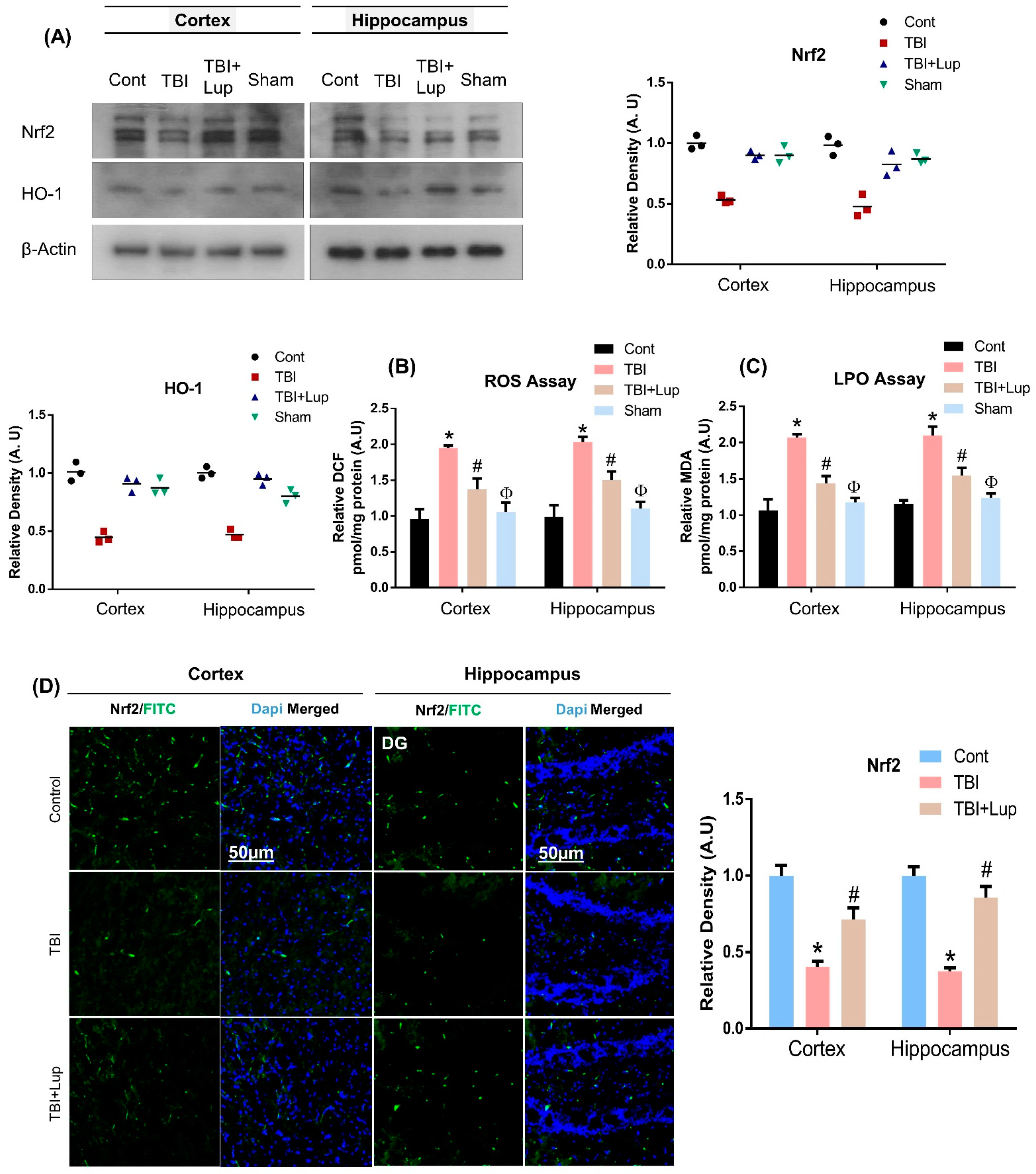
2.2. Lupeol Treatment Induces Nrf2/HO-1 Expression and Attenuates ROS and LPO in Mouse Brain with TBI

Several studies have reported that the brain is susceptible to oxidative stress [76]. Oxidative stress has been well documented in TBI. Once injury-induced excitotoxicity occurs, the production of ROS and nitric oxide (NO) is increased, where protective mechanisms such as antioxidants fail to control these radicals, leading to oxidative stress [40]. To explore the antioxidant potential of Lupeol against TBI–induced oxidative stress, we conducted ROS and LPO assays. Our results indicated elevated ROS levels and increased DCF contents in TBI mouse brains as compared with the saline-treated (control) mouse brain, which were significantly reduced by lupeol treatment (Figure 2B). Similarly, there was an enhanced expression of LPO (MDA level) in the TBI-alone group, which was significantly attenuated with lupeol treatment (Figure 2C). Moreover, the antioxidant potential of Lupeol was further evaluated by analyzing the protein expression of Nrf2 and HO-1 in the mouse brain. Our results indicated a reduced level of Nrf2 and HO-1 in TBI-treated mouse brain, but the TBI + Lupeol co-treatment group significantly reversed the Nrf2 and HO-1 level in both cortex and hippocampus the mouse brain (Figure 2A). We further confirmed our results through immunofluorescence analysis that indicated a decreased expression level of Nrf2 in the cortex and hippocampal region of the TBI mouse group, while the level was significantly raised in the TBI + Lupeol co-treatment mouse group (Figure 2D), suggesting that antioxidant lupeol treatment mitigates oxidative stress induced by TBI in mouse brain.

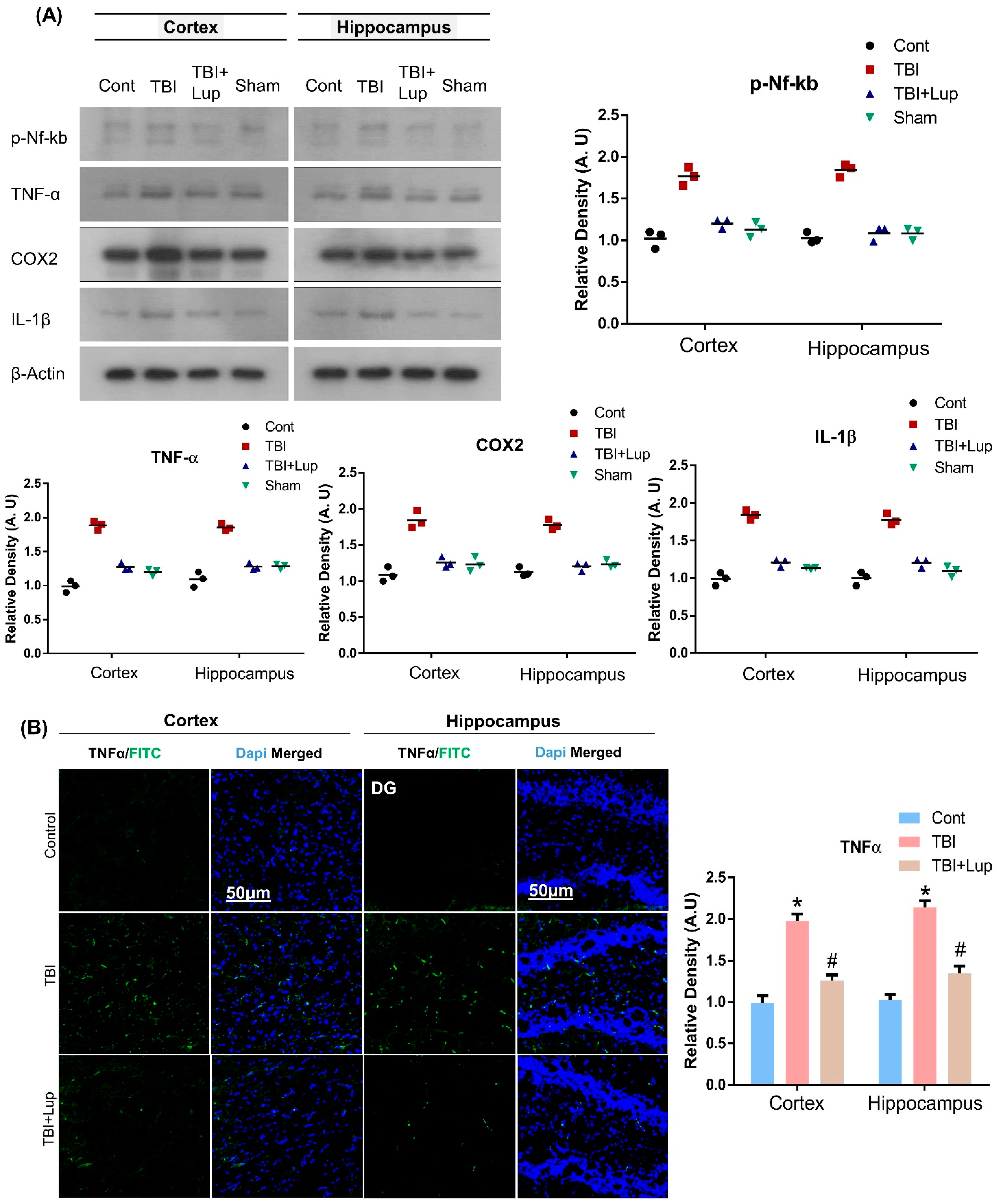
2.3. Administration of Lupeol Reduced the Expression of Inflammatory Cytokines in Mouse Brain with TBI

In response to TBI, glia activation and oxidative stress leads to neuroinflammation [77,78,79]. In the central nervous system, a variety of stimuli, such as injury, oxidative stress, and neurotoxins induce p-NF-κB, which upregulates other inflammatory cytokines such as tumor necrosis factor alpha (TNFα), cyclooxygenase-2 (COX2), and interleukin 1 beta (IL-1β) [80,81]. In order to examine the effects of lupeol on TBI-mediated neuroinflammation, we conducted immunoblot and immunofluorescence analyses. Our immunoblot results exposed that, compared with the saline-treatment (control) group of mice, the TBI-alone mice group showed significantly increased protein expression level of p-NF-κB and its associated downstream neuroinflammatory mediators, including TNFα, COX-2, and IL-1β in the cortex and hippocampus regions (Figure 3A). Interestingly, TBI + Lupeol co-treatment significantly reduced the elevated expression of p-NF-κB and its related downstream inflammatory mediators. To further strengthen these results, we also performed confocal microscopy. In accordance with our immunoblot results, the immunofluorescence analysis also suggested that TNFα immunoreactivity significantly increased in the TBI-alone group as compared with the normal saline-treated (control) mice group. Interestingly, TBI + Lupeol co-treatment significantly reduced immunofluorescence reactivity of TNFα in both the cortex and hippocampus regions (Figure 3B). Taken together, these results indicate that lupeol is effective against TBI-induced neuroinflammation in the mouse brain.

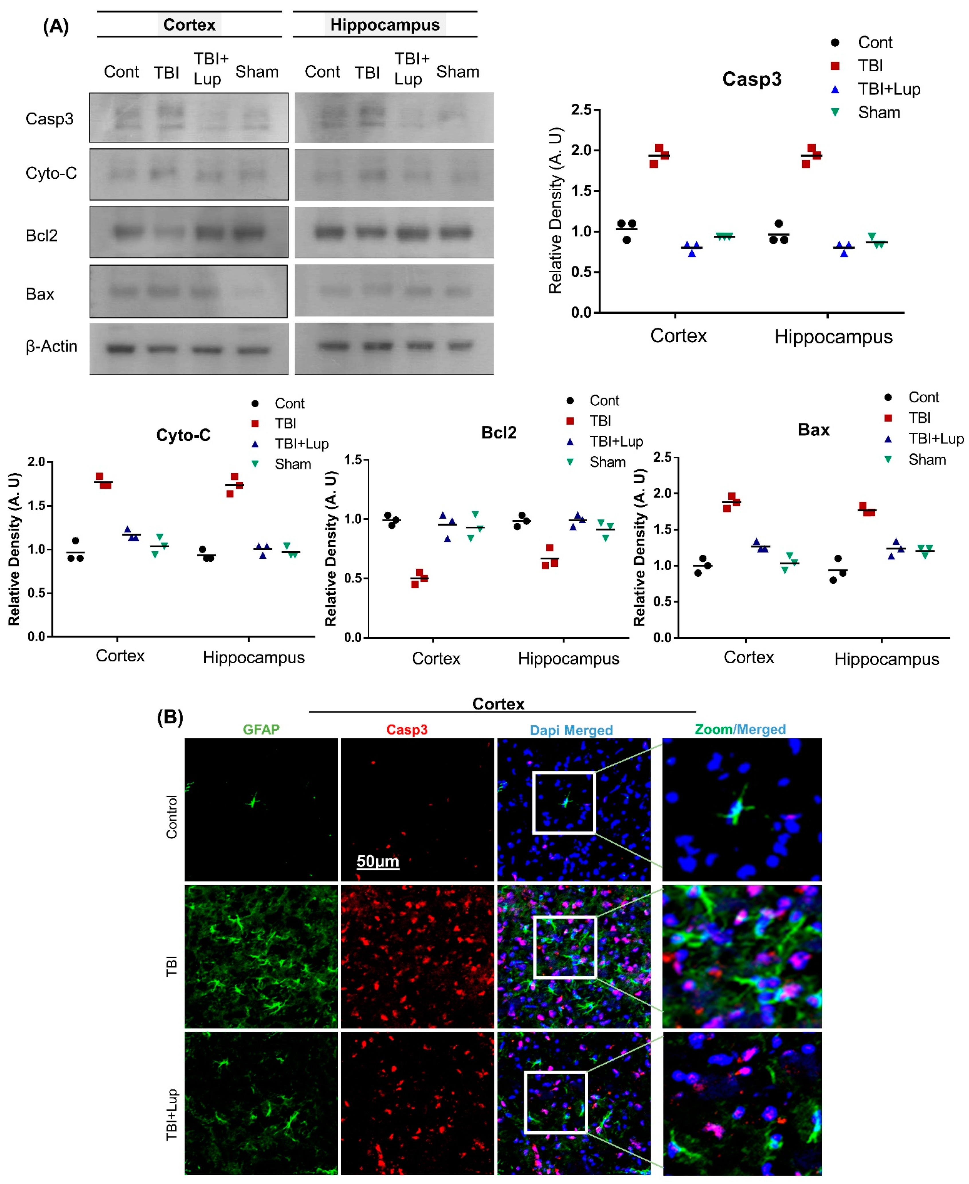
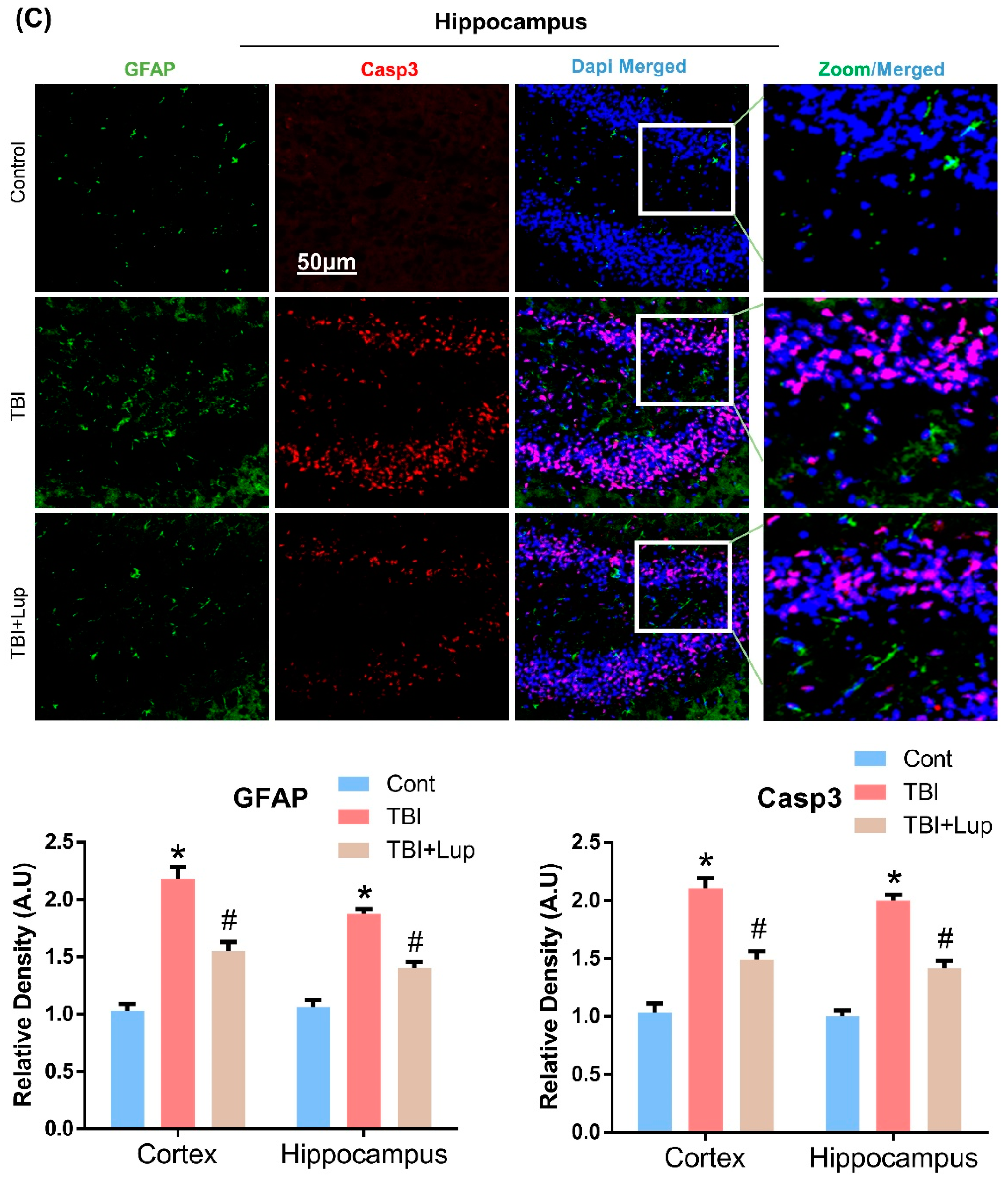
2.4. Lupeol Treatment Attenuated TBI-Induced Apoptotic Cell Death in Mouse Brain

Several studies have been reported that TBI causes apoptotic cell death and neurodegeneration [53,56], which are associated with behavioral dysfunction and histopathological changes [82]. To determine the effects of lupeol against apoptotic neurodegeneration mediated by TBI, we conducted immunoblotting analysis for proapoptotic markers (caspase-3, Bax, and Cyto-C) and an antiapoptotic marker (Bcl-2) in all the experimental groups. In comparison with the saline-treated (control) group, there was an increased expression of caspase-3, Bcl-2, Bax, and Cyto-C and downregulation of Bcl-2 in the TBI-treated group of mice (Figure 4A). Interestingly the TBI + Lupeol co-treatment regulated the apoptotic signaling molecules in both cortex and hippocampus regions the mouse brain. To strengthen our immunoblot results, we also conducted double immunofluorescence analysis of caspase-3 and GFAP. The immunofluorescence analysis of caspase-3 and GFAP also indicated an elevated expression in both cortex (Figure 4B) and hippocampus (Figure 4C) of the TBI-treated group as compared with the saline-treated (control) group. However, the TBI + Lupeol co-treatment significantly decreased the expression levels of caspase-3 and GFAP (Figure 4B,C).

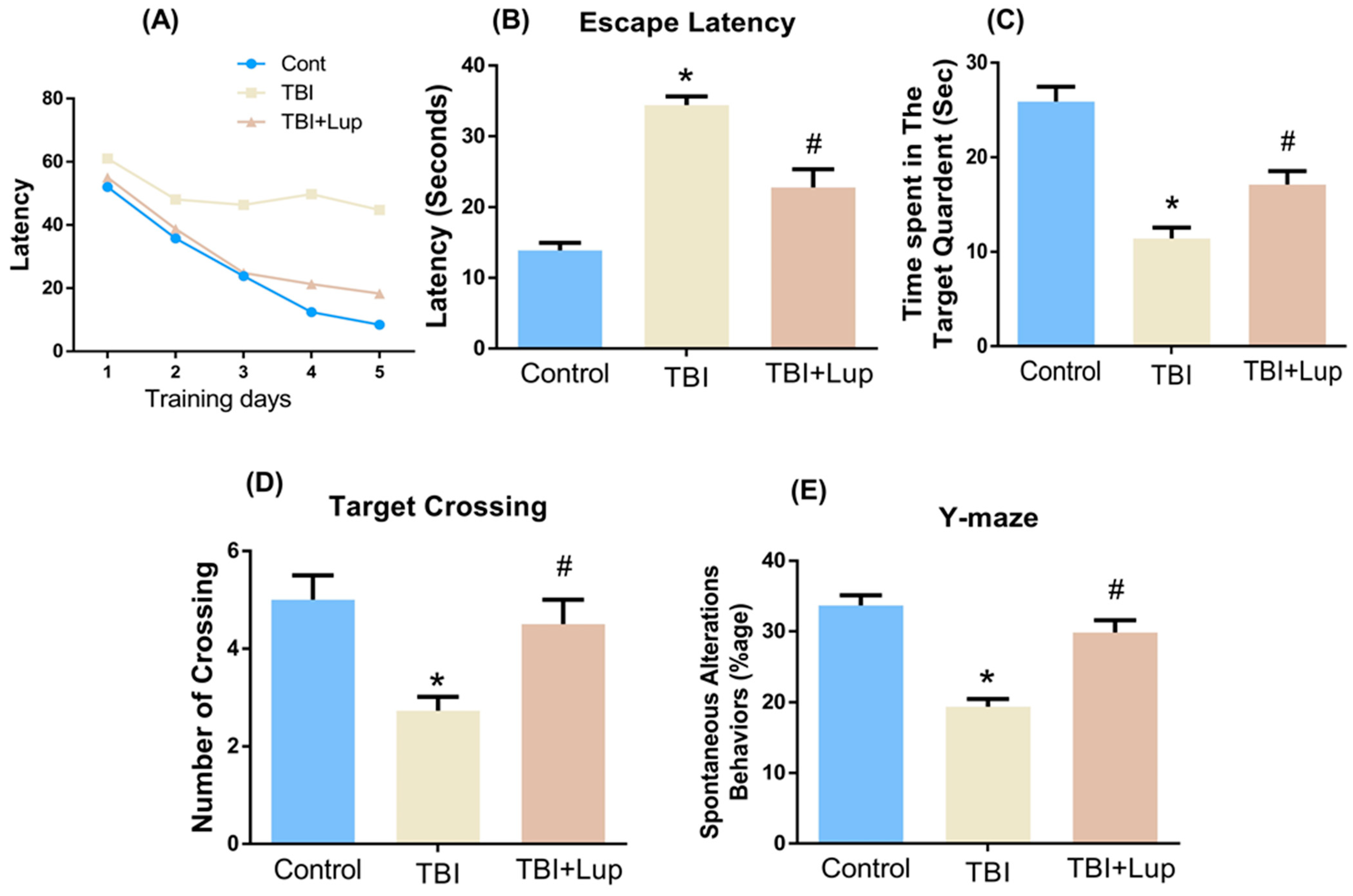
2.5. Lupeol Treatment Improved Learning Memory and Spontaneous Alteration Behavior in TBI-Induced Memory Impairment
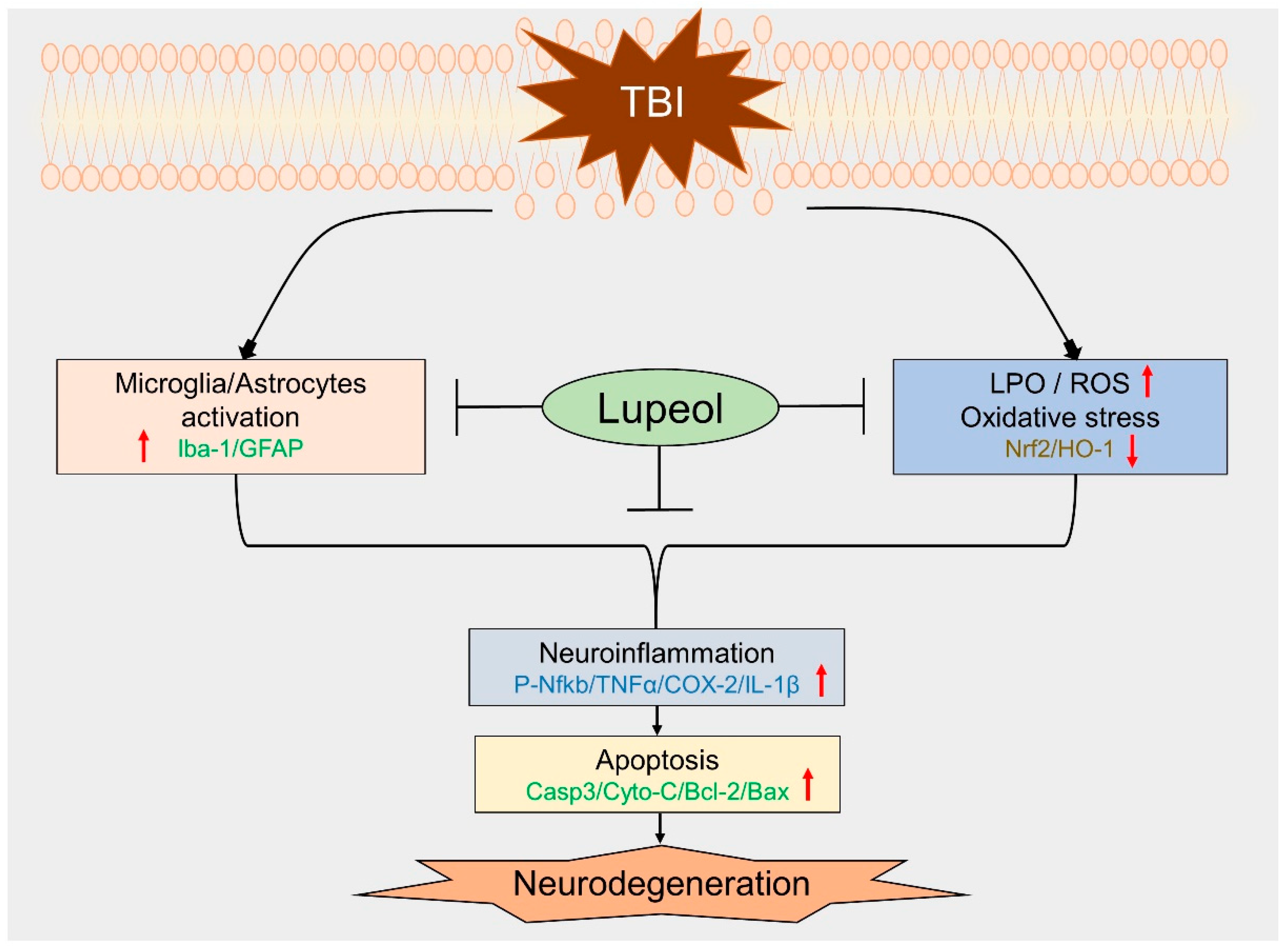
In order to analyze cognitive abnormalities in all experimental mice groups, we conducted the behavioral tests. Our tests demonstrated that the TBI-treated mice showed memory glitches, as shown in the MWM test. In the MWM test, the mean latency (to find hidden platform) slowly reduced over training days in all mouse groups, except the TBI-treated group, which showed a longer latency as compared with the saline-treated (control) group. However, in comparison with the TBI-alone group, the TBI + Lupeol co-treatment group showed significantly reduced latency in finding the platform (Figure 5A,B). After completion of the trial period, the hidden platforms were removed and the probe test was performed. We found that the number of platform crossings were significantly increased in the TBI + Lupeol co-treated group compared with the TBI-alone group (Figure 5D). Additionally, we found that TBI + Lupeol co-treated mice spent more time in the target quadrant as compared with the TBI-treated mice (Figure 5C), showing that lupeol reduced TBI-induced memory impairment. To analyze spatial working memory, we performed the Y-maze test, based on the spontaneous alteration behavior percentage (%). We found that the alternation behaviors percentage (%) was significantly reduced in the TBI-alone group as compared with the saline-treated (control) mice, indicating impaired working memory. However, the TBI + Lupeol co-treated group showed a significant increase in spontaneous alteration behavior (%) in comparison with the TBI-alone group (Figure 5E), indicating that lupeol attenuated short-term memory deficits in the TBI-treated mice.

3. Discussion

This study presents the role of lupeol in inhibiting the activation of astrocytes/microglia and ROS/LPO, providing neuroprotection after TBI in mice. The brain is extremely sensitive to lipid peroxidation (LPO) because of its high content of polyunsaturated fatty acids and iron [83]. ROS are byproducts of normal metabolism of oxygen which includes free radicals such as superoxide, hydroxyl radicals, and singlet oxygen [84]. Trauma induces excessive production of ROS, induced from excitotoxicity and depletion of the endogenous antioxidant system (e.g., superoxide dismutase, catalase, and glutathione peroxidase), and causes lipid peroxidation, protein oxidation, mitochondrial dysfunction, DNA cleavage, and changed signal transduction [85,86,87]. Current studies have proved that a key transcription factor, nuclear factor erythroid 2-related factor 2 (Nrf2), has an essential role in the induction of endogenous antioxidant enzymes against oxidative stress [88]. However, the impairment of Nrf2 signaling leads to oxidative stress [89]. HO-1, implicated in the endogenous defense response, represents an important cytoprotective mechanism against oxidative damage by breakdown of toxic heme into free iron, carbon monoxide, and biliverdin [90,91]. HO-1 also facilitates cellular responses to oxidative stress. In this study, we aimed to investigate the oxidative stress and cellular energy imbalance after TBI in mouse brain and the modulating effect of energy homeostasis through the antioxidant lupeol in the brain of TBI mice. Our ROS and LPO analyses clearly indicated that oxidative stress was increased in the mouse brain after TBI. In addition, the antioxidants Nrf-2 and HO-1 were shown to be downregulated via our Western blot results in the mouse brain that received TBI. However, it was interesting to find that oxidative stress levels were reversed in the mouse brains treated with lupeol, as revealed by ROS and LPO analyses and elevated expression of antioxidants Nrf-2 and HO-1. Our findings suggest that lupeol may mitigate TBI-induced oxidative stress by inducing Nrf2 and HO-1 expression.
A vital feature of TBI is the development of neuroinflammation, primarily due to glial activation [92]. Glial activation may be either advantageous or detrimental, depending on the degree of activity and longevity. Thus, the expression and release of neurotrophic or noxious substances in response to nerve damage is likely a determinant of neuronal viability [93]. An important feature of TBI is increased GFAP expression, signifying activation, and/or proliferation of astrocytes [94]. When injury occurs, microglia are rapidly activated, modifying their normal morphology to forked cell structures and larger cell bodies [95]. Early microglia activation in TBI promotes astrogliosis and persistent inflammation [96]. Astrocytes and microglia link with each other through the cytokines and other extracellular mediators they release [97,98]. They play critical roles in the brain’s ion and water homeostasis, energy metabolism, blood–brain barrier, and immune response, and they change their morphology and protein expression, releasing both pro- and anti-inflammatory mediators [99]. In the present study, our results elucidated that lupeol reduced the number of activated astrocytes and microglia in the adult mouse cortex and hippocampus regions. Activated microglia/astrocytes and oxidative stress are responsible for the release of inflammatory molecules, such as TNF-α, IL-1β, and COX-2, that cause neuroinflammation. It is broadly known that the inflammatory response of the brain after TBI contributes to widespread apoptosis and chronic tissue degeneration. Neuroinflammation is one of the important factors among the pathophysiological signs of TBI [100]. The transcription factor NF-κB is a known immunomodulatory factor in various cell types [101]. Astrocytes activation and Nrf2 downregulation by TBI also activate NF-κB [89], which mediates inflammation through upregulation of the Na/K/2Cl cotransporter (NKCC) [102]. Likewise, TNF-α, which is produced by microglia, astrocytes, and neurons [103], is an important mediator of the inflammatory response in TBI [104]. It also appears to be released earlier than other proinflammatory cytokines and initiates the activation of several cytokines, growth factors, and the recruitment of immune cells [105,106]. Moreover, another inflammatory cytokine, COX2, has been associated with worse outcomes after brain injury as well as early-onset dementia [107,108]. Elevated COX2 expression has been observed in head trauma and several progressive neurodegenerative conditions, e.g., Alzheimer’s disease and Parkinson’s disease [109,110]. Similarly, an increasing number of studies have reported that immune-reactive IL-1β cells are overexpressed in pathogenic conditions, brain injury, and degeneration. Overexpressed IL-1β disturbs both neuronal and non-neuronal cells of the CNS [111]. Moderate and severe TBI both increase IL-1β mRNA and protein levels in the cortical and deep brain structures after injury [112]. We therefore analyzed the expression levels of these inflammatory mediators through Western blot and confocal microscopy. Our findings illuminated that the TBI+ Lupeol co-treatment suppressed p-NF-κB, and its associated proinflammatory mediators (TNF-α, IL-1β, and COX-2) in the indicated region (cortex and hippocampus) of mouse brain, as described in the diagram (Figure 6).
Proapoptotic-related proteins, such as Bax and caspase-3, can also be induced by TBI [56]. Therefore, we also investigated the protein expression of active caspase-3, which leads to cleavage of many proteins, ultimately leading to DNA fragmentation and apoptosis [113]. Activation of caspase-3 is also supported by studies demonstrating cellular modifications including neuronal cell death within the brain tissue of TBI patients [114], and has been considered a key feature of neurodegenerative diseases [115]. Caspase-dependent and caspase-independent apoptosis are both regulated by proteins of the Bcl-2 family, which include both pro-death and pro-survival members [90,116]. Bcl-2 family proteins regulate the permeability of the outer mitochondrial membrane and the formation of permeable transition pores [117]. They contain highly conserved Bcl-2 homology domains (BH 1–4), which are important for homo- and heterocomplex formations. Complexes formed between proteins containing BH-3 domains, such as Bax, can promote the release of cytochrome-c from mitochondria [90]. It has also been reported that, in the acute post-traumatic period, damaged neurons showed a sharp decrease in Bcl-2 expression, followed later by an increase in Bax mRNA and protein [118]. In our study, we revealed that lupeol downregulated the expression of Cyto-C, Bax, and Casp-3, while upregulating Bcl2 expression. Several recent studies have confirmed the therapeutic potential of lupeol, and it has been shown that lupeol provides neuroprotection via suppressing inflammation and apoptosis [68]. Similarly, our behavior analysis (MWM and Y-maze tests) also indicated that TBI significantly impaired cognitive and learning behavior. However, the TBI + Lupeol co-treatment considerably reversed these effects and improved cognition, spatial learning, and memory processing. Taken together, our findings prove that lupeol is effective in reversing TBI-induced memory impairment in mouse brain.

4. Materials and Methods

4.1. Animals

Male wild-type C57BL/6 N mice (8 weeks old, body weight 25–30 g, n = 42) were purchased from Samtako Bio (Osan, Republic of Korea). The animals were kept in Ostrich’s mouse cages and were acclimatized for 7 days; they were maintained under controlled temperature (25 °C) and lighting (12/12 dark/light cycle). Each mouse group (12 mice/group) was kept in two cages (6 mice/cage) and were allowed free access to water and food as previously described [119]. All the experimental techniques were performed according to the protocol approved by the Animal Ethics Committee of the Division of Applied Life Sciences, Gyeongsang National University, South Korea (Approval ID: 125, 3 June 2020).

4.2. Traumatic Brain Injury of Mice and Drug Treatment

Mice were randomly arranged into four groups: (1) a control saline-treated group (control, n = 12), which received saline for one week as a vehicle; (2) traumatic brain injury group (TBI, n = 12); (3) traumatic brain injury group with lupeol treatment (TBI + Lup, n = 12) (lupeol dose: 50 mg/kg/day/mice/p.o.); and (4) sham-treated group (Lup, n = 6). The sham-treated group received a daily injection of lupeol (50 mg/kg/day/mice/p.o.) for 7 days without head injury (Figure 7). Lupeol dosages were selected according to previously published studies [67,68]. Lupeol (CAS Number*: 545-47-1) was purchased from Sigma Co. (St. Louis, MO, USA). For the surgical procedures, the mice were anesthetized with Zoletil (0.1 mL/100 g body weight) and Rompun (0.05 mL/100 g body weight) and placed on a stereotaxic frame. Surgery was performed on the animals in a temperature-controlled environment. The skull was exposed by removing the skin (mid-longitudinal incision). TBI was induced by a sharp edge scalpel blade inserted 3 mm into right hemisphere of the brain. The scalpel blade remained in the brain for 1 min and was slowly removed. The skull was covered with bone wax and the skin was closed with a silk suture. The experiments were performed under continuous heating with a heating lamp and mice were monitored visually until full recovery, as previously described [57,120].

4.3. Morris Water Maze (MWM) Test

For behavior analysis, The Morris water maze (MWM) test was performed as previously described [121,122]. The MWM apparatus consisted of a round tank (100 cm in diameter, 40 cm high, and 15.5 cm deep) filled with water and made opaque with white ink. A transparent platform 20 cm in height and 10 cm in diameter was hidden 1 cm below the water surface in one quadrant of the tank during the experiment. After three days, following TBI and lupeol treatment, all mice were brought into the MWM apparatus to assess the cognitive ability of the treated mice. The behavior study (training session) was carried out for four days. After the training session, a probe test was conducted by removing the hidden platform. In the probe trial, the number of crossings, the latency to the platform, and the time spent in the target quadrant were calculated. All the data were recorded by a video tracking system (SMART, Panlab Harvard Apparatus, Bioscience Company, Holliston, MA, USA).

4.4. Y-Maze Test

For the assessment of spatial working memory, Y-maze tests were performed, made of black painted plastic. The Y-maze test apparatus contains three arms, each arm of the maze was 20 cm high, 50 cm long, and 10 cm wide at both the bottom and top. The mice were placed at the center of the apparatus and were permitted to move freely in the apparatus for 8 min in each arm. The number of arm entries were observed visually. The spontaneous alternation was defined as the successive entry of mice in three arms in an overlapping set of triplets. The spontaneous alternation behavior percentage (%) was calculated using the following formula: successive triplet sets (entries in three different arms consecutively)/total number of arm entries-2) × 100. The higher percentage of spontaneous alternation behavior was considered to show an improved cognitive performance, and vice versa.

4.5. Homogenization of Mouse Brain

Homogenization of the mouse brain was performed as described previously [123,124]. After behavioral analysis, all the mice were anesthetized with ketamine/xylazine and then sacrificed. The brain tissues were removed immediately, and the cortex and hippocampus were carefully separated. The tissues were homogenized within PRO-PREPTM extraction solution (iNtRON Biotechnology, Burlington, NJ, USA) and an equal volume of 20–30 μg of protein was mixed with 2× Sample Buffer (Invitrogen, Waltham, MA, USA), then centrifuged at a speed of 13,000 rpm for 25 min at 4 °C. The supernatant was collected and stored at −80 °C.

4.6. ROS Assay

The ROS assay was conducted to evaluate the reactive oxygen species (ROS) level in the brain of the experimental mice groups (n = 6 mice/group). The assay was mainly based on the formation of 2′7′ dichlorofluorescein (DCF) from oxidation of 2′7′-dichlorodihydrofluorescein diacetate (DCFH-DA). Briefly, the brain homogenates of the normal and treated mice groups were diluted 1:20 in ice-cold Lock’s buffer to produce 2.5 mg tissue/500 μL as the final concentration. The final reaction mixture of 1 mL of the Lock’s buffer (pH ± 7.4), 0.2 mL of homogenates, and 10 mL of DCFH-DA (5 mM) were incubated for 15 min at room temperature to form fluorescent DCF from DCFH-DA. The conversion of 2′-7′dichlorofluorescin diacetate DCFH-DA to DCF was evaluated using spectrofluorimeter at an excitation wavelength of 484 nm and an emission wavelength of 530 nm. To measure the conversion of DCFH-DA to DCF in the absence of homogenate (background fluorescence), parallel blanks were used. The ROS levels were measured and expressed as relative pmol DCF/mg protein.

4.7. LPO Assay

A lipid peroxidation (LPO) assay was performed as previously described with some modifications [36]. Evaluation of LPO is important for the assessment of oxidative stress. The LPO marker free malondialdehyde (MDA), in the cortical and hippocampal protein lysates, was assessed using a thiobarbituric-acid-reactive substance (TBARS) assay kit (Bio Vision, Milpitas, CA, USA) according to the manufacturer’s instructions.

4.8. Antibodies and Reagents

The following antibodies were used in immunoblotting and immunofluorescence studies: anti-Iba-1, anti-GFAP, Nrf2, HO-1, anti-TNF-α, anti-IL-1β, anti-p-NF-κB, Cox2, anti-caspase-3, anti-cytochrome-C, anti-Bax, anti-Bcl2, and anti-β-actin. These antibodies were obtained from Santa Cruz Biotechnology (Dallas, TX, USA). Primary antibodies were diluted in 1× TBST (Tris-buffered saline plus Tween) (1:1000), while secondary conjugated anti-mouse horseradish peroxidase (HRP) and conjugated anti-rabbit HRP were diluted 1:10,000 in 1× TBST, all these were purchased from Promega, Madison, WI, USA. For immunofluorescence studies, the secondary fluorescent antibodies goat anti-mouse and goat anti-rabbit were used, and diluted in 1 × 100 phosphate-buffered saline (PBS).

4.9. Immunoblotting

Expression levels of proteins were quantified by using a Bradford assay (Bio-Rad Protein Assay kit, Bio-Rad Laboratories, Hercules, CA, USA) as described previously, with some modification [47,125,126]. The protein samples were electrophoresed on SDS-PAGE and then transferred to polyvinylidene difluoride membranes (PVDF) (Millipore, Burlington, MA, USA). A protein marker (GangNam-STAIN, iNtRON Biotechnology, CA, USA) was loaded parallel determine the molecular weight of the protein. All the membranes were blocked in 5% skimmed milk to reduce the nonspecific bindings, and then incubated overnight with the primary antibodies at 4 °C. The next day, the membranes were washed with 1xTBST (3 × 10 min) and blocked with horseradish-peroxidase-conjugated secondary antibodies, diluted in 1× TBST for 1 h, and then washed with 1× TBST (4 × 12 min). After washing, the membranes were developed in a dark room and the bands were detected using an enhanced chemiluminescent (ECL) detection reagent (EzWestLumiOne, ATTO, Tokyo, Japan). The immunoblots bands were obtained using X-ray films. For band quantification, ImageJ software (v. 1.50, NIH, Bethesda, MD, USA) was used and the graphs were evaluated using GraphPad Prism 6 software (San Diego, CA, USA).

4.10. Immunofluorescence Analysis

Immunofluorescence analysis was performed as described previously [127,128,129,130]. Briefly, the brain sections were dehydrated overnight and then rehydrated on next with 1% 1x phosphate-buffered saline (PBS) for 10 min. The slides were then treated with proteinase K for 5 min, then washed again with PBS and blocked with 5% normal serum for 1 h. After blocking, the slides were carefully incubated with primary antibodies (1:100) for 24 h at 4 °C. After incubation with primary antibodies, the slides were washed with 1% PBS and treated with fluorescein isothiocyanate (FITC)-labeled secondary antibodies for 90 min at room temperature. The slides were then washed and treated with 4′,6-diamidino-2-phenylindole (DAPI) for detection of the nucleus. The slides were covered using coverslips with a fluorescent mounting medium (FluoView FV 1000; Olympus, Tokyo, Japan) and the images were captured by a confocal scanning microscope (FV1000MPE). The captured results were evaluated by the relative integrated densities using ImageJ software (version 1.50, NIH, 1 March 2016; https://imagej.nih.gov/ij/, USA) and the graphs were created through PRISM6 software.

4.11. Statistical Analysis

The Western blot bands were scanned and analyzed using densitometry in the computer-based Sigma Gel System (SPSS Inc., Chicago, IL, USA). The immunofluorescence results were analyzed using ImageJ software, and the densities were calculated in arbitrary units. The data are presented as the mean ± standard error of mean (SEM). One-way analysis of variance (ANOVA) and Student’s t-test were used for comparison of the different groups. Statistical analyses were performed using Graph-Pad Prism 6 software. P values less than 0.05 were considered to show a significant difference between the groups; Western blots and graph bars are represented as Control, TBI (TBI-treated group), TBI + Lupeol (TBI and lupeol-treated group), and Sham (Lupeol-alone treatment group).

5. Conclusions

In summary, the findings of the current study demonstrated that lupeol is a potential neurotherapeutic candidate that markedly reversed TBI-induced glial cells activation, oxidative stress, neuroinflammation, apoptosis, and memory impairment in mouse brains. We propose that these underlying potent neuroprotective effects of lupeol against TBI-induced neurotoxicity might be due to the inhibition of glial cells activation and oxidative stress. Based on these findings, we suggest that lupeol is a safe, effective, and a promising neurotherapeutic agent. However, future studies are highly encouraged to further evaluate the underlying molecular mechanism and role of lupeol in neuroinflammation and in various age-related neurodegenerative disorders, particularly in AD.

Author Contributions

R.A. designed the model and wrote the manuscript; A.K. helped in sample collection and provided critical suggestions in manuscript writing, while helping in statistical analysis and analyses of the data; I.K. performed the confocal microscopy; I.U.R. and H.J.L. performed the Western blot experiments; M.O.K. supervised, organized, provided critical instructions, and approved the final version of the manuscript. All authors reviewed and approved the paper and provided feedback. All authors have read and agreed to the published version of the manuscript.

Funding

This research was supported by the Neurological Disorder Research Program of the National Research Foundation (NRF) funded by the Korean Government (MSIT) (2020M3E5D9080660).

Institutional Review Board Statement

This study was carried out in animals in accordance with approved guidelines (Approval ID: 125) by the animal ethics committee (IACUC) of the Division of Applied Life Science, Gyeongsang National University, South Korea.

Informed Consent Statement

Not applicable.

Data Availability Statement

The authors hereby declares that the data presented in this study will be presented upon request from the corresponding author.

Conflicts of Interest

The authors declare no conflict of interest.

References

  1. Andelic, N. The epidemiology of traumatic brain injury. Lancet Neurol. 2013, 12, 28–29. [Google Scholar] [CrossRef]
  2. Rosenfeld, J.V.; Maas, A.I.; Bragge, P.; Morganti-Kossmann, M.C.; Manley, G.T.; Gruen, R.L. Early management of severe traumatic brain injury. Lancet 2012, 380, 1088–1098. [Google Scholar] [CrossRef]
  3. Faden, A.I.; Wu, J.; Stoica, B.A.; Loane, D.J. Progressive inflammation-mediated neurodegeneration after traumatic brain or spinal cord injury. Br. J. Pharmacol. 2016, 173, 681–691. [Google Scholar] [CrossRef] [PubMed] [Green Version]
  4. Gardner, R.C.; Burke, J.F.; Nettiksimmons, J.; Kaup, A.; Barnes, D.E.; Yaffe, K. Dementia risk after traumatic brain injury vs nonbrain trauma: The role of age and severity. JAMA Neurol. 2014, 71, 1490–1497. [Google Scholar] [CrossRef] [PubMed]
  5. Das, M.; Mohapatra, S.; Mohapatra, S.S. New perspectives on central and peripheral immune responses to acute traumatic brain injury. J. Neuroinflamm. 2012, 9, 236. [Google Scholar] [CrossRef] [PubMed] [Green Version]
  6. Stoica, B.A.; Faden, A.I. Cell death mechanisms and modulation in traumatic brain injury. Neurotherapeutics 2010, 7, 3–12. [Google Scholar] [CrossRef]
  7. Plesnila, N.; von Baumgarten, L.; Retiounskaia, M.; Engel, D.; Ardeshiri, A.; Zimmermann, R.; Hoffmann, F.; Landshamer, S.; Wagner, E.; Culmsee, C. Delayed neuronal death after brain trauma involves p53-dependent inhibition of NF-kappaB transcriptional activity. Cell Death Differ. 2007, 14, 1529–1541. [Google Scholar] [CrossRef] [Green Version]
  8. Shafi, S.; Barnes, S.A.; Millar, D.; Sobrino, J.; Kudyakov, R.; Berryman, C.; Rayan, N.; Dubiel, R.; Coimbra, R.; Magnotti, L.J.; et al. Suboptimal compliance with evidence-based guidelines in patients with traumatic brain injuries. J. Neurosurg. 2014, 120, 773–777. [Google Scholar] [CrossRef]
  9. Timmons, S.D.; Waltzman, D.; Duhaime, A.C.; Spinks, T.J.; Sarmiento, K. Considerations for neurosurgeons: Recommendations from the CDC Pediatric Mild Traumatic Brain Injury Guideline. J. Neurosurg. 2019, 131, 979–983. [Google Scholar] [CrossRef]
  10. Hutchinson, P.J.; Kolias, A.G.; Timofeev, I.S.; Corteen, E.A.; Czosnyka, M.; Timothy, J.; Anderson, I.; Bulters, D.O.; Belli, A.; Eynon, C.A.; et al. Trial of Decompressive Craniectomy for Traumatic Intracranial Hypertension. N. Engl. J. Med. 2016, 375, 1119–1130. [Google Scholar] [CrossRef] [Green Version]
  11. Dusick, J.R.; Wang, C.; Cohan, P.; Swerdloff, R.; Kelly, D.F. Pathophysiology of hypopituitarism in the setting of brain injury. Pituitary 2012, 15, 2–9. [Google Scholar] [CrossRef] [PubMed] [Green Version]
  12. Ichkova, A.; Rodriguez-Grande, B.; Bar, C.; Villega, F.; Konsman, J.P.; Badaut, J. Vascular impairment as a pathological mechanism underlying long-lasting cognitive dysfunction after pediatric traumatic brain injury. Neurochem. Int. 2017, 111, 93–102. [Google Scholar] [CrossRef] [PubMed]
  13. Hayes, J.P.; Logue, M.W.; Sadeh, N.; Spielberg, J.M.; Verfaellie, M.; Hayes, S.M.; Reagan, A.; Salat, D.H.; Wolf, E.J.; McGlinchey, R.E.; et al. Mild traumatic brain injury is associated with reduced cortical thickness in those at risk for Alzheimer’s disease. Brain 2017, 140, 813–825. [Google Scholar] [CrossRef] [PubMed] [Green Version]
  14. Bains, M.; Hall, E.D. Antioxidant therapies in traumatic brain and spinal cord injury. Biochim. Biophys. Acta 2012, 1822, 675–684. [Google Scholar] [CrossRef] [Green Version]
  15. Chao, H.; Anthonymuthu, T.S.; Kenny, E.M.; Amoscato, A.A.; Cole, L.K.; Hatch, G.M.; Ji, J.; Kagan, V.E.; Bayir, H. Disentangling oxidation/hydrolysis reactions of brain mitochondrial cardiolipins in pathogenesis of traumatic injury. JCI Insight 2018, 3, e97677. [Google Scholar] [CrossRef] [Green Version]
  16. Ji, J.; Tyurina, Y.Y.; Tang, M.; Feng, W.; Stolz, D.B.; Clark, R.S.; Meaney, D.F.; Kochanek, P.M.; Kagan, V.E.; Bayir, H. Mitochondrial injury after mechanical stretch of cortical neurons in vitro: Biomarkers of apoptosis and selective peroxidation of anionic phospholipids. J. Neurotrauma 2012, 29, 776–788. [Google Scholar] [CrossRef] [Green Version]
  17. Kabadi, S.V.; Faden, A.I. Neuroprotective strategies for traumatic brain injury: Improving clinical translation. Int. J. Mol. Sci. 2014, 15, 1216–1236. [Google Scholar] [CrossRef] [Green Version]
  18. Lin, C.; Chao, H.; Li, Z.; Xu, X.; Liu, Y.; Hou, L.; Liu, N.; Ji, J. Melatonin attenuates traumatic brain injury-induced inflammation: A possible role for mitophagy. J. Pineal Res. 2016, 61, 177–186. [Google Scholar] [CrossRef]
  19. Menge, T.; Zhao, Y.; Zhao, J.; Wataha, K.; Gerber, M.; Zhang, J.; Letourneau, P.; Redell, J.; Shen, L.; Wang, J.; et al. Mesenchymal stem cells regulate blood-brain barrier integrity through TIMP3 release after traumatic brain injury. Sci. Transl. Med. 2012, 4, 161ra150. [Google Scholar] [CrossRef] [Green Version]
  20. Simon, D.W.; McGeachy, M.J.; Bayir, H.; Clark, R.S.; Loane, D.J.; Kochanek, P.M. The far-reaching scope of neuroinflammation after traumatic brain injury. Nat. Rev. Neurol. 2017, 13, 171–191. [Google Scholar] [CrossRef] [Green Version]
  21. Streit, W.J.; Mrak, R.E.; Griffin, W.S. Microglia and neuroinflammation: A pathological perspective. J. Neuroinflamm. 2004, 1, 14. [Google Scholar] [CrossRef] [PubMed] [Green Version]
  22. Block, M.L.; Zecca, L.; Hong, J.S. Microglia-mediated neurotoxicity: Uncovering the molecular mechanisms. Nat. Rev. Neurosci. 2007, 8, 57–69. [Google Scholar] [CrossRef] [PubMed]
  23. Woodcock, T.; Morganti-Kossmann, M.C. The role of markers of inflammation in traumatic brain injury. Front. Neurol. 2013, 4, 18. [Google Scholar] [CrossRef] [PubMed] [Green Version]
  24. Block, M.L.; Hong, J.S. Microglia and inflammation-mediated neurodegeneration: Multiple triggers with a common mechanism. Prog. Neurobiol. 2005, 76, 77–98. [Google Scholar] [CrossRef]
  25. Loane, D.J.; Kumar, A.; Stoica, B.A.; Cabatbat, R.; Faden, A.I. Progressive neurodegeneration after experimental brain trauma: Association with chronic microglial activation. J. Neuropathol. Exp. Neurol. 2014, 73, 14–29. [Google Scholar] [CrossRef] [Green Version]
  26. Marklund, N.; Hillered, L. Animal modelling of traumatic brain injury in preclinical drug development: Where do we go from here? Br. J. Pharmacol. 2011, 164, 1207–1229. [Google Scholar] [CrossRef] [Green Version]
  27. Suliman, H.B.; Piantadosi, C.A. Mitochondrial Quality Control as a Therapeutic Target. Pharmacol. Rev. 2016, 68, 20–48. [Google Scholar] [CrossRef]
  28. Waldbaum, S.; Patel, M. Mitochondrial dysfunction and oxidative stress: A contributing link to acquired epilepsy? J. Bioenergy Biomembr. 2010, 42, 449–455. [Google Scholar] [CrossRef] [Green Version]
  29. Clausen, F.; Lundqvist, H.; Ekmark, S.; Lewen, A.; Ebendal, T.; Hillered, L. Oxygen free radical-dependent activation of extracellular signal-regulated kinase mediates apoptosis-like cell death after traumatic brain injury. J. Neurotrauma 2004, 21, 1168–1182. [Google Scholar] [CrossRef]
  30. Darwish, R.S.; Amiridze, N.; Aarabi, B. Nitrotyrosine as an oxidative stress marker: Evidence for involvement in neurologic outcome in human traumatic brain injury. J. Trauma 2007, 63, 439–442. [Google Scholar] [CrossRef]
  31. Naseem, M.; Parvez, S. Role of melatonin in traumatic brain injury and spinal cord injury. Sci. World J. 2014, 2014, 586270. [Google Scholar] [CrossRef] [PubMed]
  32. Fleminger, S.; Oliver, D.L.; Lovestone, S.; Rabe-Hesketh, S.; Giora, A. Head injury as a risk factor for Alzheimer’s disease: The evidence 10 years on; a partial replication. J. Neurol. Neurosurg. Psychiatry 2003, 74, 857–862. [Google Scholar] [CrossRef] [PubMed]
  33. Gardner, R.C.; Byers, A.L.; Barnes, D.E.; Li, Y.; Boscardin, J.; Yaffe, K. Mild TBI and risk of Parkinson disease: A Chronic Effects of Neurotrauma Consortium Study. Neurology 2018, 90, e1771–e1779. [Google Scholar] [CrossRef] [PubMed]
  34. Lee, Y.K.; Hou, S.W.; Lee, C.C.; Hsu, C.Y.; Huang, Y.S.; Su, Y.C. Increased risk of dementia in patients with mild traumatic brain injury: A nationwide cohort study. PLoS ONE 2013, 8, e62422. [Google Scholar] [CrossRef] [PubMed] [Green Version]
  35. Lucas, S.M.; Rothwell, N.J.; Gibson, R.M. The role of inflammation in CNS injury and disease. Br. J. Pharmacol. 2006, 147 (Suppl. 1), S232–S240. [Google Scholar] [CrossRef] [Green Version]
  36. Rehman, S.U.; Shah, S.A.; Ali, T.; Chung, J.I.; Kim, M.O. Anthocyanins Reversed D-Galactose-Induced Oxidative Stress and Neuroinflammation Mediated Cognitive Impairment in Adult Rats. Mol. Neurobiol. 2017, 54, 255–271. [Google Scholar] [CrossRef]
  37. Gao, H.M.; Hong, J.S. Why neurodegenerative diseases are progressive: Uncontrolled inflammation drives disease progression. Trends Immunol. 2008, 29, 357–365. [Google Scholar] [CrossRef] [Green Version]
  38. Griffin, W.S.; Sheng, J.G.; Royston, M.C.; Gentleman, S.M.; McKenzie, J.E.; Graham, D.I.; Roberts, G.W.; Mrak, R.E. Glial-neuronal interactions in Alzheimer’s disease: The potential role of a ‘cytokine cycle’ in disease progression. Brain Pathol. 1998, 8, 65–72. [Google Scholar] [CrossRef]
  39. Kreutzberg, G.W. Microglia: A sensor for pathological events in the CNS. Trends Neurosci. 1996, 19, 312–318. [Google Scholar] [CrossRef]
  40. Higgins, G.C.; Beart, P.M.; Shin, Y.S.; Chen, M.J.; Cheung, N.S.; Nagley, P. Oxidative stress: Emerging mitochondrial and cellular themes and variations in neuronal injury. J. Alzheimer’s Dis. 2010, 20 (Suppl. 2), S453–S473. [Google Scholar] [CrossRef] [Green Version]
  41. Ma, Q. Role of nrf2 in oxidative stress and toxicity. Annu. Rev. Pharmacol. Toxicol. 2013, 53, 401–426. [Google Scholar] [CrossRef] [PubMed] [Green Version]
  42. Villeneuve, N.F.; Lau, A.; Zhang, D.D. Regulation of the Nrf2-Keap1 antioxidant response by the ubiquitin proteasome system: An insight into cullin-ring ubiquitin ligases. Antioxid. Redox Signal. 2010, 13, 1699–1712. [Google Scholar] [CrossRef] [PubMed] [Green Version]
  43. Kobayashi, A.; Kang, M.I.; Okawa, H.; Ohtsuji, M.; Zenke, Y.; Chiba, T.; Igarashi, K.; Yamamoto, M. Oxidative stress sensor Keap1 functions as an adaptor for Cul3-based E3 ligase to regulate proteasomal degradation of Nrf2. Mol. Cell. Biol. 2004, 24, 7130–7139. [Google Scholar] [CrossRef] [PubMed] [Green Version]
  44. de Vries, H.E.; Witte, M.; Hondius, D.; Rozemuller, A.J.; Drukarch, B.; Hoozemans, J.; van Horssen, J. Nrf2-induced antioxidant protection: A promising target to counteract ROS-mediated damage in neurodegenerative disease? Free Radic. Biol. Med. 2008, 45, 1375–1383. [Google Scholar] [CrossRef] [PubMed]
  45. Khan, M.S.; Khan, A.; Ahmad, S.; Ahmad, R.; Rehman, I.U.R.; Ikram, M.; Kim, M.O. Inhibition of JNK Alleviates Chronic Hypoperfusion-Related Ischemia Induces Oxidative Stress and Brain Degeneration via Nrf2/HO-1 and NF-kappaB Signaling. Oxid. Med. Cell. Longev. 2020, 2020, 5291852. [Google Scholar] [CrossRef]
  46. Baierle, M.; Nascimento, S.N.; Moro, A.M.; Brucker, N.; Freitas, F.; Gauer, B.; Durgante, J.; Bordignon, S.; Zibetti, M.; Trentini, C.M.; et al. Relationship between inflammation and oxidative stress and cognitive decline in the institutionalized elderly. Oxid. Med. Cell. Longev. 2015, 2015, 804198. [Google Scholar] [CrossRef] [Green Version]
  47. Kim, M.J.; Rehman, S.U.; Amin, F.U.; Kim, M.O. Enhanced neuroprotection of anthocyanin-loaded PEG-gold nanoparticles against Abeta1-42-induced neuroinflammation and neurodegeneration via the NF-KB /JNK/GSK3beta signaling pathway. Nanomedicine 2017, 13, 2533–2544. [Google Scholar] [CrossRef]
  48. Schmidt, O.I.; Heyde, C.E.; Ertel, W.; Stahel, P.F. Closed head injury--an inflammatory disease? Brain Res. Brain Res. Rev. 2005, 48, 388–399. [Google Scholar] [CrossRef]
  49. Holmin, S.; Mathiesen, T. Long-term intracerebral inflammatory response after experimental focal brain injury in rat. Neuroreport 1999, 10, 1889–1891. [Google Scholar] [CrossRef]
  50. Dalton, T.P.; Shertzer, H.G.; Puga, A. Regulation of gene expression by reactive oxygen. Annu. Rev. Pharmacol. Toxicol. 1999, 39, 67–101. [Google Scholar] [CrossRef]
  51. Glushakov, A.O.; Glushakova, O.Y.; Korol, T.Y.; Acosta, S.A.; Borlongan, C.V.; Valadka, A.B.; Hayes, R.L.; Glushakov, A.V. Chronic Upregulation of Cleaved-Caspase-3 Associated with Chronic Myelin Pathology and Microvascular Reorganization in the Thalamus after Traumatic Brain Injury in Rats. Int. J. Mol. Sci. 2018, 19, 3151. [Google Scholar] [CrossRef] [PubMed] [Green Version]
  52. Wilke, S.; Prehn, K.; Taud, B.; List, J.; Floel, A. Multimodal Assessment of Recurrent MTBI across the Lifespan. J. Clin. Med. 2018, 7, 95. [Google Scholar] [CrossRef] [PubMed] [Green Version]
  53. Rehman, S.U.; Ali, T.; Alam, S.I.; Ullah, R.; Zeb, A.; Lee, K.W.; Rutten, B.P.F.; Kim, M.O. Ferulic Acid Rescues LPS-Induced Neurotoxicity via Modulation of the TLR4 Receptor in the Mouse Hippocampus. Mol. Neurobiol. 2019, 56, 2774–2790. [Google Scholar] [CrossRef] [PubMed]
  54. Mei, Z.; Zheng, P.; Tan, X.; Wang, Y.; Situ, B. Huperzine A alleviates neuroinflammation, oxidative stress and improves cognitive function after repetitive traumatic brain injury. Metab. Brain Dis. 2017, 32, 1861–1869. [Google Scholar] [CrossRef] [PubMed]
  55. Lee, Y.J.; Choi, D.Y.; Choi, I.S.; Kim, K.H.; Kim, Y.H.; Kim, H.M.; Lee, K.; Cho, W.G.; Jung, J.K.; Han, S.B.; et al. Inhibitory effect of 4-O-methylhonokiol on lipopolysaccharide-induced neuroinflammation, amyloidogenesis and memory impairment via inhibition of nuclear factor-kappaB in vitro and in vivo models. J. Neuroinflamm. 2012, 9, 35. [Google Scholar] [CrossRef] [Green Version]
  56. Raghupathi, R.; Graham, D.I.; McIntosh, T.K. Apoptosis after traumatic brain injury. J. Neurotrauma 2000, 17, 927–938. [Google Scholar] [CrossRef]
  57. Alam, S.I.; Rehman, S.U.; Kim, M.O. Nicotinamide Improves Functional Recovery via Regulation of the RAGE/JNK/NF-kappaB Signaling Pathway after Brain Injury. J. Clin. Med. 2019, 8, 271. [Google Scholar] [CrossRef] [Green Version]
  58. Loane, D.J.; Stoica, B.A.; Faden, A.I. Neuroprotection for traumatic brain injury. Handb. Clin. Neurol. 2015, 127, 343–366. [Google Scholar] [CrossRef] [Green Version]
  59. Sharifi-Rad, M.; Lankatillake, C.; Dias, D.A.; Docea, A.O.; Mahomoodally, M.F.; Lobine, D.; Chazot, P.L.; Kurt, B.; Tumer, T.B.; Moreira, A.C.; et al. Impact of Natural Compounds on Neurodegenerative Disorders: From Preclinical to Pharmacotherapeutics. J. Clin. Med. 2020, 9, 1061. [Google Scholar] [CrossRef] [Green Version]
  60. Scheff, S.W.; Ansari, M.A. Natural Compounds as a Therapeutic Intervention following Traumatic Brain Injury: The Role of Phytochemicals. J. Neurotrauma 2017, 34, 1491–1510. [Google Scholar] [CrossRef] [Green Version]
  61. Beserra, F.P.; Vieira, A.J.; Gushiken, L.F.S.; de Souza, E.O.; Hussni, M.F.; Hussni, C.A.; Nobrega, R.H.; Martinez, E.R.M.; Jackson, C.J.; de Azevedo Maia, G.L.; et al. Lupeol, a Dietary Triterpene, Enhances Wound Healing in Streptozotocin-Induced Hyperglycemic Rats with Modulatory Effects on Inflammation, Oxidative Stress, and Angiogenesis. Oxid. Med. Cell. Longev. 2019, 2019, 3182627. [Google Scholar] [CrossRef] [PubMed]
  62. Hajialyani, M.; Hosein Farzaei, M.; Echeverria, J.; Nabavi, S.M.; Uriarte, E.; Sobarzo-Sanchez, E. Hesperidin as a Neuroprotective Agent: A Review of Animal and Clinical Evidence. Molecules 2019, 24, 648. [Google Scholar] [CrossRef] [PubMed] [Green Version]
  63. Maurya, S.K.; Shadab, G.; Siddique, H.R. Chemosensitization of Therapy Resistant Tumors: Targeting Multiple Cell Signaling Pathways by Lupeol, a Pentacyclic Triterpene. Curr. Pharm. Des. 2020, 26, 455–465. [Google Scholar] [CrossRef] [PubMed]
  64. Lee, C.; Lee, J.W.; Seo, J.Y.; Hwang, S.W.; Im, J.P.; Kim, J.S. Lupeol inhibits LPS-induced NF-kappa B signaling in intestinal epithelial cells and macrophages, and attenuates acute and chronic murine colitis. Life Sci. 2016, 146, 100–108. [Google Scholar] [CrossRef] [PubMed]
  65. Saleem, M. Lupeol, a novel anti-inflammatory and anti-cancer dietary triterpene. Cancer Lett. 2009, 285, 109–115. [Google Scholar] [CrossRef] [PubMed] [Green Version]
  66. Fernandez, M.A.; de las Heras, B.; Garcia, M.D.; Saenz, M.T.; Villar, A. New insights into the mechanism of action of the anti-inflammatory triterpene lupeol. J. Pharm. Pharmacol. 2001, 53, 1533–1539. [Google Scholar] [CrossRef] [PubMed]
  67. Ahmad, R.; Khan, A.; Lee, H.J.; Ur Rehman, I.; Khan, I.; Alam, S.I.; Kim, M.O. Lupeol, a Plant-Derived Triterpenoid, Protects Mouse brains against Abeta-Induced Oxidative Stress and Neurodegeneration. Biomedicines 2020, 8, 380. [Google Scholar] [CrossRef]
  68. Badshah, H.; Ali, T.; Shafiq-ur, R.; Faiz-ul, A.; Ullah, F.; Kim, T.H.; Kim, M.O. Protective Effect of Lupeol Against Lipopolysaccharide-Induced Neuroinflammation via the p38/c-Jun N-Terminal Kinase Pathway in the Adult Mouse Brain. J. Neuroimmune Pharmacol. 2016, 11, 48–60. [Google Scholar] [CrossRef]
  69. Kumari, A.; Kakkar, P. Lupeol prevents acetaminophen-induced in vivo hepatotoxicity by altering the Bax/Bcl-2 and oxidative stress-mediated mitochondrial signaling cascade. Life Sci. 2012, 90, 561–570. [Google Scholar] [CrossRef]
  70. Kumari, A.; Kakkar, P. Lupeol protects against acetaminophen-induced oxidative stress and cell death in rat primary hepatocytes. Food Chem. Toxicol. 2012, 50, 1781–1789. [Google Scholar] [CrossRef]
  71. Eyolfson, E.; Khan, A.; Mychasiuk, R.; Lohman, A.W. Microglia dynamics in adolescent traumatic brain injury. J. Neuroinflamm. 2020, 17, 326. [Google Scholar] [CrossRef] [PubMed]
  72. Bylicky, M.A.; Mueller, G.P.; Day, R.M. Mechanisms of Endogenous Neuroprotective Effects of Astrocytes in Brain Injury. Oxid. Med. Cell. Longev. 2018, 2018, 6501031. [Google Scholar] [CrossRef] [PubMed]
  73. Zhou, Y.; Shao, A.; Yao, Y.; Tu, S.; Deng, Y.; Zhang, J. Dual roles of astrocytes in plasticity and reconstruction after traumatic brain injury. Cell Commun. Signal. 2020, 18, 62. [Google Scholar] [CrossRef] [PubMed] [Green Version]
  74. Donat, C.K.; Scott, G.; Gentleman, S.M.; Sastre, M. Microglial Activation in Traumatic Brain Injury. Front. Aging Neurosci. 2017, 9, 208. [Google Scholar] [CrossRef] [Green Version]
  75. d’Avila, J.C.; Lam, T.I.; Bingham, D.; Shi, J.; Won, S.J.; Kauppinen, T.M.; Massa, S.; Liu, J.; Swanson, R.A. Microglial activation induced by brain trauma is suppressed by post-injury treatment with a PARP inhibitor. J. Neuroinflamm. 2012, 9, 31. [Google Scholar] [CrossRef] [Green Version]
  76. Cenini, G.; Lloret, A.; Cascella, R. Oxidative Stress in Neurodegenerative Diseases: From a Mitochondrial Point of View. Oxid. Med. Cell. Longev. 2019, 2019, 2105607. [Google Scholar] [CrossRef] [Green Version]
  77. Wofford, K.L.; Loane, D.J.; Cullen, D.K. Acute drivers of neuroinflammation in traumatic brain injury. Neural Regen. Res. 2019, 14, 1481–1489. [Google Scholar] [CrossRef]
  78. Lozano, D.; Gonzales-Portillo, G.S.; Acosta, S.; de la Pena, I.; Tajiri, N.; Kaneko, Y.; Borlongan, C.V. Neuroinflammatory responses to traumatic brain injury: Etiology, clinical consequences, and therapeutic opportunities. Neuropsychiatr. Dis. Treat. 2015, 11, 97–106. [Google Scholar] [CrossRef] [Green Version]
  79. Kempuraj, D.; Ahmed, M.E.; Selvakumar, G.P.; Thangavel, R.; Raikwar, S.P.; Zaheer, S.A.; Iyer, S.S.; Govindarajan, R.; Nattanmai Chandrasekaran, P.; Burton, C.; et al. Acute Traumatic Brain Injury-Induced Neuroinflammatory Response and Neurovascular Disorders in the Brain. Neurotox. Res. 2021, 39, 359–368. [Google Scholar] [CrossRef]
  80. Memet, S. NF-kappaB functions in the nervous system: From development to disease. Biochem. Pharmacol. 2006, 72, 1180–1195. [Google Scholar] [CrossRef]
  81. Hayden, M.S.; Ghosh, S. NF-kappaB, the first quarter-century: Remarkable progress and outstanding questions. Genes Dev. 2012, 26, 203–234. [Google Scholar] [CrossRef] [PubMed] [Green Version]
  82. Raghupathi, R.; Conti, A.C.; Graham, D.I.; Krajewski, S.; Reed, J.C.; Grady, M.S.; Trojanowski, J.Q.; McIntosh, T.K. Mild traumatic brain injury induces apoptotic cell death in the cortex that is preceded by decreases in cellular Bcl-2 immunoreactivity. Neuroscience 2002, 110, 605–616. [Google Scholar] [CrossRef]
  83. Hall, E.D.; Vaishnav, R.A.; Mustafa, A.G. Antioxidant therapies for traumatic brain injury. Neurotherapeutics 2010, 7, 51–61. [Google Scholar] [CrossRef] [PubMed] [Green Version]
  84. Cheng, G.; Kong, R.H.; Zhang, L.M.; Zhang, J.N. Mitochondria in traumatic brain injury and mitochondrial-targeted multipotential therapeutic strategies. Br. J. Pharmacol. 2012, 167, 699–719. [Google Scholar] [CrossRef] [PubMed] [Green Version]
  85. Chong, Z.Z.; Li, F.; Maiese, K. Oxidative stress in the brain: Novel cellular targets that govern survival during neurodegenerative disease. Prog. Neurobiol. 2005, 75, 207–246. [Google Scholar] [CrossRef] [PubMed]
  86. Singh, I.N.; Sullivan, P.G.; Hall, E.D. Peroxynitrite-mediated oxidative damage to brain mitochondria: Protective effects of peroxynitrite scavengers. J. Neurosci. Res. 2007, 85, 2216–2223. [Google Scholar] [CrossRef]
  87. Mustafa, A.G.; Wang, J.A.; Carrico, K.M.; Hall, E.D. Pharmacological inhibition of lipid peroxidation attenuates calpain-mediated cytoskeletal degradation after traumatic brain injury. J. Neurochem. 2011, 117, 579–588. [Google Scholar] [CrossRef] [Green Version]
  88. Sekhar, K.R.; Yan, X.X.; Freeman, M.L. Nrf2 degradation by the ubiquitin proteasome pathway is inhibited by KIAA0132, the human homolog to INrf2. Oncogene 2002, 21, 6829–6834. [Google Scholar] [CrossRef] [Green Version]
  89. Bhowmick, S.; D’Mello, V.; Caruso, D.; Abdul-Muneer, P.M. Traumatic brain injury-induced downregulation of Nrf2 activates inflammatory response and apoptotic cell death. J. Mol. Med. 2019, 97, 1627–1641. [Google Scholar] [CrossRef]
  90. Graham, S.H.; Chen, J.; Clark, R.S. Bcl-2 family gene products in cerebral ischemia and traumatic brain injury. J. Neurotrauma 2000, 17, 831–841. [Google Scholar] [CrossRef]
  91. Mancuso, C. Heme oxygenase and its products in the nervous system. Antioxid. Redox Signal. 2004, 6, 878–887. [Google Scholar] [CrossRef] [PubMed]
  92. Yang, Q.Q.; Zhou, J.W. Neuroinflammation in the central nervous system: Symphony of glial cells. Glia 2019, 67, 1017–1035. [Google Scholar] [CrossRef] [PubMed]
  93. Huang, Y.N.; Yang, L.Y.; Greig, N.H.; Wang, Y.C.; Lai, C.C.; Wang, J.Y. Neuroprotective effects of pifithrin-alpha against traumatic brain injury in the striatum through suppression of neuroinflammation, oxidative stress, autophagy, and apoptosis. Sci. Rep. 2018, 8, 2368. [Google Scholar] [CrossRef] [PubMed]
  94. Chen, Y.; Mao, H.; Yang, K.H.; Abel, T.; Meaney, D.F. A modified controlled cortical impact technique to model mild traumatic brain injury mechanics in mice. Front. Neurol. 2014, 5, 100. [Google Scholar] [CrossRef] [Green Version]
  95. Xu, H.; Wang, Z.; Li, J.; Wu, H.; Peng, Y.; Fan, L.; Chen, J.; Gu, C.; Yan, F.; Wang, L.; et al. The Polarization States of Microglia in TBI: A New Paradigm for Pharmacological Intervention. Neural Plast. 2017, 2017, 5405104. [Google Scholar] [CrossRef]
  96. Witcher, K.G.; Bray, C.E.; Dziabis, J.E.; McKim, D.B.; Benner, B.N.; Rowe, R.K.; Kokiko-Cochran, O.N.; Popovich, P.G.; Lifshitz, J.; Eiferman, D.S.; et al. Traumatic brain injury-induced neuronal damage in the somatosensory cortex causes formation of rod-shaped microglia that promote astrogliosis and persistent neuroinflammation. Glia 2018, 66, 2719–2736. [Google Scholar] [CrossRef]
  97. Laird, M.D.; Shields, J.S.; Sukumari-Ramesh, S.; Kimbler, D.E.; Fessler, R.D.; Shakir, B.; Youssef, P.; Yanasak, N.; Vender, J.R.; Dhandapani, K.M. High mobility group box protein-1 promotes cerebral edema after traumatic brain injury via activation of toll-like receptor 4. Glia 2014, 62, 26–38. [Google Scholar] [CrossRef] [Green Version]
  98. Ullah, R.; Jo, M.H.; Riaz, M.; Alam, S.I.; Saeed, K.; Ali, W.; Rehman, I.U.; Ikram, M.; Kim, M.O. Glycine, the smallest amino acid, confers neuroprotection against D-galactose-induced neurodegeneration and memory impairment by regulating c-Jun N-terminal kinase in the mouse brain. J. Neuroinflamm. 2020, 17, 303. [Google Scholar] [CrossRef]
  99. Mira, R.G.; Lira, M.; Cerpa, W. Traumatic Brain Injury: Mechanisms of Glial Response. Front. Physiol. 2021, 12, 740939. [Google Scholar] [CrossRef]
  100. Kumar, A.; Loane, D.J. Neuroinflammation after traumatic brain injury: Opportunities for therapeutic intervention. Brain Behav. Immun. 2012, 26, 1191–1201. [Google Scholar] [CrossRef]
  101. Mitchell, S.; Vargas, J.; Hoffmann, A. Signaling via the NFkappaB system. Wiley Interdiscip. Rev. Syst. Biol. Med. 2016, 8, 227–241. [Google Scholar] [CrossRef] [PubMed] [Green Version]
  102. Jayakumar, A.R.; Tong, X.Y.; Ruiz-Cordero, R.; Bregy, A.; Bethea, J.R.; Bramlett, H.M.; Norenberg, M.D. Activation of NF-kappaB mediates astrocyte swelling and brain edema in traumatic brain injury. J. Neurotrauma 2014, 31, 1249–1257. [Google Scholar] [CrossRef] [PubMed]
  103. McCoy, M.K.; Tansey, M.G. TNF signaling inhibition in the CNS: Implications for normal brain function and neurodegenerative disease. J. Neuroinflamm. 2008, 5, 45. [Google Scholar] [CrossRef] [PubMed] [Green Version]
  104. Tuttolomondo, A.; Pecoraro, R.; Pinto, A. Studies of selective TNF inhibitors in the treatment of brain injury from stroke and trauma: A review of the evidence to date. Drug Des. Dev. Ther. 2014, 8, 2221–2238. [Google Scholar] [CrossRef] [Green Version]
  105. Shohami, E.; Bass, R.; Wallach, D.; Yamin, A.; Gallily, R. Inhibition of tumor necrosis factor alpha (TNFalpha) activity in rat brain is associated with cerebroprotection after closed head injury. J. Cereb. Blood Flow Metab. 1996, 16, 378–384. [Google Scholar] [CrossRef] [Green Version]
  106. Ullah, R.; Park, T.J.; Huang, X.; Kim, M.O. Abnormal amyloid beta metabolism in systemic abnormalities and Alzheimer’s pathology: Insights and therapeutic approaches from periphery. Ageing Res. Rev. 2021, 71, 101451. [Google Scholar] [CrossRef]
  107. Jantzen, P.T.; Connor, K.E.; DiCarlo, G.; Wenk, G.L.; Wallace, J.L.; Rojiani, A.M.; Coppola, D.; Morgan, D.; Gordon, M.N. Microglial activation and beta -amyloid deposit reduction caused by a nitric oxide-releasing nonsteroidal anti-inflammatory drug in amyloid precursor protein plus presenilin-1 transgenic mice. J. Neurosci. 2002, 22, 2246–2254. [Google Scholar] [CrossRef]
  108. Jellinger, K.A. Head injury and dementia. Curr. Opin. Neurol. 2004, 17, 719–723. [Google Scholar] [CrossRef]
  109. Pasinetti, G.M.; Aisen, P.S. Cyclooxygenase-2 expression is increased in frontal cortex of Alzheimer’s disease brain. Neuroscience 1998, 87, 319–324. [Google Scholar] [CrossRef]
  110. Ali, T.; Kim, M.O. Melatonin ameliorates amyloid beta-induced memory deficits, tau hyperphosphorylation and neurodegeneration via PI3/Akt/GSk3beta pathway in the mouse hippocampus. J. Pineal Res. 2015, 59, 47–59. [Google Scholar] [CrossRef]
  111. Wyss-Coray, T.; Rogers, J. Inflammation in Alzheimer disease-a brief review of the basic science and clinical literature. Cold Spring Harb. Perspect. Med. 2012, 2, a006346. [Google Scholar] [CrossRef] [PubMed]
  112. Kinoshita, K.; Chatzipanteli, K.; Vitarbo, E.; Truettner, J.S.; Alonso, O.F.; Dietrich, W.D. Interleukin-1beta messenger ribonucleic acid and protein levels after fluid-percussion brain injury in rats: Importance of injury severity and brain temperature. Neurosurgery 2002, 51, 195–203; discussion 203. [Google Scholar] [CrossRef] [PubMed]
  113. Slee, E.A.; Harte, M.T.; Kluck, R.M.; Wolf, B.B.; Casiano, C.A.; Newmeyer, D.D.; Wang, H.G.; Reed, J.C.; Nicholson, D.W.; Alnemri, E.S.; et al. Ordering the cytochrome c-initiated caspase cascade: Hierarchical activation of caspases-2, -3, -6, -7, -8, and -10 in a caspase-9-dependent manner. J. Cell Biol. 1999, 144, 281–292. [Google Scholar] [CrossRef] [PubMed]
  114. Ang, B.T.; Yap, E.; Lim, J.; Tan, W.L.; Ng, P.Y.; Ng, I.; Yeo, T.T. Poly(adenosine diphosphate-ribose) polymerase expression in human traumatic brain injury. J. Neurosurg. 2003, 99, 125–130. [Google Scholar] [CrossRef] [PubMed]
  115. Ali, T.; Badshah, H.; Kim, T.H.; Kim, M.O. Melatonin attenuates D-galactose-induced memory impairment, neuroinflammation and neurodegeneration via RAGE/NF-K B/JNK signaling pathway in aging mouse model. J. Pineal Res. 2015, 58, 71–85. [Google Scholar] [CrossRef]
  116. Ali, T.; Rehman, S.U.; Shah, F.A.; Kim, M.O. Acute dose of melatonin via Nrf2 dependently prevents acute ethanol-induced neurotoxicity in the developing rodent brain. J. Neuroinflamm. 2018, 15, 119. [Google Scholar] [CrossRef]
  117. Harris, M.H.; Thompson, C.B. The role of the Bcl-2 family in the regulation of outer mitochondrial membrane permeability. Cell Death Differ. 2000, 7, 1182–1191. [Google Scholar] [CrossRef]
  118. Raghupathi, R.; Strauss, K.I.; Zhang, C.; Krajewski, S.; Reed, J.C.; McIntosh, T.K. Temporal alterations in cellular Bax:Bcl-2 ratio following traumatic brain injury in the rat. J. Neurotrauma 2003, 20, 421–435. [Google Scholar] [CrossRef] [Green Version]
  119. Ahmad, A.; Ali, T.; Kim, M.W.; Khan, A.; Jo, M.H.; Rehman, S.U.; Khan, M.S.; Abid, N.B.; Khan, M.; Ullah, R.; et al. Adiponectin homolog novel osmotin protects obesity/diabetes-induced NAFLD by upregulating AdipoRs/PPARalpha signaling in ob/ob and db/db transgenic mouse models. Metab. Clin. Exp. 2019, 90, 31–43. [Google Scholar] [CrossRef]
  120. Rehman, S.U.; Ikram, M.; Ullah, N.; Alam, S.I.; Park, H.Y.; Badshah, H.; Choe, K.; Kim, M.O. Neurological Enhancement Effects of Melatonin against Brain Injury-Induced Oxidative Stress, Neuroinflammation, and Neurodegeneration via AMPK/CREB Signaling. Cells 2019, 8, 760. [Google Scholar] [CrossRef] [Green Version]
  121. Muhammad, T.; Ikram, M.; Ullah, R.; Rehman, S.U.; Kim, M.O. Hesperetin, a Citrus Flavonoid, Attenuates LPS-Induced Neuroinflammation, Apoptosis and Memory Impairments by Modulating TLR4/NF-kappaB Signaling. Nutrients 2019, 11, 648. [Google Scholar] [CrossRef] [PubMed] [Green Version]
  122. Khan, A.; Ali, T.; Rehman, S.U.; Khan, M.S.; Alam, S.I.; Ikram, M.; Muhammad, T.; Saeed, K.; Badshah, H.; Kim, M.O. Neuroprotective Effect of Quercetin Against the Detrimental Effects of LPS in the Adult Mouse Brain. Front. Pharmacol. 2018, 9, 1383. [Google Scholar] [CrossRef] [PubMed]
  123. Shah, S.A.; Yoon, G.H.; Chung, S.S.; Abid, M.N.; Kim, T.H.; Lee, H.Y.; Kim, M.O. Novel osmotin inhibits SREBP2 via the AdipoR1/AMPK/SIRT1 pathway to improve Alzheimer’s disease neuropathological deficits. Mol. Psychiatry 2017, 22, 407–416. [Google Scholar] [CrossRef]
  124. Amin, F.U.; Shah, S.A.; Badshah, H.; Khan, M.; Kim, M.O. Anthocyanins encapsulated by PLGA@PEG nanoparticles potentially improved its free radical scavenging capabilities via p38/JNK pathway against Abeta1-42-induced oxidative stress. J. Nanobiotechnol. 2017, 15, 12. [Google Scholar] [CrossRef] [PubMed] [Green Version]
  125. Ali, T.; Kim, M.J.; Rehman, S.U.; Ahmad, A.; Kim, M.O. Anthocyanin-Loaded PEG-Gold Nanoparticles Enhanced the Neuroprotection of Anthocyanins in an Abeta1-42 Mouse Model of Alzheimer’s Disease. Mol. Neurobiol. 2017, 54, 6490–6506. [Google Scholar] [CrossRef]
  126. Zhou, P.; Xie, W.; Meng, X.; Zhai, Y.; Dong, X.; Zhang, X.; Sun, G.; Sun, X. Notoginsenoside R1 Ameliorates Diabetic Retinopathy through PINK1-Dependent Activation of Mitophagy. Cells 2019, 8, 213. [Google Scholar] [CrossRef] [PubMed] [Green Version]
  127. Badshah, H.; Ali, T.; Kim, M.O. Osmotin attenuates LPS-induced neuroinflammation and memory impairments via the TLR4/NFkappaB signaling pathway. Sci. Rep. 2016, 6, 24493. [Google Scholar] [CrossRef]
  128. Ali, T.; Kim, T.; Rehman, S.U.; Khan, M.S.; Amin, F.U.; Khan, M.; Ikram, M.; Kim, M.O. Natural Dietary Supplementation of Anthocyanins via PI3K/Akt/Nrf2/HO-1 Pathways Mitigate Oxidative Stress, Neurodegeneration, and Memory Impairment in a Mouse Model of Alzheimer’s Disease. Mol. Neurobiol. 2018, 55, 6076–6093. [Google Scholar] [CrossRef]
  129. Ali, T.; Rehman, S.U.; Khan, A.; Badshah, H.; Abid, N.B.; Kim, M.W.; Jo, M.H.; Chung, S.S.; Lee, H.G.; Rutten, B.P.F.; et al. Adiponectin-mimetic novel nonapeptide rescues aberrant neuronal metabolic-associated memory deficits in Alzheimer’s disease. Mol. Neurodegener. 2021, 16, 23. [Google Scholar] [CrossRef]
  130. Park, J.S.; Kim, S.T.; Kim, S.Y.; Jo, M.G.; Choi, M.J.; Kim, M.O. A novel kit for early diagnosis of Alzheimer’s disease using a fluorescent nanoparticle imaging. Sci. Rep. 2019, 9, 13184. [Google Scholar] [CrossRef] [Green Version]
Figure 1. Lupeol ameliorates TBI-induced microglia/astrocytes activation in mouse brain: (A) Western blot analysis showing the expression of GFAP and Iba-1 in the experimental mice (cortex and hippocampus regions); (B,C) confocal photomicrographs showing the immunoreactivity of GFAP and Iba-1 (FITC—green), stained with DAPI (blue) in the cortex and DG region of hippocampus in different mice groups. The cropped bands were quantified using Image software, and the differences are represented in the histogram. Asterisk (*) sign indicates significant difference from the normal saline-treated group; hash (#) sign indicates significant difference from the TBI-treated group. Bar = 50µm. The density values are expressed in arbitrary units as the mean ± SEM of the indicated proteins (n = 6 animals per group). Significance = p ≤ 0.05.
Ijms 23 06086 g001
Figure 2. Lupeol treatment inhibited TBI-induced oxidative stress and ameliorated ROS/LPO production in the cortical and hippocampal region of mouse brains: (A) Western blot analysis representing the expression of Nrf2 and HO-1 in the cortex and hippocampus of mouse brain; (B,C) the analysis of the generation of ROS and LPO production in vivo in the mouse brain (cortex and hippocampus); (D) immunofluorescence analysis of Nrf2 immunoreactivity (FITC—green), stained with DAPI (blue) in the cortex and DG region of hippocampus in different experimental mice groups. The cropped bands were quantified using Image software, and the differences are represented in the histogram. Asterisk (*) sign indicates significant difference from the normal saline-treated group; hash (#) sign indicates significant difference from the TBI-treated group, while the phi (Φ) sign indicates no significant from normal saline-treated control group. Bar = 50 µm. The density values are expressed in arbitrary units as the mean ± SEM of the indicated proteins (n = 6 animals per group). Significance = p ≤ 0.05.
Ijms 23 06086 g002
Figure 3. Lupeol alleviates the expression of p-NF-kB and inflammatory cytokines in TBI-treated mouse brain: (A) Western blot analysis of p-NF-kB, TNF-α, COX2, and IL-1β expression in the cortex and hippocampus of mouse; (B) immunofluorescence analysis of TNF-α immunoreactivity (FITC—green), stained with DAPI (blue) in the cortex and DG region of hippocampus in different experimental mice groups. The cropped bands were quantified using Image software, and the differences are represented in the histogram. Asterisk (*) sign indicates significant difference from the normal saline-treated group; hash (#) sign indicates significant difference from the TBI-treated group. Bar = 50µm. The density values are expressed in arbitrary units as the mean ± SEM of the indicated proteins (n = 6 animals per group). Significance = p ≤ 0.05.
Ijms 23 06086 g003
Figure 4. Lupeol reversed the TBI-induced apoptotic cell death in the mouse brain. (A) Western blot analysis of proapoptotic protein caspase-3, Bax, cytochrome-C, and antiapoptotic protein expression (Bcl2) in the cortex and hippocampus of mice; (B) immunofluorescence images of the colocalized reactivity of GFAP (FITC—green), Casp3 (red), stained with DAPI (blue) in the cortex region; (C) immunofluorescence images of the colocalized reactivity of GFAP (FITC—green), Casp3 (red), stained with DAPI (blue) in the hippocampus region. The zoom represents high magnification. The cropped bands were quantified using Image software, and the differences are represented in the histogram. Asterisk (*) sign indicates significant difference from the normal saline-treated group; hash (#) sign indicates significant difference from the TBI-treated group. Bar = 50µm. The density values are expressed in arbitrary units as the mean ± SEM of the indicated proteins (n = 6 animals per group). Significance = p ≤ 0.05.
Ijms 23 06086 g004aIjms 23 06086 g004b
Figure 5. Lupeol improved memory, learning, and cognitive behavior in TBI-treated mice: (A) mean escape latency to reach the hidden platform during training (5 days) with its representative trajectories at day 6 and (B) at the 6th day after training; (C,D) the time spent in the target quadrant where the hidden platform was previously present and the number of crossings over that location in the absence of a platform; (E) the Y-maze analysis representing the spontaneous alteration behaviors of mice and its representative trajectories. Asterisk (*) sign indicates significant difference from the normal saline-treated group; hash (#) sign indicates significant difference from the TBI-treated group. The density values are expressed in arbitrary units as the mean ± SEM of the indicated proteins (n = 6 animals per group). Significance = * p ≤ 0.05, # p ≤ 0.05.
Ijms 23 06086 g005
Figure 6. Suggested graphical representation of possible Lupeol neuroprotective effect against TBI-induced neurotoxicity. Lupeol (a triterpenoid) reduced TBI-induced neuroinflammation, oxidative stress, neuroapoptosis, memory impairment, and neurodegeneration induced by TBI in mouse brain. (Upward direction of arrow indicates activation/upregulation, while downward direction indicates downregulation).
Ijms 23 06086 g006
Figure 7. Schematic diagram of the experimental design showing the duration of the TBI and/or lupeol administration in adult mice and the behavioral analysis.
Ijms 23 06086 g007
Publisher’s Note: MDPI stays neutral with regard to jurisdictional claims in published maps and institutional affiliations.

