
Mechanism of Bile Acid-Induced Programmed Cell Death and Drug Discovery against Cancer: A Review


Abstract
:1. Introduction
2. Role of Natural Bile Acids in Cancer
3. Effect of Natural Bile Acids on Apoptosis
3.1. Apoptosis
3.2. Types of Apoptosis
3.3. Bile Acid-Related Apoptosis
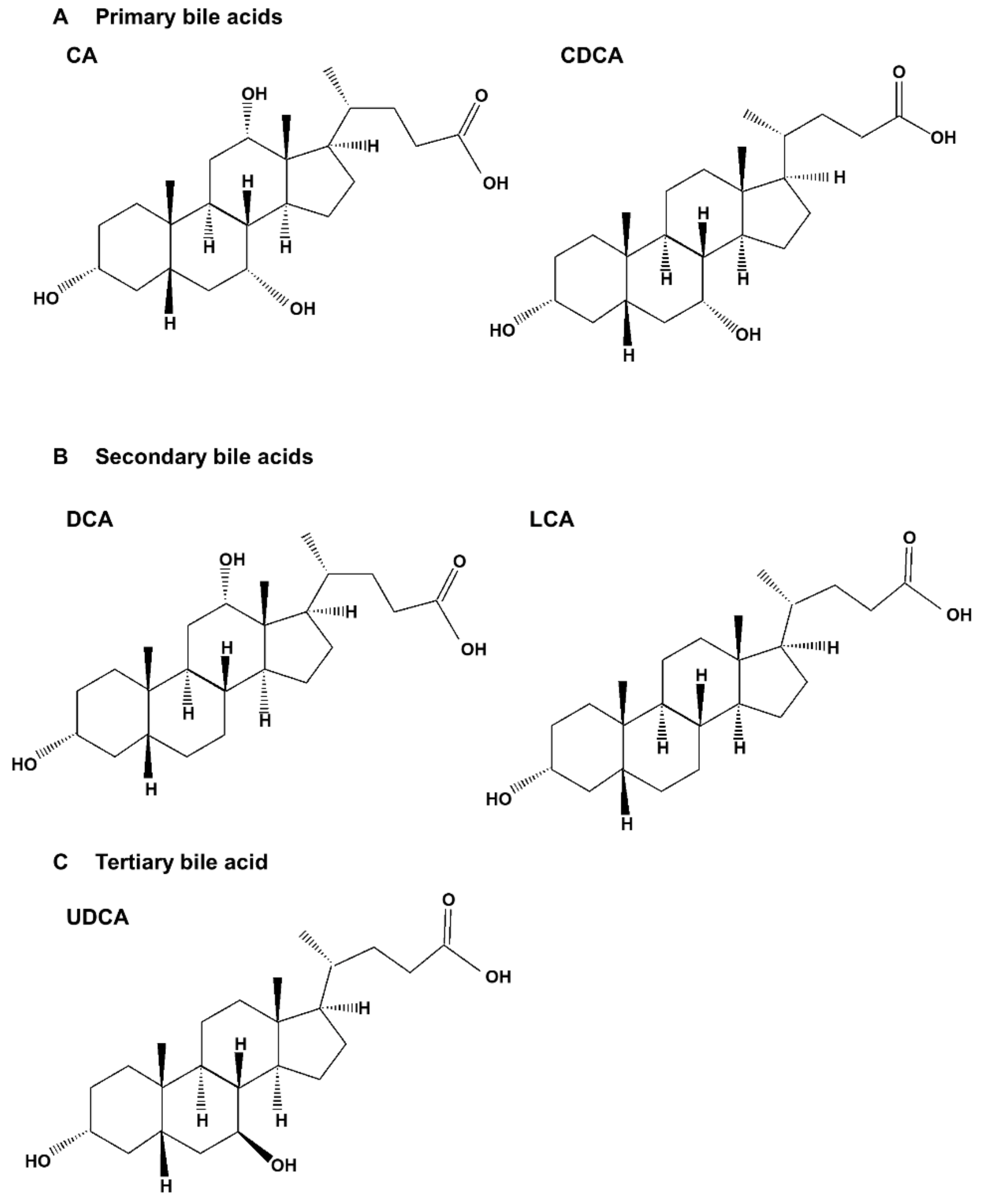
3.3.1. Effects of Primary Bile Acids on Apoptosis
3.3.2. Effects of Secondary and Tertiary Bile Acids on Apoptosis
4. Effect of Natural Bile Acids on Autophagy
4.1. Autophagy
4.2. Types of Autophagy
4.3. Bile Acid-Related Autophagy
5. Effect of Natural Bile Acids on Necroptosis
5.1. Necroptosis
5.2. Necroptosis in Cancer
5.3. Bile Acid-Related Necroptosis
6. Cell Death Mechanism of Synthetic Bile Acid Derivatives in Cancer
7. Conclusions
Author Contributions
Funding
Acknowledgments
Conflicts of Interest
References
- Wei, S.; Ma, X.; Zhao, Y. Mechanism of hydrophobic bile acid-induced hepatocyte injury and drug discovery. Front. Pharmacol. 2020, 11, 1084. [Google Scholar] [CrossRef] [PubMed]
- Kundu, S.; Kumar, S.; Bajaj, A. Cross-talk between bile acids and gastrointestinal tract for progression and development of cancer and its therapeutic implications. IUBMB life 2015, 67, 514–523. [Google Scholar] [CrossRef] [PubMed] [Green Version]
- Martinez, J.D.; Stratagoules, E.D.; LaRue, J.M.; Powell, A.A.; Gause, P.R.; Craven, M.T.; Payne, C.M.; Powell, M.B.; Gerner, E.W.; Earnest, D.L. Different bile acids exhibit distinct biological effects: The tumor promoter deoxycholic acid induces apoptosis and the chemopreventive agent ursodeoxycholic acid inhibits cell proliferation. Nutr. Cancer 1998, 31, 111–118. [Google Scholar] [CrossRef] [PubMed]
- Bajor, A.; Gillberg, P.G.; Abrahamsson, H. Bile acids: Short and long term effects in the intestine. Scand. J. Gastroenterol. 2010, 45, 645–664. [Google Scholar] [CrossRef]
- Martinot, E.; Sèdes, L.; Baptissart, M.; Lobaccaro, J.M.; Caira, F.; Beaudoin, C.; Volle, D.H. Bile acids and their receptors. Mol. Asp. Med. 2017, 56, 2–9. [Google Scholar] [CrossRef]
- Fracchia, M.; Pellegrino, S.; Secreto, P.; Pera, A.; Galatola, G. Biliary lipid composition in idiopathic bile acid malabsorption. Gut 1998, 43, 812–816. [Google Scholar] [CrossRef] [Green Version]
- Blanchet, M.; Brunel, J.M. Bile acid derivatives: From old molecules to a new potent therapeutic use: An overview. Curr. Med. Chem. 2018, 25, 3613–3636. [Google Scholar] [CrossRef]
- Di Ciaula, A.; Garruti, G.; Lunardi Baccetto, R.; Molina-Molina, E.; Bonfrate, L.; Wang, D.Q.; Portincasa, P. Bile acid physiology. Ann. Hepatol. 2017, 16, s4–s14. [Google Scholar] [CrossRef]
- Di Ciaula, A.; Wang, D.Q.; Molina-Molina, E.; Lunardi Baccetto, R.; Calamita, G.; Palmieri, V.O.; Portincasa, P. Bile acids and cancer: Direct and environmental-dependent effects. Ann. Hepatol. 2017, 16, s87–s105. [Google Scholar] [CrossRef]
- Zeng, H.; Umar, S.; Rust, B.; Lazarova, D.; Bordonaro, M. Secondary bile acids and short chain fatty acids in the colon: A focus on colonic microbiome, cell poliferation, inflammation, and cancer. Int. J. Mol. Sci. 2019, 20, 1214. [Google Scholar] [CrossRef] [Green Version]
- Rust, C.; Karnitz, L.M.; Paya, C.V.; Moscat, J.; Simari, R.D.; Gores, G.J. The bile acid taurochenodeoxycholate activates a phosphatidylinositol 3-kinase-dependent survival signaling cascade. J. Biol. Chem. 2000, 275, 20210–20216. [Google Scholar] [CrossRef] [PubMed] [Green Version]
- Ðanić, M.; Stanimirov, B.; Pavlović, N.; Goločorbin-Kon, S.; Al-Salami, H.; Stankov, K.; Mikov, M. Pharmacological applications of bile acids and their derivatives in the treatment of metabolic syndrome. Front. Pharmacol. 2018, 9. [Google Scholar] [CrossRef] [PubMed]
- Cook, J.; Kennaway, E.; Kennaway, N. Production of tumours in mice by deoxycholic acid. Nature 1940, 145, 627. [Google Scholar] [CrossRef]
- Park, M.J.; Kim, K.H.; Kim, H.Y.; Kim, K.; Cheong, J. Bile acid induces expression of COX-2 through the homeodomain transcription factor CDX1 and orphan nuclear receptor SHP in human gastric cancer cells. Carcinogenesis 2008, 29, 2385–2393. [Google Scholar] [CrossRef] [Green Version]
- Hu, Y.; Chau, T.; Liu, H.X.; Liao, D.; Keane, R.; Nie, Y.; Yang, H.; Wan, Y.J. Bile acids regulate nuclear receptor (Nur77) expression and intracellular location to control proliferation and apoptosis. Mol. Cancer Res. 2015, 13, 281–292. [Google Scholar] [CrossRef] [Green Version]
- Sharma, R.; Quilty, F.; Gilmer, J.F.; Long, A.; Byrne, A.M. Unconjugated secondary bile acids activate the unfolded protein response and induce golgi fragmentation via a src-kinase-dependant mechanism. Oncotarget 2017, 8, 967–978. [Google Scholar] [CrossRef] [Green Version]
- Farhana, L.; Nangia-Makker, P.; Arbit, E.; Shango, K.; Sarkar, S.; Mahmud, H.; Hadden, T.; Yu, Y.; Majumdar, A.P. Bile acid: A potential inducer of colon cancer stem cells. Stem Cell Res. Ther. 2016, 7, 181. [Google Scholar] [CrossRef] [Green Version]
- Liu, H.; Xu, H.W.; Zhang, Y.Z.; Huang, Y.; Han, G.Q.; Liang, T.J.; Wei, L.L.; Qin, C.Y.; Qin, C.K. Ursodeoxycholic acid induces apoptosis in hepatocellular carcinoma xenografts in mice. World J. Gastroenterol. 2015, 21, 10367–10374. [Google Scholar] [CrossRef]
- Wu, Y.C.; Chiu, C.F.; Hsueh, C.T.; Hsueh, C.T. The role of bile acids in cellular invasiveness of gastric cancer. Cancer Cell Int. 2018, 18, 75. [Google Scholar] [CrossRef]
- Pang, L.; Zhao, X.; Liu, W.; Deng, J.; Tan, X.; Qiu, L. Anticancer effect of ursodeoxycholic acid in human oral squamous carcinoma HSC-3 cells through the caspases. Nutrients 2015, 7, 3200–3218. [Google Scholar] [CrossRef] [Green Version]
- Phelan, J.P.; Reen, F.J.; Caparros-Martin, J.A.; O’Connor, R.; O’Gara, F. Rethinking the bile acid/gut microbiome axis in cancer. Oncotarget 2017, 8, 115736–115747. [Google Scholar] [CrossRef] [PubMed] [Green Version]
- Park, S.E.; Choi, H.J.; Yee, S.B.; Chung, H.Y.; Suh, H.; Choi, Y.H.; Yoo, Y.H.; Kim, N.D. Synthetic bile acid derivatives inhibit cell proliferation and induce apoptosis in HT-29 human colon cancer cells. Int. J. Oncol. 2004, 25, 231–236. [Google Scholar] [CrossRef]
- Katona, B.W.; Anant, S.; Covey, D.F.; Stenson, W.F. Characterization of enantiomeric bile acid-induced apoptosis in colon cancer cell lines. J. Biol. Chem. 2009, 284, 3354–3364. [Google Scholar] [CrossRef] [PubMed] [Green Version]
- Agarwal, D.S.; Mazumdar, S.; Italiya, K.S.; Chitkara, D.; Sakhuja, R. Bile-acid-appended triazolyl aryl ketones: Design, synthesis, in vitro anticancer activity and pharmacokinetics in rats. Molecules 2021, 26, 5741. [Google Scholar] [CrossRef] [PubMed]
- Melloni, E.; Marchesi, E.; Preti, L.; Casciano, F.; Rimondi, E.; Romani, A.; Secchiero, P.; Navacchia, M.L.; Perrone, D. Synthesis and biological investigation of bile acid-paclitaxel hybrids. Molecules 2022, 27, 471. [Google Scholar] [CrossRef] [PubMed]
- Brossard, D.; El Kihel, L.; Clément, M.; Sebbahi, W.; Khalid, M.; Roussakis, C.; Rault, S. Synthesis of bile acid derivatives and in vitro cytotoxic activity with pro-apoptotic process on multiple myeloma (KMS-11), glioblastoma multiforme (GBM), and colonic carcinoma (HCT-116) human cell lines. Eur. J. Med. Chem. 2010, 45, 2912–2918. [Google Scholar] [CrossRef]
- Singh, M.; Singh, A.; Kundu, S.; Bansal, S.; Bajaj, A. Deciphering the role of charge, hydration, and hydrophobicity for cytotoxic activities and membrane interactions of bile acid based facial amphiphiles. Biochim. Biophys. Acta 2013, 1828, 1926–1937. [Google Scholar] [CrossRef] [Green Version]
- El Kihel, L.; Clement, M.; Bazin, M.A.; Descamps, G.; Khalid, M.; Rault, S. New lithocholic and chenodeoxycholic piperazinylcarboxamides with antiproliferative and pro-apoptotic effects on human cancer cell lines. Bioorg. Med. Chem. 2008, 16, 8737–8744. [Google Scholar] [CrossRef]
- Singh, M.; Bansal, S.; Kundu, S.; Bhargava, P.; Singh, A.; Motiani, R.K.; Shyam, R.; Sreekanth, V.; Sengupta, S.; Bajaj, A. Synthesis, structure-activity relationship, and mechanistic investigation of lithocholic acid amphiphiles for colon cancer therapy. Medchemcomm 2015, 6, 192–201. [Google Scholar] [CrossRef] [Green Version]
- Sreekanth, V.; Bansal, S.; Motiani, R.K.; Kundu, S.; Muppu, S.K.; Majumdar, T.D.; Panjamurthy, K.; Sengupta, S.; Bajaj, A. Design, synthesis, and mechanistic investigations of bile acid-tamoxifen conjugates for breast cancer therapy. Bioconjug. Chem. 2013, 24, 1468–1484. [Google Scholar] [CrossRef]
- Tang, Y.; Blomenkamp, K.S.; Fickert, P.; Trauner, M.; Teckman, J.H. NorUDCA promotes degradation of α1-antitrypsin mutant Z protein by inducing autophagy through AMPK/ULK1 pathway. PLoS ONE 2018, 13, e0200897. [Google Scholar] [CrossRef] [PubMed]
- Markov, A.V.; Babich, V.O.; Popadyuk, I.I.; Salomatina, O.V.; Logashenko, E.B.; Salakhutdinov, N.F.; Zenkova, M.A. Novel derivatives of deoxycholic acid bearing linear aliphatic diamine and aminoalcohol moieties and their cyclic analogs at the C3 position: Synthesis and evaluation of their in vitro antitumor potential. Molecules 2019, 24, 2644. [Google Scholar] [CrossRef] [Green Version]
- Yan, G.; Elbadawi, M.; Efferth, T. Multiple cell death modalities and their key features (Review). World Acad. Sci. J. 2020, 2, 39–48. [Google Scholar] [CrossRef] [Green Version]
- Galluzzi, L.; Vitale, I.; Aaronson, S.A.; Abrams, J.M.; Adam, D.; Agostinis, P.; Alnemri, E.S.; Altucci, L.; Amelio, I.; Andrews, D.W.; et al. Molecular mechanisms of cell death: Recommendations of the Nomenclature Committee on Cell Death 2018. Cell Death Differ. 2018, 25, 486–541. [Google Scholar] [CrossRef]
- Nguyen, T.T.; Ung, T.T.; Kim, N.H.; Jung, Y.D. Role of bile acids in colon carcinogenesis. World J. Clin. Cases 2018, 6, 577–588. [Google Scholar] [CrossRef]
- Bernstein, H.; Bernstein, C.; Payne, C.M.; Dvorakova, K.; Garewal, H. Bile acids as carcinogens in human gastrointestinal cancers. Mutat. Res. 2005, 589, 47–65. [Google Scholar] [CrossRef]
- Schwabe, R.F.; Jobin, C. The microbiome and cancer. Nat. Rev. Cancer 2013, 13, 800–812. [Google Scholar] [CrossRef] [Green Version]
- Bernstein, C.; Holubec, H.; Bhattacharyya, A.K.; Nguyen, H.; Payne, C.M.; Zaitlin, B.; Bernstein, H. Carcinogenicity of deoxycholate, a secondary bile acid. Arch. Toxicol. 2011, 85, 863–871. [Google Scholar] [CrossRef] [Green Version]
- Payne, C.M.; Bernstein, C.; Dvorak, K.; Bernstein, H. Hydrophobic bile acids, genomic instability, Darwinian selection, and colon carcinogenesis. Clin. Exp. Gastroenterol. 2008, 1, 19–47. [Google Scholar] [CrossRef] [Green Version]
- Kazumori, H.; Ishihara, S.; Rumi, M.A.; Kadowaki, Y.; Kinoshita, Y. Bile acids directly augment caudal related homeobox gene Cdx2 expression in oesophageal keratinocytes in Barrett’s epithelium. Gut 2006, 55, 16–25. [Google Scholar] [CrossRef] [Green Version]
- Redlak, M.J.; Power, J.J.; Miller, T.A. Prevention of deoxycholate-induced gastric apoptosis by aspirin: Roles of NF-kappaB and PKC signaling. J. Surg. Res. 2008, 145, 66–73. [Google Scholar] [CrossRef] [PubMed]
- Wu, J.; Gong, J.; Geng, J.; Song, Y. Deoxycholic acid induces the overexpression of intestinal mucin, MUC2, via NF-kB signaling pathway in human esophageal adenocarcinoma cells. BMC Cancer 2008, 8, 333. [Google Scholar] [CrossRef] [PubMed] [Green Version]
- Tucker, O.N.; Dannenberg, A.J.; Yang, E.K.; Fahey, T.J., 3rd. Bile acids induce cyclooxygenase-2 expression in human pancreatic cancer cell lines. Carcinogenesis 2004, 25, 419–423. [Google Scholar] [CrossRef] [PubMed] [Green Version]
- Barrasa, J.I.; Olmo, N.; Lizarbe, M.A.; Turnay, J. Bile acids in the colon, from healthy to cytotoxic molecules. Toxicol. Vitr. 2013, 27, 964–977. [Google Scholar] [CrossRef] [PubMed]
- Sun, J.; Kato, I. Gut microbiota, inflammation and colorectal cancer. Genes Dis. 2016, 3, 130–143. [Google Scholar] [CrossRef] [Green Version]
- Zimber, A.; Chedeville, A.; Abita, J.P.; Barbu, V.; Gespach, C. Functional interactions between bile acids, all-trans retinoic acid, and 1,25-dihydroxy-vitamin D3 on monocytic differentiation and myeloblastin gene down-regulation in HL60 and THP-1 human leukemia cells. Cancer Res. 2000, 60, 672–678. [Google Scholar]
- Wu, Z.; Lu, Y.; Wang, B.; Liu, C.; Wang, Z.R. Effects of bile acids on proliferation and ultrastructural alteration of pancreatic cancer cell lines. World J. Gastroenterol. 2003, 9, 2759–2763. [Google Scholar] [CrossRef]
- Pyo, J.S.; Ko, Y.S.; Kang, G.; Kim, D.H.; Kim, W.H.; Lee, B.L.; Sohn, J.H. Bile acid induces MUC2 expression and inhibits tumor invasion in gastric carcinomas. J. Cancer Res. Clin. Oncol. 2015, 141, 1181–1188. [Google Scholar] [CrossRef]
- Yu, H.; Fu, Q.R.; Huang, Z.J.; Lin, J.Y.; Chen, Q.X.; Wang, Q.; Shen, D.Y. Apoptosis induced by ursodeoxycholic acid in human melanoma cells through the mitochondrial pathway. Oncol. Rep. 2019, 41, 213–223. [Google Scholar] [CrossRef] [Green Version]
- Vandewynckel, Y.P.; Laukens, D.; Devisscher, L.; Paridaens, A.; Bogaerts, E.; Verhelst, X.; Van den Bussche, A.; Raevens, S.; Van Steenkiste, C.; Van Troys, M.; et al. Tauroursodeoxycholic acid dampens oncogenic apoptosis induced by endoplasmic reticulum stress during hepatocarcinogen exposure. Oncotarget 2015, 6, 28011–28025. [Google Scholar] [CrossRef] [Green Version]
- Kim, Y.H.; Kim, J.H.; Kim, B.G.; Lee, K.L.; Kim, J.W.; Koh, S.J. Tauroursodeoxycholic acid attenuates colitis-associated colon cancer by inhibiting nuclear factor kappaB signaling. J. Gastroenterol. Hepatol. 2019, 34, 544–551. [Google Scholar] [CrossRef] [PubMed]
- Phelan, J.P.; Reen, F.J.; Dunphy, N.; O’Connor, R.; O’Gara, F. Bile acids destabilise HIF-1alpha and promote anti-tumour phenotypes in cancer cell models. BMC Cancer 2016, 16, 476. [Google Scholar] [CrossRef] [Green Version]
- Debruyne, P.R.; Bruyneel, E.A.; Li, X.; Zimber, A.; Gespach, C.; Mareel, M.M. The role of bile acids in carcinogenesis. Mutat. Res. 2001, 480–481, 359–369. [Google Scholar] [CrossRef]
- Alberts, D.S.; Martínez, M.E.; Hess, L.M.; Einspahr, J.G.; Green, S.B.; Bhattacharyya, A.K.; Guillen, J.; Krutzsch, M.; Batta, A.K.; Salen, G.; et al. Phase III trial of ursodeoxycholic acid to prevent colorectal adenoma recurrence. J. Natl. Cancer Inst. 2005, 97, 846–853. [Google Scholar] [CrossRef] [Green Version]
- Tung, B.Y.; Emond, M.J.; Haggitt, R.C.; Bronner, M.P.; Kimmey, M.B.; Kowdley, K.V.; Brentnall, T.A. Ursodiol use is associated with lower prevalence of colonic neoplasia in patients with ulcerative colitis and primary sclerosing cholangitis. Ann. Intern. Med. 2001, 134, 89–95. [Google Scholar] [CrossRef] [PubMed]
- Serfaty, L.; De Leusse, A.; Rosmorduc, O.; Desaint, B.; Flejou, J.F.; Chazouilleres, O.; Poupon, R.E.; Poupon, R. Ursodeoxycholic acid therapy and the risk of colorectal adenoma in patients with primary biliary cirrhosis: An observational study. Hepatology 2003, 38, 203–209. [Google Scholar] [CrossRef]
- Huang, W.K.; Hsu, H.C.; Liu, J.R.; Yang, T.S.; Chen, J.S.; Chang, J.W.; Lin, Y.C.; Yu, K.H.; Kuo, C.F.; See, L.C. The association of ursodeoxycholic acid use wth colorectal cancer risk: A nationwide cohort study. Medicine 2016, 95, e2980. [Google Scholar] [CrossRef]
- Kerr, J.F.; Wyllie, A.H.; Currie, A.R. Apoptosis: A basic biological phenomenon with wide-ranging implications in tissue kinetics. Br. J. Cancer 1972, 26, 239–257. [Google Scholar] [CrossRef] [Green Version]
- D’Arcy, M.S. Cell death: A review of the major forms of apoptosis, necrosis and autophagy. Cell Biol. Int. 2019, 43, 582–592. [Google Scholar] [CrossRef]
- Zhao, J.; Hu, Y.; Peng, J. Targeting programmed cell death in metabolic dysfunction-associated fatty liver disease (MAFLD): A promising new therapy. Cell. Mol. Biol. Lett. 2021, 26, 17. [Google Scholar] [CrossRef]
- Schwabe, R.F.; Luedde, T. Apoptosis and necroptosis in the liver: A matter of life and death. Nat. Rev. Gastroenterol. Hepatol. 2018, 15, 738–752. [Google Scholar] [CrossRef] [PubMed]
- Su, Z.; Yang, Z.; Xu, Y.; Chen, Y.; Yu, Q. Apoptosis, autophagy, necroptosis, and cancer metastasis. Mol. Cancer 2015, 14, 48. [Google Scholar] [CrossRef] [PubMed] [Green Version]
- Erekat, N.S. Apoptosis and its therapeutic implications in neurodegenerative diseases. Clin. Anat. 2022, 35, 65–78. [Google Scholar] [CrossRef]
- Obeng, E. Apoptosis (programmed cell death) and its signals—A review. Braz. J. Biol. 2021, 81, 1133–1143. [Google Scholar] [CrossRef]
- Mishra, A.P.; Salehi, B.; Sharifi-Rad, M.; Pezzani, R.; Kobarfard, F.; Sharifi-Rad, J.; Nigam, M. Programmed cell death, from a cancer perspective: An overview. Mol. Diagn. Ther. 2018, 22, 281–295. [Google Scholar] [CrossRef] [PubMed]
- Bertheloot, D.; Latz, E.; Franklin, B.S. Necroptosis, pyroptosis and apoptosis: An intricate game of cell death. Cell Mol. Immunol. 2021, 18, 1106–1121. [Google Scholar] [CrossRef] [PubMed]
- Fu, J.; Yu, M.; Xu, W.; Yu, S. Research progress of bile acids in cancer. Front. Oncol. 2021, 11, 778258. [Google Scholar] [CrossRef]
- Perez, M.J.; Briz, O. Bile-acid-induced cell injury and protection. World J. Gastroenterol. 2009, 15, 1677–1689. [Google Scholar] [CrossRef]
- Ignacio Barrasa, J.; Olmo, N.; Pérez-Ramos, P.; Santiago-Gómez, A.; Lecona, E.; Turnay, J.; Antonia Lizarbe, M. Deoxycholic and chenodeoxycholic bile acids induce apoptosis via oxidative stress in human colon adenocarcinoma cells. Apoptosis 2011, 16, 1054–1067. [Google Scholar] [CrossRef]
- Rolo, A.P.; Palmeira, C.M.; Holy, J.M.; Wallace, K.B. Role of mitochondrial dysfunction in combined bile acid-induced cytotoxicity: The switch between apoptosis and necrosis. Toxicol. Sci. 2004, 79, 196–204. [Google Scholar] [CrossRef] [Green Version]
- Shen, D.; Zeng, Y.; Zhang, W.; Li, Y.; Zhu, J.; Liu, Z.; Yan, Z.; Huang, J.A. Chenodeoxycholic acid inhibits lung adenocarcinoma progression via the integrin α5β1/FAK/p53 signaling pathway. Eur. J. Pharmacol. 2022, 923, 174925. [Google Scholar] [CrossRef] [PubMed]
- Higuchi, H.; Bronk, S.F.; Takikawa, Y.; Werneburg, N.; Takimoto, R.; El-Deiry, W.; Gores, G.J. The bile acid glycochenodeoxycholate induces trail-receptor 2/DR5 expression and apoptosis. J. Biol. Chem. 2001, 276, 38610–38618. [Google Scholar] [CrossRef] [PubMed] [Green Version]
- Bernt, C.; Vennegeerts, T.; Beuers, U.; Rust, C. The human transcription factor AP-1 is a mediator of bile acid-induced liver cell apoptosis. Biochem. Biophys. Res. Commun. 2006, 340, 800–806. [Google Scholar] [CrossRef]
- Iizaka, T.; Tsuji, M.; Oyamada, H.; Morio, Y.; Oguchi, K. Interaction between caspase-8 activation and endoplasmic reticulum stress in glycochenodeoxycholic acid-induced apoptotic HepG2 cells. Toxicology 2007, 241, 146–156. [Google Scholar] [CrossRef] [PubMed]
- Yang, H.B.; Song, W.; Cheng, M.D.; Fan, H.F.; Gu, X.; Qiao, Y.; Lu, X.; Yu, R.H.; Chen, L.Y. Deoxycholic acid inhibits the growth of BGC-823 gastric carcinoma cells via a p53-mediated pathway. Mol. Med. Rep. 2015, 11, 2749–2754. [Google Scholar] [CrossRef]
- Song, W.; Yang, H.B.; Chen, P.; Wang, S.M.; Zhao, L.P.; Xu, W.H.; Fan, H.F.; Gu, X.; Chen, L.Y. Apoptosis of human gastric carcinoma SGC-7901 induced by deoxycholic acid via the mitochondrial-dependent pathway. Appl. Biochem. Biotechnol. 2013, 171, 1061–1071. [Google Scholar] [CrossRef]
- Qiao, D.; Stratagouleas, E.D.; Martinez, J.D. Activation and role of mitogen-activated protein kinases in deoxycholic acid-induced apoptosis. Carcinogenesis 2001, 22, 35–41. [Google Scholar] [CrossRef] [Green Version]
- Yui, S.; Kanamoto, R.; Saeki, T. Deoxycholic acid can induce apoptosis in the human colon cancer cell line HCT116 in the absence of Bax. Nutr. Cancer 2008, 60, 91–96. [Google Scholar] [CrossRef]
- Zeng, H.; Safratowich, B.D.; Wang, T.T.Y.; Hamlin, S.K.; Johnson, L.K. Butyrate inhibits deoxycholic-acid-resistant colonic cell proliferation via cell cycle arrest and apoptosis: A potential pathway linking dietary fiber to cancer prevention. Mol. Nutr. Food Res. 2020, 64, e1901014. [Google Scholar] [CrossRef]
- Yui, S.; Saeki, T.; Kanamoto, R.; Iwami, K. Characteristics of apoptosis in HCT116 colon cancer cells induced by deoxycholic acid. J. Biochem. 2005, 138, 151–157. [Google Scholar] [CrossRef]
- Shiraki, K.; Ito, T.; Sugimoto, K.; Fuke, H.; Inoue, T.; Miyashita, K.; Yamanaka, T.; Suzuki, M.; Nabeshima, K.; Nakano, T.; et al. Different effects of bile acids, ursodeoxycholic acid and deoxycholic acid, on cell growth and cell death in human colonic adenocarcinoma cells. Int. J. Mol. Med. 2005, 16, 729–733. [Google Scholar] [PubMed]
- Luu, T.H.; Bard, J.M.; Carbonnelle, D.; Chaillou, C.; Huvelin, J.M.; Bobin-Dubigeon, C.; Nazih, H. Lithocholic bile acid inhibits lipogenesis and induces apoptosis in breast cancer cells. Cell. Oncol. 2018, 41, 13–24. [Google Scholar] [CrossRef] [PubMed]
- Goldberg, A.A.; Titorenko, V.I.; Beach, A.; Sanderson, J.T. Bile acids induce apoptosis selectively in androgen-dependent and -independent prostate cancer cells. PeerJ 2013, 1, e122. [Google Scholar] [CrossRef]
- Trah, J.; Arand, J.; Oh, J.; Pagerols-Raluy, L.; Trochimiuk, M.; Appl, B.; Heidelbach, H.; Vincent, D.; Saleem, M.A.; Reinshagen, K.; et al. Lithocholic bile acid induces apoptosis in human nephroblastoma cells: A non-selective treatment option. Sci. Rep. 2020, 10, 20349. [Google Scholar] [CrossRef] [PubMed]
- Goldberg, A.A.; Beach, A.; Davies, G.F.; Harkness, T.A.; Leblanc, A.; Titorenko, V.I. Lithocholic bile acid selectively kills neuroblastoma cells, while sparing normal neuronal cells. Oncotarget 2011, 2, 761–782. [Google Scholar] [CrossRef] [PubMed] [Green Version]
- Zhao, M.X.; Cai, Z.C.; Zhu, B.J.; Zhang, Z.Q. The apoptosis effect on liver cancer cells of gold nanoparticles modified with lithocholic acid. Nanoscale Res. Lett. 2018, 13, 304. [Google Scholar] [CrossRef] [PubMed]
- Yao, Z.; Zhang, X.; Zhao, F.; Wang, S.; Chen, A.; Huang, B.; Wang, J.; Li, X. Ursodeoxycholic acid inhibits glioblastoma progression via endoplasmic reticulum stress related apoptosis and synergizes with the proteasome inhibitor bortezomib. ACS Chem. Neurosci. 2020, 11, 1337–1346. [Google Scholar] [CrossRef]
- Lee, W.S.; Jung, J.H.; Panchanathan, R.; Yun, J.W.; Kim, D.H.; Kim, H.J.; Kim, G.S.; Ryu, C.H.; Shin, S.C.; Hong, S.C.; et al. Ursodeoxycholic acid induces death receptor-mediated apoptosis in prostate cancer cells. J. Cancer Prev. 2017, 22, 16–21. [Google Scholar] [CrossRef] [Green Version]
- Zhu, L.; Shan, L.J.; Liu, Y.J.; Chen, D.; Xiao, X.G.; Li, Y. Ursodeoxycholic acid induces apoptosis of hepatocellular carcinoma cells in vitro. J. Dig. Dis. 2014, 15, 684–693. [Google Scholar] [CrossRef]
- Tsagarakis, N.J.; Drygiannakis, I.; Batistakis, A.G.; Kolios, G.; Kouroumalis, E.A. A concentration-dependent effect of ursodeoxycholate on apoptosis and caspases activities of HepG2 hepatocellular carcinoma cells. Eur. J. Pharmacol. 2010, 640, 1–7. [Google Scholar] [CrossRef]
- Lim, S.C.; Duong, H.Q.; Parajuli, K.R.; Han, S.I. Pro-apoptotic role of the MEK/ERK pathway in ursodeoxycholic acid-induced apoptosis in SNU601 gastric cancer cells. Oncol. Rep. 2012, 28, 1429–1434. [Google Scholar] [CrossRef] [Green Version]
- Lim, S.C.; Duong, H.Q.; Choi, J.E.; Lee, T.B.; Kang, J.H.; Oh, S.H.; Han, S.I. Lipid raft-dependent death receptor 5 (DR5) expression and activation are critical for ursodeoxycholic acid-induced apoptosis in gastric cancer cells. Carcinogenesis 2011, 32, 723–731. [Google Scholar] [CrossRef] [PubMed] [Green Version]
- Jung, H.W.; Hwang, J.H. Anticancer effects of ursi fel extract and its active compound, ursodeoxycholic acid, in FRO anaplastic thyroid cancer cells. Molecules 2021, 26, 5309. [Google Scholar] [CrossRef] [PubMed]
- Vlahcevic, Z.R.; Heuman, D.M.; Hylemon, P.B. Regulation of bile acid synthesis. Hepatology 1991, 13, 590–600. [Google Scholar] [CrossRef]
- Chiang, J.Y.L. Bile acids: Regulation of synthesis. J. Lipid Res. 2009, 50, 1955–1966. [Google Scholar] [CrossRef] [PubMed] [Green Version]
- Grant, S.M.; DeMorrow, S. Bile acid signaling in neurodegenerative and neurological disorders. Int. J. Mol. Sci. 2020, 21, 5982. [Google Scholar] [CrossRef]
- Chiang, J.Y.L. Recent advances in understanding bile acid homeostasis. F1000Research 2017, 6, 2029. [Google Scholar] [CrossRef]
- Chiang, J.Y.L.; Ferrell, J.M. Bile acids as metabolic regulators and nutrient sensors. Annu. Rev. Nutr. 2019, 39, 175–200. [Google Scholar] [CrossRef]
- Jia, W.; Xie, G.; Jia, W. Bile acid-microbiota crosstalk in gastrointestinal inflammation and carcinogenesis. Nat. Rev. Gastroenterol. Hepatol. 2018, 15, 111–128. [Google Scholar] [CrossRef] [Green Version]
- Jang, J.Y.; Sung, B.; Kim, N.D. Role of induced programmed cell death in the chemopreventive potential of apigenin. Int. J. Mol. Sci. 2022, 23, 3757. [Google Scholar] [CrossRef]
- Su, T.; Li, X.; Yang, M.; Shao, Q.; Zhao, Y.; Ma, C.; Wang, P. Autophagy: An intracellular degradation pathway regulating plant survival and stress response. Front. Plant Sci. 2020, 11, 164. [Google Scholar] [CrossRef] [PubMed] [Green Version]
- Li, X.; He, S.; Ma, B. Autophagy and autophagy-related proteins in cancer. Mol. Cancer 2020, 19, 12. [Google Scholar] [CrossRef]
- Ravanan, P.; Srikumar, I.F.; Talwar, P. Autophagy: The spotlight for cellular stress responses. Life Sci. 2017, 188, 53–67. [Google Scholar] [CrossRef] [PubMed]
- Zada, S.; Hwang, J.S.; Ahmed, M.; Lai, T.H.; Pham, T.M.; Elashkar, O.; Kim, D.R. Cross talk between autophagy and oncogenic signaling pathways and implications for cancer therapy. Biochim. Biophys. Acta Rev. Cancer 2021, 1876, 188565. [Google Scholar] [CrossRef] [PubMed]
- Yang, Q.; Wang, R.; Zhu, L. Chaperone-mediated autophagy. Adv. Exp. Med. Biol. 2019, 1206, 435–452. [Google Scholar] [CrossRef] [PubMed]
- Menon, M.B.; Dhamija, S. Beclin 1 phosphorylation - at the center of autophagy regulation. Front. Cell Dev. Biol. 2018, 6, 137. [Google Scholar] [CrossRef] [Green Version]
- Wijshake, T.; Zou, Z.; Chen, B.; Zhong, L.; Xiao, G.; Xie, Y.; Doench, J.G.; Bennett, L.; Levine, B. Tumor-suppressor function of Beclin 1 in breast cancer cells requires E-cadherin. Proc. Natl. Acad. Sci. USA 2021, 118. [Google Scholar] [CrossRef]
- Vega-Rubín-de-Celis, S. The role of beclin 1-dependent autophagy in cancer. Biology 2019, 9, 4. [Google Scholar] [CrossRef] [Green Version]
- Roesly, H.B.; Khan, M.R.; Chen, H.D.; Hill, K.A.; Narendran, N.; Watts, G.S.; Chen, X.; Dvorak, K. The decreased expression of Beclin-1 correlates with progression to esophageal adenocarcinoma: The role of deoxycholic acid. Am. J. Physiol. Gastrointest. Liver Physiol. 2012, 302, G864–G872. [Google Scholar] [CrossRef]
- Gafar, A.A.; Draz, H.M.; Goldberg, A.A.; Bashandy, M.A.; Bakry, S.; Khalifa, M.A.; AbuShair, W.; Titorenko, V.I.; Sanderson, J.T. Lithocholic acid induces endoplasmic reticulum stress, autophagy and mitochondrial dysfunction in human prostate cancer cells. PeerJ 2016, 4, e2445. [Google Scholar] [CrossRef]
- Lim, S.C.; Han, S.I. Ursodeoxycholic acid effectively kills drug-resistant gastric cancer cells through induction of autophagic death. Oncol. Rep. 2015, 34, 1261–1268. [Google Scholar] [CrossRef] [PubMed] [Green Version]
- Zanetti, L.C.; Weinlich, R. Necroptosis, the other main caspase-independent dell death. Adv. Exp. Med. Biol. 2021, 1301, 123–138. [Google Scholar] [CrossRef] [PubMed]
- Choi, M.E.; Price, D.R.; Ryter, S.W.; Choi, A.M.K. Necroptosis: A crucial pathogenic mediator of human disease. JCI Insight 2019, 4, e128834. [Google Scholar] [CrossRef]
- Linkermann, A.; Green, D.R. Necroptosis. N. Engl. J. Med. 2014, 370, 455–465. [Google Scholar] [CrossRef] [PubMed] [Green Version]
- Kaczmarek, A.; Vandenabeele, P.; Krysko, D.V. Necroptosis: The release of damage-associated molecular patterns and its physiological relevance. Immunity 2013, 38, 209–223. [Google Scholar] [CrossRef] [Green Version]
- Tonnus, W.; Meyer, C.; Paliege, A.; Belavgeni, A.; von Mässenhausen, A.; Bornstein, S.R.; Hugo, C.; Becker, J.U.; Linkermann, A. The pathological features of regulated necrosis. J. Pathol. 2019, 247, 697–707. [Google Scholar] [CrossRef] [PubMed]
- Kaiser, W.J.; Sridharan, H.; Huang, C.; Mandal, P.; Upton, J.W.; Gough, P.J.; Sehon, C.A.; Marquis, R.W.; Bertin, J.; Mocarski, E.S. Toll-like receptor 3-mediated necrosis via TRIF, RIP3, and MLKL. J. Biol. Chem. 2013, 288, 31268–31279. [Google Scholar] [CrossRef] [Green Version]
- Weinlich, R.; Oberst, A.; Beere, H.M.; Green, D.R. Necroptosis in development, inflammation and disease. Nat. Rev. Mol. Cell Biol. 2017, 18, 127–136. [Google Scholar] [CrossRef]
- Chen, J.; Kos, R.; Garssen, J.; Redegeld, F. Molecular insights into the mechanism of necroptosis: The necrosome as a potential therapeutic target. Cells 2019, 8, 1486. [Google Scholar] [CrossRef] [Green Version]
- Silke, J.; Rickard, J.A.; Gerlic, M. The diverse role of RIP kinases in necroptosis and inflammation. Nat. Immunol. 2015, 16, 689–697. [Google Scholar] [CrossRef]
- Xu, Y.; Han, J. The necrosome in acute kidney injury. Semin. Nephrol. 2016, 36, 199–207. [Google Scholar] [CrossRef] [PubMed]
- Zhang, J.; Yang, Y.; He, W.; Sun, L. Necrosome core machinery: MLKL. Cell. Mol. Life Sci. 2016, 73, 2153–2163. [Google Scholar] [CrossRef] [PubMed]
- Seifert, L.; Miller, G. Molecular pathways: The necrosome-a target for cancer therapy. Clin. Cancer. Res. 2017, 23, 1132–1136. [Google Scholar] [CrossRef] [PubMed] [Green Version]
- Cai, Z.; Jitkaew, S.; Zhao, J.; Chiang, H.C.; Choksi, S.; Liu, J.; Ward, Y.; Wu, L.G.; Liu, Z.G. Plasma membrane translocation of trimerized MLKL protein is required for TNF-induced necroptosis. Nat. Cell Biol. 2014, 16, 55–65. [Google Scholar] [CrossRef]
- Maelfait, J.; Liverpool, L.; Bridgeman, A.; Ragan, K.B.; Upton, J.W.; Rehwinkel, J. Sensing of viral and endogenous RNA by ZBP1/DAI induces necroptosis. EMBO J. 2017, 36, 2529–2543. [Google Scholar] [CrossRef] [PubMed]
- Riegler, A.N.; Brissac, T.; Gonzalez-Juarbe, N.; Orihuela, C.J. Necroptotic cell death promotes adaptive immunity against colonizing pneumococci. Front. Immunol. 2019, 10, 615. [Google Scholar] [CrossRef] [Green Version]
- Galluzzi, L.; Kepp, O.; Chan, F.K.; Kroemer, G. Necroptosis: Mechanisms and relevance to disease. Annu. Rev. Pathol. 2017, 12, 103–130. [Google Scholar] [CrossRef]
- Gong, Y.; Fan, Z.; Luo, G.; Yang, C.; Huang, Q.; Fan, K.; Cheng, H.; Jin, K.; Ni, Q.; Yu, X.; et al. The role of necroptosis in cancer biology and therapy. Mol. Cancer 2019, 18, 100. [Google Scholar] [CrossRef] [Green Version]
- Stoll, G.; Ma, Y.; Yang, H.; Kepp, O.; Zitvogel, L.; Kroemer, G. Pro-necrotic molecules impact local immunosurveillance in human breast cancer. Oncoimmunology 2017, 6, e1299302. [Google Scholar] [CrossRef] [Green Version]
- Li, X.; Guo, J.; Ding, A.P.; Qi, W.W.; Zhang, P.H.; Lv, J.; Qiu, W.S.; Sun, Z.Q. Association of mixed lineage kinase domain-like protein expression with prognosis in patients with colon cancer. Technol. Cancer Res. Treat. 2017, 16, 428–434. [Google Scholar] [CrossRef] [Green Version]
- Seifert, L.; Werba, G.; Tiwari, S.; Giao Ly, N.N.; Alothman, S.; Alqunaibit, D.; Avanzi, A.; Barilla, R.; Daley, D.; Greco, S.H.; et al. The necrosome promotes pancreatic oncogenesis via CXCL1 and Mincle-induced immune suppression. Nature 2016, 532, 245–249. [Google Scholar] [CrossRef] [PubMed] [Green Version]
- Ertao, Z.; Jianhui, C.; Kang, W.; Zhijun, Y.; Hui, W.; Chuangqi, C.; Changjiang, Q.; Sile, C.; Yulong, H.; Shirong, C. Prognostic value of mixed lineage kinase domain-like protein expression in the survival of patients with gastric caner. Tumour. Biol. 2016, 37, 13679–13685. [Google Scholar] [CrossRef] [PubMed]
- Strilic, B.; Yang, L.; Albarrán-Juárez, J.; Wachsmuth, L.; Han, K.; Müller, U.C.; Pasparakis, M.; Offermanns, S. Tumour-cell-induced endothelial cell necroptosis via death receptor 6 promotes metastasis. Nature 2016, 536, 215–218. [Google Scholar] [CrossRef] [PubMed]
- Fulda, S. Targeting apoptosis for anticancer therapy. Semin. Cancer Biol. 2015, 31, 84–88. [Google Scholar] [CrossRef]
- Qin, X.; Ma, D.; Tan, Y.X.; Wang, H.Y.; Cai, Z. The role of necroptosis in cancer: A double-edged sword? Biochim. Biophys. Acta Rev. Cancer 2019, 1871, 259–266. [Google Scholar] [CrossRef]
- Wada, N.; Kawano, Y.; Fujiwara, S.; Kikukawa, Y.; Okuno, Y.; Tasaki, M.; Ueda, M.; Ando, Y.; Yoshinaga, K.; Ri, M.; et al. Shikonin, dually functions as a proteasome inhibitor and a necroptosis inducer in multiple myeloma cells. Int. J. Oncol. 2015, 46, 963–972. [Google Scholar] [CrossRef] [Green Version]
- Huang, C.; Luo, Y.; Zhao, J.; Yang, F.; Zhao, H.; Fan, W.; Ge, P. Shikonin kills glioma cells through necroptosis mediated by RIP-1. PLoS ONE 2013, 8, e66326. [Google Scholar] [CrossRef] [Green Version]
- Brown, M.F.; Leibowitz, B.J.; Chen, D.; He, K.; Zou, F.; Sobol, R.W.; Beer-Stolz, D.; Zhang, L.; Yu, J. Loss of caspase-3 sensitizes colon cancer cells to genotoxic stress via RIP1-dependent necrosis. Cell Death Dis. 2015, 6, e1729. [Google Scholar] [CrossRef] [Green Version]
- Tenev, T.; Bianchi, K.; Darding, M.; Broemer, M.; Langlais, C.; Wallberg, F.; Zachariou, A.; Lopez, J.; MacFarlane, M.; Cain, K.; et al. The Ripoptosome, a signaling platform that assembles in response to genotoxic stress and loss of IAPs. Mol. Cell 2011, 43, 432–448. [Google Scholar] [CrossRef]
- Xu, Y.; Ma, H.B.; Fang, Y.L.; Zhang, Z.R.; Shao, J.; Hong, M.; Huang, C.J.; Liu, J.; Chen, R.Q. Cisplatin-induced necroptosis in TNFα dependent and independent pathways. Cell. Signal. 2017, 31, 112–123. [Google Scholar] [CrossRef]
- Hannes, S.; Abhari, B.A.; Fulda, S. Smac mimetic triggers necroptosis in pancreatic carcinoma cells when caspase activation is blocked. Cancer Lett. 2016, 380, 31–38. [Google Scholar] [CrossRef] [PubMed]
- Fulda, S. Promises and challenges of Smac mimetics as cancer therapeutics. Clin. Cancer. Res. 2015, 21, 5030–5036. [Google Scholar] [CrossRef] [PubMed] [Green Version]
- Cekay, M.J.; Roesler, S.; Frank, T.; Knuth, A.K.; Eckhardt, I.; Fulda, S. Smac mimetics and type II interferon synergistically induce necroptosis in various cancer cell lines. Cancer Lett. 2017, 410, 228–237. [Google Scholar] [CrossRef]
- Safferthal, C.; Rohde, K.; Fulda, S. Therapeutic targeting of necroptosis by Smac mimetic bypasses apoptosis resistance in acute myeloid leukemia cells. Oncogene 2017, 36, 1487–1502. [Google Scholar] [CrossRef] [PubMed]
- Weber, K.; Roelandt, R.; Bruggeman, I.; Estornes, Y.; Vandenabeele, P. Nuclear RIPK3 and MLKL contribute to cytosolic necrosome formation and necroptosis. Commun. Biol. 2018, 1, 6. [Google Scholar] [CrossRef] [PubMed]
- Petrie, E.J.; Sandow, J.J.; Jacobsen, A.V.; Smith, B.J.; Griffin, M.D.W.; Lucet, I.S.; Dai, W.; Young, S.N.; Tanzer, M.C.; Wardak, A.; et al. Conformational switching of the pseudokinase domain promotes human MLKL tetramerization and cell death by necroptosis. Nat. Commun. 2018, 9, 2422. [Google Scholar] [CrossRef] [Green Version]
- Zhu, K.; Liang, W.; Ma, Z.; Xu, D.; Cao, S.; Lu, X.; Liu, N.; Shan, B.; Qian, L.; Yuan, J. Necroptosis promotes cell-autonomous activation of proinflammatory cytokine gene expression. Cell Death Dis. 2018, 9, 500. [Google Scholar] [CrossRef]
- De Almagro, M.C.; Goncharov, T.; Izrael-Tomasevic, A.; Duttler, S.; Kist, M.; Varfolomeev, E.; Wu, X.; Lee, W.P.; Murray, J.; Webster, J.D.; et al. Coordinated ubiquitination and phosphorylation of RIP1 regulates necroptotic cell death. Cell Death Differ. 2017, 24, 26–37. [Google Scholar] [CrossRef]
- Dhuriya, Y.K.; Sharma, D. Necroptosis: A regulated inflammatory mode of cell death. J. Neuroinflamm. 2018, 15, 199. [Google Scholar] [CrossRef] [Green Version]
- Meng, H.; Liu, Z.; Li, X.; Wang, H.; Jin, T.; Wu, G.; Shan, B.; Christofferson, D.E.; Qi, C.; Yu, Q.; et al. Death-domain dimerization-mediated activation of RIPK1 controls necroptosis and RIPK1-dependent apoptosis. Proc. Natl. Acad. Sci. USA 2018, 115, E2001–E2009. [Google Scholar] [CrossRef] [Green Version]
- Chen, X.; He, W.T.; Hu, L.; Li, J.; Fang, Y.; Wang, X.; Xu, X.; Wang, Z.; Huang, K.; Han, J. Pyroptosis is driven by non-selective gasdermin-D pore and its morphology is different from MLKL channel-mediated necroptosis. Cell Res. 2016, 26, 1007–1020. [Google Scholar] [CrossRef] [PubMed]
- Hoff, J.; Xiong, L.; Kammann, T.; Neugebauer, S.; Micheel, J.M.; Ghait, M.; Deshmukh, S.; Gaßler, N.; Bauer, M.; Press, A.T. RIPK3 promoter hypermethylation in hepatocytes protects from bile acid induced inflammation and necroptosis. bioRxiv 2021. [Google Scholar] [CrossRef]
- Cao, M.; Chen, F.; Xie, N.; Cao, M.Y.; Chen, P.; Lou, Q.; Zhao, Y.; He, C.; Zhang, S.; Song, X.; et al. c-Jun N-terminal kinases differentially regulate TNF- and TLRs-mediated necroptosis through their kinase-dependent and -independent activities. Cell Death Dis. 2018, 9, 1140. [Google Scholar] [CrossRef] [PubMed] [Green Version]
- Umebashi, K.; Tokito, A.; Yamamoto, M.; Jougasaki, M. Interleukin-33 induces interleukin-8 expression via JNK/c-Jun/AP-1 pathway in human umbilical vein endothelial cells. PLoS ONE 2018, 13, e0191659. [Google Scholar] [CrossRef] [PubMed] [Green Version]
- Afonso, M.B.; Rodrigues, P.M.; Simão, A.L.; Ofengeim, D.; Carvalho, T.; Amaral, J.D.; Gaspar, M.M.; Cortez-Pinto, H.; Castro, R.E.; Yuan, J.; et al. Activation of necroptosis in human and experimental cholestasis. Cell Death Dis. 2016, 7, e2390. [Google Scholar] [CrossRef] [Green Version]
- Yang, H.; Duan, Z. Bile acids and the potential role in primary biliary cirrhosis. Digestion 2016, 94, 145–153. [Google Scholar] [CrossRef]
- Lan, W.; Chen, Z.; Chen, Y.; Tan, M.; Chen, Y.; Chen, J.; Chi, X.; Chen, Y. Glycochenodeoxycholic acid impairs transcription factor E3-dependent autophagy-lysosome machinery by disrupting reactive oxygen species homeostasis in L02 cells. Toxicol. Lett. 2020, 331, 11–21. [Google Scholar] [CrossRef]
- Van de Wiel, S.M.W.; Porteiro, B.; Belt, S.C.; Vogels, E.W.M.; Bolt, I.; Vermeulen, J.L.M.; de Waart, D.R.; Verheij, J.; Muncan, V.; Oude Elferink, R.P.J.; et al. Differential and organ-specific functions of organic solute transporter α and β in experimental cholestasis. JHEP Rep. 2022, 4, 100463. [Google Scholar] [CrossRef]
- Zhou, X.; Xie, L.; Bergmann, F.; Endris, V.; Strobel, O.; Büchler, M.W.; Kroemer, G.; Hackert, T.; Fortunato, F. The bile acid receptor FXR attenuates acinar cell autophagy in chronic pancreatitis. Cell Death Discov. 2017, 3, 17027. [Google Scholar] [CrossRef]
- Muili, K.A.; Wang, D.; Orabi, A.I.; Sarwar, S.; Luo, Y.; Javed, T.A.; Eisses, J.F.; Mahmood, S.M.; Jin, S.; Singh, V.P.; et al. Bile acids induce pancreatic acinar cell injury and pancreatitis by activating calcineurin. J. Biol. Chem. 2013, 288, 570–580. [Google Scholar] [CrossRef] [Green Version]
- Choi, Y.H.; Im, E.O.; Suh, H.; Jin, Y.; Lee, W.H.; Yoo, Y.H.; Kim, K.W.; Kim, N.D. Apoptotic activity of novel bile acid derivatives in human leukemic T cells through the activation of caspases. Int. J. Oncol. 2001, 18, 979–984. [Google Scholar] [CrossRef] [PubMed]
- Im, E.; Choi, S.H.; Suh, H.; Choi, Y.H.; Yoo, Y.H.; Kim, N.D. Synthetic bile acid derivatives induce apoptosis through a c-Jun N-terminal kinase and NF-kappaB-dependent process in human cervical carcinoma cells. Cancer Lett. 2005, 229, 49–57. [Google Scholar] [CrossRef] [PubMed]
- Im, E.O.; Choi, Y.H.; Paik, K.J.; Suh, H.; Jin, Y.; Kim, K.W.; Yoo, Y.H.; Kim, N.D. Novel bile acid derivatives induce apoptosis via a p53-independent pathway in human breast carcinoma cells. Cancer Lett. 2001, 163, 83–93. [Google Scholar] [CrossRef]
- Choi, Y.H.; Im, E.O.; Suh, H.; Jin, Y.; Yoo, Y.H.; Kim, N.D. Apoptosis and modulation of cell cycle control by synthetic derivatives of ursodeoxycholic acid and chenodeoxycholic acid in human prostate cancer cells. Cancer Lett. 2003, 199, 157–167. [Google Scholar] [CrossRef]
- Jeong, J.H.; Park, J.S.; Moon, B.; Kim, M.C.; Kim, J.K.; Lee, S.; Suh, H.; Kim, N.D.; Kim, J.M.; Park, Y.C.; et al. Orphan nuclear receptor Nur77 translocates to mitochondria in the early phase of apoptosis induced by synthetic chenodeoxycholic acid derivatives in human stomach cancer cell line SNU-1. Ann. N. Y. Acad. Sci. 2003, 1010, 171–177. [Google Scholar] [CrossRef]
- Moon, B.; Kim, M.C.; Park, J.S. Synthetic CDCA derivatives-induced apoptosis of stomach cancer cell line SNU-1 cells. Cancer Res. Treat. 2004, 36, 132–139. [Google Scholar] [CrossRef] [Green Version]
- Yee, S.B.; Yeo, W.J.; Park, B.S.; Kim, J.Y.; Baek, S.J.; Kim, Y.C.; Seo, S.Y.; Lee, S.H.; Kim, J.H.; Suh, H.; et al. Synthetic chenodeoxycholic acid derivatives inhibit glioblastoma multiform tumor growth in vitro and in vivo. Int. J. Oncol. 2005, 27, 653–659. [Google Scholar]
- Yee, S.B.; Song, Y.S.; Jeong, S.H.; Lee, H.S.; Seo, S.Y.; Kim, J.H.; Suh, H.; Kim, N.D.; Yoo, Y.H. A novel chenodeoxycholic derivative HS-1200 enhances radiation-induced apoptosis in MCF-7 cells. Oncol. Rep. 2007, 17, 919–923. [Google Scholar] [CrossRef] [Green Version]
- Park, S.E.; Lee, S.W.; Hossain, M.A.; Kim, M.Y.; Kim, M.N.; Ahn, E.Y.; Park, Y.C.; Suh, H.; Kim, G.Y.; Choi, Y.H.; et al. A chenodeoxycholic derivative, HS-1200, induces apoptosis and cell cycle modulation via Egr-1 gene expression control on human hepatoma cells. Cancer Lett. 2008, 270, 77–86. [Google Scholar] [CrossRef]
- Liu, H.; Qin, C.K.; Han, G.Q.; Xu, H.W.; Ren, W.H.; Qin, C.Y. Synthetic chenodeoxycholic acid derivative, HS-1200, induces apoptosis of human hepatoma cells via a mitochondrial pathway. Cancer Lett. 2008, 270, 242–249. [Google Scholar] [CrossRef]
- Kim, T.H.; Yoo, Y.H.; Kang, D.Y.; Suh, H.; Park, M.K.; Park, K.J.; Kim, S.H. Efficacy on anaplastic thyroid carcinoma of valproic acid alone or in combination with doxorubicin, a synthetic chenodeoxycholic acid derivative, or lactacystin. Int. J. Oncol. 2009, 34, 1353–1362. [Google Scholar] [CrossRef] [PubMed] [Green Version]
- Kim, N.D.; Im, E.; Yoo, Y.H.; Choi, Y.H. Modulation of the cell cycle and induction of apoptosis in human cancer cells by synthetic bile acids. Curr. Cancer Drug Targets 2006, 6, 681–689. [Google Scholar] [CrossRef]
- Aubrey, B.J.; Kelly, G.L.; Janic, A.; Herold, M.J.; Strasser, A. How does p53 induce apoptosis and how does this relate to p53-mediated tumour suppression? Cell Death Differ. 2018, 25, 104–113. [Google Scholar] [CrossRef] [PubMed] [Green Version]
- Unnisa, A.; Greig, N.H.; Kamal, M.A. Inhibition of caspase 3 and caspase 9 mediated apoptosis: A multimodal therapeutic target in traumatic brain injury. Curr. Neuropharmacol. 2022, 20, 121898. [Google Scholar] [CrossRef]
- Lopez, J.; Tait, S.W. Mitochondrial apoptosis: Killing cancer using the enemy within. Br. J. Cancer 2015, 112, 957–962. [Google Scholar] [CrossRef] [PubMed] [Green Version]
- Boice, A.; Bouchier-Hayes, L. Targeting apoptotic caspases in cancer. Biochim. Biophys. Acta Mol. Cell Res. 2020, 1867, 118688. [Google Scholar] [CrossRef] [PubMed]
- Zaman, S.; Wang, R.; Gandhi, V. Targeting the apoptosis pathway in hematologic malignancies. Leuk. Lymphoma 2014, 55, 1980–1992. [Google Scholar] [CrossRef]
- Zhao, D.Y.; Jacobs, K.M.; Hallahan, D.E.; Thotala, D. Silencing Egr1 attenuates radiation-induced apoptosis in normal tissues while killing cancer cells and delaying tumor growth. Mol. Cancer Ther. 2015, 14, 2343–2352. [Google Scholar] [CrossRef] [Green Version]
- Gitenay, D.; Baron, V.T. Is EGR1 a potential target for prostate cancer therapy? Future Oncol. 2009, 5, 993–1003. [Google Scholar] [CrossRef] [Green Version]
- Oh, S.; Kim, H.; Nam, K.; Shin, I. Egr-1 is required for neu/HER2-induced mammary tumors. Cell. Signal. 2018, 45, 102–109. [Google Scholar] [CrossRef]
- Lee, S.M.; Park, M.S.; Park, S.Y.; Choi, Y.D.; Chung, J.O.; Kim, D.H.; Jung, Y.D.; Kim, H.S. Primary bile acid activates Egr-1 expression through the MAPK signaling pathway in gastric cancer. Mol. Med. Rep. 2022, 25, 129. [Google Scholar] [CrossRef] [PubMed]
- Kim, N.D.; Moon, J.O.; Slitt, A.L.; Copple, B.L. Early growth response factor-1 is critical for cholestatic liver injury. Toxicol. Sci. 2006, 90, 586–595. [Google Scholar] [CrossRef] [PubMed] [Green Version]
- Sui, X.; Kong, N.; Ye, L.; Han, W.; Zhou, J.; Zhang, Q.; He, C.; Pan, H. p38 and JNK MAPK pathways control the balance of apoptosis and autophagy in response to chemotherapeutic agents. Cancer Lett. 2014, 344, 174–179. [Google Scholar] [CrossRef]
- Czaja, M.J. The future of GI and liver research: Editorial perspectives. III. JNK/AP-1 regulation of hepatocyte death. Am. J. Physiol. Gastrointest. Liver Physiol. 2003, 284, G875–G879. [Google Scholar] [CrossRef] [PubMed]
- Glinghammar, B.; Holmberg, K.; Rafter, J. Effects of colonic lumenal components on AP-1-dependent gene transcription in cultured human colon carcinoma cells. Carcinogenesis 1999, 20, 969–976. [Google Scholar] [CrossRef] [Green Version]
- Brady, L.M.; Beno, D.W.; Davis, B.H. Bile acid stimulation of early growth response gene and mitogen-activated protein kinase is protein kinase C-dependent. Biochem. J. 1996, 316 Pt 3, 765–769. [Google Scholar] [CrossRef] [Green Version]
- Qiao, D.; Chen, W.; Stratagoules, E.D.; Martinez, J.D. Bile acid-induced activation of activator protein-1 requires both extracellular signal-regulated kinase and protein kinase C signaling. J. Biol. Chem. 2000, 275, 15090–15098. [Google Scholar] [CrossRef] [Green Version]
- Higuchi, H.; Grambihler, A.; Canbay, A.; Bronk, S.F.; Gores, G.J. Bile acids up-regulate death receptor 5/TRAIL-receptor 2 expression via a c-Jun N-terminal kinase-dependent pathway involving Sp1. J. Biol. Chem. 2004, 279, 51–60. [Google Scholar] [CrossRef] [Green Version]
- Yin, L.; Yu, X. Arsenic-induced apoptosis in the p53-proficient and p53-deficient cells through differential modulation of NFkB pathway. Food Chem. Toxicol. 2018, 118, 849–860. [Google Scholar] [CrossRef]
- Abu Rmilah, A.; Fencl, R.; Watt, K.; Krowka, M.; Wiesner, R.; Murray, D.; Nyberg, S.; Leise, M. Association of α 1 antitrypsin phenotype and development of advanced liver disease and pulmonary complications before and after liver transplantation. Transplantation 2021, 105, 1576–1584. [Google Scholar] [CrossRef]


| Types | Target Molecules | Model(s) | Refs. | |
|---|---|---|---|---|
| Up-Regulation | Down-Regulation | |||
| CDCA | mitochondrial transition permeability, ROS, caspase-3 and -9, cleavage of Bcl-2, Bax | ΔΨm | Colon cancer cells (BCS-TC2) | [69] |
| cleavage of PARP, mitochondrial depolarization, Cyt c (cytosolic) | Hepatocellular carcinoma cells (HepG2) | [70] | ||
| E-cadherin, p53, p21, Bax, GADD45, P2xm, Mcl-1 | N-cadherin, Snail, integrin α5, integrin β1, p-FAK, IGFBP3, Bcl-2 | Lung cancer cells (A549, H1650), xenograft (A549) | [71] | |
| GCDCA | Cyt c, DR5, TNF-R1, cleaved caspase-3, -7, -8, and BAP31, AP-1, p-JNK, p-p38, Bax, Bip, CHOP, LDH | DR6 | Hepatocellular carcinoma cells (HepG2, HepG2-Ntcp, HuH-BAT) | [72,73,74] |
| DCA | NF-κB (nuclear), caspase-3, -6, and -9, cleavage of PARP and PKC ε, PKC β1, ratio of Bax to Bcl-2, p53, cyclin D1 | Bcl-2, cyclin D1, Cdk2, ΔΨm | Gastric cancer cells (AGS, BGC-823, SGC-7901) | [41,75,76] |
| p-ERK, p-p38, p-Elk-1, p-ATF2, Cyt c release, caspase-3, -8, and -9, cleavage of PARP, ROS, p-p38, p-ERK1/2, cleavage of Bcl-2, Bax | c-Myc, ΔΨm, Bid | Colon cancer cells (HCT-116, BCS-TC2, HT-29) | [69,77,78,79,80,81] | |
| LCA | p53 | Bcl-2, p-Akt, SREBP-1c, FASN, ACACA, ERα | Breast cancer cells (MCF-7, MDA-MB-231) | [82] |
| caspase-3, -8, and -9 activity, cleavage of PARP, Bid, Bax | Bcl-2, ΔΨm | Prostate cancer cells (PC-3, LNCaP) | [83] | |
| TGR5, caspase-3, -6, -7, -8, and-9 activity | Neuroblastoma cells (WT-CLS1, SK-NEP1, BE(2)-m17, SK-n-SH, SK-n-MCIXC, Lan-1) | [84,85] | ||
| ROS | ΔΨm | Hepatocellular carcinoma cells (HepG2) | [86] | |
| UDCA | ROS, cleaved caspase-3, -9, and PARP-1, Bax/Bcl-2 ratio, Apaf-1, p21, p53, Cyt c | ΔΨm, Cdk1, cyclin B1, Bcl-2, MMP-2 and -9, | Melanoma cells (M14 and A375) | [49] |
| ROS, Bip, IRE1α, ATF4, ATF6, p-PERK, CHOP, p21, p53, p-ERK | ΔΨm, Cdk2, Cdk4, Cdk6, pRb, cyclin D1, RIP3, Bcl-2 | Glioblastoma multiforme cells (A172, LN229) | [87] | |
| TRAIL, DR4, DR5, Bax, Cyt c, cleavage of PARP | Bcl-xL, pro-caspase-3 and -8 | Prostate cancer cells (DU145) | [88] | |
| Bax, Samc, caspase-2, -3, -8, and -9, Apaf-1 | Bcl-2, Livin | Hepatocellular carcinoma cells (HepG2) | [89,90] | |
| Bax, Apaf-1, cleavage of caspase-3 and -9, Cyt c (cytosolic) | Bcl-2, Cyt c (mitochondrial) | Hepatocellular carcinoma xenografts (BEL7402) | [18] | |
| p-ERK1/2, p-MEK1/2, caspase-3, -6, and -8, cleavage of PARP, DR5, TRAIL, ROS, PKCδ | Gastric cancer cells (SNU601, SNU638) | [91,92] | ||
| DR5 | Gastric cancer cell xenografts (SNU601) | [92] | ||
| Bax, caspase-3, Cyt c, PARP | Bcl-2, TGF-β, VEGF, N-cadherin, SIRT-1, p-Akt, p-mTOR | Anaplastic thyroid cancer (FRO) | [93] | |
| caspase-3, -8, and -9, Bax, Fas, FasL, TRAIL, DR4, DR5, IκB-α | Bcl-2, Bcl-xL, XIAP, cIAP-1, cIAP-2, survival, NF-κB | Oral squamous carcinoma cells (HSC-3) | [20] | |
| Types | Target Molecules | Model (s) | Refs. | |
|---|---|---|---|---|
| Up-Regulation | Down-Regulation | |||
| DCA | Beclin-1 | Esophageal adenocarcinoma (CP-A) | [109] | |
| LCA | p-JNK, p-eIF2α, CHOP, ROS, caspase-3, LC3BⅡ, ATG5 | BIM, PUMA, | Prostate cancer cells (PC-3, DU-145) | [110] |
| UDCA | Cleaved caspase-3, Cyt c, cleavage of PARP, LC3Ⅱ, caspase-3, -6, and -8, DR5, c-FLIP(L) | ATG5 | Gastric cancer subline (SNU601/WT, SNU601/R) | [111] |
| Types | Target Molecules | Model (s) | Refs. | |
|---|---|---|---|---|
| Up-Regulation | Down-Regulation | |||
| CA | RIPK3, p-RIPK3, IL-8, p-JNK | Hepatocellular carcinoma cells (HepG2) | [152] | |
| TCA | FXR | ATG7 | Rat pancreatic acinar-like cancer cell line (AR42J) | [159] |
| FXR, SQSTM1/p62, FOXO3 (cytosolic), MLKL, caspase-3, -8, and -9, Bax, RIPK3, p-MLKL | ATG5, ATG7, LC3 (LC3-II), Beclin-1 | Chronic pancreatitis tissue | [159] | |
| UDCA | RIPK3, p-RIPK3, p-JNK | Hepatocellular carcinoma cells (HepG2) | [152] | |
| CDCA | p-RIPK3, IL-8 | Hepatocellular carcinoma cells (HepG2) | [152] | |
| GCDCA | MLKL, p-MLKL, RIPK3 | Liver of patients with PBC | [155] | |
| MLKL, p-MLKL, RIPK1, RIPK3 | Liver of mice after BDL | [155] | ||
| FXR | ATG5, ATG7 | Pancreatic cancer cell lines (MIA PaCa-2, BxPC-3) | [159] | |
| FXR | ATG5, ATG7 | Rat pancreatic acinar-like cancer cell line (AR42J) | [159] | |
| FXR, SQSTM1/p62, FOXO3 (cytosolic), MLKL, caspase-3, -8, and -9, Bax, RIPK3, p-MLKL | ATG5, ATG7, LC3 (LC3-II), Beclin-1 | Chronic pancreatitis tissue | [159] | |
| TCDCA | RIPK3, p-RIPK3, | Hepatocellular carcinoma cells (HepG2) | [152] | |
| LCA GLCA TLCA | RIPK3, p-RIPK3 | Hepatocellular carcinoma cells (HepG2) | [152] | |
| Derivatives | Types | Mechanism | Target Molecules | Model (s) | Refs. | |
|---|---|---|---|---|---|---|
| Up-Regulation | Down-Regulation | |||||
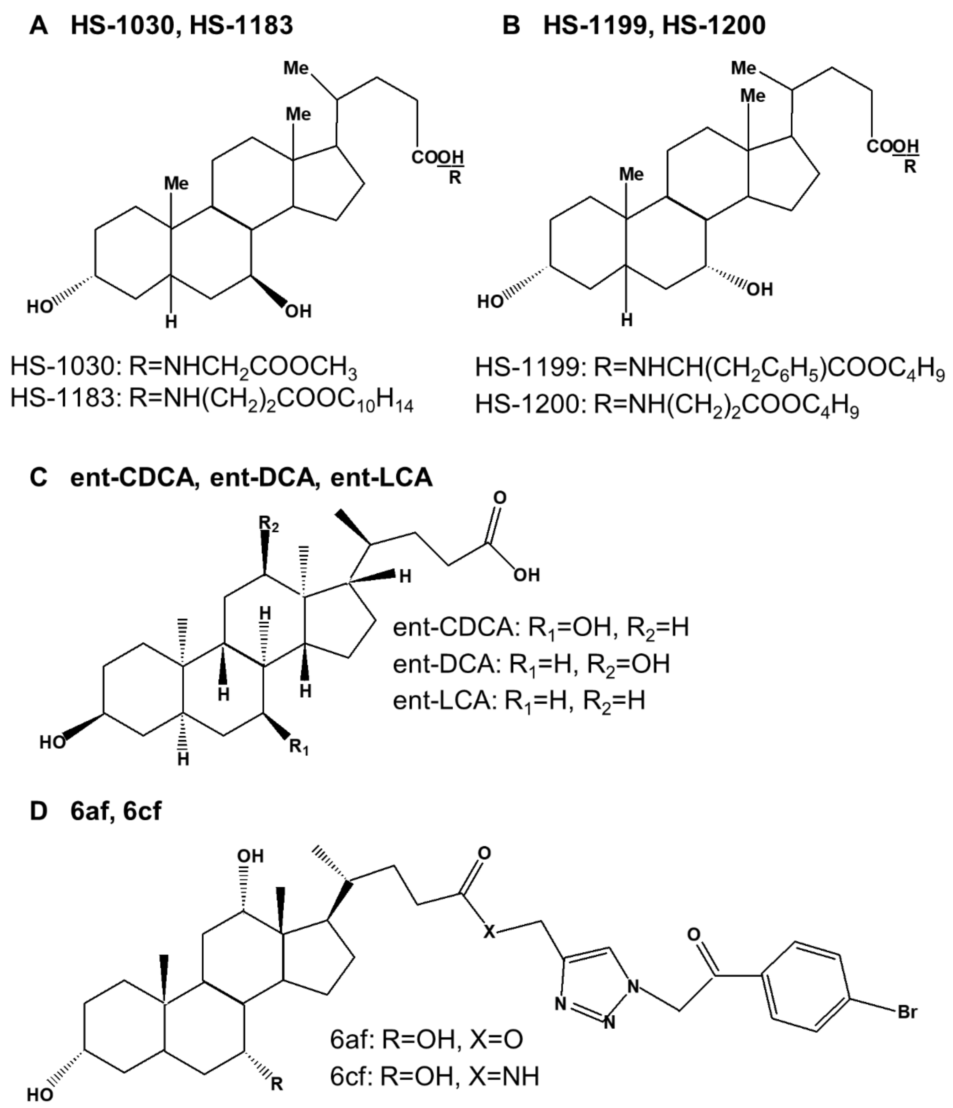
| HS-1030 | UDCA derivative | apoptosis | p21 | cyclin E, Cdk2, Cdk4, Cdk6, E2F-1 | Colon cancer cells (HT-29) | [22] |
| HS-1183 | UDCA derivative | apoptosis | cleavage of PARP | pro-caspase-3 and -8 | Leukemic T cells (Jurkat cells) | [161] |
| apoptosis | p21 | cyclin D1, Cdk4, Cdk6, E2F-1 | Colon cancer cells (HT-29) | [22] | ||
| apoptosis | cleavage of PARP, Bax, c-Jun, p-JNK, AP-1 | p-p38, p50, p65, IkB-α | Cervical carcinoma cells (SiHa) | [162] | ||
| apoptosis | Bax, cleavages of lamin B and PARP, p21, p53 | Bcl-2, cyclin D3, pRb | Breast cancer cells (MCF-7, MDA-MB-231) | [163] | ||
| apoptosis | p21 | pRb | Prostate cancer cells (PC-3) | [164] | ||
| HS-1199 | CDCA derivative | apoptosis | cleavage of PARP | pro-caspase-3 and -8 | Leukemic T cells (Jurkat cells) | [161] |
| apoptosis | p21 | cyclin D1, cyclin E, Cdk2, Cdk4, Cdk6, E2F-1 | Colon cancer cells (HT-29) | [22] | ||
| apoptosis | cleavage of PARP, Bax, c-Jun, p-JNK, AP-1 | p-p38, p-ERK | Cervical carcinoma cells (SiHa) | [162] | ||
| apoptosis | Bax, cleavages of lamin B and PARP, p21, p53 | Bcl-2, cyclin D1, cyclin D3, pRb | Breast cancer cells (MCF-7, MDA-MB-231) | [163] | ||
| apoptosis | cleavage of PARP, p21 | pRb, cyclin D1, cyclin D3 | Prostate cancer cells (PC-3) | [164] | ||
| apoptosis | Cyt c, cleavage of PARP and DFF45, AIF(N), Nur77 | ΔΨm, pro-caspase-3, XIAP | Stomach cancer cells (SNU-1) | [165,166] | ||
| apoptosis | cleavage of PARP, Cyt c | pro-caspase-3, ΔΨm | Malignant glioblastoma cells (U-118MG, U-87MG, T98G, U-373MG) | [167] | ||
| HS-1200 | CDCA derivative | apoptosis | cleavage of PARP | pro-caspase-3 and -8 | Leukemic T cells (Jurkat cells) | [161] |
| apoptosis | cleavage of PARP, p21 | cyclin D1, cyclin E, Cdk2, Cdk4, Cdk6, pRb, E2F-1 | Colon cancer cells (HT-29) | [22] | ||
| apoptosis | cleavage of PARP, Bax, c-Jun, p-JNK, AP-1 | p-p38, p-ERK, p65(total), p50(total), IkB-α(total) | Cervical carcinoma cells (SiHa) | [162] | ||
| apoptosis | Bax, cleavages of lamin B and PARP, p21, p53, AIF | Bcl-2, cyclin D1, cyclin D3, pRb | Breast cancer cells (MCF-7, MDA-MB-231) | [163,168] | ||
| apoptosis | cleavage of PARP, p21 | pRb, Cdk2, cyclin D1, cyclin D3 | Prostate cancer cells (PC-3) | [164] | ||
| apoptosis | Cyt c, cleavage of PARP and DFF45, AIF(N), Nur77 | ΔΨm, pro-caspase-3, XIAP | Stomach cancer cells (SNU-1) | [165,166] | ||
| apoptosis | cleavage of PARP, Cyt c | pro-caspase-3, ΔΨm | Malignant glioblastoma cells (U-118MG, U-87MG, T98G, U-373MG), Xenografts (U87MG) | [167] | ||
| apoptosis | Bax, p53, p21, p27, Egr-1, caspase-3 and 9, cleavage of PARP, Cyt c (cytosolic) | Bcl-2, cyclin D1, Cdk2, cyclin A, E2F-1, Mdm2, COX-2, Cyt c (mitochondrial), ΔΨm | Hepatoma cells (HepG2, Hep3B, BEL7402) | [169,170] | ||
| apoptosis | AIF, CAD, cleavage of PARP | ΔΨm, pro-caspase-3 and -7, PARP | Thyroid carcinoma (KAT 18) | [171] | ||
| ent-CDCA | enantiomers of CDCA | apoptosis | pro-caspase-3 and -9 | Colon cancer cells (HT-29, HCT-116) | [23] | |
| ent-DCA | enantiomers of DCA | apoptosis | pro-caspase-3 and -9 | Colon cancer cells (HT-29, HCT-116) | [23] | |
| ent-LCA | enantiomers of LCA | apoptosis | CD95, ROS, | pro-caspase-2, -3, -8, and -9, Bid | Colon cancer cells (HT-29, HCT-116) | [23] |
| 6af and 6cf | bile-acid-appended triazolyl aryl ketones | apoptosis | Breast cancer cells (MCF-7) | [24] | ||
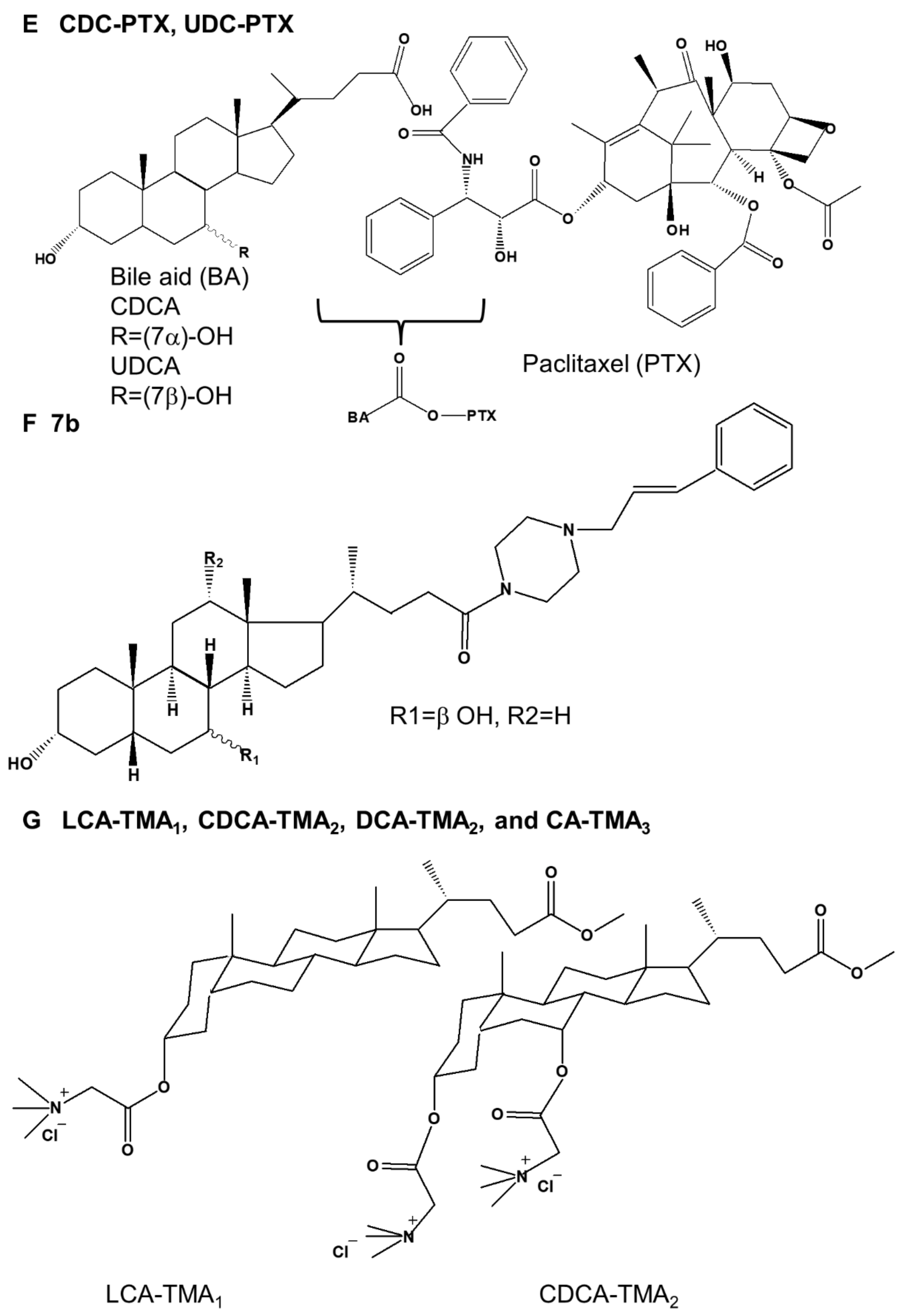
| CDC-PTX UDC-PTX | CDCA derivative UDCA derivative | apoptosis | Acute promyelocytic leukemia cells (HL60, NB4) | [25] | ||
| CDCA derivative UDCA derivative | apoptosis | Colon cancer cells (RKO, HCT-116) | [25] | |||
| 7b | Piperazinyl bile acid derivative | apoptosis | Multiple myeloma (KMS-11), Colonic cancer cells (HCT-116) | [26] | ||
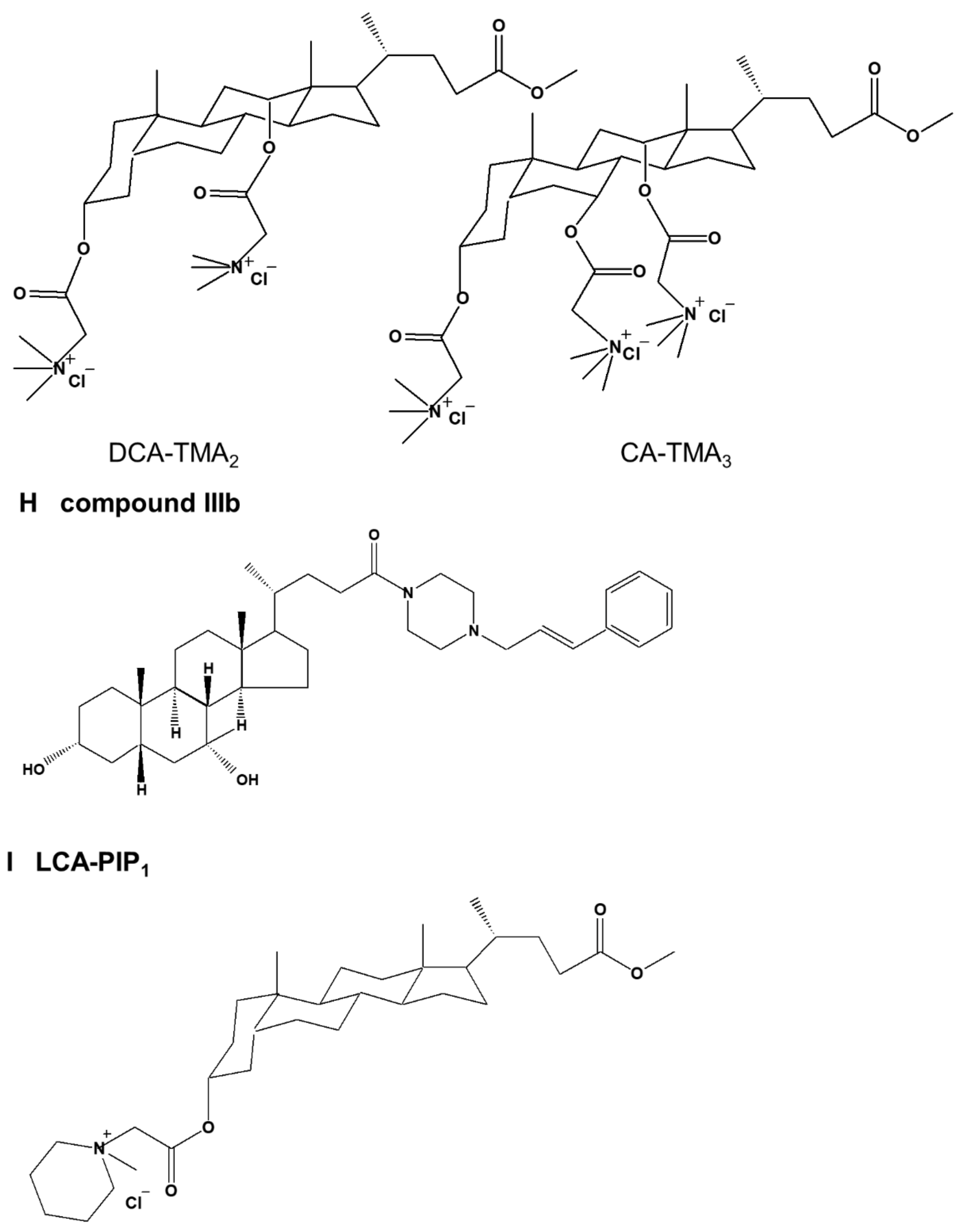
| LCA-TMA1, CDCA-TMA2, DCA-TMA2, and CA-TMA3 | cationic bile acid based facial amphiphiles featuring trimethyl ammonium head groups | apoptosis | Colon cancer cells (HCT-116, DLD-1) | [27] | ||
| compound IIIb | CDCA-substituted piperazine conjugate | apoptosis | cleavage of Mcl-1 and PARP-1, Ip-IκBα, DNA fragmentation | IκBα | Multiple myeloma (KMS-11) | [28] |
| LCA-PIP1 | LCA amphiphile | apoptosis | pro-caspase-3, -7, and -8 | Colon cancer cells (HCT-116) Xenograft (HCT-116) | [29] | |
| CA-Tam3-Am | CA−tamoxifen conjugate | apoptosis | Bax, Bid, Bad, caspase-9, cleaved caspase-3 and -8, Cyt c, ROS | Bcl-2, Bcl-xL, survivin | Breast cancer cells (MCF-7, T47D, MDA-MB 231) | [30] |
| norUDCA | UDCA derivative | autophagy | ratio of LC3-II to LC3-I, ATG5, ATG5/ATG12, p-AMPK, p-ULK1(Ser317), p-ULK1(Ser555), p-ULK1(Ser777) | p62, α1ATZ, p-mTOR, p-ULK1(Ser757) | Cervical cancer cells (HTOZ) | [31] |
| compound 9 | DCA derivative | apoptosis, autophagy | caspase-3 and -7, ROS | Duodenal carcinoma cells (HuTu 80) | [32] | |
Publisher’s Note: MDPI stays neutral with regard to jurisdictional claims in published maps and institutional affiliations. |
© 2022 by the authors. Licensee MDPI, Basel, Switzerland. This article is an open access article distributed under the terms and conditions of the Creative Commons Attribution (CC BY) license (https://creativecommons.org/licenses/by/4.0/).
Share and Cite
Jang, J.Y.; Im, E.; Choi, Y.H.; Kim, N.D. Mechanism of Bile Acid-Induced Programmed Cell Death and Drug Discovery against Cancer: A Review. Int. J. Mol. Sci. 2022, 23, 7184. https://doi.org/10.3390/ijms23137184
Jang JY, Im E, Choi YH, Kim ND. Mechanism of Bile Acid-Induced Programmed Cell Death and Drug Discovery against Cancer: A Review. International Journal of Molecular Sciences. 2022; 23(13):7184. https://doi.org/10.3390/ijms23137184
Chicago/Turabian StyleJang, Jung Yoon, Eunok Im, Yung Hyun Choi, and Nam Deuk Kim. 2022. "Mechanism of Bile Acid-Induced Programmed Cell Death and Drug Discovery against Cancer: A Review" International Journal of Molecular Sciences 23, no. 13: 7184. https://doi.org/10.3390/ijms23137184