Article

Evolutionary and Expression Analysis of MOV10 and MOV10L1 Reveals Their Origin, Duplication and Divergence

1
College of Marine Life Sciences, and Institute of Evolution & Marine Biodiversity, Ocean University of China, Qingdao 266003, China
2
School of Basic Medicine, Qingdao University, Qingdao 266071, China
3
Laboratory for Marine Biology and Biotechnology, Pilot National Laboratory for Marine Science and Technology (Qingdao), Qingdao 266003, China
*
Authors to whom correspondence should be addressed.
Int. J. Mol. Sci. 2022, 23(14), 7523; https://doi.org/10.3390/ijms23147523
Submission received: 23 May 2022 / Revised: 30 June 2022 / Accepted: 5 July 2022 / Published: 7 July 2022
(This article belongs to the Section Biochemistry)

Abstract

:
MOV10 and MOV10L1 both encode ATP-dependent RNA helicases. In mammals, MOV10 and MOV10L1 participate in various kinds of biological contexts, such as defense of RNA virus invasion, neuron system, germ cell and early development. However, mov10 and mov10l1 in zebrafish are obscure and the evolutionary relationships of mov10 among different species remain unclear. In this study, we found MOV10 and MOV10L1 had some variations despite they possessed the conserved feature of RNA helicase, however, they may originate from a single ancestor although they shared limited homology. A single MOV10L1 gene existed among all species, while MOV10 gene experienced lineage-specific intra-chromosomal gene duplication in several species. Interestingly, the mov10 gene expanded to three in zebrafish, which originating from a duplication by whole genome specific duplication of teleost lineage followed by a specific intra-chromosome tandem duplication. The mov10 and mov10l1 showed distinct expression profiles in early stages, however, in adult zebrafish, three mov10 genes exhibited similar diverse expression patterns in almost all tissues. We also demonstrated mov10 genes were upregulated upon virus challenge, highlighting they had redundant conserved roles in virus infection. These results provide valuable data for the evolution of MOV10 and MOV10L1 and they are important to the further functional exploration.

1. Introduction

RNA helicases are implicated in all steps of RNA metabolism. MOV10 (Moloney leukemia virus 10) and MOV10L1 (Mov10 like 1) are RNA helicase homologs implicated in wide variety of biological processes such as mRNA translation, miRNA-mediated post-transcription, piRNA biogenesis, and so on [1]. MOV10, first identified in cell lines derived from mouse strains in which Moloney leukemia virus (M-MuLV) was inserted into the germ line, was known as a protein that prevents M-MuLV infection in mice [2]. MOV10L1, also known as CHAMP (cardiac helicase activated by MEF2 protein), with high homology to MOV10, was initially identified as a protein that expressed in testis specifically [3].
MOV10 and MOV10L1 both encode ATP-dependent helicases, which are enzymes that mediate ATP-dependent unwinding of RNA duplexes thus promoting structural rearrangements of RNP complexes [4]. There are six superfamily helicases classified by sequences, structures and mechanisms [5]. Eukaryotic helicases exclusively belong to the SF1 and SF2 superfamilies, which share a conserved core composed of two tandem domains with RNA binding ability and ATPase activity [6,7]. SF1 helicases have a (aspartic acid (D)-glutamic acid (E)-alanine (A)-glycine (G)) DEAG motif, rather than the more common (aspartic acid (D)-glutamic acid (E)-alanine (A)-aspartic acid (D)/Histidine (H)) DEAD/H box, which is the symbol of the SF2 helicases. To date, a lot of the identified RNA helicases belong to the SF2, only a dozen of SF1 RNA helicases have been identified. Among them, MOV10 and MOV10L1 both encode SF1 RNA helicases, which involved in various kinds of biological contexts, such as defense of RNA virus invasion, early development, neuron system, germ cell [8,9].
MOV10 is a versatile RNA helicase implicated in wide RNA regulation network. MOV10 is a nucleus/cytoplasmic protein and ubiquitously expressed in almost all tissues. MOV10 normally localizes to the RNA processing bodies (PB) in the cytoplasm. MOV10 has been reported in few organisms. In Drosophila, CG6967 or dMOV10 is induced specifically during the mid-gut regeneration, and inhibits intestinal stem cell proliferation, thus regulates the termination of mid-gut degeneration [10]. Knockdown of maternal mov10 in xenopus embryos led to gastrulation defects, likely resulting from the large scales changes in mRNA expression due to Mov10’s role in miRNA mediated regulation of maternal-zygotic transition. In addition, zygotic Mov10 is necessary for development of the central nervous system [9]. In mouse, the Mov10 knockout one led to embryonic lethality, suggesting MOV10 is critical for early development. The heterozygous mouse has less dendritic complexity in hippocampal neurons, implying a role of MOV10 in the development of brain [11]. MOV10 is also a nucleocytoplasmic protein expressed in spermatogonia and a determinant of spermiogenesis [1]. In Hela cell, MOV10 could interact with argonaute proteins and localizes to mRNA-degrading cytoplasmic P bodies, participating in the RNAi pathway and mediating miRNA-guided mRNA cleavage [12]. Later, it is found that MOV10 has a dual role in regulating translation, which facilitates the translation of some RNAs, but also increases the expression of other RNAs when bound to FMRP [13]. MOV10 interacts with UPF1, the crucial component of the nonsense-mediated mRNA decay pathway, promotes mRNA degradation [14]. Accumulating evidences suggest the key role of MOV10 is antivirus activity against HIV1, Influenza, HBV and HCV virus invasion [15,16,17,18,19]. MOV10 primarily executes the antiviral responses by two different ways. First, MOV10 regulates antiviral gene expressions including IFN signal and autophagy pathways. Second, MOV10 participates RNA degradation via miRNA pathways [8,20].
Contrary to the diverse expression patterns and versatile roles of MOV10, numerous studies demonstrate that MOV10L1 exhibits a restricted germ cell-specific expression patterns, emerges as a conserved key regulator of piRNA biogenesis [21,22,23,24,25]. While drosophila armitage (armi), supposed to be the common ancestor of vertebrate MOV10 and MOV10L1, is another MOV10/MOV10L1 homolog. Armi interacts with piRNA pathway factor Yb, tethered to piRNA precursors transcript, thus facilitating its processing into primary piRNAs in drosophila ovaries [22]. In mouse, the specific expression in germ cells of MOV10L1 increases during the development of sperms. MOV10L1 interacts with HSPA2 and Piwi proteins, participates in piRNA-directed retrotransposons silencing, thus implicates in male fertility [21]. Conditional knockout of MOV10L1 in mouse disrupted the RNA helicase domain. The Mov10l1 mutant male mice were sterile owing to meiotic arrest before the pachytene stage. In addition, MOV10L1 is associated with piRNA ribonucleoprotein particles (piRNPs), the Mov10l1 mutant expresses Piwi proteins but devoid of piRNA, implying MOV10L1 is involved in piRNA biogenesis [25]. Disruption of MOV10L1 helicase activity leads to loss of pre-pachytene piRNAs, resulting in retrotransposons activation and early meiotic arrest [21,25,26]. MOV10L1 acts upstream of Piwi proteins and is also required for the biogenesis of pachytene piRNAs, a poorly characterized piRNA class expressed in the pachytene stage of meiosis, which has unique function in the maintenance of post-meiotic genomic integrity [24]. MOV10L1 possesses RNA helicase activity and exhibits 5′-3′ directional RNA unwinding activity. MOV10L1 binds piRNA precursors and is essential to activate piRNA processing [23]. Very recently, a yama mutant, which harbors V229E substitution in the N-terminal region instead of the helicase domain, leads to meiotic arrest, loss of pachytene piRNA and male infertility. The results reveal an important role of the MOV10L1 in piRNA biogenesis during male germ cell development. This accumulating evidence suggest that MOV10L1 act as a major regulator of piRNA biogenesis, and both the N-terminal and helicase domain are required for the function of MOV10L1.
So far, the MOV10 and MOV10L1 have been identified in several species, while MOV10 and MOV10L1 members in teleost are undetermined, and the evolutionary relationships of MOV10 among metazoan taxa remain unclear. In this study, we first analyzed the molecular evolution of MOV10 and MOV10L1 in representative species of metazoan, then examined the expression profiles of mov10 and mov10l1 in early development and adult tissues as well as in response to virus challenge in zebrafish.

2. Results

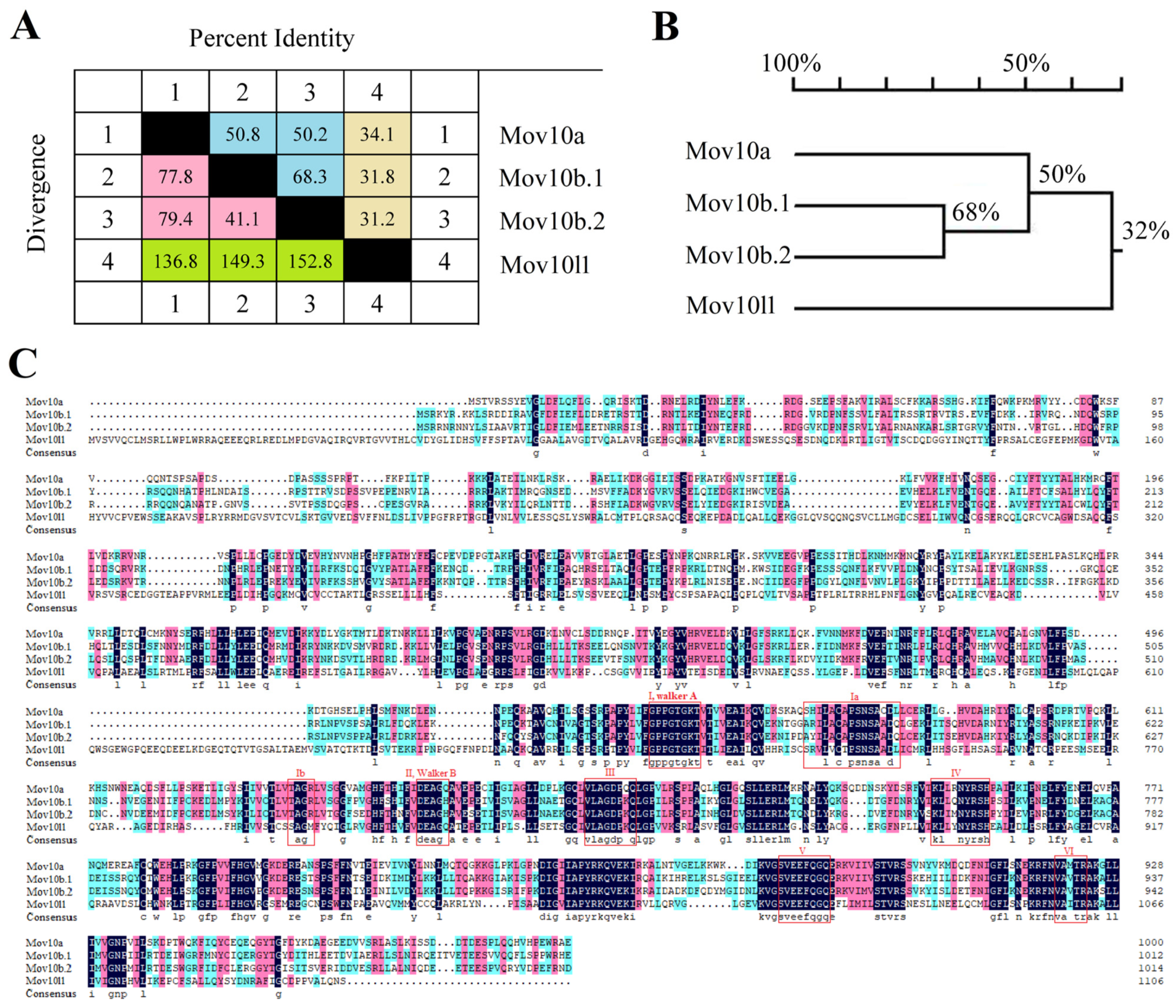
2.1. Three mov10 Genes and mov10l1 in Zebrafish

The MOV10 and MOV10L1 of human were used to search the Mov10 and Mov10l1 in zebrafish genome by BLAST. As a result, a mov10l1 gene and three mov10 homologs, designated as mov10a, mov10b.1 and mov10b.2, respectively, were identified. This is consistent with the nomenclature in NCBI database. Next, we obtained their cDNA by RT-PCR, suggesting the identified candidate genes are expressed in vivo. The ORF of mov10a coded for a protein of 1001 amino acids, with a molecular mass of ~113.92 kDa and a pI value of ~9.02. The ORF of mov10b.1 encoded a 1013 aa protein, with a molecular mass of ~116.41 kDa and a pI of ~8.87. The predicted Mov10b.2 protein was quite similar to Mov10b.1, consisted of 1015 aa, with a molecular mass of ~116.51 kDa and a pI of ~8.53. The ORF of mov10l1 coded for a protein of 1106 aa, with a molecular mass of ~122.61 kDa and a pI of ~5.89 (Table 1).
The sequence similarity and divergence between Mov10s (zebrafish Mov10a, Mov10b.1 and Mov10b.2) and Mov10l1 were shown in Figure 1. In specific, the Mov10s shared higher homology, ranging from 50.2% to 68.3%, while either of Mov10s and Mov10l1 shared lower homology but diverged greatly, ranging from 31.2% to 34.1% (Figure 1A,B). The sequence alignment revealed the higher homology among Mov10s (Figure 1C). The sequence alignment also showed that the c-terminal parts of the proteins are more conserved compared to the N-terminal part of the proteins (Figure 1C).

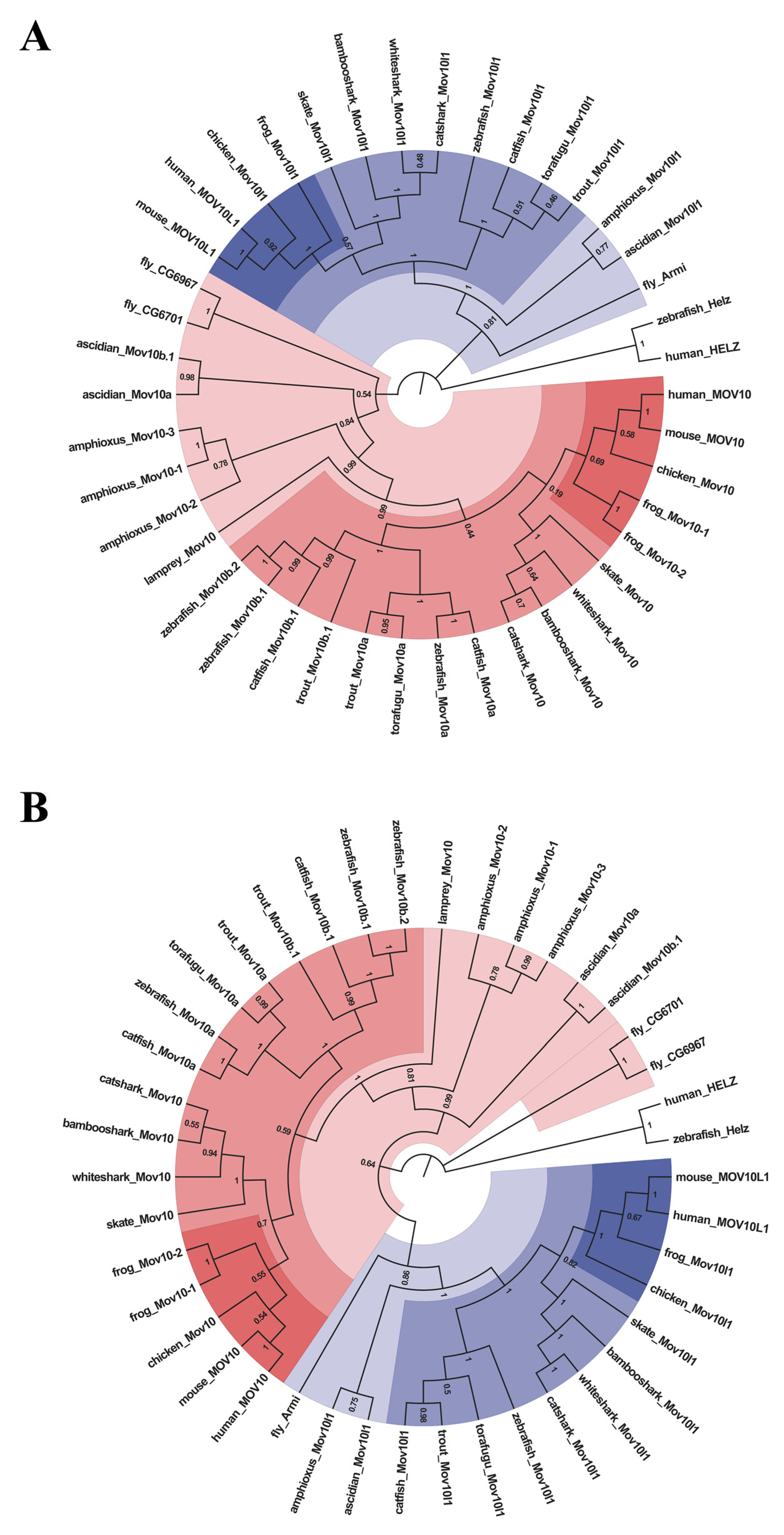
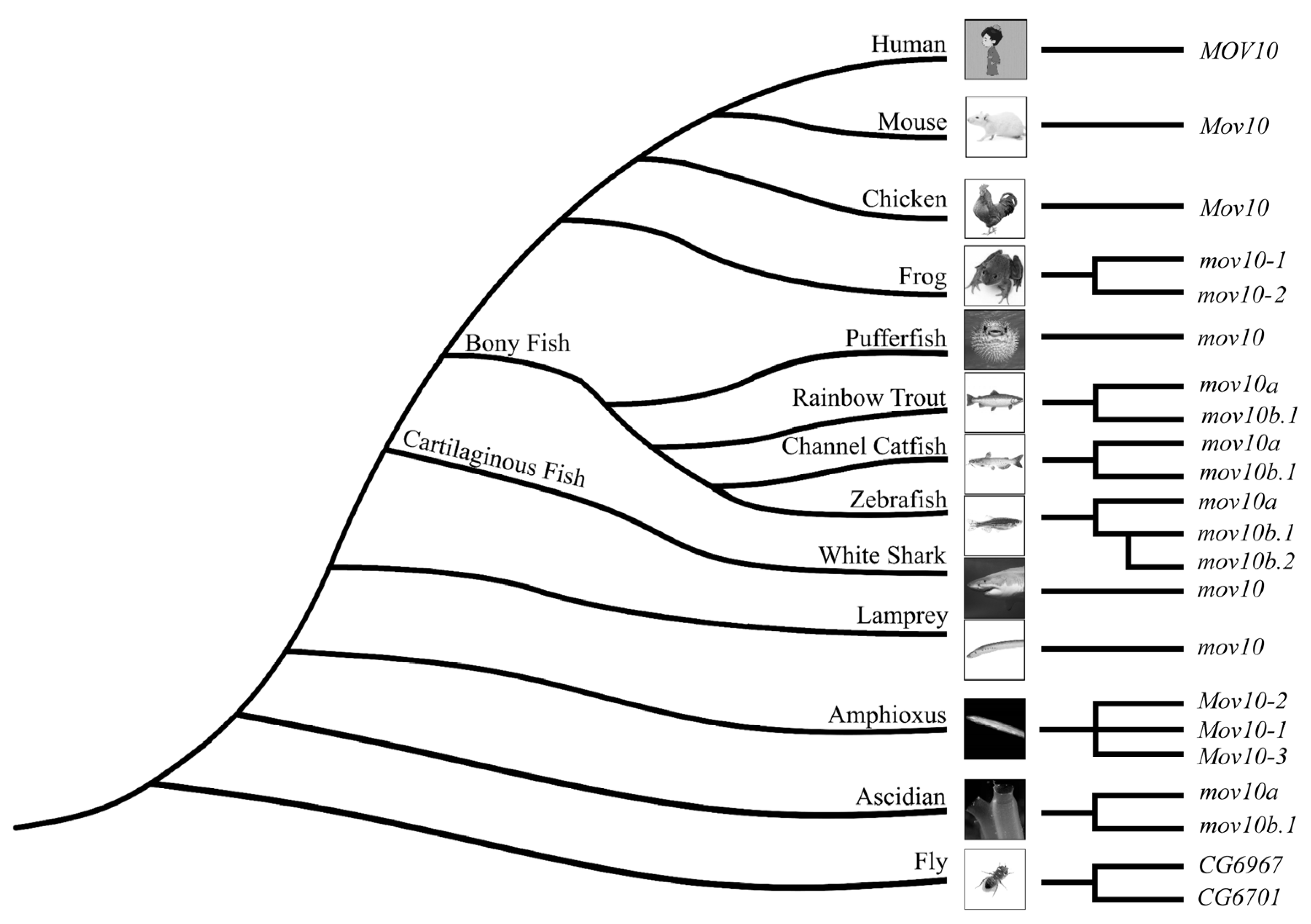
2.2. Lineage-Specific Gene Duplication of MOV10 in Multiple Species

Integrated MOV10 and MOV10L1 sequences in representative species from invertebrates to vertebrates, including fly, ascidian, amphioxus, lamprey, cartilaginous fishes, bony fishes, frog, chicken, mouse and human, were identified from NCBI and Ensembl databases by BLAST. Intriguingly, there were distinct MOV10 members in specific species, in contrast to only one MOV10L1 in all selected species. In specific, lamprey, cartilaginous fishes (Chondrichthyes), chicken, mouse and human possessed one MOV10, while as fly, frog and ascidian had two Mov10 members. Additionally, amphioxus contained three Mov10. In bony fishes (Osteichthyes), torafugu only had one single copy of Mov10, channel catfish and rainbow trout possessed two Mov10, the most peculiar one was zebrafish, which had three Mov10 (Table 2, Supplemental Information: Sequence Information). More than one MOV10 members existed in several species, demonstrating possible gene duplication events during evolution.
To explore the evolutionary relationship of MOV10 and MOV10L1 in metazoan, the sequences obtained were first aligned by ClustalW algorithm for further phylogenetic analysis. We constructed phylogenetic trees of MOV10 and MOV10L1 of selected species by Maximum-Likelihood and Neighbor-Joining methods of MEGA 7 (Figure 2, Supplemental Information: Sequence Information). The two constructed phylogenetic trees were consistent. In detail, All the MOV10L1 proteins clustered together, while MOV10 proteins formed another clade. In the MOV10 clade, the Mov10 proteins in the fly, ascidian, amphioxus, were clustered on the outermost side in turn, respectively, demonstrating that they were more primitive and conservative during the evolution process. It could be speculated the novel MOV10 members possibly derived from the species lineage-specific gene duplication. Four Mov10 of cartilaginous fish clubbed together indicating they had high homology compared to the counterpart of other species. All the bony fish Mov10 members were clustered into two branches: Mov10a and Mov10b. The torafugu Mov10 was clustered with the Mov10a of other bonny fishes. It could be inferred the late evolved Mov10b might be generated from teleost-specific genome duplication. Interestingly, zebrafish Mov10b.1 and Mov10b.2 formed a more closed sub-branch, indicating they shared high homology compared with Mov10b of the other teleost. We proposed that zebrafish Mov10b.1 and Mov10b.2 may be originated from zebrafish lineage-specific gene duplication. Overall, although the numbers of MOV10 diverged among species, MOV10L1 were only single in all the representative species.

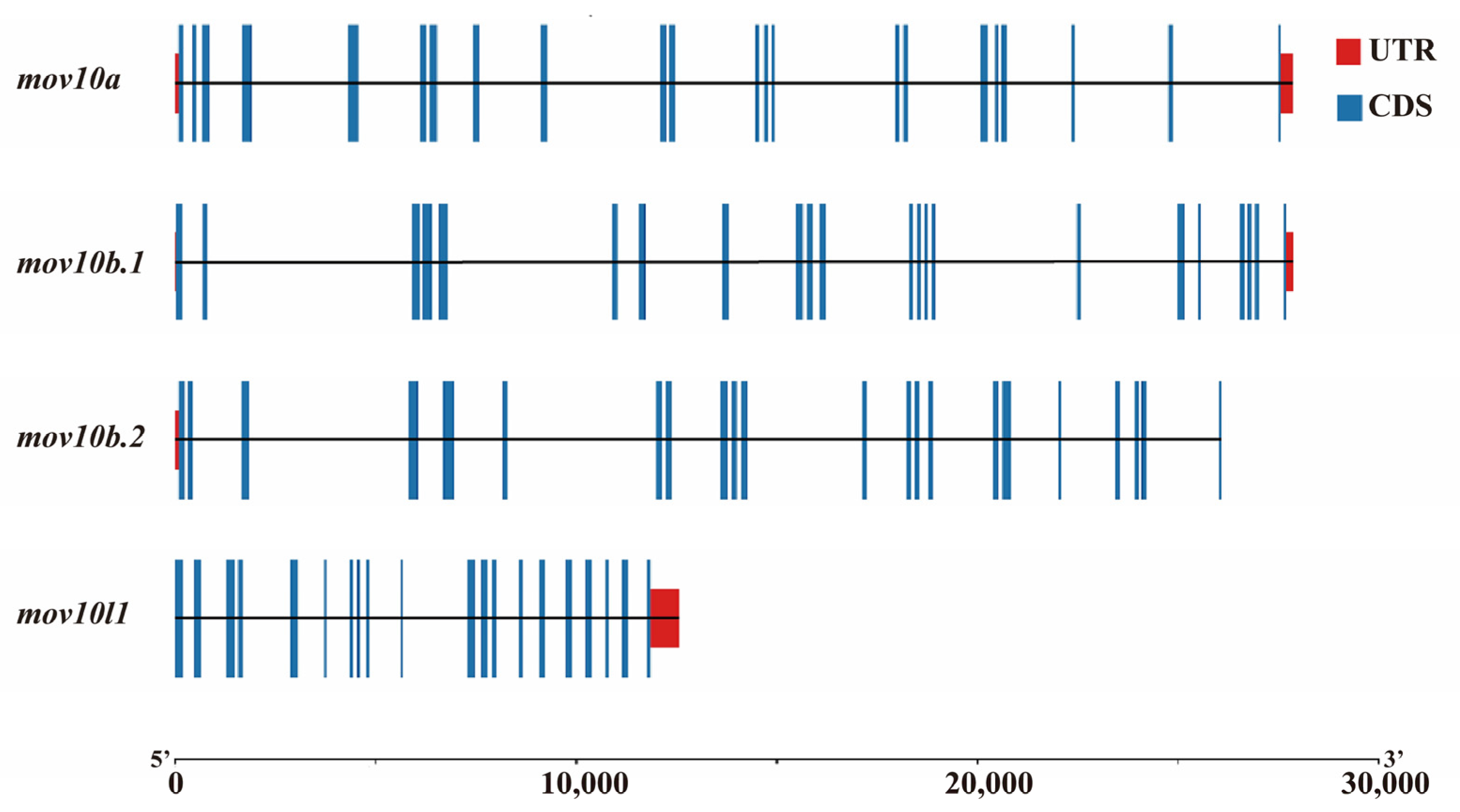
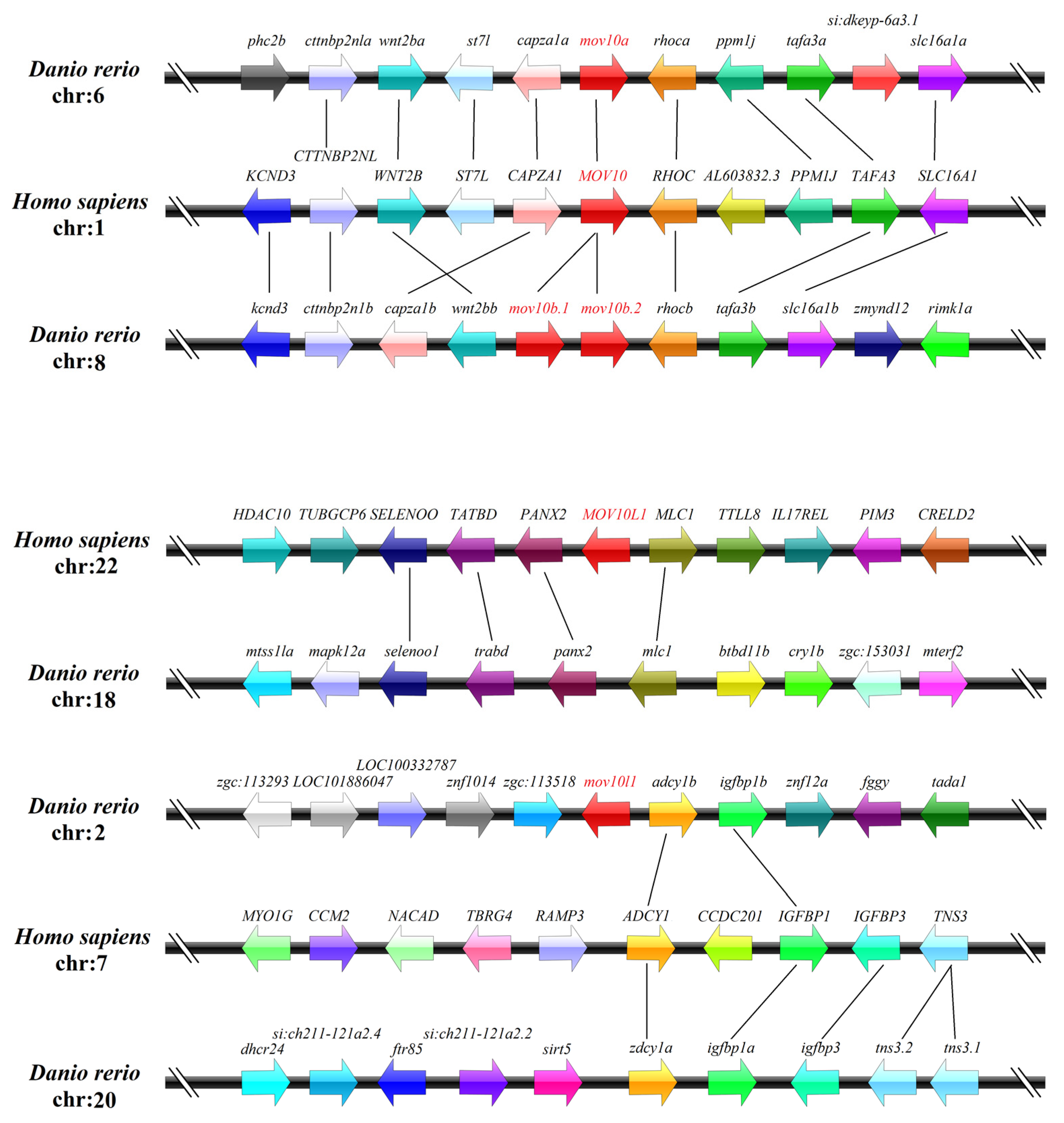
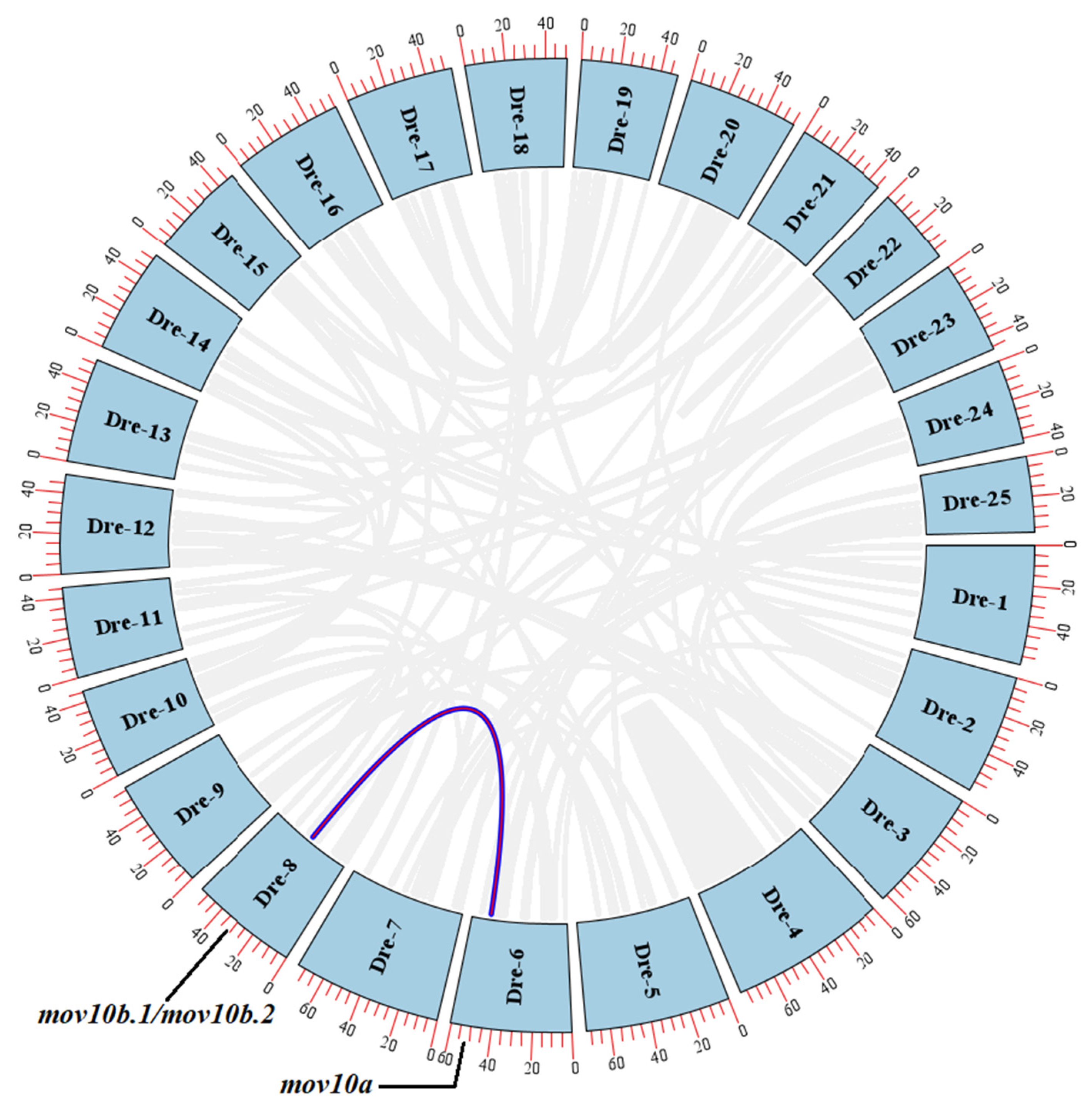
2.3. Genomic Structure and Synteny of MOV10 Genes

We analyzed the gene structures of zebrafish mov10s and mov10l1 using TBtools. The exon-intron genomic structure of zebrafish mov10s and mov10l1 were shown (Figure 3). The three mov10s in zebrafish shared similar intron-exon structure consisting of 22 exons, while zebrafish mov10l1 possessed 21 exons. It also showed that zebrafish mov10l1 gene was more compact than the three mov10s of zebrafish, which have similar gene length.
To explore the synteny, we analyzed the genes surrounding MOV10 and MOV10L1 genes in human and zebrafish. While zebrafish mov10a located on chromosome 6, mov10b.1, mov10b.2 and mov10l1 were all mapped to chromosome 8, on which mov10b.1 and mov10b.2 were linked to each other. Further analysis of the surrounding genes revealed that the upstream and downstream genes of mov10a had their orthologs located near to the human MOV10. Similarly, the flanking genes of mov10b.1 and mov10b.2 could be found in the neighbor around human MOV10. The conserved synteny suggested that mov10a, mov10b.1 and mov10b.2 were co-orthologs of the human MOV10. However, zebrafish mov10l1 had no synteny to the human MOV10L1 gene (Figure 4) though the MOV10L1 were well co-linearized among human, rat, mouse, chicken frog and some fishes (Figure S1). Thus, synteny analysis showed that MOV10 was relatively conserved while MOV10L1 was not.

2.4. Gene Duplication and Selective Pressure Analysis

The phylogenetic trees of MOV10 and MOV10L1 in different species and the synteny among the zebrafish mov10s and the human MOV10 suggested that the intra-species gene duplication of MOV10 may occur in some species. To verify whether it’s happening in zebrafish, we mapped the Circos plot of the inter-chromosomal relationships by TBtools. In zebrafish, mov10a had the conserved synteny with mov10b.1 as well as mov10b.2 while mov10l1 had no synteny with the three genes (Figure 5). This suggested that mov10b (either mov10b.1 or mov10b.2) was originated from duplication of mov10a during evolution.
The value of the nonsynonymous substitutions per nonsynonymous site (Ka)/ the number of synonymous substitutions per synonymous site (Ks) ratios was used to explore the selection pressures influencing sequence divergence for the tandem duplication events of MOV10 and MOV10L1 (Table 3). The Ka/Ks ratios of zebrafish mov10s and mov10l1 ranged from 0.18 to 0.35, indicating that the zebrafish mov10s and mov10l1 may have experienced purifying selective pressure during evolution. The Ka/Ks ratio of mov10b.2 and mov10l1 was worked out but it of mov10b.1 and mov10l1 did not, reminded us that mov10b.2 may be duplicated from mov10a first, then mov10b.1 originated from a tandem duplication event of mov10b.2 during evolution. In addition, the Ka/Ks ratios of MOV10S and MOV10L1 between zebrafish and human or zebrafish and catfish were between 0.12 and 0.23, showed that the MOV10 and MOV10L1 genes may have undergone purifying selective pressure during the evolution of species. In conclusion, the above analyses showed that the MOV10S and MOV10L1 were conservative during the evolutionary process, and its function may be redundant to some extent.

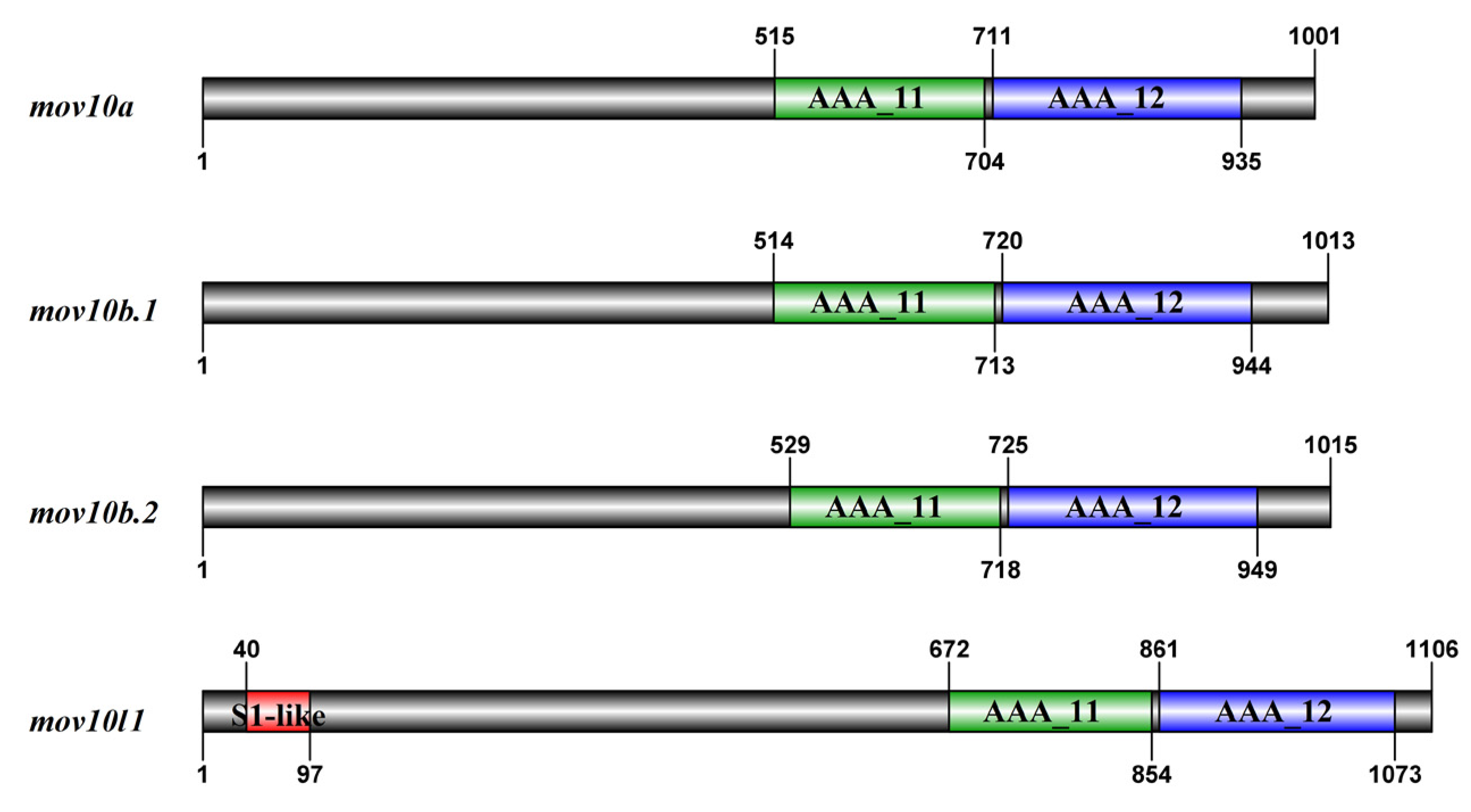
2.5. Domain and Motif Distribution Analysis

To elucidate the characteristic of Mov10s and Mov10l1, we used SMART and Pfam to predict the conserved domain Mov10s and Mov10l1 in zebrafish. All the four proteins shared two conserved RecA-like helicase core domains: AAA_11 (Domain I) and AAA_12 (Domain II), which located in the c-terminal region (Figure 6). AAA_11 is a ATPase domain associated with a varied cellular activities, AAA_12 is a characteristic domain of RNA helicases belonging to superfamily 1. Instead of the common DEAD/H box, SF1 helicases normally possessed a DEAG motif. Based on the characteristics of protein sequences, all Mov10s and Mov10l1 definitely belonged to SF1 helicases. Mov10l1 also possessed another S1-like RNA binding domain in the N-terminal, which were absent from all Mov10s (Figure 6).
We applied MEME online tools to analyze the motif structure of zebrafish Mov10s and Mov10l1. All Mov10 proteins shared similar motif structures, containing ten motifs from motif 1 to motif 10. While motif 2, 4, 5, 6, 7, 8, 9, 10 were presented in zebrafish Mov10l1, in which motif 1 and 3 were missing (Figure 7). Specifically, the motif 4 and 5 were in the AAA_11 domain, and the AAA_12 domain has the motif 7, 8, 9 and 10, while the motif 6 was shared by the two motifs. The DEAG motif, the signature feature sequence of SF1 helicases, was located in the motif 5. In addition, the other specific amino acid sequences that Mov10 had ubiquitously in other species were also present in these motifs (Figure 1C). Zebrafish Mov10s and Mov10l1 proteins had conserved functional domain and similar motifs as other species, suggesting their functions may be conserved.

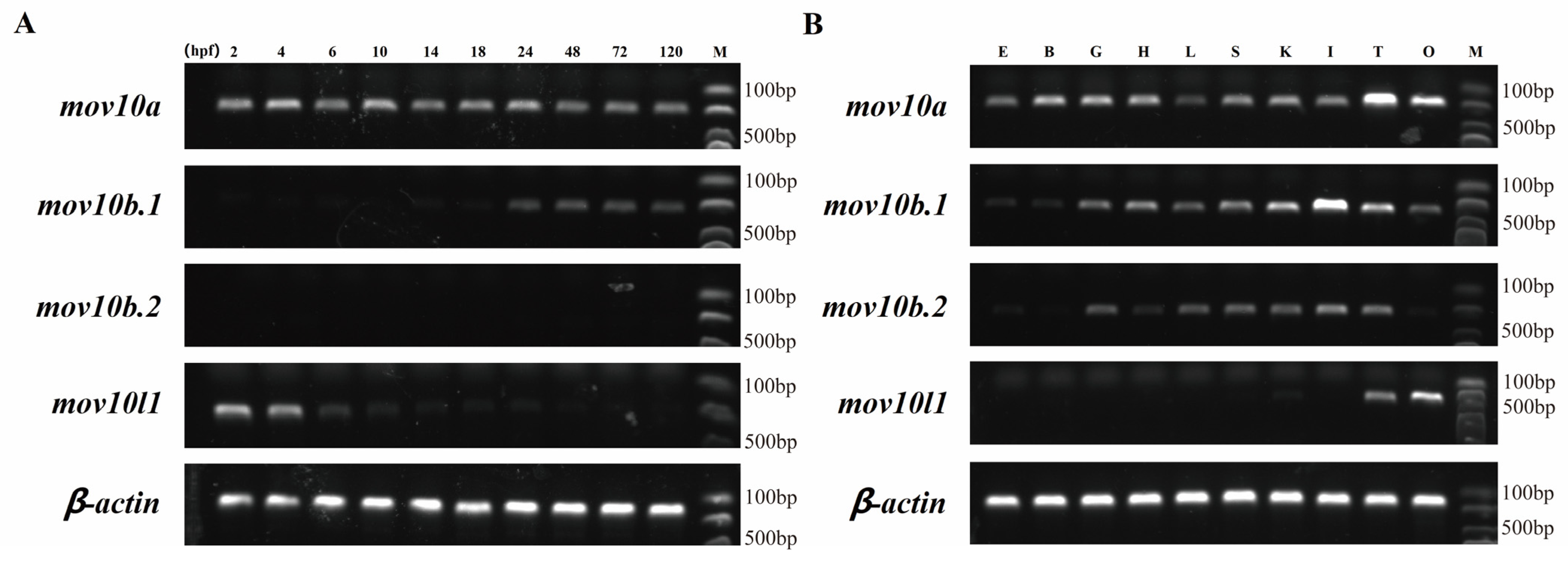
2.6. Expression Pattern of mov10s and mov10l1 in Zebrafish

The gene expression is generally related to its function. Therefore, we detected the expression pattern of mov10s and mov10l1 in early embryonic development and adult tissues of zebrafish. All the four genes had quite different expression patterns in early stages of zebrafish (Figure 8A). Under the same number of PCR cycles, mov10a had a relatively stable robust expression during all the embryonic development stages, both maternal and zygotic. The mov10b.1 has no maternal expression, and the slightly significant expression begun to be observed from 24 hpf. However, mov10b.2 had no transcript in the early embryos. mov10l1 only had maternal but not zygotic expression. In conclusion, the two mov10 genes and mov10l1 gene were expressed in early stages, suggesting they were involved in early development of zebrafish.
We also measured the expression of mov10s and mov10l1 in adult tissues of zebrafish. The mov10a had a relatively abundant expression in all the tissues with the highest expression in testis, followed by ovary. The expression level of mov10b.2 was lower than that of mov10b.1, although they had similar expression patterns in tissues except ovary in which mov10b.2 is undetectable. Compared to mov10a, they both had weak expression in eye and brain and the highest expression in intestine (Figure 8B). Different from the diverse expression patterns of mov10s, it was obvious that mov10l1 was exclusively expressed in the testis and ovary, especially in the ovary. mov10l1 was specifically expressed in germ cells resembling of the expression pattern of mov10l1 in human and mouse, implying Mov10l1 in zebrafish had similar roles. The three zebrafish mov10s had similar but distinct expression patterns, indicating that the function of three Mov10s may be redundant but diverged to some extent.

2.7. Expression Profiles of mov10s and mov10l1 upon Virus Challenge

MOV10 has been proved to be antiviral player in human and mouse. To explore whether all the three Mov10 have antiviral properties in zebrafish, we conducted the following experiments. First, we tested and determined the lethal concentration of GCRV viruses. We injected the 9-month-old zebrafish with 30 microliters of the virus through the intraperitoneal injection, then the tissues were gutted, and total RNA was extracted at 24, 48, 96 h post injection.
Considering mov10l1 specifically expressed in testis and ovary, so we only detected the expression of three mov10 genes. The qRT-PCR results showed that the three mov10 genes were upregulated upon GCRV virus challenge compared to the uninjected group though the level of upregulation were different at all the three time points upon virus infection (Figure 9A–I). When the virus invaded, the increase level of mov10b.1 expression was the highest among the three mov10, especially in liver, a key immune organ. The results imply that all three mov10 shared the redundant roles and were involved in virus defenses in zebrafish.

3. Discussion

MOV10 and MOV10L1, belonging to SF1 RNA helicases, are implicated in diverse biological processes [1,8,20]. MOV10 and MOV10L1 homologs have been identified in human, mouse, frog and fly. However, in the lower animals including teleost, cartilaginous fish, protochordates, agnatha and invertebrates, no definite orthologs determined.
Previously, MOV10 and MOV10L1 helicases supposed to be evolved from an common ancestor, drosophila Mov10/Mo10l1 homolog Armitage [2], while our phylogenetic tree proved that drosophila Armitage was the ortholog of Mov10l1. In addition, we did not obtain any homolog of MOV10 and MOV10L1 in nematode. ERI6/7, which share the same function as MOV10, has been proposed to be a MOV10 homolog [8], however, we found it was not an authentic homolog of MOV10 (data not shown). Interestingly, contrary to a single copy of MOV10L1 in all the selected species, the numbers of MOV10 differs among species, which implies that gene duplication events occurred for MOV10. All the tetrapod, except frog, possess only one mov10 gene in each species, lamprey and cartilaginous fish also have a single mov10 gene. In invertebrate lineage, such as fruit fly, ascidian, there are two mov10 genes. While in teleost lineage, the number of mov10 differs from one to three among different fishes. The phylogenetic analysis revealed that all the MOV10 family members were clubbed into one clade, it could be divided into eight subclades. Clearly, some of the Mov10 members clustered in a species-specific manner in several species, including fly, ascidian, amphioxus, frog. These results implies that the species lineage duplication occurred in these species. In addition, the mov10 genes in each of these species are resided in the same chromosome, which suggesting that the duplication is intra-chromosomal duplication but not tandem duplication. In teleost species, we found Mov10a and Mov10b clustered into different subclades, suggesting that this duplication of mov10 in teleost lineage due to the third rounds of WGD in teleost [27]. Zebrafish Mov10b.1 and Mov10b.2 formed a small clade, furthermore, these two genes are located to each other, which implies an extra tandem duplication occurred in the zebrafish lineage. These results are not consistent with the well-established two rounds (2R) of whole genome duplication (WGD) theory [28,29]. A lot of new genes are originated from gene duplication, which is important to the evolution of genome and genetic robustness [30].
Although MOV10 and MOV10L1 genes differ greatly in evolution among different species, Mov10 and Mov10l1 share many common features. They are similar in size and have the characteristic domain of SF1 helicases: two conserved RecA-like helicase core domains located in the c-terminal region, however, Mov10 and Mov10L1 also have variations to some extent. The zebrafish Mov10s and Mov10l1 share eight conserved motifs, in which the last seven corresponding to the conserved domain, while the 1st and 3nd motif are only existed in mov10 proteins. The 1st, 2nd and 3rd motif are not included in the characterized domain; therefore, it is difficult to predict the role of these motifs, which merits further exploration. In addition, the three Mov10 members in zebrafish are alkaline, while Mov10l1 in zebrafish is acidic with PI values less than 6. This implies that Mov10 and Mov10L1 have distinct roles in vivo although they share homology. As expected, the mov10l1 and mov10 homologs among species fall into two clades separately in the phylogenetic tree, though they may originate from the same ancestor. The divergence between mov10 and mov10l1 is consistent with that mov10 and mov10l1 implicated in distinct intracellular pathways. Mov10L1 displays a specific expression in germ cell and has a key regulatory role in piRNA biogenesis [21,23,24,31]. Contrarily, MOV10 shows diverse expression pattern and versatile roles [1,8,9,10,20,32,33].
The prediction of the subcellular localization is important for the understanding of the mechanism of the proteins. The prediction of the putative localization of Mov10 and Mov10l1 in zebrafish showed that they may localize in nucleus, cytoplasm, mitochondrial and other organelles, suggesting Mov10 and Mov10l1 could display dynamic subcellular localization under different contexts. The cellular localizations of mouse and human MOV10 have previously been analyzed. Consistent with the prediction, MOV10 and MOV10L1 display versatile subcellular localization under different circumstances. Most research has shown that MOV10 accumulated in the cytoplasm, normally co-localized with the RNA processing body. MOV10 is restricted in the nuclei of several human cell strains and postnatal mouse brain [11,34,35]. While MOV10L1 is a nucleocytoplasmic protein in germ cells [1]. Drosophila Mov10L1 homolog, Armitage, known to shuttle between nuage (germline-specific membrane-less organelles) and mitochondria, facilitates stepwise RNA processing within these two compartments ovaries [36,37]. MOV10L1 could interact with PIWI, in which the piRNAs are processed and tethered, then the formed complexes are transported into the nucleus to exert TE silencing [38]. Therefore, we speculate that MOV10L1 also could shuttle between cytoplasm and nucleus. The MOV10 are predicted in the mitochondria with high possibilities especially for MOV10B.1 and MOV10B.2, however, both MOV10 hasn’t been found in any organelles before. This implies that the functions of MOV10 are far more complex. The localization of MOV10 and MOV10L1 in organelles and the roles depending on are worth of further exploration. The diverse cellular localization of MOV10 and MOV10L1 hint the possibility that they perform multiple and dynamic functions through different mechanisms in vivo. Despite of the conservation of zebrafish Mov10 proteins, the subcellular localizations of them show similar but different diversified patterns, implying they may have redundant but distinct functions. Detailed subcellular localization and the corresponding roles of MOV10 is intriguing and worth further investigation.
A striking different feature of MOV10 and MOV10L1 is the expression pattern. MOV10S exhibit a diverse expression pattern, while MOV10L1 is specifically expressed in germ cells. In agreement, three mov10 members share similar but slightly different expression patterns in adult zebrafish, they ubiquitously expressed in almost all the tissues except for in few ones. mov10l1 is restricted on ovary/testis. The duplicated genes have three main fates, non-functionalization, sub-functionalization and neo-functionalization. Similar expression patterns of mov10s suggested that the duplicated mov10s might have redundant functions in adult zebrafish, whereas distinct expression of mov10s in certain tissues suggested their unique functions in these tissues. Surprisingly, mov10 members show distinct expression profiles in early embryos: robust expression of mov10a in all tested stages and weak expression of mov10b.1 from 24 hpf but no expression of mov10b.2 in all the stages. As for mov10l1, the only maternal expression is observed in early stage. The diverged expression of mov10s and mov10l1 in embryos implies they have different roles to be explored in future. One critical role of MOV10 is antiviral activity in human and mouse, the infected cells upregulate the expression of MOV10 in response to virus and MOV10 could inhibit viral infection by several different strategies [17,18,39]. Similarly, we also demonstrated that the expression of three mov10 genes are induced by GCRV in several immune organs, suggesting duplicated mov10 may have redundant conserved role in virus defenses.
In summary, we performed the phylogenetic analysis of MOV10 and MOV10L1 in this study and found contrary to single MOV10L1 genes among species, lineage-specific intra-chromosomally duplications and tandem duplication of MOV10 occurred among species (Figure 10), suggesting the origin of independent and continuous duplication. Different from the specific expression of MOV10L1, MOV10 genes exhibit similar but distinct expression patterns. We also demonstrated MOV10 genes are upregulated by virus challenge, highlighting they have redundant conserved roles in virus infection.

4. Materials and Methods

4.1. Zebrafish Strain

Zebrafish AB strain that was purchased from China Zebrafish Resource Center (http://www.zfish.cn/, accessed on 12 September 2019), was cultured in the Haisheng Zebrafish Circulation Culture Tank at 28 °C following the standard protocol. Embryos were cultured in E3 medium.

4.2. Sequence Retrieval and Phylogenetics Analysis

To identify mov10 and mov10l1 genes in zebrafish, we searched GenBank (http://www.ncbi.nlm.nih.gov, accessed on 18 October 2021) and Ensembl (http://www.ensembl.org, accessed on 18 October 2021) using homo sapiens MOV10 and MOV10L1 as queries by local BLAST programs. To verify the accuracy of the retrieval candidate genes, we predicted the conserved domain of the predicted proteins with Smart program (http://smart.embl-heidelberg.de, accessed on 5 November 2021) and Pfam 35.0 (http://pfam.xfam.org/, accessed on 5 November 2021). Then, we predicted some other characteristics of these proteins. The predictive molecular weight and isoelectric point (PI) for the Mov10s and Mov10l1 proteins were calculated from Compute pI/Mw tool (https://web.expasy.org/compute_pi, accessed on 10 December 2021). The sequence similarity and divergence of them were align by MegAlign. In addition, the four proteins’ subcellular localization was studied using WoLF PSORT (https://www.genscript.com/wolf-psort.html, accessed on 10 December 2021).
To motif analysis, we submitted the four proteins to Multiple Em for Motif Elicitation (https://meme-suite.org/meme/tools/meme, accessed on 11 December 2021) in the neighbourhood of homology, in which the number of expected motifs was set to 10, and the rest parameters were all default values.
The intron-exon structures of the mov10s and mov10l1 genes were generated by TBtools, Visualize Gene Structure (https://github.com/CJ-Chen/TBtools, accessed on 13 December 2021) [40]. In addition, the zebrafish genomic data was download from RefSeq: NCBI Reference Sequence Database (ftp://ftp.ncbi.nlm.nih.gov/genomes/refseq/, accessed on 18 December 2021).
The MOV10 and MOV10L1 protein sequences of human and mouse were used to BLAST the MOV10 and MOV10L1 protein homologs of other species with GenBank (http://www.ncbi.nlm.nih.gov, accessed on 18 October 2021) and Ensembl (http://www.ensembl.org, accessed on 18 October 2021). After that, we removed the redundant transcript sequence of the same gene, and then checked one by one to acquire all the MOV10 and MOV10L1 protein sequences. Multiple alignment of the MOV10 and MOV10L1 proteins of all species or the zebrafish were performed using ClustalW method in MegAlign V7.0.26. The Maximum-Likelihood and Neighbor-Joining method was used to construct the phylogenetic tree with a bootstrap of 1000 replicates and the evolutionary distances were computed using the JTT matrix-based method.

4.3. Synteny Analysis and Selective Pressure Analysis

The gene syntenic data of the genes of the zebrafish and human was collected from Genome Date Viewer (https://www.ncbi.nlm.nih.gov/genome/gdv, accessed on 27 December 2021) and Genomicus v100.01 (https://www.genomicus.bio.ens.psl.eu/genomicus-100.01/cgi-bin/search.pl, accessed on 27 December 2021). After that, we drawn the picture using the above information by the software IBS (http://ibs.biocuckoo.org/, accessed on 5 December 2021).
The zebrafish inter-chromosomal synteny analyses of the mov10s genes were generated by TBtools, Advanced Circos. In addition, the zebrafish genomic data was download from Ensembl Database (ftp://ftp.ensembl.org/pub/current_fasta/danio_rerio/, accessed on 5 December 2021). The transcript sequences of zebrafish, catfish and human, which were corresponding to the protein sequences in phylogenetic tree, were download from NCBI. In addition, the Ka/Ks ratios of them were generated by TBtools, Simple Ka/Ks Calculator (NG).

4.4. Virus Amplification, Adult Zebrafish Injection and Expression Analysis

The EPC cell strains were cultured with standard medium conditions at 28 °C, 5% CO2 following the standard protocol. First, the GCRV virus strain was inoculated into the EPC cells to expand the number of viruses. After that, the virus collected above was used to determine its lethal concentration. Finally, twelve 9-month-old adult zebrafish were inoculated with half the lethal concentration of virus, and the control group was injected with the standard medium. Twenty-four, forty-eight and ninety-six hours after injection, they were dissected to extract total RNA from specific tissues.
Twelve healthy zebrafish, which included six females and six males at the age of six months, were anaesthetized on ice. Then they were killed and twelve tissues in the order of eye, brain, gills, heart, liver, spleen, kidney, intestine, testis, and ovary, were sampled. Total RNA was extracted from the twelve tissues samples that were ground and preserved in RNAiso Plus (Takara, Shinjuku City, Tokyo) and purified using Total RNA Kit I (OMEGA Bio-Tek, Winooski, VT, USA). The cDNAs were reverse transcribed using HiScript III RT SuperMix for qPCR (+gDNA wiper) (Vazyme, Nanjing, China) as guided by the manufacturer’s instructions and the amount of RNA were 1 μg for each tissue. The specific primers (Supplemental information: primers) for the zebrafish mov10s and mov10l1 genes were designed using Primer Premier 6.0 based on existing sequences from NCBI, and synthesized by the company (Sangon, Shanghai, China).
The RT-PCR analysis was conducted using the Taq Master Mix (Dye Plus) (Vazyme, Nanjing, China). Reaction conditions were: 94 °C for 30 s as stage 1, then 30 cycles of 94 °C for 30 s, 60 °C for 30 s, 72 °C for 30sas stage 2, and 72 °C for 7 min as stage 3. Nucleic acid electrophoresis was performed using 0.75% agarose gel. To ensure the accuracy and authenticity of experimental data, three times of the above experiments were repeated.
The qRT-PCR analysis was conducted using the ChamQ SYBR qPCR Master Mix (Low ROX Premixed) (Vazyme, Nanjing, China) on the ABI 7500 real-time PCR system (Applied Biosystem, Singapore). The reaction conditions was performed as described previously [41], and the figures of the relative expression levels of each gene were generated by the GraphPad Prism 7.

Supplementary Materials

The following supporting information can be downloaded at: https://www.mdpi.com/article/10.3390/ijms23147523/s1.

Author Contributions

H.L. conceived the project, G.J. searched the sequence by local BLAST programs. S.Y., X.Z. and X.L. performed the experiments and analyzed the data. S.Y., X.Y. and L.T. prepared the figures and wrote the manuscript. H.L. and G.J. revised the manuscript. All authors have read and agreed to the published version of the manuscript.

Funding

This research was supported by the grants (31872187, 32171139) of Natural Science Foundation of China (NSFC) and the grant (2018YFD0900502) of the Ministry of Science and Technology (MOST) of China.

Institutional Review Board Statement

The study was approved by the Ocean University of China Institutional Animal Care and Use Committee (OUC-IACUC) prior to the initiation of the study. All experiments and relevant methods were carried out in accordance with the approved guidelines and regulations of OUC-IACUC.

Informed Consent Statement

Not applicable.

Data Availability Statement

The transcript sequences and protein sequences of all species, which was needed for the research content in this paper, was searched and download from NCBI. The zebrafish genomic data was download from Ensembl Database (ftp://ftp.ensembl.org/pub/current_fasta/danio_rerio/, accessed on 5 December 2021; ftp://ftp.ensembl.org/pub/current_gff3/danio_rerio/, accessed on 5 December 2021) and RefSeq: NCBI Reference Sequence Database (ftp://ftp.ncbi.nlm.nih.gov/genomes/refseq/, accessed on 5 December 2021).

Acknowledgments

We thank the members of the Laboratory for Evolution and Development, including Lihui Chen and Meng Li for the provision of the GCRV virus and the guidance of cell line related experiments.

Conflicts of Interest

The authors declare no conflict of interest.

References

  1. Fu, K.; Tian, S.; Tan, H.; Wang, C.; Wang, H.; Wang, M.; Wang, Y.; Chen, Z.; Wang, Y.; Yue, Q.; et al. Biological and Rna Regulatory Function of Mov10 in Mammalian Germ Cells. BMC Biol. 2019, 17, 39. [Google Scholar] [CrossRef] [PubMed]
  2. Jaenisch, R.; Jahner, D.; Nobis, P.; Simon, I.; Lohler, J.; Harbers, K.; Grotkopp, D. Chromosomal Position and Activation of Retroviral Genomes Inserted into the Germ Line of Mice. Cell 1981, 24, 519–529. [Google Scholar] [CrossRef]
  3. Wang, P.J.; McCarrey, J.R.; Yang, F.; Page, D.C. An Abundance of X-Linked Genes Expressed in Spermatogonia. Nat. Genet. 2001, 27, 422–426. [Google Scholar] [CrossRef]
  4. Hanet, A.; Rasch, F.; Weber, R.; Ruscica, V.; Fauser, M.; Raisch, T.; Kuzuoglu-Ozturk, D.; Chang, C.T.; Bhandari, D.; Igreja, C.; et al. Helz Directly Interacts with Ccr4-Not and Causes Decay of Bound Mrnas. Life Sci. Alliance 2019, 2, e201900405. [Google Scholar] [CrossRef] [PubMed]
  5. Singleton, M.R.; Dillingham, M.S.; Wigley, D.B. Structure and Mechanism of Helicases and Nucleic Acid Translocases. Annu. Rev. Biochem. 2007, 76, 23–50. [Google Scholar] [CrossRef]
  6. Wang, Y.; Yuan, S.C.; Jia, X.; Ge, Y.; Ling, T.; Nie, M.; Lan, X.H.; Chen, S.W.; Xu, A.L. Mitochondria-Localised Znfx1 Functions as a Dsrna Sensor to Initiate Antiviral Responses through Mavs. Nat. Cell Biol. 2019, 21, 1346. [Google Scholar] [CrossRef]
  7. Fairman-Williams, M.E.; Guenther, U.P.; Jankowsky, E. Sf1 and Sf2 Helicases: Family Matters. Curr. Opin. Struct. Biol. 2010, 20, 313–324. [Google Scholar] [CrossRef] [Green Version]
  8. Nawaz, A.; Shilikbay, T.; Skariah, G.; Ceman, S. Unwinding the Roles of Rna Helicase Mov10. Wiley Interdiscip. Rev. RNA 2022, 13, e1682. [Google Scholar] [CrossRef]
  9. Skariah, G.; Perry, K.J.; Drnevich, J.; Henry, J.J.; Ceman, S. RNA Helicase Mov10 Is Essential for Gastrulation and Central Nervous System Development. Dev. Dyn. 2018, 247, 660–671. [Google Scholar] [CrossRef]
  10. Takemura, M.; Bowden, N.; Lu, Y.S.; Nakato, E.; O’Connor, M.B.; Nakato, H. Drosophila Mov10 Regulates the Termination of Midgut Regeneration. Genetics 2021, 218, iyab031. [Google Scholar] [CrossRef]
  11. Skariah, G.; Seimetz, J.; Norsworthy, M.; Lannom, M.C.; Kenny, P.J.; Elrakhawy, M.; Forsthoefel, C.; Drnevich, J.; Kalsotra, A.; Ceman, S. Mov10 Suppresses Retroelements and Regulates Neuronal Development and Function in the Developing Brain. BMC Biol. 2017, 15, 54. [Google Scholar] [CrossRef] [PubMed] [Green Version]
  12. Meister, G.; Landthaler, M.; Peters, L.; Chen, P.Y.; Urlaub, H.; Luhrmann, R.; Tuschl, T. Identification of Novel Argonaute-Associated Proteins. Curr. Biol. 2005, 15, 2149–2155. [Google Scholar] [CrossRef] [PubMed] [Green Version]
  13. Kenny, P.J.; Zhou, H.; Kim, M.; Skariah, G.; Khetani, R.S.; Drnevich, J.; Arcila, M.L.; Kosik, K.S.; Ceman, S. Mov10 and Fmrp Regulate Ago2 Association with Microrna Recognition Elements. Cell Rep. 2014, 9, 1729–1741. [Google Scholar] [CrossRef] [PubMed] [Green Version]
  14. Gregersen, L.H.; Schueler, M.; Munschauer, M.; Mastrobuoni, G.; Chen, W.; Kempa, S.; Dieterich, C.; Landthaler, M. Mov10 Is a 5’ to 3’ Rna Helicase Contributing to Upf1 Mrna Target Degradation by Translocation Along 3’ Utrs. Mol. Cell 2014, 54, 573–585. [Google Scholar] [CrossRef] [Green Version]
  15. Huang, F.; Zhang, J.; Zhang, Y.; Geng, G.; Liang, J.; Li, Y.; Chen, J.; Liu, C.; Zhang, H. Rna Helicase Mov10 Functions as a Co-Factor of Hiv-1 Rev to Facilitate Rev/Rre-Dependent Nuclear Export of Viral Mrnas. Virology 2015, 486, 15–26. [Google Scholar] [CrossRef] [PubMed] [Green Version]
  16. Li, J.; Hu, S.; Xu, F.; Mei, S.; Liu, X.; Yin, L.; Zhao, F.; Zhao, X.; Sun, H.; Xiong, Z.; et al. Mov10 Sequesters the Rnp of Influenza a Virus in the Cytoplasm and Is Antagonized by Viral Ns1 Protein. Biochem. J. 2019, 476, 467–481. [Google Scholar] [CrossRef]
  17. Liu, D.; Ndongwe, T.P.; Puray-Chavez, M.; Casey, M.C.; Izumi, T.; Pathak, V.K.; Tedbury, P.R.; Sarafianos, S.G. Effect of P-Body Component Mov10 on Hcv Virus Production and Infectivity. FASEB J. 2020, 34, 9433–9449. [Google Scholar] [CrossRef]
  18. Liu, T.; Sun, Q.; Liu, Y.; Cen, S.; Zhang, Q. The Mov10 Helicase Restricts Hepatitis B Virus Replication by Inhibiting Viral Reverse Transcription. J. Biol. Chem. 2019, 294, 19804–19813. [Google Scholar] [CrossRef]
  19. Song, Z.W.; Ma, Y.X.; Fu, B.Q.; Teng, X.; Chen, S.J.; Xu, W.Z.; Gu, H.X.; Participants Antibiotic Resistance Surveillance Program in Portugal. Altered Mrna Levels of Mov10, A3g, and Ifn-Alpha in Patients with Chronic Hepatitis B. J. Microbiol. 2014, 52, 510–514. [Google Scholar] [CrossRef]
  20. Su, F.; Liu, X.; Jiang, Y. Roles of Mov10 in Animal Rna Virus Infection. Front. Vet. Sci. 2020, 7, 569737. [Google Scholar] [CrossRef]
  21. Frost, R.J.; Hamra, F.K.; Richardson, J.A.; Qi, X.; Bassel-Duby, R.; Olson, E.N. Mov10l1 Is Necessary for Protection of Spermatocytes against Retrotransposons by Piwi-Interacting Rnas. Proc. Natl. Acad. Sci. USA 2010, 107, 11847–11852. [Google Scholar] [CrossRef] [PubMed] [Green Version]
  22. Pandey, R.R.; Homolka, D.; Chen, K.M.; Sachidanandam, R.; Fauvarque, M.O.; Pillai, R.S. Recruitment of Armitage and Yb to a Transcript Triggers Its Phased Processing into Primary Pirnas in Drosophila Ovaries. PLoS Genet. 2017, 13, e1006956. [Google Scholar] [CrossRef] [PubMed] [Green Version]
  23. Vourekas, A.; Zheng, K.; Fu, Q.; Maragkakis, M.; Alexiou, P.; Ma, J.; Pillai, R.S.; Mourelatos, Z.; Wang, P.J. The RNA Helicase Mov10l1 Binds Pirna Precursors to Initiate Pirna Processing. Genes Dev. 2015, 29, 617–629. [Google Scholar] [CrossRef] [PubMed] [Green Version]
  24. Zheng, K.; Wang, P.J. Blockade of Pachytene Pirna Biogenesis Reveals a Novel Requirement for Maintaining Post-Meiotic Germline Genome Integrity. PloS Genet. 2012, 8, e1003038. [Google Scholar] [CrossRef] [Green Version]
  25. Zheng, K.; Xiol, J.; Reuter, M.; Eckardt, S.; Leu, N.A.; McLaughlin, K.J.; Stark, A.; Sachidanandam, R.; Pillai, R.S.; Wang, P.J. Mouse Mov10l1 Associates with Piwi Proteins and Is an Essential Component of the Piwi-Interacting Rna (Pirna) Pathway. Proc. Natl. Acad. Sci. USA 2010, 107, 11841–11846. [Google Scholar] [CrossRef] [Green Version]
  26. Fu, Q.; Pandey, R.R.; Leu, N.A.; Pillai, R.S.; Wang, P.J. Mutations in the Mov10l1 Atp Hydrolysis Motif Cause Pirna Biogenesis Failure and Male Sterility in Mice. Biol. Reprod. 2016, 95, 103. [Google Scholar] [CrossRef]
  27. Braasch, I.; Gehrke, A.R.; Smith, J.J.; Kawasaki, K.; Manousaki, T.; Pasquier, J.; Amores, A.; Desvignes, T.; Batzel, P.; Catchen, J.; et al. The Spotted Gar Genome Illuminates Vertebrate Evolution and Facilitates Human-Teleost Comparisons. Nat. Genet. 2016, 48, 427–437. [Google Scholar] [CrossRef] [Green Version]
  28. Dehal, P.; Boore, J.L. Two Rounds of Whole Genome Duplication in the Ancestral Vertebrate. PLoS Biol. 2005, 3, e314. [Google Scholar] [CrossRef] [Green Version]
  29. Holland, P.W.; Garcia-Fernandez, J.; Williams, N.A.; Sidow, A. Gene Duplications and the Origins of Vertebrate Development. Dev. Suppl. 1994, 125–133. Available online: https://journals.biologists.com/dev/article/1994/Supplement/125/49474/Gene-duplications-and-the-origins-of-vertebrate (accessed on 5 December 2021). [CrossRef]
  30. Gu, Z.; Steinmetz, L.M.; Gu, X.; Scharfe, C.; Davis, R.W.; Li, W.H. Role of Duplicate Genes in Genetic Robustness against Null Mutations. Nature 2003, 421, 63–66. [Google Scholar] [CrossRef]
  31. Zhu, C.; Yin, Z.; Ren, J.; McCormick, R.J.; Ford, S.P.; Guo, W. Rbm20 Is an Essential Factor for Thyroid Hormone-Regulated Titin Isoform Transition. J. Mol. Cell Biol. 2015, 7, 88–90. [Google Scholar] [CrossRef] [PubMed] [Green Version]
  32. Goodier, J.L.; Cheung, L.E.; Kazazian, H.H., Jr. Mov10 RNA Helicase Is a Potent Inhibitor of Retrotransposition in Cells. PLoS Genet. 2012, 8, e1002941. [Google Scholar] [CrossRef] [PubMed] [Green Version]
  33. Li, X.; Zhang, J.; Jia, R.; Cheng, V.; Xu, X.; Qiao, W.; Guo, F.; Liang, C.; Cen, S. The Mov10 Helicase Inhibits Line-1 Mobility. J. Biol. Chem. 2013, 288, 21148–21160. [Google Scholar] [CrossRef] [Green Version]
  34. Choi, J.; Hwang, S.Y.; Ahn, K. Interplay between Rnaseh2 and Mov10 Controls Line-1 Retrotransposition. Nucleic. Acids Res. 2018, 46, 1912–1926. [Google Scholar] [CrossRef]
  35. El Messaoudi-Aubert, S.; Nicholls, J.; Maertens, G.N.; Brookes, S.; Bernstein, E.; Peters, G. Role for the Mov10 Rna Helicase in Polycomb-Mediated Repression of the Ink4a Tumor Suppressor. Nat. Struct. Mol. Biol. 2010, 17, 862–868. [Google Scholar] [CrossRef] [PubMed] [Green Version]
  36. Ge, D.T.; Wang, W.; Tipping, C.; Gainetdinov, I.; Weng, Z.; Zamore, P.D. The Rna-Binding Atpase, Armitage, Couples Pirna Amplification in Nuage to Phased Pirna Production on Mitochondria. Mol. Cell 2019, 74, 982–995.e6. [Google Scholar] [CrossRef] [PubMed]
  37. Ishizu, H.; Kinoshita, T.; Hirakata, S.; Komatsuzaki, C.; Siomi, M.C. Distinct and Collaborative Functions of Yb and Armitage in Transposon-Targeting Pirna Biogenesis. Cell Rep. 2019, 27, 1822–1835.e8. [Google Scholar] [CrossRef] [PubMed] [Green Version]
  38. Ishizu, H.; Nagao, A.; Siomi, H. Gatekeepers for Piwi-Pirna Complexes to Enter the Nucleus. Curr. Opin. Genet. Dev. 2011, 21, 484–490. [Google Scholar] [CrossRef]
  39. Furtak, V.; Mulky, A.; Rawlings, S.A.; Kozhaya, L.; Lee, K.; Kewalramani, V.N.; Unutmaz, D. Perturbation of the P-Body Component Mov10 Inhibits Hiv-1 Infectivity. PLoS ONE 2010, 5, e9081. [Google Scholar] [CrossRef]
  40. Chen, C.; Chen, H.; Zhang, Y.; Thomas, H.R.; Frank, M.H.; He, Y.; Xia, R. Tbtools: An Integrative Toolkit Developed for Interactive Analyses of Big Biological Data. Mol. Plant 2020, 13, 1194–1202. [Google Scholar] [CrossRef]
  41. Wang, S.; Yang, Q.; Wang, Z.; Feng, S.; Li, H.; Ji, D.; Zhang, S. Evolutionary and Expression Analyses Show Co-Option of Khdrbs Genes for Origin of Vertebrate Brain. Front. Genet. 2017, 8, 225. [Google Scholar] [CrossRef] [PubMed] [Green Version]
Figure 1. The protein sequences alignment of the Mov10s and Mov10l1 in zebrafish. (A) The percent identity and divergence of Mov10s and Mov10l1. The blue and pink areas, respectively, represent the percent identity and divergence between the three Mov10s, and the yellow and green areas show the percentage between either of Mov10s and Mov10l1. (B) The phylogenetic tree of Mov10s and Mov10l1. Numbers at each branches mean the percentage of their similarity. (C) The sequences alignment of Mov10s and Mov10l1. The dark blue area means 100% similarity; pale red, 75%; light blue, 50%; colorless, 0. The red boxes represents conserved sequences from previous studies.
Ijms 23 07523 g001
Figure 2. Phylogenetic tree analysis of the MOV10 and MOV10L1 in the selected species above. They are constructed by Maximum-Likelihood method (A) and Neighbor-Joining method (B). Different colored areas represent different evolutionary status, with darker colors indicating higher evolutionary status. Numbers at each branches mean the percentage of replicate trees in which the associated taxa clustered together in the bootstrap test (1000 replicates). Both trees are rooted with human and zebrafish helz.
Ijms 23 07523 g002
Figure 3. The intron-exon structures of the mov10s and mov10l1 genes in zebrafish. The blue boxes denote the gene coding regions, the red boxes mean the untranslated region, and the lines represent the introns. The graduated line at the bottom shows the length of the genes.
Ijms 23 07523 g003
Figure 4. Synteny analyses of the MOV10 and MOV10L1 genes in human and zebrafish. The MOV10 and MOV10L1 genes are marked in red. The same color and the line between the genes of the two species mean they are homologous genes. The direction of the arrow represents the direction of the gene. The spatial distribution between different genes on the chromosome is indicated by bold black lines.
Ijms 23 07523 g004
Figure 5. The circos plot of mov10s in zebrafish inter-chromosomal synteny analyses. The blue boxes represent the numbered zebrafish chromosomes. The outer red scales indicate the lengths of the chromosome sequences, with each scale representing 5 MB. The gray lines inside between the two chromosomes indicate the homologies between the genes, while the other colored lines indicated them between mov10s. Dre, Danio rerio.
Ijms 23 07523 g005
Figure 6. The schematic diagrams of domain organization in zebrafish Mov10s and Mov10l1. The bold black lines mean the amino acid sequence of the proteins. The higher squares represent the predicted domains of Mov10s and Mov10l1.
Ijms 23 07523 g006
Figure 7. The schematic diagram of motifs of zebrafish Mov10s and Mov10l1 proteins. The motifs are arranged according to the position on the protein sequence, the letters in each motif are amino abbreviation. The size of the letter represents the saliency of the amino acid in the motif. The larger the letter, the higher the saliency, which is, the higher the frequency at which the amino acid appears in the same position in the same motif in different sequences.
Ijms 23 07523 g007
Figure 8. The Expression electrophoretogram of zebrafish mov10s and mov10l1 genes. The transcript expression level of each mov10s and mov10l1 gene was determined by RT-PCR in 10 embryonic development stages (A) and 10 tissues from six adult zebrafish (B). The number of PCR amplification cycles was 30. E, eye; B, brain; G, gills; H, heart; L, liver; S, spleen; K, kidney; I, intestine; T, testis; O, ovary; hpf: hour post fertilization; M, DNA Markers.
Ijms 23 07523 g008
Figure 9. The antiviral properties of mov10s in various zebrafish tissues at different times upon virus infection. Compared to the control groups, the expression levels of mov10a, mov10b.1 and mov10b.2 at 24 hours (AC), 48 hours (DF), 96 hours (GI) after GCRV virus injection. ** means p < 0.01; *** means p < 0.001; **** means p < 0.0001; ns, not significant. hpi, hours post injection.
Ijms 23 07523 g009
Figure 10. The lineage-specific intra-chromosomally duplications and tandem duplication of mov10 occurred among species.
Ijms 23 07523 g010
Table 1. Summary of the features of the mov10s and mov10l1 in zebrafish.
GeneChromosomeNo.
Exon
Coding ExonCDS
(bp)
Amino
Acids (aa)
Molecular
Weight (kDa)
pILocalization
mov10a6222230061001113.929.02C, N, P
mov10b.18222230421013116.418.87C, N, M
mov10b.28222230481015116.518.53C, N, M
mov10l18212033211106122.615.89C, N, M, E
Abbreviations for protein locations are: C, cytoplasm; N, nucleus; M, mitochondrion; E, extracellular; P, Integral membrane protein.
Table 2. Comparison of identified the MOV10 and MOV10L1 gene copy numbers across different representative species.
ClassSpeciesMOV10MOV10L1
Mammaliahuman11
mouse11
Aveschicken11
Amphibiafrog2 (1, 2)1
Osteichthyestorafugu11
catfish2 (a, b.1)1
trout2 (a, b.1)1
zebrafish3 (a, b.1, b.2)1
Chondrichthyes 1shark11
Cyclostomatalamprey11
Leptocardiiamphioxus3 (1, 2, 3)1
Ascidiaceaascidian2 (a, b.1)1
Insectafly2 *1
1 The cartilaginous fishes (Chondrichthyes) include whiteshark (Carcharodon carcharias), bambooshark (Chiloscyllium plagiosum), skate (Amblyraja radiata), catshark (Scyliorhinus canicula). The * means that the two mov10 genes of fly are CG6701 and CG6967.
Table 3. The Ka/Ks ratios of the MOV10 and MOV10L1 genes in zebrafish and other three representative species.
Seq_1Seq_2KaKsKa/Ks
Dre-mov10aDre-mov10b.10.4290101532.3064448270.186004949
Dre-mov10aDre-mov10b.20.4592203252.2284871540.206068195
Dre-mov10b.1Dre-mov10b.20.2157645680.6419670280.336099143
Dre-mov10aDre-mov10l10.735818652.1637356820.34006864
Dre-mov10b.1Dre-mov10l10.782334787NaNNaN
Dre-mov10b.2Dre-mov10l10.7791313563.8648421080.201594615
Dre-mov10aHsa-MOV100.528536372.3939452690.220780473
Dre-mov10b.1Hsa-MOV100.5585100732.6545571870.2103967
Dre-mov10b.2Hsa-MOV100.572195229NaNNaN
Dre-mov10l1Hsa-MOV10L10.4406567963.4958083630.126052904
Dre-mov10aIpu-mov10a0.202992311.4316213560.141791898
Dre-mov10b.1Ipu-mov10b.10.3096220351.9206366680.16120802
Dre-mov10b.2Ipu-mov10b.10.3008912192.0605775890.146022756
Dre-mov10l1Ipu-mov10l10.2166275131.3145320380.164794396
Abbreviations for the species names are: Dre, Danio rerio; Hsa, Homo sapiens; Ipu, Ictalurus punctatus. Ka: nonsynonymous substitution rate; Ks: synonymous substitution rate; Ka/Ks: the nonsynonymous substitutions per nonsynonymous site/the number of synonymous substitutions per synonymous site ratios; NaN: Not a Number.
Publisher’s Note: MDPI stays neutral with regard to jurisdictional claims in published maps and institutional affiliations.

