Article

The Single-Stranded DNA-Binding Gene Whirly (Why1) with a Strong Pathogen-Induced Promoter from Vitis pseudoreticulata Enhances Resistance to Phytophthora capsici

1
Institute of Agricultural Engineering Technology, Fujian Academy of Agricultural Sciences, Fuzhou 350003, China
2
Institute of Agricultural Bio-Resources Research, Fujian Academy of Agricultural Sciences, Fuzhou 350003, China
*
Authors to whom correspondence should be addressed.
Int. J. Mol. Sci. 2022, 23(14), 8052; https://doi.org/10.3390/ijms23148052
Submission received: 8 June 2022 / Revised: 11 July 2022 / Accepted: 19 July 2022 / Published: 21 July 2022
(This article belongs to the Section Molecular Plant Sciences)

Abstract

:
Vitis vinifera plants are disease-susceptible while Vitis pseudoreticulata plants are disease-resistant; however, the molecular mechanism remains unclear. In this study, the single-stranded DNA- and RNA-binding protein gene Whirly (VvWhy1 and VpWhy1) were cloned from V. vinifera “Cabernet Sauvignon” and V. pseudoreticulata “HD1”. VvWhy1 and VpWhy1 promoter sequences (pVv and pVp) were also isolated; however, the identity of the promoter sequences was far lower than that between the Why1 coding sequences (CDSs). Both Why1 gene sequences had seven exons and six introns, and they had a C-terminal Whirly conserved domain and N-terminal chloroplast transit peptide, which was then verified to be chloroplast localization. Transcriptional expression showed that VpWhy1 was strongly induced by Plasmopara viticola, while VvWhy1 showed a low expression level. Further, the GUS activity indicated pVp had high activity involved in response to Phytophthora capsici infection. In addition, Nicotiana benthamiana transiently expressing pVp::VvWhy1 and pVp::VpWhy1 enhanced the P. capsici resistance. Moreover, Why1, PR1 and PR10 were upregulated in pVp transgenic N. benthamiana leaves. This research presented a novel insight into disease resistance mechanism that pVp promoted the transcription of Why1, which subsequently regulated the expression of PR1 and PR10, further enhancing the resistance to P. capsici.

1. Introduction

Grapevine (Vitis spp.) is an important fruit and beverage crop with extensive adaptability, and it is widely distributed and cultivated all over the world. Vitis plants are assigned to species originated in three regions, Eurasian, East Asian and North American [1], among which, Vitis vinifera is the only Eurasian species. However, table and wine grape varieties are mainly limited to V. vinifera conferring a high yield, good fruit quality and high economic value [2]. However, V. vinifera is confronted with major disease threats such as downy mildew, powdery mildew and anthracnose [3,4,5,6]. Downy mildew is one of the most serious diseases in grapevine [7,8], which is caused by Plasmopara viticola, and it could lead to the abuse of pesticides and fertilizers and bring great damage to the grape industry, consumers and the environment [9]. On the contrary, Vitis pseudoreticulata belonging to the East Asian species is a wild germplasm involved in the resistance to downy mildew [10]. So far, more than 31 Rpv (resistance to P. viticola) loci were identified from East Asian and North American grapevine species [11,12,13,14]. Moreover, an increasing number of disease resistance (or related) genes were identified and reported in several wild species including V. pseudoreticulata. A transcriptomic analysis of V. pseudoreticulata laid a foundation for further analysis of key genes involved in the resistance to downy mildew [15]. The overexpression of VpSTS29/STS2 in V. vinifera and Arabidopsis revealed that VpSTS29/STS2 enhanced fungal tolerance through a positive feedback loop [16]. VpRPW8 from V. pseudoreticulata was induced by P. viticola, and transgenic Nicotiana benthamiana improved the resistance to Phytophthora capsici [17,18]. Therefore, there is an immediate need for disease-resistance-associated gene discovery in V. pseudoreticulata.
Transcription factors play important roles in plant disease resistance via regulating the expression of resistance genes (R gene) or other constitutive genes. Whirly (Why) is a small family as single-stranded DNA-binding transcription factors in plants [19], and recent studies have shown that Why also had RNA-binding function [20]. Why proteins are dual-located on both the organelle (chloroplast/mitochondria) and the nucleus with DNA- and RNA-binding features [21,22]; among these proteins, Why1 has versatile functions involved in pathogen-induced transcription [14], embryonic development [23], abiotic stress response [24,25,26], genome repair [27], telomere maintenance [28] and leaf senescence [29]. Potato PBF-2 (PR10a binding factor 2), renamed as StWhy1 subsequently, was the first identified Why family member in plants [19]. StWhy1 has been implicated in the PR-10a activation binding to the ERE (elicitor response element) [19,30], and is required for SA-dependent disease resistance. Pathogenesis-related (PR) proteins were first reported in tobacco-mosaic-virus-infected Nicotiana tabacum plants [31]. Pathogenesis-related genes encoding PR proteins were induced by pathogen or abiotic stimulation in host plants [32]. The PR gene can be regulated by Why1 involved in disease resistance, but its function differentiation remains unknown between susceptible V. vinifera and resistant V. pseudoreticulata.
Here, we report two differential expressions of Why1 genes identified from V. vinifera and V. pseudoreticulata. Our work shows that both Why1 CDSs have a high identity (99.38%), but with different length of promoter sequences and a lower identity (95.16%). It is the VpWhy1 promoter (pVp) instead of the VvWhy1 promoter (pVv) that drives the Why1 gene enhanced disease resistance and increases the expression of pathogenesis-related genes (PR1 and PR10) in Nicotiana benthamiana. This work adds to the knowledge of the roles of noncoding region regulation in the Why1 gene and provides a novel insight into disease resistance mechanism.

2. Results

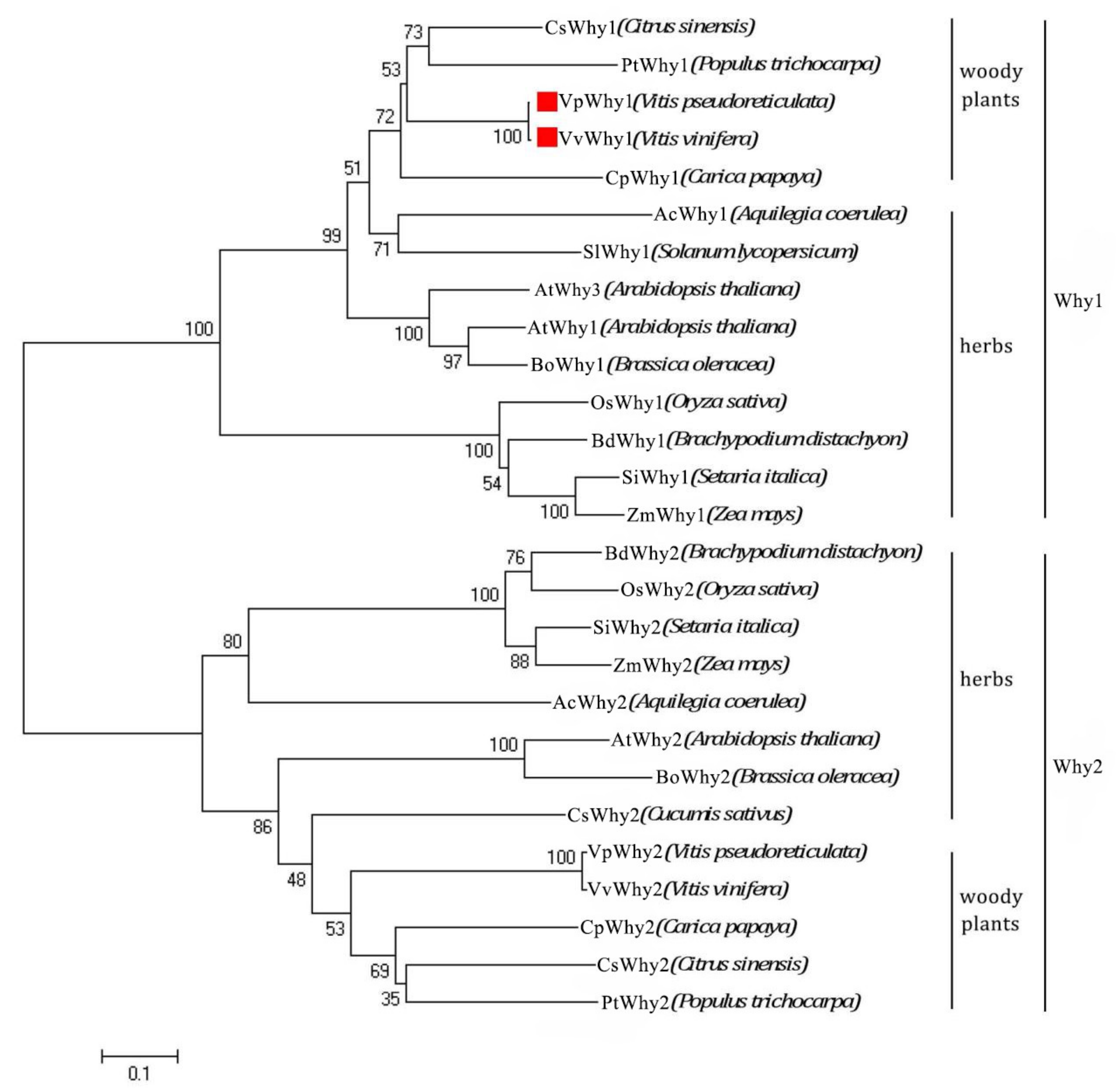
2.1. Isolation and Characterization of Why1 Genes and Their Promoters

Two Why1 genes and their 5′-upstream promoter sequences were amplified. VvWhy1 (GenBank: MN395403) and its promoter sequence (GenBank: MN397251) were isolated from V. vinifera “Cabernet Sauvignon” and VpWhy1 (GenBank: MN395402) and its promoter sequence (GenBank: MN397250) were isolated from V. pseudoreticulata “HD1”. Both Why1 genes had a total length of 862 bp with 807 bp length ORF, encoding 268 amino acid residues (Figure 1a). The VvWhy1 and VpWhy1 promoter sequences had 1127 bp and 1136 bp. Interestingly, the VvWhy1 promoter sequence showed nine bp missing compared with the VpWhy1 promoter sequence (Supplementary Materials sequence). The sequence alignment showed that the identity between the two Why1 genes was 99.38%, and their CDS only encoded three different amino acid residues. However, two Why1 promoters showed a lower identity of 95.16%. In consequence, the sequence alignments showed the Why1 CDS sequences were more conserved than the Why1 promoter sequences.
The gene structure analysis showed that both Why1 sequences had seven exons and six introns (Figure 1b), they had a C-terminal Whirly conserved domain and N-terminal chloroplast transit peptide (Figure 1c), indicating a potential chloroplast subcellular localization. Phylogenetic analyses were conducted to estimate the evolution relationship and an unrooted phylogenetic tree was constructed from an aligned dataset of 27 homologous Why protein sequences (Figure 2). These proteins were grouped into two classes, Why1 and Why2, respectively. Both Why1 and Why2 could be divided into two subgroups, which were derived from woody and herb plants. VvWhy1 from V. vinifera and VpWhy1 from V. pseudoreticulata were clustered together with other Why1 of woody plants.

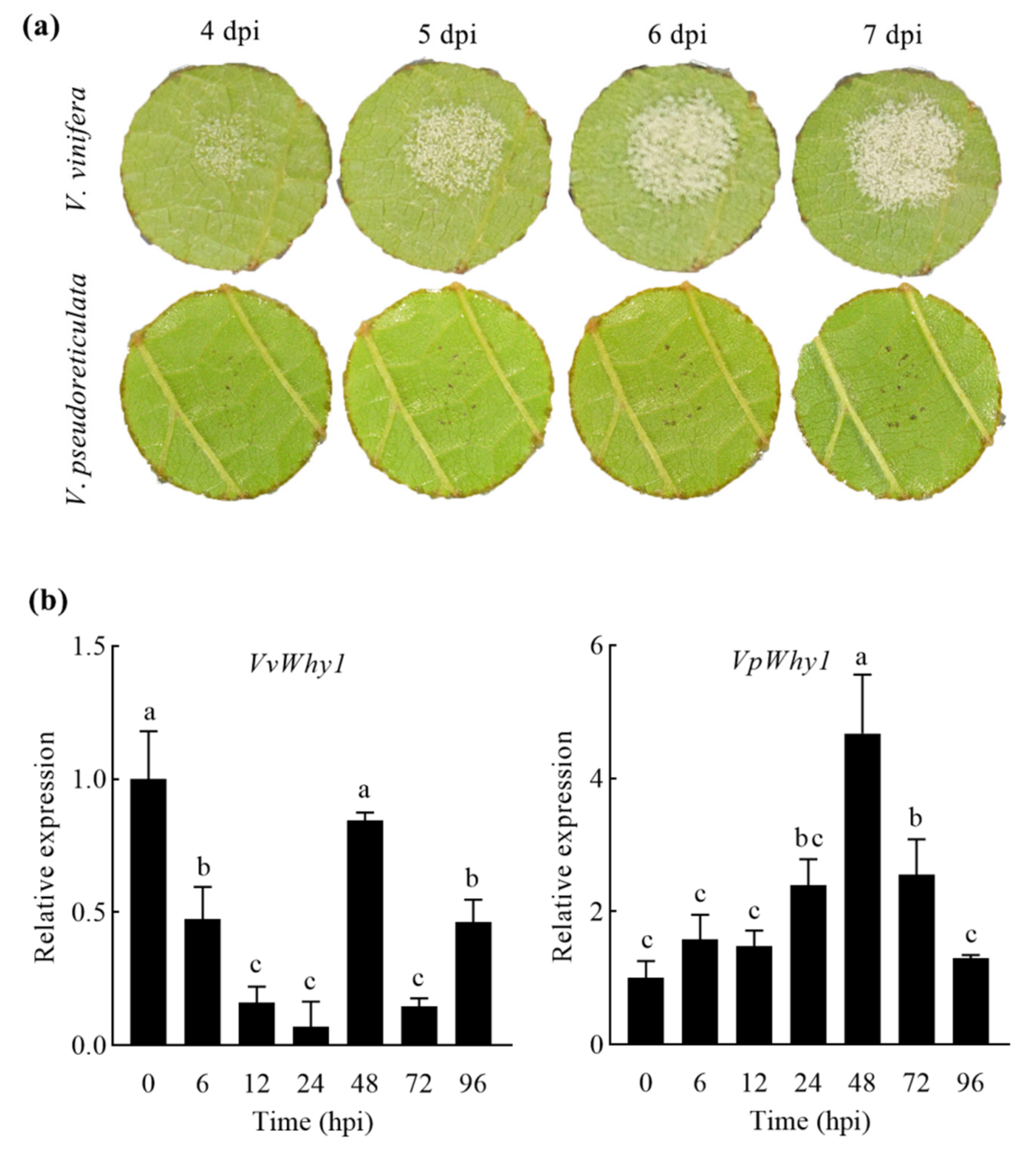
2.2. Why1 Expression under P. viticola Induction in V. vinifera and V. pseudoreticulata

To investigate the Why1 expression model under pathogen infection, the leaves of V. vinifera and V. pseudoreticulata were inoculated with P. viticola. As shown in Figure 3a, the leaf symptoms were significantly different between V. vinifera and V. pseudoreticulata. P. viticola was observed obviously in V. vinifera leaves at 4 days postinoculation (dpi), and the leaf symptoms of P. viticola aggravated with the extension of the treatment time. However, only a few disease spots appeared in V. pseudoreticulata leaves without P. viticola growth. The results indicated that V. vinifera “Cabernet Sauvignon” was downy-mildew-susceptible, whereas V. pseudoreticulata “HD1” was resistant. A transcriptional expression was further performed by using qRT-PCR and the results showed that VvWhy1 remained at a low expression level (Figure 3b). On the contrary, VpWhy1 was significantly upregulated under P. viticola infection and reached a maximum at 48 h postinoculation (hpi), then rapidly decreased when it was 72 and 96 hpi. A transcriptional expression analysis revealed that VpWhy1’s expression was induced by P. viticola, while VvWhy1 remained insensitive to P. viticola. VpWhy1 may play an important role in P. viticola resistance.

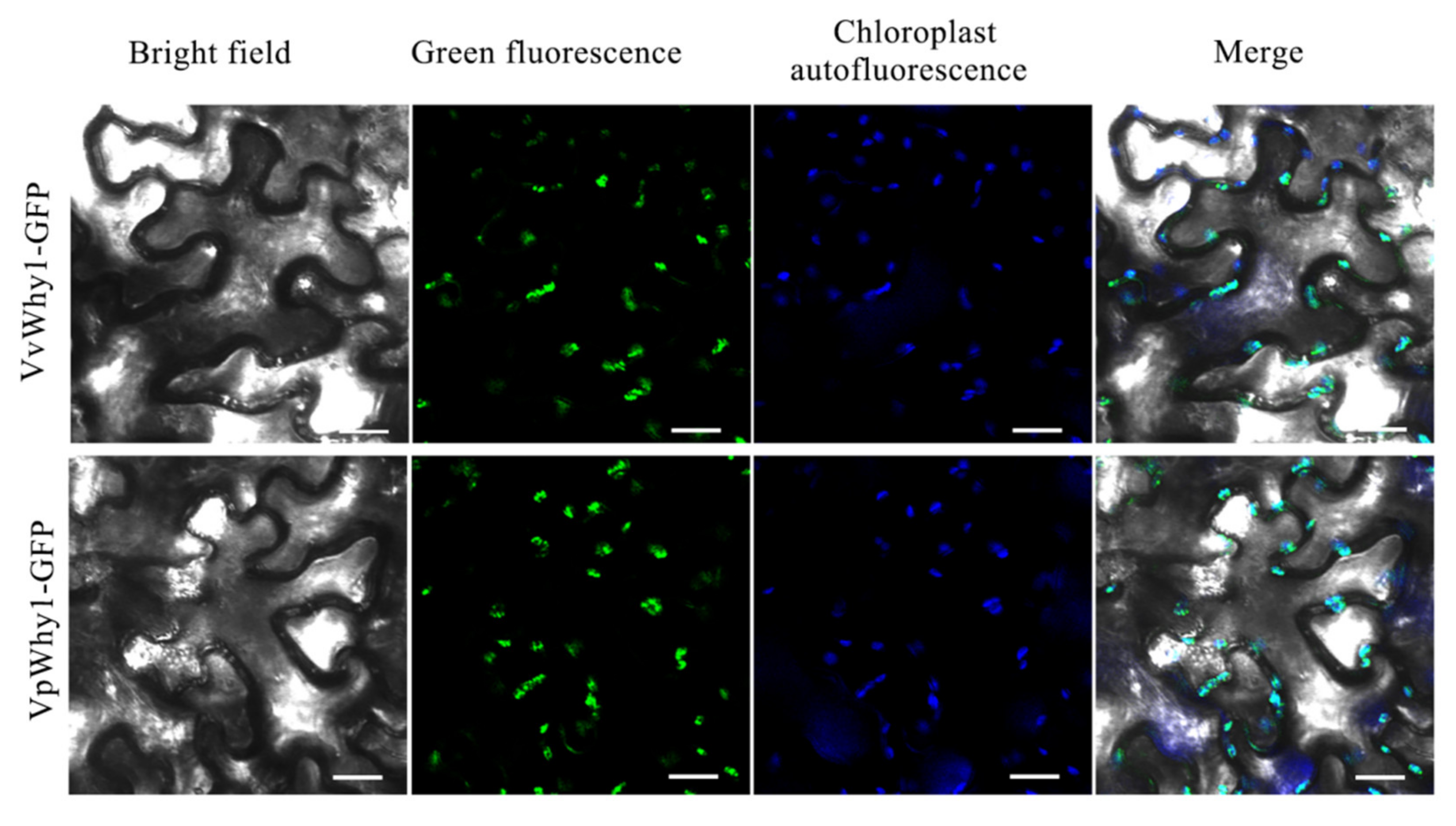
2.3. Subcellular Localization Analysis of Why1

A conserved domain analysis showed the Why1 proteins had an N-terminal chloroplast transit peptide. To verify the subcellular localization of Why1, the plant expression vectors of pBI121-VvWhy1-GFP and pBI121-VpWhy1-GFP were constructed. Agrobacterium tumefaciens strain GV3101 harboring a recombinant plasmid was injected into N. benthamiana leaves to transiently express the Why1-GFP fusion protein. Results showed that the GFP fluorescence signal of VvWhy1 and VpWhy1 infusion proteins were overlapped with chloroplast autofluorescence signal (Figure 4). Therefore, a subcellular localization indicated that both VvWhy1 and VpWhy1 proteins were chloroplast localization, and may play important roles in chloroplast.

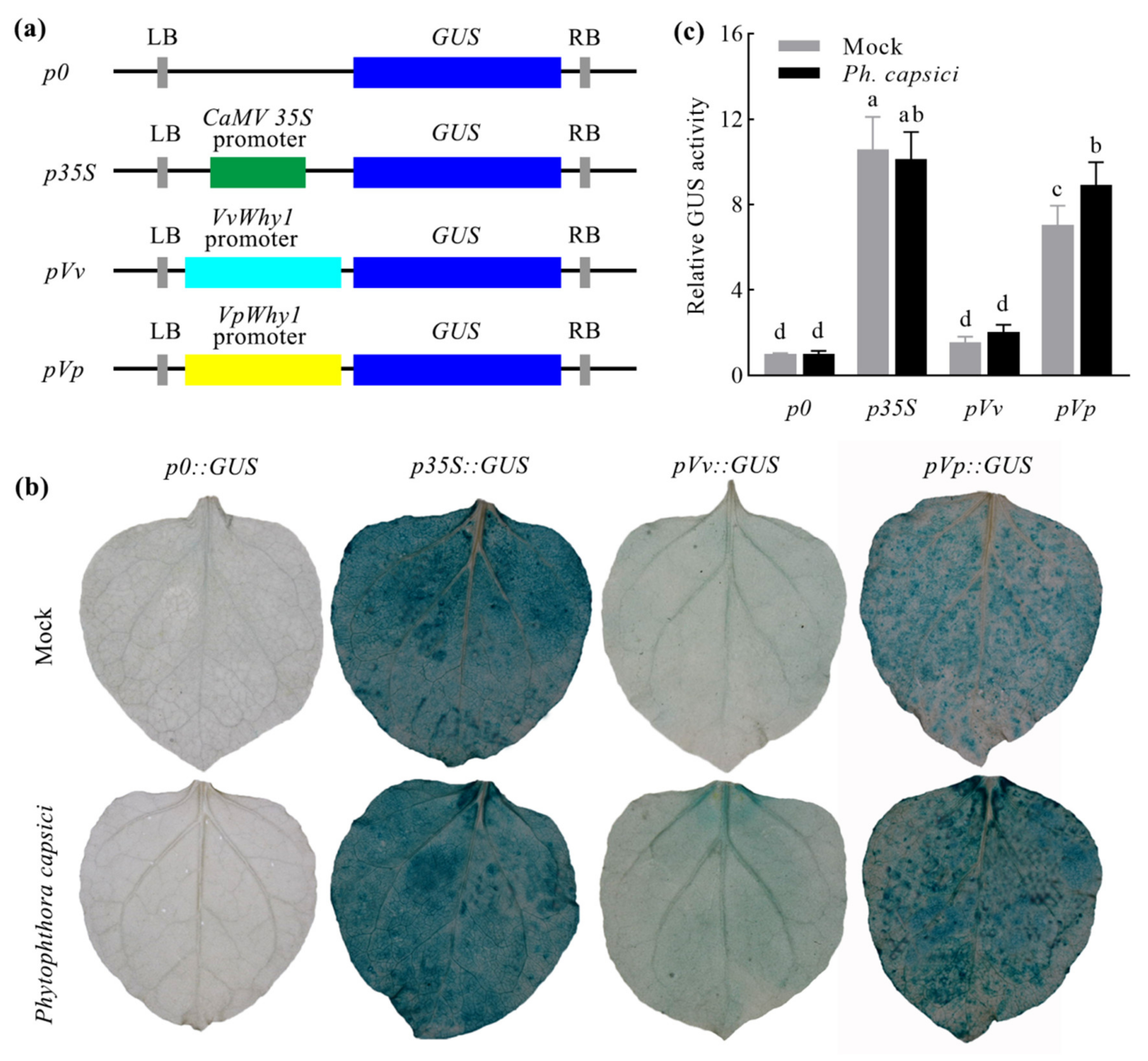
2.4. Why1 Promoter Activity Analysis

A previous sequence analysis of two Why1 genes showed a 99.38% identity and the CDS only encoded three different amino acid residues. The promoter identity (95.16%) was far lower than that in CDS. Moreover, a transcriptional expression analysis showed a different expression pattern of VvWhy1 and VpWhy1. We supposed that the VvWhy1 promoter (pVv) and VpWhy1 promoter (pVp) may play important roles in response to P. viticola. To determine the activity of the two promoters, pBI121-pVv::GUS and pBI121-pVp::GUS were generated and p0::GUS and p35S::GUS were negative and positive controls (Figure 5a). A GUS histochemical stain showed that the negative control leaves had no staining, and the positive control leaves were stained with deep blue, both controls were not induced by P. capsici. The pVv::GUS treatment leaves had no staining under the mock treatment and a slight blue stain appeared when it was under P. capsici stress. For the pVp::GUS treatment, it showed a high GUS activity, as shown by the deep blue staining (Figure 5b). The relative quantitative GUS activity of the pVv samples showed no significant difference between the mock and P. capsici treatments, but it was significantly induced in the pVp treatments (Figure 5c). This study indicated that pVp had a high promoter activity and was involved in the response to P. capsici.

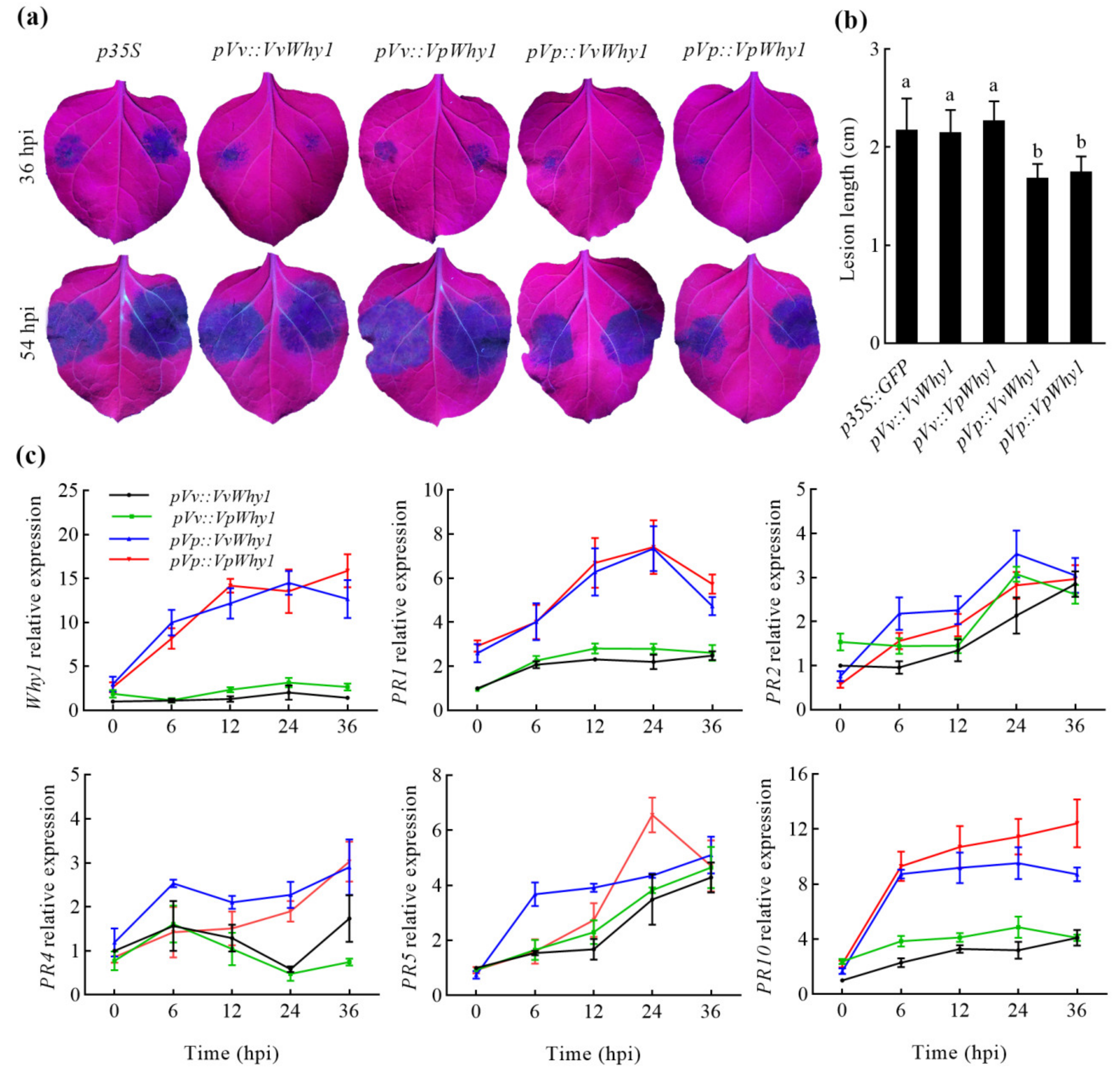
2.5. P. capsici Resistance of Why1 Genes Driven by Native and Exotic Promoters

A transcriptional expression and promoter activity showed that VpWhy1 and its promoter were strongly induced in response to P. viticola and P. capsici. To investigate whether the Why1 and its promoter had P. capsici resistance, we generated four Why1 vectors driven by native and exotic promoters (Figure 6a) for the transient expression in N. benthamiana. A P. capsici resistance assay after trypan blue staining in N. benthamiana leaves shown in Figure 6b (left side for p35 controls, right side for experimental treatments). The leaves exhibited obvious lesion symptoms in the control (left side) and experimental (right side) treatments. For VvWhy1 and VpWhy1 driven by pVp, the lesion areas were significantly smaller than that in the controls. A further quantitative examination showed that the relative lesion areas in pVv::VvWhy1 and pVv::VpWhy1 were smaller than that in pVp::VvWhy1 and pVp::VpWhy1. The result of the lesion areas calculation was consistent with the findings when observing the disease symptoms, which implied that pVp significantly enhanced the resistance to P. capsici.

2.6. Why1 Heterologous Expression and Pathogenesis-Related Genes in Response to P. capsici

The recombinant vectors were described as 2.5 and the transient expression was conducted on both leaf sides. N. benthamiana leaf lesion symptoms were observed by using a UV light under P. capsici infection at 36 and 54 hpi (Figure 7a). All treatments of N. benthamiana displayed varying degrees of disease symptoms. The pVp treatments of transient transgenic leaves had a smaller lesion area than the control (p35S) and pVv treatments at 36 hpi. The lesion area expanded in all leaf samples with different treatments at 54 hpi. Then, a disease resistance evaluation was conducted by calculating the lesion length in N. benthamiana. The lesion length of p35S and the two pVv treatments were between 2.14 and 2.27 cm, while the two pVp treatments were between 1.68 and 1.75 cm, respectively. The lesion length of the two pVv treatments were significantly longer than that in the two pVp treatments, these results were consistent with those of the above trypan blue staining assay, indicating that pVp enhanced P. capsici resistance.
To further explore the molecular mechanism of pVp in the resistance to P. capsici, a transcriptional expression of Why1 and PR genes was performed in transient transgenic N. benthamiana. Why1 and PR genes could be divided into two groups; Why1, PR1 and PR10 were strongly induced in pVp transient transgenic N. benthamiana, whereas PR2, PR4 and PR5 remained at a relative low expression level. The expression of PR2, PR4 and PR5 showed an increasing trend with time extension and were not significantly induced by different transgenic treatments under P. capsici infection. Why1, PR1 and PR10 were significantly induced in pVp transient transgenic treatments, while the expression remained at a lower level in the pVv treatments. VvWhy1 and VpWhy1 were upregulated, and reached a maximum expression level exceeding 14- and 15-fold change at 24 and 36 hpi in the pVp::VvWhy1 and pVp::VpWhy1 transgenic treatments, respectively. The PR1 gene was upregulated and reached a maximum at 24 dpi, then declined at 36 dpi. The expression of PR10 increased dramatically at 6 hpi in both pVp treatments. For the pVp::VvWhy1 treatment, PR10 continued to increase and reached a maximum expression level. However, the PR10 expression stayed at a stable level, and even decreased at 36 hpi. As a result, pVp enhanced the expression of Why1, PR1 and PR10; these findings were in accordance with the disease symptoms observation in the P. capsici resistant assay. In conclusion, these experiments suggested that pVp promoted the Why1 transcription. Furthermore, Why1 regulated the expression of PR1 and PR10, which finally enhanced the resistance to P. capsici.

3. Discussion

3.1. Dual Localization of Why1

Whirly (Why) proteins appeared to have multiple functions in plant growth and development, biotic and abiotic stress, which may attribute to their complicated subcellular localization. Desveaux [19] provided clear evidence for an insight into a novel single-stranded DNA-binding transcription activator PBF-2 (StWhy1), which was identified from Solanum tuberosum. The plant did possess at least two Why members, and a previous study indicated that in the cases of two members, the Why proteins were putatively chloroplast- (Why1) and mitochondrial-localized (Why2), respectively [21]. The occurrence of the third Why was revealed to be nucleus-localized and may be restricted to Arabidopsis [33].
However, recent studies have revealed that Why1 translocated from chloroplast to nucleus [34]. Proteins with dual subcellular localization mediate diverse intercellular signaling processes and various functions [35,36,37]. As a consequence, Why1 is dual-localized to the chloroplast and nucleus [38,39]. The translocation function may result from stress-associated redox changes in the photosynthetic apparatus [40]. Moreover, the phosphorylation and oxidization of the Why1 protein also lead to different subcellular localization in the nucleus or plastid [38,41]. Why1 is an excellent candidate for communication between chloroplasts and nucleus due to its dual localization in chloroplasts and nucleus [42]. Why1 has even been shown to be relocated from one compartment to another upon environmental or developmental clues [43]. In our study, VvWhy1 and VpWhy1 showed chloroplast-localization in N. benthamiana, and no fluorescence signal was detected in the nucleus. Similarly, it raised the question of whether it is dual-targeted in vivo since a Why1-GFP protein in potato mesophyll protoplasts localized to the chloroplasts but not to the nucleus in an earlier study. According to the translocation and relocalization functions, Why1 is a sequestered nuclear transcription factor that can be released from plastids under certain conditions [43]. We hypothesized that stress treatments may lead to the dual localization of VvWhy1 and VpWhy1 proteins.

3.2. Promoter Differentiation between VvWhy1 and VpWhy1

Vitis plants including Eurasian, East Asian and North American species share a close genetic relationship and conservative genomic sequences. The genome sequencing completion of the Eurasian grapevine V. vinifera “Pinot Noir” marks a new stage of grapevine research [44]. The “Pinot Noir” genome is regarded as a reference for other grapevine species in genome assembly, gene annotation, gene cloning and function verification based on their close genetic backgrounds. However, different grapevines show a varied degree of disease resistance; V. vinifera “Cabernet Sauvignon” is downy-mildew-susceptible, while V. pseudoreticulata “HD1” is resistant [45].
In this study, both grapevines showed a different response to P. viticola. However, Why1 CDS from V. vinifera and V. pseudoreticulata showed a high identity of 99.38% with five different bases, which encoded three different amino acids. The VvWhy1 and VpWhy1 proteins had similar physical and chemical properties. In contrast, the upstream promoter regions of both grapevines possessed different sequence lengths: 1127 bp and 1136 bp promoter sequences were obtained from V. vinifera and V. pseudoreticulata, respectively. The VvWhy1 promoter (pVv) sequence was nine bp shorter than that in the VpWhy1 promoter (pVp). Moreover, the promoter sequence identity was 95.16%, which was far lower than that in CDSs. Therefore, VvWhy1 and VpWhy1 had conserved CDS but with promoter sequence differentiation. The different response to P. viticola between VvWhy1 and VpWhy1 may result from the promoter differentiation, and further lead to the disease resistance differentiation in different grapevines. In general, promoter sequence alterations provided an effective manner to regulate gene expression and function. Interestingly, the homologous genes VvSTS and VpSTS from V. vinifera “Carigane” and V. pseudoreticulata share >99% identity on the amino acid level, but with significant difference in promoter regions; VpSTS conferred powdery mildew resistance with an elevated responsiveness of the promoter [46]. In V. labrusca “Concord”, the absence of a 426 bp and/or a 42 bp sequence in the acyltransferase gene (AMAT) promoter highly associated with high levels of AMAT expression, and further regulated the methyl anthranilate (MA) accumulation, which caused a special “foxy” aroma [47]. We claim that promoter differentiation between VvWhy1 and VpWhy1 may lead to disease resistance differentiation in V. vinifera and V. pseudoreticulata.

3.3. Why1 Driven by VpWhy1 Promoter (pVp) Enhanced Disease Resistance via Regulating the Expression of PR Genes

The Why1 gene function has been elucidated in multiple biological functions, but the transcriptional regulation of its noncoding region has rarely been reported. Based on the high conservation of Why1 CDS and promoter differentiation between VvWhy1 and VpWhy1, the differential expression of VvWhy1 and VpWhy1 in response to pathogen may result from their promoter sequence. The activity of the VpWhy1 promoter was significantly higher than that of the VvWhy1 promoter and was induced by P. viticola and P. capsici. VvWhy1 and VpWhy1 driven by the VpWhy1 promoter (pVp) increased P. capsici resistance in N. benthamiana leaves. StWhy1 participated in interactions with the elicitor response element (ERE) of PR-10a, its binding to the ERE correlating with the expression of PR-10a [19]. The transcription activator StWhy1 encoding a protein of 24 kD, critical for the interaction of PBF-2 with single-stranded DNA or RNA is a motif consisting of KGKAAL. Mutations in this domain have occurred in the absence of DNA-binding activity [48]. Why1 also plays a new role in salicylic acid (SA) biosynthesis via the coordination of isochorismate synthase1 (ICS1), phenylalnine ammonialyase (PAL1) and S-adenosyl-L-Met-dependent methyltransferase1 (BSMT1) [39]. SA plays important roles in disease resistance and pathogenesis-related response.
Pathogenesis-related genes (PR) are among the best characterized genes induced by pathogens [19]; it is one of the key components of plant innate immune system especially systemic acquired resistance (SAR) [49]. Rossarolla [13] indicated the stronger and earlier activation of the defense pathway in the genotypes containing pyramided Rpv was evidenced in the expression of the evaluated PR genes, among which PR1 and PR10, involved in the defense reaction, were mainly associated with the SA signaling pathway and JA signaling pathway, respectively [50,51]. The overexpression of VpPR10.1 from V. pseudoreticulata enhanced downy mildew resistance in V. vinifera [52]. PR genes, including PR-1, PR-3, PR-10, contributed to powdery mildew resistance in wheat [53]. Arabidopsis Why1 was reported to be involved in disease defense signaling and required for pathogen-induced PR1 expression [33]. To further investigate whether the PR genes were regulated by VvWhy1 and VpWhy1 under P. capsici infection, a transcriptional expression of PR1, PR2, PR4, PR5 and PR10 was conducted in N. benthamiana transiently expressing VvWhy1 and VpWhy1 driven by pVv and pVp. The results showed that pVp promoted VvWhy1 and VpWhy1 correlated with the expression of PR1 and PR10. As a result, Why1 driven by the VpWhy1 promoter (pVp) enhanced disease resistance via regulating the expression of PR1 and PR10 genes.

4. Materials and Methods

4.1. Plant Materials, Pathogen and Treatments

One-year-old V. vinifera “Cabernet Sauvignon” and V. pseudoreticulata “HD1” from Shangzhuang experimental station of China Agricultural University, were grown in pots at 25 ± 2 °C under a 16/8 h light/dark photoperiod in a greenhouse. For real-time PCR, grapevine leaves were inoculated with P. viticola sporangia suspension liquid (105 sporangia mL−1). Five grapevine leaf samples were collected at 0, 6, 12, 24, 48, 72, and 96 hpi. One-month-old N. benthamiana seedlings were used for transient gene expression and disease resistance evaluation, the third to fifth unfolded leaves from the shoot apex were injected with A. tumefaciens transient suspension liquid harboring a recombinant plastid. After 48 h, five N. benthamiana leaves were excised for each P. capsici inoculation treatment. Three biological replicates were performed for P. viticola and P. capsici inoculation.

4.2. Gene Cloning and Sequence Analysis

V. vinifera “Cabernet Sauvignon” and V. pseudoreticulata “HD1” genomic DNA and total RNA were isolated according to the CTAB method with minor modifications. Reverse transcription was performed using RevertAid TM First Strand cDNA Synthesis Kit (Invitrogen, Waltham, MA, USA). Genomic DNA and cDNA were used for promoter and gene cloning, respectively. Primers were designed according to predicted Why1 gene sequences and their upstream regulatory sequences downloaded from Phytozome V. vinifera “Pinot Noir” reference genome; primer sequences are listed in the Supplementary Materials Table S1. Why1 genes and promoters were cloned by using PCR amplification, electrophoretic band purification, cloning vector ligation, Escherichia coli transformation and finally sequencing validation with four E. coli clones. The gene structure was analyzed by online software Gene Structure Display Server (GSDS) [54]. The conserved domain was predicted using online NCBI-CDD [55]. The phylogenetic tree was constructed using MEGA 5.0 with the neighbor-joining (NJ) method. The promoter sequences were analyzed using PlantCARE [56] to search for cis-acting elements.

4.3. Quantitative Real-Time PCR Analysis of Why1 under P. viticola Induction

Total RNA was extracted from grapevine using the CTAB method with minor modification. The cDNA synthesis was conducted according to PrimeScript™ RT reagent Kit (Takara, Kusatsu, Japan). Three biological cDNA replicates were mixed for next amplification. Specific primers listed in Supplementary Materials Table S1 were designed for real-time PCR. The amplification was performed on the QIAGEN Rotor-Gene Q system (QIAGEN, Hilden, Germany) using the SuperReal PreMix Plus (SYBR Green) kit (TIANGEN, Beijing, China). Constitutively expressed elongation factor1-α (EF1-α) was used as reference gene to calculate relative expression levels with the 2−∆∆Ct method. All reactions were performed with three technical replicates.

4.4. Subcellular Localization Analysis

Plant expression vectors pBI121-VpWhy1-GFP and pBI121-VvWhy1-GFP driven by the CaMV 35S promoter were constructed for the subcellular localization analysis. Primers with BamHI and KpnI (2 bp insertion was required to avoid frameshift mutation) restriction sites flanking the VvWhy1 and VpWhy1 sequences were designed (Supplementary Materials Table S2). Ten positive E. coli clones harboring recombinant plasmids were validated by PCR, among which four clones were further sequenced. The recombinant plasmids were introduced into Agrobacterium tumefaciens (GV3101) via electroporation. The GV3101 was harvested when the OD600 reached ~1.0 grown with rifampicin (50 mg/L) and kanamycin (60 mg/L) in LB liquid medium, and then resuspended with MMA solution supplemented with 10 mM MgCl2, 10 mM MES, pH 5.7 and 100 μM acetosyringone, to a final OD600 of 0.6. The Why1-GFP fusion proteins were transiently expression in N. benthamiana using the Agrobacterium-mediated leaf injection method as previously described [57]. Fluorescence observation was conducted using an FV1000 confocal laser-scanning microscope (Olympus, Tokyo, Japan).

4.5. Promoter Activity Analysis

To estimate the Why1 promoter activity, primers with HindIII and SmaI restriction sites flanking the VvWhy1 promoter (pVv) and VpWhy1 promoter (pVp) were designed to construct pBI121-pVv::GUS and pBI121-pVp::GUS, while pBI121-p0::GUS and pBI121-35S::GUS were negative and positive controls, respectively. The method of GUS transiently expression in N. benthamiana was described as above. The pathogen stress was conducted with P. capsici zoospore suspensions according to [58]. The GUS histochemical staining and quantitative detection were carried out to qualitatively and quantitatively assess the promoter activity, respectively. A fluorescence spectrophotometer was used for the GUS quantitative determination as described in [59]. The total protein concentration was measured with the Bradford method [60]. Three biological replicates were performed for the GUS histochemical staining and quantitative detection.

4.6. P. capsici Resistance Assay Transiently Expressing Why1 Driven by Native and Exotic Promoter

For the P. capsici resistance assay, based on previous constructs of pBI121-VvWhy1-GFP and pBI121-VpWhy1-GFP in subcellular localization, primers with SbfI and BamHI flanking pVv and pVp were designed to construct 4 pBI121 plant expression vectors containing pVv::VvWhy1, pVv::VpWhy1, pVp::VvWhy1 and pVp::VpWhy1, and the CaMV 35S promoter was used as control. Transient expression was conducted as mentioned above. A P. capsici resistance assay was performed with the disk inoculation method according to a previous protocol [17]. To estimate the resistance to P. capsici of Why1, trypan blue staining was carried out and the disease lesion area was calculated. The relative lesion area is the ratio of the experimental lesion areas to those on the control. Three biological replicates were used, each of which contained five leaf samples.

4.7. Why1 Heterologous Expression and Pathogenesis-Related Genes in Response to P. capsici

To further illustrate the molecular mechanism of disease resistance, the expression of Why1 and pathogenesis-relative genes (PR) was performed. The plant expression vectors and transient transformation procedure were described as the above transient expression in the P. capsici resistance assay, the difference being that N. benthamiana was injected with the same Agrobacterium suspensions on both leaf sides. P. capsici infection was conducted after 48 h of transient expression. Five detached leaves were collected when it was 0, 6, 12, 24 and 36 hpi for the transcriptional expression analysis, using three biological replicates. The qRT-PCR of Why1 associated with PR1, PR2, PR4, PR5 and PR10 in transgenic N. benthamiana was performed; the primer sequences are listed in Supplementary Materials Table S1. Lesion symptoms were observed by using a UV light, and the lesion lengths were calculated to estimate the disease resistance with five leaf samples for each treatment and three biological replicates.

4.8. Statistical Analysis

The quantitative results for the gene expression analysis, GUS activity, lesion area and lesion length are presented as the means ± standard deviations (SD). Tukey’s multiple comparisons test was conducted by using GraphPad Prism 8.0. Different letters on the columns indicate statistically significant differences at the 0.05 level. Figures were prepared using GraphPad Prism 8.0.

5. Conclusions

The single-stranded DNA- and RNA-binding protein genes VvWhy1 and VpWhy1 with promoter sequences were isolated from V. vinifera “Cabernet Sauvignon” and V. pseudoreticulata “HD1”. VpWhy1 was strongly induced by P. viticola, while VvWhy1 showed a low expression level. VvWhy1 and VpWhy1 shared 99.38% of identity encoded in only three different amino acid residues. However, the identity of the promoter sequences was far lower than that between Why1 CDSs. Therefore, we proposed that the differentiation of Why1 promoters played important roles in pathogen response. Further, the GUS activity indicated the VpWhy1 promoter (pVp) had a high activity involved in response to P. capsici infection. N. benthamiana transiently expressing pVp::Why1 (pVp::VvWhy1 and pVp::VpWhy1) enhanced the P. capsici resistance. Moreover, Why1, PR1 and PR10 were upregulated under P. capsici infection in pVp transient transgenic N. benthamiana leaves. PR genes were reported to be regulated by Why1; in consequence, PR1 and PR10 were promoted by pVp::Why1. In conclusion, this study provides new insight into the novel disease resistance mechanism of VpWhy1 with a strong pathogen-induced promoter.

Supplementary Materials

The following supporting information can be downloaded at: https://www.mdpi.com/article/10.3390/ijms23148052/s1.

Author Contributions

G.L. and J.C. designed the experiments, Q.W., H.G. and X.G. prepared the materials. C.L., Q.Q. and R.P. performed the experiments. C.L., J.C. and G.L. analyzed the data and wrote the manuscript. All authors have read and agreed to the published version of the manuscript.

Funding

This research was funded by Fujian Provincial Department of Science and Technology of Special Public-funded Projects (grant no. 2021R1032009), Freely Explore Scientific and Technological Innovation Projects of Fujian Academy of Agricultural Sciences (grant no. ZYTS2021008), Fujian Academy of Agricultural Sciences Research Projects (grant no. DWHZ2021-01).

Institutional Review Board Statement

Not applicable.

Informed Consent Statement

Not applicable.

Data Availability Statement

Not applicable.

Conflicts of Interest

The authors declare no conflict of interest.

Abbreviations

P. ViticolaPlasmopara Viticola
P. capsicaPhytophthora capsici
pVvVvWhy1 promoter
pVpVpWhy1 promoter
PR genePathogenesis-related gene
R geneResistance gene
qRT-PCRQuantitative real-time PCR
GFPGreen fluorescent protein
CDSCoding sequence
dpiDays postinoculation
hpiHours postinoculation

References

  1. Jiang, H.E.; Zhang, Y.B.; Li, X.; Yao, Y.F.; Fergusond, D.K.; Lü, E.G.; Li, C.S. Evidence for early viticulture in China: Proof of a grapevine (Vitis vinifera L., Vitaceae) in the Yanghai Tombs, Xinjiang. J. Archaeol. Sci. 2009, 36, 1458–1465. [Google Scholar] [CrossRef]
  2. Figueiredo, J.; Cunha, J.; Eiras-Dias, J.; Silva, M.S.; Figueiredo, A. Pathogen-related specificity of subtilase VVISBT4.19 X1 in the Vitis vinifera defence response. Cienc. Tec. Vitivinic. 2020, 35, 42–48. [Google Scholar] [CrossRef]
  3. Cavaco, A.R.; Laureano, G.; Cunha, J.; Eiras-Dias, J.; Figueiredo, A. Fatty acid modulation and desaturase gene expression are differentially triggered in grapevine incompatible interaction with biotrophs and necrotrophs. Plant Physiol. Biochem. 2021, 163, 230–238. [Google Scholar] [CrossRef] [PubMed]
  4. Marcianò, D.; Ricciardi, V.; Fassolo, E.M.; Passera, A.; Toffolatti, S.L. RNAi of a putative grapevine susceptibility gene as a possible downy mildew control strategy. Front. Plant Sci. 2021, 12, 667319. [Google Scholar] [CrossRef] [PubMed]
  5. Yin, W.; Wang, X.; Liu, H.; Wang, Y.; Nocker, S.V.; Tu, M.; Fang, J.; Guo, J.; Li, Z.; Wang, X. Overexpression of VqWRKY31 enhances powdery mildew resistance in grapevine by promoting salicylic acid signaling and specific metabolite synthesis. Hortic. Res. 2022, 9, uhab064. [Google Scholar] [CrossRef]
  6. Yan, X.; Qiao, H.; Zhang, X.; Guo, C.; Wang, M.; Wang, Y.; Wang, X. Analysis of the grape (Vitis vinifera L.) thaumatin-like protein (TLP) gene family and demonstration that TLP29 contributes to disease resistance. Sci. Rep. 2017, 7, 4269. [Google Scholar] [CrossRef] [Green Version]
  7. Palmieri, M.C.; Perazzolli, M.; Matafora, V.; Moretto, M.; Bachi, A.; Pertot, I. Proteomic analysis of grapevine resistance induced by Trichoderma harzianum T39 reveals specific defence pathways activated against downy mildew. J. Exp. Bot. 2012, 63, 6237–6251. [Google Scholar] [CrossRef]
  8. Li, M.Y.; Jiao, Y.T.; Wang, Y.T.; Zhang, N.; Wang, B.B.; Liu, R.Q.; Yin, X.; Xu, Y.; Liu, G.T. CRISPR/Cas9-mediated VvPR4b editing decreases downy mildew resistance in grapevine (Vitis vinifera L.). Hortic. Res. 2020, 7, 149. [Google Scholar] [CrossRef]
  9. Gessler, C.; Pertot, I.; Perazzolli, M. Plasmopara viticola: A review of knowledge on downy mildew of grapevine and effective disease management. Phytopathol. Mediterr. 2011, 50, 3–44. [Google Scholar] [CrossRef]
  10. Ma, H.; Xiang, G.Q.; Li, Z.Q.; Wang, Y.T.; Dou, M.R.; Su, L.; Yin, X.; Liu, R.Q.; Wang, Y.J.; Xu, Y. Grapevine VpPR10.1 functions in resistance to Plasmopara viticola through triggering a cell death-like defence response by interacting with VpVDAC3. Plant Biotechnol. J. 2018, 16, 1488–1501. [Google Scholar] [CrossRef] [Green Version]
  11. Sargolzaei, M.; Maddalena, G.; Bitsadze, N.; Maghradze, D.; Bianco, P.A.; Failla, O.; Toffolatti, S.L.; Lorenzis, G.D. Rpv29, Rpv30 and Rpv31: Three novel genomic loci associated with resistance to Plasmopara viticola in Vitis vinifera. Front. Plant Sci. 2020, 11, 562432. [Google Scholar] [CrossRef] [PubMed]
  12. Fu, P.; Wu, W.; Lai, G.; Li, R.; Peng, Y.; Yang, B.; Wang, B.; Yin, L.; Qu, J.; Song, S.; et al. Identifying Plasmopara viticola resistance Loci in grapevine (Vitis amurensis) via genotyping-by-sequencing-based QTL mapping. Plant Physiol. Biochem. 2020, 154, 75–84. [Google Scholar] [CrossRef] [PubMed]
  13. Ciubotaru, R.M.; Franceschi, P.; Zulini, L.; Stefanini, M.; Škrab, D.; Rossarolla, M.D.; Robatscher, P.; Oberhuber, M.; Vrhovsek, U.; Chitarrini, G. Mono-locus and pyramided resistant grapevine cultivars reveal early putative biomarkers upon artificial inoculation with Plasmopara viticola. Front. Plant Sci. 2021, 12, 693887. [Google Scholar] [CrossRef] [PubMed]
  14. Rossarolla, M.D.; Tomazetti, T.C.; Welter, L.J.; Santos, H.P.; Stefanini, M.; Trapp, O.; Guerra, M.P.; Nodari, R.O. The interplay between hormone signaling and defense gene expression in grapevine genotypes carrying genetic resistance against Plasmopara viticola. Vitis 2021, 60, 195–206. [Google Scholar] [CrossRef]
  15. Liu, R.; Weng, K.; Dou, M.; Chen, T.; Xu, Y. Transcriptomic analysis of Chinese wild Vitis pseudoreticulata in response to Plasmopara viticola. Protoplasma 2019, 256, 1409–1424. [Google Scholar] [CrossRef] [PubMed]
  16. Xu, W.; Ma, F.; Li, R.; Zhou, Q.; Yao, W.; Jiao, Y.; Zhang, C.; Zhang, J.; Wang, X.; Xu, Y. VpSTS29/STS2 enhances fungal tolerance in grapevine through a positive feedback loop. Plant Cell Environ. 2019, 42, 2979–2998. [Google Scholar] [CrossRef]
  17. Lai, G.; Fu, P.; Liu, Y.; Jiang, X.; Lu, J. Molecular characterization and overexpression of VpRPW8s from Vitis pseudoreticulata enhances resistance to Phytophthora capsici in Nicotiana benthamiana. Int. J. Mol. Sci. 2018, 19, 839. [Google Scholar] [CrossRef] [Green Version]
  18. Lai, G.; Song, S.; Liu, Y.; Fu, P.; Xiang, J.; Lu, J. RPW8 promoter is involved in pathogen- And stress-inducible expression from Vitis pseudoreticulata. J. Phytopathol. 2019, 167, 65–74. [Google Scholar] [CrossRef]
  19. Desveaux, D.; Després, C.; Joyeux, A.; Brisson, S.N. PBF-2 is a novel single-stranded DNA binding factor implicated in PR-10a gene activation in potato. Plant Cell 2000, 12, 1477–1489. [Google Scholar] [CrossRef] [Green Version]
  20. Prikryl, J.; Watkins, W.K.; Friso, G.; van Wijk, K.J.; Barkan, A. A member of the Whirly family is a multifunctional RNA- and DNA-binding protein that is essential for chloroplast biogenesis. Nucleic Acids Res. 2008, 36, 5152–5165. [Google Scholar] [CrossRef]
  21. Krause, K.; Kilbienski, I.; Mulisch, M.; Rdiger, A.; Schfer, A.; Krupinska, K. DNA-binding proteins of the Whirly family in Arabidopsis thaliana are targeted to the organelles. FEBS Lett. 2005, 579, 3707–3712. [Google Scholar] [CrossRef] [PubMed] [Green Version]
  22. Grabowski, E.; Miao, Y.; Mulisch, M.; Krupinska, K. Single-stranded DNA-binding protein Whirly1 in barley leaves is located in plastids and the nucleus of the same cell. Plant Physiol. 2008, 147, 1800–1804. [Google Scholar] [CrossRef] [PubMed] [Green Version]
  23. McCarty, D.R.; Mark Settles, A.; Suzuki, M.; Tan, B.C.; Latshaw, S.; Porch, T.; Robin, K.; Baier, J.; Avigne, W.; Lai, J. Steady-state transposon mutagenesis in inbred maize. Plant J. 2005, 44, 52–61. [Google Scholar] [CrossRef] [PubMed]
  24. Zhuang, K.; Kong, F.; Zhang, S.; Meng, C.; Yang, M. Whirly1 enhances tolerance to chilling stress in tomato via protection of photosystem II and regulation of starch degradation. New Phytol. 2018, 221, 1998–2012. [Google Scholar] [CrossRef]
  25. Zhuang, K.; Gao, Y.; Liu, Z.; Diao, P.; Sui, N.; Meng, Q.; Meng, C.; Kong, F. WHIRLY1 regulates HSP21.5A expression to promote thermotolerance in tomato. Plant Cell Physiol. 2020, 61, 169–177. [Google Scholar] [CrossRef]
  26. Zhuang, K.; Wang, J.; Jiao, B.; Chen, C.; Zhang, J.; Ma, N.; Meng, Q. WHIRLY1 maintains leaf photosynthetic capacity in tomato by regulating the expression of RbcS1 under chilling stress. J. Exp.Bot. 2020, 71, 3653–3663. [Google Scholar] [CrossRef]
  27. Cappadocia, L.; Marechal, A.; Parent, J.S.; Lepage, E.; Sygusch, J.; Brisson, N. Crystal structures of DNA-Whirly complexes and their role in Arabidopsis organelle genome repair. Plant Cell 2010, 22, 1849–1867. [Google Scholar] [CrossRef] [Green Version]
  28. Yoo, H.H.; Kwon, C.; Lee, M.M.; Chung, I.K. Single-stranded DNA binding factor AtWHY1 modulates telomere length homeostasis in Arabidopsis. Plant J. 2007, 49, 442–451. [Google Scholar] [CrossRef]
  29. Miao, Y.; Jiang, J.; Ren, Y.; Zhao, Z. The single-stranded DNA-binding protein WHIRLY1 represses WRKY53 expression and delays leaf senescence in a developmental stage-dependent manner in Arabidopsis. Plant Physiol. 2013, 163, 746–756. [Google Scholar] [CrossRef] [Green Version]
  30. Desveaux, D.; Subramaniam, R.; Després, C.; Mess, J.N.; Lévesque, C.; Fobert, P.R.; Dangl, J.L.; Brisson, N. A “Whirly” transcription factor is required for salicylic acid-dependent disease resistance in Arabidopsis. Dev. Cell 2004, 6, 229–240. [Google Scholar] [CrossRef] [Green Version]
  31. Van Loon, L.; Van Kammen, A. Polyacrylamide disc electrophoresis of the soluble leaf proteins from Nicotiana tabacum var. ‘Samsun’ and ‘Samsun NN’: II. Changes in protein constitution after infection with tobacco mosaic virus. Virology 1970, 40, 199–211. [Google Scholar] [CrossRef]
  32. Wang, X.; Bi, W.; Jing, G.; Yu, X.; Wang, H.; Liu, D. Systemic acquired resistance, NPR1, and pathogenesis-related genes in wheat and barley. J. Integr. Agric. 2018, 17, 2468–2477. [Google Scholar] [CrossRef] [Green Version]
  33. Desveaux, D.; Maréchal, A.; Brisson, N. Whirly transcription factors: Defense gene regulation and beyond. Trends Plant Sci. 2005, 10, 95–102. [Google Scholar] [CrossRef]
  34. Isemer, R.; Krause, K.; Grabe, N.; Kitahata, N.; Asami, T.; Krupinska, K. Plastid located WHIRLY1 enhances the responsiveness of Arabidopsis seedlings toward abscisic acid. Front. Plant Sci. 2012, 3, 283. [Google Scholar] [CrossRef] [PubMed] [Green Version]
  35. Sun, X.; Feng, P.; Xu, X.; Guo, H.; Ma, J.; Chi, W.; Lin, R.; Lu, C.; Zhang, L. A chloroplast envelope-bound PHD transcription factor mediates chloroplast signals to the nucleus. Nat. Commun. 2011, 2, 477. [Google Scholar] [CrossRef] [PubMed] [Green Version]
  36. Pesaresi, P.; Kim, C. Current understanding of GUN1: A key mediator involved in biogenic retrograde signaling. Plant Cell Rep. 2019, 38, 819–823. [Google Scholar] [CrossRef] [PubMed]
  37. Wu, G.Z.; Meyer, E.H.; Richter, A.S.; Schuster, M.; Ling, Q.; Schttler, M.A.; Walther, D.; Zoschke, R.; Grimm, B.; Jarvis, R.P.; et al. Control of retrograde signalling by protein import and cytosolic folding stress. Nat. Plants 2019, 5, 525–538. [Google Scholar] [CrossRef]
  38. Lin, W.; Huang, D.; Shi, X.; Deng, B.; Miao, Y. H2O2 as a feedback signal on dual-located WHIRLY1 associates with leaf senescence in Arabidopsis. Cells 2019, 8, 1585. [Google Scholar] [CrossRef] [Green Version]
  39. Lin, W.; Zhang, H.; Huang, D.; Schenke, D.; Cai, D.; Wu, B.; Miao, Y. Dual-localized WHIRLY1 affects salicylic acid biosynthesis via coordination of ISOCHORISMATE SYNTHASE1, PHENYLALANINE AMMONIA LYASE1, and S-ADENOSYL-L-METHIONINE-DEPENDENT METHYLTRANSFERASE1. Plant Physiol. 2020, 184, 1884–1899. [Google Scholar] [CrossRef]
  40. Foyer, C.H.; Karpinska, B.; Krupinska, K. The functions of WHIRLY1 and REDOX-RESPONSIVE TRANSCRIPTION FACTOR 1 in cross tolerance responses in plants: A hypothesis. Philos. Trans. R. Soc. Lond. B Biol. Sci. 2014, 369, 20130226. [Google Scholar] [CrossRef] [Green Version]
  41. Ren, Y.; Li, Y.; Jiang, Y.; Wu, B.; Ying, M. Phosphorylation of WHIRLY1 by CIPK14 shifts its localization and dual functions in Arabidopsis. Mol. Plant 2017, 10, 749–763. [Google Scholar] [CrossRef] [PubMed] [Green Version]
  42. Nia, M.S.; Repnik, U.; Krupinska, K.; Bilger, W. The plastid-nucleus localized DNA-binding protein WHIRLY1 is required for acclimation of barley leaves to high light. Planta 2022, 255, 84. [Google Scholar] [CrossRef]
  43. Krause, K.; Krupinska, K. Nuclear regulators with a second home in organelles. Trends Plant Sci. 2009, 14, 194–199. [Google Scholar] [CrossRef]
  44. Jaillon, O.; Aury, J.M.; Noel, B.; Policriti, A.; Clepet, C.; Casagrande, A.; Choisne, N.; Aubourg, S.; Vitulo, N.; Jubin, C.; et al. The grapevine genome sequence suggests ancestral hexaploidization in major angiosperm phyla. Nature 2007, 449, 463–470. [Google Scholar] [CrossRef]
  45. Cui, K.C.; Liu, M.; Ke, G.H.; Zhang, X.Y.; Wen, Y.Q. Transient Silencing of VvCSN5 enhances powdery mildew resistance in grapevine (Vitis vinifera). Plant Cell Tissue Organ Cult. (PCTOC) 2021, 146, 621–633. [Google Scholar] [CrossRef]
  46. Jiao, Y.; Xu, W.; Duan, D.; Wang, Y.; Nick, P. A stilbene synthase allele from a Chinese wild grapevine confers resistance to powdery mildew by recruiting salicylic acidsignalling for efficient defence. J. Exp. Bot. 2016, 67, 5841–5856. [Google Scholar] [CrossRef] [Green Version]
  47. Yang, Y.; Cuenca, J.; Wang, N.; Liang, Z.; Sun, H.; Gutierrez, B.; Xi, X.; Arro, J.; Wang, Y.; Fan, P.; et al. A key ‘foxy’ aroma gene is regulated by homology-induced promoter indels in the iconic juice grape ‘Concord’. Hortic. Res. 2020, 7, 67. [Google Scholar] [CrossRef] [Green Version]
  48. Desveaux, D.; Allard, J.; Brisson, N.; Sygusch, J. A new family of plant transcription factors displays a novel ssDNA-binding surface. Nat. Struct. Mol. Biol. 2002, 9, 512–517. [Google Scholar] [CrossRef] [PubMed]
  49. Ali, S.; Ganai, B.A.; Kamili, A.N.; Bhat, A.A.; Mir, Z.A.; Bhat, J.A.; Tyagi, A.; Islam, S.T.; Mushtaq, M.; Yadav, P.; et al. Pathogenesis-related proteins and peptides as promising tools for engineering plants with multiple stress tolerance. Microbiol. Res. 2018, 212, 29–37. [Google Scholar] [CrossRef]
  50. Bari, R.; Jones, J.D.G. Role of plant hormones in plant defence responses. Plant Mol. Biol. 2009, 69, 473–488. [Google Scholar] [CrossRef]
  51. Sinha, R.K.; Verma, S.S.; Rastogi, A. Role of pathogen-related protein 10 (PR 10) under abiotic and biotic stresses in plants. Phyton-Int. J. Exp. Bot. 2020, 89, 167–182. [Google Scholar] [CrossRef]
  52. Su, H.; Jiao, Y.; Wang, F.; Liu, Y.; Niu, W.; Liu, G.; Xu, Y. Overexpression of VpPR10.1 by an efficient transformation method enhances downy mildew resistance in V. vinifera. Plant Cell Rep. 2018, 37, 819–832. [Google Scholar] [CrossRef] [PubMed]
  53. Li, Y.; Qiu, L.; Liu, X.; Zhang, Q.; Zhuansun, X.; Fahima, T.; Krugman, T.; Sun, Q.; Xie, C. Glycerol-induced powdery mildew resistance in wheat by regulating plant fatty acid metabolism, plant hormones cross-talk, and pathogenesis-related genes. Int. J. Mol. Sci. 2020, 21, 673. [Google Scholar] [CrossRef] [Green Version]
  54. Hu, B.; Jin, J.; Guo, A.Y.; Zhang, H.; Luo, J.; Gao, G. GSDS 2.0: An upgraded gene feature visualization server. Bioinformatics 2015, 31, 1296–1297. [Google Scholar] [CrossRef] [PubMed] [Green Version]
  55. Lu, S.; Wang, J.; Chitsaz, F.; Derbyshire, M.K.; Geer, R.C.; Gonzales, N.R.; Gwadz, M.; Hurwitz, D.I.; Marchler, G.H.; Song, J.S. CDD/SPARCLE: The conserved domain database in 2020. Nucleic Acids Res. 2020, 48, D265–D268. [Google Scholar] [CrossRef] [Green Version]
  56. Lescot, M.; Déhais, P.; Thijs, G.; Marchal, K.; Moreau, Y.; Van de Peer, Y.; Rouz, P.; Rombauts, S. PlantCARE, a database of plant cis-acting regulatory elements and a portal to tools for in silico analysis of promoter sequences. Nucleic Acids Res. 2002, 30, 325–327. [Google Scholar] [CrossRef]
  57. Xiang, J.; Li, X.; Wu, J.; Yin, L.; Zhang, Y.; Lu, J. Studying the mechanism of Plasmopara viticola RxLR effectors on suppressing plant immunity. Front. Microbiol. 2016, 7, 709. [Google Scholar] [CrossRef]
  58. Li, X.; Zhang, Y.; Yin, L.; Lu, J. Overexpression of pathogen-induced grapevine TIR-NB-LRR gene VaRGA1 enhances disease resistance and drought and salt tolerance in Nicotiana benthamiana. Protoplasma 2017, 254, 957–969. [Google Scholar] [CrossRef]
  59. Jefferson, A.R. Assaying chimeric genes in plants: The GUS gene fusion system. Plant Mol. Biol. Rep. 1987, 5, 387–405. [Google Scholar] [CrossRef]
  60. Bradford, M.M. A rapid and sensitive method for the quantitation of microgram quantities of protein utilizing the principle of protein-dye binding. Anal. Biochem. 1976, 72, 248–254. [Google Scholar] [CrossRef]
Figure 1. Sequence alignment, gene structure and conserved domain of VvWhy1 and VpWhy1. (a) Sequence alignment of VvWhy1 and VpWhy1 shows an identity of 99.38% with five different nucleotides encoding three different amino acid residues. The red frame shows the encoding of different amino acids, and the blue frame shows the encoding of the same amino acids. (b) Gene structure of VvWhy1 and VpWhy1, which consisted of seven exons and six introns. (c) Conserved domain of VvWhy1 and VpWhy1 reveals C-terminal Whirly conserved domain and N-terminal chloroplast transit peptide.
Ijms 23 08052 g001
Figure 2. Phylogenetic tree construction of Why family. A dataset of 27 homologous Why protein sequences were constructed using MEGA 5.0 with neighbor-joining; the numbers at the nodes represent the bootstrap values based on 1000 replications. VvWhy1 from V. vinifera and VpWhy1 from V. pseudoreticulata were clustered together with other Why1 proteins from woody plants.
Ijms 23 08052 g002
Figure 3. Disease symptoms and relative expression of VvWhy1 and VpWhy1 in V. vinifera “Cabernet Sauvignon” and V. pseudoreticulata “HD1” under P. viticola infection. (a) Disease symptoms show V. vinifera and V. pseudoreticulata are downy-mildew-susceptible and resistant, respectively. (b) Relative expression of VvWhy1 and VpWhy1. VpWhy1 is strongly induced in response to P. viticola, while VvWhy1 is insensitive to P. viticola. hpi indicates hours postinoculation. Tukey’s multiple comparisons test was conducted using GraphPad Prism 8.0. Vertical bars represent standard deviations; different letters indicate significant differences at the 0.05 level.
Ijms 23 08052 g003
Figure 4. Subcellular localization of VvWhy1 and VpWhy1. N. benthamiana leaves were used to express Why1-GFP fusion protein. Fluorescence observation was conducted using an FV1000 confocal laser-scanning microscope (Olympus, Tokyo, Japan). The green signal indicates GFP fluorescence, the blue signal indicates chloroplast autofluorescence and the cyan signal indicates merge result. Both Why1 proteins were chloroplast-localized. Scale bar = 20 μm.
Ijms 23 08052 g004
Figure 5. Construction of plant expression vectors and analysis of promoter activity. (a) Schematic representation of Why1 promoter-GUS constructs. p0 and p35S refer to negative and positive controls, respectively. (b) Histochemical staining analysis of GUS activity in transiently transformed N. benthamiana leaves. P. capsici treatments were prepared with zoospores (100 zoospores/μL), while mock treatments were sprayed with sterile water. (c) Relative quantitative GUS activity of fluorometric analysis. Tukey’s multiple comparisons test was conducted by using GraphPad Prism 8.0. Vertical bars represent standard deviations; different letters indicate significant differences at the 0.05 level.
Ijms 23 08052 g005
Figure 6. Construction of plant expression vectors and trypan blue staining assay in transient expression N. benthamiana leaves under P. viticola. (a) Schematic representation of Why1 driven by native and exotic promoters. (b) Trypan blue staining assay in transient expression N. benthamiana leaves at 54 hpi. The left side of leaves represent control treatments (p35S), those on the right side represent transient expression treatments. (c) Relative lesion area of transient expression N. benthamiana leaves. The relative lesion area was the ratio of the lesion area on transient expression leaves to those on the controls. Vertical bars represent standard deviations; different letters indicate significant differences at the 0.05 level.
Ijms 23 08052 g006
Figure 7. Lesion symptoms of transient transgenic N. benthamiana and transcriptional expression of Why1 and PR genes in response to P. capsici. (a) Lesion symptoms of transient transgenic N. benthamiana at 36 and 54 hpi. (b) Lesion length of transient transgenic N. benthamiana leaves (different letters indicate significant difference at 0.05 level). (c) Transcriptional expression of Why1 and PR genes in response to P. capsici. Why1, PR1 and PR10 are significantly induced in pVp transient transgenic treatments, while the expression remains at a lower level in pVv treatments. Vertical bars represent standard deviations; different letters indicate significant differences at the 0.05 level. hpi indicates hours postinoculation.
Ijms 23 08052 g007
Publisher’s Note: MDPI stays neutral with regard to jurisdictional claims in published maps and institutional affiliations.

