Article

First Experimental Evidence for Reversibility of Ammonia Loss from Asparagine

1
Department of Medical Biochemistry and Biophysics, Karolinska Institutet, SE-171 65 Stockholm, Sweden
2
Department of Surgical Sciences, Uppsala University, SE-751 05 Uppsala, Sweden
3
Department of Cell Biology, Harvard Medical School, Boston, MA 02115, USA
4
Endocrinology Research Centre, 115478 Moscow, Russia
5
Department of Pharmacological & Technological Chemistry, I.M. Sechenov First Moscow State Medical University, 119146 Moscow, Russia
*
Author to whom correspondence should be addressed.
Int. J. Mol. Sci. 2022, 23(15), 8371; https://doi.org/10.3390/ijms23158371
Submission received: 21 June 2022 / Revised: 14 July 2022 / Accepted: 20 July 2022 / Published: 28 July 2022
(This article belongs to the Special Issue Small Molecules, Influence of Molecular Pathways 2.0)

Abstract

:
Ammonia loss from L-asparaginyls is a nonenzymatic reaction spontaneously occurring in all proteins and eventually resulting in damaging isoaspartate residues that hamper protein function and induce proteinopathy related to aging. Here, we discuss theoretical considerations supporting the possibility of a full repair reaction and present the first experimental evidence of its existence. If confirmed, the true repair of L-asparaginyl deamidation could open new avenues for preventing aging and neurodegenerative diseases.

1. Introduction

Deamidation is a spontaneous, nonenzymatic loss of ammonia (NH3) from asparaginyls (L-Asn) and, to a lesser extent, glutamyl residues in proteins. Asn deamidation as well as water loss from aspartyls (L-Asp) in proteins result in succinimidyl (Succ). The latter cyclic group, being an unstable intermediate, quickly attaches a water molecule to become a β-isomer of Asp (isoaspartyl, isoAsp) [1,2]. IsoAsp differs from Asp by the rearrangement to the backbone of a CH2 side chain group, which distorts the secondary protein structure and can hamper protein function [3]. Moreover, deamidated proteins, such as human serum albumin (HSA), tend to lose their native conformation and start to aggregate [4,5], eventually contributing to the onset of neurodegenerative diseases [6,7,8].
Nature has designed several ways of dealing with deamidation (Figure 1a). In most organisms, the enzyme protein-L-isoaspartyl (D-aspartyl) O-methyltransferase (PIMT) can methylate isoAsp residues, using S-adenosylmethionine (SAM) as a cofactor [9,10,11]. Upon spontaneous methanol loss, methylated isoAsp converts back to Succ, which, upon water attachment, can convert to L-Asp in a minority (<25%) of cases. In the literature, such an inefficient two-stage process is usually referred to as isoAsp repair [10,12]. However, this process is more of a quick fix, as the final product of this “repair” is not the original L-Asn residue, but the biochemically different L-Asp. Here, we will call this process “partial repair” to distinguish it from the conceivable true repair, i.e., the conversion of isoAsp back to L-Asn.
Asn deamidation leading to isoAsp formation is ubiquitous, and it is one of the most common post-translational modifications (PTMs) in proteins. For example, in shotgun proteomics, Asn deamidation always occurs during sample preparation, typically during the trypsin digestion step that is performed overnight at 37 °C. Therefore, any competent proteomics data processing includes Asn deamidation as a variable modification [13]. However, likely because of the mundane chemical origin of isoAsp, there is a low awareness among biochemists of the existence and biochemical significance of deamidation. A notable exception is practicing biotechnologists, for whom Asn deamidation in recombinant proteins is a major nemesis to fight in their daily work [14,15].
Does the “quick fix” of Asn deamidation in the form of the PIMT + SAM “repair” of isoAsp represent a semi-efficient wrench in nature’s toolbox, or it is a first step of true repair leading to L-Asn? Until now, the mechanism of the latter process has remained unknown, and even its existence has not been postulated in literature. Furthermore, there is no enzyme known to catalyze Asn deamidation in humans (unlike glutamyl deamidation [16]), with only such deamidases found in viruses [17,18]. If there were such an enzyme in human cells, its reverse reaction would restore Asn from Succ or isoAsp by ammonia ligation.

2. Results and Discussion

Is the true repair of Asn deamidation possible, and if yes, does such a process occur in nature? From the theoretical perspective, one big obstacle in converting isoAsp or Succ to L-Asn could conceivably be the large enthalpy change associated with such a reaction. However, high-level simulations showed that even though the conversion of Asn to Succ and Succ to isoAsp involves overcoming large energy barriers, the overall enthalpy change in each of these steps is close to zero [19].
Of course, there are entropy considerations speaking against full repair. Since the concentration of ammonia (NH3) in the cellular environment is orders of magnitude lower than the water concentration [20], spontaneous NH3 attachment to Succ to form asparaginyl is far less likely than water attachment to form (iso)aspartyl. However, the microscopic reversibility principle still dictates that both a direct reaction (NH3 loss + H2O attachment) and a reverse reaction (H2O loss + NH3 attachment) take place. Therefore, at some level of deamidation, equilibrium specific to a given aspartyl and experimental conditions would be reached. Indeed, we found that in the artificial deamidation of HSA, Asn deamidation in the partial sequence LVNTEFAK saturates at ≈60%, with this level not changing between 28 and 42 days of incubation under deamidation-promoting conditions [21]. We also analyzed the deamidation degrees in other HSA asparaginyls and found that all of them reached the maximum after four weeks of incubation (Figure 1b). Therefore, it stands to reason that both the deamidation of Asn and the reverse activity (ligating NH3 to Succ or directly exchanging -OH to -NH2 from isoAsp) are possible via as yet unknown enzymatic activity. We named such a hypothetical enzyme protein succinimide/isoaspartate ammonia ligase (PSIAL). Isoaspartate is added to the enzyme name because the one-step conversion of isoAsp to Asn is also possible, as a one-step Asn deamidation process leading to isoAsp has been postulated [22].
If PSIAL exists, it should be active in cells that are known for their self-renewal properties, such as stem cells, including cancer stem cells. We embarked on a search of the evidence for PSIAL activity, and here report the first promising results.
First, we designed an assay for detecting the PSIAL activity, consisting of a mixture of artificially deamidated peptide LVGNGVL containing 99% isoAsp (Figure 2a,b), with added SAM to assist partial isoAsp repair, ATP as a source of possibly needed energy, and a nitrogen-rich amino acid. This mixture was added to a tested cell lysate and incubated for 4 h at 37 °C and, in parallel, at 4 °C for control. After incubation, the mixture was filtered through a 3 kDa membrane and the filtrate was analyzed by LC-MS/MS (Figure 2c). The peptide LVGNGVL in three forms (with Asn, Asp, and isoAsp, Figure 2b) was quantified as customary in label-free proteomics, and the fold-change α of the relative abundance of the Asn form in 37 °C incubation versus 4 °C was taken as a measure of the PSIAL activity. All experiments were performed in n ≥ 3 replicates.
In the first round of experiments on the HCT116 cell lysate, the most significant PSIAL activity was obtained using 100 µM SAM and 100 µM L-Gln as nitrogen-rich amino acid (α = 7.8 ± 0.8; Figure 2d and Figure S1). Importantly, using 100 µM D-Gln instead of L-Gln did not show a PSIAL effect (α = 0.9 ± 0.1), as would be expected if L-Gln were a nitrogen source for isoAsp repair. The relative PSIAL activity at 100 µM SAM and 100 µM L-Gln was measured for lysates of A498, RKO, HCT116, A549, MCF-7, and HT29 cell lines (Figure 2e), with the highest activity found for HT29 cells. To localize the PSIAL activity, we separated the lysed HT29 cells into cytoplasmic, membrane, nuclear, chromatin-bound, and cytoskeletal fractions. The highest PSIAL activity was detected in the cytoplasmic fraction (α = 1.9 ± 0.2), and the lowest in the nuclear fraction (α = 1.2 ± 0.1, Figure 2f).
To verify these findings, we performed a second series of experiments with artificially aged HSA (aHSA) as a deamidated polypeptide (Figure 3A–C), incubating it with SAM, ATP, and nitrogen donors (Figure 3D). The ETD MS/MS spectrum confirms the isoAsp presence by the z’8-57 ion (Figure 3C). As a source of nitrogen, Gln was added in either isotopically normal form or as 15N-enriched in either the amide or amine groups. The tryptic peptide LVNTEFAK and its deamidated variants produced in trypsin digestion of aHSA were monitored by LC-MS/MS. Besides the higher abundance of the Asn form, its molecular mass increase was expected if isotopically normal ammonia was lost from the asparaginyl of LVNTEFAK, and then 15N atoms in ammonia (from both Amine-15N and Amide-15N Gln) was added to Succ or -15NH2 was exchanged with –OH in isoAsp. The relative mass shift was monitored as a centroid of the mass distribution for an isotopic cluster of the MH+ molecular ion encompassing its monoisotopic peak M as well as two subsequent isotopic peaks M + 1 and M + 2. The results provided statistically significant evidence for PSIAL activity (p < 0.05), with the largest mass shift occurring with amide 15N (Figure 3E). Both Amine-15N and Amide-15N Gln contributed to the reamination reaction, which could be due to the existence of multiple reamination enzymes. In addition, since deamidation is a slow process, the repair may also be slow, and slow-acting enzymes may be less specific as they explore a wide range of different molecular configurations over a long time, each with its own specificity. Moreover, the specificity of enzymes is often overestimated, while for evolutionary reasons they must preserve wide specificity [23].
As the highest PSIAL activity was obtained in largely soluble cytoplasmic proteome fraction (Figure 3C, we performed thermal proteome profiling experiments [22] on cell lysate to determine which proteins interact with an artificially deamidated peptide Gly-isoAsp-Gly-isoAsp-Gly-isoAsp-Gly-isoAsp-Gly, using the L-Asp version of the same peptide as a control. Among the 2796 proteins with well-fitting melting curves (p < 0.05) from three independent experiments, cytoplasmic aspartate aminotransferase (GOT1) was at the top of the list based on the combination of the melting temperature shift ∆T and p value of the shift (Table 1). To verify PSIAL activity in that protein, recombinant enzymes GOT1 and, as a negative control, bleomycin hydrolase BLMH, were incubated with aHSA, ATP, SAM, and L-Gln at 37 °C and 4 °C, respectively. The cofactor pyridoxal 5’-phosphate known for GOT1 enzyme was also added to the GOT1 samples. The analysis supported PSIAL activity for GOT1, but not for bleomycin hydrolase, as expected (Figure 3F). The α for GOT1 was 1.26, which is a relatively small value, probably due to the slow kinetics of cellular deamidation and the reversed repair reaction.
Summarizing, here we present the first experimental evidence that Asn deamidation in proteins may be reversed through enzymatic activity. The combined evidence appears to surpass the threshold for discovery. Among the six cancer cell lines, the highest PSIAL activity was found in HT29 cells known to contain a high percentage of cancer stem cells [24]. If confirmed by further research, the true repair of L-asparaginyl deamidation could open new avenues to preventing deamidation-related aging and diseases (e.g., neurodegenerative diseases), thus prolonging healthy lives.

3. Materials and Methods

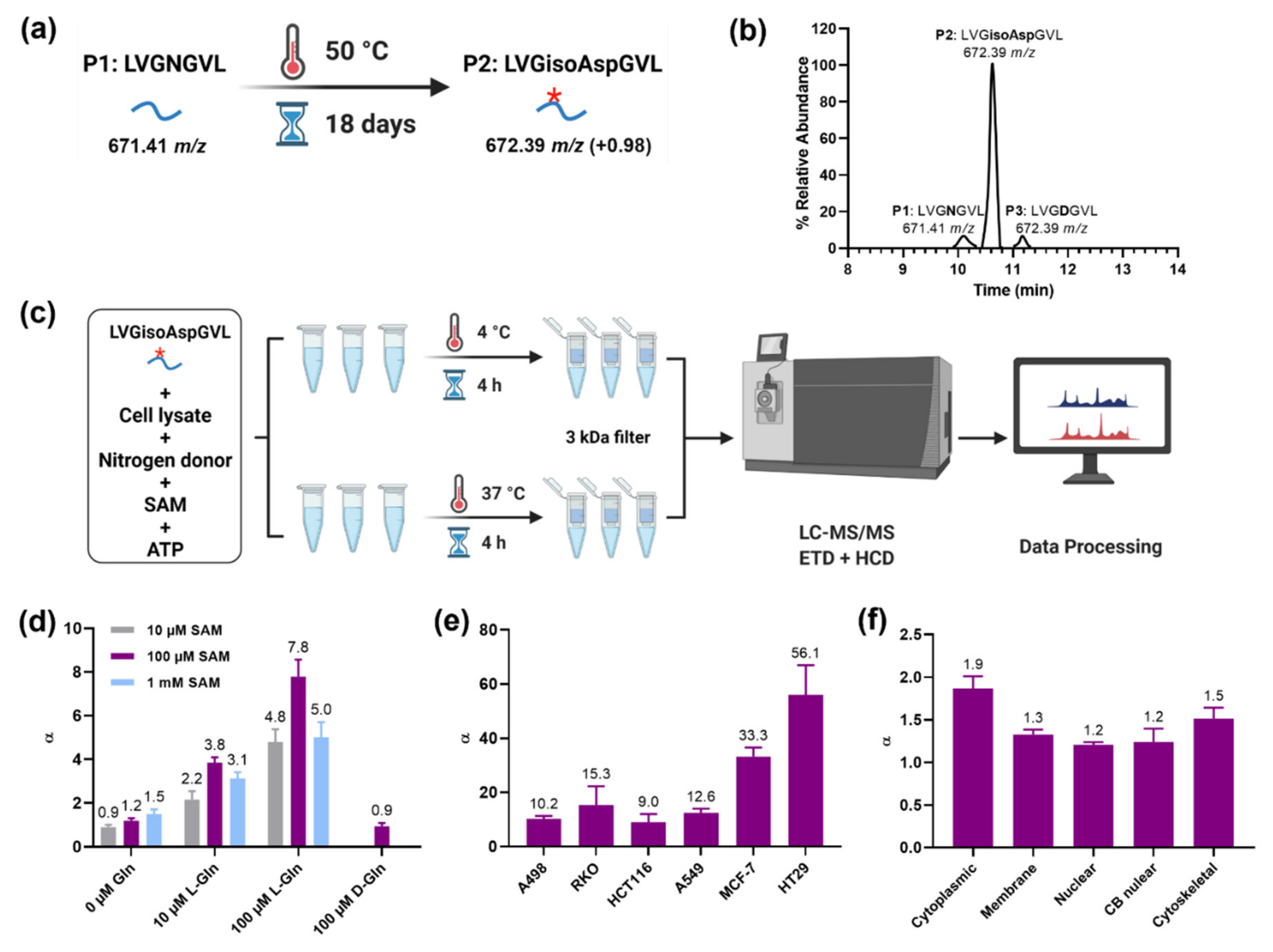
3.1. Artificial Aging of the Synthetic Peptide

The synthetic peptide LVGNGVL (SynPeptide) was incubated in 50 mM Tris buffer (pH 8.5) at 50 °C for 18 days. The aged peptide was then analyzed by an Easy-nLCII high-pressure liquid chromatography (LC) system (Thermo Fisher Scientific, Waltham, MA, USA) coupled with a Q Exactive HF mass spectrometer (Thermo Fisher Scientific, Waltham, MA, USA). The Easy-nLCII system was equipped with a self-packed column (100 μm × 200 mm, 3 μm C8 beads (Dr. Maisch, Ammerbuch, Germany)). Peptide elution was performed using a binary solvent system consisting of 0.1% (v/v) formic acid, 2% (v/v) acetonitrile (solvent A) and 98% acetonitrile (v/v), 0.1% (v/v) formic acid (solvent B) with a flow rate of 300 nL/min. The gradient was of 10–35% B in 20 min followed by 35–95% B in 3 min. The isoaspartate (isoAsp) occupancy was 99.9% after 18-day incubation, and this sample was used as “aged peptide” in the following PSIAL activity experiment.

3.2. PSIAL Activity Experiment on “Aged Peptide”

One million of the HCT 116 cells (ATCC, p = 6) were washed with cold phosphate-buffered saline (PBS) (Cytiva, Marlborough, MA, USA) three times. The cell pellet was dissolved in PBS buffer supplemented with EDTA-free protease inhibitor (Roche, Basel, Switzerland) and phosphatase inhibitor (Roche, Basel, Switzerland), snap-frozen in liquid nitrogen, and thawed at room temperature. After repeating the “freeze and thaw” procedure three times, the cell suspension was centrifuged at 14,000× g and the supernatant was collected as cell lysate. The mixture in six replicates was prepared with 20 μL of each component in PBS: 10 μM “aged peptide”, 1 mg/mL cell lysate, 100 μM ATP, together with nitrogen donor (L-Gln or D-Gln) and S-adenosylmethionine (SAM) with different concentrations (as shown in Figure 2a). Three replicates were incubated at 37 °C for 4 h, while the rest of the replicates were incubated at 4 °C for 4 h as controls. All samples were filtered using the Amicon Ultra 3 kDa filter (Millipore, Burlington, MA, USA) and the filtrate was analyzed via mass spectrometry as described above. The fold-change α of each sample was calculated as the relative abundance of the Asn form (P1) at 37 °C incubation versus 4 °C.

3.3. PSIAL Activity Experiment in Different Cell Lines

Cell lysate from different cancer cell lines (A498, RKO, HCT116, A549, MCF-7, HT29) in the same passage (p = 6) was extracted from six replicates using the same procedures described above, and 1 mg/mL of each cell lysate was incubated respectively with the mixture as above. The fold-change α of each cell lysate was calculated.

3.4. PSIAL Activity Experiment in Subcellular Fractions

The five subcellular fractions (cytoplasmic, membrane, nuclear, chromatin-bound nuclear, cytoskeletal) were extracted from HT29 cell lysate using a Subcellular Protein Fractionation Kit (Thermo Fisher Scientific, Waltham, MA, USA) in six replicates, according to manufacturer’s instructions. Then, 1 mg/mL of different fractions were incubated respectively with the mixture of 10 μM “aged peptide”, 100 μM L-Gln, 100 μM SAM, and 100 μM ATP similar to the procedures above, and the fold-change α of each fraction was calculated.

3.5. Thermal Proteome Profiling–Temperature Range (TPP-TR) Experiments

To determine which proteins specifically interact with a short isoAsp-containing peptide, the thermal proteome profiling–temperature range (TPP-TR) experiments were performed according to the reported method [25]. In short, the specific melting temperatures for each protein from the “treatment” and “control” samples were compared. Here, the peptide GisoAspGisoAspG (SynPeptide, Shanghai, China) was used as a “drug” and GDGDG (SynPeptide, Shanghai, China) as a “control” to treat the HCT116 cells. The other procedures were followed as previously described [1]. The temperature shift of each protein was calculated, as well as the p-value between two groups. For proteins with a p-value less than 0.05, we generated the score using the formula: S c o r e =   | Median T | × Log 10   ( P   Value   of   T ) .

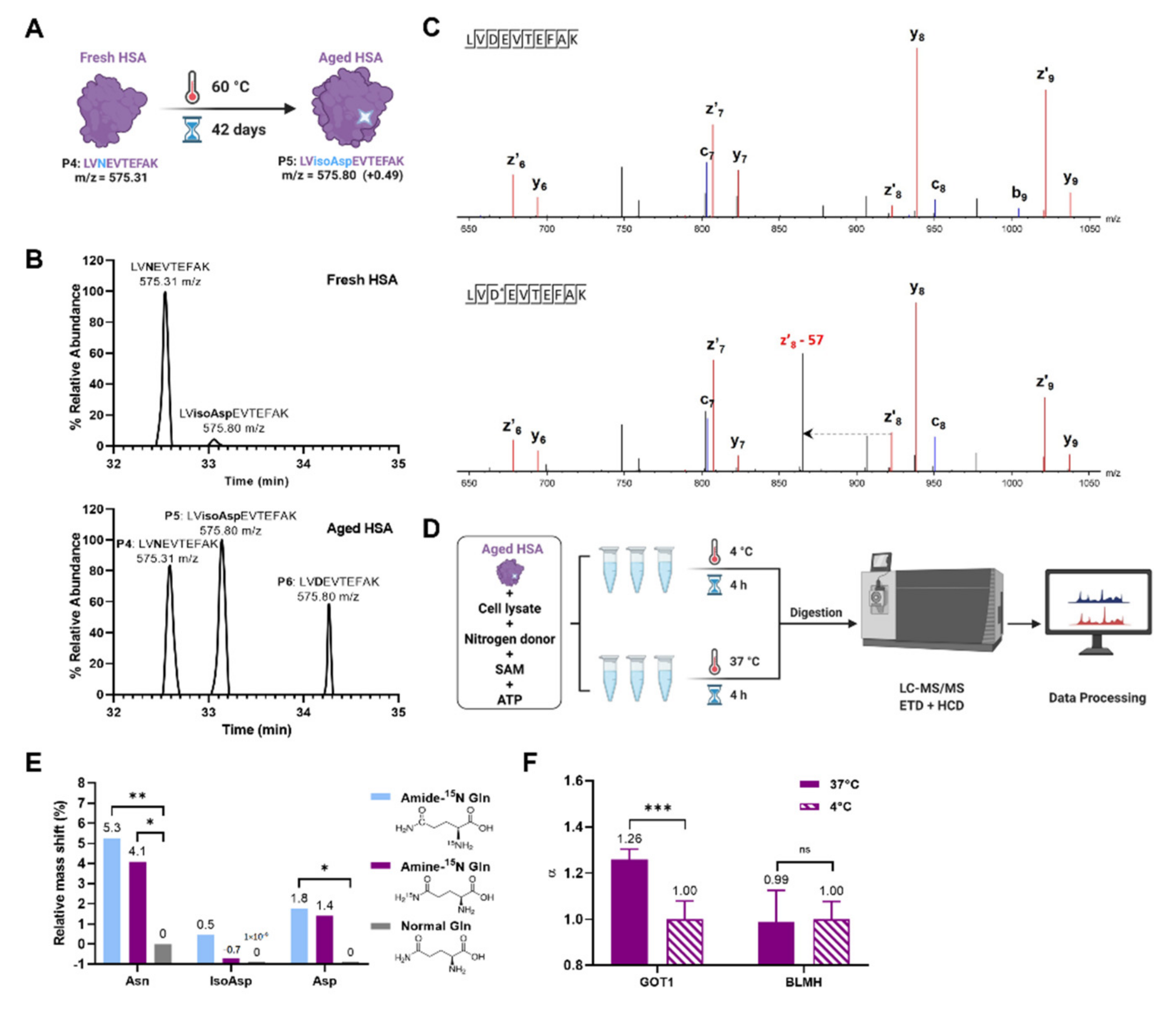
3.6. Artificial Aging of Human Serum Albumin (HSA)

The HSA (Sigma Aldrich, Saint Louis, MO, USA) was incubated in 50 mM Tris buffer (pH 8.0) at 60 °C for 42 days, digested to peptides, and analyzed by an LC-MS/MS, as described [2]. The isoAsp occupancy of the peptide sequence LVNTEFAK showed the highest (~60%) among the asparagine-containing peptides. The “aged HSA”, after 42 days of incubation, was used in the following PSIAL activity experiment.

3.7. PSIAL Activity Experiment on “Aged HSA”

The 0.1 mg/mL “aged HSA” in eight replicates was mixed with 1 mg/mL HT29 cell lysate, 100 μM SAM, 100 μM ATP, and 100 μM nitrogen donor (Amide-15N-enriched Gln, Amine-15N-enriched Gln, or normal Gln), and each component was prepared in 20 μL PBS. After incubation of 37 °C and 4 °C for 4 h, respectively, the samples were subjected to a nano-LC system Ultimate 3000 (Thermo Fisher Scientific, Waltham, MA, USA) connected in-line with a Fusion Orbitrap mass spectrometer (Thermo Fisher Scientific, Waltham, MA, USA). Peptide separation was performed on a 50 cm long EASY spray column (PepMap, C18, 3 μm, 100 Å) with a gradient of 5–30% B in 75 min, and 30–95% B in 5 min (Gradient A: water with 2% acetonitrile and 0.1% formic acid, and Gradient B: acetonitrile with 2% water and 0.1% formic acid). Precursor ion fragmentation was performed with simultaneous higher-energy collision dissociation (HCD; collision energy: 27%, resolution 70,000, AGC target 5.0e4, maximum injection time 200 ms) and electron-transfer/higher-energy collision dissociation (EtHCD; collision energy: 40%, resolution 17,500, AGC target 5.0e4, maximum injection time 200 ms). The relative mass shift of each sample was calculated according to the formula:
Relative   Mass   Shift   ( % ) = I n t e n s i t y   o f   M + 1 + 2 × I n t e n s i t y   o f   M + 2 I n t e n s i t y   o f   M × 100 % .

Supplementary Materials

The following supporting information can be downloaded at: www.mdpi.com/article/10.3390/ijms23158371/s1.

Author Contributions

Conceptualization, R.A.Z.; methodology, R.A.Z., J.W., S.R., A.A.S. and X.Z.; validation, J.W.; formal analysis, J.W.; investigation, J.W.; resources, R.A.Z. and S.R.; data curation, J.W.; writing—original draft preparation, J.W. and R.A.Z.; writing—review and editing, S.R., A.A.S. and X.Z.; visualization, J.W.; supervision, R.A.Z.; project administration, R.A.Z.; funding acquisition, R.A.Z. All authors have read and agreed to the published version of the manuscript.

Funding

This research was funded by the China Scholarship Council (KI-CSC program) and internal resources of Chemistry I Division (Department of Molecular Biochemistry and Biophysics, Karolinska Institutet, Stockholm, Sweden).

Institutional Review Board Statement

Not applicable.

Informed Consent Statement

Not applicable.

Data Availability Statement

Not applicable.

Acknowledgments

The authors thank Elif Değerli for help with the comparisons of PSIAL activity among the different cell lines.

Conflicts of Interest

The authors declare no conflict of interest.

References

  1. Geiger, T.; Clarke, S. Deamidation, Isomerization, and Racemization at Asparaginyl and Aspartyl Residues in Peptides—Succinimide-Linked Reactions That Contribute to Protein-Degradation. J. Biol. Chem. 1987, 262, 785–794. [Google Scholar] [CrossRef]
  2. Johnson, B.A.; Murray, E.D.; Clarke, S.; Glass, D.B.; Aswad, D.W. Protein Carboxyl Methyltransferase Facilitates Conversion of Atypical L-Isoaspartyl Peptides to Normal L-Aspartyl Peptides. J. Biol. Chem. 1987, 262, 5622–5629. [Google Scholar] [CrossRef]
  3. Roher, A.E.; Lowenson, J.D.; Clarke, S.; Wolkow, C.; Wang, R.; Cotter, R.J.; Reardon, I.M.; Zurcherneely, H.A.; Heinrikson, R.L.; Ball, M.J.; et al. Structural Alterations in the Peptide Backbone of Beta-Amyloid Core Protein May Account for Its Deposition and Stability in Alzheimers-Disease. J. Biol. Chem. 1993, 268, 3072–3083. [Google Scholar] [CrossRef]
  4. Goldberg, A.L. Protein degradation and protection against misfolded or damaged proteins. Nature 2003, 426, 895–899. [Google Scholar] [CrossRef]
  5. Böhme, L.; Bär, J.W.; Hoffmann, T.; Manhart, S.; Ludwig, H.-H.; Rosche, F.; Demuth, H.-U. Isoaspartate residues dramatically influence substrate recognition and turnover by proteases. Biol. Chem. 2008, 389, 1043–1053. [Google Scholar] [CrossRef]
  6. Yang, H.; Lyutvinskiy, Y.; Soininen, H.; Zubarev, R.A. Alzheimer’s disease and mild cognitive impairment are associated with elevated levels of isoaspartyl residues in blood plasma proteins. J. Alzheimers Dis. 2011, 27, 113–118. [Google Scholar] [CrossRef] [Green Version]
  7. Chatterjee, T.; Das, G.; Chatterjee, B.K.; Dhar, J.; Ghosh, S.; Chakrabarti, P. The role of isoaspartate in fibrillation and its prevention by Protein-L-isoaspartyl methyltransferase. Biochim. Biophys. Acta Gen. Subj. 2020, 1864, 129500. [Google Scholar] [CrossRef]
  8. Wang, J.; Guo, C.; Meng, Z.; Zwan, M.D.; Chen, X.; Seelow, S.; Lundström, S.L.; Rodin, S.; Teunissen, C.E.; Zubarev, R.A. Testing the link between isoaspartate and Alzheimer’s disease etiology. bioRxiv 2022. [Google Scholar] [CrossRef]
  9. Johnson, B.A.; Shirokawa, J.M.; Geddes, J.W.; Choi, B.H.; Kim, R.C.; Aswad, D.W. Protein L-Isoaspartyl Methyltransferase in Postmortem Brains of Aged Humans. Neurobiol. Aging 1991, 12, 19–24. [Google Scholar] [CrossRef]
  10. Robinson, N.E.; Robinson, A.B. Molecular clocks. Proc. Natl. Acad. Sci. USA 2001, 98, 944–949. [Google Scholar] [CrossRef] [Green Version]
  11. O’Connor, C.M. Protein L-isoaspartyl, D-aspartyl O-methyltransferases: Catalysts for protein repair. Enzymes 2006, 24, 385–433. [Google Scholar] [CrossRef]
  12. Mishra, P.K.K.; Mahawar, M. PIMT-Mediated Protein Repair: Mechanism and Implications. Biochem. Mosc. 2019, 84, 453–463. [Google Scholar] [CrossRef]
  13. Hao, P.L.; Ren, Y.; Alpert, A.J.; Sze, S.K. Detection, Evaluation and Minimization of Nonenzymatic Deamidation in Proteomic Sample Preparation. Mol. Cell. Proteom. 2011, 10. [Google Scholar] [CrossRef] [Green Version]
  14. Chelius, D.; Rehder, D.S.; Bondarenko, P.V. Identification and characterization of deamidation sites in the conserved regions of human Immunoglobulin Gamma antibodies. Anal. Chem. 2005, 77, 6004–6011. [Google Scholar] [CrossRef]
  15. Gervais, D. Protein deamidation in biopharmaceutical manufacture: Understanding, control and impact. J. Chem. Technol. Biotechnol. 2016, 91, 569–575. [Google Scholar] [CrossRef]
  16. Vaintraub, I.A.; Kotova, L.V.; Shaha, R. Protein Deamidase from Germinating Wheat Grains. FEBS Lett. 1992, 302, 169–171. [Google Scholar] [CrossRef] [Green Version]
  17. Zhao, J.; Zeng, Y.; Xu, S.; Chen, J.; Shen, G.; Yu, C.; Knipe, D.; Yuan, W.; Peng, J.; Xu, W.; et al. A Viral Deamidase Targets the Helicase Domain of RIG-I to Block RNA-Induced Activation. Cell Host Microbe 2016, 20, 770–784. [Google Scholar] [CrossRef] [Green Version]
  18. Zhang, J.; Zhao, J.; Xu, S.; Li, J.; He, S.; Zeng, Y.; Xie, L.; Xie, N.; Liu, T.; Lee, K.; et al. Species-Specific Deamidation of cGAS by Herpes Simplex Virus UL37 Protein Facilitates Viral Replication. Cell Host Microbe 2018, 24, 234–248.e235. [Google Scholar] [CrossRef] [Green Version]
  19. Konuklar, F.A.S.; Aviyente, V.; Sen, T.Z.; Bahar, I. Modeling the deamidation of asparagine residues via succinimide intermediates. J. Mol. Model. 2001, 7, 147–160. [Google Scholar] [CrossRef]
  20. Russell, J.B.; Strobel, H.J. Concentration of Ammonia across Cell-Membranes of Mixed Rumen Bacteria. J. Dairy Sci. 1987, 70, 970–976. [Google Scholar] [CrossRef]
  21. Wang, J.; Lundström, S.L.; Seelow, S.; Rodin, S.; Meng, Z.; Astorga-Wells, J.; Jia, Q.; Zubarev, R.A. First Immunoassay for Measuring Isoaspartate in Human Serum Albumin. Molecules 2021, 26, 6709. [Google Scholar] [CrossRef] [PubMed]
  22. Catak, S.; Monard, G.; Aviyente, V.; Ruiz-Lopez, M.F. Deamidation of asparagine residues: Direct hydrolysis versus succinimide-mediated deamidation mechanisms. J. Phys. Chem. A 2009, 113, 1111–1120. [Google Scholar] [CrossRef] [PubMed]
  23. Peracchi, A. The Limits of Enzyme Specificity and the Evolution of Metabolism. Trends Biochem. Sci. 2018, 43, 984–996. [Google Scholar] [CrossRef] [PubMed]
  24. Cioffi, M.; D’Alterio, C.; Camerlingo, R.; Tirino, V.; Consales, C.; Riccio, A.; Ierano, C.; Cecere, S.C.; Losito, N.S.; Greggi, S.; et al. Identification of a distinct population of CD133+CXCR4+ cancer stem cells in ovarian cancer. Sci. Rep. 2015, 5, 10357. [Google Scholar] [CrossRef] [Green Version]
  25. Mateus, A.; Maatta, T.A.; Savitski, M.M. Thermal proteome profiling: Unbiased assessment of protein state through heat-induced stability changes. Proteome Sci. 2017, 15, 13. [Google Scholar] [CrossRef] [Green Version]
Figure 1. Proposed protein succinimide/isoaspartate ammonia (PSIAL) activity. (a) The mechanism of isoAsp formation and repair by PMIT and the proposed amidation via enzymes with PSIAL activity. (b) The deamidation degree of asparaginyl peptides in HSA after artificial deamidation for 42 days.
Ijms 23 08371 g001
Figure 2. Discovery of PSIAL activity on synthetic isoAsp-containing peptide. (a) Artificial deamidation of the synthetic peptide LVGNGVL (P1) to LVGisoAspGVL (P2) via incubating at 50 °C for 18 days. *: isoAsp. (b) The chromatogram of the aged peptide above in three forms with Asn (P1), isoAsp (P2), and Asp (P3). (c) The scheme of testing PSIAL activity in cell lysate using aged P1 and cofactors. (d) The effects of cofactors (Gln and SAM) with different concentrations on PSIAL activity. (e) Comparison of PSIAL activities among different cancer cell lines by measuring the fold-change α of the relative abundance of the Asn form of peptide in 37 °C incubation versus 4 °C. (f) Comparisons of PSIAL activities among different subfractions of HT29 cell lysate.
Ijms 23 08371 g002
Figure 3. Discovery of PSIAL activity on aged HSA. (A) Artificial deamidation of fresh HSA to aged HSA via incubating at 60 °C for 42 days. (B) The chromatogram of fresh (up) and aged (down) HSA (D* = isoAsp). (C) ETD MS/MS spectrum confirms the isoAsp presence by the z8′-57 ion (up), and its absence in the corresponding peptide with Asp (down). (D) The scheme of testing PSIAL activity in cell lysate using aged HSA and cofactors. (E) The mass shift in the Asn form of an HSA reporter peptide after adding Amine-15N L-Gln, Amide-15N L-Gln, and isotopically normal L-Gln. (F) Validation of PSIAL activity in GOT1 protein with BLMH as a negative control. * p < 0.05, ** p < 0.01, *** p < 0.001.
Ijms 23 08371 g003
Table 1. Top 10 protein candidates interacting with isoAsp in a peptide.
Protein IDProtein NameGene Name|Median △T|−Log10 (p Value)Score *
P17174Aspartate aminotransferase, cytoplasmicGOT19.84.645.6
P36959GMP reductase 1GMPR9.63.129.9
Q9UFN0Protein NipSnap homolog 3ANIPSNAP3A12.42.125.6
E2QRF9GemininGMNN7.72.620.3
O94992Protein HEXIM1HEXIM17.72.419.0
Q13126S-methyl-5-thioadenosine phosphorylaseMTAP4.34.217.9
P41236Protein phosphatase inhibitor 2PPP1R210.21.717.4
P61956-2Small ubiquitin-related modifier 2SUMO28.12.016.3
O43852CalumeninCALU7.62.116.2
P00390-2Glutathione reductase, mitochondrialGSR8.61.815.5
* S c o r e =   | Median T | × Log 10   ( P   Value   of   T )
Publisher’s Note: MDPI stays neutral with regard to jurisdictional claims in published maps and institutional affiliations.

