Article

CpMAX1a, a Cytochrome P450 Monooxygenase Gene of Chimonanthus praecox Regulates Shoot Branching in Arabidopsis

Chongqing Engineering Research Center for Floriculture, Key Laboratory of Horticulture Science for Southern Mountainous Regions of Ministry of Education, College of Horticulture and Landscape Architecture, Southwest University, Chongqing 400715, China
*
Author to whom correspondence should be addressed.
These authors contributed equally to this work.
Int. J. Mol. Sci. 2022, 23(18), 10888; https://doi.org/10.3390/ijms231810888
Submission received: 11 August 2022 / Revised: 14 September 2022 / Accepted: 15 September 2022 / Published: 17 September 2022

Abstract

:
Strigolactones (SLs) are a class of important hormones in the regulation of plant branching. In the model plant ArabidopsisAtMAX1 encodes a cytochrome P450 protein and is a crucial gene in the strigolactone synthesis pathway. Yet, the regulatory mechanism of MAX1 in the shoot branching of wintersweet (Chimonanthus praecox) remains unclear. Here we identified and isolated three MAX1 homologous genes, namely CpMAX1a, CpMAX1b, and CpMAX1c. Quantitative real-time PCR (qRT-PCR) revealed the expression of CpMAX1a in all tissues, being highest in leaves, whereas CpMAX1b was only expressed in stems, while CpMAX1c was expressed in both roots and stem tips. However, CpMAX1a’s expression decreased significantly after decapitation; hence, we verified its gene function. CpMAX1a was located in Arabidopsis chloroplasts. Overexpressing CpMAX1a restored the phenotype of the branching mutant max1–3, and reduced the rosette branch number, but resulted in no significant phenotypic differences from the wild type. Additionally, expression of AtBRC1 was significantly upregulated in transgenic lines, indicating that the CpMAX1a gene has a function similar to the homologous gene of Arabidopsis. In conclusion, our study shows that CpMAX1a plays a conserved role in regulating the branch development of wintersweet. This work provides a molecular and theoretical basis for better understanding the branch development of wintersweet.

1. Introduction

In plants, the development of side shoots begins in the axillary meristems (AMs) at the leaf axils, with shoot branching usually divided into two developmental stages: the generation of an AM in each leaf axil and the ensuing outward growth process. After their generation, the axillary buds may either remain dormant or grow to form branches [1,2,3]. Branch formation is regulated by a variety of factors, including hormones, developmental status, and the environment, in which phytohormones play an extremely important role [4,5]. Auxins and cytokinins have long been thought to figure prominently in controlling shoot branching, but recent studies suggest that SLs may be involved in plant branching regulation by interacting with auxin and cytokinin as second messengers of auxin [6,7,8,9].
Strigolactones (SLs) were originally isolated from plant root exudates as germination stimulants for root parasitic plants of the Orobanchaceae family, including witchweeds (Striga spp.), broomrapes (Orobanche and Phelipanche spp.), and Alectra spp. [10,11]. They were later shown to be indispensable chemical signals for root colonization by arbuscular mycorrhizal fungi (AMF) and therefore considered as beneficial plant metabolites [12]. Furthermore, SLs mediate plant resistance in response to various abiotic stresses (including drought, salinity, temperature) [13,14,15]. Several branching mutants deficient in SLs synthesis and signaling have been well studied: rms (ramosus) 1–5 mutants in pea (Pisum sativum) [16,17,18], max (more axillary growth) 1–4 mutants in Arabidopsis [19,20,21,22], decreasedapical dominance (dad) mutants of petunia (Petunia hybrida) [23]. Accordingly, SLs or their metabolites are now recognized as a new class of phytohormones whose participation in the inhibition of shoot germination is pivotal.
Presently, the biosynthesis pathway of SLs has been basically elucidated and the key enzymes involved in this pathway identified. Specifically, the carotenoid isomerase DWARF27 (D27) converts all-trans-β-carotene to 9-cis-β-carotene [24,25], after which CAROTENOID CLEAV AGE DIOXYGENASE 7 (CCD7) catalyzes the cleavage of 9-cis-β-carotene to form 9-cis-β-apo-10′-carotenal, whose cleavage and oxidation mediated by CAROTENOID CLEAV AGE DIOXYGENASE 8 (CCD8) then forms carlactone [26,27,28]. The cytochrome P450 oxygenase encoded by MORE AXILLARY GROWTH 1 (MAX1) synthesizes the precursor of SLs needed to further synthesize them [29,30]. The α/β hydrolase D14 specifically recognizes and hydrolyzes SLs. Subsequently, the receptor SLs D14 protein undergoes a conformational change to form a SCF protein complex with D3 (MAX2) [31,32,33], and the newly SCF formed complex subsequently mediates the ubiquitin-dependent degradation of the transcriptional repressor D53 (SMXL6, SMXL7, SMXL8) and transduces SLs signaling [34,35]. For the biosynthesis or signaling of SLs, these genes are required to properly regulate plant axillary bud growth and shoot branching.
MAX1 was first discovered in Arabidopsis and acts downstream of the MAX3 and MAX4 genes, encoding cytochrome P450 monooxygenase (Cyt P450) to convert CL into carlactonoic acid (CLA) [29]. To date, the MAX1 homologous gene has been studied in many plant species. In rice, deletion of the MAX1 homolog cytochrome P450 genes slb1 and slb2 led to the reduced secretion of SLs. Both rice genes are able to rescue the Arabidopsis max1–1 highly branched mutant phenotype, and they increase the production of the SL, ent-2′-epi-5-deoxystrigol when overexpressed in Oryza sativa [36]. In tomato, MAX1 knockout mutants exhibit a strong multi-branched phenotype and reduced SL content, which suggests the biosynthesis of most SLs is accomplished through MAX1 [37]. Collectively, these studies suggest that MAX1 plays a key role in regulating plant branch development by affecting SLs’ synthesis.
However, investigations of MAX1 gene’s functioning in ornamental plants are limited, especially in wintersweet (Chimonanthus praecox). As a critical agronomic trait, branching can greatly influence the structure and yield of higher plants. The number of branches in the production of wintersweet will directly affect the yield and ornamental quality of this plant’s fresh cut flowers. Therefore, it is of great significance to study the molecular mechanism of CpMAX1 for regulating the process of branching in wintersweet. Here, we identified and isolated three MAX1 homologous genes: CpMAX1a, CpMAX1b, and CpMAX1c. The three genes are highly similar, but CpMAX1a is expressed more in axillary buds, so we focused on CpMAX1a as the research object. To verify the function of CpMAX1a, we heterologously expressed CpMAX1a in Arabidopsis. The overexpression of CpMAX1a restored the multi-branched phenotype of the Arabidopsis mutant max1–3. Further, the number of branches in CpMAX1a overexpressed lines decreased but not significantly, suggesting that CpMAX1a plays a conserved role in regulating branch germination. Overall, the results of this study enhance our understanding of the role of CpMAX1a in the development of lateral branches in wintersweet and provide a basis for exploring the molecular mechanism of branching in this plant and closely related species.

2. Results

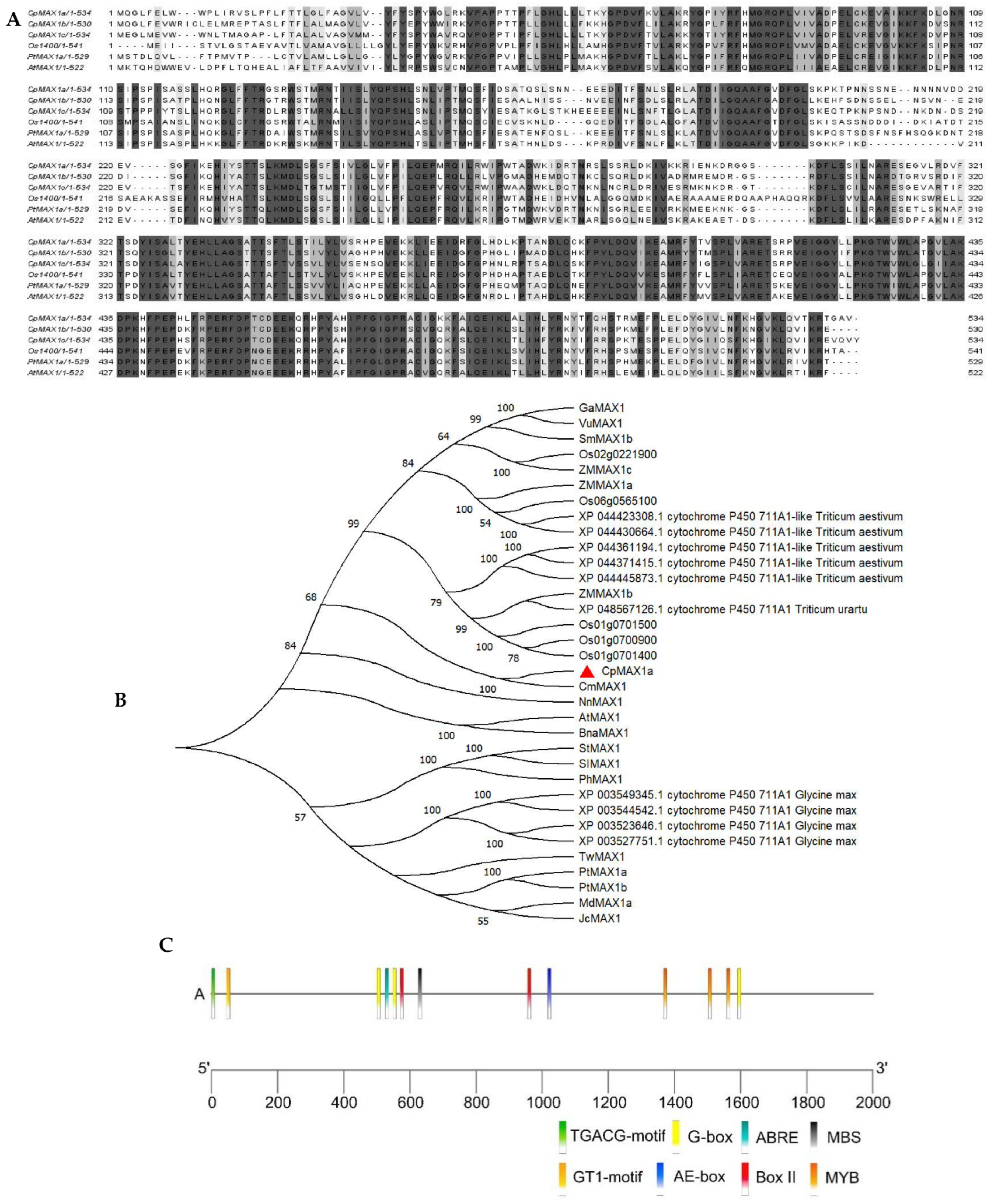
2.1. CpMAX1a Cloning and Phylogenetic Analysis

The CpMAX1a was successfully isolated from the leaf samples of wintersweet. The cDNA sequence of CpMAX1a was obtained from the wintersweet flower transcriptome database. According to its sequence analysis, CpMAX1a has an open reading frame (ORF) of 1605 bp, encoding a protein of 534 amino acids (aa) (Figure 1A), in which leucine (Leu) constitutes the largest proportion (11.8%) of the amino acid composition. Multiple sequence alignment showed that CpMAX1a had the highest sequence similarity with the homologous genes OS1400, AtMAX1, and PtMAX1a in rice, Arabidopsis, and P. trichocarpa, respectively (Figure 1B). The phylogenetic tree results indicated that CpMAX1a and the Cinnamomum micranthum CYP711 subfamily homologous genes clustered together and had the closest genetic relationship. In addition, we cloned the promoter region 2000-bp upstream of CpMAX1a gene, using the genomic walking method. This promoter analysis showed that the CpMAX1a promoter region harbored seven light-responsive elements (three G-boxes, two Box II, one AE-box, and one GT1-motif), one MeJA responsive element (TGACG-motif), one ABA-responsive element (ABRE), one MYB binding site (MBS), and three stress-responsive elements (MYB), indicating that CpMAX1a may be induced by light and stress (Figure 1C).

2.2. Expression Pattern of CpMAX1a

Here we identified and isolated three MAX1 homologous genes. Among them, CpMAX1b is only expressed in stems (Figure 2B), CpMAX1c is only expressed in roots and stem tips (Figure 2C), but CpMAX1a is expressed in all tissue types (Figure 2A). Based on tissue specificity, it can be concluded that CpMAX1a is constitutively regulated, while CpMAX1b and CpMAX1c are non-constitutively regulated. To analyze the role of CpMAX1a in the shoot branching of wintersweet, we used qRT-PCR technology to detect the expression levels of CpMAX1a in the six-leaf stage of wintersweet’s axillary buds. Axillary bud samples were collected at 0 h, 2 h, 6 h, 12 h, 24 h, 36 h, and 48 h after decapitation; axillary buds of seedlings without decapitation served as the control. Real-time fluorescence quantitative results showed that the expression of CpMAX1a was significantly downregulated in decapitated axillary buds, being lowest at 12 h. Within 48 h after decapitation, CpMAX1a was consistently expressed at a low level during the critical period of axillary bud germination (Figure 2D). Further, the GR24 treatment significantly inhibited CpMAX1a’s expression. Compared with the decapitation control, the expression level of CpMAX1a in axillary buds treated with GR24 remained low throughout the treatment period (Figure 2E).

2.3. Subcellular Localization of CpMAX1a

To determine the subcellular localization of the protein encoded by CpMAX1a, it was fused to the N-terminus of the GFP (green fluorescent protein) gene. Then 35S::CpMAX1a-GFP and 35S::GFP (control) were separately transformed into Arabidopsis leaf protoplasts. In the 35S::GFP-transformed protoplasts, the GFP signal was dispersed throughout the cytoplasm, whereas in 35S::CpMAX1a-GFP the signal was localized in chloroplasts (Figure 3). Hence, these results suggested that the CpMAX1a protein is localized in chloroplasts.

2.4. Effect of Environmental Factors on the Expression of CpMAX1a

To explore the effect of environmental factors on the expression of CpMAX1a, we used qRT-PCR to determine the transcription level of CpMAX1a in wintersweet. These treatments were selected based on the cis-acting elements in the CpMAX1a promoter region. CpMAX1a expression was significantly reduced under dark conditions and was consistently lower than the control (Figure 4A). Under ABA treatment, CpMAX1a expression decreased at 2 h, and then increased to the highest value at 12 h (Figure 4B). Under high temperature treatment, CpMAX1a expression gradually decreased, but significantly increased at 24 h (Figure 4C). For low temperature treatment, CpMAX1a expression decreased at 2 h, and then gradually increased to reach the maximum at 12 h (Figure 4D). In PEG treatment, CpMAX1a expression increased significantly at 2 h and then decreased gradually to reach the minimum at 12 h. Interestingly, CpMAX1a expression showed an increasing trend again at 24 h (Figure 4E). In Nacl treatment, CpMAX1a expression was significantly increased and was significantly higher than the control from 0–12 h (Figure 4F).

2.5. Effects of Overexpression of the CpMAX1a Gene on Branching in Arabidopsis

To investigate the function of the CpMAX1a gene, we transformed the 35S:: CpMAX1a construct into wild-type (WT) Arabidopsis. Six transgenic lines expressing 35S:: CpMAX1a were obtained by wetomycin screening and PCR identification. Three pure-hybrid overexpression lines (i.e., OE1, OE2, and OE3) were selected for phenotypic analysis (Figure 5F). We counted the number of rosette leaves of the CpMAX1a-OE lines grown under long day (LD) conditions for 15 days and the number of branches at 35 days of growth. These results showed that the mean (±SD) numbers of rosette leaves of the CpMAX1a-OE strain were as follows: WT, 9.85 ± 0.72; OE1, 9.62 ± 0.74; OE2, 9.46 ± 1.34; OE3, 9.69 ± 0.82 (Figure 5D); for the number of branches of the CpMAX1a-OE strain, the corresponding values were: WT, 1.87 ± 0.34; OE1, 1.8 ± 0.4; OE2, 1.73 ± 0.44; OE3, 1.8 ± 0.4 (Figure 5E). Evidently, the number of rosettes of the CpMAX1a-OE strains had slightly fewer rosette leaves and branches than WT strains, but there were no significant differences between them and WT plants. Previous studies have suggested the MAX1 gene may control Arabidopsis meristems by influencing the transcription of BRC1 [38,39]. Therefore, we examined the expression levels of AtBRC1 in both WT and CpMAX1a-OE strains, finding that AtBRC1 was more highly expressed in the CpMAX1a-OE strains compared to the WT strains. (Figure 5G). The extraction solution of CpMAX1a-OE lines induced Orobanche aegyptiaca seeds germination experiments showed that the germination rate of CpMAX1a-OE lines was higher than that of wt, suggesting that overexpression of CpMAX1a in Arabidopsis does improve the germination of Orobanche aegyptiaca seeds (Figure 5H). These results suggest that CpMAX1a may inhibit the growth of Arabidopsis rosette branches through the synthesis of SLs by modulating the transcriptional regulation of AtBRC1.

2.6. Overexpression of the CpMAX1a Gene Restores the Branching Phenotype of Arabidopsis max1 Mutants

To further investigate the functioning of the CpMAX1a gene, we transformed 35S::CpMAX1a into the Arabidopsis branch mutant max1 to obtain nine CpMAX1a complementary lines, and selected three independent mutant restorer lines for subsequent experimentation (Figure 6F). We counted the number of rosette leaves of the mutant restorer lines grown under long day (LD) conditions for 15 days and the number of branches at 35 days of growth. Both the number of rosette leaves and branches of the mutant significantly exceeded those of WT, whereas those the restored mutant strain were similar to WT. Mean values for the number of rosette leaves were as follows: WT, 9.85 ± 0.72; max1–3, 25.61 ± 1.21; Line1, 10.07 ± 0.73; Line2, 9.54 ± 0.93; Line3, 10.39 ± 0.62 (Figure 6D); for the number of branches, the values were WT, 1.87 ± 0.34; max1–3, 5.53 ± 0.71; Line1, 2.07 ± 0.44; Line2, 2 ± 0.52; Line3, 2.33 ± 0.47 (Figure 6E). In addition, we also examined the expression levels of AtBRC1, a key gene in the controlling of branching, in the WT lines, mutant lines, and complementary lines. These results showed that CpMAX1a significantly upregulated the expression level of AtBRC1 in the overexpression and complemented lines (Figure 6G). The extraction solution of CpMAX1a restored lines induced Orobanche aegyptiaca seeds germination experiment showed that the restored lines and wt germination rate were similar, both being significantly higher than max1–3 (Figure 6H). Altogether, the above results indicated the function of CpMAX1a is relatively conserved in the branch development of wintersweet and that its functioning is achieved by regulating the transcription of BRC1.

3. Discussion

Here, we isolated and identified three MAX1 homologous genes—CpMAX1a, CpMAX1b, and CpMAX1c—in wintersweet. Given that these three genes are quite similar, yet CpMAX1a undergoes high expression in axillary buds, we focused on studying gene as it may be directly involved in controlling branch development. Multiple sequence alignment revealed that the CpMAX1a protein shared a high sequence similarity with the MAX1 protein sequences of other plants. Further, the cytochrome P450 domain motifs were found in the CpMAX1a sequence (Figure 1A), which is consistent with the characteristics of MAX1 protein members. Phylogenetic analysis showed that CpMAX1a clustered with the Cinnamomum micranthum CYP711 protein, indicating that CpMAX1a is most closely related to AtMAX1 (Figure 1B). We found that subcellular localization of the CpMAX1a protein is in the chloroplasts, thus it was speculated CpMAX1a could be involved in the synthesis of SLs in plastids. In addition, analysis of the cis-elements of the CpMAX1a promoter indicated it harbors multiple elements related to light and stress responses (Figure 1C). CpMAX1a expression was significantly reduced under dark conditions (Figure 4A), indicating that light induces CpMAX1a expression, which is consistent with the finding that light induces an increase in the content of solanum endolipid in Arabidopsis [40].In addition, ABA, high temperature, low temperature, Nacl and PEG induce an increase in CpMAX1a expression(Figure 4B–F),this result is consistent with that adversity can induce the synthesis of SLs in rice, grapevine, submerged plants, and tomato [41,42,43,44].
To the best of our knowledge, there has been no research published on the SL biosynthesis gene CpMAX1a in wintersweet. In this study, we examined the expression level of CpMAX1a during branch development after decapitation of wintersweet. Compared with the control group, CpMAX1a was significantly downregulated at 2 h post-decapitation and maintained a low level of expression during the critical period of axillary bud germination. Further, when GR24 was applied to decapitated wintersweet axillary buds, the expression of CpMAX1a was significantly lower than in the control group, a result consistent with the finding that applying GR24 to Arabidopsis and sugar beet leads to the diminished expression of genes related to SLs’ synthesis [45,46]. This probably occurred because the negative feedback regulation caused by the substrate GR24 in excess amounts inhibited the expression of CpMAX1a, which lends further support to the inference that CpMAX1a is involved in the synthesis of SLs. Therefore, we speculate that CpMAX1a may inhibit the axillary buds’ growth of wintersweet by regulating the synthesis of SLs. The tissue-specific expression pattern of the CpMAX1a gene in wintersweet differs from that of other plants’ homologous genes. In Arabidopsis, rice, and tomato, their corresponding AtMAX1, OsMAX1a, OsMAX1e, and SIMAX1 genes were mainly expressed in the roots and stems [37,47]. In rapeseed (Brassica napus L.), the expression of BnaMAX1 was greatest in axillary buds, followed by the roots [48], though in chrysanthemum its CmMAX1 gene was highly expressed in leaves, axillary buds, and stems, as well as roots [49]. Here, the CpMAX1a gene of wintersweet was expressed most in the leaves and stems, but least expressed in the roots (Figure 2A). Differential expression patterns of MAX1 in true cotyledons versus monocotyledons suggest that the regulation of branching by SLs is a species-specific process.
To test our speculations and to better understand the functioning of CpMAX1a, we heterologously expressed CpMAX1a in Arabidopsis. We found that in the CpMAX1a-OE strains, the number of rosette leaves and rosette branches were not inhibited as expected (Figure 5D,E). Therefore, we performed experiments to induce Orobanche seeds germination by extracts of the CpMAX1a-OE strains. The experimental results showed that extracts from the CpMAX1a-OE strains indeed promotes the germination, but not to the same extent compared to the exogenous GR24 application(Figure 5H), this means that CpMAX1a was involved in the biosynthesis of SLs but did not significantly increase the SLs content, which may also be the reason why overexpression of CpMAX1a in Arabidopsis did not inhibit branching. When there are not enough SLs in Arabidopsis to inhibit auxin transport, rosette branching is not significantly inhibited [9].In addition, overexpression of CpMAX1a in Arabidopsis could not significantly increase SLs content may be that the complex genetic backgrounds of perennial woody plants and herbaceous plants may also be different, and exogenous expression may not accurately reflect the phenotype of the species studied. Numerous MAX1 knockout experiments have demonstrated that the functional deletion of the MAX1 gene results in more branches in Arabidopsis [50], tomato [37], rapeseed [48], and rice [47]. Moreover, under the control of the cauliflower mosaic virus (CaMV) 35S promoter, rapeseed BnaMAX1 was overexpressed in the Arabidopsis MAX1 branching mutant, restoring the multibranching phenotype [48], and likewise, overexpression of chrysanthemum CmMAX1 in the Arabidopsis max1–1 mutant restored its multibranching phenotype [49]. This would suggest MAX1 plays a conserved role in the control of branch development. To test this conjecture, we performed complementation experiments with the Arabidopsis max1 mutant, finding here that the CpMAX1a gene was indeed capable of restoring the multibranching phenotype of the Arabidopsis mutant max1–3 (Figure 6B); the extraction solution of CpMAX1a restored lines induced Orobanche aegyptiaca seeds germination experiments showed that Overexpression of CpMAX1a increased the content of SLs in max1–3 mutants(Figure 6H); hence, it is likely the regulation of plant branching by CpMAX1a is conserved in wintersweet. To further analyze the CpMAX1a action pathway, we investigated the expression of the gene BRC1, which belongs to the TCP gene family and is known for being central how a variety of environmental and developmental factors function locally to inhibit branching [38,51,52]. BRC1 reportedly acts the downstream of SLs to encode a key transcription factor that inhibits shoot growth, and treatment with SLs can upregulate the expression of BRC1 [53,54]. The effect of MAX1 on branching is mainly attributable to the transcriptional control of BRC1. Here. we analyzed the relative expression levels of AtBRC1 in the overexpression and recovery mutant lines, finding them significantly upregulated in both, which strongly suggests that CpMAX1a suppresses axillary buds growth in Arabidopsis by upregulating its downstream gene BRC1. Therefore, we conclude that the role of CpMAX1a is conserved in the control of branch development in wintersweet.
As a winter woody ornamental plant, wintersweet’s branching structure confers to it substantial ornamental value. So far, the CpCCD7 and CpCCD8 genes have been identified in the SLs’ synthesis pathway in wintersweet [39], but the downstream CpMAX1a had not been identified or reported on yet. The uniqueness of wintersweet’s growth and development makes it imperative to understand the contribution of CpMAX1a towards regulating branch development. In this study, we isolated CpMAX1a and analyzed its expression pattern and functional characteristics. CpMAX1a expression was downregulated during branching in wintersweet, indicating that it negatively regulates axillary buds’ growth in wintersweet. Overexpression of CpMAX1a, however, restored the multibranched phenotype of branched mutant max1–3 and up-regulated the AtBRC1 gene. These results suggest CpMAX1a plays a conserved role in the regulation of wintersweet branching via the regulation of the downstream BRC1 gene. This study strengthens our understanding of the homologous genes of CpMAX1a in wintersweet and provides a timely basis for us to further study the molecular regulatory mechanism of the CpMAX1a gene.

4. Materials and Methods

4.1. Plants

The ‘Suxin’ wintersweet cultivar was used, whose seeds were collected from the campus of Southwest University and sown in the Floriculture Laboratory of Southwest University, China. To analyze the expression pattern of the gene CpMAX1a in wintersweet, the roots, stems, leaves, shoot tips, and axillary buds were each collected. These tissues were rapidly frozen in liquid nitrogen once collected. Three biological replicates were obtained for each tissue type sample.
Wild-type Arabidopsis (ecotype Columbia−0) and Arabidopsis max1 (SALK-209654C, code N2105389) purchased from The Arabidopsis Information Resource (TAIR) database were used for the plant transformations. Arabidopsis seeds were sown on solid Murashige and Skoog (MS) medium. After vernalization at 4 °C for 3 days, the seedlings were transferred to a constant temperature culture room (22 °C, a 16-h:8-h light:dark photoperiod, 200 Lux light leevl, relative humidity of 70%). When they had four fully expanded leaves, each seedling was transferred to a nutrient bowl (vermiculite:charcoal ratio of 1:1). The results of PCR detection of CpMAX1a overexpression in Arabidopsis strains are shown in: Figure S2. Orobanche aegyptiaca seeds were presented by Prof. Zhao of Shihezi University. Orobanche aegyptiaca seed germination experiments were referred to Tsuchiya, Yuichiro et al. [55]. Arabidopsis seedlings grown for about 10 days were transplanted into 0.1xMS liquid medium with small glass beads, after 10 days of growth, all contents were washed three times with water, followed by three extractions in batches with ethyl acetate and finally concentrated. After complete evaporation of the solvent, acetone was added according to the fresh weight of Arabidopsis, with 10 µL acetone per 1 g. The concentrated root secretion, diluted with water and used for germination of Orobanche aegyptiaca seeds. After the Orobanch seeds were incubated for 3 days at 25 °C in dark and humid environment, the appropriate volume of root secretion was added and the number of germinations was counted after incubation again at 25 °C in dark environment for 10 days.

4.2. Cloning of CpMAX1a Gene

Total RNA was extracted from wintersweet leaves by following the manufacturing instructions of the EASYspin Plant RNA Rapid Extraction Kit (Aidlab, Beijing, China). To ensure their quality and quantity, the RNA extracts were visualized by 1% agarose gel electrophoresis and absorbance measured by a Nanodrop ND-1000 spectrophotometer (Thermo Fisher Scientific, Wilmington, MA, USA) at 260 nm and 280 nm optical densities for quality control (purity) and concentration determination. Synthesis of the cDNA first strand was conducted using the PrimeScript™ RT reagent Kit with the gDNA Eraser (TaKaRa, Japan) reverse transcription kit. The first-step reaction was run in a 10-µL system that contained 2 µL of 5× gDNA Eraser Buffer, 1 µL of gDNA Eraser, and 1 µg of total RNA. Next, the PCR was performed at 42 °C for 5 min. The first strand of cDNA was synthesized by reverse transcriptase in a 20-µL system containing 10 µL of the reaction solution from the first-step reaction, 1 µL of PrimeScript RT Enzyme MixI, 4 µL of RT Primer Mix, 4 µL of 5× PrimeScript Buffer 2, and 1 µL of RNase Free ddH2O; then, the PCR was implemented at 37 °C for 15 min, then at 80 °C for 5 s.
Specific primers were designed for CpMAX1a-F/R (Table S1) using Primer Premier 5.0 software, and the CpMAX1a gene was amplified from leaf cDNA according to the instructions of the TransStart FastPfu kit (TransGen Biotech, Beijing, China). The PCR procedure consisted of an initial preheating step at 95 °C for 5 min, followed by 30 cycles of denaturation at 95 °C for 30 s, annealing at 56 °C for 30 s, and an extension at 72 °C for 1 min 45 sec, with a final extension carried out at 72 °C for 10 min. The ensuing PCR products were separated by 1% agarose gel electrophoresis, and then the target DNA fragments were recovered using an agarose gel recovery kit (Both, Hangzhou, China) according to the manufacturer’s instructions. The amplified products were ligated to the cloning vector pMD19-T (Takara, Dalian, China) and sequenced externally, by the TsingKe Company (TsingKe, Chengdu, China).
The CpMAX1a gene promoter sequence was obtained via a chromosome stepping method, this implemented as described in the Universal Genome WalkerTM 2.0 User Manual kit. Specific primers (SP1 and SP2; Table S1) were designed according to the ORF (open reading frame) of the CpMAX1a gene. For the genomic DNA walking library, the enzyme digestion template was established using these steps: first, digest with the EcoRV blunt-end enzyme, then passivate the digested product, and then ligate with T4-DNA ligase, and connect the upstream and downstream genome walking adapters overnight in a 16 °C-water bath. The PCR reaction in a 25 µL system contained 2.5 µL of 10× buffer, 0.5 µL of dNTPs, 0.5 of µL primer APF1/2, 0.5 µL of primer SP1/2, 0.5 µL of Taq DNA polymerase, 1 µL of DNA, and 19.5 µL of ddH2O. The PCR reaction procedure was performed according to the manufacturer’s instructions. The obtained products were separated by 1% agarose gel electrophoresis, the target band was recovered, and the CpMAX1a promoter sequence was determined by sequencing.

4.3. Bioinformatics Analysis

Multiple sequence alignment analysis was performed using DAMAN 8 software. Amino acid sequences encoded by the CpMAX1a gene and its heterospecific homologs were analyzed via alignment, using the NCBI’s (National Center for Biotechnology Information) online BlastP tool: https://blast.ncbi.nlm.nih.gov/Blast.cgi (accessed on 11 October 2021), and subjected to an evolutionary tree analysis in MEGA 11 software, in addition to performing a protein basic information analysis online using ProtParam at http://web.expasy.org/protparam/ (accessed on 25 April 2022). The obtained promoter sequence for the CpMAX1a gene of wintersweet was analyzed using a bioinformatics online tool, PlantCARE http://bioinformatics.psb.ugent.be/webtools/plantcare/htmL (accessed on 20 May 2022), to predict the potential cis-regulatory elements and transcription start site of this promoter.

4.4. Quantitative Real-Time PCR (qRT-PCR)

The expression pattern of the CpMAX1a gene was analyzed in different tissues of wintersweet. Total RNA extraction and cDNA first strand synthesis were performed on wintersweet tissues (as described in Section 2.2). Gene expression was analyzed by qRT-PCR by using the SsoFastTMEvaGreen Supermix and the Bio-Rad CFX96 system. The Ssofast EvaGreen Supermix (50 × 20 µL reactions) includes a 2× reaction buffer containing dNTPs, an Sso7d fusion polymerase, MgCl2, EvaGreen dye, and a stabilizer [according to the manufacturer’s instructions]. Each 10 µL reaction mixture contained 5 µL of the Ssofast EvaGreen Supermix (Bio RAD, Hercules, CA, USA), 0.5 µL of each gene specific primer, and 3.5 µL of nuclease-free water. The qRT-PCR was run under the following conditions: 95 °C for 3 min, followed by 40 cycles of 95 °C for 5 s, 60 °C for 5 s and 72 °C for 5 s, and a melt cycle spanning 65 °C to 95 °C. There were three biological replicates and three technical replicates per tissue type sample. The CpActin gene served as an internal reference gene for wintersweet [56]. The qRT-PCR primers for CpMAX1a (RT-CpMAX1a-F/R) can be found in Table S1; all primers were designed in Primer Premier 5.0 software.

4.5. Construction of Expression Vectors

The CpMAX1a gene was fused into the pCAMBIA1300-GFP vector harboring CaMV35S, via a DNA recombination reaction, to generate the 35S::CpMAX1a-GFP. Specific primers (Sacl-CpMAX1a-F/BamH1-CpMAX1a-R) (Table S1) used for this can be found in Table S1, and the plasmid map of the pCAMBIA1300-GFP vector is shown in Figure S1.

4.6. Generation and Screening of Transgenic Arabidopsis

The 35S::CpMAX1a-GFP recombinant plasmid was transformed into wild-type Arabidopsis as well as Arabidopsis max1 plants by inflorescence infestation, to obtain the transgenic strains [57].The generation seeds were sown in MS medium that contained thaumatin (Hyg, 25 mg/L) resistance for their transgenic seed screening. Transgenic positive plants and wild-type plants were transferred into nutrient bowls after that screening, and then sampled at ca. 35 days of incubation. Their leaf DNA was extracted by CTAB, and this extracted DNA used for the PCR amplification of CpMAX1a-F/R with specific primers; the PCR procedure included an initial preheating step at 95 °C for 5 min, followed by 27 denaturation cycles run of 95 °C for 30 s, annealing at 56 °C for 30 s, and an extension at 72 °C for 1 min 40 s, followed by a final extension time at 72 °C for 10 min. The ensuing PCR products were detected by 1% gel electrophoresis to confirm CpMAX1a’s insertion into the transgenic plants.
The qRT-PCR was used to detect and determine the expression levels of CpMAX1a in transgenic strains with AtActin serving as the internal reference gene. Three T-3 generation transgenic lines were selected for phenotypic observations and statistics. The numbers of rosette leaf branches and stem branches were counted 35 days after transplanting.

4.7. Subcellular Localization of CpMAX1a

To determine the subcellular localization of the protein CpMAX1a, the ORFs of CpMAX1a without its stop codon were cloned into the pCAMBIA1300 vector by using the SacI and BamHI sites. The obtained plasmid 35S:CpMAX1a-GFP or an empty vector was then introduced. Protoplasts were transformed using the Arabidopsis Protoplast Preparation and Transformation Kit (Coolaber, Beijing, China), according to the manufacturer’s instructions, and the GFP signal observed by confocal microscopy (Zeiss, Germany). Primers used for the plasmid construction are listed in Table S2.

4.8. Statistical Analysis

Data were analyzed by one-way analysis of variance (ANOVA) and Duncan’s test using IBM SPSS 22 software (SPSS, Chicago, IL, USA) and GraphPad Prism 8 (Insightful Science, Republic of Chile), for which p < 0.05 and p < 0.01 were considered statistically significant and highly significant, respectively.

Supplementary Materials

The following supporting information can be downloaded at: https://www.mdpi.com/article/10.3390/ijms231810888/s1.

Author Contributions

Conceptualization, H.Z. and R.H.; Data curation, H.Z., R.H., X.W. and H.W.; Funding acquisition, D.L. and S.S.; Methodology, H.O. and X.L.; Software, H.Z. and Y.H.; Writing original draft, H.Z. and X.W.; Writing–review & editing, S.S. All authors have read and agreed to the published version of the manuscript.

Funding

This research was funded by the National Natural Science Foundation of China (Grant Nos. 31971711 and 32101575), Chongqing Science and Technology Bureau (cstc2018jscx-mszdX0004).

Institutional Review Board Statement

Not applicable.

Informed Consent Statement

Not applicable.

Data Availability Statement

Not applicable.

Conflicts of Interest

The authors declare no conflict of interest.

References

  1. Shimizu-Sato, S.; Mori, H. Control of Outgrowth and Dormancy in Axillary Buds. Plant Physiol. 2001, 127, 1405–1413. [Google Scholar] [CrossRef] [PubMed]
  2. Schmitz, G.; Theres, K. Shoot and inflorescence branching. Curr. Opin. Plant Biol. 2005, 8, 506–511. [Google Scholar] [CrossRef]
  3. Mcsteen, P.; Leyser, O. Shoot Branching. Annu. Rev. Plant Biol. 2005, 56, 353. [Google Scholar] [CrossRef] [PubMed]
  4. Domagalska, M.A.; Leyser, O. Signal integration in the control of shoot branching. Nat. Rev. Mol. Cell Biol. 2011, 12, 211–221. [Google Scholar] [CrossRef] [PubMed]
  5. Wang, Y.; Li, J. Branching in rice. Curr. Opin. Plant Biol. 2011, 14, 94–99. [Google Scholar] [CrossRef] [PubMed]
  6. Gomez-Roldan, V.; Fermas, S.; Brewer, P.B.; Puech-Pagès, V.; Dun, E.A.; Pillot, J.-P.; Letisse, F.; Matusova, R.; Danoun, S.; Portais, J.-C.; et al. Strigolactone inhibition of shoot branching. Nature 2008, 455, 189–194. [Google Scholar] [CrossRef]
  7. Brewer, P.B.; Dun, E.A.; Ferguson, B.J.; Rameau, C.; Beveridge, C.A. Strigolactone Acts Downstream of Auxin to Regulate Bud Outgrowth in Pea and Arabidopsis. Plant Physiol. 2009, 150, 482–493. [Google Scholar] [CrossRef]
  8. Dun, E.A.; de Saint Germain, A.; Rameau, C.; Beveridge, C.A. Antagonistic Action of Strigolactone and Cytokinin in Bud Outgrowth Control. Plant Physiol. 2011, 158, 487–498. [Google Scholar] [CrossRef]
  9. Crawford, S.; Shinohara, N.; Sieberer, T.; Williamson, L.; George, G.; Hepworth, J.; Müller, D.; Domagalska, M.A.; Leyser, O. Strigolactones enhance competition between shoot branches by dampening auxin transport. Development 2010, 137, 2905–2913. [Google Scholar] [CrossRef]
  10. Barnett, H.L. Annual Review of Phytopathology. Mycologia 1965, 57, 677. [Google Scholar] [CrossRef]
  11. Xie, X.N.; Yoneyama, K.; Yoneyama, K. The Strigolactone Story. In Annual Review of Phytopathology; VanAlfen, N.K., Bruening, G., Leach, J.E., Eds.; Annual Reviews: Palo Alto, CA, USA, 2010; Volume 48, pp. 93–117. [Google Scholar]
  12. Akiyama, K.; Matsuzaki, K.-I.; Hayashi, H. Plant sesquiterpenes induce hyphal branching in arbuscular mycorrhizal fungi. Nature 2005, 435, 824–827. [Google Scholar] [CrossRef] [PubMed]
  13. Zhang, X.; Zhang, L.; Sun, Y.; Zheng, S.; Wang, J.; Zhang, T. Hydrogen peroxide is involved in strigolactone induced low temperature stress tolerance in rape seedlings (Brassica rapa L.). Plant Physiol. Biochem. 2020, 157, 402–415. [Google Scholar] [CrossRef] [PubMed]
  14. Van Ha, C.; Leyva-González, M.A.; Osakabe, Y.; Tran, U.T.; Nishiyama, R.; Watanabe, Y.; Tanaka, M.; Seki, M.; Yamaguchi, S.; Van Dong, N.; et al. Positive regulatory role of strigolactone in plant responses to drought and salt stress. Proc. Natl. Acad. Sci. USA 2014, 111, 851–856. [Google Scholar] [CrossRef]
  15. Kapulnik, Y.; Koltai, H. Strigolactone Involvement in Root Development, Response to Abiotic Stress, and Interactions with the Biotic Soil Environment. Plant Physiol. 2014, 166, 560–569. [Google Scholar] [CrossRef] [PubMed]
  16. Beveridge, C.A. Long-distance signalling and a mutational analysis of branching in pea. Plant Growth Regul. 2000, 32, 193–203. [Google Scholar] [CrossRef]
  17. Foo, E.; Turnbull, C.G.N.; Beveridge, C.A. Long-Distance Signaling and the Control of Branching in the rms1 Mutant of Pea. Plant Physiol. 2001, 126, 203–209. [Google Scholar] [CrossRef]
  18. Morris, S.E.; Turnbull, C.G.N.; Murfet, I.C.; Beveridge, C.A. Mutational Analysis of Branching in Pea. Evidence That Rms1 and Rms5 Regulate the Same Novel Signal. Plant Physiol. 2001, 126, 1205–1213. [Google Scholar] [CrossRef]
  19. Stirnberg, P.; van de Sande, K.; Leyser, H.M.O. MAX1 and MAX2 control shoot lateral branching in Arabidopsis. Development 2002, 129, 1131–1141. [Google Scholar] [CrossRef]
  20. Booker, J.; Auldridge, M.; Wills, S.; McCarty, D.; Klee, H.; Leyser, O. MAX3/CCD7 Is a Carotenoid Cleavage Dioxygenase Required for the Synthesis of a Novel Plant Signaling Molecule. Curr. Biol. 2004, 14, 1232–1238. [Google Scholar] [CrossRef]
  21. Sorefan, K.; Booker, J.; Haurogné, K.; Goussot, M.; Bainbridge, K.; Foo, E.; Chatfield, S.; Ward, S.; Beveridge, C.; Rameau, C.; et al. MAX4 and RMS1 are orthologous dioxygenase-like genes that regulate shoot branching in Arabidopsis and pea. Genes Dev. 2003, 17, 1469–1474. [Google Scholar] [CrossRef] [Green Version]
  22. Turnbull, C.G.N.; Booker, J.P.; Leyser, H.M.O. Micrografting techniques for testing long-distance signalling in Arabidopsis. Plant J. 2002, 32, 255–262. [Google Scholar] [CrossRef]
  23. Napoli, C. Highly Branched Phenotype of the Petunia dad1-1 Mutant Is Reversed by Grafting. Plant Physiol. 1996, 111, 27–37. [Google Scholar] [CrossRef]
  24. Lin, H.; Wang, R.; Qian, Q.; Yan, M.; Meng, X.; Fu, Z.; Yan, C.; Jiang, B.; Su, Z.; Li, J.; et al. DWARF27, an Iron-Containing Protein Required for the Biosynthesis of Strigolactones, Regulates Rice Tiller Bud Outgrowth. Plant Cell 2009, 21, 1512–1525. [Google Scholar] [CrossRef]
  25. Waters, M.T.; Brewer, P.B.; Bussell, J.D.; Smith, S.M.; Beveridge, C.A. The Arabidopsis Ortholog of Rice DWARF27 Acts Upstream of MAX1 in the Control of Plant Development by Strigolactones. Plant Physiol. 2012, 159, 1073–1085. [Google Scholar] [CrossRef]
  26. Schwartz, S.H.; Qin, X.; Loewen, M.C. The Biochemical Characterization of Two Carotenoid Cleavage Enzymes from Arabidopsis Indicates That a Carotenoid-derived Compound Inhibits Lateral Branching. J. Biol. Chem. 2004, 279, 46940–46945. [Google Scholar] [CrossRef] [PubMed]
  27. Alder, A.; Holdermann, I.; Beyer, P.; Al-Babili, S. Carotenoid oxygenases involved in plant branching catalyse a highly specific conserved apocarotenoid cleavage reaction. Biochem. J. 2008, 416, 289–296. [Google Scholar] [CrossRef] [PubMed]
  28. Alder, A.; Jamil, M.; Marzorati, M.; Bruno, M.; Vermathen, M.; Bigler, P.; Ghisla, S.; Bouwmeester, H.; Beyer, P.; Al-Babili, S. The Path from β-Carotene to Carlactone, a Strigolactone-Like Plant Hormone. Science 2012, 335, 1348–1351. [Google Scholar] [CrossRef] [PubMed]
  29. Abe, S.; Sado, A.; Tanaka, K.; Kisugi, T.; Asami, K.; Ota, S.; Kim, H.I.; Yoneyama, K.; Xie, X.; Ohnishi, T.; et al. Carlactone is converted to carlactonoic acid by MAX1 in Arabidopsis and its methyl ester can directly interact with AtD14 in vitro. Proc. Natl. Acad. Sci. USA 2014, 111, 18084–18089. [Google Scholar] [CrossRef] [PubMed]
  30. Seto, Y.; Sado, A.; Asami, K.; Hanada, A.; Umehara, M.; Akiyama, K.; Yamaguchi, S. Carlactone is an endogenous biosynthetic precursor for strigolactones. Proc. Natl. Acad. Sci. USA 2014, 111, 1640–1645. [Google Scholar] [CrossRef]
  31. Zhao, J.; Wang, T.; Wang, M.; Liu, Y.; Yuan, S.; Gao, Y.; Yin, L.; Sun, W.; Peng, L.; Zhang, W.; et al. DWARF3 Participates in an SCF Complex and Associates with DWARF14 to Suppress Rice Shoot Branching. Plant Cell Physiol. 2014, 55, 1096–1109. [Google Scholar] [CrossRef] [Green Version]
  32. Yao, R.; Ming, Z.; Yan, L.; Li, S.; Wang, F.; Ma, S.; Yu, C.; Yang, M.; Chen, L.; Chen, L.; et al. DWARF14 is a non-canonical hormone receptor for strigolactone. Nature 2016, 536, 469–473. [Google Scholar] [CrossRef] [PubMed]
  33. Stirnberg, P.; Furner, I.J.; Leyser, H.M.O. MAX2 participates in an SCF complex which acts locally at the node to suppress shoot branching. Plant J. 2007, 50, 80–94. [Google Scholar] [CrossRef] [PubMed]
  34. Zhou, F.; Lin, Q.; Zhu, L.; Ren, Y.; Zhou, K.; Shabek, N.; Wu, F.; Mao, H.; Dong, W.; Gan, L.; et al. D14–SCFD3-dependent degradation of D53 regulates strigolactone signalling. Nature 2013, 504, 406–410. [Google Scholar] [CrossRef] [PubMed]
  35. Wang, L.; Wang, B.; Yu, H.; Guo, H.; Lin, T.; Kou, L.; Wang, A.; Shao, N.; Ma, H.; Xiong, G.; et al. Transcriptional regulation of strigolactone signalling in Arabidopsis. Nature 2020, 583, 277–281. [Google Scholar] [CrossRef]
  36. Cardoso, C.; Zhang, Y.; Jamil, M.; Hepworth, J.; Charnikhova, T.; Dimkpa, S.O.N.; Meharg, C.; Wright, M.H.; Liu, J.; Meng, X.; et al. Natural variation of rice strigolactone biosynthesis is associated with the deletion of two MAX1 orthologs. Proc. Natl. Acad. Sci. USA 2014, 111, 2379–2384. [Google Scholar] [CrossRef]
  37. Zhang, Y.; Cheng, X.; Wang, Y.; Díez-Simón, C.; Flokova, K.; Bimbo, A.; Bouwmeester, H.J.; Ruyter-Spira, C. The tomato MAX1 homolog, SlMAX1, is involved in the biosynthesis of tomato strigolactones from carlactone. New Phytol. 2018, 219, 297–309. [Google Scholar] [CrossRef]
  38. Aguilar-Martínez, J.A.; Poza-Carrión, C.; Cubas, P. Arabidopsis BRANCHED1 Acts as an Integrator of Branching Signals within Axillary Buds. Plant Cell 2007, 19, 458–472. [Google Scholar] [CrossRef] [PubMed]
  39. Wang, X.; Liu, D.; Lin, J.; Zhu, T.; Liu, N.; Yang, X.; Ma, J.; Sui, S. Carotenoid Cleavage Dioxygenase Genes of Chimonanthus praecox, CpCCD7 and CpCCD8, Regulate Shoot Branching in Arabidopsis. Int. J. Mol. Sci. 2021, 22, 8750. [Google Scholar] [CrossRef] [PubMed]
  40. Xie, Y.; Liu, Y.; Ma, M.; Zhou, Q.; Zhao, Y.; Zhao, B.; Wang, B.; Wei, H.; Wang, H. Arabidopsis FHY3 and FAR1 integrate light and strigolactone signaling to regulate branching. Nat. Commun. 2020, 11, 1955. [Google Scholar] [CrossRef]
  41. Haider, I.; Andreo-Jimenez, B.; Bruno, M.; Bimbo, A.; Floková, K.; Abuauf, H.; Ntui, V.O.; Guo, X.; Charnikhova, T.; Al-Babili, S.; et al. The interaction of strigolactones with abscisic acid during the drought response in rice. J. Exp. Bot. 2018, 69, 2403–2414. [Google Scholar] [CrossRef] [Green Version]
  42. Yu, Y.; Xu, J.; Wang, C.; Pang, Y.; Li, L.; Tang, X.; Li, B.; Sun, Q. Genome-wide analysis of the strigolactone biosynthetic and signaling genes in grapevine and their response to salt and drought stresses. PeerJ 2022, 10, e13551. [Google Scholar] [CrossRef] [PubMed]
  43. Li, Q.; Tian, X.; Gu, P.; Yang, G.; Deng, H.; Zhang, J.; Zheng, Z. Transcriptomic analysis reveals phytohormone and photosynthetic molecular mechanisms of a submerged macrophyte in response to microcystin-LR stress. Aquat. Toxicol. 2022, 245, 106119. [Google Scholar] [CrossRef] [PubMed]
  44. Chi, C.; Xu, X.; Wang, M.; Zhang, H.; Fang, P.; Zhou, J.; Xia, X.; Shi, K.; Zhou, Y.; Yu, J. Strigolactones positively regulate abscisic acid-dependent heat and cold tolerance in tomato. Hortic. Res. 2021, 8, 237. [Google Scholar] [CrossRef]
  45. Lantzouni, O.; Klermund, C.; Schwechheimer, C. Largely additive effects of gibberellin and strigolactone on gene expression in Arabidopsis thaliana seedlings. Plant J. 2017, 92, 924–938. [Google Scholar] [CrossRef] [PubMed]
  46. Ünlü, E.S.; Gürel, S.; Aflaki, F.; Pazuki, A.; Şahin, G.; Gürel, E. Identification and expressional profiling of putative MAX1 gene in sugar beet (Beta vulgaris L.). Turk. J. Bot. 2020, 44, 377–387. [Google Scholar] [CrossRef]
  47. Xiu-Mei, W.; Yue-Yang, L.; Ling, L.; Chang-Wei, G.; Hai-Peng, W.; Xiao-Xi, H.; Shuang-Cheng, L.; Qi-Ming, D.; Jun, Z.; Ai-Ping, Z.; et al. Identification and Cloning of Tillering-Related Genes OsMAX1 in Rice. Rice Sci. 2015, 22, 255–263. [Google Scholar] [CrossRef]
  48. Zheng, M.; Zhang, L.; Tang, M.; Liu, J.; Liu, H.; Yang, H.; Fan, S.; Terzaghi, W.; Wang, H.; Hua, W. Knockout of two BnaMAX1 homologs by CRISPR/Cas9-targeted mutagenesis improves plant architecture and increases yield in rapeseed (Brassica napus L.). Plant Biotechnol. J. 2020, 18, 644–654. [Google Scholar] [CrossRef]
  49. Dong, L.; Wang, Q.; Xiong, F.; Liu, N.; Zhang, S. Isolation and Functional Analysis of CmMAX1 from Chrysanthemum. J. Am. Soc. Hortic. Sci. 2018, 143, 430–435. [Google Scholar] [CrossRef]
  50. Booker, J.; Sieberer, T.; Wright, W.; Williamson, L.; Willett, B.; Stirnberg, P.; Turnbull, C.; Srinivasan, M.; Goddard, P.; Leyser, O. MAX1 Encodes a Cytochrome P450 Family Member that Acts Downstream of MAX3/4 to Produce a Carotenoid-Derived Branch-Inhibiting Hormone. Dev. Cell 2005, 8, 443–449. [Google Scholar] [CrossRef]
  51. Navaud, O.; Dabos, P.; Carnus, E.; Tremousaygue, D.; Hervé, C. TCP Transcription Factors Predate the Emergence of Land Plants. J. Mol. Evol. 2007, 65, 23–33. [Google Scholar] [CrossRef]
  52. Wang, M.; Le Moigne, M.-A.; Bertheloot, J.; Crespel, L.; Perez-Garcia, M.-D.; Ogé, L.; Demotes-Mainard, S.; Hamama, L.; Davière, J.-M.; Sakr, S. BRANCHED1: A Key Hub of Shoot Branching. Front. Plant Sci. 2019, 10, 76. [Google Scholar] [CrossRef] [PubMed]
  53. Min, Z.; Li, Z.; Chen, L.; Zhang, Y.; Liu, M.; Yan, X.; Fang, Y. Transcriptome analysis revealed hormone signaling response of grapevine buds to strigolactones. Sci. Hortic. 2021, 283, 109936. [Google Scholar] [CrossRef]
  54. Muhr, M.; Prüfer, N.; Paulat, M.; Teichmann, T. Knockdown of strigolactone biosynthesis genes in Populus affects BRANCHED1 expression and shoot architecture. New Phytol. 2016, 212, 613–626. [Google Scholar] [CrossRef] [PubMed]
  55. Tsuchiya, Y.; Vidaurre, D.; Toh, S.; Hanada, A.; Nambara, E.; Kamiya, Y.; Yamaguchi, S.; McCourt, P. A small-molecule screen identifies new functions for the plant hormone strigolactone. Nat. Chem. Biol. 2010, 6, 741–749. [Google Scholar] [CrossRef]
  56. Huang, R.; Liu, D.; Huang, M.; Ma, J.; Li, Z.; Ma, J.; Li, M.; Sui, S. CpWRKY71, a WRKY Transcription Factor Gene of Wintersweet (Chimonanthus praecox), Promotes Flowering and Leaf Senescence in Arabidopsis. Int. J. Mol. Sci. 2019, 20, 5325. [Google Scholar] [CrossRef]
  57. Clough, S.J.; Bent, A.F. Floral dip: A simplified method for Agrobacterium-mediated transformation of Arabidopsis thaliana. Plant J. 1998, 16, 735–743. [Google Scholar] [CrossRef] [Green Version]
Figure 1. Characterization of CpMAX-1a gene in wintersweet. (A) Multiple sequence alignment of CpMAX1 proteins with MAX1 proteins from Oryza sativa (JX235696), Populus trichocarpa (XP_006372016), and Arabidopsis (AK316903). Identical amino acids are shaded in black, while similar amino acids are shaded in gray. (B) Phylogenetic analysis of CpMAX-1a and CYP711As in various plants. MEGA 11 software with the neighbor-joining (NJ) method (1000 bootstrap repeats) was used to reconstruct the phylogenetic tree. (C) Cis-acting elements in the promoter of CpMAX1a. Accession numbers of the protein sequences and analysis of cis-acting elements of promoters are shown in Tables S2 and S3, respectively.
Ijms 23 10888 g001
Figure 2. Expression analysis of CpMAX1 in wintersweet. Expression levels in different tissues of (A) CpMAX1a, (B) CpMAX1b, and (C) CpMAX1c genes. (D) Expression levels of CpMAX1a in axillary buds after decapitation. (E) Expression of CpMAX1a in axillary buds after decapitation and the decapitation + GR24 treatment. Data shown are the mean ± standard deviation (SD) of three technical replicates. Different lowercase letters (a–d) above bars indicate significant differences (** p < 0.01).
Ijms 23 10888 g002
Figure 3. Subcellular localization analysis of GFP-tagged CpMAX1a. GFP-tagged CpMAX1a genes were expressed in Arabidopsis protoplasts. The 35S::GFP construct served as the control. Green coloring indicates the GFP signal; red coloring indicates chlorophyll autofluorescence; yellow indicates the merged signal. Scale bars: 10 µm.
Ijms 23 10888 g003
Figure 4. Effect of environmental factors on the expression of CpMAX1a. Six-leaf stage wintersweet was exposed to (A) Dark treatment, (B) 50 µM ABA, (C) 42 °C, (D) 4 °C, (E) 50% PEG-6000, and (F) 300 mM NaCl treatments. Data represent the mean of three biological repeats ± SD. Error bars indicate standard deviation. ** p < 0.01.
Ijms 23 10888 g004
Figure 5. Branching phenotypes of the CpMAX1a-OE lines. (A) Seedlings grown in soil for 2 weeks. (B,C) Rosette branching phenotype of CpMAX1a-OE plants at 35 days after transplanting. (D) Number of rosette leaves in the WT and CpMAX1a-OE lines. (E) Number of rosette branches in the WT and CpMAX1a-OE lines. (F) Expression levels of CpMAX1a in the transgenic and WT plants. (G) Expression levels of AtBRC1 in the CpMAX1a-OE lines and WT. (H) The extraction solution of CpMAX1a-OE lines induced Orobanche seeds germination experiment; water as a negative control, and GR24 as a positive control. Data shown are the mean of three biological repeats ± standard deviation (SD). Different lowercase letters (a–e) above the bars indicate significant differences (p < 0.05).
Ijms 23 10888 g005
Figure 6. CpMAX1a overexpression restores the phenotype of the Arabidopsis branching mutant max1–3. (A) Seedlings grown in soil for 2 weeks. (B,C) Branching phenotype of the WT, max1–3 mutant, and restored lines 1–3 grown in soil for 5 weeks. (D) Number of rosette leaves and (E) number of rosette branches in the WT, max1–3 mutant, and CpMAX1a complementation lines 1–3. (F) Expression levels of CpMAX1a and (G) expression levels of AtBRC1 in the WT, max1–3 mutant, and CpMAX1a restored lines 1–3. (H) The extraction solution of CpMAX1a restored lines induced Orobanche seeds germination experiment; water as a negative control, and GR24 as a positive control. Data shown are the mean ± SD of three biological replicates. Different lowercase (a–e) above the bars indicates significant differences (p < 0.05).
Ijms 23 10888 g006
Publisher’s Note: MDPI stays neutral with regard to jurisdictional claims in published maps and institutional affiliations.

