Review

Moderate-Risk Genes for Hereditary Ovarian Cancers Involved in the Homologous Recombination Repair Pathway

1
Department of Gynecologic Oncology, Cancer Institute Hospital of JFCR, Tokyo 135-8550, Japan
2
Aichi Cancer Center Research Institute, Nagoya 464-8681, Japan
3
Clinical Genetic Oncology, Cancer Institute Hospital, Japanese Foundation for Cancer Research, Tokyo 135-8550, Japan
*
Author to whom correspondence should be addressed.
Int. J. Mol. Sci. 2022, 23(19), 11790; https://doi.org/10.3390/ijms231911790
Submission received: 24 August 2022 / Revised: 29 September 2022 / Accepted: 30 September 2022 / Published: 4 October 2022
(This article belongs to the Special Issue Molecular Biology of Hereditary Tumors)

Abstract

:
Approximately 20% of cases of epithelial ovarian cancer (EOC) are hereditary, sharing many causative genes with breast cancer. The lower frequency of EOC compared to breast cancer makes it challenging to estimate absolute or relative risk and verify the efficacy of risk-reducing surgery in individuals harboring germline pathogenic variants (GPV) in EOC predisposition genes, particularly those with relatively low penetrance. Here, we review the molecular features and hereditary tumor risk associated with several moderate-penetrance genes in EOC that are involved in the homologous recombination repair pathway, i.e., ATM, BRIP1, NBN, PALB2, and RAD51C/D. Understanding the molecular mechanisms underlying the expression and function of these genes may elucidate trends in the development and progression of hereditary tumors, including EOC. A fundamental understanding of the genes driving EOC can help us accurately estimate the genetic risk of developing EOC and select appropriate prevention and treatment strategies for hereditary EOC. Therefore, we summarize the functions of the candidate predisposition genes for EOC and discuss the clinical management of individuals carrying GPV in these genes.

1. Introduction

In 2020, ovarian cancer ranked as the eighth most common cancer in women, with an estimate of almost 320,000 new cases worldwide [1]. More than 70% of cases are diagnosed at advanced stages [2,3], partly because of the delayed onset of disease-specific symptoms and the absence of effective screening tools, which result in high mortality rates despite initial treatment [4]. Approximately 90% of ovarian cancers are epithelial ovarian cancers (EOCs), which can be further classified into five major histological subtypes with different risk factors and molecular characteristics: high-grade serous, low-grade serous, clear cell, endometrioid, and mucinous carcinomas [5,6].
Although the pathogenesis of EOC is not well characterized, several risk factors for developing EOC have been identified, including both acquired environmental and genetic factors. Environmental factors include older age, early menarche or late menopause, smoking, and being overweight or obese [6,7,8]. Moreover, the widespread use of oral contraceptive pills, other reproductive factors such as higher parity (more children), and breastfeeding have been reported as protective factors against EOC [6,8,9]. Chronic inflammation can directly cause DNA damage related to cancer initiation and progression [10,11]. Thus, proinflammatory conditions such as pelvic inflammatory disease [12,13,14] and endometriosis [15,16] are also associated with a risk of developing EOC. Furthermore, up to 20% of EOC cases, particularly high-grade serous carcinomas, may be caused by germline pathogenic variants (GPVs) in various cancer predisposition genes [17,18]. A smaller proportion of other ovarian cancer subtypes are also likely to be related to GPVs in EOC predisposition genes. Some research suggests that oxidative stress during the menstrual cycle may play a role in ovarian tumorigenesis [19]. Moreover, the regulation of hormones, especially estrogen, appears to increase double-strand breaks (DSBs), which may explain tissue specificity [20,21,22].
The known EOC predisposition genes predominantly belong to two different DNA repair pathways [23,24,25,26,27,28,29]. Most EOC predisposition genes, including BRCA1 and BRCA2, are involved in part of the homologous recombination (HR) repair pathway, which governs the error-free DNA repair mechanism. Conversely, the mismatch repair (MMR) genes, such as MLH1, MSH2, MSH6, and PMS2, are involved in the MMR pathway, which handles erroneous misincorporations, insertions, and deletions of nucleotides. GPVs in genes encoding proteins important for the HR repair pathway increase the risk of high-grade serous carcinoma, whereas GPVs in MMR genes increase the risk of endometriosis-associated EOC, such as endometrioid and clear cell carcinomas [26,30]. In addition, a number of common variants associated with EOC susceptibility have been identified by genome-wide association studies [31,32]. Thus, the ability to accurately estimate ovarian cancer risk using genetic information from the patient may have crucial implications for EOC management in individuals.
Several screening tools have been proposed for EOC, such as serial transvaginal ultrasound and serum cancer antigen 125 (CA-125); however, there are no effective secondary prevention strategies for reducing mortality in EOC [3]. Therefore, the only strategy shown to reduce EOC mortality in women with a high risk of developing EOC is primary prevention, such as risk-reducing salpingo-oophorectomy (RRSO) and chemoprevention. For example, in BRCA1- and BRCA2-associated hereditary breast and ovarian cancer syndromes (HBOC), RRSO decreases the incidence of EOC and reduces mortality [32,33,34,35], whereas the potential benefits and dangers of oral contraceptive pills, a type of chemoprophylaxis for EOC, remain unclear [36]. In contrast to the high-penetrance BRCA1 or BRCA2 (BRCA1/2) gene, there is still substantial controversy regarding the degree of conferred risks for EOC in individuals with other EOC predisposition genes and whether those risks are sufficiently elevated above the general population to warrant consideration of RRSO [37,38]. Additionally, RRSO has potential negative aspects with regard to women’s health, such as cardiovascular events and osteoporosis. Even though RRSO prevents EOC, there is concern that cardiovascular events caused by RRSO-induced ovarian dysfunction may worsen life expectancy and reduce the quality of life due to menopausal symptoms. For this reason, several clinical trials are currently being conducted to demonstrate the risk-reducing effects of prior risk-reducing salpingectomy (RRS) [38,39,40,41]. Because most EOC-associated genes are involved in one of two DNA repair pathways [42], a fundamental understanding of disease drivers in these pathways will allow us to accurately estimate the genetic risk of developing EOC and select appropriate prevention and treatment strategies for hereditary EOC. Syndromic diseases such as Lynch syndrome, Li–Fraumeni syndrome, Cowden syndrome/PTEN hamartoma tumor syndrome (PHTS), Peutz–Jeghers syndrome, DICER1 syndrome, and rhabdoid tumor predisposition syndrome—which are caused by GPVs in MMR genes, TP53, PTEN, STK11, DICER1, and SMARCB1/SMARCA4, respectively, and may also predispose to EOC [24,43,44]—can be characterized separately as syndromes. In this review, therefore, we mainly focused on candidate EOC predisposition genes involved in the HR repair pathway (Figure 1), summarized their molecular mechanisms of cancer predisposition, and discussed the clinical management of individuals carrying GPVs in each of these genes.

2. Predisposition Genes Included in This Study

Previous research has reported many EOC-associated genes. In a recent meta-analysis, Suszynska et al. [45] reported 11 cancer predisposition genes, including BRCA1 and BRCA2, that were significantly associated with the development of EOC. Through a large-scale association analysis, they compared the frequencies of GPVs in a group of approximately 120,000 controls derived from the population-based noncancer Genome Aggregation Database (gnomAD) [45]. These statistically significant EOC-associated genes include ATM, BRIP1, BRCA1, BRCA2, MSH2, MSH6, NBN, PALB2, RAD51C, RAD51D, and TP53. Liu et al. [30] also identified ATM, BRIP1, BRCA1, BRCA2, MLH1, MSH2, MSH6, PALB2, RAD51C, and RAD51D as EOC-associated genes that exhibited a risk beyond that of the general population risk for EOC. By combining these lists with the clinical practice guidelines in oncology of the National Comprehensive Cancer Network (NCCN) [46,47], we selected ATM, BRIP1, BRCA1, BRCA2, NBN, PALB2, RAD51C, and RAD51D as key EOC-associated genes involved in the HR repair pathway (Figure 1).
Among these genes, we focused on the following six: ATM, BRIP1, NBN, PALB2, RAD51C, and RAD51D. This is because, unlike BRCA1/2, there is still substantial debate as to whether the degree of risk of EOC in individuals with GPVs in these genes is sufficiently higher than that in the general population to warrant consideration of RRSO [30]. The unreliability of risk estimates for these genes is primarily attributed to the following factors: the GPV prevalence of candidate genes is generally low; individual ovarian cancer studies typically involve fewer cases than breast cancer studies; and most previous analyses lack a comparable control group, which hinders the interpretation of results [48,49].
The process by which the genome repairs DNA damage from external or internal sources is essential for preventing cell death. One of the most serious DNA alterations can be caused by DSBs, which are lethal to cells if left unchecked [50]. DSBs describe disruptions in both reading frames of the DNA and are often caused by external sources such as ionizing radiation [51]. Two main mechanisms enable cells to repair DSBs: nonhomologous end-joining (NHEJ) and HR [52] (Figure 1). In response to DSBs induced by genotoxic agents in the S/G2 phase, either side of the DNA is lysed from 5′ to 3′ by MRE11. The MRE-RAD50-NBN complex (MRN) is recruited to DSBs and promotes ATM recruitment (Figure 1a). NHEJ causes binding proteins to attach to the open ends of DNA to stabilize and ultimately reconnect the sides of the DNA but does not consider the reading frame, which introduces errors into the DNA [53,54]. ATM phosphorylates and activates many downstream targets that are essential for DNA damage repair via NHEJ and HR (Figure 1b,c). ATM activates other kinases, such as CHEK2, and ultimately phosphorylates multiple proteins that regulate the cell cycle, resulting in cell cycle arrest. This prevents cells from dividing with residual DNA damage, passing DNA damage to daughter cells, and causing chromosomal aberrations. When the amount of DNA damage is large and exceeds the repair capacity of the cell, p53 protein and other proteins are activated to induce cell death or apoptosis. Active ATM creates a platform to recruit BRCA1, which facilitates a shift from NHEJ to HR (Figure 1b).
While this occurs, HR repairs the unaltered reading frame. CtBP-interacting protein, in conjunction with the MRN complex, catalyzes 5′-3′ resection at DSBs to generate single-stranded DNA (ssDNA). From the open ends, a single-strand 3′ opening is created, which allows a series of proteins (e.g., RAD51/BRCA2) to begin searching for a compatible sequence with which to invade and create a D-loop. This process allows both sides to faithfully reconstruct the reading frame [23] (Figure 1c). BRCA1/2 each play multiple, unique roles in HR repair.
For example, BRCA1 is thought to be part of a larger complex molecule that helps to survey DNA for DSB damage [16]. The role of BRCA2 is less clear, but it may play a more direct role in repair by helping the RAD51 complex attach to the repair site. Both BRCA1 and BRCA2 play important roles in a large framework of repair molecules. RAD51 is recruited by the BRCA1–PALB2–BRCA2 effector complex, resulting in their promotion of RPA removal and RAD51 loading [55]. The resulting RAD51-ssDNA filament invades the intact sister chromatid and extends the strand (Figure 1c), which is followed by further restoration and ligation of double strands.
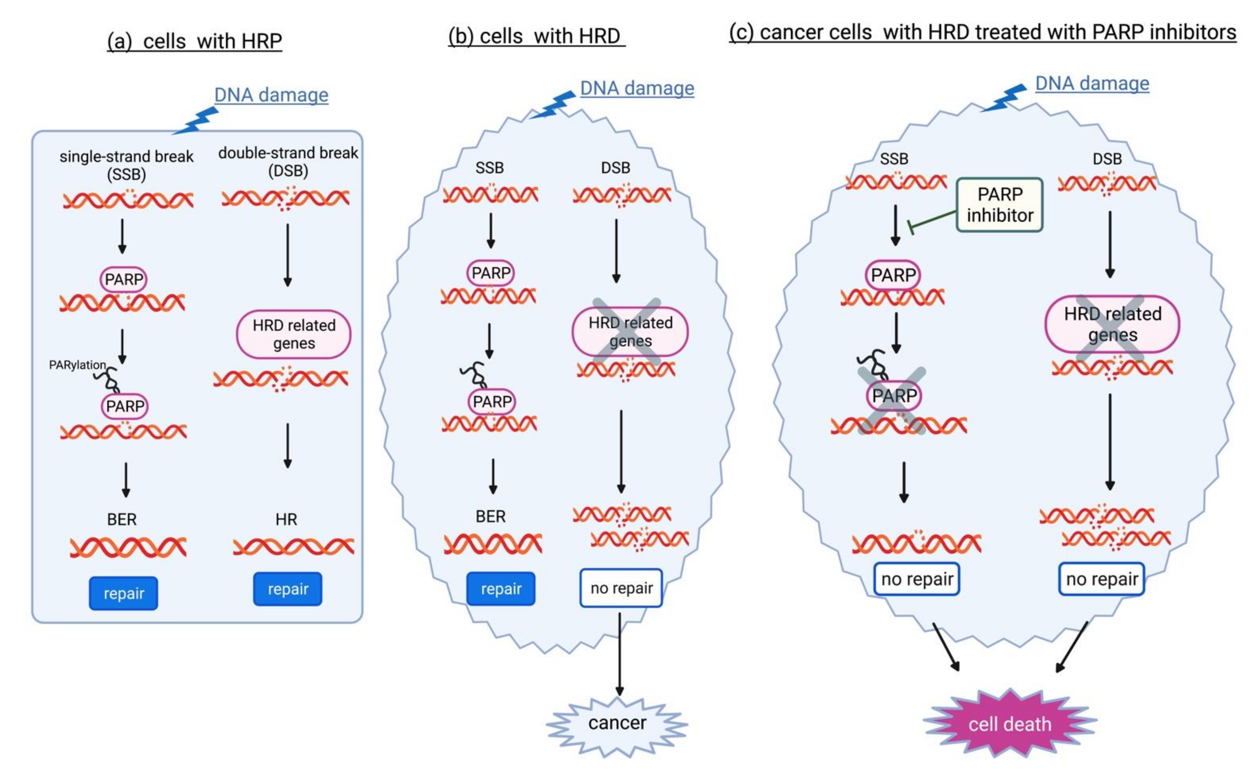
The multifunctional enzyme Poly ADP ribose polymerase (PARP) plays an important role in DNA damage repair and genome stability. Among the 18 members of the PARP family, PARP-1 is the most important and plays dominant roles in DNA repair pathways. Activated PARP-1 plays an important role in DNA base excision repair (BER) [57]. When PARP-1 fails to function, oxidized bases accumulate. The replication fork stops at the site of the damaged DNA, eventually resulting in DSBs. In normal cells which are proficient at HR (HR proficiency, HPR), both BER and HR are available for the repair of damaged DNA (Figure 2a). Meanwhile, in cells with HR deficiency (HRD), HR is nonfunctional and leads to carcinogenesis (Figure 2b). When PARP-1 is inhibited by PARP inhibitor, cancer cells with HRD are unable to repair DNA damage by HR and BER, leading to cell death (synthetic lethality, Figure 2c). PARP-1 is also involved in the regulation of nucleotide excision repair (NER), classical NHEJ (cNHEJ), alternative NHEJ (aNHEJ), microhomology-mediated end-joining (MMEJ), HR, MMR, and maintenance of replication fork stability [58]. For example, PARP-1 recruits DSB repair enzymes MRE11 and NBS1 for modulating DSB repair [59].
PARP inhibitors are the first clinically approved anti-cancer agents which specifically targeted the DNA damage response in BRCA1/2-mutated cancers [60]. In advanced-stage EOC patients with the BRCA1/2 GPVs, maintenance therapy with a PARP inhibitor (olaparib) resulted in a 70% lower risk of disease progression or death (SOLO1 clinical trial) [55]. The phase III OlympiAD trial showed that olaparib had better efficacy than standard chemotherapy for HER2-negative breast cancer patients with the BRCA1/2 GPVs [61]. Their indication has been gradually extended to prostate and pancreatic cancer patients with BRCA1/2 GPVs [56]. More recently, the effect of PARP inhibitors was also validated in HR-deficient cancers without BRCA1/2 GPVs, suggesting widespread use of PARP inhibitors for cancers caused by GPVs in other HR pathway genes [62,63].

3. ATM (Ataxia–Telangiectasia Mutated) Gene

3.1. Molecular Function in the Response to DSBs

The ATM gene encodes a protein kinase with pleiotropic functions belonging to the superfamily of phosphatidylinositol 3-kinase-related protein kinases at the peak of a cascade responding to DSBs [64]. In DSB repair, the HR repair pathway is largely restricted to the S and G2 phases of the cell cycle, when an intact sister chromatid is available as a template, whereas NHEJ can be active in any cell cycle [65] (Figure 1b). ATM, which is recruited and activated by the MRN protein complex that recognizes the free DNA ends of DSBs, phosphorylates many important proteins, e.g., BRCA1, p53, AKT, and CHEK2 proteins, thereby mediating the DNA damage response, promoting cell cycle arrest, or inducing apoptosis. In addition to playing a key role in HR, ATM also orchestrates DSB repair by preventing the toxic error-prone NHEJ pathway [66,67]. ATM GPV heterozygous carriers have an increased risk for several types of cancers, including breast, ovarian, and pancreatic cancers [46,68]. However, the carriers of biallelic ATM GPVs are affected by ataxia–telangiectasia (AT, OMIM #208900), which is a rare autosomal recessive syndrome characterized by progressive cerebellar ataxia, cutaneous telangiectasias, increased risk of developing hematologic and solid tumors, and immunodeficiency [66,68,69].

3.2. Prevalence and Risk of Developing EOC

A recent meta-analysis [45] reported the prevalence of ATM GPVs in patients with EOC to be 0.6767% (26/3842 cases) and showed a significant association between ATM GPVs and EOC (odds ratio (OR) = 1.977, 95% confidence interval (CI) = 1.330–2.939) (Table 1). Another population-based cohort study reported that the prevalence of ATM GPVs was 0.57–0.64% [18,69]. The absolute lifetime risk of EOC estimated by the NCCN clinical practice guidelines in oncology is <3% [26,46].

3.3. Medical Management for the Prevention of EOC

For heterozygote ATM GPV carriers, there is insufficient evidence available to recommend RRSO, although a large EOC study reported strong evidence for an approximately two-fold increased risk of developing EOC compared with noncarriers [46]. Therefore, RRSO should be considered according to the family history of the patient (Table 1) [46]. The detection of heterozygous ATM GPVs should not lead to a recommendation to avoid radiation therapy at this time [46]. Furthermore, the NCCN clinical practice guidelines in oncology recommend counseling for ATM GPV carriers because of the risk of autosomal recessive inheritance in their offspring [46].

4. BRIP1 (BRCA1 Interacting Helicase 1) Gene

4.1. Molecular Function in the Response to DSBs

The protein encoded by BRIP1 is a member of the RecQ DEAH helicase family and part of the Fanconi anemia group. The BRIP1 protein interacts with the BRCT repeats at the carboxyl-terminus of BRCA1 (Figure 1b). The bound complex is important for normal DSB repair by HR. BRIP1 is also physiologically essential for maintaining genomic integrity, removing proteins bound to DNA, stabilizing replication forks, and unwinding substitutive DNA structures along with RPA [70].

4.2. Prevalence and Risk of Developing EOC

GPVs in BRIP1 are the second most common pathogenic variant found in patients with EOC after those in BRCA1/2, with a frequency of approximately 1% of EOC cases (Table 1) [17,45]. In a recent meta-analysis [46], BRIP1 GPVs were significantly associated with EOC (OR = 4.878, 95% CI = 3.729–6.380). Another population-based cohort study reported that the prevalence of BRIP1 GPVs was 0.92–1.36% [18,71]. A larger meta-analysis using approximately 29,400 EOC cases from 63 studies and approximately 116,000 controls from the gnomAD database reported that the prevalence of BRIP1 GPVs in patients with EOC was 0.8891% (200/22,494 cases) and that BRIP1 was significantly associated with EOC (OR = 4.94, 95%CI = 4.07–6.00) [49]. The NCCN clinical practice guidelines in oncology estimate that the absolute lifetime risk of EOC for individuals with BRIP1 GPVs is >10% [46].

4.3. Medical Management for the Prevention of EOC

For BRIP1 GPV carriers, the NCCN clinical practice guidelines in oncology recommend that RRSO should be considered from age 45 to 50 years or earlier based on a specific family history of early-onset EOC [45,46,72] (Table 1). Although the lifetime risk of EOC in BRIP1 GPV carriers seems to be sufficient to justify considering RRSO, there is currently no evidence to make a firm recommendation on the optimal age for this procedure. Reportedly, the median age at diagnosis for BRIP1 GPV carriers with EOC is 65 years old [72]. Moreover, the age at which to begin consultation for surgery may change as more evidence is collected. Furthermore, because BRIP1 was originally identified in research on Fanconi anemia (FANCJ; OMIM #609054) [73], the NCCN clinical practice guidelines in oncology recommend counseling BRIP1 GPV carriers about the risk of autosomal recessive conditions in their offspring [46].

5. NBN (Nibrin) Gene

5.1. Molecular Function in the Response to DSBs

NBN encodes the protein NBN or nibrin, one of the components of the MRN protein complex, which is essential for DSB repair, DNA recombination, maintenance of telomere integrity, cell cycle checkpoint regulation, and meiosis (Figure 1a) [74]. The MRN protein complex is composed of two heterodimers of RAD50 and MRE11, as well as a single NBN, and possesses single-strand endonuclease activity and double-strand-specific 3′-5′ exonuclease activity provided by MRE11. In DSB repair, RAD50 is required to bind DNA ends and hold them in close proximity [75]. NBN modulates DNA damage signal sensing by recruiting ATM, ATR, and DNA-dependent protein kinase catalytic subunits to the sites of DNA damage and activating their functions [76]. NBN can also recruit MRE11 and RAD50 to the proximity of DSBs via its interaction with the histone H2AX [77]. NBN also functions in telomere length maintenance by generating the 3′ overhang which serves as a primer for telomerase-dependent telomere elongation [78].
GPVs at the homozygous or compound heterozygous status within NBN are responsible for Nijmegen breakage syndrome (NBS; OMIM #251260), a rare autosomal recessive disorder characterized by microcephaly, growth retardation, humoral and cellular immunodeficiency, radiosensitivity, and cancer predisposition. By the age of 20 years, more than 40% of patients with NBS develop a malignant disease, primarily of lymphoid origin [79].

5.2. Prevalence and Risk of Developing EOC

As NBN has an essential function in the DNA repair pathway, several case–control studies have investigated its status as an EOC susceptibility gene. However, most studies have provided insufficient evidence of a significant association with the risk of developing EOC [42,46]. In a recent meta-analysis [45], the reported prevalence of NBN GPVs in patients with EOC was 0.2837% (20/7050 EOC cases), and NBN GPVs were significantly associated with EOC (OR = 2.166, 95% CI = 1.346–3.488) (Table 1). Another population-based cohort study including 6,001 patients with EOC reported a prevalence of NBN GPVs of 0.35% [71]. According to the NCCN clinical practice guidelines in oncology, the absolute lifetime risk of EOC in NBN GPV carriers is relatively low (<3%); however, the evidence strength is limited, and insufficient data are available [46].

5.3. Medical Management for the Prevention of EOC

According to the NCCN clinical practice guidelines in oncology, there is currently insufficient evidence to recommend RRSO in NBN GPV carriers at this time [46]. Medical management for EOC risk should be considered based on family history [46]. Because the NBN gene is associated with the development of NBS, the NCCN clinical practice guidelines in oncology recommend counseling NBN GPV carriers about the risk of autosomal recessive conditions in their offspring [46].

6. PALB2 (Partner and Localizer of BRCA2) Gene

6.1. Molecular Function in the Response to DSBs

PALB2 was originally identified as the gene encoding protein immunoprecipitated with the BRCA2 protein. PALB2 colocalizes with BRCA2 in nuclear foci; promotes the stable association of BRCA2 with nuclear structures, allowing BRCA2 to escape the effects of proteasome-mediated degradation; and enables the HR repair of DSBs and the maintenance of G2/M checkpoint functions (Figure 1b) [80,81].

6.2. Prevalence and Risk of Developing EOC

Although the previous NCCN clinical practice guidelines in oncology described “ovarian cancer risk and management” for PALB2 GPVs as insufficient evidence (ver1.2022), the latest version (ver2.2022) has been updated to state that the evidence is strong [46]. A recent meta-analysis reported that the prevalence of PALB2 GPVs in patients with EOC was 0.4226% (30/7099 EOC cases), and that PALB2 GPVs were significantly associated with EOC (OR = 2.134, 95% CI = 1.420–3.207) (Table 1) [45]. However, the relationship between PALB2 GPVs and EOC susceptibility is debated and exhibits low statistical significance. Another population-based cohort study reported that the prevalence of PALB2 GPVs was 0.40–0.62% [18,71].
A recent international study of 524 families with PALB2 GPVs estimated the relative and cumulative risks using complex segregation analysis to model the cancer inheritance patterns in families while adjusting for the mode of ascertainment of each family [82]. This study demonstrated that the estimated risk of female PALB2 GPV carriers developing EOC by the age of 80 was 5%. Based on this result, the NCCN clinical practice guidelines in oncology estimate an absolute lifetime risk of EOC in heterozygote PALB2 GPV carriers of 3–5%, with strong evidence [46].

6.3. Medical Management for the Prevention of EOC

Although ACMG guidance showed that PALB2 GPV carriers had a small to moderate risk for EOC [83], the clinical benefit of RRSO was not sufficiently proven to reduce morbidity and mortality. For all PALB2 GPV carriers, there is insufficient evidence available to recommend RRSO. Therefore, RRSO should be considered based on family history for EOC (Table 1) [46]. As PALB2 is a Fanconi anemia gene (FANCN; OMIM #610832), the NCCN clinical practice guidelines in oncology recommend counseling PALB2 GPV carriers about the risk of autosomal recessive conditions in their offspring [46].

7. RAD51C/RAD51D Gene

7.1. Molecular Function in the Response to DSBs

RAD51C and RAD51D encode the RAD51 paralog proteins, RAD51C and RAD51D, which are structurally similar to the RAD51 recombinase. The Rad51 paralogs associate with one another in two distinct complexes: RAD51B-RAD51C-RAD51D-XRCC2 (BCDX2) and RAD51C-XRCC3 (CX3) [56]. The RAD51 paralogs participate in the assembly and stabilization of the ssDNA/RAD51 filament and the HR intermediates. They are also involved in the process downstream of the homology search.

7.2. Prevalence and Risk of Developing EOC

A recent meta-analysis [45] reported that the prevalence of RAD51C and RAD51D GPVs in patients with EOC was 0.5539% (21/3791 EOC cases) and 0.5832 (19/3258 EOC cases), respectively, and that RAD51C and RAD51D were significantly associated with EOC (OR = 4.241, 95% CI = 2.562–7.022, and OR = 7.276, 95% CI = 4.028–13.140, respectively) (Table 1). Another population-based cohort study reported that the prevalence of RAD51C and RAD51D GPVs was 0.57% and 0.57%, respectively [18]. In a larger meta-analysis using 29,400 EOC cases and 116,000 controls from the noncancer gnomAD database, the prevalence of RAD51C and RAD51D GPVs with EOC was 0.6260% (149/23,802 cases) and 0.4125% (94/22,787 cases), respectively, and RAD51C and RAD51D were significantly associated with EOC (OR = 5.59, 95%CI = 4.42–7.07 and OR = 6.94, 95%CI = 5.10–9.44, respectively) [48].
A recent study including 6,178 and 6,690 families with known RAD51C and RAD51D GPVs, respectively, estimated the relative and cumulative risks using complex segregation analysis to model the cancer inheritance patterns in families while adjusting for the mode of ascertainment of each family [84]. According to the results of this relatively large case–control study, the cumulative risk of developing EOC by the age of 80 years was 11% and 13% for RAD51C and RAD51D GPV carriers, respectively. Thus, the NCCN clinical practice guidelines in oncology estimate the absolute lifetime risk of EOC in heterozygote RAD51C/RAD51D GPV carriers as >10% [46].

7.3. Medical Management for the Prevention of EOC

For RAD51C/RAD51D GPV carriers, the NCCN clinical practice guidelines in oncology recommend considering RRSO from age 45 to 50 years or earlier based on a specific family history of early-onset EOC [46,72]. Although the lifetime risk of EOC in RAD51C/RAD51D GPV carriers seems to be sufficient to justify considering RRSO, there is insufficient evidence to make a firm recommendation regarding the optimal age for this procedure. Reportedly, the median age at diagnosis for RAD51C/RAD51D GPV carriers with EOC is 62 and 57 years old [72]. Therefore, the age at which to begin consultation for surgery may change as more evidence is accumulated. As RAD51C is a Fanconi anemia gene (FANCO; OMIM # 613390), the NCCN clinical practice guidelines in oncology recommend counseling RAD51C GPV carriers about the risk of autosomal recessive conditions in their offspring [46].
RAD51C- [85] and RAD51D-deficient [86] cells, or those expressing pathogenic variants in these genes [86,87], have been shown to render sensitivity to PARP inhibitors, such as olaparib, which is the first PARP inhibitor to be approved for EOC treatment [62,63,88,89,90]. However, it remains unclear whether identifying RAD51C/RAD51D GPVs in patients with EOC is useful for identifying patients that might benefit from treatment with protocols using PARP inhibitors [50].

8. Conclusions

Compared to other cancers, EOC includes a relatively high percentage of hereditary tumors. Approximately 50% of patients with hereditary EOC harbor GPVs in the BRCA1/2 genes contributing to the HR repair pathway. Moreover, other genes participating in the HR repair pathway, such as ATM, BRIP1, NBN, PALB2, and RAD51C/D, are also known as predisposition genes related to hereditary EOC with moderate penetrance. This review has outlined the current knowledge of these moderate-risk genes for EOC involved in the HR repair pathway. In addition to the molecular functions of these EOC-associated genes, we discussed the recommended clinical strategies for preventing EOC in individuals carrying GPVs in these genes. This review can improve our ability to estimate the genetic risk of developing EOC and select appropriate preventive and treatment strategies for hereditary EOC.
However, there are still some issues limiting the effective medical management for EOC based on individual genetic analysis. First, the penetrance of hereditary tumors is not 100%, and the significance of detecting moderate-risk genes for medical management remains unclear [91]. Second, further data registration is important because the GPV frequency prevalence of each cancer predisposition gene varies among populations. Third, understanding the relationship between genotype and phenotype may be extremely useful in a clinical setting. However, if the results are not properly interpreted and explained, there is a risk of inappropriate treatment. Fourth, along with the education of clinicians, it is crucial to ensure collaboration among clinicians, researchers, and companies that provide genetic testing for providing medical care based on genetic information.

Author Contributions

A.A. and I.I. contributed to searching the literature and drafting. A.U., H.N., and H.K. edited the manuscript. All authors have read and agreed to the published version of the manuscript.

Funding

This research received no external funding.

Institutional Review Board Statement

Not applicable.

Informed Consent Statement

Not applicable.

Data Availability Statement

Not applicable.

Acknowledgments

Figure 1 and Figure 2 were created with BioRender.com (accessed on 12 May 2022).

Conflicts of Interest

The authors declare no conflict of interest.

References

  1. Sung, H.; Ferlay, J.; Siegel, R.L.; Laversanne, M.; Soerjomataram, I.; Jemal, A.; Bray, F. Global cancer statistics 2020: GLOBOCAN estimates of incidence and mortality worldwide for 36 cancers in 185 countries. CA Cancer J. Clin. 2021, 71, 209–249. [Google Scholar] [CrossRef] [PubMed]
  2. Howlader, N.; Noone, A.M.; Krapcho, M.; Miller, D.; Brest, A.; Yu, M.; Ruhl, J.; Tatalovich, Z.; Mariotto, A.; Lewis, D.R.; et al. (Eds.) SEER Cancer Statistics [Review]; National Cancer Institute: Bethesda, MD, USA. Available online: https://seer.cancer.gov/archive/csr/1975_2017/ (accessed on 12 May 2022).
  3. Lheureux, S.; Gourley, C.; Vergote, I.; Oza, A.M. Epithelial ovarian cancer. Lancet 2019, 393, 1240–1253. [Google Scholar] [CrossRef] [Green Version]
  4. Henderson, J.T.; Webber, E.M.; Sawaya, G.F. Screening for ovarian cancer: Updated evidence report and systematic review for the US Preventive Services Task Force. JAMA 2018, 319, 595–606. [Google Scholar] [CrossRef] [PubMed] [Green Version]
  5. Prat, J. Ovarian carcinomas: Five distinct diseases with different origins, genetic alterations, and clinicopathological features. Virchows Arch. 2012, 460, 237–249. [Google Scholar] [CrossRef] [PubMed]
  6. Cabasag, C.J.; Arnold, M.; Butler, J.; Inoue, M.; Trabert, B.; Webb, P.M.; Bray, F.; Soerjomataram, I. The influence of birth cohort and calendar period on global trends in ovarian cancer incidence. Int. J. Cancer 2020, 146, 749–758. [Google Scholar] [CrossRef]
  7. Moyer, V.A.; U.S. Preventive Services Task Force. Risk assessment, genetic counseling, and genetic testing for BRCA-related cancer in women: U.S. Preventive Services Task Force recommendation statement. Ann. Intern. Med. 2014, 160, 271–281. [Google Scholar] [CrossRef]
  8. Tanha, K.; Mottaghi, A.; Nojomi, M.; Moradi, M.; Rajabzadeh, R.; Lotfi, S.; Janani, L. Investigation on factors associated with ovarian cancer: An umbrella review of systematic review and meta-analyses. J. Ovarian Res. 2021, 14, 153. [Google Scholar] [CrossRef]
  9. Abdulaziz, G.; Welc, N.A.; Gąsiorowska, E.; Nowak-Markwitz, E. Assessment of gynecological and lifestyle-related risk factors of ovarian cancer. Prz. Menopauzalny 2021, 20, 184–192. [Google Scholar] [CrossRef]
  10. Ferguson, L.R. Chronic inflammation and mutagenesis. Mutat. Res. 2010, 690, 3–11. [Google Scholar] [CrossRef]
  11. Kawanishi, S.; Ohnishi, S.; Ma, N.; Hiraku, Y.; Murata, M. Crosstalk between DNA damage and inflammation in the multiple steps of carcinogenesis. Int. J. Mol. Sci. 2017, 18, 1808. [Google Scholar] [CrossRef]
  12. Zhou, Z.; Zeng, F.; Yuan, J.; Tang, J.; Colditz, G.A.; Tworoger, S.S.; Trabert, B.; Su, X. Pelvic inflammatory disease and the risk of ovarian cancer: A meta-analysis. Cancer Causes Control 2017, 28, 415–428. [Google Scholar] [CrossRef] [PubMed] [Green Version]
  13. Trabert, B.; Ness, R.B.; lo Ciganic, W.-H.; Murphy, M.A.; Goode, E.L.; Poole, E.M.; Brinton, L.A.; Webb, P.M.; Nagle, C.M.; Jordan, S.J.; et al. Aspirin, nonaspirin nonsteroidal anti-inflammatory drug, and acetaminophen use and risk of invasive epithelial ovarian cancer: A pooled analysis in the Ovarian Cancer Association Consortium. J. Natl. Cancer Inst. 2014, 106, djt431. [Google Scholar] [CrossRef] [PubMed] [Green Version]
  14. Risch, H.A.; Howe, G.R. Pelvic inflammatory disease and the risk of epithelial ovarian cancer. Cancer Epidemiol. Biomark. Prev. 1995, 4, 447–451. [Google Scholar]
  15. Ness, R.B.; Grisso, J.A.; Cottreau, C.; Klapper, J.; Vergona, R.; Wheeler, J.E.; Morgan, M.; Schlesselman, J.J. Factors related to inflammation of the ovarian epithelium and risk of ovarian cancer. Epidemiology 2000, 11, 111–117. [Google Scholar] [CrossRef]
  16. Brilhante, A.V.M.; Augusto, K.L.; Portela, M.C.; Sucupira, L.C.G.; Oliveira, L.A.F.; Pouchaim, A.J.M.V.; Nobrega, L.R.M.; Magalhaes, T.F.M.; Sobreira, L.R.P. Endometriosis and ovarian cancer: An integrative review (endometriosis and ovarian cancer). Asian Pac. J. Cancer Prev. 2017, 18, 11–16. [Google Scholar] [CrossRef]
  17. Pennington, K.P.; Walsh, T.; Harrell, M.I.; Lee, M.K.; Pennil, C.C.; Rendi, M.H.; Thornton, A.; Norquist, B.M.; Casadei, S.; Nord, A.S.; et al. Germline and somatic mutations in homologous recombination genes predict platinum response and survival in ovarian, Fallopian tube, and peritoneal carcinomas. Clin. Cancer Res. 2014, 20, 764–775. [Google Scholar] [CrossRef] [PubMed] [Green Version]
  18. Norquist, B.M.; Harrell, M.I.; Brady, M.F.; Walsh, T.; Lee, M.K.; Gulsuner, S.; Bernards, S.S.; Casadei, S.; Yi, Q.; Burger, R.A.; et al. Inherited mutations in women with ovarian carcinoma. JAMA Oncol. 2016, 2, 482–490. [Google Scholar] [CrossRef] [Green Version]
  19. Calaf, G.M.; Urzua, U.; Termini, L.; Aguayo, F. Oxidative stress in female cancers. Oncotarget 2018, 9, 23824–23842. [Google Scholar] [CrossRef] [Green Version]
  20. Savage, K.I.; Matchett, K.B.; Barros, E.M.; Cooper, K.M.; Irwin, G.W.; Gorski, J.J.; Orr, K.S.; Vohhodina, J.; Kavanagh, J.N.; Madden, A.F.; et al. BRCA1 deficiency exacerbates estrogen-induced DNA damage and genomic instability. Cancer Res. 2014, 74, 2773–2784. [Google Scholar] [CrossRef] [Green Version]
  21. Alayev, A.; Salamon, R.S.; Manna, S.; Schwartz, N.S.; Berman, A.Y.; Holz, M.K. Estrogen induces RAD 51C expression and localization to sites of DNA damage. Cell Cycle 2016, 15, 3230–3239. [Google Scholar] [CrossRef]
  22. Zach, L.; Yedidia-Aryeh, L.; Goldberg, M. Estrogen and DNA damage modulate mRNA levels of genes involved in homologous recombination repair in estrogen-deprived cells. J. Trans. Genet. Genom. 2022, 6, 266–280. [Google Scholar] [CrossRef]
  23. Yamamoto, H.; Hirasawa, A. Homologous recombination deficiencies and hereditary tumors. Int. J. Mol. Sci. 2022, 23, 348. [Google Scholar] [CrossRef]
  24. Ueki, A.; Hirasawa, A. Molecular features and clinical management of hereditary gynecological cancers. Int. J. Mol. Sci. 2020, 21, 9504. [Google Scholar] [CrossRef] [PubMed]
  25. Bono, M.; Fanale, D.; Incorvaia, L.; Cancellierando, D.; Fiorino, A.; Calo, V.; Dimino, A.; Filorizzo, C.; Corsini, L.R.; Brando, C.; et al. Impact of deleterious variants in other genes beyond BRCA1/2 detected in breast/ovarian and pancreatic cancer patients by NGS-based multi-gene panel testing: Looking over the hedge. ESMO Open 2021, 6, 100235. [Google Scholar] [CrossRef] [PubMed]
  26. Domchek, S.M.; Robson, M.E. Update on genetic testing in gynecologic cancer. J. Clin. Oncol. 2019, 37, 2501–2510. [Google Scholar] [CrossRef] [PubMed]
  27. Tsaousis, G.N.; Papadopoulou, E.; Apessos, A.; Agiannitopoulos, K.; Pepe, G.; Kampouri, S.; Diamatopoulos, N.; Floros, T.; Iosifidou, R.; Katopodi, O.; et al. Analysis of hereditary cancer syndromes by using a panel of genes: Novel and multiple pathogenic mutations. BMC Cancer 2019, 19, 535. [Google Scholar] [CrossRef] [Green Version]
  28. Samadder, N.J.; Riegert-Johnson, D.; Boardman, L.; Rhodes, D.; Wick, M.; Okuno, S.; Kunze, K.L.; Golafshar, M.; Uson, P.L.S.; Mountjoy, L.; et al. Comparison of universal genetic testing vs guideline-directed targeted testing for patients with hereditary cancer syndrome. JAMA Oncol. 2021, 7, 230–237. [Google Scholar] [CrossRef]
  29. Vietri, M.T.; D’Elia, G.; Caliendo, G.; Casamassimi, A.; Federico, A.; Passariello, L.; Cioffi, M.; Molinari, A.M. Prevalence of mutations in BRCA and MMR genes in patients affected with hereditary endometrial cancer. Med. Oncol. 2021, 38, 13. [Google Scholar] [CrossRef]
  30. Liu, Y.L.; Breen, K.; Catchings, A.; Ranganathan, M.; Latham, A.; Goldfrank, D.J.; Grisham, R.N.; Long Roche, K.; Frey, M.K.; Chi, D.S.; et al. Risk-reducing bilateral salpingo-oophorectomy for ovarian cancer: A review and clinical guide for hereditary predisposition genes. JCO Oncol. Pract. 2022, 18, 201–209. [Google Scholar] [CrossRef]
  31. Phelan, C.M.; Kuchenbaecker, K.B.; Tyrer, J.P.; Kar, S.P.; Lawrenson, K.; Winham, S.J.; Dennis, J.; Pirie, A.; Riggan, M.J.; Chornokur, G.; et al. Identification of 12 new susceptibility loci for different histotypes of epithelial ovarian cancer. Nat. Genet. 2017, 49, 680–691. [Google Scholar] [CrossRef] [Green Version]
  32. Kar, S.P.; Berchuck, A.; Gayther, S.A.; Goode, E.L.; Moysich, K.B.; Pearce, C.L.; Ramus, S.J.; Schildkraut, J.M.; Sellers, T.A.; Pharoah, P.D.P. Common genetic variation and susceptibility to ovarian cancer: Current insights and future directions. Cancer Epidemiol. Biomark. Prev. 2018, 27, 395–404. [Google Scholar] [CrossRef] [PubMed]
  33. Eleje, G.U.; Eke, A.C.; Ezebialu, I.U.; Ikechebelu, J.I.; Ugwu, E.O.; Okonkwo, O.O. Risk-reducing bilateral salpingo-oophorectomy in women with BRCA1 or BRCA2 mutations. Cochrane Database Syst. Rev. 2018, 8, CD012464. [Google Scholar] [CrossRef] [PubMed]
  34. Rebbeck, T.R.; Lynch, H.T.; Neuhausen, S.L.; Narod, S.A.; Van’t Veer, L.; Garber, J.E.; Evans, G.; Isaacs, C.; Daly, M.B.; Matloff, E.; et al. Prophylactic oophorectomy in carriers of BRCA1 or BRCA2 mutations. N. Engl. J. Med. 2002, 346, 1616–1622. [Google Scholar] [CrossRef] [PubMed]
  35. Kauff, N.D.; Satagopan, J.M.; Robson, M.E.; Scheuer, L.; Hensley, M.; Hudis, C.A.; Ellis, N.A.; Boyd, J.; Borgen, P.I.; Barakat, R.R.; et al. Risk-reducing salpingo-oophorectomy in Women with a BRCA1 or BRCA2 Mutation. N. Engl. J. Med. 2002, 346, 1609–1615. [Google Scholar] [CrossRef]
  36. Neff, R.T.; Senter, L.; Salani, R. BRCA mutation in ovarian cancer: Testing, implications and treatment considerations. Ther. Adv. Med. Oncol. 2017, 9, 519–531. [Google Scholar] [CrossRef] [Green Version]
  37. Tung, N.; Domchek, S.M.; Stadler, Z.; Nathanson, K.L.; Couch, F.; Garber, J.E.; Offit, K.; Robson, M.E. Counselling framework for moderate-penetrance cancer-susceptibility mutations. Nat. Rev. Clin. Oncol. 2016, 13, 581–588. [Google Scholar] [CrossRef] [Green Version]
  38. Samuel, D.; Diaz-Barbe, A.; Pinto, A.; Schlumbrecht, M.; George, S. Hereditary ovarian carcinoma: Cancer pathogenesis looking beyond BRCA1 and BRCA2. Cells 2022, 11, 539. [Google Scholar] [CrossRef]
  39. Leblanc, E.; Narducci, F.; Hudry, D.; Bresson, L.; Charvolin, J.Y.; Ferron, G.; Guyon, F.; Fourchotte, V.; Lambaudie, E.; Baron, M.; et al. First results of a prospective national controlled study: Prophylactic radical fimbriectomy (NCT01608074), in women with a hereditary familial risk of breast/ovarian cancer—Tolerance and pathological findings. J. Clin. Oncol. 2018, 36, 5574. [Google Scholar] [CrossRef]
  40. Gaba, F.; Robbani, S.; Singh, N.; McCluggage, W.G.; Wilkinson, N.; Ganesan, R.; Bryson, G.; Rowlands, G.; Tyson, C.; Arora, R.; et al. Preventing ovarian cancer through early excision of tubes and late ovarian removal (PROTECTOR): Protocol for a prospective non-randomised multi-center trial. Int. J. Gynecol. Cancer 2021, 31, 286–291. [Google Scholar] [CrossRef]
  41. Steenbeek, M.P.; Bommel, M.V.; Swisher, E.; Lu, K.; Hermens, R.; Hullu, J.D. Tubectomy with delayed oophorectomy in high risk women to assess the safety of prevention (TUBA-WISP-II). Int. J. Gynecol. Cancer 2021, 31, A314. [Google Scholar] [CrossRef]
  42. Pavanello, M.; Chan, I.H.; Ariff, A.; Pharoah, P.D.; Gayther, S.A.; Ramus, S.J. Rare germline genetic variants and the risks of epithelial ovarian cancer. Cancers 2020, 12, 3046. [Google Scholar] [CrossRef] [PubMed]
  43. Watanabe, T.; Soeda, S.; Endo, Y.; Okabe, C.; Sato, T.; Kamo, N.; Ueda, M.; Kojima, M.; Furukawa, S.; Nishigori, H.; et al. Rare hereditary gynecological cancer syndromes. Int. J. Mol. Sci. 2022, 23, 1563. [Google Scholar] [CrossRef] [PubMed]
  44. Pietragalla, A.; Arcieri, M.; Marchetti, C.; Fagotti, A. Ovarian cancer predisposition beyond BRCA1 and BRCA2 genes. Int. J. Gynecol. Cancer 2020, 30, 1803–1810. [Google Scholar] [CrossRef] [PubMed]
  45. Suszynska, M.; Klonowska, K.; Jasinska, A.J.; Kozlowski, P. Large-scale meta-analysis of mutations identified in panels of breast/ovarian cancer-related genes—Providing evidence of cancer predisposition genes. Gynecol. Oncol. 2019, 153, 452–462. [Google Scholar] [CrossRef] [PubMed] [Green Version]
  46. National Comprehensive Cancer Network (NCCN) in Oncology. Genetic/Familial High-Risk Assessment: Breast, Ovarian and Pancreatic. Version 2. 2022. Available online: https://www.nccn.org/professionals/physician_gls/pdf/genetics_bop.pdf (accessed on 12 May 2022).
  47. National Comprehensive Cancer Network (NCCN) in Oncology. Genetic/Familial High-Risk Assessment: Colorectal. Version 2. 2021. Available online: https://www.nccn.org/professionals/physician_gls/pdf/genetics_colon.pdf (accessed on 12 May 2022).
  48. Suszynska, M.; Ratajska, M.; Kozlowski, P. BRIP1, RAD51C, and RAD51D mutations are associated with high susceptibility to ovarian cancer: Mutation prevalence and precise risk estimates based on a pooled analysis of ~30,000 cases. J. Ovarian Res. 2020, 13, 50. [Google Scholar] [CrossRef] [PubMed]
  49. Momozawa, Y.; Sasai, R.; Usui, Y.; Shiraishi, K.; Iwasaki, Y.; Taniyama, Y.; Parsons, M.T.; Mizukami, K.; Sekine, Y.; Hirata, M.; et al. Expansion of Cancer Risk Profile for BRCA1 and BRCA2 Pathogenic Variants. JAMA Oncol. 2022, 8, 871–878. [Google Scholar] [CrossRef]
  50. Huertas, P. DNA resection in eukaryotes: Deciding how to fix the break. Nat. Struct. Mol. Biol. 2010, 17, 11–16. [Google Scholar] [CrossRef] [Green Version]
  51. Huang, R.X.; Zhou, P.K. DNA damage response signaling pathways and targets for radiotherapy sensitization in cancer. Signal Transduct. Target Ther. 2020, 5, 60. [Google Scholar] [CrossRef]
  52. Pannunzio, N.R.; Watanabe, G.; Lieber, M.R. Nonhomologous DNA end-joining for repair of DNA double strand breaks. J. Biol. Chem. 2018, 293, 10512–10523. [Google Scholar] [CrossRef] [Green Version]
  53. Pardo, B.; Gómez-González, B.; Aguilera, A. DNA repair in mammalian cells: DNA double-strand break repair—How to fix a broken relationship. Cell. Mol. Life Sci. 2009, 66, 1039–1056. [Google Scholar] [CrossRef]
  54. Hartlerode, A.J.; Scully, R. Mechanisms of double-strand break repair in somatic mammalian cells. Biochem. J. 2009, 423, 157–168. [Google Scholar] [CrossRef] [PubMed]
  55. Moore, K.; Colombo, N.; Scambia, G.; Kim, B.G.; Oaknin, A.; Friedlander, M.; Lisyanskaya, A.; Floquet, A.; Leary, A.; Sonke, G.S.; et al. Maintenance Olaparib in patients with newly diagnosed advanced ovarian cancer. N. Eng. J. Med. 2018, 379, 2495–2505. [Google Scholar] [CrossRef] [PubMed]
  56. Matos-Rodrigues, G.; Guirouilh-Barbat, J.; Martini, E.; Lopez, B.S. Homologous recombination, cancer and the ‘RAD51 paradox’. NAR Cancer 2021, 3, zcab016. [Google Scholar] [CrossRef] [PubMed]
  57. Burkle, A. Poly(APD-ribosyl)ation, a DNA damage-driven protein modification and regulator of genomic instability. Cancer Lett. 2001, 163, 1–5. [Google Scholar] [CrossRef]
  58. Pazzaglia, S.; Pioli, C. Multifaceted role of PARP-1 in DNA repair and inflammation: Pathological and therapeutic implications in cancer and non-cancer diseases. Cells 2020, 9, 41. [Google Scholar] [CrossRef] [Green Version]
  59. Haince, J.F.; McDonald, D.; Rodrigue, A.; Déry, U.; Masson, J.Y.; Hendze, M.J.; Poirier, G.G. PARP1-dependent kinetics of recruitment of MRE11 and NBS1 proteins to multiple DNA damage sites. J. Biol. Chem. 2008, 283, 1197–1208. [Google Scholar] [CrossRef] [Green Version]
  60. Griguolo, G.; Dieci, M.V.; Guarneri, V.; Conte, P. Olaparib for the treatment of breast cancer. Expert Rev. Anticancer Ther. 2018, 18, 519–530. [Google Scholar] [CrossRef]
  61. Robson, M.; Im, S.-A.; Senkus, E.; Xu, B.; Domchek, S.M.; Masuda, N.; Delaloge, S.; Li, W.; Tung, N.; Armstrong, A.; et al. Olaparib for Metastatic Breast Cancer in Patients with a Germline BRCA Mutation. N. Engl. J. Med. 2017, 377, 523–533. [Google Scholar] [CrossRef]
  62. Curtin, N.J.; Szabo, C. Poly(ADP-ribose) polymerase inhibition: Past, present and future. Nat. Rev. Drug Discov. 2020, 19, 711–736. [Google Scholar] [CrossRef]
  63. Paulet, L.; Trecourt, A.; Leary, A.; Peron, J.; Descotes, F.; Devouassoux-Shisheboran, M.; Leroy, K.; You, B.; Lopez, J. Cracking the homologous recombination deficiency code: How to identify responders to PARP inhibitors. Eur. J. Cancer 2022, 166, 87–99. [Google Scholar] [CrossRef]
  64. Stucci, L.S.; Internò, V.; Tucci, M.; Perrone, M.; Mannavola, F.; Palmirotta, R.; Porta, C. The ATM gene in breast cancer: Its relevance in clinical practice. Genes 2021, 12, 727. [Google Scholar] [CrossRef]
  65. Yu, W.; Lescale, C.; Babin, L.; Bedora-Faure, M.; Lenden-Hasse, H.; Baron, L.; Demangel, C.; Yelamos, J.; Brunet, E.; Deriano, L. Repair of G1 induced DNA double-strand breaks in S-G2/M by alternative NHEJ. Nat. Commun. 2020, 11, 5239. [Google Scholar] [CrossRef] [PubMed]
  66. Balmus, G.; Pilger, D.; Coates, J.; Demir, M.; Sczaniecka-Clift, M.; Barros, A.C.; Woods, M.; Fu, B.; Yang, F.; Chen, E.; et al. ATM orchestrates the DNA-damage response to counter toxic non-homologous end-joining at broken replication forks. Nat. Commun. 2019, 10, 87. [Google Scholar] [CrossRef] [PubMed] [Green Version]
  67. Britton, S.; Chanut, P.; Delteil, C.; Barboule, N.; Frit, P.; Calsou, P. ATM antagonizes NHEJ proteins assembly and DNA-ends synapsis at single-ended DNA double strand breaks. Nucleic Acids Res. 2020, 48, 9710–9723. [Google Scholar] [CrossRef]
  68. Ueno, S.; Sudo, T.; Hirasawa, A. ATM: Functions of ATM kinase and its relevance to hereditary tumors. Int. J. Mol. Sci. 2022, 23, 523. [Google Scholar] [CrossRef]
  69. Putti, S.; Giovinazzo, A.; Merolle, M.; Falchetti, M.L.; Pellegrini, M. ATM kinase dead: From ataxia telangiectasia syndrome to cancer. Cancers 2021, 13, 5498. [Google Scholar] [CrossRef] [PubMed]
  70. Ouhtit, A.; Gupta, I.; Shaikh, Z. BRIP1, a potential candidate gene in development of non-BRCA1/2 breast cancer. Front. Biosci. (Elite Ed.) 2016, 8, 289–298. [Google Scholar] [CrossRef] [PubMed]
  71. Kurian, A.W.; Ward, K.C.; Howlader, N.; Deapen, D.; Hamilton, A.S.; Mariotto, A.; Miller, D.; Penberthy, L.S.; Katz, S.J. Genetic testing and results in a population-based cohort of breast cancer patients and ovarian cancer patients. J. Clin. Oncol. 2019, 37, 1305–1315. [Google Scholar] [CrossRef] [PubMed] [Green Version]
  72. Cummings, S.; Roman, S.S.; Saam, J.; Bernhisel, R.; Brown, K.; Lancaster, J.M.; Usha, L. Age of ovarian cancer diagnosis among BRIP1, RAD51C, and RAD51D mutation carriers identified through multi-gene panel testing. J. Ovarian Res. 2021, 14, 61. [Google Scholar] [CrossRef]
  73. Levitus, M.; Waisfisz, Q.; Godthelp, B.C.; de Vries, Y.; Hussain, S.; Wiegant, W.W.; Elghalbzouri-Maghrani, E.; Steltenpool, J.; Rooimans, M.A.; Pals, G.; et al. The DNA helicase BRIP1 is defective in Fanconi anemia complementation group. J. Nat. Genet. 2005, 37, 934–935. [Google Scholar] [CrossRef]
  74. Wen, J.; Cerosaletti, K.; Schultz, K.J.; Wright, J.A.; Concannon, P. NBN phosphorylation regulates the accumulation of MRN and ATM at sites of DNA double-strand breaks. Oncogene 2013, 32, 4448–4456. [Google Scholar] [CrossRef] [PubMed]
  75. Syed, A.; Tainer, J.A. The MRE11-RAD50-NBS1 complex conducts the orchestration of damage signaling and outcomes to stress in DNA replication and repair. Annu. Rev. Biochem. 2018, 87, 263–294. [Google Scholar] [CrossRef] [PubMed]
  76. Menolfi, D.; Zha, S. ATM, ATR and DNA-PKcs Kinases-the lessons from the mouse models: Inhibition ≠ deletion. Cell Biosci. 2020, 10, 8. [Google Scholar] [CrossRef] [PubMed]
  77. Yuan, J.; Chen, J. MRE11-RAD50-NBS1 complex dictates DNA repair independent of H2AX. J. Biol. Chem. 2010, 285, 1097–1104. [Google Scholar] [CrossRef] [PubMed] [Green Version]
  78. Zhang, Y.; Zhou, J.; Lim, C.U.K. The role of NBS1 in DNA double strand break repair, telomere stability, and cell cycle checkpoint control. Cell Res. 2006, 16, 45–54. [Google Scholar] [CrossRef] [Green Version]
  79. Chrzanowska, K.H.; Gregorek, H.; Dembowska-Bagińska, B.; Kalina, M.A.; Digweed, M. Nijmegen breakage syndrome (NBS). Orphanet J. Rare Dis. 2012, 7, 13. [Google Scholar] [CrossRef] [Green Version]
  80. Simhadri, S.; Vincelli, G.; Huo, Y.; Misenko, S.; Foo, T.K.; Ahlskog, J.; Sørensen, C.S.; Oakley, G.G.; Ganesan, S.; Bunting, S.F.; et al. PALB2 connects BRCA1 and BRCA2 in the G2/M checkpoint response. Oncogene 2019, 38, 1585–1596. [Google Scholar] [CrossRef]
  81. Zhuang, J.; Zhang, J.; Willers, H.; Wang, H.; Chung, J.H.; van Gent, D.C.; Hallahan, D.E.; Powell, S.N.; Xia, F. Checkpoint kinase 2-mediated phosphorylation of BRCA1 regulates the fidelity of nonhomologous end-joining. Cancer Res. 2006, 66, 1401–1408. [Google Scholar] [CrossRef] [Green Version]
  82. Yang, X.; Leslie, G.; Doroszuk, A.; Schneider, S.; Allen, J.; Decker, B.; Dunning, A.M.; Redman, J.; Scarth, J.; Plaskocinska, I.; et al. Cancer risks associated With germline PALB2 pathogenic variants: An international study of 524 families. J. Clin. Oncol. 2020, 38, 674–685. [Google Scholar] [CrossRef]
  83. Tischkowitz, M.; Balmana, J.; Foulkes, W.D.; James, P.; Ngeow, J.; Schmutzler, R.; Voian, N.; Wick, M.J.; Stewart, D.R.; Pal, T.; et al. Management of individuals with germline variants in PALB2: A clinical practice resource of the American College of Medical Genetics and Genomics (ACMG). Genet. Med. 2021, 23, 1416–1423. [Google Scholar] [CrossRef]
  84. Yang, X.; Song, H.; Leslie, G.; Engel, C.; Hahnen, E.; Auber, B.; Horváth, J.; Kast, K.; Niederacher, D.; Turnbull, C.; et al. Ovarian and breast cancer risks associated with pathogenic variants in RAD51C and RAD51D. J. Natl. Cancer Inst. 2020, 112, 1242–1250. [Google Scholar] [CrossRef]
  85. Min, A.; Im, S.A.; Yoon, Y.K.; Song, S.H.; Nam, H.J.; Hur, H.S.; Kim, H.P.; Lee, K.H.; Han, S.W.; Oh, D.Y.; et al. RAD51C-deficient cancer cells are highly sensitive to the PARP inhibitor olaparib. Mol. Cancer Ther. 2013, 12, 865–877. [Google Scholar] [CrossRef] [PubMed] [Green Version]
  86. Loveday, C.; Turnbull, C.; Ramsay, E.; Hughes, D.; Ruark, E.; Frankum, J.R.; Bowden, G.; Kalmyrzaev, B.; Warren-Perry, M.; Snape, K.; et al. Germline mutations in RAD51D confer susceptibility to ovarian cancer. Nat. Genet. 2011, 43, 879–882. [Google Scholar] [CrossRef]
  87. Gayarre, J.; Martín-Gimeno, P.; Osorio, A.; Paumard, B.; Barroso, A.; Fernández, V.; de La Hoya, M.; Rojo, A.; Caldés, T.; Palacios, J.; et al. Characterisation of the novel deleterious RAD51C p.Arg312Trp variant and prioritisation criteria for functional analysis of RAD51C missense changes. Br. J. Cancer 2017, 117, 1048–1062. [Google Scholar] [CrossRef] [PubMed]
  88. Slade, D. PARP and PARG inhibitors in cancer treatment. Genes Dev. 2020, 34, 360–394. [Google Scholar] [CrossRef] [PubMed] [Green Version]
  89. Hodgson, D.R.; Dougherty, B.A.; Lai, Z.; Fielding, A.; Grinsted, L.; Spencer, S.; O’Connor, M.J.; Ho, T.W.; Robertson, J.A.; Lanchbury, J.S.; et al. Candidate biomarkers of PARP inhibitor sensitivity in ovarian cancer beyond the BRCA genes. Br. J. Cancer 2018, 119, 1191–1409. [Google Scholar] [CrossRef] [Green Version]
  90. Chian, Y.C.; Lin, P.H.; Chen, W.F. Homologous recombination deficiency assays in epithelial ovarian cancer: Current status and future direction. Front. Oncol. 2021, 11, 675972. [Google Scholar] [CrossRef]
  91. Yoshimura, A.; Imoto, I.; Iwata, H. Functions of breast cancer predisposition genes: Implications for clinical management. Int. J. Mol. Sci. 2022, 23, 7481. [Google Scholar] [CrossRef]
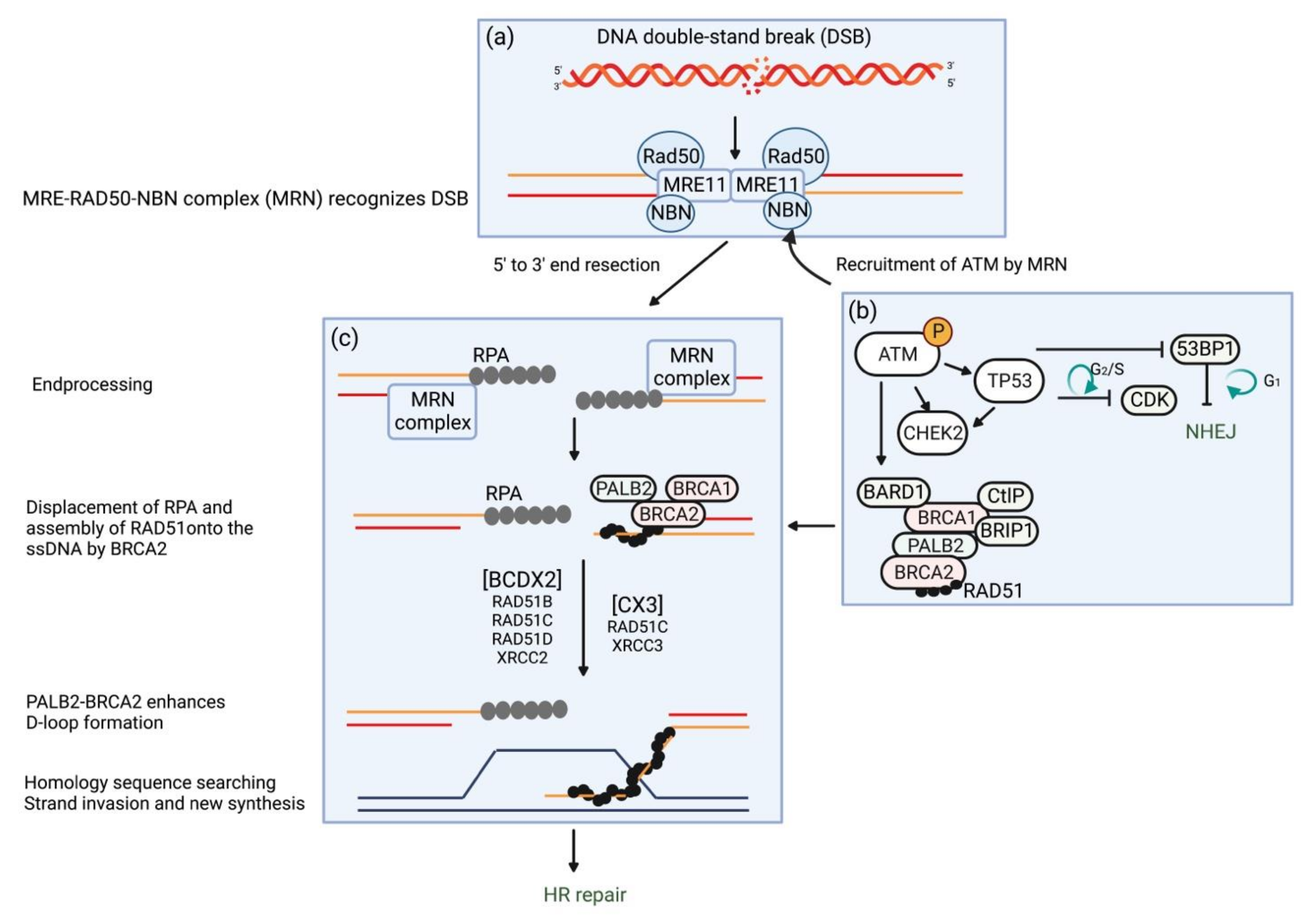
Figure 1. Schematic of DNA double-strand break (DSB) repair by homologous recombination (HR) and key molecules: MRE-RAD50-NBN (MRN) protein complex, ATM, BARD, BRCA1, BRCA2, BRIP1, CHEK2, PALB2, and RAD51C/D. The free DNA ends produced by DSBs are recognized by the MRN protein complex (a). The MRN protein complex recruits and activates ATM, which in turn phosphorylates and activates many downstream targets essential for DNA damage repair via nonhomologous end-joining (NHEJ) and HR (b,c). The replication protein A (RPA) is recruited by the BRCA1–PALB2–BRCA2 effector complex and is loaded on a long 3′ single-stranded DNA (ssDNA) tail to form RAD51–ssDNA nucleofilament. BRCA2 mediates displacement of RPA with RAD51. PALB2-BRCA2 enhances D-loop formation, which is followed by HR repair. The RAD51 paralogs associated in protein complexes (RAD51B-RAD51C-RAD51D-XRCC2 (BCDX2) and RAD51C-XRCC3 (CX3)) participate in the assembly and stabilization of the ssDNA/RAD51 filament and the HR intermediates as well as in the steps downstream of the homology search (not represented) [56].
Ijms 23 11790 g001
Figure 2. In normal cells which are proficient in homologous recombination (HRP), both base excision repair (BER) and HR are available for the repair of damaged DNA via poly (ADP-ribose) polymerase (PARP) and proteins encoded by HR deficiency (HRD)-related genes, respectively (a). In cells with HRD, HR is nonfunctional and leads to carcinogenesis (b). When PARP is inhibited by PARP inhibitor, cancer cells with HRD are unable to repair DNA damage by HR and BER, resulting in cell death (synthetic lethality) (c).
Ijms 23 11790 g002
Table 1. Frequency of germline pathogenic variants in patients with epithelial ovarian cancers (EOC), relative and absolute risks for EOC, and risk reduction for EOC in each predisposition gene.
GeneSuszynska et al. [45]NCCN Guidelines [46,47]
Frequency of GPV in EOC Patients (%)Relative Risk for EOCAbsolute Risk for EOCEvidence for AssociationManagement for Risk Reduction
OR (95% CI)p-Value
BRCA18.60735.26
(29.56–42.05)
<0.000139–58%very strongRRSO recommended for patients aged 35–40 yrs
BRCA24.52011.91
(9.87–14.39)
<0.000113–29%very strongRRSO recommended for patients aged 40–45 yrs
BRIP11.0574.88
(3.73–6.38)
<0.0001>10%strongRRSO considered for patients aged 45–50 yrs
CHEK20.7030.43
(0.29–0.63)
<0.0001not establishednot establishednot established
ATM0.6771.98
(1.33–2.94)
0.001<3%insufficientmanage based on family history
RAD51C0.5544.24
(2.56–7.02)
<0.0001>10%strongRRSO considered for patients aged 45–50 yrs
RAD51D0.5837.28
(4.03–13.14)
<0.0001>10%strongRRSO considered for patients aged 45–50 yrs
MSH60.4444.08
(2.43–6.85)
<0.0001<13%insufficient, mixed-
PALB20.4232.13
(1.42–3.21)
0.00033–5%insufficientmanage based on family history
TP530.2945.05
(2.41–10.58)
<0.0001not establishednot establishednot established
NBN0.2842.17
(1.35–3.49)
0.0020insufficient datalimitedmanage based on family history
MSH20.2383.98
(1.18–8.69)
0.0007>10%strongRRSO should be individualized after childbearing
PMS20.1830.71
(0.29–1.72)
0.5633<3%limited-
MLH10.1041.44
(0.53–3.91)
0.6815>10%strongRRSO should be individualized after childbearing
BARD10.1421.41
(0.69–2.89)
0.4706not establishednot establishednot established
PTEN0.0635.47
(1.26–23.82)
0.0799not establishednot establishednot established
CI, confidence interval; EOC, epithelial ovarian cancer; GPV, germline pathogenic variant; OR, odds ratio; RRSO, risk-reducing salpingo-oophorectomy; yrs, years. Genes in boldface indicate those described in this review article.
Publisher’s Note: MDPI stays neutral with regard to jurisdictional claims in published maps and institutional affiliations.