Article

Resveratrol Improves the Digestive Ability and the Intestinal Health of Siberian Sturgeon

1
Department of Aquaculture, College of Animal Science & Technology, Sichuan Agricultural University, Chengdu 611130, China
2
Fisheries Institute, Sichuan Academy of Agricultural Sciences, Chengdu 610066, China
3
Department of Engineering and Applied Biology, College of Life Science, Sichuan Agricultural University, Ya’an 625014, China
*
Author to whom correspondence should be addressed.
Int. J. Mol. Sci. 2022, 23(19), 11977; https://doi.org/10.3390/ijms231911977
Submission received: 22 September 2022 / Revised: 7 October 2022 / Accepted: 7 October 2022 / Published: 9 October 2022
(This article belongs to the Section Molecular Microbiology)

Abstract

:
The lack of detailed information on nutritional requirement results in limited feeding in Siberian sturgeon. In this study, resveratrol, a versatile natural extract, was supplemented in the daily diet, and the digestive ability and microbiome were evaluated in the duodena and valvular intestines of Siberian sturgeon. The results showed that resveratrol increased the activity of pepsin, α-amylase, and lipase, which was positively associated with an increase in the digestive ability, but it did not influence the final body weight. Resveratrol improved the digestive ability probably by distinctly enhancing intestinal villus height. Microbiome analysis revealed that resveratrol changed the abundance and composition of the microbial community in the intestine, principally in the duodenum. Random forests analysis found that resveratrol significantly downregulated the abundance of potential pathogens (Citrobacter freundii, Vibrio rumoiensis, and Brucella melitensis), suggesting that resveratrol may also improve intestinal health. In summary, our study revealed that resveratrol improved digestive ability and intestinal health, which can contribute to the development of functional feed in Siberian sturgeon.

1. Introduction

The Siberian sturgeon (Acipenser baerii) is one of the oldest surviving vertebrates on Earth [1]. It has been widely farmed because of its high growth performance, disease resistance, and short reproductive cycle [2,3]. However, the controlled feeding of Siberian sturgeon is receiving increasing attention, since the formulation of any artificial diet requires detailed information on the specific nutritional requirements and digestive physiology of the candidate species [4]. Its intestinal tissue, a main digestive organ, has also evolved an ecosystem where a host of microbes live [5]. Unlike the external environment, the intestine provides a steady living environment that stably supplies nutrients to the microbes [6]. In return, the microbiota provides the intestine with a wide range of metabolites that maintain the normal metabolic and immune barrier function of the intestine [7]. Hence, the intestine and its microbiota are usually considered as a whole and play an important role in maintaining the body’s healthy.
Previous studies have found that stresses, such as temperature, salinity, starvation, and dissolved oxygen, drive adverse effects on the intestinal microbiota and, ultimately, affect the growth performance of fish species [8,9,10,11]. Amongst these, temperature is a universal and significant factor in affecting the health of fish species, as they are poikilothermic vertebrates and particularly vulnerable to changes in environmental temperature. In cobia, high temperature has been found to affect growth performance by decreasing digestion efficiency [12]. Hani et al. found that the elevated temperature induced reduction in the activity of intestinal amylase and trypsin in three-spined stickleback, thereby influencing its digestive processes [13]. High temperature also affected the composition of the intestinal microbiota, which in turn might negatively regulate digestive processes [14].
Resveratrol (trans-3,4,5-trihydroxystilbene), an antitoxin secreted by plants, shows versatile therapeutic potential for treating human diseases because of its biological activities, e.g., antioxidant, anti-inflammatory, anti-cancer, and antibacterial [15,16,17,18,19,20]. Resveratrol also plays an important role in modulation of intestinal microbiota. In high-fat mice, resveratrol was reported to reduce obesity by enhancing the abundance of Bacteroides, Blautia, Lachnoclostridium, Parabacteroides, and Ruminiclostridium in the intestine [21]. In piglets, resveratrol downregulated the abundance of the phyla Actinobacteria and Firmicutes, and within Firmicutes, the genus Ruminococcaceae UCG-005 to reduce diquat-induced oxidative stress [22]. Studies in Tilapia (Oreochromis niloticus) found that resveratrol increased the proportion of beneficial microbial taxa (Acetobacteriaceae and Methylbacteraceae), while the proportion of harmful microbial taxa (Streptococcaceae) was decreased [23]. These studies have shown that resveratrol can improve the health of intestinal microbiota.
Our study aimed to explore the effect of resveratrol on the intestinal health of Siberian sturgeon. Resveratrol was supplied into the daily diet and after 45 days, morphology, digestive ability and microbiome of the duodenum and valvular intestine were evaluated. This study revealed the positive effects of resveratrol on intestinal health, which contributes to the application of resveratrol in aquaculture and provides a reference for the functional development of resveratrol in humans.

2. Results

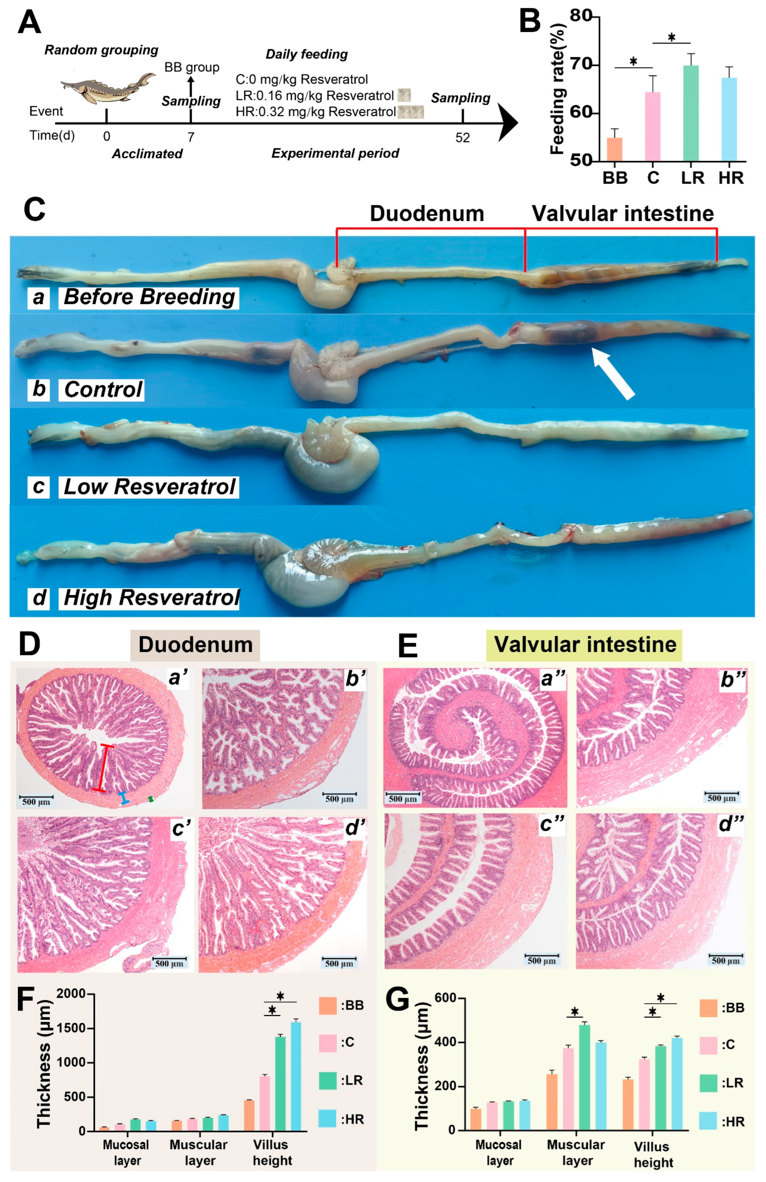
2.1. Resveratrol Improved Intestinal Digestion and Protected the Intestinal Structure in Siberian Sturgeon

To visualize the entire experimental design, a concise experimental flow chart was drawn (Figure 1A). During the experiment, the feeding rates of the four groups were tallied daily. The results showed that sturgeon from the LR group showed a significantly higher feeding rate compared to those in group C (p < 0.05), while the feeding rate of the HR group showed an increasing trend compared to group C (Figure 1B). However, no significant differences were observed in final body weight (FBW) and percentage weight gain (PWG) among the groups (Table 1).
The duodenum and valvular fragments of the intestinal tissue were sampled to observe morphological and histological changes. The valvular intestinal contents were significantly less in the LR and HR groups than in group C (Figure 1C). Histological observations showed a significant increase in the height of intestinal villus in both the duodenum and valvular intestine of Siberian sturgeon fed with resveratrol in groups LR and HR compared to group C (p < 0.05) (Figure 1D–G). Additionally, in the valvular intestine, the muscular layer thickness in the LR group was markedly increased compared to that of the C group (p < 0.05), but no significance was observed in the HR group (Figure 1G).

2.2. Resveratrol Increased Digestive Enzyme Activity of Siberian Sturgeon

To further explore the effects of resveratrol on the intestinal digestion capacity of Siberian sturgeon, the activity of digestive enzymes (pepsin, α-amylase, lipase) was detected in the duodenum and valvular fragments. As shown in Figure 2A–C, in the duodenum, the activity of all three digestive enzymes tended to decline with the increase of farming time (C group vs. BB group), while the activity of three digestive enzymes was enhanced in the LR and HR groups compared to that of group C, especially in the LR group that showed statistical significance (p < 0.01). This result was also found in pepsin activity of the valvular intestine (Figure 2D). Of α-amylase and lipase activity in the valvular intestine, no significant change was observed between BB and C groups, despite α-amylase activity in the C group being higher than in the BB group (Figure 2E,F). In the valvular intestine, the α-amylase and lipase activities were also increased in the LR and HR groups relative to the C group, but only lipase activity in the HR group showed significance (p < 0.05, Figure 2E,F).

2.3. Global Analysis of OTUs

To investigate the influence of resveratrol on the gut microbiota of Siberian sturgeon, 24 samples from the duodenum and valvular intestine of Siberian sturgeon were analyzed (Table S1). A total of 600,418 valid sequences were obtained with an average length of 1,413 bp (Table S1). A total of 1,656 OTUs were classified at the level of species (73%), genus (89%), family (94%), order (97%), class (99%), and phylum (99%). We assessed the diversity and depth of microbial community structure. The Shannon’s index and Simpson’s index were used to compare community diversity among samples. No significant difference (p > 0.05) was found in the diversity among the samples of the duodenum or valvular intestine (Table S1, Figure S1A,D). The community richness was measured by Chao index, which indicated no difference (p > 0.05) among samples of the duodenum or valvular intestine (Table S1). The coverage index showed that the community coverage of all samples was more than 99% (Table S1). Rank-abundance and dilution curves that illustrate the difference in community richness and evenness among the samples, respectively, did not show significant difference (Figure S1B,C,E,F). The raw 16S rRNA sequencing data have been submitted to the Sequence Read Archive (SRA) database of the National Center for Biotechnology Information (NCBI), with accession number PRJNA863445.
Beta diversity analysis showed that the variability of microbial communities in the duodenum samples was higher than in the valvular intestine samples (Figure 3). The microbial community was dominated by Pseudomonadaceae in the valvular intestine samples, and the abundance of other families accounted for a relatively low percentage. In the duodenum samples, Pseudomonadaceae was also the dominant community, but unlike the valvular intestine, other communities, such as Enterobacteriaceae and Rhodobacteraceae, also showed higher abundance. Principal coordinate analysis revealed marked differences in the microbial community structure of different groups, particularly in the valvular intestine (Figure S2).

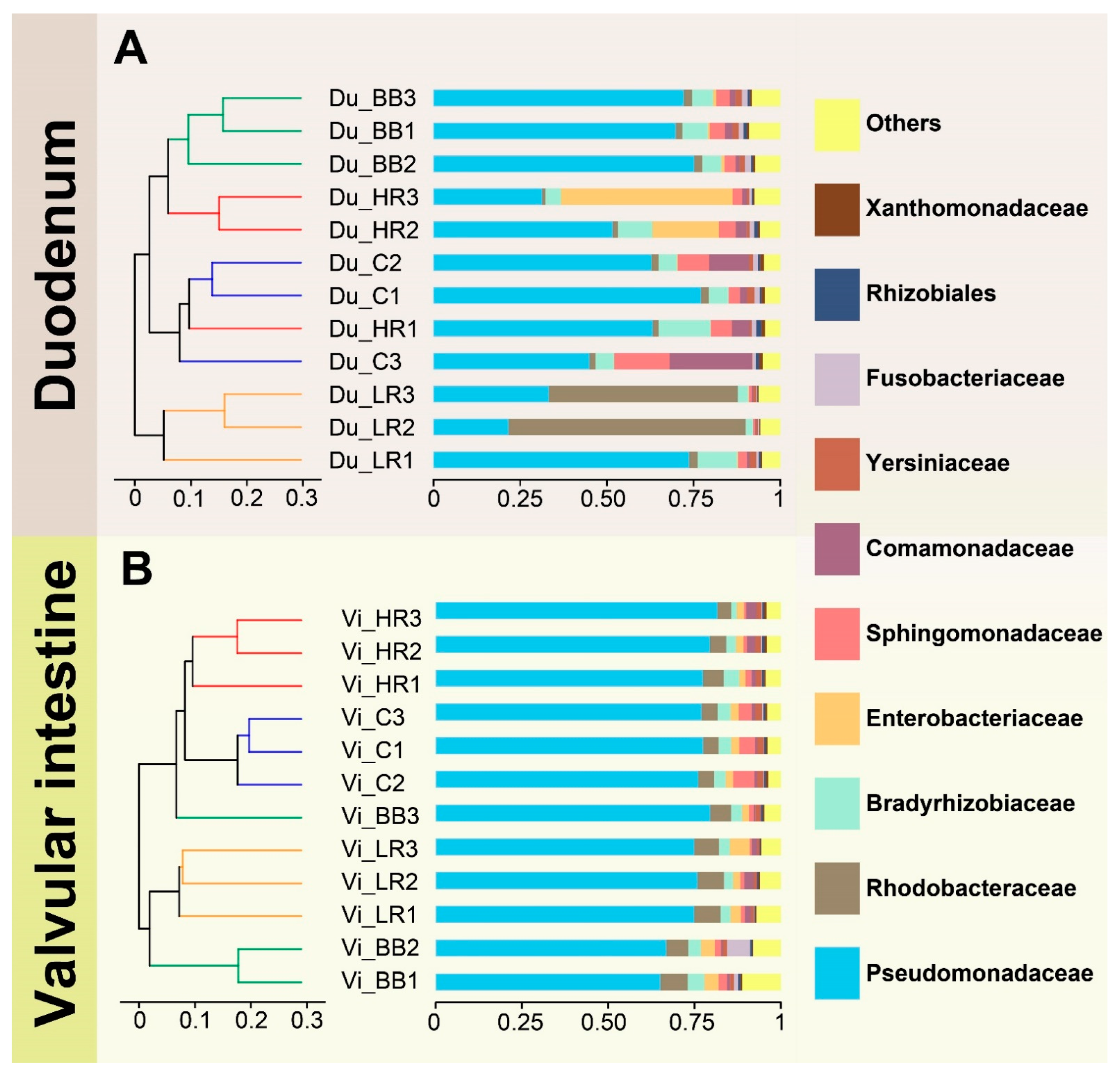
2.4. Resveratrol Alters the Community Composition in the Duodenum of Siberian Sturgeon

OTU levels among different groups were analyzed to compare the species distinctiveness (Figure 4). The Venn annotation showed that the community composition of both the duodenum and valvular intestine presented distinct diversity among different groups. In the duodenum samples, a total of 895 OTUs were identified, of which only 224 OTUs were shared in BB, C, LR and HR groups (Figure 4A). A total of 739 OTUs were identified in the valvular intestine, but only 183 OTUs were shared in the BB, C, LR, and HR groups (Figure 4B). Additionally, in both the duodenum and valvular intestine, the number of microbial species was decreased significantly with the increasing of culturing time (BB group vs. C group), while diets supplemented with different doses of resveratrol increased the number of microbial species, in comparison to the C group (Figure 4C,D).
In the community composition chart, the microbiota of Siberian sturgeon in the duodenum and valvular intestine mainly belonged to five phyla: Proteobacteria, Fusobacteria, Firmicutes, Actinobacteria, and Bacteroidetes (Figures S3 and S4). The Proteobacteria occupied the largest proportion in all groups. In the duodenum of the HR group, the ratio of Fusobacteria was greater than in other groups, but this event did not happen in the valvular intestine (Figures S3D and S4D). Further analysis revealed that Fusobacterium was identified as the differential microbiota in the duodenum between the HR group and the C group, while Escherichia coli was the differential microbiota in the duodenum of LR group (Figure S3E). However, marked diversity was found in the microbiota composition among the groups in the valvular intestine (Figure S4).

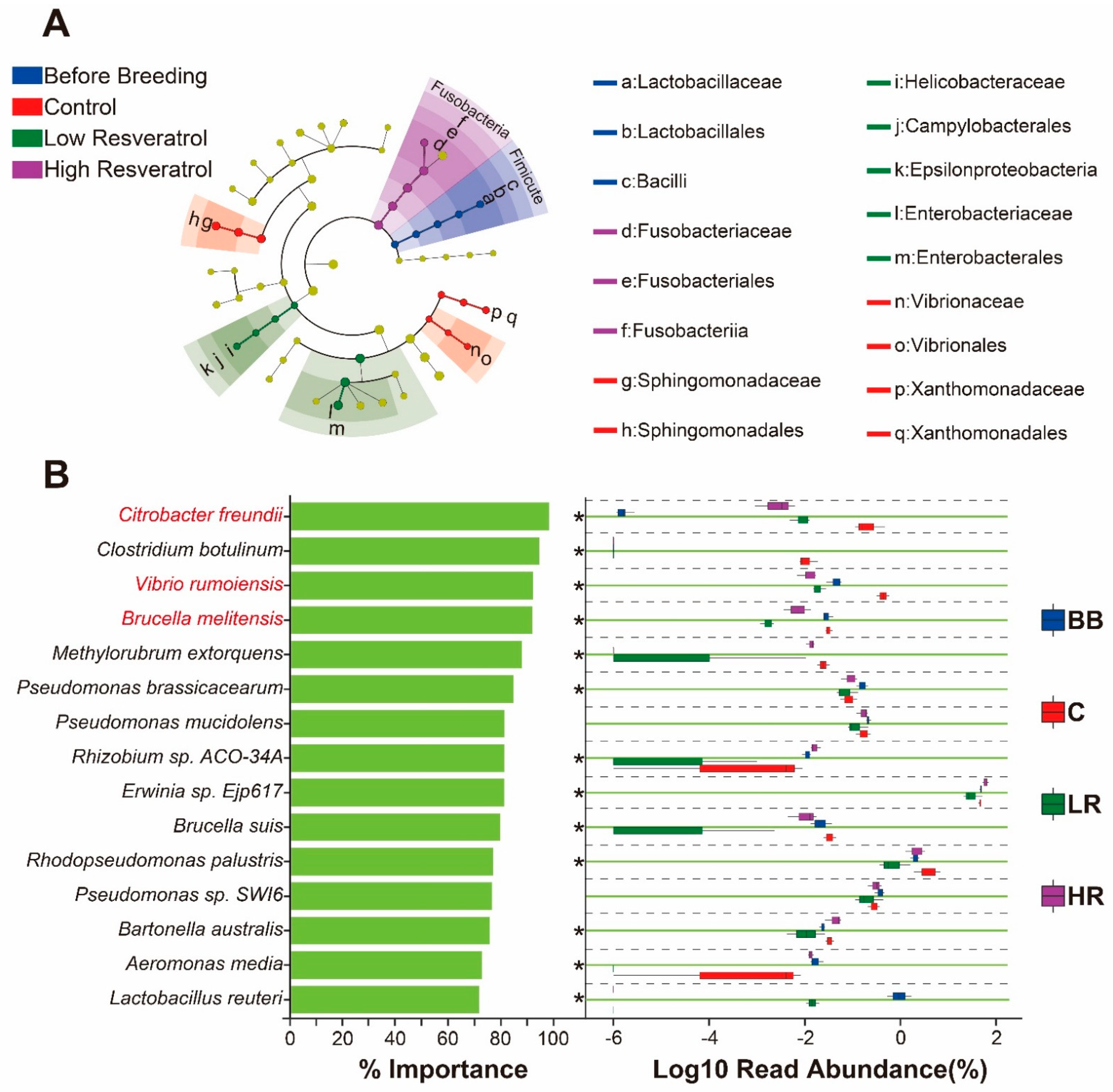
2.5. Resveratrol Influenced the Abundance of Duodenum Microbiota of Siberian Sturgeon

The community abundance of the BB/LR/HR groups vs. the C group was analyzed in the duodenum and valvular intestine (Figure 5). In the duodenum, the abundance of Sphingomonas hengshuiensis, Sphingomonas sp. LK11 in the BB group was significantly decreased compared to that in the C group, but no community with the increased abundance was found. In the LR vs. C group, the abundance of Escherichia coli was significantly increased, while the abundance of Salmonella enterica and unclassified Enterobacterriaceae was observably decreased. In the HR vs. C group, the communities with the significantly increased abundance were unclassified Rhodobacter, Rhodopseudomonas palustris, and Ilyobacter polytropus, while the abundance of unclassified Fusobacterium was markedly decreased. By contrast, only unclassified Rhizobiaceae in the LR vs. C group showed significant increase in the community abundance in the valvular intestine.

2.6. Resveratrol Reduced the Abundance of Potential Pathogens in the Duodenum of Siberian Sturgeon

A cladogram of LEfSe was constructed to find the most crucial microbiota (microbial biomarkers) in the duodenum among different groups (Figure 6A). The most dominant microbe was the following families: Lactobacillaceae in the BB group; Sphingomonadaceae, Vibrionaceae, and Xanthomonadaceae in group C; Helicobacteraceae and Enterobacteriaceae in the LR group; and Fusobacteriaceae in the HR group. Random forest analysis was carried out to determine variable importance of the samples from the BB, C, LR, and HR groups (Figure 6B). The results indicated that the potential pathogens (Citrobacter freundii, Vibrio rumoiensis, and Brucella melitensis) were the critical bacterial species in all groups, and their abundances were markedly reduced in the LR and HR groups compared to the C group (p < 0.05).

3. Discussion

Resveratrol has been reported to function as a regulator for the intestinal microbial community and as a protector of the integrity of the intestinal barrier [24,25,26], protecting mainly through its anti-inflammatory modulation [27,28,29]. In aquatic animals [30], resveratrol improved the absorption of nutrients in the intestinal tissues of zebrafish by increasing the number of goblet cells as well as the width and height of the villi. This study showed that diet supplemented with resveratrol increased the feeding rate of Siberian sturgeon but did not influence the final body weight. Generally, increase in food intake directly relates body- weight gain, even obesity [31,32]. By contrast, one study reported that diet supplemented with resveratrol lessened the body mass and whole-body fat in blunt snout bream, highlighting the hypolipidemic effect of resveratrol [33]. Thus, we speculated that resveratrol, being a versatile dietary additive, not only improves the nutrient uptake but also prevents obesity in fish.
Digestive capacity of the intestine is critical for energy absorption [34], however, we currently have limited knowledge about the digestive capacity of the intestine in aquaculture species, especially in cold-water species. Digestive enzymes directly reflect the digestive capacity, and in this study, the activity of pepsin, α-amylase, and lipase was assayed in the duodenum and valvular intestine. The results showed that supplementing the diet with resveratrol significantly increased the activity of pepsin, α-amylase, and lipase in both the duodenum and valvular intestine of Siberian sturgeon. This result indicates that resveratrol supplementation improved digestive capacity by enhancing the activity of digestive enzymes, which led to the increased feeding rate (Figure 1B). Tagliazucchi et al., found that resveratrol could bind pepsin and change its three-dimensional conformation to make it more active in pork [35]; this may be the reason why resveratrol increased pepsin activity in this study, while the reason for the ability of resveratrol to increase α-amylase and lipase activity remains to be further investigated.
The intestinal lining, which is in direct contact with the external environment, may become a major site for colonization and growth of microbiota [36,37]. In aquaculture species, intestinal microbiota is of significance to maintaining homeostasis [38,39]. In this study, the composition and abundance of microbial communities were evaluated and showed distinct diversity among different groups (Figure 3, Figure 4 and Figure 5). More specifically, supplementing the diet with resveratrol significantly influenced the abundance and composition of the bacterial community in the duodenum of Siberian sturgeon. After feeding with resveratrol, the community abundance of unclassified Rhodobacter, Rhodopseudomonas palustris, Ilyobacter polytropus, and Escherichia coli were significantly elevated, while the community abundance of unclassified Fusobacterium, Salmonella enterica, and unclassified Enterobacteriaceae were distinctly reduced in the duodenum (Figure 5). Among the microbiota of significantly elevated abundance, unclassified Rhodobacter was not annotated to any species, but its members can exert hypocholesterolemic effects by assimilating cholesterol into bile and producing bile acids [40]. Rhodopseudomonas palustris has been applied as one of the potential probiotics in the feed industry due to its extraordinary metabolic versatility [41]. Ilyobacter polytropus may be related to the degradation of monomeric 3-hydroxybutyrate, thus promoting the utilization of butyrate in the organism [42]. Among the significantly reduced components of the microbiota, the genus Fusobacterium has been proven to be a potentially pathogenic bacterium and implicated in human colorectal cancer [43,44]; Salmonella enterica is a well-versed pathogenic that can cause death in humans and animals through intestinal infection [45,46]; and unclassified Enterobacteriaceae was not mapped to any species, but Enterobacteriaceae has also received much concern for its extensive drug-resistance [47]. Therefore, our study revealed that resveratrol increased several intestinal probiotic bacteria (unclassified Rhodobacter, Rhodopseudomonas palustris, and Ilyobacter polytropus) and decreased several potentially pathogenic bacteria (Fusobacterium, Salmonella enterica, etc.), and was thereby associated with positive regulation of the microbial community in the duodenum of Siberian sturgeon.
In this study, the LEfSe algorithm and random forest analysis were constructed to determine microbial markers of each group and variable importance of all the samples, respectively, in the duodenum. The results revealed that the Citrobacter freundii, Vibrio rumoiensis, and Brucella melitensis were the most important bacterial species (could distinguish resveratrol-feeding groups from control group with the highest accuracy), and their abundance was significantly decreased in the duodenum after feeding with resveratrol (Figure 6B). These components of the microbiota were identified as the potential pathogens [48,49,50], indicating that resveratrol could reduce the composition of the pathogen community, consistent with previous studies [51,52]. The valvular intestine is a special structure of sturgeon, alongside the duodenum, to digest and absorb nutrients from food [53], but no significant influence of resveratrol was found on the valvular intestine microbiota of Siberian sturgeon. Thus, resveratrol may specifically influence the microbial community of the duodenum, but the mechanism is needed to be investigated in the future.

4. Materials and Methods

4.1. Fish and Experimental Design

A total of 300 fish (10-month-old, 248.1 ± 5.9 g) were purchased from a sturgeon farm (Tianquan County Chuanze Fishery Co., Ltd., Ya’an, China) and randomly assigned to circular plastic tanks (1.5 m in diameter and 1 m in height) for one week of acclimatization. The sturgeons were reared in flowing water, the water temperature, dissolved oxygen, pH, ammonia nitrogen, and nitrite were maintained at 16.0 ± 0.5 °C, 8.0 ± 0.6 mg/L, 7.6 ± 0.2, ≤ 0.01 mg/L, and ≤ 0.05 mg/L, respectively. The sturgeon were fed with commercial feed (Haida Group Co., Ltd., Guangdong, China) 3 times per day (at 8:00 am, 14:00 pm, and 20:00 pm, respectively), each fed 1% of the total fish weight, and after 1 h, the residual feed was collected and weighed after drying to calculate the feeding rate. Feeding rate (%) = (1 − average residual feed/average feeding feed) × 100; average residual feed (g/fish/day) = [ k = 1 n residual   feed   per   day / fish   number k]/n, n = day number of each group; average feeding feed (g/fish/day) = [ k = 1 n feeding   feed   per   day / fish   number k]/n, n = day number of each group. The fish number was dynamically counted based on the actual survival number.
After the acclimatization, 30 fishes (divided into 3 replicates) were randomly selected from the 300 fish and set as the before breeding (BB) group, while the rest of the fish were randomly assigned to 3 groups: control (C) group, fed with commercial feed; LR group, diet supplemented with low-dose resveratrol (0.16 mg/kg); HR group, diet supplemented with high-dose resveratrol (0.32 mg/kg). Each group contained 3 tanks, 30 fishes per tank (Figure 1A). Resveratrol (purity ≥ 99%) was purchased from Macklin Biochemical Co., Ltd (Shanghai, China). All animal handling procedures were approved by the Animal Care and Use Committee of Sichuan Agricultural University, following the guidelines of animal experiments of Sichuan Agricultural University under permit number 015-01521300.

4.2. Sample Collection, DNA Isolation, Library Construction, and Sequencing

At the end of the feeding, the duodena and valvular intestines of nine fish from each group were collected and fixed in 10% neutral formalin buffer for at least 24 h for histological observation. For microbiome analysis, nine fish were randomly selected from each group and anesthetized using 200 mg/L MS-222 (Jinjiang Aquatic Supplies Co., Ltd., Fujian, China). Intestinal tissues, containing duodena and valvular fragments, were collected under aseptic condition and then stored at −80 °C. Genomic DNA was extracted from the intestinal bacteria using a bacterial DNA isolation kit (Foregene, Chengdu, China). After the extraction, DNA fragments from the samples were amplified using specific primers: 8F: 5′-AGAGTTTGATCATGGCTCAG-3′, 1492R: 5′-CGGTTACCTTGTTACGACTT-3′. The PCR products were detected by electrophoresis in a 1% agarose gel, purified using a Zymoclean Gel Recovery Kit (D4008), quantified by the QuantiFluorTM-ST Blue Quantitative Fluorescence System (Promega, Beijing, China), and then subjected to next-generation sequencing. V3-V4 amplicon libraries were constructed and pair-end (PE) sequencing was performed on the MiSeq system (Illumina, San Diego, CA, USA). Based on the overlapping PE reads, pairs of reads were merged into sequences using Flash software (version: 1.2.11, http://ccb.jhu.edu/software/FLASH/index.shtml (accessed on 9 August 2021)). To further analyse, the following sequences were excluded from the dataset: (i) shorter than 1400 bp, (ii) greater than 1600 bp in the library, or (iii) contained 10 consecutive identical bases.

4.3. Taxonomy Classification and Differential Abundance Analysis

The acquired sequences were clustered into operational taxonomic units (OTUs) using UPARSE (http://drive5.com/uparse (accessed on 15 August 2021)). Each OTU was compared with the 16s rRNA database (Silva) to obtain classification information of the species. The RDP Classifier Bayes algorithm was used to classify and analyze the OTU representative sequences with 97% similarity, and the corresponding sequences were assigned to each classification level: kingdom, phylum, class, order, family, genus, and species. In addition, alpha diversity, including Shannon, Simpson, Chao and Coverage indices were calculated by Mothur. Hierarchical clustering analysis was performed based on the beta diversity distance matrix, and the UPGMA algorithm was used to construct clustering trees to compare the similarity of community composition. Species with relative abundance less than 0.01 in all samples were categorized as “other”. Differential species analysis was performed by LEfse tool (https://bitbucket.org/biobakery/biobakery/wiki/Home (accessed on 18 August 2021)), random forest analysis was carried out with the randomForest package in R studio, and community function prediction was conducted based on the functional gene profile of microorganisms in the KEGG database.

4.4. Histological Observation

The fixed intestinal tissues were dehydrated, transparentized with xylene, and embedded in paraffin wax. The solidified wax blocks were cut into 5-mm slices and mounted on slides for hematoxylin and eosin (H&E) staining. After staining, the slides were observed under optical microscope (Nikon, Tokyo, Japan). To evaluate the effects of resveratrol on the intestine of Siberian sturgeon, the intestinal parameters (mucosal thickness, muscular layer thickness, and villus height) were assessed with reference to the previous study. The thicknesses of the mucosal layer and muscular layer were defined as the average of five measurements per fish. The villus height was measured from the tip of the villus to the base, five measurements per sample.

4.5. Digestive Enzyme Activity Assay of Intestine

The duodenum and valvular fragments of the intestinal tissue were ground with buffer solution using commercial kits (A080-1-1, C016-1-1, A054-1-1, Nanjing Jiancheng Bioengineering Institute, Nanjing, China). The activity of pepsin, α-amylase, and lipase was assayed with the above-mentioned commercial kits. All operations were performed in strict accordance with the manufacturer’s description.

4.6. Statistical Analysis

Data were expressed as mean ± SD. Statistical analysis was performed using one-way ANOVA in SPSS version 27.0 software. Least Significant Difference (LSD) was used to analyze the differences between BB/LR/HR and C groups. Principal coordinates analysis was used to cluster different samples according to explanatory parameters. The p-value less than 0.05 was considered as the statistical significance.

5. Conclusions

In conclusion, the elevated activity of digestive enzymes and the increased height of intestinal villi were found after feeding with resveratrol in Siberian sturgeon. The function of resveratrol to inhibit the abundance of potentially pathogenic bacteria in the duodenum was also revealed. These results indicate that resveratrol may improve intestinal digestion and protect the microbial barrier. The information presented in the present work will be helpful to further the study of the intestinal function of Siberian sturgeon and expand the application of resveratrol in aquaculture.

Supplementary Materials

The following supporting information can be downloaded at: https://www.mdpi.com/article/10.3390/ijms231911977/s1.

Author Contributions

Conceptualization, S.Y.; methodology, S.Y.; software, Y.L.; validation, L.F.; investigation, W.X., L.F., C.Z., C.Y., and J.Z.; resources, J.L., T.Y., Z.H., X.D., and Z.D.; resources; writing—original draft preparation, W.X.; writing—review and editing, W.L., Y.L.; supervision, X.H., J.W., Y.L.; project administration, Y.L.; funding acquisition, S.Y. All authors have read and agreed to the published version of the manuscript.

Funding

This study was supported by the Natural Science Foundation of Sichuan Province of China (2022NSFSC0070), the Sichuan Science and Technology Program (2021YFYZ0015), Project of Sichuan Innovation Team of National Modern Agricultural Industry Technology System, Science and Technology Achievement Transfer and Transformation Demonstration Project of Sichuan Province (2021ZHCG0065), Youth Foundation of Natural Science of Sichuan Province (2022NSFSC1723), and Project of Sichuan Innovation Team of National Modern Agricultural Industry Technology System: 2022-2025.

Institutional Review Board Statement

All animal handling procedures were approved by the Animal Care and Use Committee of Sichuan Agricultural University, following the guidelines of animal experiments of Sichuan Agricultural University under permit number 015-01521300.

Informed Consent Statement

Not applicable.

Data Availability Statement

The raw 16S rRNA sequencing data have been submitted to the Sequence Read Archive (SRA) database of the National Center for Biotechnology Information (NCBI) with accession number PRJNA863445.

Acknowledgments

We are grateful to the Rhonin Biosciences Co., Ltd. for performing high throughput sequencing and assisting with bioinformatics analysis. Thanks to all authors for their contribution to this article. All authors read and approved the final manuscript.

Conflicts of Interest

The authors declare no conflict of interest.

References

  1. Biltueva, L.S.; Prokopov, D.Y.; Romanenko, S.A.; Interesova, E.A.; Schartl, M.; Trifonov, V.A. Chromosome Distribution of Highly Conserved Tandemly Arranged Repetitive DNAs in the Siberian Sturgeon (Acipenser baerii). Genes 2020, 11, 1375. [Google Scholar] [CrossRef] [PubMed]
  2. Bronzi, P.; Rosenthal, H.; Gessner, J. Global sturgeon aquaculture production: An overview. J. Appl. Ichthyol. 2011, 27, 169–175. [Google Scholar] [CrossRef]
  3. Abdolahnejad, Z.; Pourkazemi, M.; Khoshkholgh, M.R.; Yarmohammadi, M. Expression of growth hormone gene during early development of Siberian sturgeon (Acipenser baerii). Mol. Biol. Res. Commun. 2015, 4, 181–188. [Google Scholar] [PubMed]
  4. Pavasovic, M.; Richardson, N.A.; Anderson, A.J.; Mann, D.; Mather, P.B. Effect of pH, temperature and diet on digestive enzyme profiles in the mud crab, Scylla serrata. Aquaculture 2004, 242, 641–654. [Google Scholar] [CrossRef]
  5. Garrett, W.S.; Gordon, J.I.; Glimcher, L.H. Homeostasis and inflammation in the intestine. Cell 2010, 140, 859–870. [Google Scholar] [CrossRef] [Green Version]
  6. Saad, R.; Rizkallah, M.R.; Aziz, R.K. Gut Pharmacomicrobiomics: The tip of an iceberg of complex interactions between drugs and gut-associated microbes. Gut Pathog. 2012, 4, 16. [Google Scholar] [CrossRef] [Green Version]
  7. Wahlström, A.; Sayin, S.I.; Marschall, H.U.; Bäckhed, F. Intestinal Crosstalk between Bile Acids and Microbiota and Its Impact on Host Metabolism. Cell Metab. 2016, 24, 41–50. [Google Scholar] [CrossRef] [Green Version]
  8. Yang, S.; Zhang, C.; Xu, W.; Li, D.; Feng, Y.; Wu, J.; Luo, W.; Du, X.; Du, Z.; Huang, X. Heat Stress Decreases Intestinal Physiological Function and Facilitates the Proliferation of Harmful Intestinal Microbiota in Sturgeons. Front. Microbiol. 2022, 13, 755369. [Google Scholar] [CrossRef]
  9. Hieu, D.Q.; Hang, B.T.B.; Lokesh, J.; Garigliany, M.M.; Huong, D.T.T.; Yen, D.T.; Liem, P.T.; Tam, B.M.; Hai, D.M.; Son, V.N.; et al. Salinity significantly affects intestinal microbiota and gene expression in striped catfish juveniles. Appl. Microbiol. Biotechnol. 2022, 106, 3245–3264. [Google Scholar] [CrossRef]
  10. Deng, Y.; Mao, C.; Lin, Z.; Su, W.; Cheng, C.; Li, Y.; Gu, Q.; Gao, R.; Su, Y.; Feng, J. Nutrients, temperature, and oxygen mediate microbial antibiotic resistance in sea bass (Lateolabrax maculatus) ponds. Sci. Total Environ. 2022, 819, 153120. [Google Scholar] [CrossRef]
  11. Xia, J.H.; Lin, G.; Fu, G.H.; Wan, Z.Y.; Lee, M.; Wang, L.; Liu, X.J.; Yue, G.H. The intestinal microbiome of fish under starvation. BMC Genom. 2014, 15, 266. [Google Scholar] [CrossRef] [PubMed] [Green Version]
  12. Yúfera, M.; Nguyen, M.V.; Navarro-Guillén, C.; Moyano, F.J.; Jordal, A.O.; Espe, M.; Conceição, L.E.C.; Engrola, S.; Le, M.H.; Rønnestad, I. Effect of increased rearing temperature on digestive function in cobia early juvenile. Comp. Biochem. Physiology. Part A Mol. Integr. Physiol. 2019, 230, 71–80. [Google Scholar] [CrossRef] [PubMed]
  13. Hani, Y.M.I.; Marchand, A.; Turies, C.; Kerambrun, E.; Palluel, O.; Bado-Nilles, A.; Beaudouin, R.; Porcher, J.M.; Geffard, A.; Dedourge-Geffard, O. Digestive enzymes and gut morphometric parameters of threespine stickleback (Gasterosteus aculeatus): Influence of body size and temperature. PLoS ONE 2018, 13, e0194932. [Google Scholar] [CrossRef] [PubMed] [Green Version]
  14. Butt, R.L.; Volkoff, H. Gut Microbiota and Energy Homeostasis in Fish. Front. Endocrinol. 2019, 10, 9. [Google Scholar] [CrossRef] [Green Version]
  15. Jeandet, P.; Bessis, R.; Maume, B.F.; Meunier, P.; Peyron, D.; Trollat, P. Effect of Enological Practices on the Resveratrol Isomer Content of Wine. J. Agric. Food Chem. 1995, 43, 316–319. [Google Scholar] [CrossRef]
  16. Piotrowska, H.; Kucinska, M.; Murias, M. Biological activity of piceatannol: Leaving the shadow of resveratrol. Mutat. Res. 2012, 750, 60–82. [Google Scholar] [CrossRef]
  17. Brockmueller, A.; Shayan, P.; Shakibaei, M. Evidence That β1-Integrin Is Required for the Anti-Viability and Anti-Proliferative Effect of Resveratrol in CRC Cells. Int. J. Mol. Sci. 2022, 23, 4714. [Google Scholar] [CrossRef]
  18. Moreira, H.; Szyjka, A.; Grzesik, J.; Pelc, K.; Żuk, M.; Kulma, A.; Emhemmed, F.; Muller, C.D.; Gąsiorowski, K.; Barg, E. Celastrol and Resveratrol Modulate SIRT Genes Expression and Exert Anticancer Activity in Colon Cancer Cells and Cancer Stem-like Cells. Cancers 2022, 14, 1372. [Google Scholar] [CrossRef]
  19. Cesmeli, S.; Goker Bagca, B.; Caglar, H.O.; Ozates, N.P.; Gunduz, C.; Biray Avci, C. Combination of resveratrol and BIBR1532 inhibits proliferation of colon cancer cells by repressing expression of LncRNAs. Med. Oncol. 2021, 39, 12. [Google Scholar] [CrossRef]
  20. Brockmueller, A.; Mueller, A.L.; Shayan, P.; Shakibaei, M. β1-Integrin plays a major role in resveratrol-mediated anti-invasion effects in the CRC microenvironment. Front. Pharmacol. 2022, 13, 978625. [Google Scholar] [CrossRef]
  21. Wang, P.; Li, D.; Ke, W.; Liang, D.; Hu, X.; Chen, F. Resveratrol-induced gut microbiota reduces obesity in high-fat diet-fed mice. Int. J. Obes. 2020, 44, 213–225. [Google Scholar] [CrossRef] [PubMed]
  22. Fu, Q.; Tan, Z.; Shi, L.; Xun, W. Resveratrol Attenuates Diquat-Induced Oxidative Stress by Regulating Gut Microbiota and Metabolome Characteristics in Piglets. Front. Microbiol. 2021, 12, 695155. [Google Scholar] [CrossRef] [PubMed]
  23. Zheng, Y.; Wu, W.; Hu, G.; Qiu, L.; Meng, S.; Song, C.; Fan, L.; Zhao, Z.; Bing, X.; Chen, J. Gut microbiota analysis of juvenile genetically improved farmed tilapia (Oreochromis niloticus) by dietary supplementation of different resveratrol concentrations. Fish Shellfish Immunol. 2018, 77, 200–207. [Google Scholar] [CrossRef]
  24. Zhang, B.; Xu, Y.; Lv, H.; Pang, W.; Wang, J.; Ma, H.; Wang, S. Intestinal pharmacokinetics of resveratrol and regulatory effects of resveratrol metabolites on gut barrier and gut microbiota. Food Chem. 2021, 357, 129532. [Google Scholar] [CrossRef]
  25. Pan, H.H.; Zhou, X.X.; Ma, Y.Y.; Pan, W.S.; Zhao, F.; Yu, M.S.; Liu, J.Q. Resveratrol alleviates intestinal mucosal barrier dysfunction in dextran sulfate sodium-induced colitis mice by enhancing autophagy. World J. Gastroenterol. 2020, 26, 4945–4959. [Google Scholar] [CrossRef] [PubMed]
  26. Cai, T.T.; Ye, X.L.; Li, R.R.; Chen, H.; Wang, Y.Y.; Yong, H.J.; Pan, M.L.; Lu, W.; Tang, Y.; Miao, H.; et al. Resveratrol Modulates the Gut Microbiota and Inflammation to Protect Against Diabetic Nephropathy in Mice. Front. Pharmacol. 2020, 11, 1249. [Google Scholar] [CrossRef] [PubMed]
  27. Buhrmann, C.; Shayan, P.; Brockmueller, A.; Shakibaei, M. Resveratrol Suppresses Cross-Talk between Colorectal Cancer Cells and Stromal Cells in Multicellular Tumor Microenvironment: A Bridge between In Vitro and In Vivo Tumor Microenvironment Study. Molecules 2020, 25, 4292. [Google Scholar] [CrossRef]
  28. Qian, Y.; Wang, R.; Wei, W.; Wang, M.; Wang, S. Resveratrol reverses the cadmium-promoted migration, invasion, and epithelial-mesenchymal transition procession by regulating the expression of ZEB1. Hum. Exp. Toxicol. 2021, 40, S331–S338. [Google Scholar] [CrossRef]
  29. Alrafas, H.R.; Busbee, P.B.; Chitrala, K.N.; Nagarkatti, M.; Nagarkatti, P. Alterations in the Gut Microbiome and Suppression of Histone Deacetylases by Resveratrol Are Associated with Attenuation of Colonic Inflammation and Protection Against Colorectal Cancer. J. Clin. Med. 2020, 9, 1796. [Google Scholar] [CrossRef]
  30. Faal, M.; Manouchehri, H.; Changizi, R.; Bootorabi, F.; Khorramizadeh, M.R. Assessment of resveratrol on diabetes of zebrafish (Danio rerio). J. Diabetes Metab. Disord. 2022, 21, 823–833. [Google Scholar] [CrossRef]
  31. Maruyama, K.; Sato, S.; Ohira, T.; Maeda, K.; Noda, H.; Kubota, Y.; Nishimura, S.; Kitamura, A.; Kiyama, M.; Okada, T.; et al. The joint impact on being overweight of self reported behaviours of eating quickly and eating until full: Cross sectional survey. BMJ 2008, 337, a2002. [Google Scholar] [CrossRef] [PubMed] [Green Version]
  32. Kim, D.; Hou, W.; Wang, F.; Arcan, C. Factors Affecting Obesity and Waist Circumference among US Adults. Prev. Chronic Dis. 2019, 16, E02. [Google Scholar] [CrossRef] [PubMed] [Green Version]
  33. Zhang, D.; Yan, Y.; Tian, H.; Jiang, G.; Li, X.; Liu, W. Resveratrol supplementation improves lipid and glucose metabolism in high-fat diet-fed blunt snout bream. Fish Physiol. Biochem. 2018, 44, 163–173. [Google Scholar] [CrossRef] [PubMed]
  34. Wierdsma, N.J.; Peters, J.H.; van Bokhorst-de van der Schueren, M.A.; Mulder, C.J.; Metgod, I.; van Bodegraven, A.A. Bomb calorimetry, the gold standard for assessment of intestinal absorption capacity: Normative values in healthy ambulant adults. J. Hum. Nutr. Diet. 2014, 27, 57–64. [Google Scholar] [CrossRef] [PubMed]
  35. Tagliazucchi, D.; Verzelloni, E.; Conte, A. Effect of some phenolic compounds and beverages on pepsin activity during simulated gastric digestion. J. Agric. Food Chem. 2005, 53, 8706–8713. [Google Scholar] [CrossRef] [PubMed]
  36. Paone, P.; Cani, P.D. Mucus barrier, mucins and gut microbiota: The expected slimy partners? Gut 2020, 69, 2232–2243. [Google Scholar] [CrossRef]
  37. Vaishnava, S.; Yamamoto, M.; Severson, K.M.; Ruhn, K.A.; Yu, X.; Koren, O.; Ley, R.; Wakeland, E.K.; Hooper, L.V. The antibacterial lectin RegIIIgamma promotes the spatial segregation of microbiota and host in the intestine. Science 2011, 334, 255–258. [Google Scholar] [CrossRef] [Green Version]
  38. Ni, J.; Yan, Q.; Yu, Y.; Zhang, T. Factors influencing the grass carp gut microbiome and its effect on metabolism. FEMS Microbiol. Ecol. 2014, 87, 704–714. [Google Scholar] [CrossRef]
  39. Semova, I.; Carten, J.D.; Stombaugh, J.; Mackey, L.C.; Knight, R.; Farber, S.A.; Rawls, J.F. Microbiota regulate intestinal absorption and metabolism of fatty acids in the zebrafish. Cell Host Microbe 2012, 12, 277–288. [Google Scholar] [CrossRef] [Green Version]
  40. Afrose, S.; Hossain, M.S.; Salma, U.; Miah, A.G.; Tsujii, H. Dietary Karaya Saponin and Rhodobacter capsulatus Exert Hypocholesterolemic Effects by Suppression of Hepatic Cholesterol Synthesis and Promotion of Bile Acid Synthesis in Laying Hens. Cholesterol 2010, 2010, 272731. [Google Scholar] [CrossRef]
  41. Chen, Y.Y.; Wang, Y.L.; Wang, W.K.; Zhang, Z.W.; Si, X.M.; Cao, Z.J.; Li, S.L.; Yang, H.J. Beneficial effect of Rhodopseudomonas palustris on in vitro rumen digestion and fermentation. Benef. Microbes 2020, 11, 91–99. [Google Scholar] [CrossRef] [PubMed]
  42. Sikorski, J.; Chertkov, O.; Lapidus, A.; Nolan, M.; Lucas, S.; Del Rio, T.G.; Tice, H.; Cheng, J.F.; Tapia, R.; Han, C.; et al. Complete genome sequence of Ilyobacter polytropus type strain (CuHbu1). Stand. Genom. Sci. 2010, 3, 304–314. [Google Scholar] [CrossRef] [PubMed] [Green Version]
  43. Brennan, C.A.; Garrett, W.S. Fusobacterium nucleatum—symbiont, opportunist and oncobacterium. Nat. Rev. Microbiol. 2019, 17, 156–166. [Google Scholar] [CrossRef] [PubMed]
  44. Allen-Vercoe, E.; Strauss, J.; Chadee, K. Fusobacterium nucleatum: An emerging gut pathogen? Gut Microbes 2011, 2, 294–298. [Google Scholar] [CrossRef] [PubMed]
  45. Heymans, R.; Vila, A.; van Heerwaarden, C.A.M.; Jansen, C.C.C.; Castelijn, G.A.A.; van der Voort, M.; Biesta-Peters, E.G. Rapid detection and differentiation of Salmonella species, Salmonella Typhimurium and Salmonella Enteritidis by multiplex quantitative PCR. PLoS ONE 2018, 13, e0206316. [Google Scholar] [CrossRef] [Green Version]
  46. Jajere, S.M. A review of Salmonella enterica with particular focus on the pathogenicity and virulence factors, host specificity and antimicrobial resistance including multidrug resistance. Vet. World 2019, 12, 504–521. [Google Scholar] [CrossRef] [Green Version]
  47. Magiorakos, A.P.; Srinivasan, A.; Carey, R.B.; Carmeli, Y.; Falagas, M.E.; Giske, C.G.; Harbarth, S.; Hindler, J.F.; Kahlmeter, G.; Olsson-Liljequist, B.; et al. Multidrug-resistant, extensively drug-resistant and pandrug-resistant bacteria: An international expert proposal for interim standard definitions for acquired resistance. Clin. Microbiol. Infect. 2012, 18, 268–281. [Google Scholar] [CrossRef] [Green Version]
  48. Yang, J.; Tian, T.; Xiao, K.; Zeng, Q.; Tan, C.; Du, H. Pathogenic infection and immune-related gene expression of Chinese sturgeon (Acipenser sinensis) challenged by Citrobacter freundii. Dev. Comp. Immunol. 2021, 114, 103872. [Google Scholar] [CrossRef]
  49. Ji, Q.; Wang, S.; Ma, J.; Liu, Q. A review: Progress in the development of fish Vibrio spp. vaccines. Immunol. Lett. 2020, 226, 46–54. [Google Scholar] [CrossRef]
  50. El-Tras, W.F.; Tayel, A.A.; Eltholth, M.M.; Guitian, J. Brucella infection in fresh water fish: Evidence for natural infection of Nile catfish, Clarias gariepinus, with Brucella melitensis. Vet. Microbiol. 2010, 141, 321–325. [Google Scholar] [CrossRef]
  51. Vestergaard, M.; Ingmer, H. Antibacterial and antifungal properties of resveratrol. Int. J. Antimicrob. Agents 2019, 53, 716–723. [Google Scholar] [CrossRef] [PubMed]
  52. Okamoto-Shibayama, K.; Yoshida, A.; Ishihara, K. Inhibitory Effect of Resveratrol on Candida albicans Biofilm Formation. Bull. Tokyo Dent. Coll. 2021, 62, 1–6. [Google Scholar] [CrossRef] [PubMed]
  53. Chen, D.; Peng, S.; Chen, D.; Yang, F.; Liu, J.; Wang, J.; Liu, Q.; Huang, X.; Ouyang, P.; Wang, K.; et al. Low lethal doses of Streptococcus iniae caused enteritis in Siberian sturgeon (Acipenser baerii). Fish Shellfish Immunol. 2020, 104, 654–662. [Google Scholar] [CrossRef] [PubMed]
Figure 1. Experimental flowchart, feeding rate, and histological observation ofSiberian sturgeon. (A) Experimental flowchart, sampling at day 7 indicates before breeding (BB) group, sampling at day 52 includes three groups: control (C), low resveratrol (LR), and high resveratrol (HR). (B) Feeding rate (%) = (1 − average residual feed/average feeding amount) × 100. (C) Representation of intestinal tissues from different groups. Intact tissues, containing duodenum and valvular intestine fragments, were sampled from each group at the termination of the feeding administration. The white arrow indicates intestinal contents. (D,E) Histological changes in the duodenum and valvular intestine. Sampled intestinal tissues from B were divided into duodenum and valvular intestine fragments that underwent H&E staining to observe histological change (n = 3). Representation of histological change is shown as follows: BB (a’ and a’’), C (b’ and b’’), LR (c’ and c’’), and HR (d’ and d’’). Red scale bars indicate length of intestinal villus; blue scale bars represent muscular layer thickness; and green scale bars indicate mucosal layer. (F,G) Thickness of mucosal layer, muscular layer, and intestinal villus length in different groups. * p < 0.05 vs. the control group; two-way ANOVA plus Bonferroni post-tests.
Ijms 23 11977 g001
Figure 2. Effects of resveratrol on the activity of intestinal digestive enzymes. (AC) Pepsin, α-amylase, lipase activity in the duodenum. (DF) Pepsin, α-amylase, lipase activity in the valvular intestine. * p < 0.05, ** p < 0.01 vs. the control group; one-way ANOVA plus Bonferroni post-tests.
Ijms 23 11977 g002
Figure 3. (A,B) represent hierarchical clustering trees of Siberian sturgeon duodenum and valvular families, respectively. Clustering analysis based on Bray–Curtis dissimilarity.
Ijms 23 11977 g003
Figure 4. Community composition analysis among different groups. (A,B) Venn diagrams of OTU numbers in different groups. (C,D) Statistics of species number among different groups, * p < 0.05 vs. the control group; one-way ANOVA plus Bonferroni post-tests.
Ijms 23 11977 g004
Figure 5. Comparison of microbial community between the treatment and control groups. The chart lists the top 10 abundant communities in the duodenum and valvular intestine in BB vs. C, LR vs. C, and HR vs. C groups. The data are presented as means ± standard deviation, Wilcoxon rank-sum test was applied to determine if differences between groups were significant (* p < 0.05). The communities with red and green fonts indicate significant increase and decrease in abundance, respectively.
Ijms 23 11977 g005
Figure 6. Identification of microbial markers for different groups by LEfSe analysis and random forest analysis for the samples from all groups in the duodenum. (A) Taxonomic cladogram generated from LEfSe analysis. Taxonomic characterization of differences in intestinal tissues among different groups; concentric circles from outside to inside indicate different taxonomic classes (phylum to family), and different color areas point to different groups. The abundance of intestinal microbiota was revealed by the size of each node. (B) Variable importance plot from random forest classifier. Left graph shows top 15 bacteria with % importance (mean decrease in Gini) ≥ 70 among different groups, bacteria marked in red font indicate potential pathogenic bacteria. Right figure represents abundance of corresponding communities by boxplots in different groups, with “*” on left side representing the difference significance of BB/LR/HR compared to C (p < 0.05).
Ijms 23 11977 g006
Table 1. The effect of resveratrol on the growth performance of Siberian sturgeon.
Sample CategoryReplicateIBW (g/fish)FBW (g/fish)PWG (%)
Before breedingBB1 248.3 ± 2.6 a//
BB2249.8 ± 5.3 a//
BB3251.2 ± 4.3 a//
ControlC1247.9 ± 5.1 a357.6 ± 8.6 b44.53 c
C2250.5 ± 2.9 a364.8 ± 7.0 b45.63 c
C3249.9 ± 6.5 a371.0 ± 15.6 b48.46 c
Low resveratrolLR1253.2 ± 3.8 a372.4 ± 13.5 b47.08 c
LR2252.3 ± 4.3 a369.1 ± 9.5 b46.29 c
LR3249.4 ± 1.6 a360.5 ± 6.3 b44.55 c
High resveratrolHR1255.1 ± 5.6 a378.5 ± 10.1 b48.37 c
HR2249.7 ± 5.8 a374.8 ± 13.0 b50.01 c
HR3251.9 ± 6.5 a366.1 ± 6.5 b45.34 c
Data are expressed as mean ± SD, and the different letters in the same column mean the significance (p < 0.05). IBW: initial body weight; FBW: final body weight; PWG: percent weight gain = (FBW − IBW)/IBW.
Publisher’s Note: MDPI stays neutral with regard to jurisdictional claims in published maps and institutional affiliations.

