Article

Genetic Study of Four Candidate Holliday Junction Processing Proteins in the Thermophilic Crenarchaeon Sulfolobus acidocaldarius

Department of Environmental Engineering for Symbiosis, Graduate School of Science and Engineering, Soka University, 1-236 Tangi-machi, Hachioji, Tokyo 192-8577, Japan
*
Author to whom correspondence should be addressed.
Current Address: Department of Bioscience and Biotechnology, Graduate School of Bioresource and Bioenvironmental Sciences, Kyushu University, Fukuoka 819-0395, Japan.
Int. J. Mol. Sci. 2022, 23(2), 707; https://doi.org/10.3390/ijms23020707
Submission received: 15 November 2021 / Revised: 24 December 2021 / Accepted: 6 January 2022 / Published: 9 January 2022
(This article belongs to the Special Issue Mechanisms of DNA Replication Fork Progression, Stalling, and Rescue)

Abstract

:
Homologous recombination (HR) is thought to be important for the repair of stalled replication forks in hyperthermophilic archaea. Previous biochemical studies identified two branch migration helicases (Hjm and PINA) and two Holliday junction (HJ) resolvases (Hjc and Hje) as HJ-processing proteins; however, due to the lack of genetic evidence, it is still unclear whether these proteins are actually involved in HR in vivo and how their functional relation is associated with the process. To address the above questions, we constructed hjc-, hje-, hjm-, and pina single-knockout strains and double-knockout strains of the thermophilic crenarchaeon Sulfolobus acidocaldarius and characterized the mutant phenotypes. Notably, we succeeded in isolating the hjm- and/or pina-deleted strains, suggesting that the functions of Hjm and PINA are not essential for cellular growth in this archaeon, as they were previously thought to be essential. Growth retardation in Δpina was observed at low temperatures (cold sensitivity). When deletion of the HJ resolvase genes was combined, Δpina Δhjc and Δpina Δhje exhibited severe cold sensitivity. Δhjm exhibited severe sensitivity to interstrand crosslinkers, suggesting that Hjm is involved in repairing stalled replication forks, as previously demonstrated in euryarchaea. Our findings suggest that the function of PINA and HJ resolvases is functionally related at lower temperatures to support robust cellular growth, and Hjm is important for the repair of stalled replication forks in vivo.

1. Introduction

Thermophiles inhabit a hot environment that accelerates the rate of DNA damage [1]. It has been proposed that DNA repair in thermophilic archaea has unique properties in order to be robust in extreme environments [2,3]. For example, no nucleotide excision repair function, which removes a broad spectrum of helix-distorting DNA lesions (such as UV-induced DNA damage, intrastrand crosslinks, and bulky adducts) has been identified in hyperthermophilic archaea; however, another DNA repair process has been proposed to have a homologous function [3,4,5]. An endonuclease XPF/Hef- and NucS/EndMS-deleted strain exhibits sensitivity to helix-distorting DNA lesions, suggesting that both endonucleases participate in homologous recombination (HR)-mediated stalled-fork DNA repair [3,4,5]. For these reasons, an opinion that this HR-mediated DNA repair serves as the major pathway for the removal of a wide variety of DNA lesions in hyperthermophilic archaea and is unusually efficient and reliable in comparison with other organisms has been argued [3,4,5]. Therefore, exploring the HR process in hyperthermophilic archaea is expected to expand our knowledge about the DNA repair mechanism in order to maintain genetic information.
HR is a fundamental mechanism that plays an important role in repairing stalled replication forks. The mechanism involves genetically exchanging homologous sequence processes to significantly enhance genome stability and genetic diversity. The process comprises four main steps. The first process is end resection catalyzed by the Rad50-Mre11-HerA-NurA complex producing 3′-single-stranded DNAs (ssDNAs). After end resection, ssDNA binding protein binds to 3′-ssDNA and protects the formation of the secondary structure of ssDNA [6,7]. In the second step, 3′-ssDNA is used for strand invasion, and the formation of four-stranded DNA is called the Holliday junction (HJ) catalyzed by recombinase RadA [8,9,10]. The third step is branch migration, in which the branch point of the HJ is moved by the activity of the branch migration helicases. In the final step, HJ resolvases bind to and cleave HJ [11,12], resulting in HJ resolution, and subsequent ligation ends the HR process. The role of putative branch migration helicases and HJ resolvases in HR in vivo and the functional relation of these proteins in vivo are not understood in detail (discussed in more detail below) [13,14,15,16].
To date, three helicases have been characterized as helicases with a branch migration activity (i.e., dissociation of a synthetic HJ to half junctions) in vitro in hyperthermophilic archaea. The orthologs of these proteins have also been found in most crenarchaeal and euryarchaeal species. Holliday junction migration (Hjm) from the hyperthermophilic euryarchaeon Pyrococcus furiosus was first discovered as a putative branch migration helicase with branch migration activity against a synthetic HJ [14]. In a previous study on the thermophilic crenarchaeon Sulfurisphaera tokodaii (formerly Sulfolobus tokodaii), Hjm has a fork regression activity against a chicken-foot structure known as the HJ in vitro [17]. Disruption of hjm led to lethality in the crenarchaeon “Sulfolobus islandicus” REY15A [18] (the double quotation marks indicate this species name has not been validated). In the hyperthermophilic euryarchaeon Thermococcus kodakarensis, the hjm knockout strain was constructed and was sensitive to UV irradiation and mitomycin C (MMC), respectively [4], suggesting that Hjm works at stalled replication forks to repair them. The second candidate is archaeal-long-helicase-related (Lhr1) [19,20]. Lhr1 from the euryarchaeon Methanothermobacter thermautotrophicus catalyzes branch migration of a synthetic HJ [20], and the thermophilic crenarchaeon Sulfolobus acidocaldarius Lhr1 (Saci_1500) unwinds a synthetic HJ, producing ssDNA [19]. However, the recombination frequency of the lhr1-deficient strain is identical to that of the parent strain [19], suggesting that the function of Lhr1 is not required for the HR process in vivo. As a third candidate, a recent study identified a novel helicase PilT N-terminal-(PIN)-domain-containing ATPase (PINA) from “S. islandicus” that promotes branch migration of a synthetic HJ in vitro [16]. PINA is likely to be responsible for branch migration in archaea, because it promotes branch migration, and its crystal structure is similar to RuvB, which is a branch migration helicase in bacteria [16]. A previous in vitro study proposed a putative role for Hjm and PINA in the regression of stalled replication fork, the formation of the chicken-foot structure, and HJ cleavage [21]. The branch-migration activity of the three candidates leads us to imagine that these helicases are involved in the repair of the stalled replication fork in vivo. However, it is unclear whether these candidates (especially Hjm and PINA) actually participate in the HR process in vivo because no genetic evidence has been provided.
HJ resolution is processed by two HJ resolvases, Hjc and Hje, in crenarchaeon. Hjc is conserved in all archaea, and Hje is a paralog of Hjc and is found in many species of the order Sulfolobales [12,22]. In thermophilic crenarchaeon, a biochemical study showed that the substrate specificity of Hjc and Hje are slightly different in Saccharolobus solfataricus (formerly Sulfolobus solfataricus), and a genetic study showed the hje-deficient strain of “S. islandicus” exhibited a high sensitivity to DNA-damaging agents HU, cisplatin, and MMS, which can cause replication fork-stalled and DNA double-strand breaks, while the hjc-deficient strain exhibited no sensitivity [22,23]. The cellular roles of Hjc and Hje seem to be different for HR-mediated DNA repair. However, similar to candidates for branch-migration helicase, it is still unclear whether both HJ resolvases are actually required for processive HR in vivo because no genetic evidence has been reported.
An in vitro study of Hjm and PINA has been reported; however, currently, hjm- and pina-deficient strains cannot be constructed in “S. islandicus”, and the sensitivity of mutants to DNA damage agents has not been examined [16,18]. In other words, the impact of Hjm and PINA on genome integrity has not been determined in detail. In addition, the relationship between Hje and these helicases is unclear [21]. The genetic study of two helicases (Hjm and PINA) and two nucleases (Hjc and Hje) will be useful for enhancing the understanding of the importance of branch migration and HJ resolution. In this study, we constructed hjm-, pina-, hjc-, and hje-knockout strains and double-knockout strains in S. acidocaldarius to investigate the functional role of Hjm and PINA in vivo, and the relationship between two helicases and two nucleases in archaeal HR.

2. Results

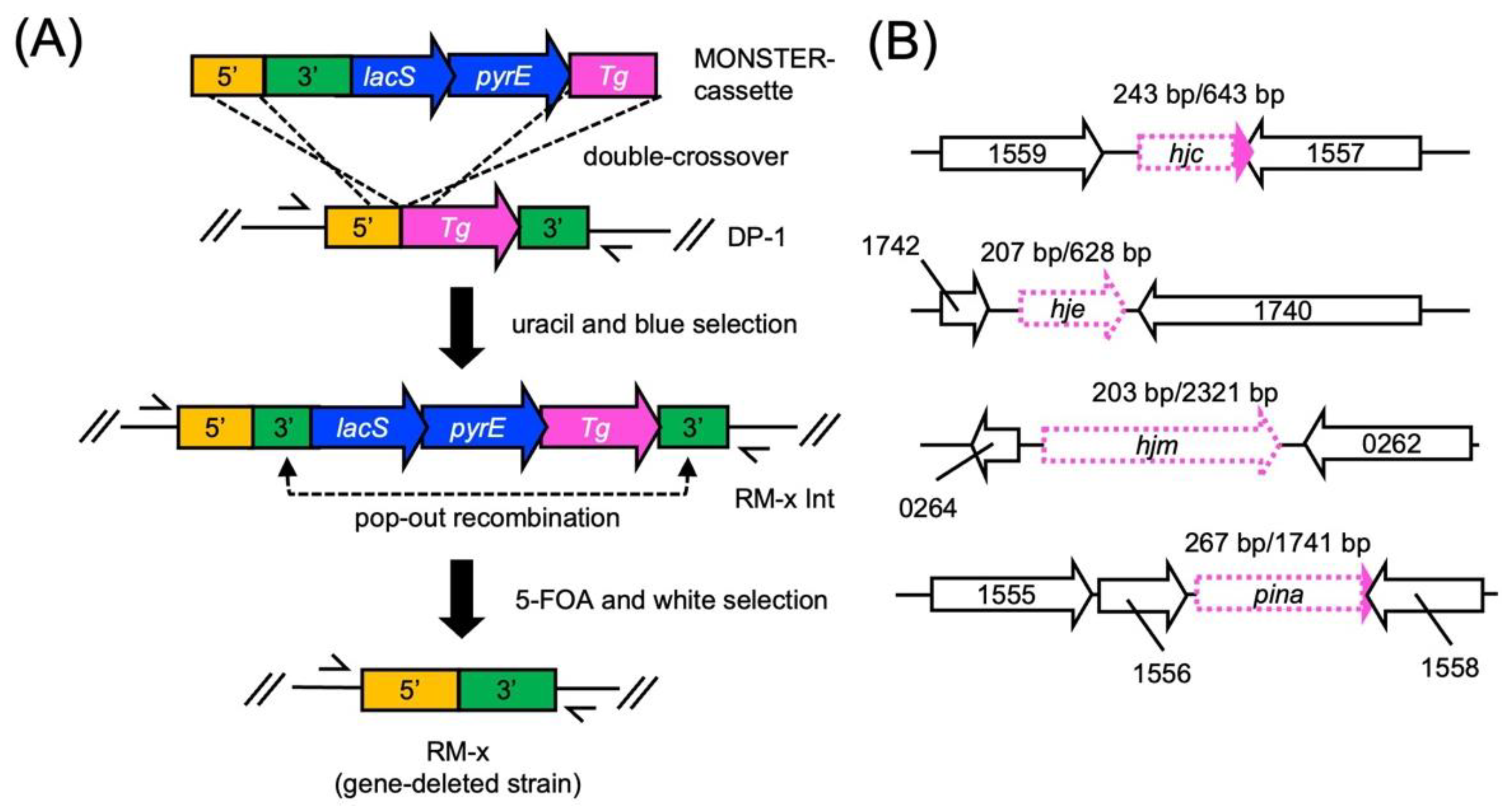
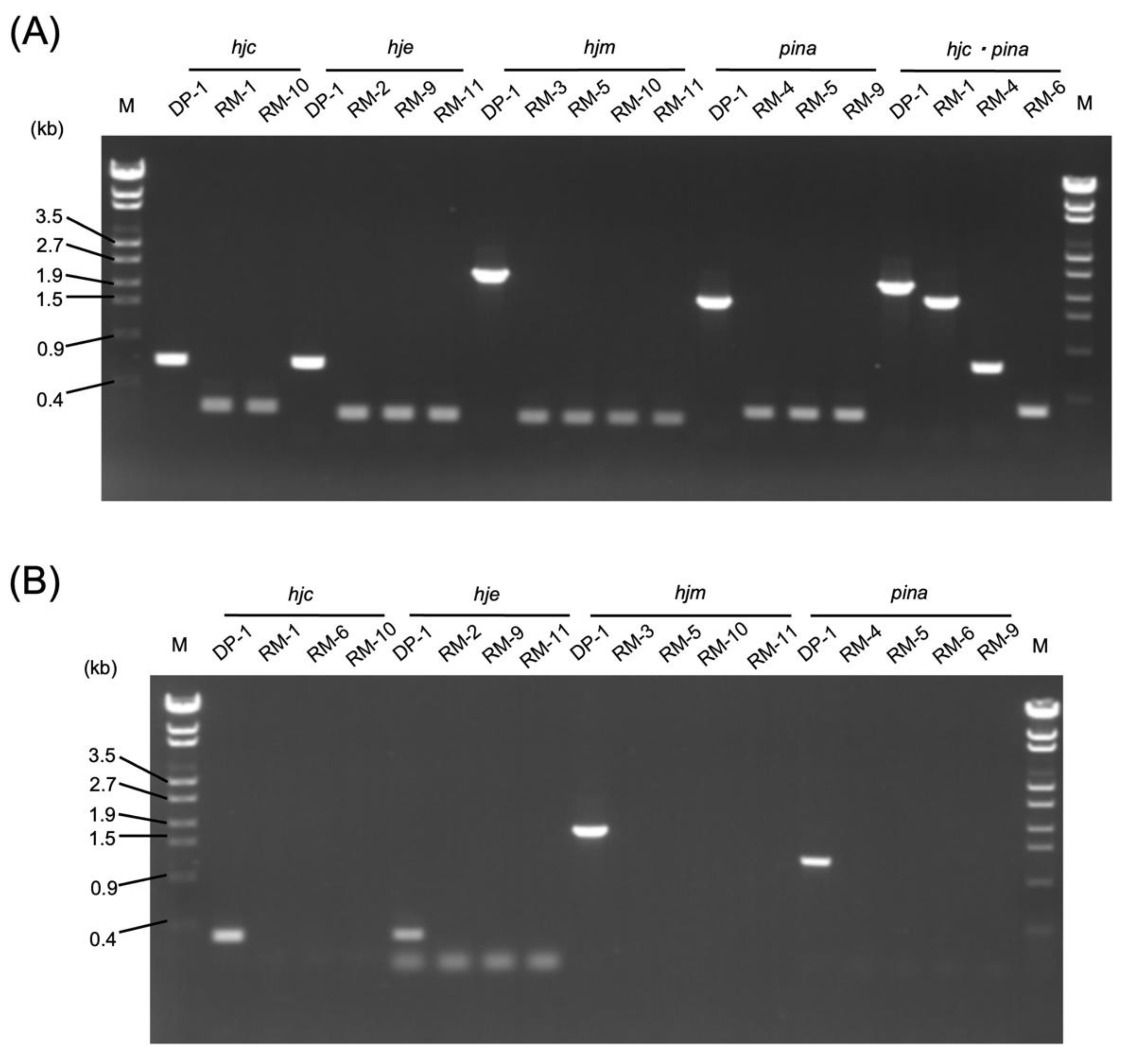
2.1. Construction of the hjc-, hje-, hjm-, and pina-Deleted Strains

The gene-deleted strains were constructed by MONSTER as an unmarked-gene-deletion method (Figure 1) [24]. After transformation, 57 colonies/µg MONSTER-hjc, 31 colonies/µg MONSTER-hje, 18 colonies/µg MONSTER-hjm, and 71 colonies/µg MONSTER-pina grew under uracil selection. After blue visualization using X-gal solution, one blue colony was purified using single-colony isolation.
PCR analysis using outer primers confirmed that the blue colonies were intermediate transformants (named RM-1 Int, RM-2 Int, RM-3 Int, and RM-4 Int). A total of 2–3 × 10⁸ RM-x Int cells were spread on an XTUF plate for pop-out recombination. X-gal visualization revealed that 12, 8, 20, and 11 white colonies grew by plating RM-1 Int, RM-2 Int, RM-3 Int, and RM-4 Int cells, respectively. Five white colonies were randomly selected for the PCR analysis using outer primers. The genotypes of these colonies demonstrated the expected 0.3 kb, 0.5 kb, 2.1 kb, and 1.4 kb deletions in the hjc, hje, hjm, and pina loci, respectively (Figure 2A). In addition, PCR analyses using inner primers yielded no product from the gene-deleted strains (Figure 2B), indicating that the target genes were deleted from the original genomic locus and were not translocated on their genomes.
The gene-deleted strains were designated the S. acidocaldarius strains RM-1 (ΔpyrE ΔsuaI Δphr Δhjc), RM-2 (ΔpyrE ΔsuaI Δphr Δhje), RM-3 (ΔpyrE ΔsuaI Δphr Δhjm), and RM-4 (ΔpyrE ΔsuaI Δphr Δpina). In addition, double-knockout strains RM-5 (ΔpyrE ΔsuaI Δphr Δpina Δhjm), RM-6 (ΔpyrE ΔsuaI Δphr Δpina Δhjc), RM-9 (ΔpyrE ΔsuaI Δphr Δpina Δhje), RM-10 (ΔpyrE ΔsuaI Δphr Δhjm Δhjc), and RM-11 (ΔpyrE ΔsuaI Δphr Δhjm Δhje) were constructed. However, we could not construct the Δhjc Δhje (double-knockout) strain. Thus, the redundant function of Hjc and Hje in the HJ resolution and the essentiality of HJ resolution for cellular viability is evident in S. acidocaldairus. This result is compatible with a previous knockout study of the hjc and hje genes in “S. islandicus” [23].

2.2. Growth Characteristics of the Gene-Deleted Strains

The growth of deletion strains (RM-1 (Δhjc), RM-2 (Δhje), RM-3 (Δhjm), RM-4 (Δpina), RM-5 (Δpina Δhjm), RM-6 (Δpina Δhjc), RM-9 (Δpina Δhje), RM-10 (Δhjm Δhjc), and RM-11 (Δhjm Δhje)) and a parental strain DP-1 in the liquid culture was compared over a wide temperature range (55, 65, and 75 °C) (Figure 3).
The growth of all deficient strains was almost normal (final cell density; 0.54 ± 0.043–0.48 ± 0.043, the cultivation time for OD600 = 0.1; 15 ± 3.0 h–27 ± 6.8 h) at 75 °C in comparison with that of the parent strain (final cell density; 0.64 ± 0.011, the cultivation time for OD600 = 0.1; 14 ± 3.9 h) (Figure 3A). Similarly, at 65 °C, the growth of all deficient strains was also the same as that of the parent strain (Figure 3B). Similarly, at 55 °C, no marked difference was observed between the growth of Δhjc, Δhje, Δhjm, Δhjm Δhjc, and Δhjm Δhje, and that of the parental strain (Figure 3C). In contrast, at 55 °C, the growth rate of Δpina was lower than that of the parental strain (the cultivation time for OD600 = 0.1; 80 ± 11 h and 239 ± 84 h for DP-1 and Δpina, respectively), but that of the final cell density remained almost normal (0.45 ± 0.023 and 0.35 ± 0.036 for DP-1 and Δpina, respectively) (Figure 3C). The growth of Δpina Δhjm was similar to that of Δpina (Figure 3C); notably, at 55 °C, the growth of Δpina Δhjc and Δpina Δhje was significantly retarded and was clearly lower than those of the parent strain and Δpina (final cell density; 0.10 ± 0.020 and 0.11 ± 0.046 for Δpina Δhjc and Δpina Δhje, respectively; the cultivation time for OD600 = 0.1; 1013 ± 398 h and 704 ± 241 h for Δpina Δhjc and Δpina Δhje, respectively) (Figure 3C). This result demonstrated that none of the genes are solely essential for cellular growth under optimal growth temperatures; in contrast, Δpina exhibited cold sensitivity, and double-knockout strains Δpina Δhjc and Δpina Δhje exhibited severe cold sensitivity in comparison with that of Δpina.

2.3. Sensitivity of the Gene-Deleted Strains to Interstrand Crosslinker MMC

We investigated the sensitivity of the gene-deleted strains to interstrand crosslinks induced by MMC by monitoring the growth in liquid culture at optimal growth temperatures after MMC treatment. Interstrand crosslinks cause severe damage to duplex DNA and stalled replication forks and strand breaks [25,26]. When mock treatment was performed, the growth of all gene deletion strains was similar to that of the parental strain (Figure 4A). After MMC treatment, a clear difference in growth between the gene-deleted strains and DP-1 was detected (Figure 4). Δhjc, Δhje, and Δpina were not sensitive to MMC. In contrast, notably, Δhjm exhibited a high sensitivity to MMC. The growth of Δpina Δhje became slightly lower after MMC treatment than that of DP-1 and Δpina. These results show that Δhjm has a high sensitivity compared to Δpina.

3. Discussion

To explore the functional role and relationship of putative branch migration helicases (Hjm and PINA) and HJ endonucleases (Hjc and Hje) in the thermophilic crenarchaeon S. acidocaldarius, we constructed hjc-, hje-, hjm-, and pina single- and double-knockout strains and characterized the mutant phenotypes (growth characteristics and MMC sensitivity). Most disrupted strains were constructed, except for Δhjc Δhje. This exception is compatible with a previous genetic study in “S. islandicus” [19], suggesting that the function of Hjc and Hje is redundant and essential for cellular viability in Sulfolobales (discussed in more detail below). Interestingly, the pina-deleted strain (but not the hjm-deleted strain) exhibited growth retardation at a lower temperature (55 °C) (cold sensitivity), but not at a higher temperature (above 65 °C) (Figure 3A–C). This cold-sensitive phenotype was accelerated by additional deletion of the HJ resolvases Hjc or Hje (Figure 3C). These cold-sensitive phenotypes indicate that PINA is important for cellular growth at lower temperatures and that PINA and HJ endonucleases are functionally linked at lower temperatures in vivo. In addition, the hjm-deleted strain (but not the pina-deleted strain) exhibited sensitivity to the interstrand crosslinker MMC (Figure 4), indicating that Hjm is important for the DNA repair of interstrand crosslinks. This result is compatible with a previous genetic study in the euryarchaeon T. kodakarensis [3], suggesting that a function of Hjm in DNA repair is important among euryarchaea and crenarchaea.
Recent genetic studies point out that the HR process seems to be essential for hyperthermophilic archaea [3,4,16,18,27]. Our and previous knockout studies [23] demonstrated that the disruption of both Hjc and Hje may lead to lethality in crenarchaeon, suggesting that HJ is not resolved in the absence of either Hjc or Hje. In euryarchaeon Haloferax volcanii, double knockout of hjc and hef (not conserved in crenarchaea) led to lethality [28]. It is highly possible that Hjc, Hje, and Hef are responsible for the HJ resolvase function in this archaeon. Thus, HJ resolution is thought to be essential for cellular viability in both Sulfolobales and haloarchaeon [23,28]. Previous genetic studies reported that hjm and pina are essential for cellular viability in “S. islandicus” strain REY15A [16,18,29]. Notably, our results indicated that hjm and/or pina are not essential in S. acidocaldarius, suggesting that the functional roles of Hjm and PINA in cellular viability are different among the species/strains in the order Sulfolobales. In fact, hjm is not essential in the “S. islandicus” strain M.16.4 [30]. It is possible that another protein that complements the role of branch migration helicase is present in S. acidocaldarius but not conserved between the strains of “S. islandicus”. In addition, the reader should be aware that “Sulfolobus islandicu”’ is not a valid species (there is no paper describing its detailed morphological, physiological, biochemical, and chemotaxonomic characteristics) and is more closely related with Saccharolobus spp. rather than S. acidocaldarius. [31]. Even in euryarchaea, the essentiality of Hjm differs depending on the species. For example, Hjm (Hel308a) is essential in Haloferax vocalnii but not in T. kodakarensis [4,32]. Therefore, the importance of Hjm for cell survival is also different among the archaeal species. To elucidate the essentiality of hjm and pina in hyperthermophilic archaea, further knockout studies are needed.
Branch migration and HJ cleavages are performed by the relationship between helicase and nuclease. Previous biochemical studies have indicated that PINA enhances the cleavage activity of Hjc on a fixed HJ, and Hjm inhibits binding to HJ and the cleavage activity of Hjc [16,18]. It has also been reported that the branch-migration activity of PINA is suppressed by the binding of Hjc to HJ, and Hjm inhibits the binding of Hjc to HJ [16,18]. Although helicase can translocate along the molecule and separate base-paired regions, denaturation of the DNA at high temperatures supplemented helicase function. In our previous study, a single-strand binding protein (SSB) deleted strain of S. acidocaldarius grew well at a high temperature, but less grew at a lower temperature [33]. This suggested that the thermal destabilization of double-strand DNA (dsDNA) may complement the function of SSB at high temperatures, but not at lower temperatures. Taken together with previous findings and our results, it appears that an enhancement of HJ nuclease activity by the branch migration activity of PINA is especially required for normal growth at lower temperatures, but not optimal growth temperatures.
The pina-, hjc-, and hje knockout strains exhibited no sensitivity to the interstrand crosslinker MMC, indicating that repair of the stalled replication fork remains normal in the absence of PINA, Hjc, and Hje. On the other hand, Hjm is important for the DNA repair of interstrand crosslinks in S. acidocaldarius. In the euryarchaeon T. kodakarensis, disruption of hjm also led to a higher sensitivity to MMC [4]. Zhai et al. provided a model of the Hjm−PINA−Hjc interaction to repair stalled replication forks and HJ migration [21]. Taken together with our results, reversal of the replication fork by Hjm may be most important for the repair of stalled replication forks in both euryarchaea and crenarchaea [4,21]. However, Δhjm sensitivity was increased in comparison with those of Δhjm Δhjc and Δhjm Δhje after MMC treatment (Figure 4B). It is not possible to state clearly why this phenomenon was observed, but it was thought that the collapse of the stalled replication fork led to the removal of interstrand crosslinks in the absence of Hjm and HJ nucleases. Hef (not conserved in crenarchaea) and XPF are known as endonucleases that cleave stalled replication forks [3]. If Hjc and Hje inhibit the activity on the collapse of a stalled replication fork by an endonuclease (such as Hef and XPF), that will be the reason the sensitivity of Δhjm was higher than those of Δhjm Δhjc and Δhjm Δhje.
To date, the RecQ-like helicase Saci_1500, which unwinds HJ DNA in vitro, has been identified as another candidate putative branch migration helicase; however, the HR function in the Saci_1500 knockout strain is proficient [19]. It is interesting whether the RecQ-like helicase Saci_1500 does not participate in the HR process or whether other proteins (possibly Hjm and PINA) mask the HR-deficient phenotype in ΔSaci_1500. Further study to investigate the functional relation between Hjm, PINA, and the RecQ-like helicase Saci_1500 will be required.

4. Materials and Methods

4.1. Strains and Growth Conditions

The strains used in this study are listed in Table 1. The growth conditions were previously reported [24]. The S. acidocaldarius pyrimidine-auxotrophic and restriction endonuclease SuaI- and the DNA photolyase Phr-deficient strain DP-1 (ΔpyrE ΔsuaI Δphr) were used as the parent strains [24,34]. This strain and its derivatives were cultivated in xylose and tryptone (XT) medium (pH 3) [35] at 75 °C with or without shaking (160 rpm), as previously described [24]. For growth of the uracil (pyrimidine)-auxotrophic strain, 0.02 g/L uracil was added to XT medium (XTU). The XTU medium was supplemented with 50 μg/mL 5-FOA (XTUF) and used for counterselection with the pop-out recombination method. The solid plate medium was prepared as previously described [24].

4.2. General DNA Manipulation

The reagents used in these experiments were prepared as previously described [24].

4.3. Construction of Gene-Deleted Strains

The plasmids and DNAs used in this study are shown in Table 1, and the PCR primers used in this study are listed in Table 2. A multiple-gene-knockout system with one-step PCR (MONSTER) was used to prepare the hjc (Saci_1558), hje (Saci_1741), hjm (Saci_0263), and pina (Saci_1557) MONSTER cassettes (MONSTER-hjc, MONSTER-hje, MONSTER-hjm, and MONSTER-pina, respectively) and to construct the hjc-, hje-, hjm-, and pina-disrupted strains and double-knockout strains [24]. In brief, the MONSTER-pina cassette was amplified from placSpyrE as a template using MONSTER-pina-F/R primers (containing the 38-bp 5′ and 30-bp 3′ sequences of the pina flanking region and a 38-bp region of the pina as the target gene (Tg)-arm at the 5′ ends of the primers) and Emerald Amp MAX PCR Master mix (Takara Bio, Kusatsu, Shiga, Japan) under the following conditions: 94 °C for 3 min; 30 cycles of 94 °C for 30 s, 50 °C for 30 s, and 72 °C for 3 min; and a final extension at 72 °C for 3 min. Similarly, the other MONSTER cassettes, i.e., MONSTER-hjc, MONSTER-hje, and MONSTER-hjm, were amplified using the primers MONSTER-hjc-F/R, MONSTER-hje-F/R, and MONSTER-hjm-F/R. The purified PCR products (200 ng/μL in 5 mM Tris-HCl, pH 8.5) were used for the subsequent electrotransformation. Preparation of the electrocompetent cells and the transformation protocol were previously described in detail [24]. Gene deletion was performed using an optimal transformation protocol [24]. To disrupt the target gene, 2.0 μg of the MONSTER cassette was electroporated (15 kV/cm, 9 ms) into 200 μL of competent cells in a 2 mm electroporation cuvette (NEPA GENE, Ichikawa-shi, Chiba, Japan). Electroporation was performed using Gene Pulser II (Bio-Rad Hercules, CA, USA). After electroporation, 800 µL of MBS (modified Brock’s basal salt mixture) (pH 4.7) [24,36] was added and incubated for 30 min at 77–78 °C. The sample was spread onto an XT plate. After seven days of cultivation at 75 °C, the transformant colonies were stained blue by spraying a 10 mg/mL X-gal solution on the plate and incubating at 75 °C for one day. After the blue selection, several blue colonies were purified by single-colony isolation and analyzed by PCR screening using outer/inner primers (Table 2) that anneal with the outer/inner regions of target genes (deleted region).

4.4. Growth Temperature Range Test

To characterize the range of growth temperatures, overnight cultures (late-log to stationary phase) were inoculated into 6 mL of XTU liquid medium to yield an initial OD600 = 0.005. Inoculation was performed in triplicate using the same overnight culture. Cells were cultivated at 55, 65, and 75 °C without shaking on a block heater. Then, a cap of the test tube was loosely opened. The cell growth was monitored thereafter (Figure 3A–C).

4.5. Growth Curve after Treatment with DNA-Damaging Agent

For the mitomycin C (MMC) (Wako, Osaka, Japan) survival test, 200 µL of each overnight culture was collected by centrifugation and was resuspended in 100 µL of MMC in Milli-Q (zero and 240 µM). The cells were incubated in PCR tubes (ASTEC, Kasuya-gun, Fukuoka, Japan) at 75 °C for 2 h on a thermal cycler (GeneAtlas G, ASTEC). Then, the cells were harvested using centrifugation, washed once in 1 mL of 20 mM sucrose, and suspended in 200 µL of 20 mM sucrose. The diluted samples were inoculated with 6 mL of XTU liquid medium to yield an initial OD600 = 0.005. The cells were cultivated at 75 °C without shaking in an air incubator. Then, the cap of the test tube was closed. The cell growth was monitored thereafter.

5. Conclusions

To explore the functional role and relationship of putative branch migration helicases (Hjm and PINA) and HJ endonucleases (Hjc and Hje) in S. acidocaldarius, we constructed hjc-, hje-, hjm-, and pina-single- and double-deletion strains; examined the growth properties under high and low temperatures; and investigated the sensitivities of MMC. These results suggest that a function of Hjm and PINA is not essential for cellular growth in this archaeon. PINA is important for cellular growth at lower temperatures, and PINA and HJ endonucleases are functionally linked at lower temperatures. Hjm is important for the DNA repair of interstrand crosslinks, as previously demonstrated in euryarchaea. This study provides new insights into HR processes in thermophilic crenarchaeon.

Author Contributions

Conceptualization and methodology, R.M., S.S. and N.K.; formal analysis and investigation, R.M.; resources and data curation, N.K.; writing—original draft preparation, R.M.; writing—review and editing, R.M., S.S. and N.K.; supervision, project administration, and funding acquisition, N.K. All authors have read and agreed to the published version of the manuscript.

Funding

This work was supported by the Special Grant of the Faculty of Science and Engineering, Soka University (NK).

Institutional Review Board Statement

Not applicable.

Informed Consent Statement

Not applicable.

Data Availability Statement

The original contributions presented in the study are included in the article, and further inquiries can be directed to the corresponding author.

Acknowledgments

We are grateful to Yoshizumi Ishino and Sonoko Ishino for valuable discussions regarding the study results. We also thank Kiyomasa Takami for his technical assistance with the experiments.

Conflicts of Interest

The authors declare no conflict of interest.

References

  1. Lindahl, T. Instability and decay of the primary structure of DNA. Nature 1993, 362, 709–715. [Google Scholar] [CrossRef]
  2. White, M.F.; Allers, T. DNA repair in the archaea-an emerging picture. FEMS Microbiol. Rev. 2018, 42, 514–526. [Google Scholar] [CrossRef]
  3. Grogan, D.W. Understanding DNA repair in hyperthermophilic archaea: Persistent gaps and other reasons to focus on the fork. Archaea 2015, 2015, 942605. [Google Scholar] [CrossRef]
  4. Fujikane, R.; Ishino, S.; Ishino, Y.; Forterre, P. Genetic analysis of DNA repair in the hyperthermophilic archaeon, Thermococcus kodakaraensis. Genes Genet. Syst. 2010, 85, 243–257. [Google Scholar] [CrossRef] [PubMed] [Green Version]
  5. Suzuki, S.; Kurosawa, N. Endonucleases responsible for DNA repair of helix-distorting DNA lesions in the thermophilic crenarchaeon Sulfolobus acidocaldarius in vivo. Extremophiles 2019, 23, 613–624. [Google Scholar] [CrossRef] [PubMed]
  6. Muniyappa, K.; Shaner, S.L.; Tsang, S.S.; Radding, C.M. Mechanism of the concerted action of recA protein and helix- destabilizing proteins in homologous recombination. Proc. Natl. Acad. Sci. USA 1984, 81, 2757–2761. [Google Scholar] [CrossRef] [Green Version]
  7. Sugiyama, T.; Zaitseva, E.M.; Kowalczykowski, S.C. A single-stranded DNA-binding protein is needed for efficient presynaptic complex formation by the Saccharomyces cerevisiae Rad51 protein. J. Biol. Chem. 1997, 272, 7940–7945. [Google Scholar] [CrossRef] [Green Version]
  8. Seitz, E.M.; Brockman, J.P.; Sandler, S.J.; Clark, A.J.; Kowalczykowski, S.C. RadA protein is an archaeal RecA protein homolog that catalyzes DNA strand exchange. Genes Dev. 1998, 12, 1248–1253. [Google Scholar] [CrossRef] [PubMed]
  9. Kerr, I.D.; Wadsworth, R.I.M.; Cubeddu, L.; Blankenfeldt, W.; Naismith, J.H.; White, M.F. Insights into ssDNA recognition by the OB fold from a structural and thermodynamic study of Sulfolobus SSB protein. EMBO J. 2003, 22, 2561–2570. [Google Scholar] [CrossRef] [Green Version]
  10. Holliday, R. A mechanism for gene conversion in fungi. Genet. Res. 2008, 89, 285–307. [Google Scholar] [CrossRef] [Green Version]
  11. Komori, K.; Sakae, S.; Shinagawa, H.; Morikawa, K.; Ishino, Y. A holliday junction resolvase from Pyrococcus furiosus: Functional similarity to Escherichia coli RuvC provides evidence for conserved mechanism of homologous recombination in Bacteria, Eukarya, and Archaea. Proc. Natl. Acad. Sci. USA 1999, 96, 8873–8878. [Google Scholar] [CrossRef] [PubMed] [Green Version]
  12. Kvaratskhelia, M.; White, M.F. An archaeal Holliday junction resolving enzyme from Sulfolobus solfataricus exhibits unique properties. J. Mol. Biol. 2000, 295, 193–202. [Google Scholar] [CrossRef] [PubMed]
  13. Guy, C.P.; Bolt, E.L. Archaeal Hel308 helicase targets replication forks in vivo and in vitro and unwinds lagging strands. Nucleic Acids Res. 2005, 33, 3678–3690. [Google Scholar] [CrossRef] [PubMed]
  14. Fujikane, R.; Komori, K.; Shinagawa, H.; Ishino, Y. Identification of a novel helicase activity unwinding branched DNAs from the hyperthermophilic archaeon, Pyrococcus furiosus. J. Biol. Chem. 2005, 280, 12351–12358. [Google Scholar] [CrossRef] [Green Version]
  15. White, M.F. Homologous recombination in the archaea: The means justify the ends. Biochem. Soc. Trans. 2011, 39, 15–19. [Google Scholar] [CrossRef]
  16. Zhai, B.; DuPrez, K.; Doukov, T.I.; Li, H.; Huang, M.; Shang, G.; Ni, J.; Gu, L.; Shen, Y.; Fan, L. Structure and Function of a Novel ATPase that Interacts with Holliday Junction Resolvase Hjc and Promotes Branch Migration. J. Mol. Biol. 2017, 429, 1009–1029. [Google Scholar] [CrossRef] [Green Version]
  17. Li, Z.; Lu, S.; Hou, G.; Ma, X.; Sheng, D.; Ni, J.; Shen, Y. Hjm/Hel308a DNA Helicase from Sulfolobus tokodaii promotes replication fork regression and interacts with Hjc endonuclease in vitro. J. Bacteriol. 2008, 190, 3006–3017. [Google Scholar] [CrossRef] [Green Version]
  18. Hong, Y.; Chu, M.; Li, Y.; Ni, J.; Sheng, D.; Hou, G.; She, Q.; Shen, Y. Dissection of the functional domains of an archaeal Holliday junction helicase. DNA Repair 2012, 11, 102–111. [Google Scholar] [CrossRef]
  19. van Wolferen, M.; Ma, X.; Albers, S.V. DNA processing proteins involved in the UV-induced stress response of sulfolobales. J. Bacteriol. 2015, 197, 2941–2951. [Google Scholar] [CrossRef] [Green Version]
  20. Buckley, R.J.; Kramm, K.; Cooper, C.D.O.; Grohmann, D.; Bolt, E.L. Mechanistic insights into Lhr helicase function in DNA repair. Biochem. J. 2020, 477, 2935–2947. [Google Scholar] [CrossRef]
  21. Zhai, B.; DuPrez, K.; Han, X.; Yuan, Z.; Ahmad, S.; Xu, C.; Gu, L.; Ni, J.; Fan, L.; Shen, Y. The archaeal ATPase PINA interacts with the helicase Hjm via its carboxyl terminal KH domain remodeling and processing replication fork and Holliday junction. Nucleic Acids Res. 2018, 46, 6627–6641. [Google Scholar] [CrossRef] [Green Version]
  22. Kvaratskhelia, M.; White, M.F. Two holliday junction resolving enzymes in Sulfolobus solfataricus. J. Mol. Biol. 2000, 297, 923–932. [Google Scholar] [CrossRef]
  23. Huang, Q.; Li, Y.; Zeng, C.; Song, T.; Yan, Z.; Ni, J.; She, Q.; Shen, Y. Genetic analysis of the Holliday junction resolvases Hje and Hjc in Sulfolobus islandicus. Extremophiles 2015, 19, 505–514. [Google Scholar] [CrossRef] [PubMed]
  24. Suzuki, S.; Kurosawa, N. Development of the Multiple Gene Knockout System with One-Step PCR in Thermoacidophilic Crenarchaeon Sulfolobus acidocaldarius. Archaea 2017, 2017, 7459310. [Google Scholar] [CrossRef] [Green Version]
  25. Branzei, D.; Foiani, M. The DNA damage response during DNA replication. Curr. Opin. Cell Biol. 2005, 17, 568–575. [Google Scholar] [CrossRef]
  26. Michel, B.; Boubakri, H.; Baharoglu, Z.; LeMasson, M.; Lestini, R. Recombination proteins and rescue of arrested replication forks. DNA Repair 2007, 6, 967–980. [Google Scholar] [CrossRef] [PubMed]
  27. Zhang, C.; Tian, B.; Li, S.; Ao, X.; Dalgaard, K.; Gökce, S.; Liang, Y.; She, Q. Genetic manipulation in Sulfolobus islandicus and functional analysis of DNA repair genes. Biochem. Soc. Trans. 2013, 41, 405–410. [Google Scholar] [CrossRef] [PubMed] [Green Version]
  28. Lestini, R.; Duan, Z.; Allers, T. The archaeal Xpf/Mus81/FANCM homolog Hef and the Holliday junction resolvase Hjc define alternative pathways that are essential for cell viability in Haloferax volcanii. DNA Repair 2010, 9, 994–1002. [Google Scholar] [CrossRef]
  29. Sakai, H.D.; Kurosawa, N. Sulfodiicoccus acidiphilus gen. nov., sp. nov., a sulfur-inhibited thermoacidophilic archaeon belonging to the order Sulfolobales isolated from a terrestrial acidic hot spring. Int. J. Syst. Evol. Microbiol. 2017, 67, 1880–1886. [Google Scholar] [CrossRef]
  30. Zhang, C.; Phillips, A.P.R.; Wipfler, R.L.; Olsen, G.J.; Whitaker, R.J. The essential genome of the crenarchaeal model Sulfolobus islandicus. Nat. Commun. 2018, 9, 4908. [Google Scholar] [CrossRef] [Green Version]
  31. Sakai, H.D.; Kurosawa, N. Saccharolobus caldissimus gen. nov., sp. nov., a facultatively anaerobic iron-reducing hyperthermophilic archaeon isolated from an acidic terrestrial hot spring, and reclassification of Sulfolobus solfataricus as Saccharolobus solfataricus comb. nov. and Sulfolobus shibatae as Saccharolobus shibatae comb. nov. Int. J. Syst. Evol. Microbiol. 2018, 68, 1271–1278. [Google Scholar] [CrossRef]
  32. Woodman, I.L.; Bolt, E.L. Molecular biology of Hel308 helicase in archaea. Biochem. Soc. Trans. 2009, 37, 74–78. [Google Scholar] [CrossRef] [PubMed]
  33. Suzuki, S.; Kurosawa, N. Robust growth of archaeal cells lacking a canonical single-stranded DNA-binding protein. FEMS Microbiol. Lett. 2019, 366, fnz124. [Google Scholar] [CrossRef] [PubMed]
  34. Suzuki, S.; Kurosawa, N. Disruption of the gene encoding restriction endonuclease SuaI and development of a host–vector system for the thermoacidophilic archaeon Sulfolobus acidocaldarius. Extremophiles 2016, 20, 139–148. [Google Scholar] [CrossRef] [PubMed]
  35. Grogan, D.W. Isolation of Sulfolobus acidocaldarius mutants. In Archaea: A Laboratory Manual—Thermophiles; Cold Spring Harbor Laboratory Press: Cold Spring Harbor, NY, USA, 1995; pp. 125–132. [Google Scholar]
  36. Kurosawa, N.; Itoh, Y.H.; Iwai, T.; Sugai, A.; Uda, I.; Kimura, N.; Horiuchi, T.; Itoh, T. Sulfurisphaera ohwakuensis gen. nov., sp. nov., a novel extremely thermophilic acidophile of the order Sulfolobales. Int. J. Syst. Bacteriol. 1998, 85507, 451–456. [Google Scholar] [CrossRef] [Green Version]
Figure 1. Construction of the gene-deleted strains using MONSTER. (A) Construction of genes (hjm, pina, hjc, and/or hje) encoding gene-deletion mutants. A plasmid-borne pyrE-lacS marker served as the PCR template, which attached S. acidocaldarius chromosomal sequences (5′, 3′, and partial sequences of target genes at the 5′ ends of the primers) to the ends of the selectable dual marker. After one-step construction, the MONSTER cassette was electroporated into strain DP-1. A double crossover between the MONSTER cassette and the chromosome at the 5′ and Tg regions results in the pyrE-lacS marker and 3′ region insertion at the target gene locus. The resulting uracil prototroph transformants that exhibit blue colonies can be selected on uracil-free plates. A target gene-deletion mutant with the marker removed was generated by pop-out recombination at two duplicated 3′ regions, which can be selected by 5-FOA counterselection in combination with X-gal staining. Arrows show the positions of the outer prime sets. (B) Setting deletion regions of the target genes. Magenta arrows indicate the target genes. The white region (dotted line) in magenta arrows indicates the locus of the deleted regions. The number above the target genes (magenta) indicates the length (bp) of the deleted sequence and whole sequence. The number in arrows indicates the gene number of Sac.
Ijms 23 00707 g001
Figure 2. (A) PCR analysis of the hjc, hje, hjm, and pina of the locus of the gene-deleted strains using outer primers (Hjc-out-F/R, Hje-out-F/R, Hjm-out-F/R, PINA-out-F/R, and Hjc-out-F/PINA-out-F). The expected sizes of the PCR bands were as follows: 0.7 kb (DP-1), 0.2 kb (RM-1), and 0.2 kb (RM-10) in the hjc locus; 0.6 kb (DP-1), 0.2 kb (RM-2), 0.2 kb (RM-9), and 0.2 kb (RM-11) in the hje locus; 2.3 kb (DP-1), 0.2 kb (RM-3). 0.2 kb (RM-5), 0.2 kb (RM-10), and 0.2 kb (RM-11) in the hjm locus; and 1.7 kb (DP-1), 0.3 kb (RM-4), 0.3 kb (RM-5), and 0.3 kb (RM-9) in the pina locus; 2.4 kb (DP-1), 1.9 kb (RM-1), 1.0 kb (RM-4), and 0.2 kb (RM-6) in the hjc and pina locus. A λ-EcoT14 ladder was loaded in lane M. (B) PCR analysis of the hjc, hje, hjm, and pina of the locus of the gene-deleted strains using inner primers (Hjc-in-F/R, Hje-in-F/R, Hjm-in-F/R, and PINA-in-F/R). The expected sizes of the PCR bands were as follows: 0.3 kb (DP-1), no band (RM-1), no band (RM-6), and no band (RM-10) in the hjc locus; 0.3 kb (DP-1), no band (RM-2), no band (RM-9), and no band (RM-11) in the hje locus; 1.7 kb (DP-1), no band (RM-3), no band (RM-5), no band (RM-10), and no band (RM-11) in the hjm locus; and 1.2 kb (DP-1), no band (RM-4), no band (RM-5), no band (RM-6), and no band (RM-9) in the pina locus. A λ-EcoT14 ladder was loaded in lane M.
Ijms 23 00707 g002
Figure 3. Growth curves of the gene-deleted strains. Overnight cultures of the gene-deleted strains and DP-1 strain were cultivated at 75 °C (A), 65 °C (B), and 55 °C (C). The error bars indicate ±SD calculated using three biological replicates.
Ijms 23 00707 g003
Figure 4. Growth curves of the knockout strains after MMC treatment. Mock treatment (A), MMC 240 µM (B). Error bars indicate ±SD calculated using three biological replicates.
Ijms 23 00707 g004
Table 1. Strains or DNA used in this study.
Strains or DNAsRelevant Characteristic(s)Source or Reference
Strains
S. acidocaldarius
 DP-1SK-1 with ΔphrpyrE ΔsuaI Δphr)[24]
 RM-1DP-1 with ΔhjcpyrE ΔsuaI Δphr Δhjc)This study
 RM-2DP-1 with ΔhjepyrE ΔsuaI Δphr Δhje)This study
 RM-3DP-1 with ΔhjmpyrE ΔsuaI Δphr Δhjm)This study
 RM-4DP-1 with ΔpinapyrE ΔsuaI Δphr Δpina)This study
 RM-5RM-4 with ΔhjmpyrE ΔsuaI Δphr Δpina Δhjm)This study
 RM-6RM-4 with ΔhjcpyrE ΔsuaI Δphr Δpina Δhjc)This study
 RM-9RM-4 withΔhjepyrE ΔsuaI Δphr Δpina Δhje)This study
 RM-10RM-3 with ΔhjcpyrE ΔsuaI Δphr Δhjm Δhjc)This study
 RM-11RM-3 with ΔhjepyrE ΔsuaI Δphr Δhjm Δhje)This study
Plasmid DNA
placSpyrEPlasmid DNA carrying 0.8 kb of 5′ and 3′ homologous regions of suaI locus at both ends of pyrE-lacS dual marker[24]
PCR products
MONSTER-hjcLinear DNA containing the 38-bp 5′ and 30-bp 3′ sequences of the hjc flanking regions, and a 38-bp region of hjc as the Tg-arm at both ends of pyrE-lacS dual marker, respectivelyThis study
MONSTER-hjeLinear DNA containing the 38-bp 5′ and 30-bp 3′ sequences of the hje flanking regions, and a 38-bp region of hje as the Tg-arm at both ends of pyrE-lacS dual marker, respectivelyThis study
MONSTER-hjmLinear DNA containing the 38-bp 5′ and 30-bp 3′ sequences of the hjm flanking regions, and a 38-bp region of hjm as the Tg-arm at both ends of pyrE-lacS dual marker, respectivelyThis study
MONSTER-pinaLinear DNA containing the 38-bp 5′ and 30-bp 3′ sequences of the pina flanking regions, and a 38-bp region of pina as the Tg-arm at both ends of pyrE-lacS dual marker, respectivelyThis study
Table 2. Primers used in this study.
PrimersSequence (5′-3′) *
MONSTER-hjc-FttagagatataattctgagaggaaaacctaaatcctaagtaaggtatgttgagggaaaaataagtaaaTGTTTTTCTCTATATCAATCTC
MONSTER-hjc-RcctttagttttattactcacataataaaataaaaacacACTCCTAGATCTAAAACTAAAG
MONSTER-hje-FcactcctttttaaggcttatcagacaattttggtgcaatatttatttttcctgttagcgagatgtaagTGTTTTTCTCTATATCAATCTC
MONSTER-hje-RagttccctttcagcactttttccaatgtctctattcatACTCCTAGATCTAAAACTAAAG
MONSTER-hjm-FcttataaatcctacaaaataatgggtaacgtttaggatttacttaaacggtaaagtgacatttaaggaTGTTTTTCTCTATATCAATCTC
MONSTER-hjm-RctatcgacaggtaaatcttctacggtaatttcttccatACTCCTAGATCTAAAACTAAAG
MONSTER-pina-FgtatgaaataaaatcctattcagaagggtattttagtctatgagaaaaagtacaagataaagataagaTGTTTTTCTCTATATCAATCTC
MONSTER-pina-RagagcagacttatctggaagtaaatctcttgcaggcaaACTCCTAGATCTAAAACTAAAG
Hjc-out-Fgcaaatactatcaaagaagg
Hjc-out-Rtgtttaataaaaaagttgtctc
Hje-out-Ftaggaagcaaataaatctatc
Hje-out-Raaagagttaggaactcattg
Hjm-out-Faaaggaaaagcttattaatgg
Hjm-out-Rtctatacgacttttcttacc
PINA-out-Fttcatcctgaattatcagag
PINA-out-Rattatgttgcggatttagag
Hjc-in-Ftgagagatatcttgtttcaag
Hjc-in-Rtgacttaattgtctctaaatcc
Hje-in-Fctgtggtatgtagttctctagg
Hje-in-Rcctagagaactacataccacag
Hjm-in-Fttatggcagaattaggtatg
Hjm-in-Ractctgaccaactctaacaac
PINA-in-Ftttcgaagtacattgagaac
PINA-in-Rttctccaaaaacgtatatctc
* A common sequence for amplification of the pyrE-lacS dual marker, and 5′, 3′, and Tg regions were indicated by capital letter, underline, bold, and double line, respectively.
Publisher’s Note: MDPI stays neutral with regard to jurisdictional claims in published maps and institutional affiliations.

