Article

GABAergic Regulation of Astroglial Gliotransmission through Cx43 Hemichannels

by
Ivanka Jiménez-Dinamarca
1,
Rachel Reyes-Lizana
1,
Yordan Lemunao-Inostroza
1,
Kevin Cárdenas
1,
Raimundo Castro-Lazo
1,
Francisca Peña
1,2,
Claudia M. Lucero
3,
Juan Prieto-Villalobos
3,
Mauricio Antonio Retamal
2,
Juan Andrés Orellana
3 and
Jimmy Stehberg
1,*
1
Laboratorio de Neurobiología, Instituto de Ciencias Biomédicas, Facultad de Medicina, Universidad Andres Bello, Santiago 8370186, Chile
2
Centro de Fisiología Celular e Integrativa, Facultad de Medicina, Universidad del Desarrollo–Clínica Alemana, Santiago 7780272, Chile
3
Departamento de Neurología, Escuela de Medicina and Centro Interdisciplinario de Neurociencias, Facultad de Medicina, Pontificia Universidad Católica de Chile, Santiago 8330024, Chile
*
Author to whom correspondence should be addressed.
Int. J. Mol. Sci. 2022, 23(21), 13625; https://doi.org/10.3390/ijms232113625
Submission received: 8 June 2022 / Revised: 30 September 2022 / Accepted: 9 October 2022 / Published: 7 November 2022
(This article belongs to the Special Issue State-of-the-Art Molecular Neurobiology in Chile)

Abstract

:
Gamma-Aminobutyric acid (GABA) is the primary inhibitory neurotransmitter in the brain. It is produced by interneurons and recycled by astrocytes. In neurons, GABA activates the influx of Cl- via the GABAA receptor or efflux or K+ via the GABAB receptor, inducing hyperpolarization and synaptic inhibition. In astrocytes, the activation of both GABAA and GABAB receptors induces an increase in intracellular Ca2+ and the release of glutamate and ATP. Connexin 43 (Cx43) hemichannels are among the main Ca2+-dependent cellular mechanisms for the astroglial release of glutamate and ATP. However, no study has evaluated the effect of GABA on astroglial Cx43 hemichannel activity and Cx43 hemichannel-mediated gliotransmission. Here we assessed the effects of GABA on Cx43 hemichannel activity in DI NCT1 rat astrocytes and hippocampal brain slices. We found that GABA induces a Ca2+-dependent increase in Cx43 hemichannel activity in astrocytes mediated by the GABAA receptor, as it was blunted by the GABAA receptor antagonist bicuculline but unaffected by GABAB receptor antagonist CGP55845. Moreover, GABA induced the Cx43 hemichannel-dependent release of glutamate and ATP, which was also prevented by bicuculline, but unaffected by CGP. Gliotransmission in response to GABA was also unaffected by pannexin 1 channel blockade. These results are discussed in terms of the possible role of astroglial Cx43 hemichannel-mediated glutamate and ATP release in regulating the excitatory/inhibitory balance in the brain and their possible contribution to psychiatric disorders.

1. Introduction

Gamma-Aminobutyric acid (GABA) is the primary inhibitory neurotransmitter in the adult brain, having a critical function in maintaining its inhibitory/excitatory balance, producing brain oscillations, and allowing several cognitive processes, including memory [1,2]. The dysfunction of GABAergic activity has been linked to many psychiatric disorders, including anxiety-associated disorders [3] and depression [4], and its agonism is the target of the most common anxiolytics, benzodiazepines.
GABA is released by inhibitory neurons known as interneurons. It acts by activating post-synaptic receptors belonging to two types; A and B. Type A receptors are ionotropic receptors that allow Cl influx, inducing a membrane hyperpolarization in neurons. Type B receptors are metabotropic receptors coupled to G-proteins linked to potassium channels, whose activation in neurons also induces hyperpolarization, but through the efflux of K+. Although GABA is mainly produced and released by interneurons, it is recycled from the synaptic cleft by astrocytes through transporters [5], and current evidence suggests that it can also be released by astrocytes into synapses as a gliotransmitter [6,7,8,9].
Astrocytes express both GABAA [10,11,12] and GABAB receptors [8,13,14]. Unlike neurons, GABA induces astroglial activation by increasing intracellular Ca2+ concentration [Ca2+]i [15,16,17,18,19]. Studies have shown that the activation of GABAA receptors leads to Cl- efflux (instead of influx) from astrocytes, inducing depolarizing currents and triggering astrocytic Ca2+ signaling via the activation of voltage-sensitive Ca2+ channels [11,20,21,22,23]. The activation of GABAB receptors in astrocytes has been reported to induce increases in [Ca2+]i involving Gi/o proteins and Ca2+ release from intracellular stores via IP3 signaling [19,24].
Previous studies have shown that GABA triggers the [Ca2+]i-dependent release of glutamate [25,26] and ATP/adenosine [25,27,28] from astrocytes. Even though both glutamate and ATP are excitatory, the net result of astroglial glutamate and ATP release from astrocytes after GABA exposure is inhibitory. The latter occurs via the neuronal conversion of ATP into adenosine, and concomitant activation of neuronal adenosine receptors, which appears to be critical for the modulation of inhibitory interneuron activity [25,27,28].
The [Ca2+]i-dependent release of gliotransmitters in astrocytes takes place via different cellular mechanisms, among which, connexin 43 (Cx43) hemichannels appear to be important for the release of both glutamate and ATP [29]. No study to date has evaluated the effect of GABA on astroglial Cx43 hemichannel activity and Cx43 hemichannel-mediated gliotransmission. Here we evaluated the effects of GABA on Cx43 hemichannel activity and gliotransmission in DI TNC1 rat astrocytes, by measuring DAPI uptake, and in rat hippocampal slices, by measuring Etd+ uptake, as well as extracellular glutamate and ATP content, after incubation with GABA and the GABAA and GABAB receptor antagonists bicuculline and CGP55845, respectively.

2. Results

Astroglial Cx43 hemichannels open in response to increments in [Ca2+]I or when the incubating medium lacks Ca2+ [30]. When incubating cells in a medium lacking Ca2+, to avoid potential interference from extracellular Mg2+, which can decrease Cx43 hemichannel opening [31], the solution is made free of divalent cations and includes EGTA as an extracellular Ca2+ chelator (Ca2+-free condition) [32]. In such condition, Cx43 hemichannels have a high opening probability, and the capacity of neurotransmitters to induce their closure can be assessed. In contrast, when incubating astrocytes in normal [Ca2+] (~1–2 mM), the Cx43 hemichannel opening probability is low, so neurotransmitters can be evaluated for their capacity to increase hemichannel activity.
Under the extracellular Ca2+-free condition, as expected, a significant increase in DAPI uptake can be observed in DI TNC1 rat astrocytes (Figure 1A,B, control), effect that was blocked by the selective Cx43 hemichannel blocker peptide TAT-Cx43L2 (Figure 1A,B, TAT-L2) and the cell-permeable Ca2+ chelator BAPTA-AM (Figure 1A,B, BAPTA). The astroglial response to the lack of extracellular Ca2+ is known to be dependent on the release of Ca2+ from intracellular stores [33]. As the extracellular Ca2+-free solution already has a non-cell permeable Ca2+ chelator (EGTA), the effects of the cell-permeable BAPTA-AM are considered an effect of chelating intracellular Ca2+. Hence, the cell-permeable Ca2+ chelator BAPTA-AM prevents the increase in [Ca2+]i induced by the lack of extracellular Ca2+, which is needed to trigger Cx43 hemichannel opening. Thus, our results confirm that astroglial DAPI uptake under extracellular Ca2+-free conditions is dependent on Cx43 hemichannels and [Ca2+]i.
Incubation with 1 μM GABA did not affect the increase in DAPI uptake in astrocytes incubated in the extracellular Ca2+-free solution (Figure 1A,B, GABA), which was also unaffected by GABAA receptor antagonist bicuculline (Figure 1A,B, Bic). The above suggests that GABA does not induce the closure of Cx43 hemichannels.
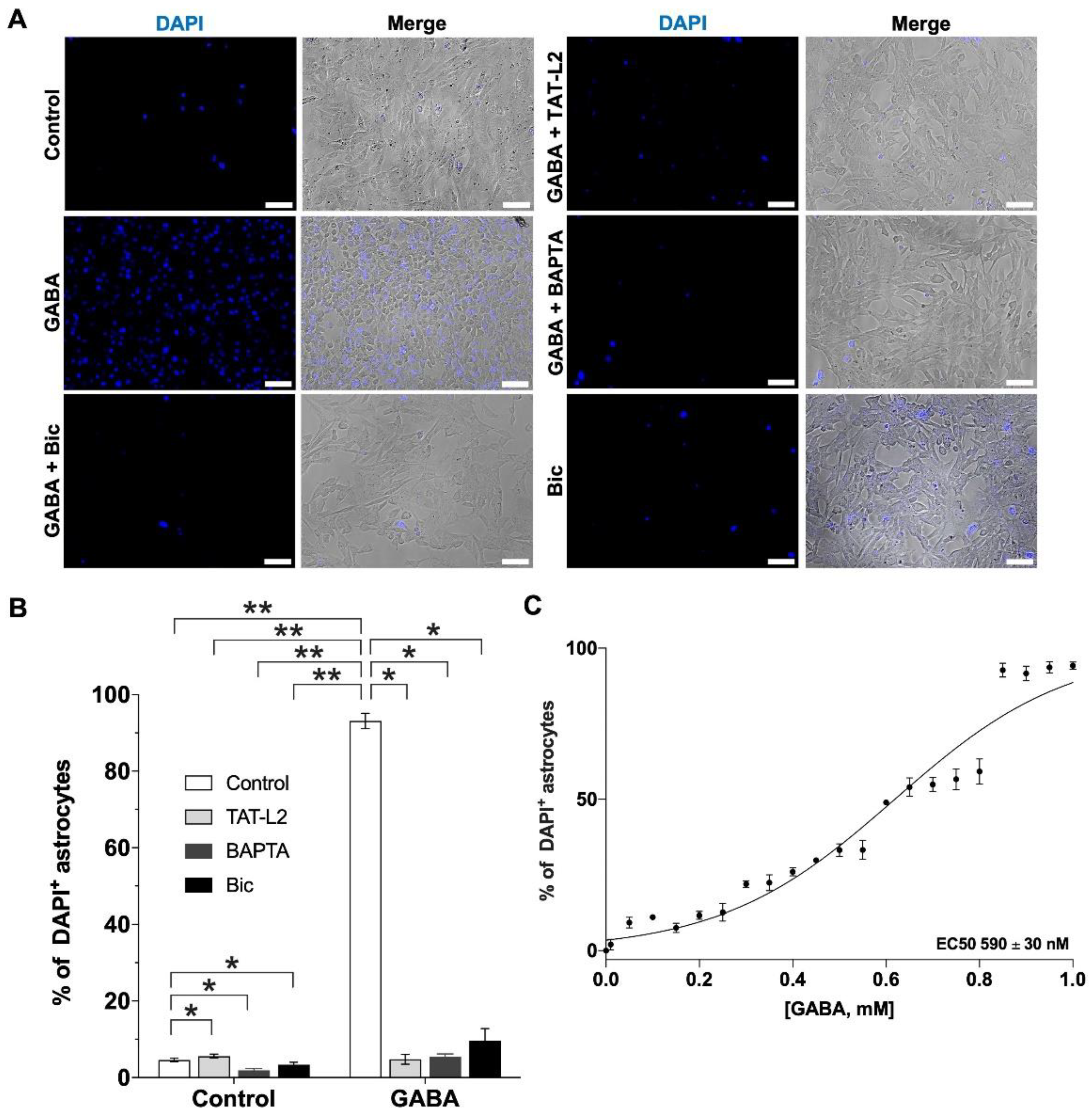
When DI TNC1 astrocytes were incubated in a solution with normal Ca2+ (1 mM), as expected, DAPI uptake was very low (Figure 2A,B, control). However, 1 μM GABA induced a large and significant increase in DAPI uptake (Figure 2A,B, GABA). The increased uptake in response to GABA was prevented by preincubation with the selective Cx43 hemichannel blocker TAT-Cx43L2 (Figure 2A,B, GABA + TAT-L2), suggesting that GABA induced an increase in Cx43 hemichannel activity. The effect of GABA on Cx43 hemichannel activity was [Ca2+]i-dependent, as it was prevented by the cell-permeable Ca2+ chelator BAPTA-AM (Figure 2A,B, GABA + BAPTA). To determine whether the effect of GABA was mediated by GABAA or GABAB receptors, cells were preincubated with either GABAA receptor antagonist bicuculline or GABAB receptor antagonist CGP55845 (CGP) before incubation with GABA. Bicuculline prevented the increase in DAPI uptake induced by GABA (Figure 2A, GABA + Bic). Bicuculline at the concentration used to block the effects of GABA did not affect Cx43 hemichannel activity by itself (Figure 2A,B, control + Bic). GABA showed an EC50 of 590 ± 30 nM (Figure 2C), and in total, 86.5% of the cells showed uptake after exposure to GABA, in stark contrast to the only 4.6% of astrocytes that showed basal DAPI uptake in control conditions (without GABA). In terms of fluorescence intensity, astrocytes showed similar median intensities between the control and GABA-treated conditions, showing only significant differences in the number of labeled cells (see Supplementary Figure S1A–C).
Although the effect of GABA was completely prevented by GABAA receptor antagonist bicuculline, cells were also preincubated with GABAB receptor antagonist CGP55845 (CGP, 1 µM), to assess a possible contribution of GABAB receptors. As can be observed in Figure 3A,B, CGP), GABAB receptor blockade by 1 µM CGP55845 had no effect on the GABA-induced increase in DAPI uptake.
Pannexin channels have also been associated with gliotransmitter release (reviewed in [29]). Although GABA-induced increase in DAPI uptake was completely prevented by Cx43 hemichannel blocker TAT-Cx43L2, we decided to evaluate the possible contribution of pannexin 1 (Panx1) channels. Hence, astrocytes were preincubated with Panx1 channel blocker peptide 10Panx1 (Panx1, 150 µM). DAPI uptake in response to GABA was unaffected by pannexin channel blockade (Figure 3, Panx1).
The above results suggest that the effect of GABA on DI TNC1 astroglial uptake is mediated by GABAA receptors and by the activation of Cx43 hemichannels.
To further corroborate the specificity of the contribution of Cx43 hemichannels, DI TNC1 astrocytes were also preincubated with the mutant TAT-Cx43L2H126K/I130N peptide (TAT-L2 mut), which is a peptide that differs from TAT-Cx43L2 by 2 amino acids, losing its affinity for Cx43, lacking inhibitory properties on Cx43-hemichannels [34], and used to assess the specificity of TAT-Cx43L2 on Cx43 hemichannel activity [35,36]. The mutated peptide did not affect GABA-induced increases in Cx43 hemichannel activity (Figure 3, TAT-L2mut), corroborating the specificity of the effects of the TAT-Cx43L2 peptide.
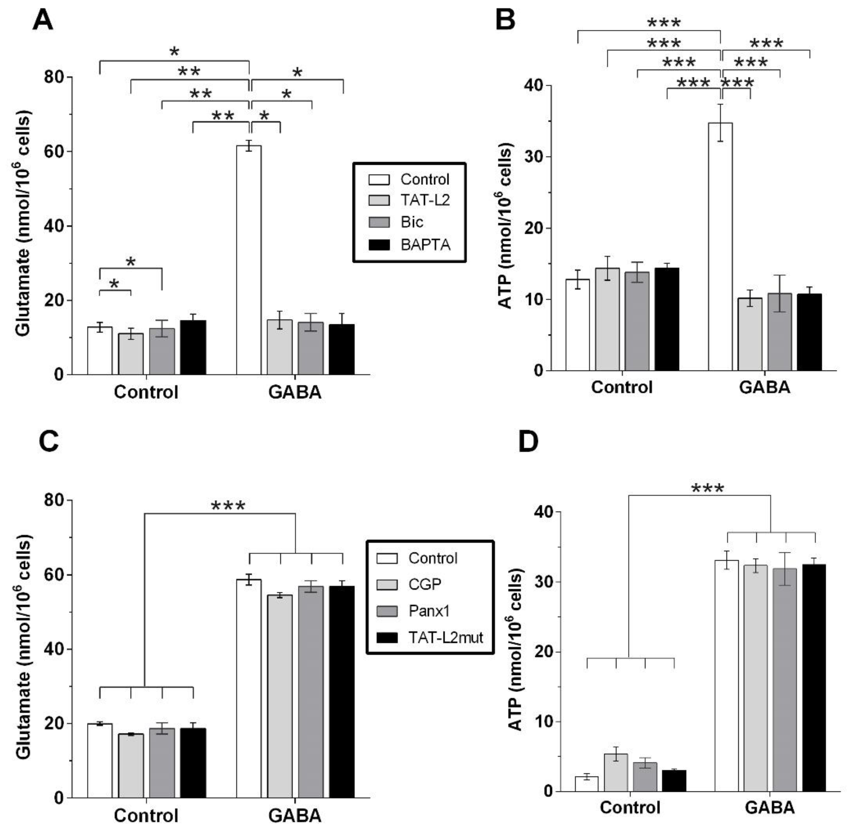
Analysis of the glutamate content in the culture medium in which DI TNC1 astrocytes were incubated with the different ligands showed that GABA induced the release of glutamate (Figure 4A, GABA), which was prevented by preincubation with TAT-Cx43L2 (Figure 4A, GABA + TAT-L2), BAPTA-AM (Figure 4A, GABA + Bapta) and GABAA receptor antagonist bicuculline (Figure 4A, GABA + Bic). The latter suggesting that GABA induces the Cx43 hemichannel-dependent release of glutamate, which is [Ca2+]i-dependent and mediated by GABAA receptors.
GABA also induced the release of ATP (Figure 4B, GABA), which was prevented by preincubation with TAT-Cx43L2 (Figure 4B, GABA + TAT-L2), BAPTA-AM (Figure 4B, GABA + Bapta), and GABAA receptor antagonist bicuculline (Figure 4B, GABA + Bic), suggesting that GABA induces Cx43 hemichannel-dependent release of ATP, which is [Ca2+]i -dependent and mediated by GABAA receptors.
The GABAB receptor antagonist CGP55845 had no effect on the astroglial release of glutamate in response to GABA (Figure 4C, CGP), suggesting that the GABA-induced release of glutamate was not mediated by the GABAB receptor. Congruently, CGP55845 also had no effect on the astroglial release of ATP induced by GABA (Figure 4D, CGP). The above results suggest that DI TNC1 astrocytes release gliotransmitters glutamate and ATP in response to GABA, via the activation of GABAA and not GABAB receptors.
As with DAPI uptake, the release of glutamate and ATP in response to GABA was also not affected by preincubation with the Panx1 channel blocker peptide 10Panx1 (Figure 4C,D, Panx1). Likewise, preincubation with the mutant TAT-Cx43L2H126K/I130N peptide also did not affect the release of glutamate or ATP in response to GABA (Figure 4C,D, TAT-L2 mut). This suggests that gliotransmission in response to GABA was mediated by Cx43 hemichannels, without a detectable contribution from Panx1 channels.
All the previous results were obtained from DI TNC1 astrocytes, which, arguably, being a cell line, may not completely recapitulate astroglia. Consequently, Cx43 hemichannel activity was measured using Etd+ in rat hippocampal slices in response to GABA, and the incubating medium was used to measure glutamate and ATP.
As can be seen in Figure 5A,B, GABA induced a large and significant increase in Etd+ uptake compared to the control condition (without GABA), an effect that was entirely blunted by preincubation with either TAT-Cx43L2 (TAT-L2) or bicuculline (Bic). The above results corroborate what was seen in DI TNC1 astrocytes and demonstrate that GABA induces an increase in Cx43 hemichannel activity in astrocytes from hippocampal slices, via activation of GABAA receptors.
Analysis of the culture medium of the hippocampal slices after GABA incubation shows a large and significant increase in extracellular glutamate (Figure 5C, GABA), which was completely prevented by preincubation with bicuculline (Figure 5C, Bic) and with TAT-Cx43L2 (Figure 5C, TAT-L2). GABA also induced an increase in extracellular ATP (Figure 5D, GABA), which was averted by preincubation with bicuculline (Figure 5D, Bic) and with TAT-Cx43L2 (Figure 5D, TAT-L2). For detailed results see Supplementary Table S1.

3. Discussion

The present study shows that both DI TNC1 astroglia and astrocytes from hippocampal brain slices respond to GABA via the activation of GABAA receptors, inducing a large increase in Cx43 hemichannel activity, and the Cx43 hemichannel-dependent release of glutamate and ATP. Experiments on DI TNC1 astrocytes suggest that GABAB receptors and Panx1 channels may not have a measurable role in GABA-induced gliotransmission. This idea is supported by the fact that in hippocampal slices, Cx43 hemichannel activity, as well as glutamate and ATP gliotransmission, are all completely prevented by GABAA antagonism and Cx43 hemichannel blockade, leaving very little room for a contribution from GABAB receptors or panx1 channels, and by the fact that neither the GABAB antagonist, nor the Panx1 blocker, affected GABA-induced gliotransmission.
Astrocytes have been reported to express both ionotropic GABAA receptors [11,12] and metabotropic GABAB receptors [8,13]. Activation of GABAA receptors in astrocytes leads to Cl- efflux from astrocytes, inducing depolarizing currents, triggering astrocytic Ca2+ signaling via the activation of voltage-sensitive Ca2+ channels [20,22,23,30,37,38]. GABAB receptor activation also induces increases in intracellular [Ca2+] involving Gi/o proteins and Ca2+ release from intracellular stores [19,24] via IP3 signaling [17,19,24,39] followed by gliotransmitter release [19,28,40]. The GABA transporter (GAT)-mediated Na+ symport also increases [Ca2+]i through the Na+/Ca2+ exchanger [41,42]. Finally, GAT-3 activation stimulates the astroglial release of ATP/adenosine, decreasing excitatory transmission via activation of presynaptic adenosine receptors [41] and increasing inhibitory transmission via activation of postsynaptic adenosine receptors [43]. The different pathways by which GABA can trigger an increase in [Ca2+]i in astrocytes have been proposed to work synergistically [43].
Glutamate release has been shown to occur in astrocytes via several pathways besides Cx43 hemichannels, including reverse flow of glutamate transporters [44,45], volume-regulated anion channels (VRACs) [9,46,47,48,49], cysteine-glutamate Xc- antiporter [50], anion channel bestrophin 1(Best1) [51,52], P2X7 receptors [53] and vesicular exocytosis [54,55,56,57,58,59,60]. Our results show that glutamate release was entirely blunted by the Cx43 hemichannel blocker TAT-Cx43L2, suggesting that, in both DI TNC1 astrocytes and hippocampal slices, glutamate release in response to GABA is almost exclusively mediated by Cx43 hemichannels, ruling out an essential contribution from the other release mechanisms. There is, however, another possibility. Cx43 hemichannels are permeable to Ca2+, so another option is that the Ca2+ released from astroglial intracellular stores is insufficient to trigger glutamate release throughout the astroglial processes but may be sufficient to increase Cx43 hemichannel activity, which could, in turn, induce the localized Ca2+ influx required to activate other complementing release mechanisms in localized astroglial compartments. Hence, Cx43 hemichannel-dependent gliotransmitter release could begin when hemichannels open but could be complemented by other Ca2+--dependent release mechanisms. This way, Cx43 hemichannel activity could be an early “upstream” step required for gliotransmission, which would explain why blocking Cx43 hemichannels completely blunts gliotransmission. Further research will be needed to assess this possibility.
Studies have shown that GABA induces the release of glutamate [25,26] and ATP/adenosine from astrocytes [25,27,28], with a net inhibitory effect. In the present study, the activation of GABAA receptors led to increased Cx43 hemichannel activity and Cx43 hemichannel-dependent release of both glutamate and ATP via GABAA, which is congruent with several studies showing intracellular [Ca2+] increases in primary astrocytes in response to GABAA activation in primary astroglial cultures [11,20,21,22]. However, a previous study found that astrocytes from adult cortical slices respond to GABA via GABAB receptors [24]. In the present report, neither the increase in Cx43 hemichannel activity nor the release of glutamate and ATP in response to GABA was sensitive to the GABAB receptor antagonist CGP55845. It may be argued that the concentration used was insufficient to attain effects, as a dose-response curve was not performed. The use of a dose-response curve was deemed unnecessary, as the blockade of GABAA receptors by bicuculline completely abolished both Cx43 hemichannel activity increases and gliotransmission in response to GABA. Hence, the present results suggest that the main mechanism by which GABA induces an increase in Cx43 hemichannel activity and astroglial release of glutamate and ATP is via the activation of GABAA receptors in both DI TNC1 astrocytes and hippocampal slices.
Previous studies have reported that astrocytes obtained from animals at post-natal P3 or P32–34 express primarily GABAA receptors, and only 10% of them responded via GABAB receptors. In contrast, those obtained from P11 and P15 showed 60% of astrocytes responding via GABAB receptors [23]. Consequently, it is possible that the timing in which astrocytes were obtained could influence their GABA receptor expression. More research is needed to address how astrocytes decide to respond to GABA, either via GABAA or GABAB receptors, but based on current literature, our results support GABAA as the main GABA receptor mediating the effects of GABA on astroglial gliotransmission.
One of the most interesting results of the present study is that GABA induces the astroglial release of both glutamate and ATP almost exclusively through Cx43 hemichannels, in both DI TNC1 and hippocampal slices. In previous studies performed in brain slices, it was reported that GABA caused the release of glutamate and ATP, but instead of inducing increased neuronal excitation, it produced an astrocyte-dependent decrease in neuronal excitability that was mediated by neuronal adenosine receptors [25,27,28]. Although it is unclear from those studies whether adenosine was released by the astrocytes or was produced by neurons from the ATP that was released from astrocytes, this result suggests that while astrocytes may release both glutamate and ATP, the synaptic effect resulting from their release may be dependent on different factors, which may include the neuronal expression of the enzyme that converts ATP into adenosine (ecto-5′-nucleotidase), the expression of adenosine receptors, or the possible release of adenosine from astrocytes, which was not measured in the present study.
This inhibitory effect found after the release of glutamate and ATP in response to GABA is particularly interesting because glutamate also induces the astroglial increase in [Ca2+]i and the release of glutamate and ATP [29]. While the ATP released by astrocytes always induces a decrease in neuronal excitability [61,62], the glutamate released by astrocytes induces an increase in intracellular cAMP and IP3, Ca2+ influx, and an increase in excitability and plasticity in neurons [63]. In the case of glutamate, the net effect of glutamate-induced gliotransmission is excitatory [64]. It is possible that the amount of glutamate and ATP released by each astrocyte may be different in response to different neurotransmitters, or it is possible that a different set of gliotransmitters be released by different astrocytes. In the central amygdala, astrocytic ATP/adenosine has been reported to depress excitatory neurotransmission by activating presynaptic A1 receptors and increasing the transmission of inhibitory inputs through the activation of presynaptic A2A receptors [65]. However, studies performed in the basolateral amygdala have shown that astroglial Cx43 hemichannels regulate post-synaptic NMDAR receptor activity via released glutamate and D-serine but not ATP [36]. Hence, the gliotransmitters released, as well as the receptors and enzymes expressed by neighboring neurons, may determine the final neuronal response to gliotransmitters. A recent review noted that this paradoxical effect might be related to mechanisms of astroglial integration [66]. How the astroglial release of glutamate, ATP, and other gliotransmitters may induce excitation and inhibition is an interesting question that remains unanswered and warrants further research.
Many psychiatric disorders, including anxiety-related disorders and depression, have been associated with a dysfunction in GABAergic transmission [67,68], and both effective antidepressant treatments and ECT have been shown to augment brain GABA [69]. Previous studies have also reported increased astroglial Cx43 hemichannel activity, both in post-mortem samples from depressed patients [70] and in animal models for depression [71]. In the latter, we found that the chronic restraint stress commonly used to induce depression-like symptoms in rodents causes an increase in Cx43 hemichannel activity in astrocytes and a Cx43 hemichannel-dependent increase in extracellular glutamate and ATP in the ventral hippocampus [71]. Whether this increase in glutamate and ATP leads to synaptic excitation or inhibition is still unknown. However, astroglial dysfunction may contribute to psychiatric disorders like depression by affecting the excitatory/inhibitory balance in different brain regions. Astrocytes can affect this balance by changing the expression and availability of GABA and glutamate transporters. They may also release glutamate and ATP, which depending on the surrounding neurons, may induce synaptic excitation or inhibition [66]. Consequently, the contribution of Cx43 hemichannel-dependent astroglial gliotransmission to the pathogenesis of psychiatric disorders, like depression, is a theme that will keep researchers busy for the next years to come.

4. Materials and Methods

4.1. DI TNC1 Astroglial Cell Culture

The rat astrocyte cell line DI TNC1 (ATCC CRL-2005) was cultured at 37 °C, and 5% CO2 in Dulbecco’s Modified Eagle’s Medium (DMEM) supplemented with 5% fetal bovine serum (Life Technologies, Carlsbad, CA, USA) and 100 µM/mL streptomycin and ampicillin (1:1, 100 IU/mL). The culture medium was replaced every other day.

4.2. DAPI Uptake

To measure Cx43 hemichannel activity, snapshots of DAPI uptake in DITNC1 astrocytes were taken, as reported in previous studies [71]. In summary, once the cells reached 80% confluence, they were transferred to 96 well plates with a HEPES -buffered salt solution either with Ca2+ (140 mM NaCl, 4 mM KCl, 1 mM CaC12, 1 mM MgC12, 5 mM glucose, 10 nΜ HEPES) or divalent cations (Ca2+)-free (140 mM NaCl, 4 mM KC1, 5 mM glucose, 2 mM EGTA, 10 nM HEPES). The Ca2+-free solution was used to assess whether GABA or bicuculline can decrease Cx43 hemichannel opening, as hemichannels open when cells are incubated in a Ca2+-free medium [30]. The standard solution containing 1 mM Ca2+ was used to determine whether GABA or bicuculline can trigger the opening of Cx43 hemichannels, as in normal-physiological Ca2+ conditions, the Cx43 hemichannel opening probability is low [30]. The ligands were added to both solutions for 5 min, followed by 5 min incubation with 4′,6-diamidino-2-phenylindole dihydrochloride (DAPI, Sigma-Aldrich, St. Louis, MO, USA, 10 µM).
Cells were then washed to eliminate extracellular DAPI and examined in fluorescence microscopy using a FLoid® Cell Imaging Station (Invitrogen, Waltham, MA, USA). Each well was photographed under white light and fluorescence (excitation, 340 nm; emission, 488 nm), using parameters preset under control conditions (no ligand). Each photograph was subdivided into four quadrants, and all cells in one quadrant were counted under white light (total cells) and fluorescence for the emission of DAPI (fluorescent nuclei). Only quadrants having at least 250 cells were included in the analysis. This way, the percentage of fluorescent cells with DAPI-labeled nuclei from the total number of cells present in the photograph was estimated for each ligand concentration. To determine the EC50 for GABA, the percentage of increase in the number of fluorescent cells was plotted from the uptake experiments for each concentration, and the concentration needed to attain a 50% effect was extrapolated. To determine whether the effects depended on intracellular Ca2+, cells were preincubated with cell-permeable Ca2+ chelator BAPTA-AM (Sigma-Aldrich, St. Louis, MO, USA) at 100 nM, before adding the ligand. To corroborate that the DAPI uptake depended specifically on Cx43 hemichannels, cells were preincubated with 10 nM of the selective Cx43 hemichannel blocker peptide TAT-Cx43L2 (YGRKKRRQRRRDGANVDMHLKQIEIKKFKYGIEEHGK; LifeTein, NJ, Somerset, USA;) or with 10 nM of its control mutant TAT-Cx43L2H126K/I130N (Biomatik, Kitchener, ON, Canada), before the addition of the ligands.

4.3. Drugs Evaluated

The effect of GABA (Sigma-Aldrich) in DI TNC1 astrocytes was evaluated using six different concentrations of GABA, ranging from 1 nM to 100 µM. The experiment was performed in three wells and repeated 2 more times to obtain triplicates (N = 3, with technical triplicates). The 1 μM concentration was the lowest concentration with a significant effect. The GABAA antagonist Bicuculline (Sigma-Aldrich) was evaluated at six concentrations from 1 nM to 100 μM for 5 min. The maximal concentration evaluated did not show an effect by itself (100 µM). Then, cells were pre-incubated with bicuculline at 100 µM and then incubated with GABA (1 μM, Sigma-Aldrich). The GABAB antagonist CGP55845 (Tocris, Bristol, UK) was evaluated at 1 µM, the panx1 blocker peptide 10Panx1 (Genscript, Piscataway, NJ, USA) at 150 µM and the mutant TAT-Cx43L2H126K/I130N (Biomatik, Kitchener, ON, Canada) was evaluated at 10 nM. All drugs were dissolved in PBS.

4.4. Measurement of Extracellular Glutamate and ATP

The cells were transferred to 12 well plates with 1,2 mL of a HEPES -buffered salt solution with Ca2+ (140 mM NaCl, 4 mM KCl, 1 mM CaC12, 1 mM MgC12, 5 mM glucose, 10 nΜ HEPES). GABA (1 µM) was added either alone, or after pretreatment with TAT-Cx43L2 (10 nM) or bicuculline (100 µM) for ten minutes. After five minutes of incubation with GABA, the medium without cells was transferred to 1 mL aliquots and frozen at −80 °C and then thawed before gliotransmitter analysis. Extracellular ATP was measured using a luciferin/luciferase bioluminescence assay kit (Sigma-Aldrich). Extracellular glutamate levels were determined using a colorimetric enzyme-linked assay (Sigma-Aldrich). In the presence of glutamate dehydrogenase (GDH) and β-nicotinamide adenine dinucleotide phosphate (NADP+), glutamate is oxidized to α-ketoglutarate, yielding NADPH, which can be determined fluorometrically for indirect quantification of glutamate concentration (excitation and emission wavelengths of 355 nm and 460 nm, respectively). For each assay, standard curves were constructed using standardized ATP and glutamate concentrations used to estimate the concentrations of ATP and glutamate in the samples, which are shown per 106 cells for in vitro experiments and per mL for hippocampal slices (ex vivo).

4.5. Animals

All procedures involving animals were performed in accordance with NIH guidelines and with the approval of the bioethical committee of the Universidad Andrés Bello (Acta 003-2020). Sprague Dawley male rats (~60-day-old, ~250 g) were caged individually at 22 °C under a 12/12 h light/dark cycle and given food chow and water ad libitum.

4.6. Hippocampal Slice Preparation

Dye uptake “snapshot” assays were performed as reported previously [36,71], with minor modifications. In brief, naïve rats were anesthetized under isoflurane. They were decapitated and their brains were extracted and cut into coronal slices (400 µm) in an ice-cold slicing solution containing (in mM): sucrose (222); KCl (2.6); NaHCO3 (27); NaHPO4 (1.5); glucose (10); MgSO4 (7); CaCl2 (0.5) and ascorbate (0.1), bubbled with 95% O2/5% CO2, using a vibratome (Leica, VT1000GS; Wetzlar, Germany). Then, the slices were transferred to a holding chamber filled with artificial cerebral spinal fluid (ACSF), at room temperature (20–22 °C), containing (in mM): 125 NaCl, 2.5 KCl, 25 glucose, 25 NaHCO3, 1.25 NaH2PO4, 2 CaCl2, and 1 MgCl2, bubbled with 95% O2/5% CO2, pH 7.4, for a stabilization period of 60 min before dye uptake experiments.

4.7. Dye Uptake Measurements in Hippocampal Slices

For dye uptake and ex vivo ‘‘snapshot’’ experiments, the slices were incubated with 5 µM ethidium (Etd+) for 10 min in a chamber oxygenated by bubbling 95% O2/5% CO2 into ACSF. To ascertain that the dye uptake was mediated by Cx43 hemichannels, some of the brain slices were incubated with 10 nM TAT-Cx43L2. To assess the contribution of GABAA receptors, some slices were preincubated with bicuculine (100 µM). Preincubation was performed for 10 min, and then GABA was added at 100 µM for 5 min, followed by Etd+ for 10 min. After the 10-min exposure to Etd+, the slices were washed three times with ACSF and were fixed at room temperature with 4% saccharose in 4% paraformaldehyde for 60 min, rinsed once for 5 min with 0.1 mM glycine in PBS, and then twice with PBS for 10 min with gentle agitation. Then the slices were sectioned using a cryostat to obtain ~ ten sections (40 µm each). The sections from the slices were incubated two times for 30 min each with a blocking solution (PBS, gelatin 0.2%, Triton-X 100 1%) at room temperature and then incubated overnight at 4 °C with a cell-specific antibody to identify astrocytes (anti-GFAP monoclonal antibody; Sigma-Aldrich). After the incubation with the antibody diluted 1:1000 in the blocking solution, the slices were washed for 10-min with the blocking solution, 3 times. The sections were then incubated for 2 h at room temperature with donkey anti-mouse Alexa Fluor 488 antibody (1:1.000, Abcam) and after 3 washes (10-min each), the slices were mounted in Fluoromount, cover-slipped, and examined in an epifluorescence microscope (ECLIPSE Ti-E, Nikon, Tokyo, Japan). Stacks of consecutive images were taken with a 60× objective at 1-µm intervals, acquired sequentially at two wavelengths (488 nm and 543 nm), and Z stack projections were reconstructed using ImageJ software (National Institute of Health, Bethesda, MD, USA). Dye uptake was calculated as: corrected total cell Etd fluorescence intensity (as arbitrary units [AU]) = integrated density − ([area of selected cell] × [mean fluorescence of background readings]). At least six cells per field were selected from at least three fields in each brain slice. The background fluorescence was set as the average value of intensity in three regions of interest (ROI) void of any fluorescence.
To measure fluorescence intensity, ROIs in GFAP positive cells (astrocytes) were set surrounding cell nuclei and the fluorescent intensity was measured using ImageJ for each cell, for all cells in each quadrant, from the same quadrants that were analyzed for counting DAPI labeled cells, from the GABA and control conditions (n = 3 with triplicates). Fluorescent intensities were plotted for all fluorescence-labeled cells, and the median number of cells and median fluorescence were compared for each condition.

4.8. Measurements of Extracellular Glutamate and ATP

The same protocol for hippocampal slices was used as above, but instead of adding Etd+, the slices were removed from the ACSF medium, which was transferred to 1 mL aliquots and frozen at −80 °C. For ATP and glutamate measurements, the aliquots were thawed and processed with the same procedure as explained above.

4.9. Statistics

All results were analyzed using the Prism software and were tested using the Kolmogorov-Smirnov test for normality and found to be normally distributed. Groups were compared either using one- or two-way ANOVA as required and a Tukey post-hoc test for multiple comparisons. Differences were considered significant when p ˂ 0.05 and shown as p ˂ 0.05 *, p ˂ 0.01 **, and p ˂ 0.001 ***.

Supplementary Materials

The following supporting information can be downloaded at: https://www.mdpi.com/article/10.3390/ijms232113625/s1.

Author Contributions

Data curation, I.J.-D.; Formal analysis I.J.-D., R.R.-L. and Y.L.-I.; investigation, I.J.-D., R.R.-L., Y.L.-I., F.P., C.M.L., J.P.-V., K.C., R.C.-L. and J.A.O.; supervision, J.A.O., M.A.R. and J.S.; writing—review and editing, I.J.-D., M.A.R. and J.A.O.; conceptualization, J.S.; writing—original draft preparation, J.S.; funding acquisition, J.S. and J.A.O. All authors have read and agreed to the published version of the manuscript.

Funding

This research was funded by FONDECYT, grant numbers 1200452 (J.S.) and 1210375 (J.A.O.), and Beca Doctorado ANID 21221905 & 21180461.

Institutional Review Board Statement

All procedures involving animals were performed in accordance with the NIH guidelines and with approval of the bioethical committee of the Universidad Andrés Bello (Acta 003-2020).

Data Availability Statement

Not applicable.

Acknowledgments

We wish to thank Lisette Leyton for contributing to the DINCT1 cell line.

Conflicts of Interest

The authors declare no conflict of interest.

References

  1. Roux, L.; Buzsáki, G. Tasks for Inhibitory Interneurons in Intact Brain Circuits. Neuropharmacology 2015, 88, 10–23. [Google Scholar] [CrossRef] [PubMed] [Green Version]
  2. Zhang, W.; Xiong, B.-R.; Zhang, L.-Q.; Huang, X.; Yuan, X.; Tian, Y.-K.; Tian, X.-B. The Role of the GABAergic System in Diseases of the Central Nervous System. Neuroscience 2021, 470, 88–99. [Google Scholar] [CrossRef] [PubMed]
  3. Goddard, A.W. Cortical and Subcortical Gamma Amino Acid Butyric Acid Deficits in Anxiety and Stress Disorders: Clinical Implications. World J. Psychiatry 2016, 6, 43. [Google Scholar] [CrossRef] [PubMed]
  4. Della Vecchia, A.; Arone, A.; Piccinni, A.; Mucci, F.; Marazziti, D. GABA System in Depression: Impact on Pathophysiology and Psychopharmacology. Curr. Med. Chem. 2022, 29, 5710–5730. [Google Scholar] [CrossRef]
  5. Scimemi, A. Structure, Function, and Plasticity of GABA Transporters. Front. Cell. Neurosci. 2014, 8, 161. [Google Scholar] [CrossRef] [PubMed] [Green Version]
  6. Jiménez-González, C.; Pirttimaki, T.; Cope, D.W.; Parri, H.R. Non-Neuronal, Slow GABA Signalling in the Ventrobasal Thalamus Targets δ-Subunit-Containing GABAA Receptors: Thalamic Glial GABAergic Signalling. Eur. J. Neurosci. 2011, 33, 1471–1482. [Google Scholar] [CrossRef] [Green Version]
  7. Kozlov, A.S.; Angulo, M.C.; Audinat, E.; Charpak, S. Target Cell-Specific Modulation of Neuronal Activity by Astrocytes. Proc. Natl. Acad. Sci. USA 2006, 103, 10058–10063. [Google Scholar] [CrossRef] [Green Version]
  8. Lee, S.; Yoon, B.-E.; Berglund, K.; Oh, S.-J.; Park, H.; Shin, H.-S.; Augustine, G.J.; Lee, C.J. Channel-Mediated Tonic GABA Release from Glia. Science 2010, 330, 790–796. [Google Scholar] [CrossRef]
  9. Liu, Q.-Y.; Schaffner, A.E.; Chang, Y.H.; Maric, D.; Barker, J.L. Persistent Activation of GABA A Receptor/Cl Channels by Astrocyte-Derived GABA in Cultured Embryonic Rat Hippocampal Neurons. J. Neurophysiol. 2000, 84, 1392–1403. [Google Scholar] [CrossRef]
  10. Bormann, J.; Kettenmann, H. Patch-Clamp Study of Gamma-Aminobutyric Acid Receptor Cl- Channels in Cultured Astrocytes. Proc. Natl. Acad. Sci. USA 1988, 85, 9336–9340. [Google Scholar] [CrossRef]
  11. MacVicar, B.; Tse, F.; Crichton, S.; Kettenmann, H. GABA-Activated Cl- Channels in Astrocytes of Hippocampal Slices. J. Neurosci. 1989, 9, 3577–3583. [Google Scholar] [CrossRef] [PubMed] [Green Version]
  12. Muller, T.; Fritschy, J.; Grosche, J.; Pratt, G.; Mohler, H.; Kettenmann, H. Developmental Regulation of Voltage-Gated K+ Channel and GABAA Receptor Expression in Bergmann Glial Cells. J. Neurosci. 1994, 14, 2503–2514. [Google Scholar] [CrossRef] [PubMed] [Green Version]
  13. Blomqvist, A.; Broman, J. Light and Electron Microscopic Immunohistochemical Demonstration of GABA-Immunoreactive Astrocytes in the Brain Stem of the Rat. J. Neurocytol. 1988, 17, 629–637. [Google Scholar] [CrossRef] [PubMed]
  14. Martínez-Rodríguez, R.; Tonda, A.; Gragera, R.R.; Paz-Doel, R.; García-Cordovilla, R.; Fernández-Fernández, E.; Fernández, A.M.; González-Romero, F.; López-Bravo, A. Synaptic and Non-Synaptic Immunolocalization of GABA and Glutamate Acid Decarboxylase (GAD) in Cerebellar Cortex of Rat. Cell. Mol. Biol. 1993, 39, 115–123. [Google Scholar] [PubMed]
  15. Mariotti, L.; Losi, G.; Lia, A.; Melone, M.; Chiavegato, A.; Gómez-Gonzalo, M.; Sessolo, M.; Bovetti, S.; Forli, A.; Zonta, M.; et al. Interneuron-Specific Signaling Evokes Distinctive Somatostatin-Mediated Responses in Adult Cortical Astrocytes. Nat. Commun. 2018, 9, 82. [Google Scholar] [CrossRef] [Green Version]
  16. Mederos, S.; Perea, G. GABAergic-astrocyte Signaling: A Refinement of Inhibitory Brain Networks. Glia 2019, 67, 1842–1851. [Google Scholar] [CrossRef] [Green Version]
  17. Nagai, J.; Rajbhandari, A.K.; Gangwani, M.R.; Hachisuka, A.; Coppola, G.; Masmanidis, S.C.; Fanselow, M.S.; Khakh, B.S. Hyperactivity with Disrupted Attention by Activation of an Astrocyte Synaptogenic Cue. Cell 2019, 177, 1280–1292. [Google Scholar] [CrossRef]
  18. Nilsson, M.; Eriksson, P.S.; Rönnbäck, L.; Hansson, E. GABA Induces Ca2+ Transients in Astrocytes. Neuroscience 1993, 54, 605–614. [Google Scholar] [CrossRef]
  19. Perea, G.; Gómez, R.; Mederos, S.; Covelo, A.; Ballesteros, J.J.; Schlosser, L.; Hernández-Vivanco, A.; Martín-Fernández, M.; Quintana, R.; Rayan, A.; et al. Activity-Dependent Switch of GABAergic Inhibition into Glutamatergic Excitation in Astrocyte-Neuron Networks. eLife 2016, 5, e20362. [Google Scholar] [CrossRef]
  20. Fraser, D.; Duffy, S.; Angelides, K.; Perez-Velazquez, J.; Kettenmann, H.; MacVicar, B. GABAA/Benzodiazepine Receptors in Acutely Isolated Hippocampal Astrocytes. J. Neurosci. 1995, 15, 2720–2732. [Google Scholar] [CrossRef]
  21. Kettenmann, H.; Schachner, M. Pharmacological Properties of Gamma-Aminobutyric Acid-, Glutamate-, and Aspartate-Induced Depolarizations in Cultured Astrocytes. J. Neurosci. 1985, 5, 3295–3301. [Google Scholar] [CrossRef] [PubMed] [Green Version]
  22. Letellier, M.; Park, Y.K.; Chater, T.E.; Chipman, P.H.; Gautam, S.G.; Oshima-Takago, T.; Goda, Y. Astrocytes Regulate Heterogeneity of Presynaptic Strengths in Hippocampal Networks. Proc. Natl. Acad. Sci. USA 2016, 113, E2685–E2694. [Google Scholar] [CrossRef] [PubMed] [Green Version]
  23. Meier, S.D.; Kafitz, K.W.; Rose, C.R. Developmental Profile and Mechanisms of GABA-Induced Calcium Signaling in Hippocampal Astrocytes. Glia 2008, 56, 1127–1137. [Google Scholar] [CrossRef] [PubMed]
  24. Mariotti, L.; Losi, G.; Sessolo, M.; Marcon, I.; Carmignoto, G. The Inhibitory Neurotransmitter GABA Evokes Long-lasting Ca2+ Oscillations in Cortical Astrocytes. Glia 2016, 64, 363–373. [Google Scholar] [CrossRef] [Green Version]
  25. Andersson, M.; Blomstrand, F.; Hanse, E. Astrocytes Play a Critical Role in Transient Heterosynaptic Depression in the Rat Hippocampal CA1 Region: Transient Heterosynaptic Depression. J. Physiol. 2007, 585, 843–852. [Google Scholar] [CrossRef]
  26. Kang, M.H.; Spigelman, I.; Olsen, R.W. Alteration in the Sensitivity of GABA(A) Receptors to Allosteric Modulatory Drugs in Rat Hippocampus after Chronic Intermittent Ethanol Treatment. Alcohol. Clin. Exp. Res. 1998, 22, 2165–2173. [Google Scholar] [CrossRef]
  27. Chen, T.; Wang, X.-L.; Qu, J.; Wang, W.; Zhang, T.; Yanagawa, Y.; Wu, S.-X.; Li, Y.-Q. Neurokinin-1 Receptor-Expressing Neurons That Contain Serotonin and Gamma-Aminobutyric Acid in the Rat Rostroventromedial Medulla Are Involved in Pain Processing. J. Pain 2013, 14, 778–792. [Google Scholar] [CrossRef]
  28. Serrano, A. GABAergic Network Activation of Glial Cells Underlies Hippocampal Heterosynaptic Depression. J. Neurosci. 2006, 26, 5370–5382. [Google Scholar] [CrossRef] [Green Version]
  29. Moraga-Amaro, R.; Jerez-Baraona, J.M.; Simon, F.; Stehberg, J. Role of Astrocytes in Memory and Psychiatric Disorders. J. Physiol.-Paris 2014, 108, 240–251. [Google Scholar] [CrossRef]
  30. Parpura, V.; Grubišić, V.; Verkhratsky, A. Ca2+ Sources for the Exocytotic Release of Glutamate from Astrocytes. Biochim. Biophys. Acta (BBA) Mol. Cell Res. 2011, 1813, 984–991. [Google Scholar] [CrossRef]
  31. Stout, C.; Charles, A. Modulation of Intercellular Calcium Signaling in Astrocytes by Extracellular Calcium and Magnesium. Glia 2003, 43, 265–273. [Google Scholar] [CrossRef] [PubMed]
  32. Schalper, K.A.; Palacios-Prado, N.; Orellana, J.A.; Sáez, J.C. Currently Used Methods for Identification and Characterization of Hemichannels. Cell Commun. Adhes. 2008, 15, 207–218. [Google Scholar] [CrossRef] [PubMed]
  33. Zanotti, S.; Charles, A. Extracellular Calcium Sensing by Glial Cells: Low Extracellular Calcium Induces Intracellular Calcium Release and Intercellular Signaling. J. Neurochem. 2002, 69, 594–602. [Google Scholar] [CrossRef] [PubMed]
  34. Ponsaerts, R.; De Vuyst, E.; Retamal, M.; D’Hondt, C.; Vermeire, D.; Wang, N.; De Smedt, H.; Zimmermann, P.; Himpens, B.; Vereecke, J.; et al. Intramolecular Loop/Tail Interactions Are Essential for Connexin 43-hemichannel Activity. FASEB J. 2010, 24, 4378–4395. [Google Scholar] [CrossRef] [Green Version]
  35. Stehberg, J.; Moraga-Amaro, R.; Salazar, C.; Becerra, A.; Echeverría, C.; Orellana, J.A.; Bultynck, G.; Ponsaerts, R.; Leybaert, L.; Simon, F.; et al. Release of Gliotransmitters through Astroglial Connexin 43 Hemichannels Is Necessary for Fear Memory Consolidation in the Basolateral Amygdala. FASEB J. 2012, 26, 3649–3657. [Google Scholar] [CrossRef] [Green Version]
  36. Linsambarth, S.; Carvajal, F.J.; Moraga-Amaro, R.; Mendez, L.; Tamburini, G.; Jimenez, I.; Verdugo, D.A.; Gómez, G.I.; Jury, N.; Martínez, P.; et al. Astroglial Gliotransmitters Released via Cx43 Hemichannels Regulate NMDAR-dependent Transmission and Short-term Fear Memory in the Basolateral Amygdala. FASEB J. 2022, 36, e22134. [Google Scholar] [CrossRef]
  37. Verkhratsky, A.; Rodríguez, J.J.; Parpura, V. Calcium Signalling in Astroglia. Mol. Cell. Endocrinol. 2012, 353, 45–56. [Google Scholar] [CrossRef]
  38. Verkhratsky, A.; Reyes, R.C.; Parpura, V. TRP Channels Coordinate Ion Signalling in Astroglia. In Reviews of Physiology, Biochemistry and Pharmacology 166; Nilius, B., Gudermann, T., Jahn, R., Lill, R., Offermanns, S., Petersen, O.H., Eds.; Reviews of Physiology, Biochemistry and Pharmacology; Springer International Publishing: Cham, Switzerland, 2013; Volume 166, pp. 1–22. ISBN 978-3-319-04905-2. [Google Scholar]
  39. Sharp, A.H.; Nucifora, F.C.; Blondel, O.; Sheppard, C.A.; Zhang, C.; Snyder, S.H.; Russell, J.T.; Ryugo, D.K.; Ross, C.A. Differential Cellular Expression of Isoforms of Inositol 1,4,5-Triphosphate Receptors in Neurons and Glia in Brain. J. Comp. Neurol. 1999, 406, 207–220. [Google Scholar] [CrossRef]
  40. Durkee, C.A.; Covelo, A.; Lines, J.; Kofuji, P.; Aguilar, J.; Araque, A. G i/o Protein-coupled Receptors Inhibit Neurons but Activate Astrocytes and Stimulate Gliotransmission. Glia 2019, 67, 1076–1093. [Google Scholar] [CrossRef]
  41. Boddum, K.; Jensen, T.P.; Magloire, V.; Kristiansen, U.; Rusakov, D.A.; Pavlov, I.; Walker, M.C. Astrocytic GABA Transporter Activity Modulates Excitatory Neurotransmission. Nat. Commun. 2016, 7, 13572. [Google Scholar] [CrossRef]
  42. Doengi, M.; Hirnet, D.; Coulon, P.; Pape, H.-C.; Deitmer, J.W.; Lohr, C. GABA Uptake-Dependent Ca2+ Signaling in Developing Olfactory Bulb Astrocytes. Proc. Natl. Acad. Sci. USA 2009, 106, 17570–17575. [Google Scholar] [CrossRef] [PubMed] [Green Version]
  43. Matos, M.; Bosson, A.; Riebe, I.; Reynell, C.; Vallée, J.; Laplante, I.; Panatier, A.; Robitaille, R.; Lacaille, J.-C. Astrocytes Detect and Upregulate Transmission at Inhibitory Synapses of Somatostatin Interneurons onto Pyramidal Cells. Nat. Commun. 2018, 9, 4254. [Google Scholar] [CrossRef] [PubMed] [Green Version]
  44. Longuemare, M.C.; Swanson, R.A. Net Glutamate Release from Astrocytes Is Not Induced by Extracellular Potassium Concentrations Attainable in Brain. J. Neurochem. 2002, 69, 879–882. [Google Scholar] [CrossRef] [PubMed] [Green Version]
  45. Rossi, D.J.; Oshima, T.; Attwell, D. Glutamate Release in Severe Brain Ischaemia Is Mainly by Reversed Uptake. Nature 2000, 403, 316–321. [Google Scholar] [CrossRef]
  46. Abdullaev, I.F.; Rudkouskaya, A.; Schools, G.P.; Kimelberg, H.K.; Mongin, A.A. Pharmacological Comparison of Swelling-Activated Excitatory Amino Acid Release and Cl Currents in Cultured Rat Astrocytes: VRACs and Excitatory Amino Acid Release in Astrocytes. J. Physiol. 2006, 572, 677–689. [Google Scholar] [CrossRef]
  47. Kimelberg, H.K.; Anderson, E.; Kettenmann, H. Swelling-Induced Changes in Electrophysiological Properties of Cultured Astrocytes and Oligodendrocytes. II. Whole-Cell Currents. Brain Res. 1990, 529, 262–268. [Google Scholar] [CrossRef]
  48. Mongin, A.A.; Kimelberg, H.K. ATP Regulates Anion Channel-Mediated Organic Osmolyte Release from Cultured Rat Astrocytes via Multiple Ca2+-Sensitive Mechanisms. Am. J. Physiol.-Cell Physiol. 2005, 288, C204–C213. [Google Scholar] [CrossRef]
  49. Ramos-Mandujano, G.; Vázquez-Juárez, E.; Hernández-Benítez, R.; Pasantes-Morales, H. Thrombin Potently Enhances Swelling-Sensitive Glutamate Efflux from Cultured Astrocytes. Glia 2007, 55, 917–925. [Google Scholar] [CrossRef]
  50. Cavelier, P.; Attwell, D. Tonic Release of Glutamate by a DIDS-Sensitive Mechanism in Rat Hippocampal Slices: Tonic Glutamate Release. J. Physiol. 2005, 564, 397–410. [Google Scholar] [CrossRef]
  51. Park, H.; Han, K.-S.; Oh, S.-J.; Jo, S.; Woo, J.; Yoon, B.-E.; Lee, C.J. High Glutamate Permeability and Distal Localization of Best1 Channel in CA1 Hippocampal Astrocyte. Mol. Brain 2013, 6, 54. [Google Scholar] [CrossRef]
  52. Woo, D.H.; Han, K.-S.; Shim, J.W.; Yoon, B.-E.; Kim, E.; Bae, J.Y.; Oh, S.-J.; Hwang, E.M.; Marmorstein, A.D.; Bae, Y.C.; et al. TREK-1 and Best1 Channels Mediate Fast and Slow Glutamate Release in Astrocytes upon GPCR Activation. Cell 2012, 151, 25–40. [Google Scholar] [CrossRef] [PubMed] [Green Version]
  53. Duan, S.; Anderson, C.M.; Keung, E.C.; Chen, Y.; Chen, Y.; Swanson, R.A. P2X 7 Receptor-Mediated Release of Excitatory Amino Acids from Astrocytes. J. Neurosci. 2003, 23, 1320–1328. [Google Scholar] [CrossRef] [Green Version]
  54. Bezzi, P.; Gundersen, V.; Galbete, J.L.; Seifert, G.; Steinhäuser, C.; Pilati, E.; Volterra, A. Astrocytes Contain a Vesicular Compartment That Is Competent for Regulated Exocytosis of Glutamate. Nat. Neurosci. 2004, 7, 613–620. [Google Scholar] [CrossRef]
  55. Bohmbach, K.; Schwarz, M.K.; Schoch, S.; Henneberger, C. The Structural and Functional Evidence for Vesicular Release from Astrocytes in Situ. Brain Res. Bull. 2018, 136, 65–75. [Google Scholar] [CrossRef] [PubMed]
  56. Bowser, D.N.; Khakh, B.S. Two Forms of Single-Vesicle Astrocyte Exocytosis Imaged with Total Internal Reflection Fluorescence Microscopy. Proc. Natl. Acad. Sci. USA 2007, 104, 4212–4217. [Google Scholar] [CrossRef] [PubMed] [Green Version]
  57. Jeftinija, S.D.; Jeftinija, K.V.; Stefanovic, G. Cultured Astrocytes Express Proteins Involved in Vesicular Glutamate Release. Brain Res. 1997, 750, 41–47. [Google Scholar] [CrossRef]
  58. Montana, V.; Malarkey, E.B.; Verderio, C.; Matteoli, M.; Parpura, V. Vesicular Transmitter Release from Astrocytes. Glia 2006, 54, 700–715. [Google Scholar] [CrossRef]
  59. Parpura, V.; Zorec, R. Gliotransmission: Exocytotic Release from Astrocytes. Brain Res. Rev. 2010, 63, 83–92. [Google Scholar] [CrossRef] [Green Version]
  60. Xu, J.; Peng, H.; Kang, N.; Zhao, Z.; Lin, J.H.-C.; Stanton, P.K.; Kang, J. Glutamate-Induced Exocytosis of Glutamate from Astrocytes. J. Biol. Chem. 2007, 282, 24185–24197. [Google Scholar] [CrossRef] [Green Version]
  61. Bennett, M.V.L.; Garré, J.M.; Orellana, J.A.; Bukauskas, F.F.; Nedergaard, M.; Giaume, C.; Sáez, J.C. Connexin and Pannexin Hemichannels in Inflammatory Responses of Glia and Neurons. Brain Res. 2012, 1487, 3–15. [Google Scholar] [CrossRef]
  62. Stout, C.E.; Costantin, J.L.; Naus, C.C.G.; Charles, A.C. Intercellular Calcium Signaling in Astrocytes via ATP Release through Connexin Hemichannels. J. Biol. Chem. 2002, 277, 10482–10488. [Google Scholar] [CrossRef] [PubMed] [Green Version]
  63. Wang, X.; Lippi, G.; Carlson, D.M.; Berg, D.K. Activation of A7-Containing Nicotinic Receptors on Astrocytes Triggers AMPA Receptor Recruitment to Glutamatergic Synapses. J. Neurochem. 2013, 127, 632–643. [Google Scholar] [CrossRef] [PubMed] [Green Version]
  64. Beppu, K.; Kubo, N.; Matsui, K. Glial Amplification of Synaptic Signals. J. Physiol. 2021, 599, 2085–2102. [Google Scholar] [CrossRef] [PubMed]
  65. Martin-Fernandez, M.; Jamison, S.; Robin, L.M.; Zhao, Z.; Martin, E.D.; Aguilar, J.; Benneyworth, M.A.; Marsicano, G.; Araque, A. Synapse-Specific Astrocyte Gating of Amygdala-Related Behavior. Nat. Neurosci. 2017, 20, 1540–1548. [Google Scholar] [CrossRef] [PubMed] [Green Version]
  66. Caudal, L.C.; Gobbo, D.; Scheller, A.; Kirchhoff, F. The Paradox of Astroglial Ca2 + Signals at the Interface of Excitation and Inhibition. Front. Cell. Neurosci. 2020, 14, 609947. [Google Scholar] [CrossRef]
  67. Choudary, P.V.; Molnar, M.; Evans, S.J.; Tomita, H.; Li, J.Z.; Vawter, M.P.; Myers, R.M.; Bunney, W.E.; Akil, H.; Watson, S.J.; et al. Altered Cortical Glutamatergic and GABAergic Signal Transmission with Glial Involvement in Depression. Proc. Natl. Acad. Sci. USA 2005, 102, 15653–15658. [Google Scholar] [CrossRef] [Green Version]
  68. Luscher, B.; Shen, Q.; Sahir, N. The GABAergic Deficit Hypothesis of Major Depressive Disorder. Mol. Psychiatry 2011, 16, 383–406. [Google Scholar] [CrossRef] [Green Version]
  69. Sanacora, G.; Mason, G.F.; Rothman, D.L.; Hyder, F.; Ciarcia, J.J.; Ostroff, R.B.; Berman, R.M.; Krystal, J.H. Increased Cortical GABA Concentrations in Depressed Patients Receiving ECT. Am. J. Psychiatry 2003, 160, 577–579. [Google Scholar] [CrossRef]
  70. Miguel-Hidalgo, J.J.; Wilson, B.A.; Hussain, S.; Meshram, A.; Rajkowska, G.; Stockmeier, C.A. Reduced Connexin 43 Immunolabeling in the Orbitofrontal Cortex in Alcohol Dependence and Depression. J. Psychiatr. Res. 2014, 55, 101–109. [Google Scholar] [CrossRef] [Green Version]
  71. Orellana, J.A.; Moraga-Amaro, R.; Dà az-Galarce, R.; Rojas, S.; Maturana, C.J.; Stehberg, J.; Sáez, J.C. Restraint Stress Increases Hemichannel Activity in Hippocampal Glial Cells and Neurons. Front. Cell. Neurosci. 2015, 9, 102. [Google Scholar] [CrossRef]
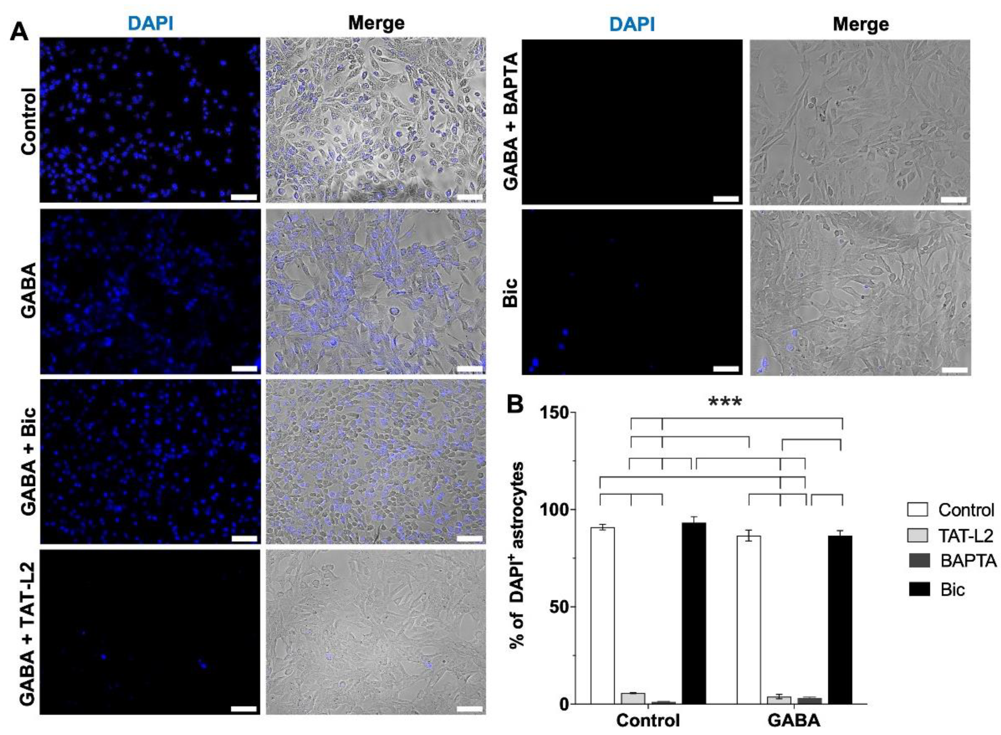
Figure 1. Increased DAPI uptake in DI TNC1 astrocytes incubated in a Ca2+-free solution is mediated by astroglial Cx43 hemichannels and depends on intracellular [Ca2+] but is unaffected by GABA or GABAA receptor antagonist Bicuculine. (A) Representative photomicrographs showing increased DAPI uptake in DI TNC1 astrocytes incubated in a Ca2+-free culture medium. Increased DAPI uptake was blunted by 10 nM TAT-Cx43L2 (TAT-L2) or 100 nM BAPTA-AM (BAPTA), but unaffected by 1 µM GABA alone (GABA) or 100 µM bicuculline (Bic). Scale bar: 50 µm. (B) Quantification of DAPI uptake. n = 3, with technical triplicates. Error bars: SE; *** p ˂ 0.001.
Ijms 23 13625 g001
Figure 2. GABA increases DAPI uptake in DI TNC1 astrocytes through Cx43 hemichannels via activation of GABAA receptors. (A) Representative photomicrographs showing DAPI uptake in DI TNC1 astrocytes under normal extracellular Ca2+ conditions (Control), show increased DAPI uptake in response to 1 µM GABA, effect that is prevented by preincubation with 100 µM bicuculline (Bic), 10 nM TAT-Cx43L2 (TAT-L2) or 100 nM BAPTA-AM (BAPTA). Scale bar: 50 µm. (B) Quantification of DAPI uptake. n = 3, with technical triplicates. (C) Concentration response curve for GABA on astroglial DAPI uptake performed to estimate the EC50 for GABA (EC50: 590 ± 30 nM). Error bars: SE; * p ˂ 0.05, ** p ˂ 0.01.
Ijms 23 13625 g002
Figure 3. The increase in DAPI uptake in DI TNC1 astrocytes in response to GABA is not mediated by GABAB receptors or Panx1 channels. (A) Representative photomicrographs showing DAPI uptake in DI TNC1 astrocytes under normal extracellular Ca2+ conditions (10 µM, control), showing increased DAPI uptake in response to 1 µM GABA, an effect that was unaffected by preincubation with 1 µM GABAB receptor antagonist CGP55845 (CGP), 10 nM mutant TAT-Cx43L2 H126K/I130N (TAT-L2mut) or 150 nM 10Panx1 (Panx1). Scale bar: 50 µm. (B) Quantification of DAPI uptake. n = 3, with technical triplicates. Error bars: SE; *** p ˂ 0.001.
Ijms 23 13625 g003
Figure 4. GABA induces the release of glutamate and ATP from DI TNC1 astrocytes, mediated by astroglial Cx43 hemichannels and the activation of GABAA receptors. (A) The extracellular glutamate concentration was determined after incubation with 1 µM GABA alone (GABA) or after preincubation with 100 µM bicuculline (Bic), 10 nM TAT-Cx43L2 (TAT-L2) or 100 nM BAPTA-AM (BAPTA). (B) Extracellular ATP concentration was determined after incubation with 1 µM GABA alone or preincubated with 100 µM bicuculline (Bic), 10 nM TAT-Cx43L2 (TAT-L2) or 100 nM BAPTA-AM (BAPTA). (C) Extracellular glutamate concentration was determined after incubation with 1 µM GABA alone or preincubated with 1 µM CGP55845 (CGP), 10 nM mutant TAT-Cx43L2 (TAT-L2mut) or 150 nM 10Panx1 (Panx1). (D) Extracellular ATP concentration was determined after incubation with 1 µM GABA alone or preincubated with 1 µM CGP55845 (CGP), 10 nM mutant TAT-Cx43L2 (TAT-L2mut) or 10Panx1 (Panx1). n = 3, with technical triplicates. Error bars: SE; * p ˂ 0.05, ** p ˂ 0.01, *** p ˂ 0.001.
Ijms 23 13625 g004
Figure 5. GABA increases astroglial Cx43 hemichannel activity in rat hippocampal slices via activation of GABAA receptors. (A) Representative photomicrographs showing ethidium (Etd+) uptake (red) and GFAP immunolabeling (green) in hippocampal slices. Scale bar: 50 µm. (B) Quantification of Etd+ uptake. Increased Etd+ uptake was found in response to 1 µM GABA (GABA), an effect that was prevented by preincubation with 100 µM bicuculline (Bic) or 10 nM TAT-Cx43L2 (TAT-L2). (C) Extracellular glutamate concentration from the culture medium of hippocampal slices without ligands (control), or after incubation with 1 µM GABA alone (GABA), or after preincubation with 100 µM bicuculline (Bic) or 10 nM TAT-Cx43L2 (TAT-L2). (D) Extracellular ATP concentration without ligand (control) or after incubation with 1 µM GABA alone (GABA) or preincubated with 100 µM bicuculline (Bic) or 10 nM TAT-Cx43L2 (TAT-L2). In all cases, n = 3, with technical triplicates. Error bars: SE; * p ˂ 0.05, ** p ˂ 0.01 and *** p ˂ 0.001.
Ijms 23 13625 g005
Publisher’s Note: MDPI stays neutral with regard to jurisdictional claims in published maps and institutional affiliations.