Article

Novel Virulence Factors Deciphering Klebsiella pneumoniae KpC4 Infect Maize as a Crossing-Kingdom Pathogen: An Emerging Environmental Threat

1
State Key Laboratory for Conservation and Utilization of Bio-Resources in Yunnan, Yunnan Agricultural University, Kunming 650201, China
2
College of Agronomy and Life Sciences and Engineering Research Center for Urban Modern Agriculture of Higher Education in Yunnan Province, Kunming University, Kunming 650214, China
*
Authors to whom correspondence should be addressed.
These authors contributed equally to this work.
Int. J. Mol. Sci. 2022, 23(24), 16005; https://doi.org/10.3390/ijms232416005
Submission received: 28 October 2022 / Revised: 21 November 2022 / Accepted: 29 November 2022 / Published: 15 December 2022
(This article belongs to the Special Issue Molecular Genetics and Genomics of Plant-Pathogen Interactions)

Abstract

:
Klebsiella pneumoniae is not only a human and animal opportunistic pathogen, but a food-borne pathogen. Cross-kingdom infection has been focused on since K. pneumoniae was identified as the pathogen of maize, banana, and pomegranate. Although the pathogenicity of K. pneumoniae strains (from ditch water, maize, and human) on plant and mice has been confirmed, there are no reports to explain the molecular mechanisms of the pathogen. This study uncovered the K. pneumoniae KpC4 isolated from maize top rot for the determination of various virulence genes and resistance genes. At least thirteen plant disease-causing genes are found to be involved in the disruption of plant defense. Among them, rcsB is responsible for causing disease in both plants and animals. The novel sequence types provide solid evidence that the pathogen invades plant and has robust ecological adaptability. It is imperative to perform further studies on the verification of these KpC4 genes’ functions to understand the molecular mechanisms involved in plant–pathogen interactions.

1. Introduction

Klebsiella pneumoniae is a notorious human and animal opportunistic pathogen for its serious nosocomial, community or healthcare-associated infections, and intractable multiple antibiotic resistance in the medical field [1,2]. It causes pneumonia, urinary tract infection, biliary tract infection, meningitis, bacteremia, and bloodstream infection in immunocompromised people and malnourished children [1,3], even in immunocompetent adults [4]. More and more attention has been drawn to the convergence of hypervirulence with multidrug-resistance of clinical K. pneumoniae [5].
Whereas, except on mucosal surfaces of mammals, K. pneumoniae also ubiquitously resides in the environment (soil, water, etc.) [6] and the surface of raw vegetables. Currently, studies revealed that K. pneumoniae frequently has been detected in fresh lettuce, arugula, cucumber, tomato, spinach, carrot, parsley, coriander, jute, and some herbs, even processed juice and canned food [7,8]. Symptoms of food poisoning such as abdominal pain, diarrhea, and vomiting appear after eating these foods contaminated with K. pneumoniae [8]. K. pneumoniae can reach and adhere to the surface of the plant, then colonize and undergo internalization. Shilpi et al. [7] revealed that K. pneumoniae (108 cfu/mL) can colonize on tomato leaves with no morphological or pathological changes. It is also likely to be related to the capsule and lipopolysaccharide of K. pneumoniae and its biofilm formation. As a food-borne pathogen, K. pneumoniae takes the plant as a reservoir or vector back to its human and animal hosts. The epiphytic or endogenous growth of K. pneumoniae is an important part of its life cycle. However, as a cross-kingdom pathogen which can infect plants, K. pneumoniae KpC4 can actively invade and exhibit cross-kingdom pathogenicity in maize [9], banana [10], and pomegranate [11], and cause top rot, leaf spot, and blight symptoms, respectively, in the field naturally, and cause sorghum leaf spot by artificial inoculation [12]. Huang et al. have illustrated that KpC4 and other K. pneumoniae strains still can survive in different environments and variant harsh conditions, such as soil, different quality water, dry filter paper, maize phylloplane, interior, kraurotic maize plant debris, poor nutrients, exposure to ultraviolet radiations, and unfit temperatures [12]. From the perspective of ecology, the extensive ecological adaptability of K. pneumoniae is the cross-kingdom infection mechanism of KpC4.
There are several virulence factors such as capsule, lipopolysaccharide, fimbriae, siderophores, outer membrane proteins, and type VI secretion systems [3,13] associated with human and animal infections that have been well described. Whether these virulence factors are also the agents of plant diseases requires further study. Previously, studies revealed that, under the experimental conditions, some human opportunistic pathogenic bacteria such as Salmonella enterica, Pseudomonas aeruginosa, and Enterococcus faecalis can infect plants using same virulence factors with humans and animals [14]. Interestingly, K. pneumoniae KpC4 has been demonstrated that it also has the same pathogenicity as the clinical strain Kp138 (K1 serotype) to infect mice; and the clinical strain (Kp138) and environmental strain E4 (K1 serotype) can cause maize top rot [9,12]. It is likely that KpC4 uses the same or different strategies (disease-related factors) to infect plants. Bioinformatics plays an important role in evaluating potential virulence factors of the microbes. In silico approaches, such as complete genome sequencing, identification of crucial genes of the pathogen responsible for the virulence and searching the homologous proteins between K. pneumoniae and plant pathogenic bacteria, could be used to reveal the pathogenesis of crossing-kingdom infection of KpC4. However, these hypothetical pathogenic mechanisms require further phenotypic and functional verification. To gain insights into the genetic elements involved in maize infection and cross-kingdom infection mechanisms, complete genome sequencing of K. pneumoniae KpC4 was performed to explore the hidden molecular mechanisms involved in its interactions inside the host.

2. Results

2.1. Genome Features

The genome of KpC4 is composed of a circular chromosome of 5,218,784 bp (Figure 1) with an overall G+C content of 57.26%, which is similar to previous reports of K. pneumoniae NTUH-K2044 (57.7%), MGH 78578 (57.5%), and 342 (57.3%) [15]. The chromosome encodes 4912 putative coding sequences (CDS) representing 87.3% coding density, and the total length of the coding region is 4,556,229 bp with G+C content of 58.87%. The average chromosomal gene length was found to be 927 nucleotides. A total of eighty-three tandem repeats and five transposons were found. Consistent with MGH 78578, Kp13, and NTUH-K2044, 86 tRNA genes with specificities were identified. The number of rRNA genes (16s-23s-5s) were eight, eight, and nine, respectively, which is same as NTUH-K2044 [16]. The characteristics of the genome and its comparison with other genomes of related K. pneumoniae are shown in Table 1.
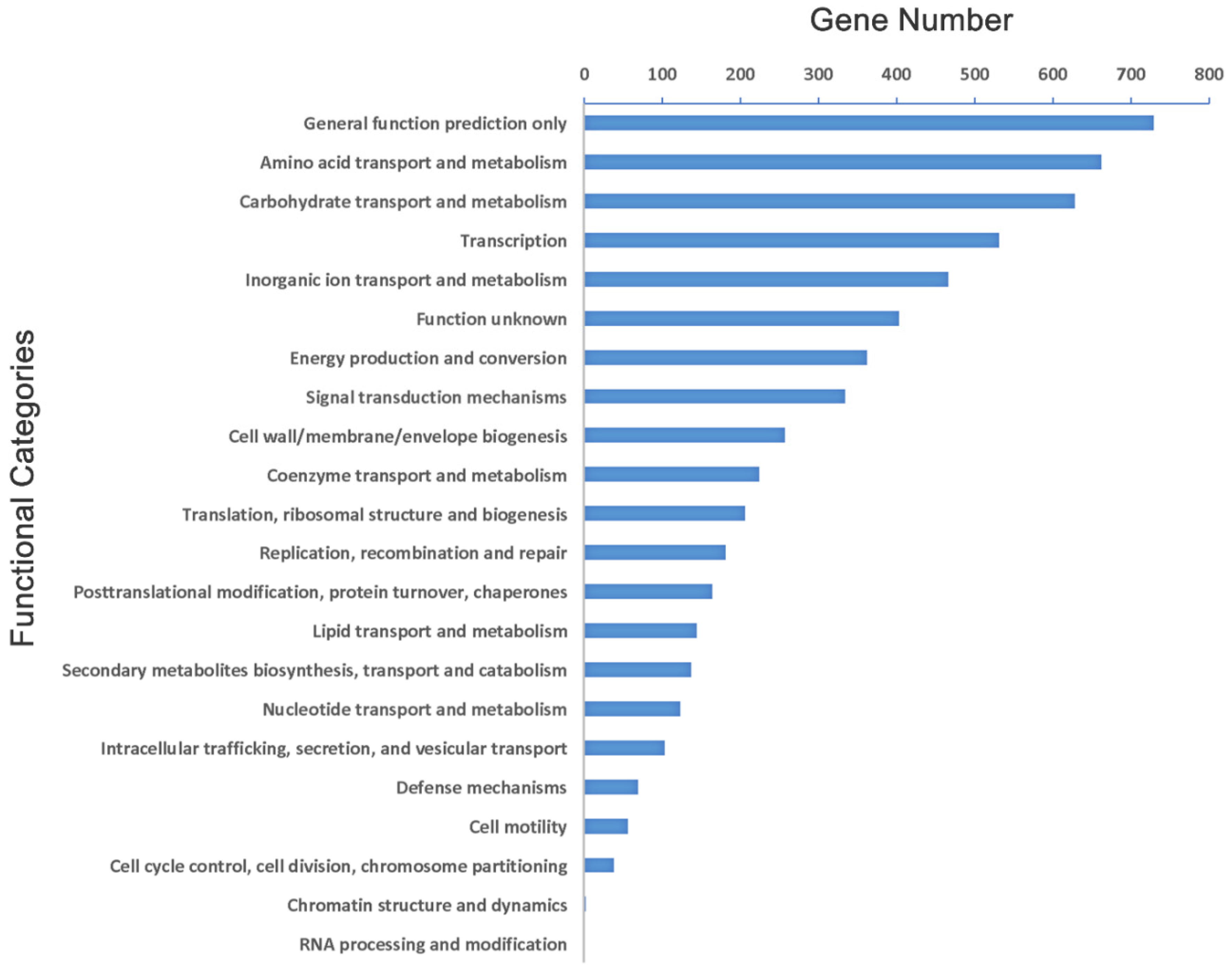
The preliminary analysis of the genome suggests that 4509 (91.8%) of the CDSs can be assigned biological role categories, while 403 (8.2%) have been annotated as enzymes of unknown function. Analysis of cluster of orthologous group (COG) showed that there were 729 genes with general function, 662 genes involved in amino acids transport and metabolism, while only a single gene involved in RNA processing and modification (Figure 2).

2.2. The Survival Mechanism of Strain KpC4 in Plants

K. pneumoniae is ubiquitous and has a wide host range in nature. There are alternative plant hosts besides humans and animals [12]. K. pneumoniae developed a survival strategy to overcome the stage of nutrient deficiency and adversity stress in the long process of evolution and adaptation. For example, K. pneumoniae could encode various transporters involved in the absorption of carbohydrates, amino acids, and iron, and enzyme systems which decompose some undegradable macromolecules in plants, or improve the tolerance to some toxic substances, such as heavy metals and chemical pesticides, in the environment.

2.3. Carbohydrate Metabolism

Like most microbes, KpC4 could use carbohydrates to produce the substances necessary for its own growth and reproduction. Carbohydrate is a kind of important organic matter widely existing in plants, accounting for more than 50% of plant dry weight. The main components of the plant cell wall are cellulose, hemicellulose, pectin, and lignin.
Cellulose is the main component of the plant cell wall and the most abundant carbohydrate in the biosphere, which has a chemical structure composed of a straight chain macromolecular compound of glucose joined by a β-1,4 glucosidic bond. The decomposition of cellulose in organisms is mainly by enzymolysis. Three enzymes, endoglucanase, exo-β-1,4-glucanases, and beta-glucoside (cellobiohydrolases), combine to break down cellulose. There are at least two genes (endoglucanase/endo-1,4-D-glucanase, KpC4_4548 and endoglucanase Y, KpC4_4556) in the KpC4 genome, which were confirmed to have the ability to degrade highly ordered forms of insoluble cellulose. Additional genes encoding enzymes with specificity towards 1,4-β-glucosidic bonds which most likely act by hydrolyzing short cellooligosaccharides include: interperimental bglX (beta-D-glucoside glucohydrolase, KpC4_0991), and similar bglA (6-phospho-beta-glucosidase, KpC4_0194), bglB (KpC4_0496, KpC4_0706, KpC4_0983), bglG (Beta-glucoside bgl operon antiterminator, BglG family, KpC4_0981; transcription antiterminator BglG, KpC4_3805), bglH (aryl-phospho-beta-D-glucosidase, KpC4_0983, KpC4_0496; cryptic outer membrane porin BglH, KpC4_4311), bglC (Aryl-phospho-beta-D-glucosidase, KpC4_1604). Additional β-glucosidase (KpC4_0991, KpC4_0331, KpC4_0480), 6-phospho-beta-glucosidase (KpC4_0706, KpC4_0983, KpC4_1604, KpC4_1875, KpC4_2197, KpC4_2324, KpC4_0194, KpC4_2726, KpC4_3798, KpC4_0301, KpC4_4418, KpC4_0480, KpC4_0496), aryl-phospho-beta-D-glucosidase (KpC4_1604). Although the gene encoding the cellobiohydrolases is absent, the enzymes above could decompose cellulose to produce oligosaccharides and cellobiose, and eventually be hydrolyzed to glucose for cell life.
Genomic analysis revealed that KpC4 has ability to catalyze many different forms of hemicellulosic substrates into fermentable sugars. For instance, the KpC4 genome possesses the genes to metabolize xylan, arabinose, and xylose, including duplications of xylA (Xylose isomerase, KpC4_4508, KpC4_3736) and xylB (xylulokinase, KpC4_4509, KpC4_1019) which are responsible for creating the phosphorylated derivative, D-xylulose 5-phosphate. β-1,4 mannanase (KpC4_0479) hydrolyzes mannosan by acting on the mannose bond of the mannitosan main chain. Xylanase (KpC4_0981, KpC4_4312) can cut the β-1,4 glucosidic bonds from the main chain of xylose to form oligoxylose and a small amount of xylose [18], which plays an important role in the hydrolysis of xylanase. Both enzymes are endonucleases, which can randomly cut off the glucosidic bond of the main chain to generate oligosaccharides. Then, the oligosaccharide is digested by different glycosidases, including β-mannosidase (KpC4_2183) or β-xylosidase (KpC4_1060, KpC4_0106, KpC4_3738) with external cutting. While glycoside hydrolases, such as galactosidase (KpC4_3154), α-galactosidase (KpC4_3944, KpC4_4237), β-galactosidase (KpC4_1604, KpC4_3153, and KpC4_3837), and β-D-galactosidase (KpC4_1958), can hydrolyze galactoside into monosaccharides. In addition, the genome also possesses β-1,4-xylosidase (KpC4_3738) responsible for the hydrolysis of 1,4-β-D-xylans, α-N-arabinofuranosidase (KpC4_3739), and arabinogalactan endo-1,4-beta-galactosidase (KpC4_3154). Arabinofuranosidases work synergistically with xylanases to degrade xylan to its component sugars.
Pectin is also a major component of plant cell walls. Pectinase is often secreted by pathogenic bacteria to degrade the cell wall during infection. Essential in pectin metabolism of oligogalacturonate lyase (pectate lyase family 22 (EC:4.2.2.6), KpC4_0283) and its oligogalacturonate transport system permease protein (OgtB, KpC4_0294), pectin methylesterase (KpC4_2787) can be found in the KpC4 genome. Pectin methylesterase can utilize the intermediate layer and the pectin of the cell wall, and eventually lead to the death of host tissue, playing a vital role in the pathogenicity of the Pectobacterium atrosepticum. The pectinesterase of Erwinia carotovora and Aspergillus niger is related to the segregation and soft rot of plant tissue. Furthermore, the KpC4 genome also encodes genes capable of degrading the α-linked glucans (primarily 1,4-α and 1,6α-linkages) of plant starches as well as the degradation of low-molecular-weight carbohydrates produced from their breakdown such as maltodextrins, pullulan, and D-galacturonate (Additional Table S1).

2.4. Aromatic Compounds Degradation

Aromatic compounds, common in plant cells, are widely and plentifully distributed in the environment and are mainly derived from the decomposition of plant lignin in nature [19]. These compounds serve as signals for plants to perceive when bacteria approach, which may play an important role in colonizing plants [20]. Genomic analysis confirmed KpC4 has the potential to oxidatively catabolize various low-molecular-weight aromatic compounds, most of which are derived from lignin degradation, including ferrulic acid, vanillate (KpC4_1900, KpC4_1901, KpC4_1649, KpC4_1529), 2-chlorobenzoate (KpC4_1702- KpC4_1700), the central aromatic ring metabolites, protochatechuate, and catechol [21,22]. Ring cleavage is mediated by 3,4-protocatechuate dioxygenase (KpC4_1617-KpC4_1618) and catechol 1,2-dioxygenase (KpC4_1699) in the protocatechuate pathway and catechol ortho cleavage pathway, respectively. A complete β-ketoadipate pathway (KpC4_2033-KpC4_2031) of the KpC4 genome is responsible for further decomposition of the ring cleavage degradation products to TCA cycle intermediates [21,22]. Additionally, genomic analyses revealed that the KpC4 genome contain genes that degrade certain aromatic compounds by reduction or non-oxidative decarboxylation, such as several 4-hydroxybenzoate decarboxylase enzymes (KpC4_0453-KpC4_0452), 3,4-dihydroxybenzoate decarboxylase (KpC4_2404), and 3-octaprenyl-4-hydroxybenzoate decarboxylase (KpC4_4106), mainly for decarboxylation of 4-hydroxybenzoic acid, generation of phenol, and CO2.

2.5. Survival against Plant Defenses

2.5.1. Evade the Plant’s Defense System

Pathogenic bacteria, in the process of infecting host plants, establish parasitism with plants, damage the normal physiological and metabolic function of host cells, regulate the adhesion, infection, colonization and expansion of plants, and finally cause the plants to show symptoms. On the other hand, plants also utilize various non-specific strategies to defend threats from bacteria, viruses, and fungi, including the production of reactive oxygen species (ROS), such as superoxide, hydrogen peroxide, hydroxyl radical, hydroperoxyl radical, nitric oxide (NO), and phytoalexins [23,24]. The genome of KpC4 encodes a powerful enzyme system that protects itself from all three plant defense mechanisms. There are three superoxide dismutases, soda (KpC4_4223), sodB (KpC4_1571) and sodC (KpC4_1580), as well as three catalases (KpC4_1752, KpC4_2322, KpC4_2412), one thioredoxin reductase (KpC4_2652), two glutathione peroxidases (KpC4_1392, KpC4_2374), sixteen NADH dehydrogenases (KpC4_0881-KpC4_0892, KpC4_2461, KpC4_0332, KpC4_4883, KpC4_0489), a NADH dehydrogenase transcriptional regulator (KpC4_1768, LysR family NADH dehydrogenase transcriptional regulator, KpC4_0879), four peroxiredoxin OsmC (KpC4_1741, KpC4_0715, KpC4_2915, KpC4_3234), two thiol peroxidases (KpC4_0715, KpC4_2194), two predicted iron-dependent peroxidases (KpC4_0762, KpC4_2514), one hydroperoxide reductase (encoded by ahpC, KpC4_2915, KpC4_3234, and ahpF, KpC4_2914), and thirteen glutathione S-transferases (GST) (KpC4_0867, KpC4_0868, KpC4_1592, KpC4_1674, KpC4_0110, KpC4_2703, KpC4_2975, KpC4_3211, KpC4_3337, KpC4_3987, KpC4_4497, KpC4_4571, KpC4_4803) (compared to seven in E. coli K12, twelve in Kv342), which protects itself against ROS. In addition, the genome encodes flavodoxin reductase (ferredoxin-NADPH reductase family 1, KpC4_2103, KpC4_2598) (compared to one in Kv342 [17]) and anaerobic nitrate reduction operon (KpC4_0505, KpC4_0503-KpC4_0502), which make KpC4 capable of detoxification of the free radical nitric oxide [25]. Additionally, it also possesses the RND-family AcrAB and efflux pump (multidrug transport protein, outer membrane (RND family), KpC4_1034, KpC4_1415, KpC4_2792, KpC4_4391, KpC4_3139, KpC4_3007, KpC4_3721, KpC4_0574), multidrug transporter AcrB (KpC4_2793, KpC4_3140), MDR efflux pump AcrAB, transcriptional activator MarA (KpC4_1941), and MDR efflux pump AcrAB transcriptional activator RobA (KpC4_3605) necessary for the export of apple tree pytoalexins by E. amylovora [26].

2.5.2. Plant-Induced and Associated Genes

Many studies have shown that plant-inducible genes of bacteria can be induced and expressed in vivo when bacterial cells colonize or grow on plants. These genes not only exist in plant pathogens [27], but also in animal pathogens [17]. Several plant-induced genes are involved in the bacterial response to oxidative stress and DNA damage caused by the plant defense response, as well as DNA damage repair. Genomic analysis showed that some genes in the genome of KpC4 are homologous with these plant-induced genes. For example, some amino acids and nucleotide biosynthesis genes of KpC4 were highly homologous with the induced genes of Ralstonia solanacearum and Pseudomonas syringae pv. tomato when they colonized the host plants. Genes with this trait include CTP synthase (pyrG, KpC4_0414), acetyl-CoA acetyltransferase (KpC4_0281, KpC4_1958), amidophosphoribosyl transferase (purF, KpC4_0855), argininosuccinate synthase (argG, KpC4_4851), diaminopimelate decarboxylase (lysA, KpC4_0298), and acetolactate synthase large subunit (ivlI, KpC4_3516) [28,29].
Some assumed stress response genes expressed in R. solanacearum which in response to plant defense during colonization in host plants were also found in KpC4. For example, a regulatory protein of the adaptive response (ada, KpC4_0926), excinuclease ABCD (uvrA, KpC4_4009, UvrB, KpC4_2773, uvrC, KpC4_1150, uvrD, KpC4_4137), DNA-damage-inducible protein F (dinF, KpC4_4023), fumarate hydratase (fumC, KpC4_2057), and acriflavin resitance protein A (acrA, KpC4_3139) [29]. Among these, ada is essential for transcriptional activation of genes that are involved in the adaptive response to DNA methylation damage and activated by NO [30]. uvrABCD are involved in UV-induced DNA repair from damage recognition to repair in concert. In addition, uvrA is also involved in the repair process of DNA damage induced by H2O2 and toxic chemicals indicating that this gene can protect bacteria from DNA-damaging compounds produced by plants [31]. These oxidative response genes are not limited to DNA repair pathways. While fumC, apart from being part of the TCA cycle, has been found to be efficiently expressed when superoxide radicals accumulate [32]. Another form of fumarate hydratase, encoded by fumA, is inactivated under oxidation [32,33]. The early stage of the plant defense response mainly involves ROS accumulation, so the induction of oxidation-stress-related genes indicates that bacteria, such as KpC4, actively evade defense mechanisms when colonizing plants. HRP-dependent type III effect of protein was also identified in the genome of KpC4, but no non-toxic proteins.

2.5.3. Plant Disease-Causing Genes

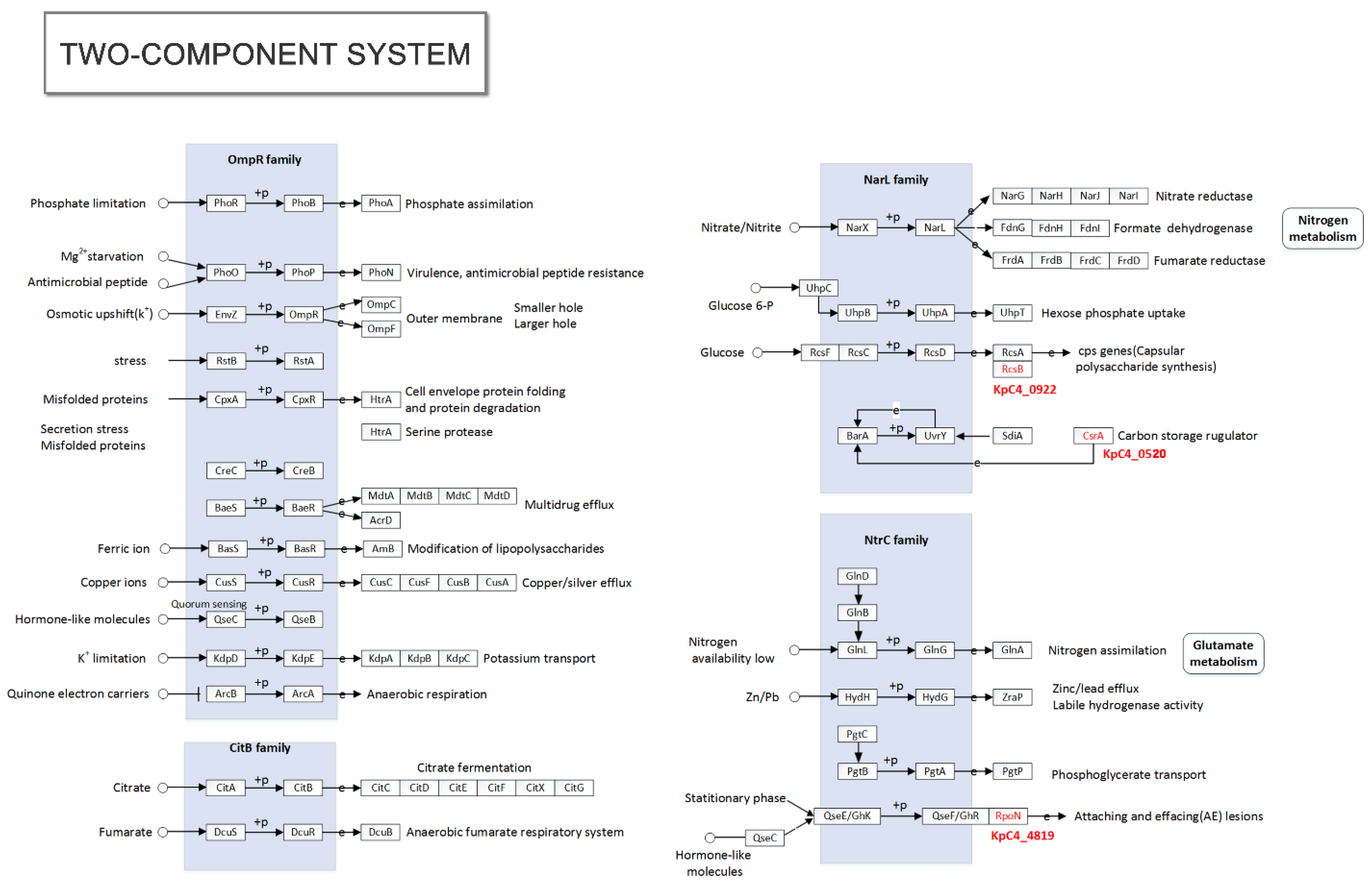
The two-component system is the most widely existing in bacteria as a combined mechanism of stimulating response to environmental change and response. The two-component system of KpC4 is composed of OmpR, CitB, NarL, and NtrC (Figure 3), including sensing and regulating nutrients (NarX-NarL), anaerobic respiration (ArcB-ArcA), osmotic pressure (EnvZ-OmpR), antibiotic (BaeS-BaeR), and gene clusters important for controlling cell growth, virulence (PhoO-PhoP), biofilms (CpxA-CpxR), and quorum sensing (QecC-QseB).
Notably, thirteen protein sequences of plant pathogenic bacteria that are more than 70% the same as the KpC4 in the PHI (pathogen host interactions) database were found (Table 2). The thirteen genes are mainly the pathogenic genes of bacteria of Pectobacterium, Erwinia, Xanthomonas, and Pantoea. Among them, the rcsB, rpoN of E. amylovora, and rsmAXoo of Xanthomonas oryzae pv. oryzae were 92.09%, 84.07%, and 82.14% identical with the query of KpC4 (KpC4_0922, KpC4_4819, and KpC4_0520) respectively, which were all related to the two-component system (Figure 3).
Interestingly, the rcsB gene, the pathogenic factor of E. amylovora (pear fire blight), is a transcriptional regulator of capsular polysaccharide synthesis [34]. Auxiliary transcriptional regulators (rcsA), are highly conserved in many bacteria, including human and animal pathogenic bacteria (E. coli, K. pneumoniae), as well as some plant pathogens, such as E. amylovora and Pectobacterium atrosepticum (rotting of tubers of potato). The alignment results showed that the homology of the RcsB protein sequences between KpC4 and K. pneumoniae clinical strains MGH78578, NTUH-K2044, Kp13, and Kp52.145 were 100% (Figure 4), and 100 K. pneumoniae strains on NCBI were also 100%.
It has been proved that RcsB has a positive regulatory effect on the high mucosity and biofilm formation of K. pneumoniae NTUH-K2044 [35]. These two characteristics are not only the important pathogenic factors of animals, but also of plants. The rcsB gene of E. amylovora was considered as the pathogenic gene of pear fire blight because it facilitates the synthesis of capsular polysaccharide. The capsule creates a favorable microenvironment for the bacterial cells and prevents it from water stress and plant defense reactions, as well as making the plants susceptible to present typical symptoms, such as wilting, necrosis, even bacterial ooze (the main component is capsule polysaccharide).

2.6. Pathogenicity of KpC4

Genomic analysis showed that KpC4 encodes the main virulence factors possessed by K. pneumoniae-infected humans and animals.

2.6.1. Capsule

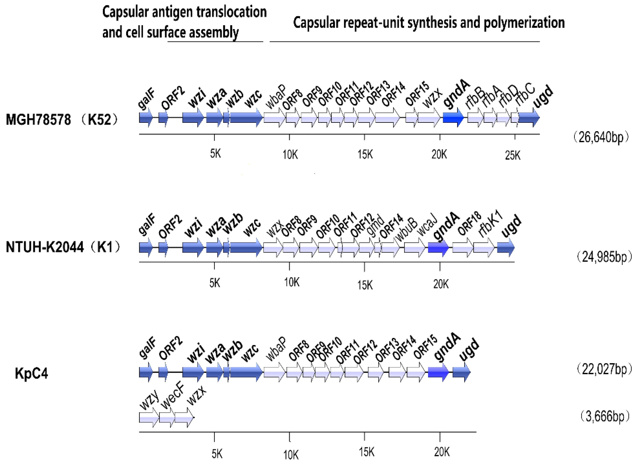
The cps (capsular polysaccharide synthesis) gene cluster of KpC4 consists of 17 genes (Figure 5, Additional Table S1), including several genes related to capsule synthesis. Similar to NTUH-K2044 and MGH78578, cps gene clusters of KpC4 are located downstream of galF and ORF2, and wzi (orfX)-wza-wzb-wzc genes cluster together. galF, OFR2, wzi(orfX), wza, wzb, wzc, and gnd are conserved in K. pneumoniae strains of different serotypes. GalF (UTP–glucose-1-phosphate uridylyltransferase, KpC4_1048), ORF2 (acid phosphatase, KpC4_1049), and gnd (gluconate-6-phosphate dehydrogenase, KpC4_1063) are involved in carbohydrate metabolism [36]. wzi (capsule assembly protein, KpC4_1050) encodes a surface protein that is related to the capsule adhesion to the outer membrane; bacteria could not form capsule if Wzi is deficient. Wza (polysaccharide export protein, KpC4_1051) is related to surface assembly. Wzb (KpC4_1052) encodes low molecular weight protein-tyrosine-phosphatase. Wzc (KpC4_1053) encodes a tyrosine protein kinase, which is responsible for translocation and surface assembly of the capsule [37,38].
The cps loci of K2044(K1), KpC4, MGH78578(K52) encode different serotypes, and some functions are similar, such as encoding glycosyl transferase (Additional Table S1). However, there are distinct differences in the sequences of orthologous genes and the deficiency or acquisition of genes. The wbap-ORF15 domain of KpC4, wzx-rfbp of K2044, and ORF8wzx of MGH78578(K52) show lower similarity (Figure 5), probably as these non-conservative regions are unique to the different serotypes. For example, wbaP (KpC4_1054) and wbaP of MGH78578 are specific in some serotypes, while rfbP is relatively specific in certain serotypes. wzy in the genome of KpC4 is highly homologous with the gene of MGM78578, which is related to the polymerization of capsular polysaccharide. wzx (O-antigen translocase, KpC4_4154) and wzy (common antigen polymerase, KpC4_4152) cluster individually in the genome of KpC4. Additionally, magA (one kind polymerase similar to wzy) involved in capsular formation, could not be found in the genome of KpC4, which coincides with the experimental result of KpC4 of the non-K1 serotype.
The synthesis of KpC4 capsular polysaccharide was mainly encoded and controlled by rcsF (KpC4_3382), rcsC (KpC4_0921), rcsD (KpC4_0923), rcsA (KpC4_1140), and rcsB (KpC4_0922), which belong to the NarL family of transcriptional regulation [39].

2.6.2. Lipopolysaccharide

Lipopolysaccharide (endotoxin), the component of the cell wall of Gram-negative bacteria, is composed of lipid A, core oligosaccharide, and O antigen polysaccharide, encoded by lpx, waa, and wb gene clusters, respectively [40,41]. The lipid A biosynthetic pathways of KpC4 are controlled by enzymes which encoded by lpxA (UDP-N-acetylglucosamine acyltransferase, KpC4_3397), lpxC (UDP-3-O-acyl N-acetylglucosamine deacetylase, KpC4_3497), lpxB (lipid-A-disaccharide synthase, KpC4_3396), lpxD (UDP-3-O-(3-hydroxymyristoyl)-glucosamine n-acyltransferase, KpC4_4042) and lpxK (tetraacyldisaccharide 4′-kinase, KpC4_2620).
The whole Waa gene cluster is located between kbl and coaD, of which E. coli k-12 contains 12 genes [42]; O-antigen, located in the outermost layer of lipopolysaccharide, is composed of oligosaccharide repeating units. Wb gene clusters (manC, manB, wzm, wzt, wbbD, wbdA, wbdB, wbdC, hisI) usually include biosynthetic genes, such as active sugar, glycosyltransferase, O antigen polymerase, and O antigen output protein [43]. The whole Waa gene cluster of KpC4, including 13 genes (Table 3), is located between kbl and coaD, while E. coli k-12 contains 12 genes [42]. Interestingly, the wb gene clusters encoding o-antigen polysaccharide were not found in the genome of KpC4. Perhaps the variation of wb gene clusters could be related to the biosynthesis of the O-antigen due to the high chemical variability of the o-antigen [43].

2.6.3. Adhesin

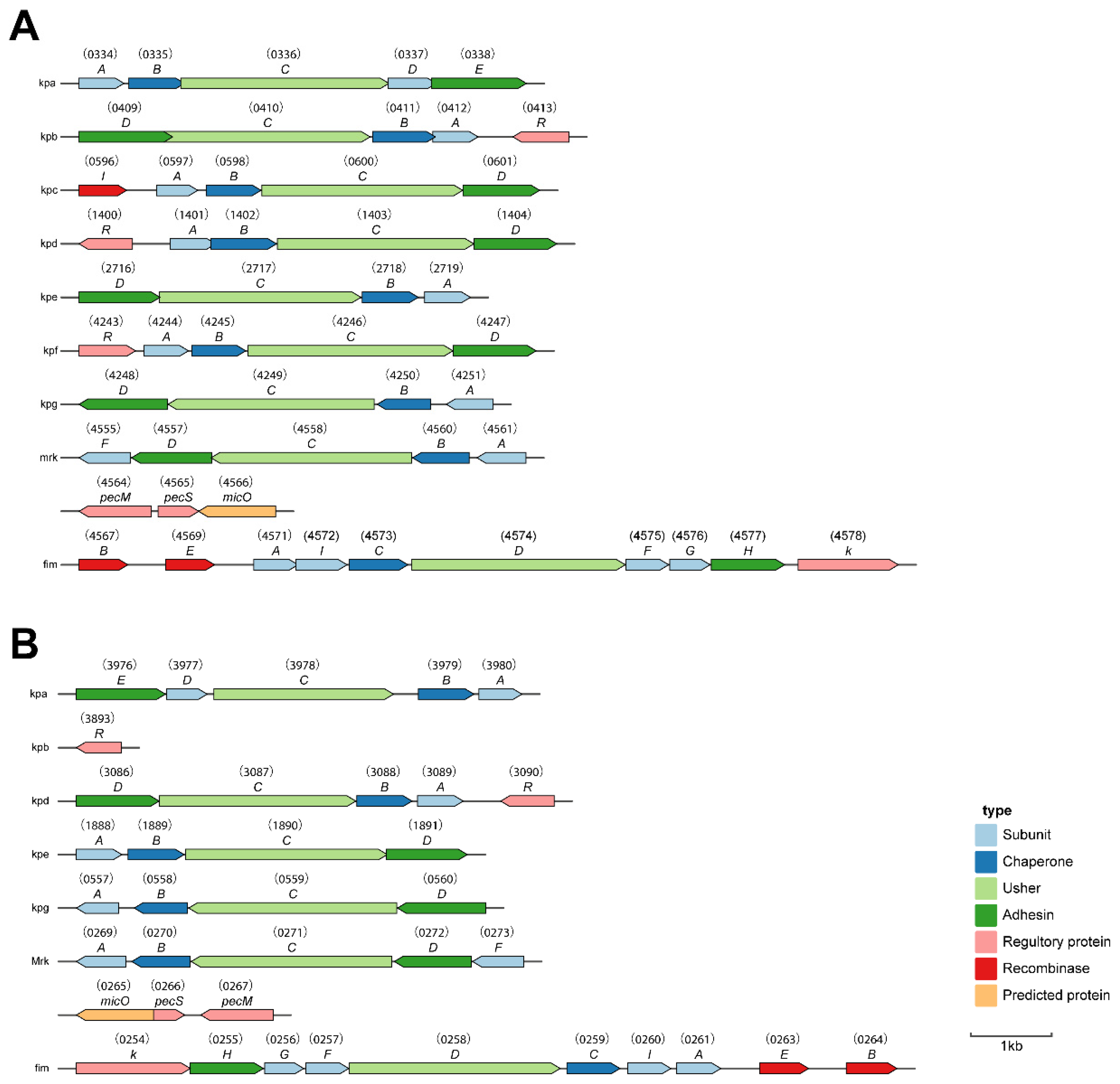
The expression of adhesin plays an important role in the colonization of bacteria and is also the first condition of the pathogenicity of an organism. The adhesive factors of K. pneumoniae mainly include type I and Ⅲ fimbriae, and non-fimbrial adhesion protein (Figure 6). Fimbriae are related to bacterial adhesion and colonization of pathogenic processes of K. pneumoniae. Currently, nine gene clusters, including fim, mrk encoding type I and III fimbriae, and seven (kpa, kpb, kpc, kpd, kpe, kpf, and kpg) gene clusters recently discovered, have been identified to be related to the biosynthesis of K. pneumoniae fimbriae. Each gene cluster contains at least four genes, which respectively encode a chaperone–usher-dependent assembly system including the fimbrial protein, molecular chaperone, usher protein, and adhesin required for fimbriae biosynthesis. Most of the genes encoding adhesins can be found in the genomes of KpC4 and its close relatives (Additional Table S2).
KpC4 encodes a complete genetic sequence of the type I fimbriae gene cluster (fimBEAICDFGH, KpC4_0254-KpC4_0264). Studies have shown that these gene products enhanced K. pneumoniae toxicity in urinary tract infections. KpC4 also carries the fimK (KpC4_0254) gene not found in E. coli, which is located downstream of the fimH. Type I fimbria could not express if fimK was deficient [44,45]. Type Ⅲ fimbriae encoded by the mrk gene cluster (mrkABCDF, KpC4_0269-KpC4_0273) can mediate the biofilms formation of K. pneumoniae on biotic and abiotic surfaces (such as the catheter in a hospital environment) [46]. This gene structure also includes homologs, including pecM (KpC4_0267), pecS (KpC4_0266), and nicO (KpC4_0265), which are conservative in KpC4, MGH78578, NTUH-k2044, and Kv342 [17,47,48].
Figure 6. Fimbrial gene clusters of the chaperone–usher-dependent assembly class in K. pneumoniae KpC4 and NTUH-K2044. (A) NTUH-K2044 [47]. (B) KpC4. The locations and orientations of the open reading frames (ORFs) are indicated by arrows. The designation of putative fimbrial genes and the locus tag of ORFs were annotated in the K. pneumoniae KpC4 and NTUH-K2044 by KpC4_number and KP1_number, respectively. Each of the putative fimbrial operons is underlined. The putative functions of the ORFs are also shown.
Ijms 23 16005 g006
Additionally, Kpa, kpd, kpe, and kpg gene clusters, which are related to the expression of these virulence factors [47] were found in the genomes of both KpC4 and NTUH-K2044 except type I (fim) and type III (mrk) adhesins. After comparison and analysis, kpc and kpf gene clusters were not found in the genome of KpC4 (Figure 7), only transcriptional regulatory proteins (KpC4_3893) were encoded by the kpb gene clusters, while these six gene clusters were conservative in the genomes of NTUH-K2044, MGH78578, 342, and KP13. Further comparative genomic analysis revealed that NTUH-K2044 and Kp13 possess nine complete fimbriae gene clusters. No gene homologous with kpc was found in KpC4, MGH78578, and 342. Wu et al. [47] analyzed 105 K. pneumoniae clinical strains with epidemiology and found that the expression of induced recombinants with the kpc ABCD genes in E. coli resulted in the formation of fimbriae and increased biofilm formation, revealing that the kpc gene was highly correlated with the K1 serotype, which is consistent with the experimental results in the literature that KpC4 [12] and MGH78578 (K52) [48] are not of the K1 serotype. Notably, kpf gene clusters, found in clinical strains MGH78578, K2044, and Kp13, were absent in both KpC4 and 342. K. variicola and K. pneumoniae are closely related sister species, and both environmental and clinical strains have extremely similar phenotypic and biochemical characteristics. The 342 (actually K. variicola [20,49], nitrogen-fixing endophyte) and KpC4 (crossing-kingdom pathogen infecting plants and animals) strains have unique ecological niches. Therefore, whether the expression of kpf is related to different ecological niches or the pathogenicity of clinical strains needs further study.
In addition, studies have shown that CDSs (poly-β-1,6-Nacetyl-D-glucosamine (PGA) adhesion, KpC4_3932-KpC4_3935) encoding the synthesis and transport of PGA adhesin in the KpC4 genome are essential for the stability of the E. coli structure [50]. Experiments showed that the regulatory factor of E. coli high adhesion phenotype, YidE [51] (KpC4_4342), was related to the survival and proliferation of pathogenic bacteria in the host of the sepsis mouse model [52]. The two-component system barA/uvrY contributes to the biofilm formation of Salmonella enterica, which is the virulence factor for E. coli urinary tract infections and was found in the KpC4 genome (KpC4_0410/KpC4_1149). The luxS (KpC4_0525) and luxR family protein (KpC4_1038), encoded the synthesis of autoinducer-2 signaling molecules of K. pneumoniae quorum sensing, which were not only important for the biofilm formation, but also conducive to the colonization of pathogens in the host and the improvement of antibiotic resistance [53].

2.7. Siderophores and Transporters

Iron is a necessary element for bacterial growth. At least twelve iron uptake systems of K. pneumoniae have been confirmed, including four main categories: Fe2+ transporters, ABC transporters, iron blood carrier absorption systems, and iron carrier absorption systems. Among them, the ABC transporter Kfu [54], Sit [55], and siderophores systems (Yersinia high-pathogenicity island [56], Iuc and IroA [57]) have proved to be essential for the virulence of K. pneumoniae (Table 4).
KpC4 has a complete iron transport system (Additional Table S3). Feo (ferrous iron transport) gene clusters (KpC4_4659-KpC4_4660) related to Fe2+ capture were found in the KpC4 genome. SitABCD system (KpC4_0468-KpC4_0471) related to the transport of bivalent cations such as Mn2+ and Fe2+, fecBDE gene clusters (KpC4_1409-KpC4_1411) related to ferric citrate transport, and Kfu gene clusters (KpC4_2567-KpC4_2569) all belong to the ABC transport protein family. It is worth mentioning that the Kfu gene cluster encoded by KpC4 is a highly pathogenic island; the iuc and iroA regions are highly correlated with the HvKP (hypervirulent K. pneumoniae) strains; iroN (outer membrane siderophore receptor, KpC4_2314) and IroE (salmochelin siderophore protein, KpC4_1881) are present in the genome of KpC4. This means that KpC4 has the potential to obtain virulence sites horizontally during the mutation from opportunistic pathogenicity to high virulence [58]. Although the iucABCD gene cluster only existed in plasmids of NTUH-K2044 (KP1_p319–KP1_p314), another ferric cell receptor (KpC4_2459) of KpC4 showed 99% similarity to the aerobactin receptor iutA (BLASTP [SwissProt:P14542]), which could also replace the function of the iucABCD gene cluster and bind exogenous chelating compounds (Additional Table S3).
Enterobactin (Ent) has the highest iron affinity compared to other iron chelators. The entABCDEF gene cluster (KpC4_2951-KpC4_2954, KpC4_2964, KpC4_2953) encoded by the genome of KpC4 is essential for Ent biosynthesis, and the fep gene cluster (KpC4_4005, KpC4_2963, KpC4_2955, KpC4_2957-KpC4_2959) is responsible for the transport of these siderophores. While the FhuABCD system (KpC4_3423-KpC4_3425, KpC4_3427) is involved in the absorption of ferrichrome, the ferrous heme transporter protein HmuRSTUV (KpC4_0458-KpC4_0462) can also be found in the KpC4.

2.8. Comparative Genome Analysis

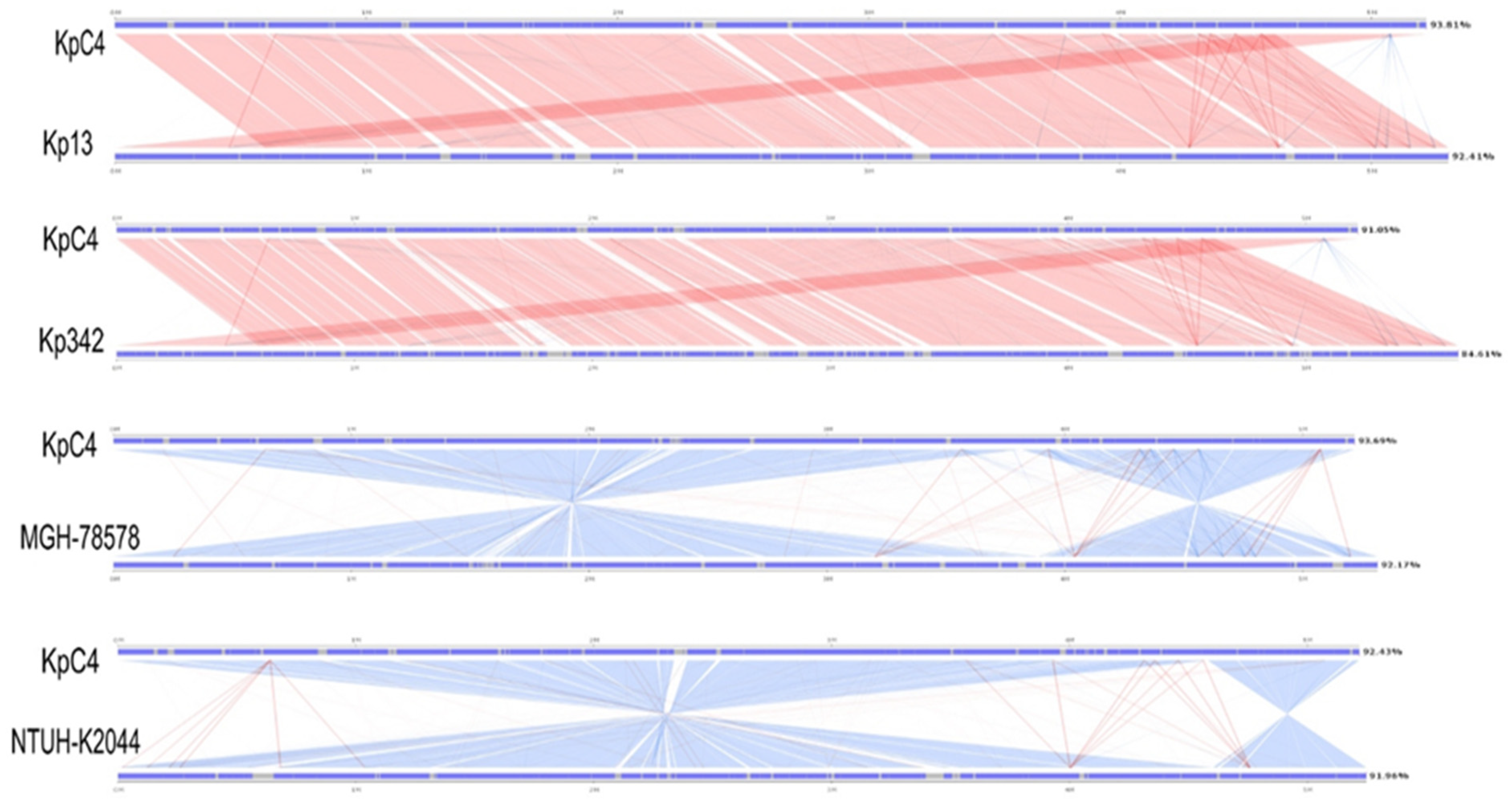
Linear analysis of KpC4, Kv342, and clinical strains MGH 78578, NTUH-K2044, and Kp13 was conducted using SBV (https://github.com/genedenovo/SBV, accessed on 13 June 2020)) software. Comparison of the genomes reveals that the gene structure of KpC4, Kv342, MGH 78578, NTUH-K2044, and Kp13 were highly conserved (Figure 7). However, the region of 4 Mb and 1 Mb (1-3816441, 3772923-5218784) in the KpC4 genome was reversed from the region of the MGH 78578 genome (5315120-3887253, 3896387-1). The same thing happened with NTUH-K2044. KpC4 was highly similar to the clinical strains Kp13 and Kv342, and the collinearity was also good.
The closer the evolutionary sequences, the greater the similarity (including sequence, structure, function, etc.). Except for the similarity of gene composition, the consistency of the sequence of genes on chromosomes in different genomes can better reflect the common origin of genomes. Genetic collinearity would be destroyed by various factors during evolution. The farther the evolutionary distance between species, the worse the genetic collinearity. The degree of collinearity between two species can be used as a measure of the evolutionary distance between them. Apparently, the strains of KpC4 and Kv342, MGH 78578, NTUH-K2044, and Kp13 have high similarity and evolutionary closeness. KpC4 not only has high similarity with Kv342 and Kp13, but also has good collinearity and higher degree of common origin.

2.9. Potential Drug Target of K. pneumoniae KpC4

The genomic data of the microbes is conducive to the identification of putative drug targets. These majority drug target candidates (Additional Table S4) which participate in essential processes such as fatty acids, LPS, peptidoglycan, pyrimidine deoxyribonucleotides, and purine nucleotide biosynthesis pathways of KpC4 were identified. It provides more efficient drug targets and therapeutic methods for ever-increasing multidrug-resistant Klebsiella and decreasing the available antimicrobial drug activity.

3. Discussion

Huang et al. [12] proved with a large number of experiments that K. pneumoniae is ubiquitous and able to survive in a variety of harsh environments for a long time, with a strong ecological adaptability. K. pneumoniae is not only a pathogen of humans and animals, but it can cross-kingdom infect plants [9]. Ecological adaptability is a survival strategy of K. pneumoniae and drives the ecological mechanism by which K. pneumoniae infects plants [12].
Here, the whole genome sequencing of KpC4 and the function analysis of some genes provide clues for further exploring the mechanism of cross-kingdom infection of K. pneumoniae in plants. KpC4 contains genes associated with survival, colonization, and capability of obtaining living energy materials in the host. Abundant K. pneumoniae strains have been isolated from the surface of vegetables [8,59,60,61], and KpC4 also can adhere to the surface of maize plants and seeds [12]. Here, genome sequencing provides evidence that the capsule, type I and Type Ⅲ fimbriae, and adhesin play an important role in KpC4 adhesion to plant surfaces, forming biofilms, and surviving. The biofilm formation is conducive to the adhesion and colonization of K. pneumoniae on plant surfaces [62]. Then, KpC4 enters the maize plant through wounds and natural orifices. Genome analyses identified that KpC4 encodes numerous genes involved in the metabolism of carbohydrate and aromatic compounds that can metabolize the cell wall components of plant hosts. The metabolism of these substances can not only promote KpC4 massive colonization and proliferation in plants, provide the material energy required for the basic life activities of KpC4, but also result in plant tissue segregation, decay, and disease. At the same time as it colonizes in the maize plant, KpC4 can protect itself from plant defense mechanisms (ROS, NO, and phytoalexins) by a powerful enzyme system. Among these, thirteen genes (twelve in Kv342, plant endophytic nitrogen-fixing bacterium [17]) encoded glutathione S-transferase (GST) against ROS; two genes (one in Kv342 [17]) encoded flavodoxin reductase, three genes encoded an anaerobic nitrate reduction operon against free radical NO. Additionally, phytoalexin can be excreted extracellular through multidrug transport protein and MDR efflux pump. This genome information implies KpC4 has stronger endophytic living ability than Kv342. This evidence is mutually confirmed with the fact that KpC4 can lead to the natural occurrence of maize top rot in the field, with typical symptoms of leaf margin incision and top rot [9].
Most noteworthy, KpC4 encodes plant pathogenic factors which include thirteen genes with highly similarity in nucleotide sequences (identity ≥ 70%) to the pathogenic genes of plant pathogens, and harbors virulence factors that have been shown to be virulent to humans and animals, such as capsular polysaccharide, lipopolysaccharide, adhesin, siderophores, antibiotic resistance and multiantibiotic-related efflux pumps. It has been demonstrated that K. pneumoniae strains from different sources have the same pathogenicity on mice [9,63,64,65,66], and the clinical strain Kp138 (K1 serotype) and environmental strain E4 (K1 serotype) also have a similar virulence on maize [9,12]. K. pneumoniae appears to use the same strategy to infect different hosts [12]. Our gene functional analysis also supports this view. For example, among the pathogenic genes of KpC4 which caused human/animal disease, the rcsB gene encoded a transcriptional regulator of capsular polysaccharide synthesis, which is also the key pathogenic gene of E. amylovora and E. stewartii, which can cause pear fire blight exhibiting bacterial ooze and maize bacterial wilt, respectively [67,68,69]. rcsB plays a vital role in promoting the synthesis of capsular polysaccharide and the biofilm of E. amylovora and E. stewartia [69]. Likewise, the main phenotypic characteristic of KpC4 infection in maize, bacterial ooze which mainly composed of capsular polysaccharide, has been observed overflowing from the whorls of the maize [9]. It can be deduced that capsular polysaccharide is the virulence factor, and rcsB is the key pathogenic gene for KpC4 cross-kingdom infection. K. pneumoniae can use same strategies to infect mammals and plants, which can make itself survive in the organisms of distinct kingdoms after leaving the human/animal host or environment. It not only expands the host range but increases the adaptability. It is the way of inexpensive cost on evolution in which it accelerates the cycle for pathogens from humans to the environment [70].

4. Materials and Methods

4.1. Strain Isolation and Verification

Klebsiella pneumoniae KpC4 was originally isolated as a plant pathogenic bacterium from the interior stems and leaves of maize bacterial top rot disease naturally infected in the fields of many areas of Yunnan Province, China [9]. Strain KpC4 was verified as K. pneumoniae using colony morphology and cultural characteristics, Gram-staining test, microscopic examination, physiological and biochemical tests, as well as 16S rRNA, rpoB, and gyrB amplification in molecular taxonomy [9].

4.2. Isolation and Purification of DNA for Library Production

Bacterial cultures were grown on Luria Bertani (LB) medium followed by the isolation of genomic DNA using the Bacterial DNA Kit D3350-01 from OMEGA.

4.3. Genome Sequencing

Genome sequencing was carried out using single molecule real-time (SMRT). KpC4 was sequenced and analyzed by Guangzhou Gene Denovo biotechnology Co., LTD., Guangzhou, China. The sequencing process mainly includes sample quality inspection, library building, BluePippin fragment screening, and on-machine sequencing.

4.4. Gene Prediction and Annotation

Genome was predicted by GeneMarkes (prokaryotic genome prediction software) to obtain detailed gene distribution and structure information. RepeatMasker software was used to predict the repeat sequence of the genome, rRNAmmer [71] software to predict rRNA, and tRNAscan software to predict the tRNA region and the secondary structure of tRNA. The predicted gene sequences were blast compared with various databases to obtain the protein with the highest similarity to the given gene sequence, and the protein functional annotation information.

4.5. Prediction and Functional Analysis of Disease-Related Genes

A BLAST (basic local alignment search tool, ftp://ftp.ncbi.nlm.nih.gov/blast/executables/blast+/LATEST/, accessed on 8 June 2020) local comparing library was built in Windows 10 platform. The protein sequences of KpC4 were used as database files for homology comparison, and 80% coverage, E < 10−5, and 90% identities were used as reference standards. When the protein sequences of different genera were searched, the identities of E < 10−5, 60% or more was considered that the gene and the matched gene had homology. When E > 10−5, the strain was unique and had no homology with other bacteria. Proteins translated from different databases were analyzed, and disease-related genes were manually searched for statistical analysis. Some key genes were compared with closely related K. pneumoniae clinical strains (MGH 78578, NTUH-K 2044, Kp13) and K. variicola 342 which is an endogenous nitrogen-fixing strain (Kv342). MGH 78578 (accession number: CP000647), a multiple drug-resistant MDR strain, isolated from pulmonary infection patients [52]; NTUH-K2044 (AP006725), hypermucoviscous, was isolated from patients with liver abscesses and meningitis [72]; Kp13 (CP003999) was isolated from the blood of patients with diabetes and cranial injury in the ICU ward [18]. Kv342 (NC011283) is an endophytic nitrogen-fixing bacteria with attenuated pathogenicity to mice [73], which had been misclassified as K. pneumoniae because of the relatively similar physiological and biochemical characteristics and genes sequences [74].

5. Conclusions

In the study, we determined that K. pneumoniae KpC4 had the ability of cross-kingdom infection of plants while maintaining the virulence factors of animal pathogens from the perspective of genomics. Like other K. pneumoniae isolates, KpC4 encodes virulence factors such as capsular polysaccharide, lipopolysaccharide, adhesin, siderophores, antibiotic resistance and multiantibiotic-related efflux pumps, which have been shown to be virulent to humans and animals. Intriguingly, KpC4 also harbors plant pathogenic factors which include thirteen genes with high similarity in nucleotide sequences (identity ≥ 70%) to the pathogenic genes of plant pathogens. KpC4 has the molecular basis to survive in animals, environment, and even in plants. Our previous studies also proved that the strong ecological adaptability of KpC4 was the ecological basis of its cross-kingdom infection [12], which opens a whole new area of biological research. The survival mechanism makes KpC4 thriving and prosperous in plants, virulence factors that cause plant disease, same or different strategies to infect organisms in different biospheres, and the mechanisms that cause cross-kingdom infection, are the key hotspots of future research. This information might supplement our understanding of the mechanisms of K. pneumoniae cross-kingdom infection. It also laid the foundation for similar microbial evolution studies. The gene function of rcsB, and other plant disease-causing genes will be investigated further in our laboratory.

Supplementary Materials

The following supporting information can be downloaded at: https://www.mdpi.com/article/10.3390/ijms232416005/s1.

Author Contributions

Conceptualization, M.H. and Y.H.; Data curation, M.H., P.H. (Pengfei He), P.H. (Pengbo He) and S.M.; Formal analysis, P.H. (Pengfei He) and P.H. (Pengbo He); Funding acquisition, M.H. and Y.H.; Investigation, P.H. (Pengfei He); Methodology, M.H., P.H. (Pengbo He) and S.M.; Project administration, Y.W.; Resources, Y.W. and Y.H.; Software, S.M.; Supervision, Y.H.; Writing—original draft, M.H. and S.M.; Writing—review and editing, S.M. and Y.H. All authors have read and agreed to the published version of the manuscript.

Funding

This study was funded by the Key Program of Local Universities Joint Project of Yunnan Province (202101BA070001-140), China; and the program “Breeding and industrialization demonstration of green and efficient new maize varieties” (202102AE090023) from the Department of Sciences and Technologies, Yunnan Province, China.

Institutional Review Board Statement

Not applicable.

Informed Consent Statement

Not applicable.

Data Availability Statement

All the data is available in the manuscript and in Supplementary section. The sequencing information is deposited into the GenBank under BioProject number: PRJNA338238.

Acknowledgments

We thank Xiaoming Wang of Institute of Crop Science, Chinese Academy of Agricultural Sciences for his beneficial suggestions. Thanks to Bingjuan Yang of college of Agronomy and Life Sciences of Kunming University for drawing Figure 6.

Conflicts of Interest

The authors have declared no conflict of interest.

References

  1. Cubero, M.; Grau, I.; Tubau, F.; Pallarés, R.; Domínguez, M.Á.; Linares, J.; Ardanuy, C. Molecular epidemiology of Klebsiella pneumoniae strains causing bloodstream infections in adults. Microb. Drug Resist. 2018, 24, 949–957. [Google Scholar] [CrossRef] [PubMed]
  2. Tsai, S.S.; Huang, J.C.; Chen, S.T.; Sun, J.H.; Wang, C.C.; Lin, S.F.; Hsu, R.S.; Lin, J.D.; Huang, S.Y.; Huang, Y.Y. Characteristics of Klebsiella pneumoniae bacteremia in community-acquired and nosocomial infections in diabetic patients. Chang Gung Med. J. 2010, 33, 532. [Google Scholar] [PubMed]
  3. Piperaki, E.-T.; Syrogiannopoulos, G.A.; Tzouvelekis, L.S.; Daikos, G.L. Klebsiella pneumoniae: Virulence, biofilm and antimicrobial resistance. Pediatr. Infect. Dis. J. 2017, 36, 1002–1005. [Google Scholar] [CrossRef] [PubMed]
  4. Cubero, M.; Grau, I.; Tubau, F.; Pallarés, R.; Dominguez, M.A.; Liñares, J.; Ardanuy, C. Hypervirulent Klebsiella pneumoniae clones causing bacteraemia in adults in a teaching hospital in Barcelona, Spain (2007–2013). Clin. Microbiol. Infect. Off. Publ. Eur. Soc. Clin. Microbiol. Infect. Dis. 2016, 22, 154. [Google Scholar] [CrossRef] [PubMed] [Green Version]
  5. Su, S.; Zhang, J.; Zhao, Y.; Yu, L.; Wang, Y.; Wang, Y.; Bao, M.; Fu, Y.; Li, C.; Zhang, X. Outbreak of KPC-2 Carbapenem-resistant Klebsiella pneumoniae ST76 and Carbapenem-resistant K2 Hypervirulent Klebsiella pneumoniae ST375 strains in northeast China: Molecular and virulent characteristics. BMC Infect. Dis. 2020, 20, 1–14. [Google Scholar] [CrossRef]
  6. Holt, K.E.; Wertheim, H.; Zadoks, R.N.; Baker, S.; Whitehouse, C.A.; Dance, D.; Jenney, A.; Connor, T.R.; Hsu, L.Y.; Severin, J. Genomic analysis of diversity, population structure, virulence, and antimicrobial resistance in Klebsiella pneumoniae, an urgent threat to public health. Proc. Natl. Acad. Sci. USA 2015, 112, E3574–E3581. [Google Scholar] [CrossRef] [Green Version]
  7. Shilpi, S.; Bhadauria, N.; Sharma, P.D.; Paul, P.K. Biofilm formation and survival of Serratia fonticola, Klebsiella pneumoniae and Chryseobacterium jejuense on tomato phylloplane. Pertanika J. Trop. Agric. Sci. 2019, 42, 1097–1110. [Google Scholar]
  8. Sylvester, W.; Son, R.; Lew, K.; Rukayadi, Y. Antibacterial activity of Java turmeric (Curcuma xanthorrhiza Roxb.) extract against Klebsiella pneumoniae isolated from several vegetables. Int. Food Res. J. 2015, 22, 1770–1776. [Google Scholar]
  9. Huang, M.; Li, L.; Wu, Y.; Ho, H.; He, P.F.; Li, G.; He, P.B.; Xiong, G.R.; Yuan, Y.; He, Y. Pathogenicity of Klebsiella pneumonia (KpC4) infecting maize and mice. J. Integr. Agric. 2016, 15, 1510–1520. [Google Scholar] [CrossRef] [Green Version]
  10. Chen, L.H.; Huang, M.; He, P.F.; Ho, H.H.; Wu, Y.X.; He, Y.Q. A new bacterial leaf spot of banana caused by Klebsiella pneumoniae subsp. pneumoniae in Yunnan province of China. Acta Phytopathol. Sin. 2016, 46, 536–542. [Google Scholar]
  11. Ajayasree, T. Survival of Klebsiella pneumoniae strain borkar in pomegranate orchard soil and its tolerance to temperature and pH. J. Appl. Biotechnol. Bioeng. 2018, 5, 299–301. [Google Scholar]
  12. Huang, M.; He, P.F.; Munir, S.; Wu, Y.; Li, X.; He, P.B.; He, Y. Ecology and etiology of bacterial top rot in maize caused by Klebsiella pneumoniae KpC4. Microb. Pathog. 2020, 139, 103906–103917. [Google Scholar] [CrossRef] [PubMed]
  13. Zhou, M.; Lan, Y.; Wang, S.; Liu, Q.; Jian, Z.; Li, Y.; Chen, X.; Yan, Q.; Liu, W. Epidemiology and molecular characteristics of the type VI secretion system in Klebsiella pneumoniae isolated from bloodstream infections. J. Clin. Lab. Anal. 2020, 34, e23459. [Google Scholar] [CrossRef] [PubMed]
  14. Huang, M.; Wu, Y.; He, P. Advances in humans and animals opportunistic pathogens from environment infecting plants by crossing kingdoms. Acta Microbiol. Sin. 2016, 56, 188–197. (In Chinese) [Google Scholar]
  15. Pereira, R.P.I.; Christina, P.R.; de Almeida, L.G.P.; Lima, N.C.B.; Raquel, G.; Vivan, A.C.P.; Xavier, D.E.; Barcellos, F.G.; Marsileni, P.; Vespero, E.C. Comparative analysis of the complete genome of KPC-2-producing Klebsiella pneumoniae Kp13 reveals remarkable genome plasticity and a wide repertoire of virulence and resistance mechanisms. BMC Genom. 2014, 15, 1–16. [Google Scholar]
  16. Wu, K.M.; Li, L.H.; Yan, J.J.; Tsao, N.; Liao, T.L.; Tsai, H.C.; Fung, C.P.; Chen, H.J.; Liu, Y.M.; Wang, J.T. Genome sequencing and comparative analysis of Klebsiella pneumoniae NTUH-K2044, a strain causing liver abscess and meningitis. J. Bacteriol. 2009, 191, 4492–4501. [Google Scholar] [CrossRef] [PubMed]
  17. DE Fouts, T.H.; De Boy, R.T.; Daugherty, S.; Ren, Q.; Badger, J.H.; Durkin, A.S.; Huot, H.; Shrivastava, S.; Kothari, S.; Dodson, R.J.; et al. Complete genome sequence of the N2-fixing broad host range endophyte Klebsiella pneumoniae 342 and virulence predictions verified in mice. PLoS Genet. 2008, 4, e1000141. [Google Scholar] [CrossRef] [PubMed] [Green Version]
  18. Chen, S.; Kaufman, M.G.; Miazgowicz, K.L.; Bagdasarian, M.; Walker, E.D. Molecular characterization of a cold-active recombinant xylanase from Flavobacterium johnsoniae and its applicability in xylan hydrolysis. Bioresour. Technol. 2013, 128, 145–155. [Google Scholar] [CrossRef] [Green Version]
  19. Masai, E.; Sasaki, M.; Minakawa, Y.; Abe, T.; Sonoki, T.; Miyauchi, K.; Katayama, Y.; Fukuda, M. A novel tetrahydrofolate-dependent O-demethylase gene is essential for growth of Sphingomonas paucimobilis SYK-6 with syringate. J. Bacteriol. 2004, 186, 2757–2765. [Google Scholar] [CrossRef] [Green Version]
  20. Danhorn, T.; Fuqua, C. Biofilm formation by plant-associated bacteria. Annu. Rev. Microbiol. 2007, 61, 401–422. [Google Scholar] [CrossRef]
  21. Eulberg, D.; Lakner, S.; Golovleva, L.A.; Schlömann, M. Characterization of a protocatechuate catabolic gene cluster from Rhodococcus opacus 1CP: Evidence for a merged enzyme with 4-carboxymuconolactone-decarboxylating and 3-oxoadipate enol-lactone-hydrolyzing activity. J. Bacteriol. 1998, 180, 1072–1081. [Google Scholar] [CrossRef] [PubMed] [Green Version]
  22. Priefert, H.; Rabenhorst, J.; Steinbüchel, A. Molecular characterization of genes of Pseudomonas sp. strain HR199 involved in bioconversion of vanillin to protocatechuate. J. Bacteriol. 1997, 179, 2595–2607. [Google Scholar] [CrossRef] [Green Version]
  23. Hammond-Kosack, K.E.; Jones, J. Resistance gene-dependent plant defense responses. Plant Cell 1996, 8, 1773–1791. [Google Scholar] [PubMed] [Green Version]
  24. Zeidler, D.; Zähringer, U.; Gerber, I.; Dubery, I.; Hartung, T.; Bors, W.; Hutzler, P.; Durner, J. Innate immunity in Arabidopsis thaliana: Lipopolysaccharides activate nitric oxide synthase (NOS) and induce defense genes. Proc. Natl. Acad. Sci. USA 2004, 101, 15811–15816. [Google Scholar] [CrossRef] [PubMed] [Green Version]
  25. Vicente, J.B.; Teixeira, M. Redox and spectroscopic properties of the Escherichia coli nitric oxide-detoxifying system involving flavorubredoxin and its NADH-oxidizing redox partner. J. Biol. Chem. 2005, 280, 34599–34608. [Google Scholar] [CrossRef] [PubMed] [Green Version]
  26. Burse, A.; Weingart, H.; Ullrich, M.S. The phytoalexin-inducible multidrug efflux pump AcrAB contributes to virulence in the fire blight pathogen, Erwinia amylovora. Mol. Plant-Microbe Interact. 2004, 17, 43–54. [Google Scholar] [CrossRef] [PubMed]
  27. Zhang, Z.; Zhang, Q.; Zhang, M.; Dong, Z.; Sun, W.; Cao, X. Inducible gene expression systems in plants. Plant Gene Trait. 2020, 11, 1–14. [Google Scholar] [CrossRef]
  28. Boch, J.; Joardar, V.; Gao, L.; Robertson, T.L.; Lim, M.; Kunkel, B.N. Identification of Pseudomonas syringae pv. tomato genes induced during infection of Arabidopsis thaliana. Mol. Microbiol. 2002, 44, 73–88. [Google Scholar] [CrossRef] [Green Version]
  29. Brown, D.; Allen, C. Ralstonia solanacearum genes induced during growth in tomato: An inside view of bacterial wilt. Mol. Microbiol. 2004, 53, 1641–1660. [Google Scholar] [CrossRef]
  30. Landini, P.; Volkert, M.R. Transcriptional activation of the Escherichia coli adaptive response gene aidB is mediated by binding of methylated Ada protein. Evidence for a new consensus sequence for Ada-binding sites. J. Biol. Chem. 1995, 270, 8285–8289. [Google Scholar] [CrossRef] [Green Version]
  31. Maria, M.; Stefania, V.; Lucia, B.; Monika, S. Genotoxic effects of the hydroxycinnamic acid derivates—Caffeic, chlorogenic and cichoric. Biologia 2005, 60, 275–279. [Google Scholar]
  32. Park, S.J.; Gunsalus, R.P. Oxygen, iron, carbon, and superoxide control of the fumarase fumA and fumC genes of Escherichia coli: Role of the arcA, fnr, and soxR gene products. J. Bacteriol. 1995, 177, 6255–6262. [Google Scholar] [CrossRef] [PubMed] [Green Version]
  33. Ueda, Y.; Yumoto, N.; Tokushige, M.; Fukui, K.; Ohyanishiguchi, H. Purification and characterization of two types of fumarase from Escherichia coli. J. Biochem. 1991, 109, 728. [Google Scholar] [CrossRef] [PubMed]
  34. Bereswill, S.; Geider, K. Characterization of the rcsB gene from Erwinia amylovora and its influence on exoploysaccharide synthesis and virulence of the fire blight pathogen. J. Bacteriol. 1997, 179, 1354–1361. [Google Scholar] [CrossRef] [Green Version]
  35. Peng, R.; Kong, X.; Zhou, X. Genotyping and drug resistance of biofilm forming strains of Klebsiella Pneumoniae. J. Clin. Transfus. Lab. Med. 2017, 19, 456–459. [Google Scholar]
  36. Whitfield, C.; Roberts, I. Structure, assembly and regulation of expression of capsules in Escherichia coli. Mol. Microbiol. 1999, 31, 1307–1319. [Google Scholar] [CrossRef]
  37. Rahn, A.; Drummelsmith, J.; Whitfield, C. Conserved organization in the cps gene clusters for expression of Escherichia coli group 1 K antigens: Relationship to the colanic acid biosynthesis locus and the cps genes from Klebsiella pneumoniae. J. Bacteriol. 1999, 181, 2307–2313. [Google Scholar] [CrossRef] [Green Version]
  38. Pan, Y.; Fang, H.; Yang, H.; Lin, T.; Hsieh, P.; Tsai, F.; Keynan, Y.; Wang, J. Capsular polysaccharide synthesis regions in Klebsiella pneumoniae serotype K57 and a new capsular serotype. J. Clin. Microbiol. 2008, 46, 2231–2240. [Google Scholar] [CrossRef]
  39. Pannen, D.; Fabisch, M.; Gausling, L.; Schnetz, K. Interaction of the RcsB response regulator with auxiliary transcription regulators in Escherichia coli. J. Biol. Chem. 2016, 291, 2357. [Google Scholar] [CrossRef] [Green Version]
  40. De, M.S.; Yu, J.; Fookes, M.; Mcateer, S.P.; Llobet, E.; Finn, S.; Spence, S.; Monahan, A.; Kissenpfennig, A.; Ingram, R.J. Elucidation of the RamA regulon in Klebsiella pneumoniae reveals a role in LPS regulation. PLoS Pathog. 2015, 11, e1004766. [Google Scholar]
  41. Raetz, C.R.; Guan, Z.; Ingram, B.O.; Six, D.A.; Song, F.; Wang, X.; Zhao, J. Discovery of new biosynthetic pathways: The lipid A story. J. Lipid Res. 2009, 50, S103–S108. [Google Scholar] [CrossRef] [PubMed] [Green Version]
  42. Regué, M.; Climent, N.; Abitiu, N.; Coderch, N.; Merino, S.; Izquierdo, L.; Altarriba, M.; Tomás, J.M. Genetic characterization of the Klebsiella pneumoniae waa gene cluster, involved in core lipopolysaccharide biosynthesis. J. Bacteriol. 2001, 183, 3564–3573. [Google Scholar] [CrossRef] [Green Version]
  43. Merino, S.; Altarriba, M.; Izquierdo, L.; Nogueras, M.M.; Regué, M.; Tomás, J.M. Cloning and sequencing of the Klebsiella pneumoniae O5 wb gene cluster and its role in pathogenesis. Infect. Immun. 2000, 68, 2435–2440. [Google Scholar] [CrossRef] [PubMed] [Green Version]
  44. Rosen, D.A.; Hilliard, J.K.; Tiemann, K.M.; Todd, E.M.; Morley, S.C.; Hunstad, D.A. Klebsiella pneumoniae FimK promotes virulence in murine pneumonia. J. Infect. Dis. 2016, 213, 649–658. [Google Scholar] [CrossRef] [PubMed] [Green Version]
  45. Struve, C.; Bojer, M.; Krogfelt, K.A. Characterization of Klebsiella pneumoniae type 1 fimbriae by detection of phase variation during colonization and infection and impact on virulence. Infect. Immun. 2008, 76, 4055–4065. [Google Scholar] [CrossRef] [Green Version]
  46. Ong, C.L.; Beatson, S.A.; Totsika, M.; Forestier, C.; Mcewan, A.G.; Schembri, M.A. Molecular analysis of type 3 fimbrial genes from Escherichia coli, Klebsiella and Citrobacter species. BMC Microbiol. 2010, 10, 183. [Google Scholar] [CrossRef] [Green Version]
  47. Wu, C.C.; Huang, Y.J.; Fung, C.P.; Peng, H.L. Regulation of the Klebsiella pneumoniae Kpc fimbriae by the site-specific recombinase KpcI. Microbiology 2010, 156 Pt 7, 1983–1992. [Google Scholar] [CrossRef] [Green Version]
  48. Chuang, Y.; Fang, C.; Lai, S.; Chang, S.; Wang, J. Genetic determinants of capsular serotype K1 of Klebsiella pneumoniae causing primary pyogenic liver abscess. J. Infect. Dis. 2006, 193, 645–654. [Google Scholar] [CrossRef] [Green Version]
  49. Martínezromero, E.; Silvasanchez, J.; Barrios, H.; Rodríguezmedina, N.; Martínezbarnetche, J.; Téllezsosa, J.; Gómezbarreto, R.E.; Garzaramos, U. Draft genome sequences of Klebsiella variicola plant isolates. Genome Announc. 2015, 3, e01015-15. [Google Scholar] [CrossRef]
  50. Agladze, K.; Wang, X.; Romeo, T. Spatial periodicity of Escherichia coli K-12 biofilm microstructure initiates during a reversible, polar attachment phase of development and requires the polysaccharide adhesin PGA. J. Bacteriol. 2005, 187, 8237–8246. [Google Scholar] [CrossRef] [Green Version]
  51. Torres, A.G.; Jeter, C.; Langley, W.; Matthysse, A.G. Differential binding of Escherichia coli O157:H7 to alfalfa, human epithelial cells, and plastic is mediated by a variety of surface structures. Appl. Environ. Microbiol. 2005, 71, 8008. [Google Scholar] [CrossRef] [PubMed] [Green Version]
  52. Khan, M.A.; Isaacson, R.E. Identification of Escherichia coli genes that are specifically expressed in a murine model of Septicemic Infection. Infect. Immun. 2002, 70, 3404. [Google Scholar] [CrossRef] [PubMed] [Green Version]
  53. Balestrino, D.; Haagensen, J.A.J.; Rich, C.; Forestier, C. Characterization of type 2 quorum sensing in Klebsiella pneumoniae and relationship with biofilm formation. J. Bacteriol. 2005, 187, 2870–2880. [Google Scholar] [CrossRef] [PubMed] [Green Version]
  54. Ma, L.; Fang, C.; Lee, C.; Shun, C.; Wang, J. Genomic heterogeneity in Klebsiella pneumoniae strains is associated with primary pyogenic liver abscess and metastatic infection [with discussion]. J. Infect. Dis. 2005, 192, 117–128. [Google Scholar] [CrossRef] [Green Version]
  55. Sun, W.; Syu, W.; Ho, W.; Lin, C.; Tsai, S.; Wang, S. SitA contributes to the virulence of Klebsiella pneumoniae in a mouse infection model. Microbes Infect. 2014, 16, 161–170. [Google Scholar] [CrossRef]
  56. Lawlor, M.; O’Connor, C.; Miller, V.L. Yersiniabactin is a virulence factor for Klebsiella pneumoniae during pulmonary infection. Infect. Immun. 2007, 75, 1463–1472. [Google Scholar] [CrossRef] [Green Version]
  57. Hsieh, P.; Lin, T.; Lee, C.; Tsai, S.; Wang, J. Serum-induced iron-acquisition systems and TonB contribute to virulence in Klebsiella pneumoniae causing primary pyogenic liver abscess. J. Infect. Dis. 2008, 197, 1717. [Google Scholar] [CrossRef] [Green Version]
  58. Chen, Z.; Liu, M.; Cui, Y.; Wang, L.; Zhang, Y.; Qiu, J.; Yang, R.; Liu, C.; Zhou, D. A novel PCR-based genotyping scheme for clinical Klebsiella pneumoniae. Future Microbiol. 2014, 9, 21–32. [Google Scholar] [CrossRef]
  59. Chi, X.; Berglund, B.; Zou, H.; Zheng, B.; Börjesson, S.; Ji, X.; Ottoson, J.; Lundborg, C.S.; Li, X.; Nilsson, L.E. Characterization of clinically relevant strains of extended-spectrum β-lactamase-producing Klebsiella pneumoniae occurring in environmental sources in a rural area of China by using whole-genome sequencing. Front. Microbiol. 2019, 10, 211–213. [Google Scholar] [CrossRef]
  60. Puspanadan, S.; Af Sah-Hejri, L.; Loo, Y.; Nillian, E.; Kuan, C.; Goh, S.; Chang, W.; Lye, Y.; John, Y.; Rukayadi, Y. Detection of Klebsiella pneumoniae in raw vegetables using most probable number-polymerase chain reaction (MPN-PCR). Int. Food Res. J. 2012, 19, 1757–1762. [Google Scholar]
  61. Rabins, L.; Bhattacharya Kumar, A.; Vijayan, V.J. Characterization of Klebsiella pneumoniae from fresh vegetables marketed in Puducherry. Int. J. Curr. Res. 2017, 8, 40598–40603. [Google Scholar]
  62. Nicolau Korres, A.M.; Aquije, G.M.d.F.V.; Buss, D.S.; Ventura, J.A.; Fernandes, P.M.B.; Fernandes, A.A.R. Comparison of biofilm and attachment mechanisms of a phytopathological and clinical isolate of Klebsiella pneumoniae subsp. pneumoniae. Sci. World J. 2013, 2013, 925375–925380. [Google Scholar]
  63. Atmani, S.M.; Messai, Y.; Alouache, S.; Fernandez, R.; Estepa, V.; Torres, C.; Bakour, R. Virulence characteristics and genetic background of ESBL-producing Klebsiella pneumoniae isolates from wastewater. Fresenius Environ. Bull. 2015, 24, 103–112. [Google Scholar]
  64. Davis, G.S.; Waits, K.; Nordstrom, L.; Weaver, B.; Aziz, M.; Gauld, L.; Grande, H.; Bigler, R.; Horwinski, J.; Porter, S. Intermingled Klebsiella pneumoniae populations between retail meats and human urinary tract infections. Clin. Infect. Dis. 2015, 61, 892–899. [Google Scholar] [CrossRef] [Green Version]
  65. Matsen, J.M.; Spindler, J.A.; Blosser, R.O. Characterization of Klebsiella isolates from natural receiving waters and comparison with human isolates. Appl. Microbiol. 1974, 28, 672–678. [Google Scholar] [CrossRef] [PubMed]
  66. Struve, C.; Krogfelt, K.A. Pathogenic potential of environmental Klebsiella pneumoniae isolates. Environ. Microbiol. 2004, 6, 584–590. [Google Scholar] [CrossRef]
  67. Gross, D.C.; Lichens-Park, A.; Kole, C. Genomics of Erwinia amylovora and related Erwinia species associated with pome fruit trees. In Genomics of Plant-Associated Bacteria; Springer: Berlin/Heidelberg, Germany, 2014; Volume 9783642553783, pp. 1–36. [Google Scholar]
  68. Pristovek, P.; Sengupta, K.; Löhr, F.; Schfer, B.; von Trebra, W.M.; Rüterjans, H.; Bernhard, F. Structural analysis of the DNA-binding domain of the Erwinia amylovora RcsB protein and its interaction with the RcsAB box. J. Biol. Chem. 2003, 278, 17752–17759. [Google Scholar] [CrossRef] [Green Version]
  69. Wang, D.; Korban, S.S.; Zhao, Y. The Rcs phosphorelay system is essential for pathogenicity in Erwinia amylovora. Mol. Plant Pathol. 2010, 10, 277–290. [Google Scholar] [CrossRef]
  70. Van Baarlen, P.; Van Belkum, A.; Summerbell, R.C.; Crous, P.W.; Thomma, B.P. Molecular mechanisms of pathogenicity: How do pathogenic microorganisms develop cross-kingdom host jumps? FEMS Microbiol. Rev. 2007, 31, 239–277. [Google Scholar] [CrossRef] [Green Version]
  71. Lowe, T.M.; Eddy, S.R. tRNAscan-SE: A program for improved detection of transfer RNA genes in genomic sequence. Nucleic Acids Res. 1997, 25, 955–964. [Google Scholar] [CrossRef]
  72. Ogawa, W.; Li, D.; Yu, P.; Begum, A.; Mizushima, T.; Kuroda, T.; Tsuchiya, T. Multidrug resistance in Klebsiella pneumoniae MGH78578 and cloning of genes responsible for the resistance. Biol. Pharm. Bull. 2005, 28, 1505–1509. [Google Scholar] [CrossRef] [PubMed]
  73. Garzaramos, U.; Silvasánchez, J.; Martínezromero, E.; Tinoco, P.; Pinagonzales, M.; Barrios, H.; Martínezbarnetche, J.; Gómezbarreto, R.E.; Tellezsosa, J. Development of a Multiplex-PCR probe system for the proper identification of Klebsiella variicola. BMC Microbiol. 2015, 15, 1–14. [Google Scholar]
  74. Pereira, R.P.I.; Cristina, P.R.; Carolina, V.E.; Marsileni, P.; Goda, Z.L.F.; Almeida, L.G.P.; Gerber, A.L.; Vasconcelos, A.T.R.; Cristina, G.A.; Fabiana, N.M. Pyrosequencing-based analysis reveals a novel capsular gene cluster in a KPC-producing Klebsiella pneumoniae clinical isolate identified in Brazil. BMC Microbiol. 2012, 12, 173–184. [Google Scholar]
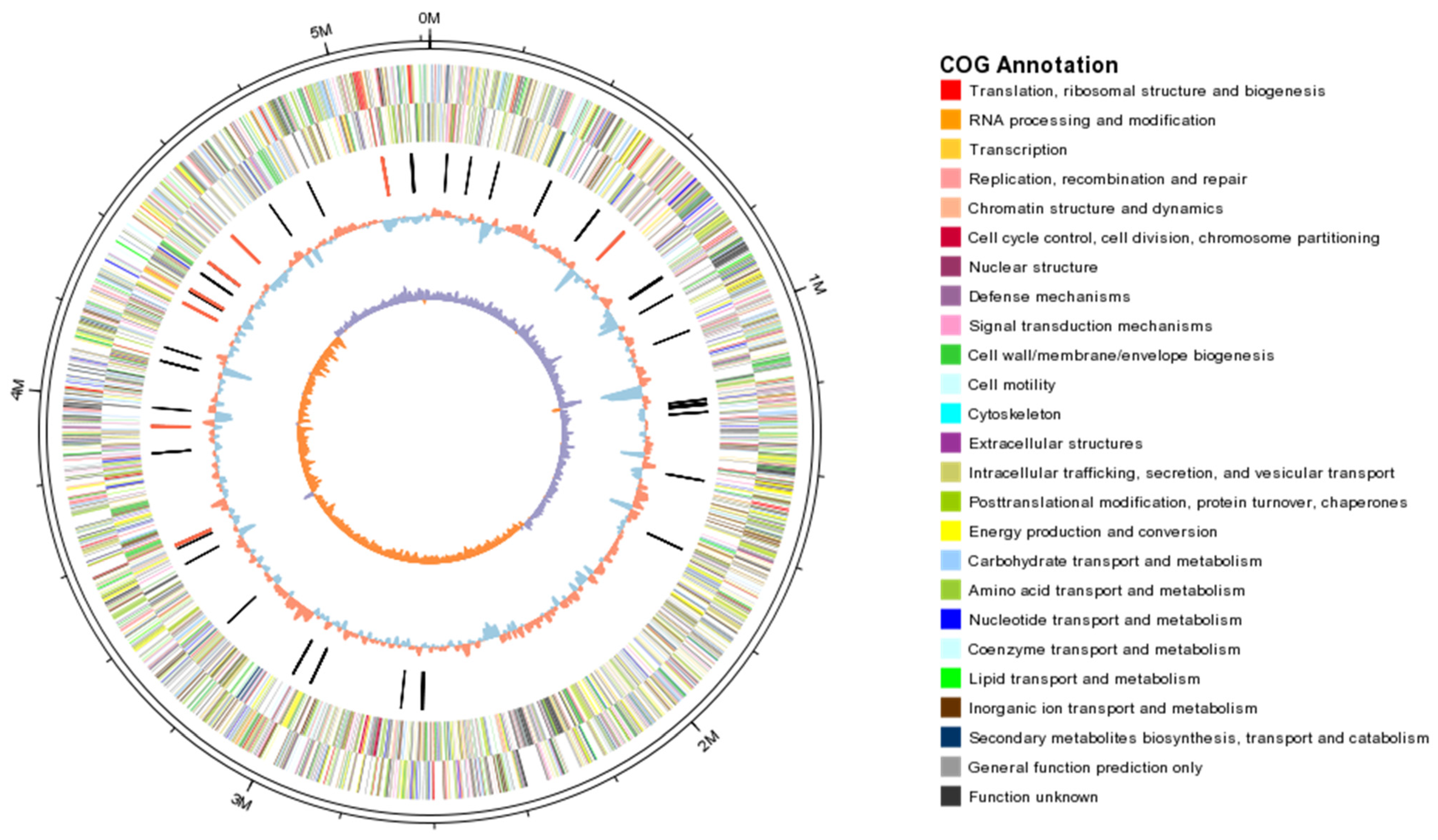
Figure 1. Circular map of the chromosome of K. pneumoniae KpC4. From the outside in, the first and second circles show the predicted protein-encoding regions on the plus and minus strands, by role, using the colors for the COG functional categories (http://www.ncbi.nlm.nih.gov/COG/grace/fiew.cgi (accessed on 3 July 2020)). The third circle shows tRNA (black) and rRNA (red). The fourth circle shows the GC content (red indicates > mean value, blue indicates < mean value). The fifth circle shows the GC skew (GC skew = (G − C)/(G + C); purple indicates > 0, orange indicates < 0).
Ijms 23 16005 g001
Figure 2. Analysis of COG functional of genome features of KpC4.
Ijms 23 16005 g002
Figure 3. Two-component system of KpC4.
Ijms 23 16005 g003
Figure 4. RcsB multi-sequence comparison results of KpC4 and K. pneumoniae clinical strains.
Ijms 23 16005 g004
Figure 5. Comparison of capsular polysaccharide synthesis (cps) gene clusters between K. pneumoniae KpC4, NTUH-K2044 (K1), and MGH78578 (K52). The locations and orientations of the open reading frames (ORFs) are indicated by arrows. ORFs are cited in order, ORFs with homologs are cited by putative gene names, and those without homologs are cited as ORFs and are numbered. Starting from galF, considered to be no. 1 (ORF** in KpC4, ORF in K2044, and ORF* in MGH78578). Solid arrows refer to ORFs with significant similarity (the aligned genes with scores > 200 and expect values < e−50 in the BLASTP search), and unshaded arrows indicate the ORFs with low similarity between any two strains (the homolog could not be found even when the expected threshold was set to be 10 in the BLASTP search). Nos. beneath each axis denote positions in kilobases.
Ijms 23 16005 g005
Figure 7. Comparative analysis of genome structures between KpC4 and the reference strains Kp13, Kv342, MGH 78578, and NTUH-K2044.
Ijms 23 16005 g007
Table 1. General features of the KpC4 genome and related analysis.
Kp StrainsSize (bp)GC Content (%)No. of CDSsMean CDS Size (bp) Coding Density (%)No. of tRNAsReference
KpC45,218,78458.87491292086.2586This study
MGH 785785,315,12057.5477695886.186[15]
Kp135,307,00357.5528889689.386[15]
NTUH-K20445,248,52057.7513093989.486[16]
Kv342 (K. variicola)5,641,23957.3542591588.088[17]
Kp52.1455,438,89456.4 5314 -88.085[17]
Table 2. Genes homologous with plant pathogens of KpC4.
Query_idGeneIdentity (%)FunctionPathogen SpeciesAccessionDisease NameExperimental Host
KpC4_4208metJ96.19Repressor of the methionine biosynthesis regulonPectobacterium atrosepticumQ6CZA0 Rotting of tubers; blackleg disease of the plant stemPotato
KpC4_4753rsmB70.09Modulates plant cell wall degrading enzymesPectobacterium atrosepticumQ6D000Rotting of tubers; blackleg disease of the plant stemPotato
KpC4_4136corA89.87Magnesium/nickel/cobalt transporter Pectobacterium carotovorumE1AP33Soft rot diseaseCelery/carrot
KpC4_3550RsmA79.41Post-transcriptional regulatorP. wasabiaeD0KML5Soft rotTobacco/potato
KpC4_0446rpoS95.76Regulation of stress and starvation responseE. amylovoraD4HX24Fire blightPear/loquat
KpC4_0922rcsB92.09Promote transcription of the genes for capsule synthesisE. amylovoraP96320Fire blightPear
KpC4_4860nlpI86.05Tetratricopeptide lipoproteinE. amylovoraD4ICB6Fire blightPear
KpC4_3140AcrB79.57Multidrug efflux pumpE. amylovoraQ7WTQ9Fire blightApple
KpC4_4819rpoN84.07Sigma factor, regulating essential virulence gene E. amylovoraD4HUY5Fire blightApple
KpC4_4766AcrB79.57Multidrug efflux pumpE. amylovoraQ7WTQ9Fire blightApple
KpC4_4696argD78.71N-acetylornithine aminotransferase enzymeE. amylovoraD4I307Fire blightPear/apple
KpC4_0520rsmAXoo82.14RNA-binding proteinXanthomonas oryzae pv. OryzaeE2J5T5Bacterial leaf blightRice
KpC4_2459iutA75.93Siderophore-mediated iron acquisitionPantoea stewartiiH3RJF2Stewart wilt of sweet cornMaize
Table 3. Annotation of open reading frames (ORFs) of lipopolysaccharide biosynthesis (lps) loci in KpC4.
Gene ClusterORF No.ORF NameORF LocationORF Homolog Characteristics
waa coaDKpC4_4461Phosphopantetheine adenylyltransferase
1waaEKpC4_4462Glucosyl transferase
2waaAKpC4_4463Kdo transferase
3ORF4KpC4_4464Glycosyl transferase
4wabHKpC4_4465Glycosyl transferase
5wabGKpC4_4466Glucuronic acid transferase
6waaQKpC4_4467Heptosyl III transferase
7wabNKpC4_4468Deacetylase
8ORF8KpC4_4469LPS 1,2-N-acetylglucosaminetransferase
9waaLKpC4_4470O-antigen ligase
10ORF10KpC4_4471Glycosyltransferase
11waaCKpC4_4472Heptosyltransferase I
12waaFKpC4_4473Heptosyltransferase II
13hldDKpC4_4474ADP-L-glycero-D-manno-heptose-6-epimerase
kblKpC4_44752-amino-3-ketobutyrate coenzyme A ligase
Lpx lpxAKpC4_3397UDP-N-acetylglucosamine acyltransferase
lpxBKpC4_3396Lipid-A-disaccharide synthase
lpxCKpC4_3497UDP-3-O-acyl N-acetylglucosamine deacetylase
lpxDKpC4_3399
KpC4_4042
UDP-3-O-(3-hydroxymyristoyl)-glucosamine n-acyltransferase
lpxKKpC4_2629Tetraacyldisaccharide 4′-kinase
Table 4. Iron uptake systems of KpC4 and related Klebsiella.
CategorySystemGeneRoleCDS Find In
KpC478578K2044Kp13342
Feo FeofeoABCFe2+ transportKpC4_4660++
ABC transporterSitsitABCDFe2+ transportKpC4_0468+++++
KfukfuABCFe3+ transportKpC4_2569
FecfecBDEFerric citrate transportKpC4_1411
fpbABCFe3+ transport ++
Hemophore-basedHmuhmuRSTUVHeme utilizationKpC4_0458+++++
Siderophore-based FepfepABCGEnterobactin transportKpC4_4005+++++
EntEntA-FEnterobactin synthesisKpC4_2951+++++
FhufhuA-CFerrichrome transportKpC4_3427
IroAiroNSalmochelin transportKpC4_2314+
iroBCDESalmochelin synthesisKpC4_1881+
AerobactiniutAAerobactin transportKpC4_2459+±+±±
iucABCDAerobactin synthesis +
Yersinia HPI ybtPQXS, ybtA-irp2-irp1- ybtUTE-fyuAYersiniabactin synthesis and transport ++
∆, initial CDS of the system relative to the KpC4 genome; −/+, absent/present in this strain; ‡, IroE appears truncated in this strain; ±, a CDS sharing 70% identity is found, although the rest of the system was not identified.
Publisher’s Note: MDPI stays neutral with regard to jurisdictional claims in published maps and institutional affiliations.