Article

Development of Bactericidal Peptides against Multidrug-Resistant Acinetobacter baumannii with Enhanced Stability and Low Toxicity

1
Marine Research Station, Institute of Cellular and Organismic Biology, Academia Sinica, 23-10 Dahuen Road., Ilan 262, Taiwan
2
Institute of Fisheries Science, National Taiwan University, 1 Roosevelt Road, Section 4, Taipei 112, Taiwan
3
Graduate Institute of Life Sciences, National Defense Medical Center, Taipei 112, Taiwan
4
Institute of Clinical Medicine, School of Medicine, National Yang-Ming University, Taipei 112, Taiwan
5
Department of Emergency Medicine, Taipei Veterans General Hospital, No. 201, Section 2, Shipai Road, Taipei 112, Taiwan
6
Faculty of Medicine, School of Medicine, National Yang-Ming University, No. 155, Section 2, Linong Street, Taipei 112, Taiwan
7
Rong Hsing Research Center for Translational Medicine, National Chung Hsing University, Taichung 402, Taiwan
8
The iEGG and Animal Biotechnology Center, National Chung Hsing University, Taichung 402, Taiwan
*
Author to whom correspondence should be addressed.
These authors contributed equally to this work.
Int. J. Mol. Sci. 2022, 23(4), 2191; https://doi.org/10.3390/ijms23042191
Submission received: 21 January 2022 / Revised: 10 February 2022 / Accepted: 15 February 2022 / Published: 16 February 2022

Abstract

:
Pathogenic superbugs are the root cause of untreatable complex infections with limited or no treatment options. These infections are becoming more common as clinical antibiotics have lost their effectiveness over time. Therefore, the development of novel antibacterial agents is urgently needed to counter these microbes. Antimicrobial peptides (AMPs) are a viable treatment option due to their bactericidal potency against multiple microbial classes. AMPs are naturally selected physiological microbicidal agents that are found in all forms of organisms. In the present study, we developed two tilapia piscidin 2 (TP2)-based AMPs for antimicrobial application. Unlike the parent peptide, the redesigned peptides showed significant antimicrobial activity against multidrug-resistant bacterial species. These peptides also showed minimal cytotoxicity. In addition, they were significantly active in the presence of physiological salts, 50% human serum and elevated temperature. The designed peptides also showed synergistic activity when combined with clinical antibiotics. The current approach demonstrates a fruitful strategy for developing potential AMPs for antimicrobial application. Such AMPs have potential for progression to further trials and drug development investigations.

1. Introduction

Antibiotics are primary agents used for neutralizing pathogenic infections. Since their discovery, they have been an integral part of clinical therapy [1]. However, there is a clear indication of a significant reduction in their activity due to multiple factors [2]. One of the key factors resulting in reduced antimicrobial potential is the recurring presence of multidrug-resistant (MDR) bacterial species. Researchers have estimated that 10 million annual deaths could occur due to microbial infection if we fail to develop effective antimicrobials [3]. The World Health Organization (WHO) has categorized critical, high-priority and medium-priority pathogens according to the need for new therapies to treat them. The critical priority group comprises Acinetobacter baumannii (carbapenem-resistant) and Pseudomonas aeruginosa. The high-priority group contains Staphylococcus aureus (methicillin-resistant) and Helicobacter pylori (clarithromycin-resistant) and the medium-priority group consists of Haemophilus influenzae (ampicillin-resistant) [4]. Therefore, the search for new antibiotics and potential alternatives is among the primary interests of the health care system [1]. To this end, antimicrobial peptides have been shown to be potential antibiotics owing to their broad-spectrum activity against multidrug-resistant bacteria [5]. Antimicrobial peptides (AMPs) are defense molecules found in all organisms [6]. They are mostly amphipathic with a net positive charge. AMPs act through membrane rupturing mechanisms. AMPs primarily interact through electrostatic interactions between the anionic bacterial membrane and the cationic peptide, causing degradation of the bacterial membrane [5].
In the current study, we used a pair of peptides known as TP2-5 and TP2-6 to develop potential bactericidal agents against multidrug-resistant A. baumannii [7]. The peptide pair was developed from tilapia piscidin 2 (TP2), an inactive antibacterial peptide found in Oreochromis niloticus (Nile tilapia) [8]. TP2-5 and TP2-6 are modified variants that have improved cationicity and amphipathic balance, which resulted in significant improvement in their antimicrobial potential in normal media [7]. Therefore, we selected this pair to assess their antimicrobial potential in the presence of human serum, lung surfactant, physiological ions and elevated temperature. Additionally, we screened the peptides against a series of MDR A. baumannii bacterial species (Supplementary Table S1). The peptides significantly retained their activity under the test conditions and against the MDR bacterial species. Additionally, the peptides were nontoxic and possessed antibiofilm potential. Both peptides showed synergistic potential against MDR bacterial species when combined with meropenem. These peptides retained significant activity under physiological conditions with potential activity against clinically important MDR A. baumannii bacterial species.

2. Results and Discussion

2.1. Peptide Design

A pair of test peptides, TP2-5 and TP2-6, were selected from a previous design based on tilapia piscidin 2 (TP2) [7]. Tilapia piscidin 2 is a peptide without antimicrobial activity obtained from Oreochromis niloticus [9]. The original peptide TP2 (GECIWDAIFHGAKHFLHRLVNP) possesses negligible or no net positive charge [8]. However, peptides TP2-5 (KKCIAKAILKKAKKLLKKLVNP) and TP2-6 (KKCIAKAILKKAKKLLKDLVNP) possess a net positive charge of +9 and +7, respectively, with amphipathic characteristics (Supplementary Figures S1–S4) [7]. Both TP2-5 and TP2-6 are nonhaemolytic and noncytotoxic and have significant antimicrobial activity [7]. Hence, we extended our study to develop a potential anti-A. baumannii peptide for future application.

2.2. Antimicrobial Activity of the Peptides in Normal Media, Serum and Lung Surfactant

TP2-5 and TP2-6 showed significant antimicrobial activity against A. baumannii bacterial species in normal media [7]. Therefore, we extended our study to assess their stability in the presence of human serum and lung surfactant. Human serum and lung surfactant were used to imitate the physiological environment. This experiment helped to clarify the extended stability of the peptides by assessing whether they retained their activity in the presence of these substances. TP2-5 and TP2-6 were both significantly active against the wild-type variant of A. baumannii in the presence of 50% human serum and 5% lung surfactant (Table 1). Both peptides were active against 12 MDR A. baumannii bacterial species (Supplementary Table S1). Additionally, TP2-5 showed significant activity against MDR A. baumannii (14B0091) in the presence of 50% human serum (Table 1). However, the presence of human serum markedly reduced the antimicrobial potential of TP2-6. The MIC and MBC values were still 2-fold lower than those of the control peptide (LL-37) and the antibiotic control (meropenem). Human serum and lung surfactants are complex mixtures of multiple enzymes and factors that can degrade or reduce the therapeutic potencies of poly-L peptides [5,6]. Unlike many naturally occurring peptides, the pair of de novo peptides retained their activities when compared with the peptide control (LL-37). However, LL-37 showed lower potency against the MDR-strains in media alone. Apart from enhanced stability, the therapeutic potencies of the peptides against MDR strains are noteworthy (Table 1). Therefore, the current pair of peptides can be further tested and explored for their possible therapeutic applications against MDR infections.

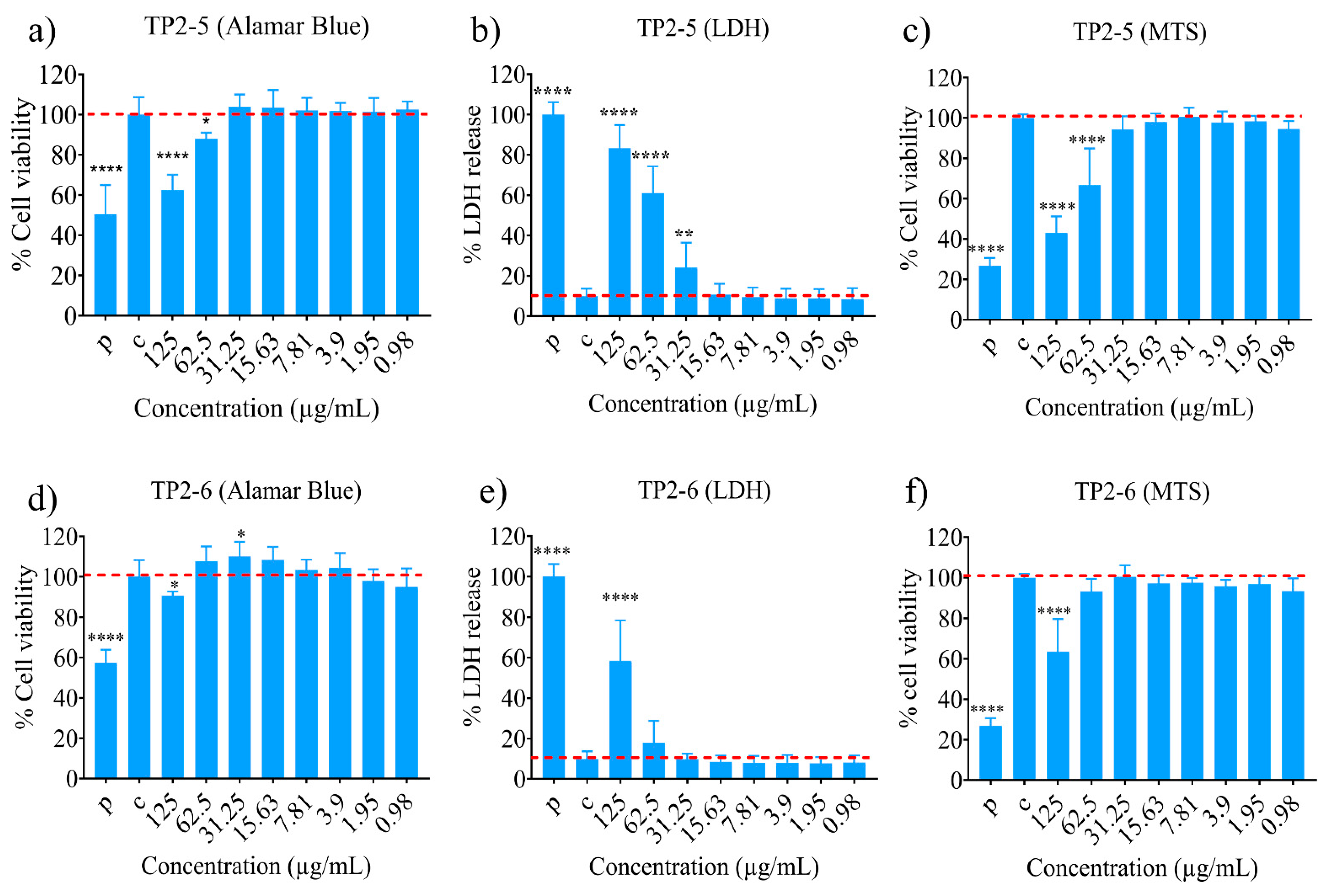
2.3. Cytotoxicity Potential of the Peptides

TP2-5 and TP2-6 showed negligible cytotoxicity against human skin fibroblast (CCD966SK) cell lines. The peptides showed relatively low toxicity even at high concentrations (Figure 1a–f). We can assume their safety profiles are acceptable, as the peptides had MIC and MBC values as low as 3.125 to 6.25 µg/mL (Table 1). Low cytotoxicity is an important parameter for any candidate peptide for continued in vivo study. This feature enhances the therapeutic index and is a favorable indicator of safety for a given set of test molecules. For our peptides, the MIC and MBC values of TP2-5 and TP2-6 were between 3.125 µg/mL and 6.25 µg/mL in the presence of human and lung surfactant, whereas toxicity was only observed at a higher dose of 62.5 µg/mL for TP2-5, as assayed by % LDH release. The alamar blue and MTS assays showed notable toxicity at a higher dose of 125 µg/mL TP2-5. TP2-6 showed notable toxicity at 125 µg/mL when measured by LDH release. The MTS and alamar assay profiles for TP2-6 were relatively better than those of TP2-5. These results demonstrate that the MIC and MBC doses of TP2-5 and TP2-6 are significantly lower than the toxic doses. Hence, the current pair of peptides may be safe and effective, making them suitable for further trials.

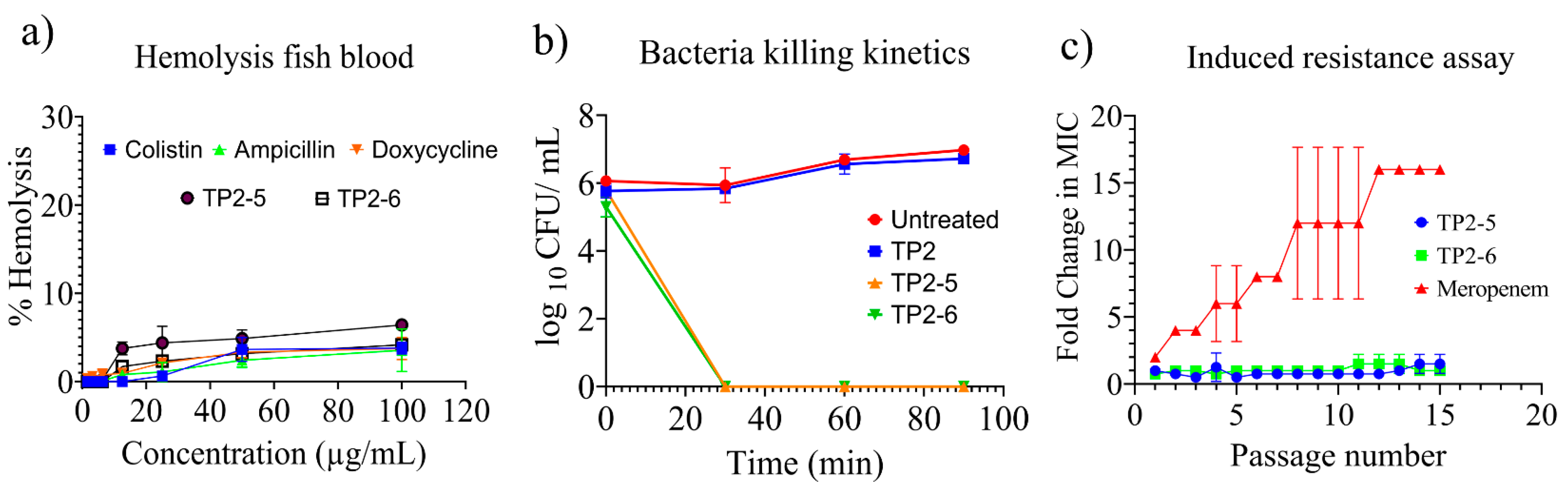
2.4. Haemolysis, Bacterial Killing Kinetics and Induced Resistance Assay

The peptides were assessed for their haemolytic potential. Both peptides showed negligible toxicity even at the highest concentration of 100 µg/mL (Figure 2a). Based on the haemolysis data, these peptides were found to be nontoxic, which is an important characteristic for continuation in the drug discovery pipeline. The antimicrobial activity of the peptides was significantly active against both wild-type and resistant variants of A. baumannii. To estimate the duration for bacterial clearance, we determined the bacterial killing kinetics of TP2-5 or TP2-6. Both peptides showed 100% bacterial killing against A. baumannii 10591 within 30 min (Figure 2b) at their respective MIC/MBC values (Table 1). Antimicrobial agents tend to show reduced activity upon prolonged exposure to microbes at low concentrations. We performed a serial passage assay of TP2-5, TP2-6 and meropenem against A. baumannii (10591) for a repetition of 15 cycles (Figure 2c). TP2-5 and TP2-6 showed negligible increases in their MIC values. However, meropenem showed a significant increase in its MIC, which was up to 16-fold higher than the initial MIC value. These results show that TP2-5 and TP2-6 are less prone to induce resistance, whereas clinical antibiotics can rapidly lose their antimicrobial activity upon prolonged exposure to bacterial species.

2.5. Antimicrobial Activity of the Peptides in the Presence of Variable Temperature and Physiological Ions

The antimicrobial activity of the peptides was also tested after incubation at high temperatures. The two peptides were incubated at 40, 60, 80 and 100 °C for 1 h prior to the assay. Both peptides showed significant antimicrobial activity even after incubation at 100 °C (Table 2), demonstrating their temperature stability. Meropenem also showed significant stability up to 80 °C; however, it lost activity after the 100 °C incubation (Table 2). TP2-5 and TP2-6 were tested for their activity in the presence of physiological ions and glucose under hyperglycaemic conditions. TP2-5 showed significant activity in the presence of physiological ions and glucose (Table 3). TP2-6 also showed notable activity in the presence of salts; however, it lost significant activity in the presence of CaCl2. AMPs tend to lose potency in the presence of ions, compromising their clinical efficacy. The results of the current study showed that the two peptides retained their activity in the presence of physiological ions, an important criterion for clinical application.

2.6. FIC Index of the Peptides and Antibiotics

The FIC index was calculated to assess potential synergistic and antagonistic antimicrobial activities of the two peptides with meropenem (Table 4). TP2-5 showed additive activity with meropenem against most of the MDR A. baumannii bacterial species, except A. baumannii 2998. The FIC index was also determined for the combination of TP2-6 and meropenem against MDR A. baumannii bacterial species (Table 5). The combination of TP2-6 and meropenem showed additive activity in most cases, except for A. baumannii 2982. Based on this assay, we confirmed the synergistic potential of the peptides and meropenem against multidrug-resistant A. baumannii bacterial species. Of note, synergistic activity can allow for the reuse of old antibiotics that were discontinued due to reduced therapeutic potential. Such repurposing of older drugs may be a faster way to develop new therapeutic options due to their well-known clinical effects. This strategy may lead to novel multiple-drug therapy (MDT) combinations that are effective against a variety of life-threatening infections. MDT is a common approach in the application of antibiotics and enzymes. Additionally, clinical studies with natural and synthetic compounds, peptides, and antibody-drug conjugates may performed to identify new therapeutic entities [1]. Hence, the synergistic activities of AMPs with antibiotics could lead to the development of new antimicrobial therapy options.

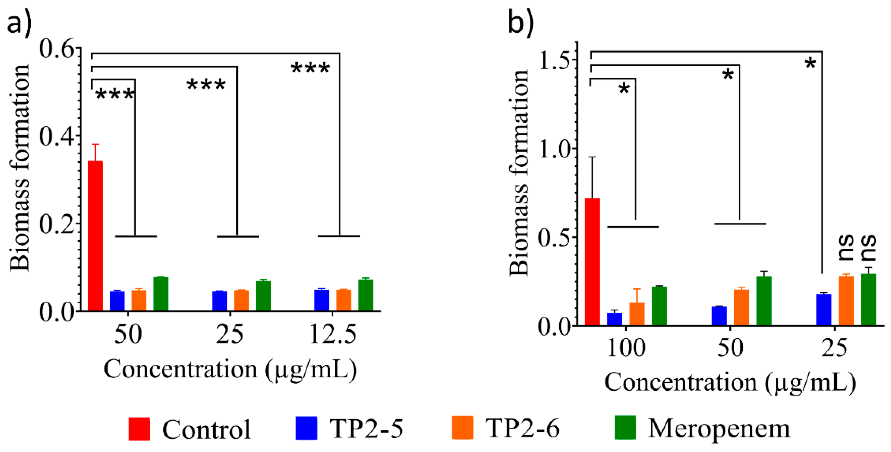
2.7. Antibiofilm Activity of the Peptides

Bacterial biofilms are the most resistant form of bacterial aggregates and are responsible for approximately 80% of bacterial infections [10]. Therefore, we tested the antibiofilm potentials of TP2-5 and TP2-6. Both peptides showed significant biofilm inhibition against A. baumannii at the low concentration of 12.5 µg/mL (Figure 3a). TP2-5 and TP2-6 showed significant biofilm rupture at all tested concentrations (Figure 3b). TP2-6 and meropenem showed nonsignificant biofilm rupture at the lowest concentration (25 µg/mL). Biofilms are mostly untreatable with current antibiotics. Therefore, potential antibiofilm molecules are of significant interest to researchers [8]. Given that our pair of AMPs showed low cytotoxic and haemolytic activities, they can be further tested as potential antibiofilm molecules for clinical applications.

2.8. Membranolytic Activity of the Peptides

AMPs are known to be membranolytic [9]; therefore, we tested the membranolytic activity of the peptides using propidium iodide reagent. The peptide-treated bacterial species showed a significant increase in fluorescence intensity compared with untreated controls (Figure 4a). Both peptides led to notable outer membrane rupture in the presence of NPN dye (Figure 4b) compared with untreated groups. The peptides were also assessed for their membrane depolarization potential using DIBAC4-3 dye. TP2-5 and TP2-6 showed significant membrane depolarization activity (Figure 4c). To infer the membrane-active nature of the peptides, microscopic analyses of peptide-treated membranes and untreated controls were performed (Figure 4d). The peptide-treated bacterial species showed deformed membrane architecture, whereas untreated controls showed intact membranes. Based on these studies, we can infer that the peptides primarily act through membranolytic activity.

3. Materials and Methods

3.1. Peptide Synthesis and Characterization

The peptides were synthesized using F-moc chemistry (GL Biochem, Shanghai, China) [11]. In brief, peptides were synthesized on a solid support (Rink amide resin) and then removed from the resin by incubating with a cleavage mixture [trifluoroacetic acid (TFA): ethanedithiol: m-cresol: thioanisole :: 20: 1: 2: 2] for 12–16 h. Later, the cleaved peptides were precipitated in ice-cold ether, followed by repeated washing. The peptides were then purified by reverse-phase high-performance liquid chromatography (RP-HPLC). A gradient was run using Solvent A (100 acetonitrile with 0.1% TFA) and Solvent B (water with 0.1% TFA) was performed at 220 nm with a flow rate of 1 mL/min. Samples were eluted using Sinochrom ODS BP-5 column over a period of 30 min. The molecular masses of the purified peptides (with purity grades of >95%) were verified by electrospray ionization mass spectrometry (ESI-MS). Samples were dissolved in a 50% mixture of Solvent A and B, followed by sample detection with a flowrate (nebulizer gas flow) of 1.5 mL/min (detector −1.5 Kv).

3.2. Antimicrobial Assay and Bacterial Killing Kinetics

Bacterial cells Acinetobacter baumannii 10591 and 14B0091 were obtained from BCRC Taiwan. All the MDR strains were obtained from the hospital as gifts. The antimicrobial assay was performed as per the standard protocol; 50 µL of 106 CFU/mL of mid-log phase bacterial suspension was treated with 50 µL of candidate peptide and meropenem (Sigma: PHR1772) incubated at 37 °C for 960~1200 min. The clear wells corresponding to the lowest concentrations are reported as the minimum inhibitory concentration (MIC). Suspensions from the clear wells were plated on Mueller–Hinton agar (MHA) plates and incubated at 37 °C overnight [6]. The lowest concentration corresponding to no growth was recorded as the minimum bactericidal concentration (MBC). To detect the effect of human serum (H3667, Sigma Aldrich, St. Louis, MO, USA) on the peptides, the media was replaced with 50% human serum-containing media. Similarly, to detect the effect of lung surfactant (SURVANTA®, beractant, AbbVie Inc., Lake Bluff, IL, USA (25 mg/mL)) on the antimicrobial activity of the peptide, the normal media was replaced with 5% lung surfactant-containing media [6]. To analyse the bacterial killing kinetics, after the addition of the peptide to the bacterial suspension, 10 µL of broth was diluted 1000 times, plated at 30 min intervals, and allowed to incubate for 90 min. The bacterial load is expressed as CFU/mL as a function of time [12].

3.3. Fish Blood Haemolysis

Fresh Nile tilapia blood was collected and washed three times in normal saline by centrifuging at 800× g. Then, 50 µL of 2% blood suspension was treated with 50 µL of test peptide for 1 h at room temperature. The incubated mixture was then centrifuged, and hemoglobin release was recorded using a spectroscope at 570 nm [13].

3.4. Cytotoxicity Assay

Cultures of 8 × 10³ cells/100 µL (human skin fibroblast (CCD966SK) were incubated for 24 h in the presence of 5% CO2, followed by peptide treatment for 24 h. Peptide-treated cell were assessed for cytotoxicity using an LDH cytotoxicity detection kit [14], Alamar blue cell viability reagent [15] and a non-radioactive cell proliferation assay (MTS) [16]. For the control for the study, 0.1% Triton X (P) and normal media (C) was used.

3.5. Antibiofilm Assay

For the biofilm inhibition assay, 106 bacterial cells were treated with peptide at various concentrations in MHB media containing 0.2% glucose for 24 h at 37 °C in 96-well plates. The wells were then washed with PBS, treated with 100% methanol for 15 min, and the washes were discarded. Crystal violet solution (0.02%) was added to the dry plates and incubated for 30 min. The wells were then washed and treated with 33% acetic acid, followed by detection at 595 nm. For the biofilm rupture assay, 106 bacterial cells were grown overnight. The residual liquid was discarded, followed by washing with PBS. Next, 100 µL of peptide at different concentrations was added as estimated according to the biofilm inhibition assay [17,18].

3.6. Peptide Sensitivity to Temperature

Here, 50 µL of 106 CFU/mL bacterial cells were treated with peptide previously incubated at 40, 60, 80, and 100 °C for 1 h [19,20]. The peptide activity was reported as per the standard MIC and MBC protocols.

3.7. Induced Resistance Assay

A total of 50 µL of 106 CFU/mL bacterial cells were treated with different concentrations of peptide. Bacterial cells from subinhibitory wells were subcultured by passaging for 15 cycles, followed by testing their MIC values. The fold change in MIC was reported in comparison with starting MIC values [21].

3.8. Fractional Inhibitory Concentration (FIC) Index Assay

A 2-fold dilution of peptide in 50 µL of media was performed along the vertical axis of a 96-well plate. Similarly, 50 µL of the next drug was diluted 2-fold along the horizontal axis. Next, 50 µL of 106 cells was added, and the standard MIC protocol was performed [22]. The FIC index was calculated according to the following formula:
FICI = FIC A + FIC B
where FIC A is MIC A & B combined/MIC A and FIC B is MIC A & B combined/MIC A.

3.9. Membrane Lysis Assay

A total of 108 bacterial cells were dispersed in MHB medium containing 20 µg/mL propidium iodide (PI). Then, 90 µL of the bacterial suspension was mixed with 10 µL of peptide in a 96-well black plate. Membrane rupture was recorded (excitation = 584, emission = 620) in the peptide-treated and untreated (blank) bacterial cells [23].

3.10. Outer Membrane Permeabilization Assay

A total of 108 bacterial cells were suspended in PBS containing 25 mM glucose, followed by incubation at 37 °C for 15 min. Post incubation, N-phenyl-1-naphthyl amine (NPN) was added to a final concentration of 10 µM. A 90 µL aliquot of the bacterial suspension was recorded as the blank (350 nm excitation, 420 emission) for 40 min. Next, 10 µL of peptide was added, and spectra were recorded for 20 min [24].

3.11. Membrane Depolarization Assay

A total of 108 bacterial cells were washed and resuspended in PBS containing 25 mM glucose. Bis-(1,3-dibarbituric acid)-trimethine oxanol (DIBAC) was then added to a final concentration of 500 nM. Ninety microlitres of the bacterial blank was recorded (excitation 490, emission 516) for 40 min, followed by peptide treatment (10 µL), and the spectra were recorded for 40 min [25].

3.12. Field Emission Scanning Electron Microscopy

The bacterial cells were treated at the respective MICs of the peptides. The cells were then washed and resuspended in 4% glutaraldehyde in PBS and incubated at 4 °C for 30 min. The cells were then fixed on glass slides for 15 min. The slides were subsequently washed with ethanol (20% to 100%). Dry slides were gold-coated, and images were recorded using an electron microscope [13].

4. Conclusions

AMPs and their derivatives are widely considered to be potential alternatives to existing antibiotics [5]. Several modified peptides, such as peptide–drug conjugates, are currently being tested in clinical trials, but only a few AMPs are in clinical use [1]. Importantly, AMPs show significant potencies against a vast array of multidrug-resistant pathogens in laboratory studies. However, their efficacies are often compromised in the presence of physiological factors, which is a major obstacle to their clinical application [5]. To overcome this and other obstacles, systematic modification of peptide sequences or peptidomimetic approach can be a possible route to develop new AMPs with enhanced activities and stabilities in vivo [5]. In the current study, we sought to design and develop potential AMPs against MDR Acinetobacter baumannii bacterial species. We successfully developed a pair of stable peptides that show significant activities in the presence of 50% human serum and 5% lung surfactants. This stability was due to targeted changes in the peptide sequences, which resulted in the development of new AMPs. Both of the peptides possess relatively higher densities of net positive charge with optimal amphipathicity, as compared to the parental TP2 peptide molecule. Most importantly, the modified peptides showed significant activity against MDR bacterial species, with low toxicity to human cells and minimal hemolytic activity. Additionally, the peptides showed significant antibiofilm activity. Both TP2-5 and TP2-6 acted synergistically with meropenem, and the designed peptides retained their activities in the presence of physiological ions and elevated temperature. Additionally, the peptides showed lower degrees of induced resistance than meropenem. The mechanism of bactericidal activity for the peptides was via lysis of the bacterial membrane. Based on our collective results, the peptides show significant potential for further study. Hence, these two peptides should be evaluated in further trials and studies, which may lead to the development of promising drug candidates to fight antimicrobial resistance.

Supplementary Materials

The following are available online at https://www.mdpi.com/article/10.3390/ijms23042191/s1.

Author Contributions

P.K.H.: methodology, investigation, formal analysis, writing—original draft, writing—review and editing. C.-C.C.: methodology, investigation, formal analysis, writing—review and editing. W.-C.L., C.-Y.H., T.-L.C., Y.-T.L. and P.-H.H.: methodology, investigation, formal analysis. J.-Y.C.: conceptualization, funding acquisition, project administration, methodology, writing—review and editing. All authors have read and agreed to the published version of the manuscript.

Funding

This research was supported by funding from the Marine Research Station (Jiaushi, Ilan), Institute of Cellular and Organismic Biology, Academia Sinica to Jyh-Yih Chen (Research Fellow). This work was also supported by the Higher Education Sprout Project from the Ministry of Education (MOE-110-S-0023-A) in Taiwan and by the Ministry of Science and Technology (110-2622-B-001-002) (108-2313-B-001-006) in Taiwan. This work was also supported by Academia Sinica funding to Jyh-Yih Chen (AS-IDR-111-01 (peptidomimetics approach in redesigning antimicrobial peptide with extended stability and activity against multidrug-resistant bacteria)).

Institutional Review Board Statement

Institutional animal ethics guidelines and supervision of the committees of Academia Sinica.

Informed Consent Statement

Not applicable.

Data Availability Statement

The data presented in this study are available on request from the corresponding author.

Acknowledgments

We thank the Institute of Cellular and Organismic Biology Core Facility for technical support during the experiments.

Conflicts of Interest

The authors declare that they have no conflicts of interest.

References

  1. Butler, M.S.; Paterson, D.L. Antibiotics in the clinical pipeline in October 2019. J. Antibiot. 2020, 73, 329–364. [Google Scholar] [CrossRef]
  2. Yoshikawa, Y.; Feldhaus, I.; Özçelik, E.; Hashiguchi, T.C.O.; Cecchini, M. Financial strategies targeting healthcare providers to promote the prudent use of antibiotics: A systematic review of the evidence. Int. J. Antimicrob. Agents 2021, 58, 106446. [Google Scholar] [CrossRef]
  3. Tan, R.; Huang, Z.; Guo, H.; Weng, Y.; Chow, A. Antibiotic expectations of emergency department patients with upper respiratory tract infections: Clinical and antibiotic use behavioral determinants. Int. J. Antimicrob. Agents 2021, 106511. [Google Scholar] [CrossRef]
  4. Venkatesan, P. WHO 2020 report on the antibacterial production and development pipeline. Lancet Microbe 2021, 2, e239. [Google Scholar] [CrossRef]
  5. Hancock, R.E.; Sahl, H.G. Antimicrobial and host-defense peptides as new anti-infective therapeutic strategies. Nat. Biotechnol. 2006, 24, 1551–1557. [Google Scholar] [CrossRef]
  6. Elliott, A.G.; Huang, J.X.; Neve, S.; Zuegg, J.; Edwards, I.A.; Cain, A.K.; Boinett, C.J.; Barquist, L.; Lundberg, C.V.; Steen, J.; et al. An amphipathic peptide with antibiotic activity against multidrug-resistant Gram-negative bacteria. Nat. Commun. 2020, 11, 3184. [Google Scholar] [CrossRef]
  7. Hazam, P.K.; Cheng, C.C.; Lin, W.C.; Hsieh, C.Y.; Chen, J.Y. Strategic modification of low-active natural antimicrobial peptides confers ability to neutralize pathogens in vitro and in vivo. 2022; Unpublished. [Google Scholar]
  8. Hazam, P.K.; Chen, J.Y. Therapeutic utility of the antimicrobial peptide Tilapia Piscidin 4 (TP4). Aquac. Rep. 2020, 17, 100409. [Google Scholar] [CrossRef]
  9. Peng, K.C.; Lee, S.H.; Hour, A.L.; Pan, C.-Y.; Lee, L.H.; Chen, J.Y. Five different piscidins from Nile Tilapia, Oreochromis niloticus: Analysis of their expressions and biological functions. PLoS ONE 2012, 7, e50263. [Google Scholar] [CrossRef]
  10. de la Fuente-Núñez, C.; Korolik, V.; Bains, M.; Nguyen, U.; Breidenstein, E.B.; Horsman, S.; Lewenza, S.; Burrows, L.; Hancock, R.E.W. Inhibition of bacterial biofilm formation and swarming motility by a small synthetic cationic peptide. Antimicrob. Agents Chemother. 2012, 56, 2696–2704. [Google Scholar] [CrossRef] [Green Version]
  11. Liu, C.W.; Su, B.C.; Chen, J.Y. Tilapia piscidin 4 (TP4) reprograms M1 macrophages to m2 phenotypes in cell models of Gardnerella vaginalis-induced vaginosis. Front. Immunol. 2021, 12, 773013. [Google Scholar] [CrossRef]
  12. Petersen, P.J.; Jones, C.H.; Bradford, P.A. In vitro antibacterial activities of tigecycline and comparative agents by time-kill kinetic studies in fresh Mueller-Hinton broth. Diagn. Microbiol. Infect. Dis. 2007, 59, 347–349. [Google Scholar] [CrossRef]
  13. Hazam, P.K.; Singh, A.; Chaudhary, N.; Ramakrishnan, V. Bactericidal potency and extended serum life of stereo-chemically engineered peptides against Mycobacterium. Int. J. Pept. Res. Ther. 2019, 25, 465–472. [Google Scholar] [CrossRef]
  14. Arechabala, B.; Coiffard, C.; Rivalland, P.; Coiffard, L.J.; de Roeck-Holtzhauer, Y. Comparison of cytotoxicity of various surfactants tested on normal human fibroblast cultures using the neutral red test, MTT assay and LDH release. J. Appl. Toxicol. 1999, 19, 163–165. [Google Scholar] [CrossRef]
  15. Nkenfou, C.N.; Mawabo, I.K.; Notedji, A.; Nkenfou, J.; Fokou, P.V.T.; Jouda, J.B.; Kuiate, J.R. In vitro antimycobacterial activity of six Cameroonian medicinal plants using microplate alamarBlue assay. Int. J. Mycobact. 2015, 4, 306–311. [Google Scholar] [CrossRef] [Green Version]
  16. Goodwin, C.J.; Holt, S.J.; Downes, S.; Marshall, N.J. Microculture tetrazolium assays: A comparison between two new tetrazolium salts, XTT and MTS. J. Immunol. Methods. 1995, 179, 95–103. [Google Scholar] [CrossRef]
  17. Au-O’Toole, G.A. Microtiter dish biofilm formation assay. J. Vis. Exp. 2011, 47, e2437. [Google Scholar]
  18. Gopal, R.; Kim, Y.G.; Lee, J.H.; Lee, S.K.; Chae, J.D.; Son, B.K.; Seo, C.H.; Park, Y. Synergistic effects and antibiofilm properties of chimeric peptides against multidrug-resistant Acinetobacter baumannii strains. Antimicrob. Agents Chemother. 2014, 58, 1622–1629. [Google Scholar] [CrossRef] [Green Version]
  19. Pan, C.Y.; Tsai, T.Y.; Su, B.C.; Hui, C.F.; Chen, J.Y. Study of the antimicrobial activity of tilapia piscidin 3 (TP3) and TP4 and their effects on immune functions in hybrid tilapia (Oreochromis spp.). PLoS ONE 2017, 12, e0169678. [Google Scholar] [CrossRef] [Green Version]
  20. Ebbensgaard, A.; Mordhorst, H.; Overgaard, M.T.; Nielsen, C.G.; Aarestrup, F.M.; Hansen, E.B. Comparative evaluation of the antimicrobial activity of different antimicrobial peptides against a range of pathogenic bacteria. PLoS ONE 2015, 10, e0144611. [Google Scholar] [CrossRef] [Green Version]
  21. Li, C.; Zhu, C.; Ren, B.; Yin, X.; Shim, S.H.; Gao, Y.; Zhu, J.; Zhao, P.; Liu, C.; Yu, R.; et al. Two optimized antimicrobial peptides with therapeutic potential for clinical antibiotic-resistant Staphylococcus aureus. Eur. J. Med. Chem. 2019, 183, 111686. [Google Scholar] [CrossRef]
  22. Li, Q.; Cebrián, R.; Montalbán-López, M.; Ren, H.; Wu, W.; Kuipers, O.P. Outer-membrane-acting peptides and lipid II-targeting antibiotics cooperatively kill Gram-negative pathogens. Commun. Biol. 2021, 4, 31. [Google Scholar] [CrossRef]
  23. Kwon, J.Y.; Kim, M.K.; Mereuta, L.; Seo, C.H.; Luchian, T.; Park, Y. Mechanism of action of antimicrobial peptide P5 truncations against Pseudomonas aeruginosa and Staphylococcus aureus. AMB Express. 2019, 9, 122. [Google Scholar] [CrossRef] [Green Version]
  24. Torres, M.D.T.; Voskian, S.; Brown, P.; Liu, A.; Lu, T.K.; Hatton, T.A.; de la Fuente-Nunez, C. Coatable and Resistance-Proof Ionic Liquid for Pathogen Eradication. ACS Nano 2021, 15, 966–978. [Google Scholar] [CrossRef]
  25. Clementi, E.A.; Marks, L.R.; Roche-Håkansson, H.; Håkansson, A.P. Monitoring changes in membrane polarity, membrane integrity, and intracellular ion concentrations in Streptococcus pneumoniae using fluorescent dyes. J. Vis. Exp. 2014, 84, e51008. [Google Scholar] [CrossRef] [Green Version]
Figure 1. Cytotoxicity potential of the peptides. Cytotoxicity potential of TP2-5 (a) Alamar blue, (b) LDH and (c) MTS assay and of TP2-6 (d) Alamar blue, (e) LDH and (f) MTS assay against human skin fibroblast (CCD966SK) cell lines. The experiments were performed twice in triplicate. The statistical analysis was performed by one-way ANOVA (Dunnett’s test) (* p < 0.05, ** p < 0.005, **** p < 0.0001).
Ijms 23 02191 g001
Figure 2. Haemolysis, bacterial killing kinetics and induced resistance assay. (a) Haemolysis activity of the peptides and control antibiotics against tilapia fish blood. (b) Bacterial killing kinetics of the peptides against A. baumannii 10591 bacterial species. (c) Induced resistance assay of the peptides and meropenem against A. baumannii 10591 for a continuous passage of 15 cycles. All experiments shown here were performed in three replicates in a set of two independent experiments.
Ijms 23 02191 g002
Figure 3. Antibiofilm activity of the peptides. (a) Biofilm inhibition and (b) rupture induced by meropenem and TP2-5 and TP2-6 at different concentrations against A. baumannii 10591. All data are reported as the mean ± SD (n = 3 per group) from two independent trials. Statistically significant differences compared to the control (vehicle) were calculated by one-way ANOVA and Dunnett’s test (* p < 0.05; *** p < 0.001; ns, not significant).
Ijms 23 02191 g003
Figure 4. Membranolytic activity of the peptides. (a) A. baumannii 10591 membrane lysis (propidium iodide-PI) caused by TP2-5 and TP2-6. (b) A. baumannii 10591 outer membrane lysis (NPN dye) caused by TP2-5 and TP2-6 at 25 µg/mL. (c) A. baumannii 10591 membrane depolarization (DIBAC4 dye) caused by TP2-5 and TP2-6 at 25 µg/mL. (d) SEM analysis of 1x and 2x MIC peptide-treated and untreated A. baumannii 10591. Scale bars are 2.5 µm, 5 µm and 10 µm. PI, NPN and DIBAC4-3 data are reported as the mean ± SD (n = 3 per group) from two independent trials. Statistically significant differences compared to the control (vehicle) were calculated by one-way ANOVA and Dunnett’s test.
Ijms 23 02191 g004
Table 1. Minimal inhibitory concentration (MIC) and minimal bactericidal concentration (MBC) of TP2-5 and TP2-6 against wild-type A. baumannii and MDR A. baumannii species in the presence of human serum and lung surfactant. All experiments were performed in triplicate in two independent experiments. Data are expressed in µg/mL.
Peptides/DrugsTP2-5 (µg/mL)TP2-6 (µg/mL)LL-37 (µg/mL)Meropenem (µg/mL)
Bacterial StrainMICMBCMICMBCMICMBCMICMBC
Wild strainA. baumannii 10591 a3.1253.1253.1253.125>100>1001.561.56
A. baumannii 10591 b3.1253.1253.1253.125>100>1001.561.56
MDR strain14B00913.1253.1253.125–6.253.125–6.2512.5–2512.5–25>100>100
20883.1253.1253.1253.1252525>100>100
9213.1253.1256.256.2512.512.5100>100
10193.1253.1256.25–12.56.25–12.512.512.52525
10333.1253.1256.256.25252550100
16071.561.566.256..2512.512.55050
17023.1253.1256.256.2512.512.512.512.5
29623.125–6.256.253.125–6.256.2525255050
29823.1253.125–6.253.1253.1252525–50>100>100
29971.56–3.1251.56–3.1256.256.2512.512.550–10050–100
29981.56–3.1251.56–3.1253.125–6.253.125–6.252550>100>100
36183.1253.1256.256.2512.512.550–10050–100
14B0091 c12.512.55050>100>100>100 >100
a, c media containing 50% human serum, b media containing 5% lung surfactant.
Table 2. Temperature-dependent antimicrobial activity of meropenem, TP2-5 and TP2-6 against A. baumannii. The data were derived from triplicate samples in two individual experiments. Data are expressed in µg/mL.
A. baumanniiTP2-5 (µg/mL)TP2-6 (µg/mL)Meropenem (µg/mL)
MICMBCMICMBCMICMBC
40 °C6.256.2512.512.51.561.56
60 °C6.256.2512.512.51.561.56
80 °C6.256.2512.512.53.1253.125
100 °C6.256.2512.512.55050
Table 3. Antimicrobial activity of meropenem, TP2-5 and TP2-6 against A. baumannii 10591 in the presence of physiological ions and glucose. The data were derived from triplicate samples in two individual experiments. Data are expressed in µg/mL.
A. baumanniiTP2-5 (µg/mL)TP2-6 (µg/mL)
MICMBCMICMBC
CaCl212.512.55050
MgCl23.1253.12512.512.5
NH4Cl1.561.563.1253.125
KCl3.1253.1253.1253.125
NaCl6.256.2512.512.5
Glucose1.566.256.256.25
Table 4. MIC values and synergistic activities of TP2-5 in combination with meropenem. MICs for drug alone and in combination were determined in two experiments performed in triplicate. Data are expressed in µg/mL.
BacteriaMIC Alone (µg/mL)MIC in Combination (µg/mL) FICIInteraction
TP2-5MeropenemTP2-5Meropenem
14B0013.1254001.562001Additive
20883.1252001.561001Additive
9213.1251000.09761000.53Additive
10193.125250.7812.50.75Additive
10333.125500.0976501.03Indifferent
16071.56250.0488250.53Additive
17023.12512.50.097612.50.53Additive
29626.2550(0.19250.53Additive
29823.1251001.561001.5Indifferent
29973.1251001.56501Additive
29983.1252000.0962001.03125Indifferent
36183.1251000.096500.28125Synergistic
Table 5. MIC values and synergistic activities of TP2-6 in combination with meropenem. MICs for drug alone and in combination were determined in two experiments performed in triplicate. Data are expressed in µg/mL.
BacteriaMIC Alone (µg/mL)MIC in Combination (µg/mL)FICIInteraction
TP2-6MeropenemTP2-6Meropenem
14B0016.254003.1252001Additive
20883.1252001.561001Additive
9216.251000.39500.5625Additive
101912.5253.12512.50.375Synergistic
10336.25503.125251Additive
16076.25251.56250.75Additive
17026.2512.53.1251.560.625Additive
29626.25500.78250.625Additive
29823.1251000.0971001.03125Indifferent
29976.251001.56/3.12550/250.75Additive
29986.252003.1251001Additive
36186.251003.125250.625Additive
Publisher’s Note: MDPI stays neutral with regard to jurisdictional claims in published maps and institutional affiliations.

