Article

Genome-Wide Identification and Expression Analysis of Pseudouridine Synthase Family in Arabidopsis and Maize

Guangdong Provincial Key Laboratory of Biotechnology for Plant Development, School of Life Science, South China Normal University, Guangzhou 510631, China
*
Author to whom correspondence should be addressed.
Int. J. Mol. Sci. 2022, 23(5), 2680; https://doi.org/10.3390/ijms23052680
Submission received: 29 December 2021 / Revised: 25 February 2022 / Accepted: 25 February 2022 / Published: 28 February 2022
(This article belongs to the Special Issue Applied and Advanced Research in Plant Bioinformatics)

Abstract

:
Pseudouridine (Ψ), the isomer of uridine (U), is the most abundant type of RNA modification, which is crucial for gene regulation in various cellular processes. Pseudouridine synthases (PUSs) are the key enzymes for the U-to-Ψ conversion. However, little is known about the genome-wide features and biological function of plant PUSs. In this study, we identified 20 AtPUSs and 22 ZmPUSs from Arabidopsis and maize (Zea mays), respectively. Our phylogenetic analysis indicated that both AtPUSs and ZmPUSs could be clustered into six known subfamilies: RluA, RsuA, TruA, TruB, PUS10, and TruD. RluA subfamily is the largest subfamily in both Arabidopsis and maize. It’s noteworthy that except the canonical XXHRLD-type RluAs, another three conserved RluA variants, including XXNRLD-, XXHQID-, and XXHRLG-type were also identified in those key nodes of vascular plants. Subcellular localization analysis of representative AtPUSs and ZmPUSs in each subfamily revealed that PUS proteins were localized in different organelles including nucleus, cytoplasm and chloroplasts. Transcriptional expression analysis indicated that AtPUSs and ZmPUSs were differentially expressed in various tissues and diversely responsive to abiotic stresses, especially suggesting their potential roles in response to heat and salt stresses. All these results would facilitate the functional identification of these pseudouridylation in the future.

1. Introduction

Up to date, more than 170 RNA modifications have been identified [1]. Among them, pseudouridine (Ψ) was the first to be discovered in 1951 [2], and later termed as ‘the fifth nucleotide’ due to its highest abundance in cellular post-transcriptionally modified RNAs [3]. Instead of the canonical C-N glycosidic bond between the base and ribose in uridine, Ψ is an isomer of uridine with a more inert C-C bond produced through enzymatic isomerization, at the N1 of which there is an extra hydrogen bond donor. Due to these structural differences, RNAs with pseudouridylation have more rigid phosphodiester backbone and more stable Ψ-A base pairs through improved base stacking and water coordination [4]. The pseudouridines have been identified in a wide range of various noncoding RNAs, such as ribosomal RNAs (rRNAs), transfer RNAs (tRNAs), small nuclear RNAs (snRNAs) and box H/ACA RNAs. The pseudouridylation in these RNAs plays essential roles in rRNA and spliceosomal small nuclear ribonucleoprotein (snSNP) biogenesis, pre-mRNA splicing and translation fidelity. In the past few years, several deep-sequencing technologies based on N-cyclohexyl-N′-[β-(N-methylmorpholino)-ethyl]-carbodiimide-p-toluene sulfonate (CMCT) labeling were developed for the high–resolution identification of transcriptome-wide pseudouridylation and novel pseudouridylation sites were found in protein-encoding mRNAs and some other non-coding RNAs as well [5,6,7,8], expanding the categories of known pseudouridylated RNAs and providing new insight for the diverse function of pseudouridylation.
Pseudouridylation is catalyzed by pseudouridine synthase (PUS) through RNA-dependent and RNA-independent mechanisms. In the RNA-dependent mechanism, box H/ACA ribonucleoproteins (RNPs), which are composed of four core proteins including Centromere-binding factor 5 (Cbf5p) in yeast, mammalian NAP57, or human dyskerin (DKC1), catalyze their targets through the guidance of box H/ACA small nucleolar RNAs (snoRNAs). Pseudouridylation also could be carried out through a single PUS in an RNA-independent manner. Based on the sequence and structures conservation, all the known pseudouridine synthases could be classified into six subfamilies, including RluA, RsuA, TruA, TruB, TruD, which are named after Escherichia coli (E. coli) PUSs, and PUS10, which exists in archaea and eukarya but not in eubacteria [9]. Although these synthases from different subfamilies have low sequence similarity, they share a structurally similar core with a catalytically active motif including a universally conserved aspartate (Asp) residue [10,11]. Besides, a variety of independent domains are located in the N- and/or C-terminal extensions of the conserved PUS core in certain pseudouridine synthases subfamilies. The pseudouridine synthases from different subfamilies are responsible for certain range of RNA substrates, according to their different features in protein structure, subcellular localization and spatio-temporal expression pattern. Pseudouridylation were previously assumed to be constitutive, especially in those constitutively expressed rRNAs and tRNAs. However, increasing evidence indicates that pseudouridylation also could be induced by certain stress responses. Some novel pseudouridylated sites of yeast U2 snRNAs were stress-specific identified in cells upon either heat shock or nutrient deprivation, different from those apparently constitutive Ψ sites [12]. Similarly, Pus1p were induced to perform the pseudouridylation of Ψ28 in U6 snRNA during yeast filamentous growth [13]. In yeast cells upon heat shock, interestingly, not only the levels of yeast Pus7p mRNA and protein were down-regulated, but also the subcellular localization of Pus7p were changed from nucleus to cytoplasm, leading to the Pus7p-dependent pseudouridylation of the novel Ψ sites [6]. In mammalian cells, the pseudouridylation in transcriptome can also be dynamically regulated in heat shock or oxidative stress-specific pattern [8]. Taken together, all the observations indicate an essential role of pseudouridylation for dynamic gene regulation in response to various stresses.
Recently, transcriptome-wide maps for RNA pseudouridylation with high-resolution and further functional analysis in bacteria, yeast, mammals, and the parasite Toxoplasma gondii provide us new insight in the roles of Ψ and the corresponding PUSs in various RNA substrates [6,7,8,12,14]. In contrast, little is known about the function of Ψ and PUSs in plant. The best characterized member in plant PUS family is SUPPRESSOR OF VARIEGATION 1 (SVR1), which encodes a chloroplast-localized pseudouridine synthase. The mutants of svr1 were deficient in chloroplast protein biosynthesis and hyposensitive to phosphorous deprivation in Arabidopsis [15,16]. Further pseudouridine-sequencing indicates some pseudouridylation of chloroplast 23S, 16S, and 4.5S rRNAs were abolished in svr1 mutant, supporting the role of SVR1 in chloroplast protein biosynthesis [15,17]. Thermo-sensitive chlorophyll-deficient (tcd3), the mutant of SVR1 ortholog in rice, displays an albino phenotype and hypersensitive to low temperature stress [18], suggesting its potential role in low temperature stress response. In plant, Arabidopsis CBF5/NAP57, the ortholog of yeast CBF5, mammalian NAP57, or human dyskerin, were shown to be located in nucleolus and physically associate with the components in H/ACA RNP and telomere RNP, respectively [19,20]. The null mutants of AtCBF5/NAP57 are embryo lethal, while T66A mutation of AtNAP57 caused a shorter telomere length and down-regulation of telomerase activity [19,20]. Although mammalian NAP57 and yeast CBF5p have been proved to be involved in the pseudouridylation of rRNAs and/or snRNAs in the guidance of H/ACA RNA, the role of its plant orthologs in RNA pseudouridylation remain unknown. Beyond that, no other plant PUSs have been functionally described.
Until now, little is known about the genome-wide organization, protein features and function for plant PUSs, even in the dicotyledonous model plant Arabidopsis. Maize (Zea mays) is one of the major cereal crops around the world, and its genome sequences have been obtained [21,22,23]. So far, the PUS protein family in maize are yet to be analyzed in detail. In this study, we identified the genes that encode PUS proteins in maize as well as Arabidopsis. Taking Arabidopsis and maize PUSs as the examples of dicotyledonous and monocotyledonous PUSs, respectively, we analyzed their phylogenetic relationship, protein features, together with other plant PUSs’, to provide a genome-wide glimpse to the organization and evolution of plant PUSs. Furthermore, as a hint for further functional analysis of plant PUSs in abiotic stress responses, their subcellular localization and spatio-temporal expression in development and various stress responses were also investigated.

2. Results

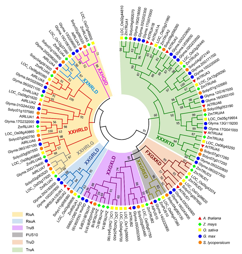
2.1. Identification and Phylogenetic Analysis of the PUS Genes in Arabidopsis thaliana and Zea mays

To identify the PUS genes in Arabidopsis and maize, we used the hidden Markov model (HMM) based search against proteome sequences of Arabidopsis and maize via HMMER (https://hmmer.org/ (11 February 2022)) taking an e-value cutoff of 1 × 10⁻⁵. The amino acid sequences were further confirmed in NCBI (https://www.ncbi.nlm.nih.gov (11 February 2022)), Ensembl plants (https://plants.ensembl.org/index.html (11 February 2022)) and maizeGDB (https://www.maizegdb.org/ (11 February 2022)). After redundant sequences and sequences without core catalytic domain were removed, a total of 20 genes in Arabidopsis and 22 genes in maize were identified and used for further analysis, respectively (Tables S1 and S2). Additionally, 22, 31, and 19 PUS genes in Oryza sativa, Glycine max, and Solanum lycopersicum, respectively, were identified using the same strategy from phytozome (https://phytozome.jgi.doe.gov/pz/portal.html (11 February 2022)) in this study (Table S3). To analyze the evolutionary relationships of the AtPUS and ZmPUS proteins, an unrooted phylogenetic tree of plant PUS genes was constructed based on the sequences of PUS catalytic domains from A. thaliana, Glycine max, Zea mays, Oryza sativa, and Solanum lycopersicum (Figure 1 and Table S4). Combined with the typical features of the conserved catalytic motif in enzymatic domain, the phylogenetic analysis showed that the PUS proteins were clustered into two groups: the first group share homological conserved catalytic domain with bacteria rRNA pseudouridine synthase, which could be further divided into two subfamilies, RluA and RsuA; the second group share homological conserved catalytic domain with bacteria tRNA pseudouridine synthase, which could be further divided into four subfamilies, TruA, TruB, TruD, and Pus10. Notably, there are only one copy of Pus10 gene in all the plant species we checked, which is consistent with the observation in animals and archaea [11], suggesting that Pus10 might play an essential role in a strict dosage-dependent manner. The RsuA subfamily is the largest PUS subfamily in E. coli, while it contracted to have only one or two members in each plant species we checked. Instead, either RluA or TruA is the largest family in Arabidopsis, maize, rice, soybean, and tomato. Therefore, all maize and Arabidopsis PUS genes were designated based their subfamily name, respectively (Figure 1. Tables S1 and S2).

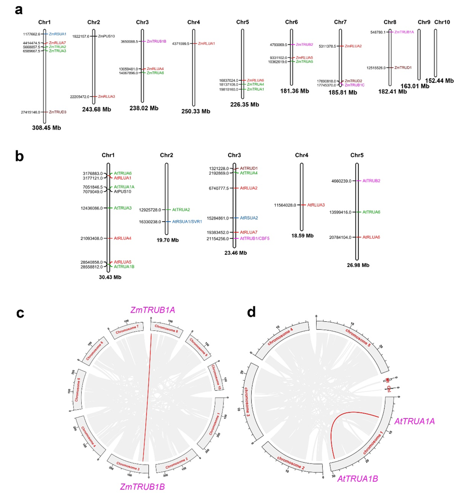
2.2. Chromosomal Distribution and Gene Synteny of AtPUS and ZmPUS Genes

According to the chromosomal location information of AtPUS and ZmPUS genes from the GFF3 reference file of Arabidopsis and maize genomes, their chromosomal maps were constructed using Mapchart, a local analytical tool (https://www.wur.nl/en/show/Mapchart.htm (13 February 2022)) (Figure 2a,b) [24]. ZmPUSs are distributed on most of chromosomes, with the exception of chromosome 9 and 10. At most, five genes including ZmRSUA1, ZmRLUA7, ZmTRUA2, ZmTRUA3, and ZmTRUD3 are located on chromosome 1. In contrast, AtPUSs are distributed on all five chromosomes in the compact genome of Arabidopsis, and chromosome 1 have the highest density of PUS genes, with 8 members. Gene synteny analysis revealed only one segmental duplication event involving two TruB genes, ZmTRUB1A and ZmTRUB1B, whereas there is no identified tandem duplication of PUS genes in maize genome (Figure 2c). Similarly, only one segmental duplication event involving two TruA genes, AtTRUA1A and AtTRUA1B, was identified on the chromosome 1 in Arabidopsis (Figure 2d). There is no whole genome duplication or segmental duplication event identified in colinear relationship analysis of PUS genes between maize and Arabidopsis (Figure S1). Instead, two colinear gene pairs were identified between Arabidopsis and rice, while 3, 2, 7 colinear PUS gene pairs were identified between Arabidopsis and tomato, Arabidopsis and soybean, rice and maize, respectively (Figure S1 and Table S5).

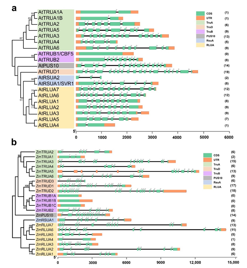
2.3. Gene Structure of AtPUS and ZmPUS Genes

As the exon-intron structure could reflect certain information in the evolution of gene families and provide additional support for phylogenetic analysis, we further analyze the exon-intron structure of the PUS genes in Arabidopsis and maize, based on their evolutionary classification (Figure 3, Tables S6 and S7). In general, the average length of ZmPUS genes is longer than the one of AtPUS genes, mainly due to the longer intron size in maize. The average intron numbers per gene are similar (7.65 per gene in Arabidopsis versus 8.00 per gene in maize). The proportion of intron phases 1, 2, and 0 in Arabidopsis are 15.03%, 26.14%, and 58.82%, respectively. In contrast, the proportion of intron phases 1, 2, and 0 in maize are 15.79%, 27.49%, and 56.73%, respectively. The intron numbers vary from 0 to 19 in different PUS members. However, the orthologous gene pairs in each subfamily between Arabidopsis and maize have comparable intron numbers and similar pattern of intron phases (Figure 1 and Figure 3). Without exception, all the orthologous genes encoding the well-described RNA-dependent pseudouridine synthase CBF5, have no intron in all the plant species surveyed. In contrast, plant TruD genes mostly contain the largest number of introns (Figure 3). Notably, in comparison with the only one TruD gene in Arabidopsis, there are three TruD genes in maize, suggesting that this subfamily in maize may have undergone gene family expansion. Among them, ZmTRUD1 and ZmTRUD2 share similar intron number and exon-intron structure, while ZmTRUD3 showed similar exon-intron structure with the 5th to 11th exons of ZmTRUD1/2 (Figure 3b).

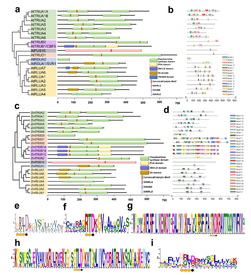
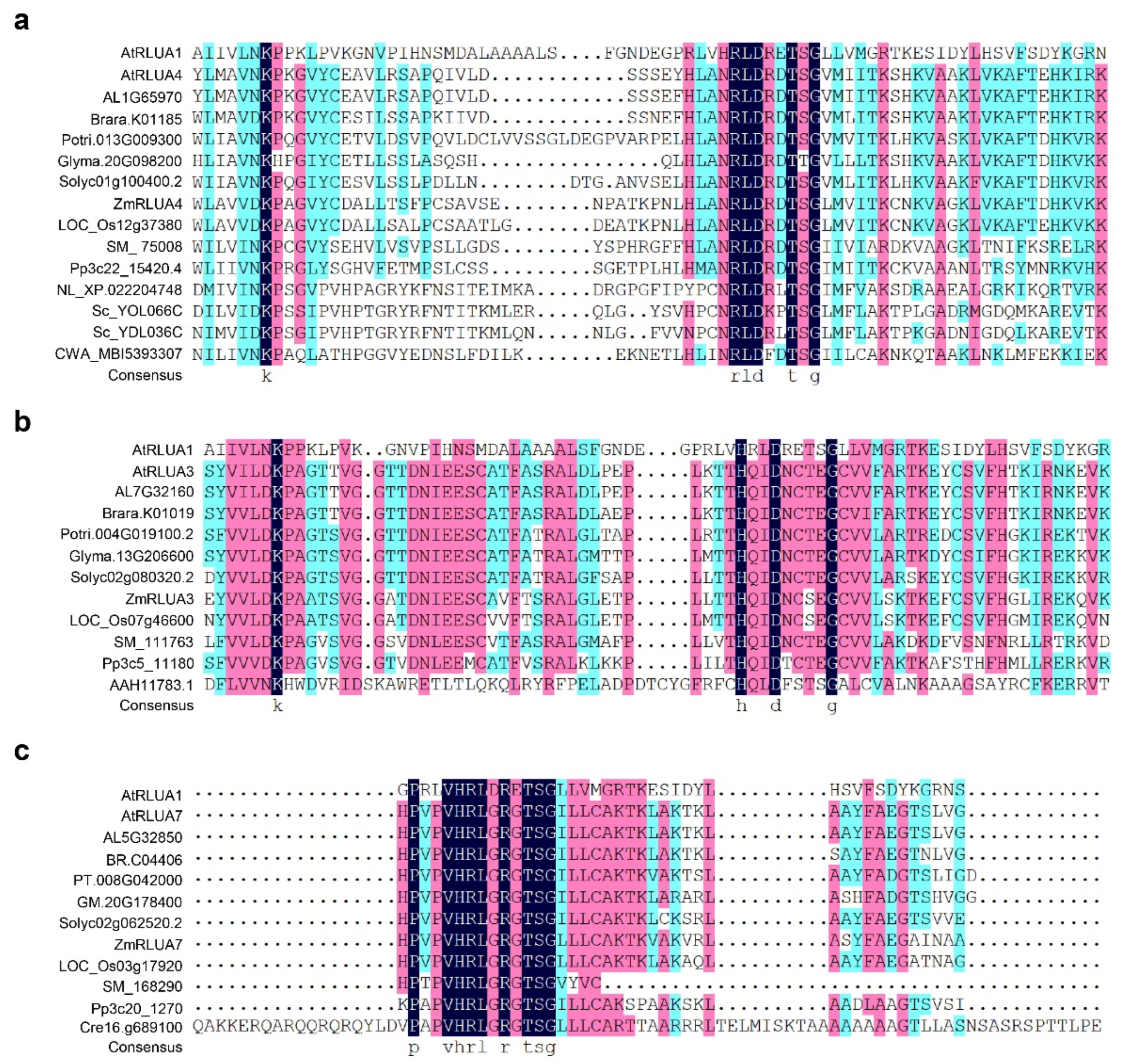
2.4. Protein Features of AtPUSs and ZmPUSs

The lengths of AtPUSs ranged from 74 to 715 amino acid residues, while the ones of ZmPUSs ranged from 167 to 701 amino acid residues. The molecular weight (MW) of AtPUS proteins ranged from 8.3 to 79.4 kDa, while the one of ZmPUS proteins ranged from 18.8 to 77.4 kDa. The isoeletric point (pI) of AtPUS proteins ranged from 5.22 to 9.95, while the one of ZmPUS proteins varied from 5.67 to 9.93 (Table 1 and Table 2). In line with the observation of the PUS proteins in E. coli, sequence alignment revealed that ZmPUS proteins diverge widely in amino acid sequence, especially between the pseudouridine synthases from different subfamilies. The high identity more than 90% only could be found between the paralogs of maize TruB subfamily (Figure S2), which might be generated from certain recent gene duplications. According to the analysis by online tool SMART (http://smart.embl-heidelberg.de/ (13 February 2022)) and sequence alignment with protein sequences in the same subfamily, the PUS catalytic domain and the core catalytic motif were annotated (Figure 4a,c) [25]. Furthermore, the protein structural diversity was analyzed and the conserved motifs were identified by the online tool MEME (http://meme-suite.org/tools/meme (13 February 2022)). Motif 2 in Arabidopsis PUSs covered conserved catalytic motifs in most of PUS subfamilies, except TruD and Pus10 (Figure 4a,e). Motif 15, 10, 2, and 11 in maize PUSs correspond to the conserved catalytic motifs in TruA, TruD, TruB, and RluA/RsuA subfamilies, respectively (Figure 4b,f–i). Eighteen members of Arabidopsis PUSs and nineteen members of maize PUSs have the universally conserved aspartic acid (Asp) (Figure 4a,c). Consistent with their PUS orthologs in E. coli, both Arabidopsis and maize PUSs have the canonical six-amino-acid motifs, such as XXHRLD in RluA subfamily, XXGRLD in RsuA subfamily, HXGXLD in TruB subfamily, XXGRED in Pus10 subfamily, and XXXRTD in TruA subfamily, which are also conserved in all dicot and monocot plants surveyed (Figure 1 and Table S4) [10,11]. Different from the canonical core catalytic motif of XAGXKD in eubacterial TruD, most of the catalytic motif in all surveyed plant TruDs is FAGTKD, and only AtTRUD1 have a serine instead of alanine in the motif (Figure 4g and Figure S3). ZmTRUD3 only contains an incomplete PUS catalytic domain lack of the core catalytic motif, and it is much shorter than the other canonical TruDs (Figure 4, Table 1 and Table 2). Likewise, sharing 94.1% identity with C-terminal of the catalytic domain in AtRSUA1/SVR1, AtRSUA2 is only around one sixth of SVR1 protein in length and likely a truncated pseudouridine synthase without core catalytic motif of XXGRLD in plant RsuA subfamily (Figure 4a and Table 1).
Notably, not all the members of RluA subfamily have the canonical six-amino-acid motif of XXHRLD, and there are three types of six-amino-acid motif variants including XXNRLD, XXHQID, and XXHRLG, in the members of both Arabidopsis and maize RluA subfamily (Figure 1 and Figure 5). Both the XXNRLD- and XXHQID-type catalytic motif variants are widely found in the RluA proteins from the close cruciferous species such as Arabidopsis lyrata and Brassca rapa, dicot and monocot plants, and even some lower-order plants such as Selaginella moellendorffii and Physcomitrella patens, whereas they are not found in algae such as Chara braunii and Chlamydomonas reinhardtii (Figure 5a,b). The universally conserved catalytic Asp is replaced with glycine in the XXHRLG-type RluA variants, such as AtRLUA7 and ZmRLUA7, probably leading to the loss of pseudouridylation activity. However, it is surprising that this putative pseudouridylation-defective RluA variants are present in alga, fern, moss, and spermatophyte (Figure 5c).

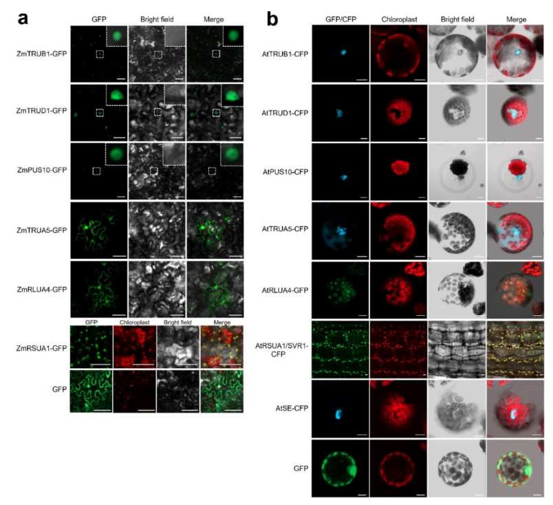
2.5. Subcellular Localization of AtPUSs and ZmPUSs

As the subcellular localization could provide us some clue to predict their potential function and target RNAs, the subcellular localizations of AtPUSs and ZmPUSs were predicted by the online tool Plant-mPLoc and WoLF PSORT (http://www.csbio.sjtu.edu.cn/bioinf/plant-multi/ (12 February 2022)) and https://wolfpsort.hgc.jp/ (12 February 2022)) (Table 1 and Table 2) [26,27,28,29]. Both AtPUSs and ZmPUSs have diverse subcellular localization in cell, e.g., nucleus, cytoplasm, chloroplast, and mitochondria, which might be correlated with the subcellular localization of their RNA substrates. We selected one PUS protein for each subfamily in maize and Arabidopsis for further analysis. Their full-length coding sequences were fused in front of Green Fluorescent Protein (GFP) or Cyan Fluorescent Protein (CFP) driven by the 35S promoter. The confocal microscope results of transient expression assay confirmed their subcellular localization (Figure 6). Most of the tested PUS proteins were localized as predicted. In consistent with previous report, ZmTRUB1A, the ortholog of AtTRUB1/CBF5/NAP57, was localized in nucleus [19]. ZmTRUD1 and ZmPUS10, together with their Arabidopsis orthologs, AtTRUD1 and AtPUS10, were also dominantly localized in nucleus, while ZmTRUA5 and AtTRUA5 showed nucleo-cytoplasmic localization. Our complementation transgenic plants of svr1-2/pSVR1-SVR1-CFP confirmed that SVR1 was co-localized with chloroplasts, in reminiscence of the transient expression result of SVR1-GFP in Arabidopsis protoplasts in previous report [15]. It is not surprising that ZmRSUA1 were co-localized with chloroplasts as well. Besides, ZmRLUA4 was localized in both nucleus and cytoplasm, whereas its Arabidopsis ortholog AtRLUA4 was localized in chloroplasts and highly accumulated in some speckles.

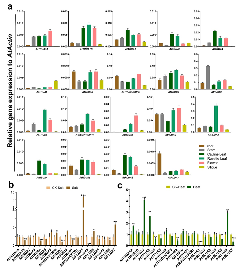
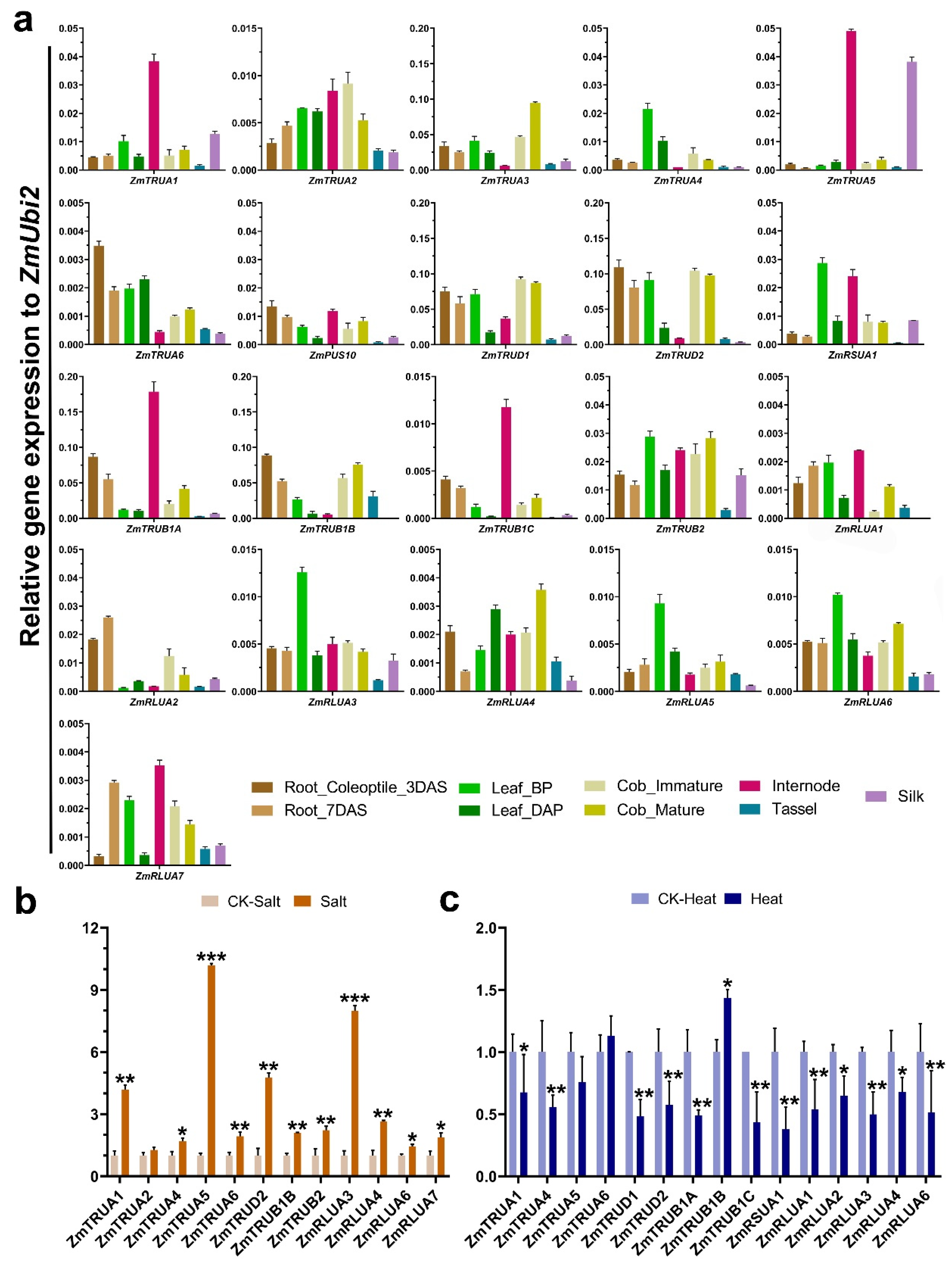
2.6. Expression Analysis of the AtPUS and ZmPUS Genes in Different Tissues and in Response to Abiotic Stresses

To analyze the expression pattern of AtPUS genes in different tissues, including root, stem, cauline leaf, rosette leaf, flower, and silique, quantitative real-time PCR (qRT-PCR) were further performed. The results showed that all the AtPUS genes except AtRSUA2 could be detected by qRT-PCR analysis (Figure 7a). The expression levels of most of AtPUS genes were relatively high in both cauline leaf and rosette leaf, in comparison with the ones in other tissues. AtTRUA1A, AtTRUB1, and AtRLUA1 were highly expressed in flower, while AtTRUA5 and AtRLUA7 has the highest expression in silique and root, respectively. In yeast and mammals, dynamic pseudouridylation and PUS subcellular localization upon various stress indicated the regulatory role of PUSs in response to stresses [1,6,12]. Here we further investigated the expression pattern of Arabidopsis seedlings under high salt stress and heat stress. AtPUSs had diverse responses to different stresses. Under salt stress, AtTRUA6, AtTRUB1/CBF5, AtPUS10, AtRLUA1, AtRLUA3, and AtRLUA7 were highly induced, whereas AtTRUA1B, AtTRUA4, AtTRUB2, AtTRUD1, and AtRLUA2 were significantly down-regulated (Figure 7b). Upon heat stress, three members of AtTRUA subfamily, AtTRUA1A, AtTRUA2, AtTRUA3, and AtRLUA6 were significantly up-regulated, and in contrast, AtTRUB2, AtRLUA1, and AtRLUA7 were significantly down-regulated (Figure 7c).
Likewise, to further analyze the expression pattern of ZmPUS genes, the expression pattern of maize tissues/organs (coleoptile, root, internode, leaf, tassel, cob, silk) at different developmental stages and maize seedlings with salt and heat stress treatments were also investigated by qRT-PCR analysis (Figure 8). In line with the observation in the expression of AtPUSs, there’s no consistent expression pattern even in each of ZmPUS subfamilies. The expression levels of most of ZmPUS genes were relatively high in leaf, internode, and cob. In particular, ZmTRUA3, ZmTRUD1, and ZmRLUA4 have the highest expression level in cob, whereas ZmTRUA5 largely expressed in internode and silk (Figure 8a). Under salt stress, ZmTRUA1, ZmTRUA5, ZmTRUA6, ZmTRUD2, ZmTRUB1B, ZmTRUB2, ZmRLUA3, and ZmRLUA4 were highly induced (Figure 8b). Under heat stress, several ZmPUS genes such as ZmTRUB1A/C and ZmRSUA1 were down-regulated, while ZmTRUB1B were moderately up-regulated (Figure 8c).

3. Discussion

In our study, although the genome sizes of all the dicots and monocots we analyzed here, including Arabidopsis, soybean, tomato, maize, and rice, varies in a large range, the numbers of PUS gene family in each species are close to each other. All the species mentioned above have members in each of PUS subfamilies, in which RluA and TruA subfamilies are the largest two subfamilies. In each PUS subfamily, both the Arabidopsis and maize PUS orthologous genes share some similar features of gene structure, such as the average intron number, the intron phases, and the size of coding sequence. Only the average intron length of ZmPUS genes is much longer than the one of AtPUS genes, which is the general difference between monocot and dicot genes. Notably, obvious gene family expansion only happened in maize TruB and TruD subfamily. Three ZmTruBs shared very high identity in protein sequences but showed diverse expression profiles in different tissues and in response to abiotic stress (Figure S2 and Figure 8), suggesting their spatio-temporal expression regulation with specificity. Among three ZmTRUDs, both ZmTRUD1 and ZmTRUD2 share similar tissue specific expression pattern (Figure 8A). Notably, both ZmTRUD3 and AtRSUA2 were supposed to encode a much shorter protein lack of the complete catalytic domain, in comparison with their paralogs (Figure 3, Figure 4A and Figure S3). In terms of their undetectable expression level in our qRT-PCR results and transcriptome data from maize and Arabidopsis eFP browsers (http://bar.utoronto.ca/efp/cgi-bin/efpWeb.cgi (7 February 2022)), ZmTRUD3 and AtRSUA2 might be pseudogenes.
Canonical pseudouridine synthase contains a structurally similar core motif including a universally conserved aspartic acid (Asp/D) residue. By our extensive searching for the core motif with the sequence alignments of PUS catalytic domain, all the canonical core motifs in bacteria could also be found in the corresponding orthologous proteins of each plant PUS subfamily. However, it’s noteworthy that not all the active site consensus sequences of plant RluA family protein are XXHRLD. There are three other conserved RluA variants in plant, in which their core motifs are XXNRLD, XXHQID, and XXHRLG, respectively. Both the XXNRLD- and XXHQID-type RluA variants are widely found in fern, moss, and spermatophyte, but not alga, suggesting that these two types may diverge after the emergence of vascular plants and have conserved function. XXNRLD-type RluAs are also present in some of eubacteria, archaea, and fungi, whereas they could hardly be found in animal but Nilaparvata lugens (Figure 5a,b). Yeast Pus8p/Rib2 and Pus9p, which are both XXNRLD-type RluA variants, are responsible for Ψ32 formation in cytoplasmic and mitochondrial tRNAs, respectively [30]. The arginine that is two amino acids N-terminal to the catalytic aspartate, is absolutely conserved in canonical pseudouridine synthase of the RluA, RsuA, and TruA subfamilies, and probably facilitate substrate stabilization and base-flipping [10]. This key arginine is replaced by glutamine in XXHQID-type RluA, probably affecting the enzyme activity. It’s notable that the core catalytic motif of human RPUSD1 and its orthologs in mammals are XXHQLD, in which the conserved isoleucine is replaced by leucine. Considering the similar identity of isoleucine and leucine, it’s not surprising that both XXHQID-type and XXHQLD-type RluAs might have similar enzyme identity. Besides, it’s interesting that XXHRLG-type RluAs, which appear to be catalytically defective PUSs, are present in plants from alga to spermatophyte but not found in other organisms (Figure 5c), suggesting that this type of RluA might play a special role in plant life cycle. The mutant of SVR1, which encodes the Arabidopsis RsuA protein, is defective in chloroplast rRNA processing and translation [15]. However, the developmental defect could be complemented by overexpression of SVR1 with the mutation in the conserved catalytic active Asp, like wild-type SVR1 [15]. In Chlamydomonas, trans splicing of group II introns in chloroplast mRNA required the physical presence but not the isomerization activity of the chloroplast-localized Maa2, a pseudouridine synthase in TruB subfamily [31]. Similar observations were also reported for other pseudouridine synthases from bacteria and yeast [32,33,34]. All these results supported that pseudouridine synthases have some function beyond their pseudouridylation activity. Likewise, XXHRLG-type RluA variants lacking the Asp catalytic active site are likely to have certain conserved functions independent of the pseudouridylation activity. Alternatively, we could not exclude another possibility that XXHRLG-type RluA variants might work cooperatively with other catalytic active partner. Anyway, further functional analysis of these RluA variants would help us understand the mechanism of RluA-mediated epigenetic regulation.
Increasing evidence in yeast and mammalian cells supported that RNA pseudouridylation play essential role in development and stress responses. However, RNA pseudouridylation in plant remains largely unknown. To investigate the role of RNA pseudouridylation in plant development and stress response, it is worth to note that the spatiotemporal expression pattern of PUS genes would provide us a hint for their function. Not surprisingly, no representative tissue-specific expression pattern for each PUS subfamily could be found either in Arabidopsis or in maize, probably due to their wide range of RNA substrates present in various tissues/organs. The diversity of expression pattern for plant PUS genes determined that detailed functional analysis for each PUS gene need to be done. Phenotypic observation and genome-wide identification of pseudouridylation sites for the loss-of-function mutants of PUS genes will help us elucidate the puzzle. It’s worth noting that the RNA pseudouridylation can be induced in response to some stresses, such as heat shock, nutrient deprivation, and oxidative stress in human or yeast [6,12,13], suggesting their regulatory role in stress responses. Interestingly, by transcriptional analysis for both Arabidopsis and maize PUS genes, here we could identify some stress responsive PUS genes, especially upon heat stress and salt stress. As some stress-responsive pseudouridylation were accompanied with the activation/repression of the corresponding PUS genes, therefore these stress-responsive PUS genes in both Arabidopsis and maize would be good candidates for elucidating the mechanism of RNA pseudouridylation in regulating the stress response. In particular, both AtRLUA3 and AtRLUA7, together with their maize orthologous genes ZmRLUA3 and ZmRLUA7, were induced by salt stress, while heat stress could repress the expression of both AtTRUA4 and AtRLUA1, as well as their maize orthologous genes. However, the expression profile of most of AtPUSs in response to either salt stress or heat stress didn’t keep pace with the one of ZmPUSs. Nevertheless, it is not always the same case that RNA pseudouridylation is positively correlated with the expression of the corresponding PUS gene. In yeast, although many pseudouridylation sites were heat-shock induced and Pus7p dependent, the levels of Pus7p mRNA and protein were down-regulated upon heat shock [6]. Not only pseudouridylation of U2 snRNA in nucleus [35,36], but also 5S rRNAs and cytoplasmic tRNAs [37,38], were all the RNA substrates of yeast Pus7p. Similarly, human PUS7 worked on cytoplasmic tRNAs as well [39,40]. Particularly, the shuttle of Pus7p from nucleus to cytoplasm upon heat shock might account for the induced pseudouridylation sites [6], which indicating that eukaryotic TruD proteins would be conditionally localized in different cellular compartments. Therefore, not only the expression level but also the subcellular localization of pseudouridine synthases need to be considered for the dynamic regulation of RNA pseudouridylation. Our preliminary study showed that both maize and Arabidopsis PUS proteins in each PUS subfamily have diverse subcellular localization pattern, which would be essential for the predication of their RNA substrates and biochemical function. Here we observed that both AtTRUD1 and ZmTRUD1 were all dominantly localized in nucleus. Whether plant TRUDs have conserved functions and localization features as well as yeast and human PUS7s remain unclear. The dynamic subcellular localization of PUS proteins still needs to be further investigated, especially upon various stress condition. A series of previous studies reported that eukaryotic and archaeal PUS10s could be localized in cytoplasm and produce pseudouridine in tRNAs [11,41,42,43,44,45]. However, it’s surprising that both AtPUS10 and ZmPUS10 were found to be dominantly localized in nucleus, probably suggesting an unexpected function in plant PUS10s. Besides, AtRLUA4 was localized in chloroplast, whereas its maize ortholog ZmRLUA4 was localized in both nucleus and cytoplasm. There might be an independent functional evolution between different species, or between dicot and monocot. Anyhow, comprehensive functional studies of plant PUSs would help us solve these mysteries.

4. Materials and Methods

4.1. Identification of the PUS Genes in Arabidopsis and Maize

To identify the PUS genes in Arabidopsis and maize, we used the protein sequences from pseudouridine synthases in E. coli and human as queries to obtain the representative PFAM IDs of six PUS subfamily except Pus10 and download their corresponding hidden Markov model (HMM) profiles from PFAM (https://pfam.xfam.org (11 February 2022)). Then we used three types of PUS10 proteins from Human, S.cerevisiae, and Archaea to constitute a hidden Markov model for Pus10. All of these HMM profiles were searched against proteome sequences of Arabidopsis and maize via HMMER (https://hmmer.org/ (11 February 2022)) taking an e-value cutoff of 1 × 10⁻⁵. The amino acid sequences and the representative domain were further confirmed in NCBI (https://www.ncbi.nlm.nih.gov (11 February 2022)), Ensembl plants (https://plants.ensembl.org/index.html (11 February 2022)) and maizeGDB (https://www.maizegdb.org/ (11 February 2022)). After redundant sequences and sequences without core catalytic domain were removed, a total of 20 Arabidopsis genes and 22 maize genes were identified and used for further analysis, respectively (Supplementary Material, Files S1 and S2). Additionally, HMMbased search against the rice, soybean, and tomato protein databases from phytozome (https://phytozome.jgi.doe.gov/pz/portal.html (11 February 2022)), including Zea mays RefGen_V5, Oryza sativa v7_JGI, Glycine max Wm82.a2.v1, and Solanum lycopersicum iTAG2.4, respectively, were performed using the same strategy.

4.2. Phylogenetic Analysis and Gene Structure

For the phylogenetic tress of PUS proteins in several organisms, the sequences of the conserved catalytic domain were used for multiple sequence alignments by ClustalW with default parameters. A maximum likelihood phylogenetic tree was constructed using MEGA 7.0 (https://www.megasoftware.net/ (22 February 2022)) with amino acid substitution model of Welan and Goldman + Freq [46] and 1000 bootstrap replicates.

4.3. Amino Acid Sequence Analysis

The domains of PUS proteins were analyzed using the online tool SMART (http://smart.embl-heidelberg.de/ (13 February 2022)) and ExPASy (https://prosite.expasy.org/ (15 February 2022)). The core catalytic motifs were identified by sequence alignments of the PUS catalytic domain in the same subfamily. The subcellular localization of the AtPUS and ZmPUS proteins were predicted using the online tool Plant-mPLoc (http://www.csbio.sjtu.edu.cn/bioinf/plant-multi/ (12 February 2022)) and WOLFPSORT (https://wolfpsort.hgc.jp/ (12 February 2022)). The molecular masses and isoelectric points of the AtPUS and ZmPUS were predicted using the online tool ExPASy (https://web.expasy.org/compute_pi/ (15 February 2022)). Multiple protein sequence alignments were performed using DNAMAN8. The conserved motifs and sequence logos of the conserved motifs of proteins were identified by the online tool MEME (http://meme-suite.org/tools/meme (23 February 2022)). The scheme of protein structures for motif annotation in PUS family proteins were constructed using TBtools [47].

4.4. Plant Materials and Growth Conditions

Seedlings of Arabidopsis Columbia-0 ecotype were grown in a greenhouse at 22 °C with a 16 h light/8 h dark cycle. The roots, stems, and rosette leaves were sampled from 3-week-old seedlings. The cauline leaves and flowers were sampled from flowering plants 5 weeks post-germination. The siliques were sampled 10 days post-pollination. The 12-day-old seedlings were used for heat stress and salt stress treatment, respectively. The plants were sampled from heat stress treatment at 37 °C for 3 h and then at 22 °C for 1 h recover, and the control plants were sampled from 22 °C for 4 h. The plants were treated and sampled for salt stress treatment at 150 mM NaCl for 24 h, while the control plants were sampled from mock treatment for 24 h.
The material of maize inbred line B73 were prepared as previously described [48]. Maize seedlings were cultured in soil at 25 °C with a 16 h light/8 h dark photoperiod in a greenhouse. Different tissues were sampled from different developmental stages as described [49]. The seedlings 14-day after sowing were used for heat stress and salt stress treatment, respectively. The plants were sampled from heat stress treatment at 55 °C for 4 h [50], and the control plants were sampled from 25 °C for 4 h. The plants were sampled from salt stress treatment with final concentration of 200 mM NaCl [51], and the control plants were sampled from mock treatment in the respective time point.

4.5. Expression of AtPUS and ZmPUS Genes Analyzed by Quantitative Real-Time PCR

Total RNA was extracted using the TRIzol reagent (Invitrogen, Beijing, China) and reverse transcribed using Takara Bio Cat. No. RR047A (Takara Bio, Tokyo, Japan) according to the manufacturer’s instructions. The quantitative real-time PCR were performed for at least three replicates and the expression of Arabidopsis ACT7 gene and maize UBI2 were used as an internal control, respectively. The sequence of the primers was listed in Table S8.

4.6. Subcellular Localization of ZmPUS and AtPUS Proteins by Confocal Imaging Analysis

The CDSs of ZmTRUB1, ZmTRUD1, ZmTRUA5, ZmRLUA4, ZmRSUA1, and AtRLUA4 were cloned into pCambia1300-221-GFP.3/GFP.1 and fused with GFP by restriction-ligation reactions. The CDS of ZmPUS10 was cloned into pCambia1300-221-GFP.1 and fused with GFP by homologous recombination reactions. These constructs were transformed into the epidermal cells of Nicotiniana benthamiana by Agrobacterium tumefaciens (strain GV3101). The CDSs of AtTRUB1, AtTRUD1, AtPUS10, and AtTRUA5 were cloned into pH7CWG2.0 and fused with CFP by Gateway LR reactions, while AtRSUA1/SVR1 were cloned and fused with CFP in C-terminal driven by SVR1 native promoter (1126 bp upstream the start codon of SVR1 gene) by restriction-ligation reaction and Gateway LR reaction. The plasmids of AtTRUB1, AtTRUD1, AtPUS10, and AtTRUA5 and AtRLUA4 were transiently expressed in Arabidopsis mesophyll protoplasts isolated from 21-day-old mature leaves according to a polyethylene glycol (PEG) transformation protocol described previously [52]. svr1-2 mutant (SALK_013085) were complemented by pSVR1-SVR1-CFP and the 7-day seedlings of complementation lines were used for confocal imaging. Signals of green fluorescent protein (GFP) and cyan fluorescent protein (CFP) were detected using a LSM800 confocal microscope (Carl Ziess GmbH, Jena, Germany). CFP fluorescence (406–470 nm) and GFP fluorescence (490–518 nm) were excited by lasers in 405nm and 488 nm, respectively. The picture processing was done by ZEN software (Carl Zeiss GmbH) and Photoshop CS6 (Adobe, San Jose, CA, USA).

5. Conclusions

In this study, 20 Arabidopsis PUS genes and 22 maize PUS genes were identified, respectively. A phylogenetic analysis revealed that both Arabidopsis and maize pseudouridine synthases could be clustered into six subfamilies, RluA, RsuA, TruB, TruA, TruD and Pus10. The chromosomal location and exon-intron structure of the genes, and the motif and domain organization of the PUS proteins were further analyzed. RluA and TruA are the largest two subfamilies in plants. Notably, there are gene expansion in TruB and TruD subfamilies in maize. According to the six amino-acid sequence of the core catalytic motif, both Arabidopsis and maize RluA family proteins could be further divided into four groups, including the canonical XXHRLD-type, and another three variants of XXNRLD-, XXHQID-, and XXHRLG-type. Representative AtPUSs and ZmPUSs in each subfamily were found to have diverse subcellular localization. Expression profiles for PUS gene families in Arabidopsis and maize suggest the potential role of pseudouridine synthase genes in the response to heat and salt stress.

Supplementary Materials

The following are available online at https://www.mdpi.com/article/10.3390/ijms23052680/s1.

Author Contributions

Conceptualization, Methodology: Y.X., Y.G. and Z.Z.; Validation, Formal analysis, Investigation: Y.X., Y.G., G.S., J.H., W.H.; Resources, Data curation: Y.X., Y.G. and Z.Z.; Writing—original draft preparation, Y.X., Y.G. and Z.Z.; Writing—review and editing, Y.X., Y.G. and Z.Z.; Supervision, Z.Z.; Project administration, Z.Z.; Funding acquisition, Z.Z. All authors have read and agreed to the published version of the manuscript.

Funding

This work was supported by the National Science Foundation of Guangdong Province, China (Grant No. 2020B1515020007), National Natural Science Foundation of China (31771349, 32170593), and Guangdong Provincial Pearl River Talent Plan (Grant NO. 2019QN01N108).

Institutional Review Board Statement

Not applicable.

Informed Consent Statement

Not applicable.

Data Availability Statement

All data generated or analyzed in this study are included in this published article and its Supplementary Material. The datasets generated and analyzed during the current study are available from the corresponding author on reasonable request.

Acknowledgments

We thank Chengwei Yang and Jianzong Nan (Guangdong Provincial Key Laboratory of Biotechnology for Plant Development, School of Life Science, South China Normal University, China) for sharing the seeds of maize B73. We thank Xiaoyan Tang and Shunquan Chen (Shenzhen Institute of Molecular Crop Design, China) for providing some materials of maize tissues.

Conflicts of Interest

The authors declare no conflict of interest.

References

  1. Frye, M.; Harada, B.T.; Behm, M.; He, C. RNA modifications modulate gene expression during development. Science 2018, 361, 1346–1349. [Google Scholar] [CrossRef] [PubMed] [Green Version]
  2. Cohn, W.E.; Volkin, E. Nucleoside-5’-Phosphates from Ribonucleic Acid. Nature 1951, 167, 483–484. [Google Scholar] [CrossRef]
  3. Davis, F.F.; Allen, F.W. Ribonucleic acids from yeast which contain a fifth nucleotide. J. Biol. Chem. 1957, 227, 907–915. [Google Scholar] [CrossRef]
  4. Charette, M.; Gray, M.W. Pseudouridine in RNA: What, where, how, and why. IUBMB Life 2000, 49, 341–351. [Google Scholar]
  5. Carlile, T.M.; Rojas-Duran, M.F.; Gilbert, W.V. Transcriptome-Wide Identification of Pseudouridine Modifications Using Pseudo-seq. Curr. Protoc. Mol. Biol. 2015, 112, 4.25.1–4.25.24. [Google Scholar] [CrossRef]
  6. Schwartz, S.; Bernstein, D.A.; Mumbach, M.R.; Jovanovic, M.; Herbst, R.H.; Leon-Ricardo, B.X.; Engreitz, J.M.; Guttman, M.; Satija, R.; Lander, E.S.; et al. Transcriptome-wide mapping reveals widespread dynamic-regulated pseudouridylation of ncRNA and mRNA. Cell 2014, 159, 148–162. [Google Scholar] [CrossRef] [Green Version]
  7. Lovejoy, A.F.; Riordan, D.P.; Brown, P.O. Transcriptome-wide mapping of pseudouridines: Pseudouridine synthases modify specific mRNAs in S. cerevisiae. PLoS ONE 2014, 9, e110799. [Google Scholar] [CrossRef] [Green Version]
  8. Li, X.; Zhu, P.; Ma, S.; Song, J.; Bai, J.; Sun, F.; Yi, C. Chemical pulldown reveals dynamic pseudouridylation of the mammalian transcriptome. Nat. Chem. Biol. 2015, 11, 592–597. [Google Scholar] [CrossRef]
  9. Spenkuch, F.; Motorin, Y.; Helm, M. Pseudouridine: Still mysterious, but never a fake (uridine)! RNA Biol. 2014, 11, 1540–1554. [Google Scholar] [CrossRef] [Green Version]
  10. Hamma, T.; Ferre-D’Amare, A.R. Pseudouridine synthases. Chem. Biol. 2006, 13, 1125–1135. [Google Scholar] [CrossRef] [Green Version]
  11. Fitzek, E.; Joardar, A.; Gupta, R.; Geisler, M. Evolution of Eukaryal and Archaeal Pseudouridine Synthase Pus10. Mol. Biol. Evol. 2018, 86, 77–89. [Google Scholar] [CrossRef] [PubMed]
  12. Carlile, T.M.; Rojas-Duran, M.F.; Zinshteyn, B.; Shin, H.; Bartoli, K.M.; Gilbert, W.V. Pseudouridine profiling reveals regulated mRNA pseudouridylation in yeast and human cells. Nature 2014, 515, 143–146. [Google Scholar] [CrossRef] [PubMed] [Green Version]
  13. Basak, A.; Query, C.C. A pseudouridine residue in the spliceosome core is part of the filamentous growth program in yeast. Cell Rep. 2014, 8, 966–973. [Google Scholar] [CrossRef] [Green Version]
  14. Nakamoto, M.A.; Lovejoy, A.F.; Cygan, A.M.; Boothroyd, J.C. mRNA pseudouridylation affects RNA metabolism in the parasite Toxoplasma gondii. RNA 2017, 23, 1834–1849. [Google Scholar] [CrossRef] [PubMed] [Green Version]
  15. Yu, F.; Liu, X.; Alsheikh, M.; Park, S.; Rodermel, S. Mutations in SUPPRESSOR OF VARIEGATION1, a factor required for normal chloroplast translation, suppress var2-mediated leaf variegation in Arabidopsis. Plant Cell 2008, 20, 1786–1804. [Google Scholar] [CrossRef] [Green Version]
  16. Lu, S.; Li, C.; Zhang, Y.; Zheng, Z.; Liu, D. Functional Disruption of a Chloroplast Pseudouridine Synthase Desensitizes Arabidopsis Plants to Phosphate Starvation. Front. Plant Sci. 2017, 8, 1421. [Google Scholar] [CrossRef] [Green Version]
  17. Sun, L.; Xu, Y.; Bai, S.; Bai, X.; Zhu, H.; Dong, H.; Wang, W.; Zhu, X.; Hao, F.; Song, C.P. Transcriptome-wide analysis of pseudouridylation of mRNA and non-coding RNAs in Arabidopsis. J. Exp. Bot. 2019, 70, 5089–5600. [Google Scholar] [CrossRef] [Green Version]
  18. Lin, D.; Kong, R.; Chen, L.; Wang, Y.; Wu, L.; Xu, J.; Piao, Z.; Lee, G.; Dong, Y. Chloroplast development at low temperature requires the pseudouridine synthase gene TCD3 in rice. Sci. Rep. 2020, 10, 8518. [Google Scholar] [CrossRef]
  19. Lermontova, I.; Schubert, V.; Bornke, F.; Macas, J.; Schubert, I. Arabidopsis CBF5 interacts with the H/ACA snoRNP assembly factor NAF1. Plant Mol. Biol. 2007, 65, 615–626. [Google Scholar] [CrossRef]
  20. Kannan, K.; Nelson, A.D.; Shippen, D.E. Dyskerin is a component of the Arabidopsis telomerase RNP required for telomere maintenance. Mol. Cell. Biol. 2008, 28, 2241–2332. [Google Scholar] [CrossRef] [Green Version]
  21. Schnable, P.S.; Ware, D.; Fulton, R.S.; Stein, J.C.; Wei, F.; Pasternak, S.; Liang, C.; Zhang, J.; Fulton, L.; Graves, T.A. The B73 Maize Genome: Complexity, Diversity, and Dynamics. Science 2009, 326, 1112–1115. [Google Scholar] [CrossRef] [PubMed] [Green Version]
  22. Sun, S.; Zhou, Y.; Chen, J.; Shi, J.; Zhao, H.; Zhao, H.; Song, W.; Zhang, M.; Cui, Y.; Dong, X.; et al. Extensive intraspecific gene order and gene structural variations between Mo17 and other maize genomes. Nat. Genet. 2018, 50, 1289–1295. [Google Scholar] [CrossRef] [PubMed] [Green Version]
  23. Springer, N.M.; Anderson, S.N.; Andorf, C.M.; Ahern, K.R.; Bai, F.; Barad, O.; Barbazuk, W.B.; Bass, H.W.; Baruch, K.; Ben-Zvi, G.; et al. The maize W22 genome provides a foundation for functional genomics and transposon biology. Nat. Genet. 2018, 50, 1282–1288. [Google Scholar] [CrossRef] [Green Version]
  24. Voorrips, R.E. MapChart: Software for the graphical presentation of linkage maps and QTLs. J. Hered. 2002, 93, 77–78. [Google Scholar] [CrossRef] [Green Version]
  25. Letunic, I.; Bork, P. 20 years of the SMART protein domain annotation resource. Nucleic Acids Res. 2018, 46, D493–D496. [Google Scholar] [CrossRef]
  26. Wang, X.; Li, G.Z.; Lu, W.C. Virus-ECC-mPLoc: A Multi-Label Predictor for Predicting the Subcellular Localization of Virus Proteins with Both Single and Multiple Sites Based on a General Form of Chou’s Pseudo Amino Acid Composition. Protein Pept. Lett. 2013, 20, 309–317. [Google Scholar]
  27. Chou, K.C.; Shen, H.B. Cell-PLoc: A package of Web servers for predicting subcellular localization of proteins in various organisms. Nat. Protoc. 2008, 3, 153–162. [Google Scholar] [CrossRef]
  28. Chou, K.C.; Shen, H.B. Plant-mPLoc: A Top-Down Strategy to Augment the Power for Predicting Plant Protein Subcellular Localization. PLoS ONE 2010, 5, e11335. [Google Scholar] [CrossRef] [Green Version]
  29. Chou, K.C.; Shen, H.B. Large-scale plant protein subcellular location prediction. J. Cell. Chem. 2007, 100, 665–678. [Google Scholar] [CrossRef]
  30. Behm-Ansmant, I.; Grosjean, H.; Massenet, S.; Motorin, Y.; Branlant, C. Pseudouridylation at position 32 of mitochondrial and cytoplasmic tRNAs requires two distinct enzymes in Saccharomyces cerevisiae. J. Biol. Chem. 2004, 279, 52998–53006. [Google Scholar] [CrossRef] [Green Version]
  31. Perron, K.; Goldschmidt-Clermont, M.; Rochaix, J.D. A factor related to pseudouridine synthases is required for chloroplast group II intron trans-splicing in Chlamydomonas reinhardtii. EMBO J. 1999, 18, 6481–6490. [Google Scholar] [CrossRef] [Green Version]
  32. Gutgsell, N.S.; Del Campo, M.; Raychaudhuri, S.; Ofengand, J. A second function for pseudouridine synthases: A point mutant of RluD unable to form pseudouridines 1911, 1915, and 1917 in Escherichia coli 23S ribosomal RNA restores normal growth to an RluD-minus strain. RNA 2001, 7, 990–998. [Google Scholar] [CrossRef] [Green Version]
  33. Gutgsell, N.; Englund, N.; Niu, L.; Kaya, Y.; Lane, B.G.; Ofengand, J. Deletion of the Escherichia coli pseudouridine synthase gene truB blocks formation of pseudouridine 55 in tRNA in vivo, does not affect exponential growth, but confers a strong selective disadvantage in competition with wild-type cells. RNA 2000, 6, 1870–1881. [Google Scholar] [CrossRef] [Green Version]
  34. Lafontaine, D.L.; Bousquet-Antonelli, C.; Henry, Y.; Caizergues-Ferrer, M.; Tollervey, D. The box H + ACA snoRNAs carry Cbf5p, the putative rRNA pseudouridine synthase. Genes Dev. 1998, 12, 527–537. [Google Scholar] [CrossRef] [Green Version]
  35. Ma, X.; Zhao, X.; Yu, Y.T. Pseudouridylation (Psi) of U2 snRNA in S. cerevisiae is catalyzed by an RNA-independent mechanism. EMBO J. 2003, 22, 1889–1897. [Google Scholar] [CrossRef] [Green Version]
  36. Wu, G.; Xiao, M.; Yang, C.; Yu, Y.T. U2 snRNA is inducibly pseudouridylated at novel sites by Pus7p and snR81 RNP. EMBO J. 2011, 30, 79–89. [Google Scholar] [CrossRef] [Green Version]
  37. Behm-Ansmant, I.; Urban, A.; Ma, X.; Yu, Y.T.; Motorin, Y.; Branlant, C. The Saccharomyces cerevisiae U2 snRNA:pseudouridine-synthase Pus7p is a novel multisite-multisubstrate RNA:Psi-synthase also acting on tRNAs. RNA 2003, 9, 1371–1382. [Google Scholar] [CrossRef] [Green Version]
  38. Decatur, W.A.; Schnare, M.N. Different mechanisms for pseudouridine formation in yeast 5S and 5.8S rRNAs. Mol. Cell. Biol. 2008, 28, 3089–3100. [Google Scholar] [CrossRef] [Green Version]
  39. Guegueniat, J.; Halabelian, L.; Zeng, H.; Dong, A.; Li, Y.; Wu, H.; Arrowsmith, C.H.; Kothe, U. The human pseudouridine synthase PUS7 recognizes RNA with an extended multi-domain binding surface. Nucleic Acids Res. 2021, 49, 11810–11822. [Google Scholar] [CrossRef]
  40. Guzzi, N.; Ciesla, M.; Ngoc, P.C.T.; Lang, S.; Arora, S.; Dimitriou, M.; Pimkova, K.; Sommarin, M.N.E.; Munita, R.; Lubas, M.; et al. Pseudouridylation of tRNA-Derived Fragments Steers Translational Control in Stem Cells. Cell 2018, 173, 1204–1216. e26. [Google Scholar] [CrossRef] [Green Version]
  41. Mukhopadhyay, S.; Deogharia, M.; Gupta, R. Mammalian nuclear TRUB1, mitochondrial TRUB2, and cytoplasmic PUS10 produce conserved pseudouridine 55 in different sets of tRNA. RNA 2021, 27, 66–79. [Google Scholar] [CrossRef]
  42. Song, J.; Zhuang, Y.; Zhu, C.; Meng, H.; Lu, B.; Xie, B.; Peng, J.; Li, M.; Yi, C. Differential roles of human PUS10 in miRNA processing and tRNA pseudouridylation. Nat. Chem. Biol. 2020, 16, 160–169. [Google Scholar] [CrossRef]
  43. Deogharia, M.; Mukhopadhyay, S.; Joardar, A.; Gupta, R. The human ortholog of archaeal Pus10 produces pseudouridine 54 in select tRNAs where its recognition sequence contains a modified residue. RNA 2019, 25, 336–351. [Google Scholar] [CrossRef] [Green Version]
  44. Gurha, P.; Gupta, R. Archaeal Pus10 proteins can produce both pseudouridine 54 and 55 in tRNA. RNA 2008, 14, 2521–2527. [Google Scholar] [CrossRef] [Green Version]
  45. Roovers, M.; Hale, C.; Tricot, C.; Terns, M.P.; Terns, R.M.; Grosjean, H.; Droogmans, L. Formation of the conserved pseudouridine at position 55 in archaeal tRNA. Nucleic Acids Res. 2006, 34, 4293–4301. [Google Scholar] [CrossRef]
  46. Whelan, S.; Goldman, N. A general empirical model of protein evolution derived from multiple protein families using a maximum-likelihood approach. Mol. Biol Evol. 2001, 18, 691–699. [Google Scholar] [CrossRef] [Green Version]
  47. Chen, C.; Chen, H.; Zhang, Y.; Thomas, H.R.; Frank, M.H.; He, Y.; Xia, R. TBtools: An Integrative Toolkit Developed for Interactive Analyses of Big Biological Data. Mol. Plant 2020, 13, 1194–1202. [Google Scholar] [CrossRef]
  48. Ludwig, Y.; Zhang, Y.X.; Hochholdinger, F. The Maize (Zea mays L.) AUXIN/INDOLE-3-ACETIC ACID Gene Family: Phylogeny, Synteny, and Unique Root-Type and Tissue-Specific Expression Patterns during Development. PLoS ONE 2013, 8, e78859. [Google Scholar] [CrossRef] [Green Version]
  49. Walley, J.W.; Sartor, R.C.; Shen, Z.; Schmitz, R.J.; Wu, K.J.; Urich, M.A.; Nery, J.R.; Smith, L.G.; Schnable, J.C.; Ecker, J.R.; et al. Integration of omic networks in a developmental atlas of maize. Science 2016, 353, 814–818. [Google Scholar] [CrossRef] [Green Version]
  50. Makarevitch, I.; Waters, A.J.; West, P.T.; Stitzer, M.; Hirsch, C.N.; Ross-Ibarra, J.; Springer, N.M. Transposable Elements Contribute to Activation of Maize Genes in Response to Abiotic Stress. PLoS Genet. 2015, 11, e1004915. [Google Scholar]
  51. Nguyen, T.X.; Sticklen, M. Barley HVA1 Gene Confers Drought and Salt Tolerance in Transgenic Maize (Zea Mays, L.). Adv. Crop Sci. Tech 2013, 1, 105. [Google Scholar] [CrossRef] [Green Version]
  52. Yoo, S.D.; Cho, Y.H.; Sheen, J. Arabidopsis mesophyll protoplasts: A versatile cell system for transient gene expression analysis. Nat. Protoc. 2007, 2, 1565–1572. [Google Scholar] [CrossRef] [PubMed] [Green Version]
Figure 1. Phylogenetic tree of pseudouridine synthase family in several plant species. Evolutionary analyses were conducted by using the Maximum Likelihood method in MEGA7, based on the pseudouridine synthase domains of the PUS proteins from A. thaliana, Z. mays, O. sativa, G. max, and S. lycopersicum. The bootstrap consensus tree inferred from 500 replicates is taken to represent the evolutionary history of the taxa analyzed. The PUS members in the branches of phylogenetic tree covered by a colored panel belong to the same PUS subfamily as indicated and the sequences of the core six-amino-acid catalytic consensus sequence were shown, respectively.
Ijms 23 02680 g001
Figure 2. Chromosomal distribution and gene synteny of ZmPUS and AtPUS genes. (a) Chromosomal distribution of ZmPUS genes. (b) Chromosomal distribution of AtPUS genes. (c) Gene synteny of ZmPUSs in maize genome. (d) Gene synteny of AtPUSs in the Arabidopsis genome. The identities of the chromosome are indicated on the top of each chromosome, while the names and chromosomal coordinates of PUSs are shown to the left and the right of each chromosome, respectively. Gray lines in the background indicate the collinear blocks within the genomes of maize/Arabidopsis, and the red lines indicate the syntenic PUS gene pairs. All chromosomes are shown to scale based on their actual lengths.
Ijms 23 02680 g002
Figure 3. Exon-intron structures and intron phases of AtPUS and ZmPUS genes. (a) Exon-intron structures and intron phases of AtPUS genes. (b) Exon-intron structures and intron phases of ZmPUS genes. The green box, orange box, and black line indicate exon, UTR, and intron, respectively. The PUS members in the branches of phylogenetic tree covered by a colored panel belong to the same PUS subfamily as indicated. The numbers in brackets are the numbers of intron of the corresponding AtPUS and ZmPUS genes. The phases of intron (0, 1, and 2) are shown on the left top of the corresponding introns.
Ijms 23 02680 g003
Figure 4. Conserved domains and motifs of AtPUS and ZmPUS proteins. (a) Conserved domains of AtPUS proteins. (b) Conserved motifs of AtPUS proteins. (c) Conserved domains of ZmPUS proteins. (d) Conserved motifs of ZmPUS proteins. (e) Frequency distribution of amino acids in the motif 2 of AtPUS proteins. (f) Frequency distribution of amino acids in the motif 15 of ZmPUS proteins from TruA subfamily. (g) Frequency distribution of amino acids in the motif 10 of ZmPUS proteins from TruD subfamily. (h) Frequency distribution of amino acids in the motif 2 of ZmPUS proteins from TruB subfamily. (i) Frequency distribution of amino acids in the motif 11 of ZmPUS proteins from RluA and RsuA subfamilies. The conserved active Asp was indicated by closed asterisk; the other conserved amino acids were indicated by open asterisk.
Ijms 23 02680 g004
Figure 5. Non-canonical catalytic motif in plant PUS proteins. (a) Sequence alignment for the XXNRLD-type catalytic motif of PUS proteins from different species. (b) Sequence alignment for the XXHQID-type catalytic motif of PUS proteins from different species. (c) Sequence alignment for the XXHRLG-type catalytic motif of PUS proteins from different species. The XXHRLD-type canonical catalytic motif of PUS proteins from A. thaliana and E. coli, the XXHQID-type of PUS proteins from H. sapiens were included for alignment control, respectively. At, Arabidopsis thaliana; AL, Arabidopsis lyrata; Brara, Brassica rapa; Potri, Populus trichocarpa; Glyma, Glycine max (Linn.) Merr; Solyc, Solanum lycopersicum; Zm, Zea mays; LOC_Os, Oryza Sativa; Sm, Selaginella moellendorffii; Pp, Physcomitrella patens; Cre, Chlamydomonas reinhardtii; Sc, Saccharomyces cerevisiae; NL, Nilaparvata lugens; Hs, Homo sapiens; CWA, Candidatus Woesearchaeota archaeon.
Ijms 23 02680 g005
Figure 6. Subcellular localization of the representative proteins in each PUS subfamily. (a) Subcellular localization of ZmPUS proteins in the epidermal cells of Nicotinana benthamiana. GFP protein serves as control. Scale bars represent 50 μm. (b) Subcellular localization of AtPUS proteins in Arabidopsis mesophyll protoplasts and the stem of svr1-2/pSVR1-SVR1-CFP transgenic plants. The subcellular localization of AtTRUB1, AtTRUD1, AtPUS10, AtTRUA5, and AtRLUA4 fused with CFP/GFP in C-terminal by PEG transformation-mediated transient expression in Arabidopsis protoplasts. The subcellular localization of AtRSUA1/SVR1 was visualized from the stem of 7-day seedlings in transgenic plants of svr1-2/pSVR1-SVR1-CFP. AtSE-CFP and GFP only serve as controls. Scale bars represent 10 μm in length.
Ijms 23 02680 g006
Figure 7. Expression profiles of AtPUS genes in different tissues/organs and in response to abiotic stresses. (a) Quantitative real-time PCR for AtPUS genes in different tissues/organs. The y axis indicated the relative expression value to AtActin. (b) Quantitative real-time PCR for AtPUS genes in salt stress. (c) Quantitative real-time PCR for AtPUS genes in heat stresses. The expression of each sample treated by different abiotic stress were normalized by the corresponding control (CK). Student t-test were applied for the significance statistical analysis. The asterisk indicated the significance of difference between the control and stress-treated samples, * p < 0.05, ** p < 0.01, *** p < 0.001.
Ijms 23 02680 g007
Figure 8. Expression profiles of ZmPUS genes in different tissues/organs and in reponse to abiotic stress. (a) Quantitative real-time PCR for ZmPUS genes in different tissues/organs. Root_Coleoptile_3DAS, primary root and coleoptile 3-day after sowing; Root_7DAS, root system 7-day after sowing; Leaf_BP, leaves before pollination; Leaf_DAP, leaves day after pollination; Internode, internode 6th–7th; Cob_immature, immature cob; Cob_mature, mature cob; Tassel, unpollinated tassel; Silk, unpollinated silk. The y axis indicated the relative expression value to ZmUbi2. (b) Quantitative real-time PCR for ZmPUS genes in salt stress. (c) Quantitative real-time PCR for ZmPUS genes in heat stress. The expression of each sample treated by different abiotic stress were normalized by the corresponding control (CK). Student T-test were applied for the significance statistical analysis. The asterisk indicated the significance of difference between the control and stress-treated samples, * p < 0.05, ** p < 0.01, *** p < 0.001.
Ijms 23 02680 g008
Table 1. Information of AtPUS genes and the predicted proteins in Arabidopsis.
GroupNameTranscript IDChromosome LocationF(+)/R(−)AA apI bMW c
(KDa)
Predicted
Subcellular Localization
TruAAtTRUA1AAT1G20370.1Chr1:7051846-70535885495.22 61.5 Nc, Cp
AtTRUA1BAT1G76120.1Chr1:28558813-285602944637.23 51.8 Cp, Cm
AtTRUA2AT2G30320.1Chr2:12925728-129278965106.02 59.0 Chl, Mt
AtTRUA5AT5G35400.2Chr5:13599416-136022404716.19 53.3 Chl,ER, Mt
AtTRUA3AT1G34150.1Chr1:12436086-12439237+5659.19 63.0 Nc, Cp
AtTRUA4AT3G06950.1Chr3:2192869-2194254+3238.96 36.2 Chl
AtTRUA6AT1G09800.1Chr1:3177121-31803363727.27 41.5 Nc
TruBAtTRUB2AT5G14460.1Chr5:4660239-46625435408.93 61.5 Chl
AtTRUB1/CBF5AT3G57150.1Chr3:21154255-211559524468.84 51.0 Nc
Pus10AtPUS10AT1G20410.1Chr1:7079049-70825045046.55 56.5 Nc
TruDAtTRUD1AT3G04820.1Chr3:1321228-13259537155.76 79.4 Chl, Mt
RsuAAtRSUA2AT3G43340.1Chr3:15284861-15285851746.468.3Chl, Cp
AtRSUA1/SVR1AT2G39140.1Chr2:16330238-16333153+4109.95 45.1 Chl
RluAAtRLUA7AT3G52260.3Chr3:19383452-193864404166.24 45.8 Chl, Cp, Nc
AtRLUA6AT5G51140.2Chr5:20784103-207867934106.29 46.5 Cp
AtRLUA1AT1G78910.1Chr1:3177121-31803364789.48 53.8 Chl, Nc, Cp
AtRLUA2AT3G19440.1Chr3:6740778-6743132+4779.46 53.0 Mt, Chl
AtRLUA3AT4G21770.1Chr4:11564028-115663454728.13 52.9 Chl
AtRLUA5AT1G76050.2Chr1:28540858-28542826+4307.04 46.7 Chl, Mt
AtRLUA4AT1G56345.1Chr1:21093409-210944543226.94 35.9 Chl, Nc
AA a, Number of amino acids; pI b, Isoelectric point; MW c, Molecular weight; ER, Endoplasmic Reticulum; Nc, Nucleus; Mt, Mitochondrial; Cp, Cytoplasm; Chl, Chloroplast.
Table 2. Information of ZmPUS genes and the predicted proteins in maize.
GroupNameTranscript IDChromosome LocationF(+)/R(−)AA apI bMW c (KDa)Predicted
Subcellular Localization
TruAZmTRUA2Zm00001eb016190_T001 1:56688574-56693320+5168.66 57.4 Chl
ZmTRUA1Zm00001eb250100_T0015:198101606-198103976+5625.67 61.6 Nc, Chl, Mt
ZmTRUA3Zm00001eb018490_T001 1:65896675-65906968+4578.42 51.7 Chl, Nc
ZmTRUA4Zm00001eb245280_T0025:181371080-181377861+3349.00 37.1 Mt, Chl
ZmTRUA5Zm00001eb274430_T001 6:103626192-103638522+4228.65 46.7 Mt, Chl
ZmTRUA6Zm00001eb138750_T005 3:140678961-140686947+3868.05 42.3 Nc,Cp,Chl
TruDZmTRUD3Zm00001eb054900_T0011:274151464-2741538811679.92 18.8 Chl, Cp
ZmTRUD1Zm00001eb352870_T005 8:125155260-1251616846826.82 75.2 Nc,Cp,Chl
ZmTRUD2Zm00001eb328420_T002 7:176908181-1769194047016.22 77.4 Chl, Cp
TruBZmTRUB1AZm00001eb333520_T001 8:5487801-5489896+5839.19 64.2 Nc
ZmTRUB1BZm00001eb127780_T0013:36500885-365039265799.17 63.8 Nc
ZmTRUB1CZm00001eb328640_T001 7:177453702-1774560635839.08 63.9 Nc
ZmTRUB2Zm00001eb267110_T001 6:47930697-479355084979.25 56.1 Chl, Mt
Pus10ZmPUS10Zm00001eb074060_T004 2:19221575-19226195+5366.34 60.6 Nc
RsuAZmRSUA1Zm00001eb004240_T0011:11776626-11783053+4069.93 43.7 Chl
RluAZmRLUA7Zm00001eb013230_T001 1:44144745-44155879+4157.04 45.6 Chl, Nc
ZmRLUA6Zm00001eb241970_T0015:168370244-1683849313867.69 42.9 Cp
ZmRLUA5Zm00001eb272250_T0036:93311018-93314911+4149.52 44.6 Chl, Mt
ZmRLUA4Zm00001eb137040_T002 3:130594814-1305981353488.66 37.5 Chl, Mt
ZmRLUA3Zm00001eb110780_T001 2:222054727-222058271+4748.82 52.6 Chl
ZmRLUA2Zm00001eb307420_T004 7:53113783-531244314669.55 51.6 Chl, Mt
ZmRLUA1Zm00001eb174620_T0014:43715995-437212264789.90 54.0 Chl, Mt
AA a, Number of amino acids; pI b, Isoelectric point; MW c, Molecular weight; Nc, Nucleus; Mt, Mitochondrial; Cp, Cytoplasm; Chl, Chloroplast.
Publisher’s Note: MDPI stays neutral with regard to jurisdictional claims in published maps and institutional affiliations.

