Review

The Role of mTOR and eIF Signaling in Benign Endometrial Diseases

by
Tatiana S. Driva
1,
Christoph Schatz
1,
Monika Sobočan
2,3,4 and
Johannes Haybaeck
1,5,*
1
Institute of Pathology, Neuropathology and Molecular Pathology, Medical University of Innsbruck, 6020 Innsbruck, Austria
2
Division of Gynecology and Perinatology, University Medical Centre Maribor, 2000 Maribor, Slovenia
3
Department of Obstetrics and Gynecology, Faculty of Medicine, University of Maribor, 2000 Maribor, Slovenia
4
Wolfson Institute of Preventative Medicine, Barts CRUK Cancer Centre, Queen Mary University of London, Charterhouse Square, London EC1M 6BQ, UK
5
Diagnostic and Research Center for Molecular Biomedicine, Institute of Pathology, Medical University of Graz, 8010 Graz, Austria
*
Author to whom correspondence should be addressed.
Int. J. Mol. Sci. 2022, 23(7), 3416; https://doi.org/10.3390/ijms23073416
Submission received: 27 February 2022 / Revised: 19 March 2022 / Accepted: 20 March 2022 / Published: 22 March 2022
(This article belongs to the Special Issue Frontiers in mTOR Signaling)

Abstract

:
Adenomyosis, endometriosis, endometritis, and typical endometrial hyperplasia are common non-cancerous diseases of the endometrium that afflict many women with life-impacting consequences. The mammalian target of the rapamycin (mTOR) pathway interacts with estrogen signaling and is known to be dysregulated in endometrial cancer. Based on this knowledge, we attempt to investigate the role of mTOR signaling in benign endometrial diseases while focusing on how the interplay between mTOR and eukaryotic translation initiation factors (eIFs) affects their development. In fact, mTOR overactivity is apparent in adenomyosis, endometriosis, and typical endometrial hyperplasia, where it promotes endometrial cell proliferation and invasiveness. Recent data show aberrant expression of various components of the mTOR pathway in both eutopic and ectopic endometrium of patients with adenomyosis or endometriosis and in hyperplastic endometrium as well. Moreover, studies on endometritis show that derangement of mTOR signaling is linked to the establishment of endometrial dysfunction caused by chronic inflammation. This review shows that inhibition of the mTOR pathway has a promising therapeutic effect in benign endometrial conditions, concluding that mTOR signaling dysregulation plays a critical part in their pathogenesis.

1. Introduction

Adenomyosis, endometriosis, endometritis, and typical endometrial hyperplasia are all non-cancerous conditions that originate from the endometrium and affect a substantial part of the female population. They are often associated with endocrine derangement and develop upon high levels of estrogen or estrogen dominance (estrogen unopposed by progesterone) [1,2,3]. Although these disorders are considered benign, they have malignant potential and may increase the risk of developing ovarian and uterine tumors [4,5,6]. Importantly, all four of these endometrial diseases are common causes of severe pelvic pain, menstrual disorders, and female infertility. Due to this, they have a serious impact on women’s quality of life [7,8,9,10].
The molecular mechanisms that underlie the pathogenesis of benign endometrial diseases have not yet been fully elucidated. Current knowledge about their biological background brings the role of the mammalian target of the rapamycin (mTOR) signaling pathway to the fore. In fact, aside from its well-known importance in malignant endometrial disorders [11,12,13], mTOR signaling and its associated eukaryotic translation initiation factors (eIFs) seem to play a critical role in the development of non-cancerous endometrial diseases as well. The purpose of this review is to illuminate the involvement of mTOR signaling in the pathogenesis of adenomyosis, endometriosis, endometritis, and typical endometrial hyperplasia and to address its potential as a therapeutic target for these conditions.

1.1. Overview of mTOR Signaling and Its Correlated eIFs

The mammalian target of rapamycin (mTOR) is a human serine/threonine protein kinase with multiple biological functions that determine cell growth, metabolism, proliferation, and survival [14]. As implied by its name, mTOR is the functional target of rapamycin, a drug firstly described as a potent antifungal metabolite that was later proven to possess immunosuppressive and antitumorigenic properties in humans [15]. The mTOR forms two structurally and functionally distinct multi-molecular complexes called the mammalian target of rapamycin complex 1 (mTORC1) and mammalian target of rapamycin complex 2 (mTORC2). mTORC1 is composed of mTOR, Raptor, mLST8/GβL, PRAS40, and DEPTOR, while mTORC2 consists of mTOR, Rictor, mLST8/GβL, PRR5, DEPTOR, and SIN1 [16].
mTORC1 is the most well-characterized mTOR complex and the one that is sensitive to rapamycin treatment [16,17]. Its function can be stimulated by various environmental signals such as growth factors, nutrients, oxygen deprivation, and energy supply [18]. The most important regulation of mTORC1 is mediated by growth factors such as insulin via the PI3K/AKT pathway [19]. Briefly, the binding of growth factors to their receptors in the cell membrane activates the phosphatidylinositol-3-kinase (PI3K), which in turn converts phosphatidylinositol-4,5-phosphate to phosphatidylinositol-3,4,5-phosphate (PIP3), a process antagonized by the tumor suppressor protein phosphatase and tensin homolog (PTEN) [20]. PIP3 then activates protein kinase B (AKT), which inhibits the complex formed by the tuberous sclerosis complex proteins 1 and 2 (TSC1-TSC2), thereby promoting mTORC1 activation [21]. Apart from PI3K/AKT pathway, another important effector upstream of mTORC1 is adenosine monophosphate (AMP)-activated protein kinase (AMPK), a kinase sensitive to changes in cellular energy levels. AMPK acts by suppressing mTORC1 signaling either by TSC1-TSC2 stimulation or via direct inhibition of Raptor [18].
After being activated, mTORC1 coordinates protein synthesis through p70S6 Kinase 1 (S6K1) and eIF4E Binding Protein 1 (4EBP1) phosphorylation. Phosphorylated 4EBP1 then dissociates from the eukaryotic translation initiation factor 4E (eIF4E), allowing the latter to participate in the formation of the eIF4F complex and thus in the translation initiation [22]. On the other hand, phosphorylated S6K1 also promotes translation initiation via eIF4B activation and additionally regulates translation elongation by modulating the function of eEF2K [23]. Apart from cell growth and proliferation induced by 4EBP1 and S6K1, respectively, mTORC1 is also well-known as an autophagy inhibitor. In a nutrient-rich environment, mTORC1 suppresses the process of autophagy via inactivation of the factors ULK1/2 and ATG13 [24].
Apart from eIF4E and eIF4B, mTORC1 is involved in translation initiation through other eukaryotic translation initiation factors (eIFs), such as eIF2a and eIF3e. In addition to the canonical pathway previously described, mTORC1 can also regulate autophagy in a non-canonical manner by interacting with eIF2a signaling. More precisely, inhibition of mTORC1 activates the phosphatase PP6C [25], which in turn forms a complex with GCN2 that stimulates eIF2a signaling and, therefore, autophagy [26]. Furthermore, mTORC1 achieves S6K1 phosphorylation after recruitment to eIF3 [27], from which S6K1 then dissociates in order to phosphorylate its substrates. Interestingly, suppression of eIF3e seems to enhance S6K1 activity and thereby mTORC1 signaling [28].
In contrast to mTORC1, the function and regulation of mTORC2 have only been minimally characterized. mTORC2’s most important function is AKT phosphorylation, while it can also activate several members of the AGC family, and thereby control the organization of the cytoskeleton and cell migration [29,30]. Unlike mTORC1, mTORC2 activity is enhanced by TSC1–TSC2 complex [21]. Additionally, mTORC2 is not sensitive to acute treatment with rapamycin, but it can be inhibited after long-term treatment [31].

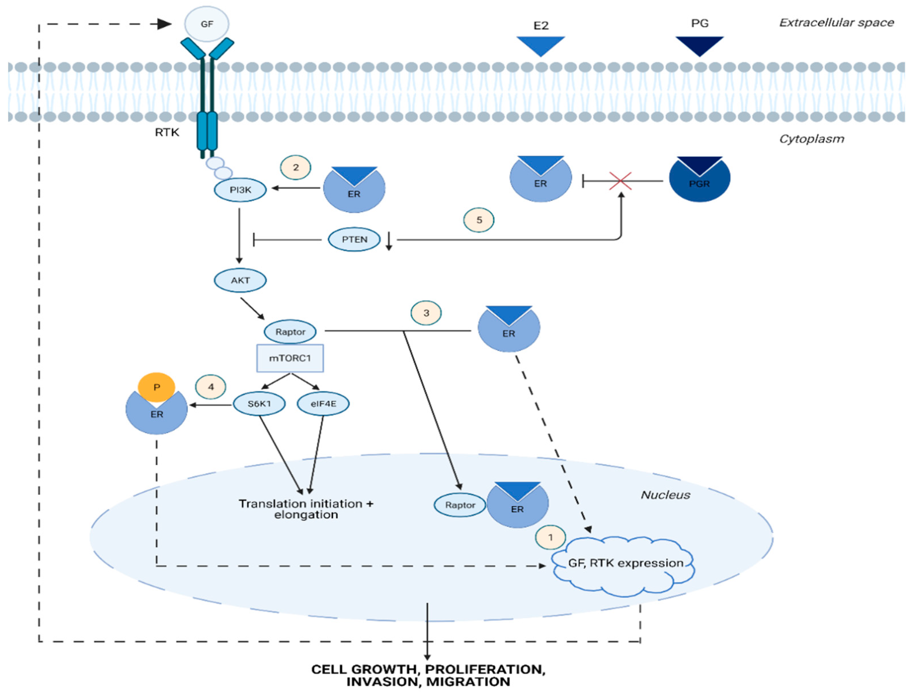
1.2. Cross-Talk between Estrogen and mTOR Signaling Pathway

The PI3K/AKT/mTOR pathway is involved in estrogen signal transduction, thus contributing to the adverse effects of abnormal estrogen levels on endometrial homeostasis [32,33]. In fact, estrogen receptors (ERs) induce the expression of several upstream regulators of the PI3K/AKT/mTOR pathway, such as receptor ligands, receptor tyrosine kinases (RTKs), and signaling adaptors [34]. Furthermore, ERs can stimulate the PI3K/AKT/mTOR pathway not only by acting as transcription factors but also by physically interacting with effectors that are involved in the cascade. For example, estrogen bound ERα directly binds to the p85α regulatory subunit of PI3K and promotes its phosphorylation [35]. In addition, activated ERα attaches to Raptor, causing the translocation of the latter to the nucleus [36]. Other than that, estrogen induces PI3K signaling via stimulation of insulin-like growth factor 1 receptor (IGF-1R) [37]. In reverse, the mTOR pathway can also promote ER signal transduction. Specifically, mTORC1 leads to ERα phosphorylation and activation via S6K1 [36,38].
Estrogen signaling plays a critical role in the pathogenesis of endometrial diseases, as it enhances the potential for proliferation, invasion, and migration of endometrial cells. [32,35]. These characteristics are not only important for the development of malignant endometrial disorders but are also requisite for benign conditions, such as adenomyosis, endometriosis and typical endometrial hyperplasia [39]. The mTOR pathway is closely linked to estrogen signaling and may therefore play an important role in the derangement of endometrial function. Thus, it is worth clarifying the association of this signaling cascade with the main benign endometrial diseases (Figure 1(1–4)).

1.3. Brief Outline of mTOR Pathway Dysregulation in Malignant Endometrial Diseases

The role of mTOR signaling in endometrial carcinogenesis has been extensively investigated in recent decades. PI3K/AKT/mTOR pathway derangement occurs in 80–95% of Type I or endometrioid endometrial carcinomas [40], with PTEN being the most commonly mutated gene in this type of cancer [41]. Interestingly, PIK3CA is frequently commutated with PTEN, implying a synergic effect of both genes on AKT activation during endometrial tumorigenesis [42]. Besides Type I, PI3K/AKT/mTOR pathway dysregulation also contributes to the development of Type II endometrial carcinoma, as the pathway has been shown to interact with p53 signaling in uterine serous carcinoma cells. Furthermore, alterations in PI3K and RTKs are often detected [43], while mTORC2 upregulation and loss of TSC2 have also been observed in endometrial carcinoma cells [44,45]. Apart from endometrial carcinomas, aberrations in mTOR signaling have also been noted in endometrial stromal sarcomas (ESSs). Recent studies [46,47] have reported upregulation of PIK3-AKT signaling and alterations of the mTOR-NF2-AKT pathway in ESS, while inhibition of PI3K and mTOR has been shown to restrain growth in an ESS cell line [48]. The prominent role of the PI3K/AKT/mTOR pathway in endometrial tumorigenesis makes it an ideal therapeutic target for this type of cancer. Inhibitors of this signaling cascade have shown promising results in preclinical models of endometrial cancer and multiple clinical trials examining their efficacy in endometrial cancer patients have been and continue to be conducted [11,40,42]. Current research data have shed light on the importance of mTOR signaling for the development of non-cancerous endometrial diseases as well, suggesting it as an effective therapeutic target for these conditions.

2. mTOR and eIF Signaling in Benign Endometrial Diseases

2.1. mTOR Signaling in Adenomyosis

Adenomyosis refers to the uterine disease in which endometrial tissue invades the myometrium. It is considered an estrogen-dependent condition, as it is characterized by high estrogen levels that contribute to its pathogenesis [1,32,49]. Estrogen acts by stimulating the PI3K/AKT/mTOR pathway (Figure 1) [32,35,50], which is known to be activated in adenomyosis, leading to endometrial epithelial cell invasion and migration [51].
Multiple studies [51,52,53,54,55,56,57] have reported the involvement of upstream regulators of the mTOR pathway in the development of adenomyosis. Protein deglycase DJ-1, a protein associated with cancer and Parkinson’s disease, is increased in adenomyotic lesions, where it controls cell proliferation, migration, and angiogenesis by inducing AKT phosphorylation [52]. Furthermore, mTOR signaling is dysregulated in adenomyosis due to the altered expression of non-coding RNAs. The term “non-coding RNAs” (ncRNAs) covers a plethora of RNA molecules that do not code for proteins [58]. In adenomyosis, the ncRNAs known to influence the disease progression via modulating the PI3K/AKT/mTOR pathway are microRNAs (miRNAs) and long non-coding RNAs (lncRNAs) [51,53,54]. Briefly, miRNAs are short RNA molecules that regulate gene expression by binding to messenger RNAs (mRNAs) and hampering their function [59]. LncRNAs, on the other hand, are RNAs of more than 200 nucleotides with a multifunctional role in cell signaling, as they are involved in various processes such as gene regulation, translation, and RNA splicing [60]. The involvement of ncRNAs in the development and progression of adenomyosis is depicted in Table 1.
Besides upstream effectors, downstream regulators of mTOR signaling seem to play a significant role in adenomyosis as well. Vascular endothelial growth factor (VEGF) and hypoxia-inducible factor-1α (HIF-1α) are highly expressed in the ectopic endometrium of patients with adenomyosis, where they promote angiogenesis, thereby contributing to the progression of the disease [55]. Both VEGF and HIF-1α are upregulated by mTORC1 [52,64]. Specifically, mTORC1 induces HIF-1α synthesis through its substrates S6K1 and STAT3 and promotes the translation of both HIF-1α and VEGF via upregulation of eIF4E. Likewise, eIF4E mediates the expression of C/EBPβ [65,66,67], a transcription factor that induces endometrial stromal proliferation and differentiation in adenomyotic lesions [56,57].
mTOR signaling may also be involved in the pathogenesis of adenomyosis by promoting epithelial to mesenchymal transition (EMT) of endometrial epithelial cells. EMT is thought to play a crucial role in the establishment of the disease, as it enhances the invasiveness of endometrial cells, thus contributing to their translocation into the myometrium [1,68]. More specifically, TGF-β is increased in adenomyotic lesions and leads to EMT [1], possibly by activating mTORC2, one of its main downstream targets in this process [69]. In addition, the PI3K/AKT/mTOR pathway stimulates TWIST, a transcription factor that promotes the EMT phenotype in endometrial cells [12]. Finally, the protein kinase FAK may mediate EMT in adenomyosis via activation of the PI3K/AKT signaling cascade [70].
It follows that targeting the PI3K/AKT/mTOR pathway may be effective for adenomyosis treatment. Interestingly, Xue et al. observed that metformin could restrain stromal cell proliferation of adenomyotic lesions by downregulating the PI3K/AKT pathway through AMPK activation [32].

2.2. mTOR-Associated eIFs in Adenomyosis

Aberrant mTOR pathway activation is also reflected in the abnormal expression of mTOR-associated eIFs in adenomyosis. Gene expression and pathway analysis applied on eutopic endometrium from adenomyosis patients revealed significant dysregulation of eIF2 as well as eIF4 signaling in comparison to normal endometrium [71]. Specifically, eIF4a2, eIF3K, and eIF4b were expressed differently between adenomyotic and normal endometrium. Furthermore, Cai et al. noted a decreased expression of eIF3e in ectopic endometrium and suggested that this reduction may promote EMT via TGF-β1 activation, as they observed a negative correlation between eIF3 and TGF-β1, Snail, vimentin, and PCNA levels [72].

2.3. mTOR Signaling in Endometriosis

Endometriosis is an estrogen-dependent disorder in which endometrial tissue develops outside the uterine cavity, most commonly in the pelvic peritoneum, ovaries, and the pouch of Douglas [73]. The association between the PI3K/AKT/mTOR pathway and the pathogenesis of endometriosis has been proposed by multiple early studies [74,75,76,77,78]. Higher levels of pAkt have been detected in both eutopic and ectopic endometrial tissue of women with endometriosis compared to normal endometrium [75]. Previous research demonstrated increased transcription of the AKT1 and 4EBP1 genes in the eutopic endometrium of endometriosis patients, suggesting a possible role for these genes in endometrial growth outside the uterus [76]. Moreover, elevated expression of the mTOR activators AXL and SHC1 [78], as well as loss of PTEN, have been earlier described in endometriosis [77].
In recent years, the investigation of mTOR signaling in endometriosis is still evolving. Madanes et al. noted elevated PI3K expression and AKT phosphorylation as well as reduced PTEN levels in both ectopic and eutopic endometrium of endometriosis patients compared to normal endometrium. Interestingly, these findings were observed mainly in women with a minimal-mild stage of the disease, suggesting that the PI3K/AKT/mTOR pathway plays a critical role in the onset of endometriosis [79]. Similar conclusions were drawn from mouse models after detecting an increased formation of endometriosis lesions in the peritoneal cavity of mice carrying a PTEN deletion in PR-positive cells [80].
According to other studies [39,81], ectopic endometrial tissue exhibits persistent mTOR activity despite changes in estrogen and progesterone levels. In fact, the PI3K/AKT/mTOR pathway seems to regulate the response of ectopic endometrial tissue to progesterone and is probably associated with progesterone resistance, which is common in endometriosis [82,83]. In particular, Li et al. noted that suppression of PTEN expression by the microRNA miR-92a (see Table 1) and thus aberrant PI3K/AKT/mTOR pathway activation can lead to progesterone resistant endometriosis [61] (Figure 1(5)). Interestingly, mTOR signaling seems to be regulated by several microRNAs in endometriosis, and their function is presented in Table 1.
The frequent activation of the PI3K/AKT/mTOR pathway in endometriosis makes it an attractive therapeutic target in this disease. Therefore, scientists have attempted to evaluate the effects of inhibiting this pathway in restraining endometriosis. Leconte et al. noted a significant and dose-dependent decrease in the proliferation rate of deep infiltrating endometriotic stromal cells in both cell culture and mouse model experiments after treatment with the mTOR-inhibitor temsirolimus [84]. In another study, dienogest, a progestin medication approved for the treatment of endometriosis, was found to promote autophagy and apoptosis by suppressing AKT and ERK1/2 activity in ectopic endometrial cells [85]. Ren et al. indicated the repressive effects of rapamycin, the well-known mTOR inhibitor, on angiogenesis in endometriotic lesions. Specifically, they noted a significant reduction in VEGF expression and microvessel density in ectopic tissues of mice with peritoneal endometriosis after 2 weeks of rapamycin treatment [86]. Similarly, Cao et al. suggested that the vasodepressor ginsenoside Rg3 may limit the rate of ectopic endometrial tissue growth via suppressing the VEGF-mediated activation of the mTOR pathway [87].
Other reports have emphasized the role of mTOR signaling in endometriosis-associated carcinogenesis [88]. Cancer develops from endometriotic tissue, usually in the ovaries, involving either clear cell or endometrioid carcinoma histotypes [89]. In those histotypes, the tumor suppressor gene ARID1A that encodes a protein involved in chromatin remodeling is often inactivated. Interestingly, the loss of ARID1A is linked to the PI3K/AKT pathway dysregulation, and several studies suggest that activation of the PI3K/AKT/mTOR pathway in combination with ARID1A mutation are key events in the malignant transformation of endometriotic lesions [89,90,91]. Furthermore, Broadway et al. observed that specific components of the mTOR complexes, such as DEPTOR, are expressed in a common pattern among endometriosis and ovarian carcinoma tissues, again supporting the connective role of mTOR signaling between tumorigenesis and endometriosis [92].

2.4. mTOR-Associated eIFs in Endometriosis

mTOR pathway-related eIFs exhibit differential expression between normal and ectopic endometrium. Specifically, Cai et al. observed that eIF3e is underexpressed in ovarian endometriosis tissue, thus promoting EMT through the preferential translation of Snail, as they noted a negative correlation between eIF3 and TGF-β1, Snail, vimentin, and PCNA levels [93]. In addition, eIFs can serve as treatment targets for endometriosis. Choi et al. suggested that the PERK/eIF2α pathway is involved in the therapeutic mechanism of dienogest in the disease [94]. According to their study, dienogest increases the expression of CHOP, an apoptosis-mediator protein, via activation of eIF2α signaling. Considering that CHOP is known to induce cell apoptosis by repressing the mTOR pathway [95], it is possible that dienogest acts by inhibiting mTOR signaling through an eIF2α-mediated CHOP activation. Moreover, the flavonoids naringenin and chrysin exhibit a suppressive role in endometriotic cell lines by enhancing endoplasmic reticulum stress via eIF2α activation and PI3K/AKT pathway inhibition [96,97].

2.5. mTOR Signaling in Endometritis

Endometritis can be classified as acute or chronic, depending on the predominance of a neutrophilic or a lymphoplasmacytic infiltrate, respectively. Chronic endometritis (CE) often leads to endometrial dysfunction and therefore increases the risk of infertility and embryo implantation failure. Wang et al. demonstrated through a case-control study including women with CE and recurrent implantation failure (RIF) that mTORC1 is significantly under-expressed in inflamed endometrium [98]. It has also been suggested that mTOR pathway downregulation and subsequent increase in autophagy reduces endometrial receptivity and can be the root of the unsuccessful implantation that is observed in CE patients [99]. However, the role of the mTOR pathway in endometrial inflammation has not been fully elucidated. Contrary to the above-mentioned data, a double-blind, phase II randomized clinical trial revealed the beneficial effect of Sirolimus treatment on RIF patients with elevated Th17/Treg cell ratio [100]. Considering that CE leads to the predominance of Th17 over Treg immunity [98], the latter study suggests that mTOR pathway inhibition could potentially have an immunosuppressive impact on inflamed endometrium.

2.6. mTOR Signaling in Typical Endometrial Hyperplasia

Endometrial hyperplasia (EH) is an irregular proliferation of the endometrium that comprises alterations in the glandular architecture and endometrial gland-to-stroma ratio. According to the simplified World Health Organization (WHO) classification, EH can be categorized as hyperplasia with or without atypia, depending on whether cytologic atypia is present or not, respectively [101]. For the purpose of this review, only EH without atypia, which is presumed benign, will be analyzed.
EH arises from endometrial exposure to excessive levels of estrogen that are not opposed by progesterone, as can be seen in various conditions such as post-menopause, obesity, and polycystic ovary syndrome (PCOS) [3,101]. Considering that estrogen promotes DNA and protein synthesis in endometrial cells through mTOR signaling, EH could probably be associated with aberrant mTOR pathway activation [102]. Interestingly, the latter hypothesis has been suggested by multiple recent studies.
Bajwa et al. indicated the importance of mTOR signaling in advanced age-related EH. They observed an increased pS6 expression in hyperplastic endometrium collected from post-menopausal women compared to normal post-menopausal endometrium and noted a similar expression pattern of pS6 between hyperplastic and normal uterine tissue collected from aged mice [103]. Interestingly, treatment of aged mice with rapamycin led to a significant decrease in EH features. The association between EH and mTOR was strengthened in the same study by reporting the establishment of hyperplastic epithelium and abnormal glandular architecture in the uteri of mouse models carrying a PTEN deletion. Besides post-menopausal EH, the mTOR pathway also contributes to the pathogenesis of obesity-induced EH. Studying the uteri of obese mice, Sahoo et al. revealed elevated phosphorylation of VEGFR2 at Tyr1175 and increased pS6 expression in the hyperplastic endometrium. This observation was interpreted as upregulation of the mTOR pathway by VEGF since it is known that VEGF activates mTOR signaling via the VEGFR2 receptor [104].
mTOR signaling derangement also seems to have a pivotal role in the development of EH induced by PCOS. PCOS is a disorder characterized by chronic anovulation and aberrant androgen biosynthesis, conditions that both increase unopposed steroid levels and can therefore promote EH [105]. The mTOR pathway plays an important role in multiple pathways that are dysregulated in PCOS, including androgen action, insulin axis and cell apoptosis. High androgen levels in PCOS restrain apoptosis and increase the proliferative potential of endometrial cells by stimulating mTOR signaling via AMPK inactivation [106] (Figure 2(4)). Moreover, increased AKT phosphorylation has been observed in endometrial tissue coming from PCOS patients [74]. Another study has demonstrated that EH in patients with PCOS results from abnormal autophagy mechanisms [107], implying another possible link between mTOR and PCOS-induced EH since mTOR is a major autophagy regulator.
Glucose metabolism in the uterus is of essential importance for normal endometrial function. Hyperinsulinemia and insulin resistance are major disorders involved in the pathogenesis of PCOS and are believed to contribute to the development of EH in women with this condition [106,108]. Insulin resistance is known to derive from abnormal function of the mTOR pathway and in particular from overactive mTORC1 and aberrant mTORC2 signaling [105,109] (Figure 2(1,2)). Furthermore, Li et al. reported impaired glucose intake in hyperplastic PCOS endometrium, as they observed lower GLUT-4 expression in comparison to normal endometrium [106]. According to their study, this dysregulation of glucose metabolism results from overactivity of the insulin receptor/PI3K/Akt/mTOR signaling cascade (Figure 2(3)). Specifically, they noted an increase in GLUT-4 levels in combination with reduced levels of the main mTOR pathway components after treating EH tissues with metformin.
It has been well established that metformin restrains EH via mTOR pathway modulation [110], as it activates AMPK, a significant negative regulator of mTOR signaling [111]. In PCOS, metformin can increase GLUT-4 and reduce androgen receptor (AR) levels in the endometrium, probably through the mTOR pathway [106,112]. In addition, compared to normal rat endometrium, uteri of rat models of PCOS exhibit high mTORC1 accumulation, which can be restored to normal levels via metformin treatment [113] (Figure 2(5)). As reported by another study, metformin attenuates estrogen and tamoxifen induced EH by inhibiting S6K1 activation by mTOR [114]. Data derived from in vivo studies as well as clinical trials show that metformin is comparable to progestins for the treatment of simple endometrial hyperplasia [115,116]. The fact that metformin acts in the endometrium via downregulating the mTOR pathway highlights the importance of this signaling cascade in EH.
Apart from exposure to unopposed estrogen, EH can also result from treatment with tamoxifen, a selective estrogen receptor modulator (SERM) used for breast cancer therapy. Upregulation of mTOR signaling might be part of the mechanism by which tamoxifen induces abnormal endometrial growth [3]. Sequestosome1/p62 (SQSTM1/p62) is an autophagy-related adaptor protein and its expression is enhanced by mTORC1 [119]. Interestingly, a study showed higher expression of SQSTM1 in tamoxifen induced EH, in comparison to hyperplastic endometrium derived from women who had not received tamoxifen. In addition, the same study demonstrated a significant retreat of tamoxifen induced EH after inhibition of Nrf2, the upstream activator of SQSTM1 [120].

2.7. mTOR-Associated eIFs in Typical Endometrial Hyperplasia

It is noteworthy that eIF2α signaling, which is in close interaction with the mTOR pathway, seems to be involved in the pathogenesis of PCOS-induced endometrial hyperplasia. In fact, endoplasmic reticulum stress (ER) and consequent eIF2α pathway activation directly contribute to the pathogenesis of PCOS by affecting the total health of ovarian follicles and oocytes in several ways, such as induction of interstitial fibrosis, follicular atresia, and accumulation of advanced glycation end products (AGEs) in ovarian granulosa cells [121,122]. Furthermore, the eIF2α pathway is involved in insulin resistance, which enhances the development of EH in PCOS patients, as previously mentioned. In PCOS, high androgen levels may lead to impaired insulin secretion via promoting ER stress in β-cells and activating the eIF2α signaling cascade. Furthermore, under excess androgen conditions, eIF2α stimulates CHOP expression in insulin-secreting cells, thereby suppressing mTOR signaling and leading to apoptosis [123].

3. Conclusions

The importance of the PI3K/AKT/mTOR signaling cascade in endometrial carcinogenesis has been well established and research has proceeded on evaluating the efficacy of inhibiting this pathway for the treatment of endometrial malignancies. Based on this knowledge, we aimed to investigate the current data about the involvement of the mTOR pathway in the development of benign endometrial conditions. From this review, it follows that mTOR and eIF signaling are deranged in adenomyosis, endometriosis, endometritis, and typical endometrial hyperplasia and may serve as druggable targets in these life-impacting diseases. However, further studies are still needed to determine the full spectrum of mTOR function in non-cancerous endometrial conditions.

Author Contributions

Conceptualization, T.S.D. and J.H.; methodology, T.S.D., J.H. and M.S.; investigation, T.S.D., C.S. and M.S.; resources, J.H.; data curation, T.S.D.; writing—original draft preparation, T.S.D.; writing—review and editing, M.S., J.H. and C.S.; visualization, C.S.; supervision, J.H.; project administration, T.S.D.; funding acquisition, J.H. All authors have read and agreed to the published version of the manuscript.

Funding

This research was partially funded by Slovenian Research Agency, grant number J3-3069 and the Internal Research Project of the University Medical Centre Maribor IRP 2021/01-21.

Institutional Review Board Statement

Not applicable.

Informed Consent Statement

Not applicable.

Data Availability Statement

Not applicable.

Acknowledgments

M.S. acknowledges support by the L’Oréal-UNESCO 2021 Slovenia Fellowship for Women in Science.

Conflicts of Interest

The authors declare no conflict of interest.

References

  1. Stratopoulou, C.A.; Donnez, J.; Dolmans, M.M. Origin and Pathogenic Mechanisms of Uterine Adenomyosis: What Is Known So Far. Reprod. Sci. 2021, 28, 2087–2097. [Google Scholar] [CrossRef] [PubMed]
  2. Kitawaki, J.; Kado, N.; Ishihara, H.; Koshiba, H.; Kitaoka, Y.; Honjo, H. Endometriosis: The Pathophysiology as an Estrogen-Dependent Disease. J. Steroid Biochem. Mol. Biol. 2002, 83, 149–155. [Google Scholar] [CrossRef]
  3. Chandra, V.; Kim, J.J.; Benbrook, D.M.; Dwivedi, A.; Rai, R. Therapeutic Options for Management of Endometrial Hyperplasia. J. Gynecol. Oncol. 2016, 27, e8. [Google Scholar] [CrossRef] [PubMed] [Green Version]
  4. Habiba, M.; Pluchino, N.; Petignat, P.; Bianchi, P.; Brosens, I.A.; Benagiano, G. Adenomyosis and Endometrial Cancer: Literature Review. Gynecol. Obstet. Investig. 2018, 83, 313–328. [Google Scholar] [CrossRef]
  5. Melin, A.; Sparén, P.; Persson, I.; Bergqvist, A. Endometriosis and the Risk of Cancer with Special Emphasis on Ovarian Cancer. Hum. Reprod. 2006, 21, 1237–1242. [Google Scholar] [CrossRef] [PubMed] [Green Version]
  6. Mutter, G.L.; Zaino, R.J.; Baak, J.P.A.; Bentley, R.C.; Robboy, S.J. Benign Endometrial Hyperplasia Sequence and Endometrial Intraepithelial Neoplasia. Int. J. Gynecol. Pathol. 2007, 26, 103–114. [Google Scholar] [CrossRef]
  7. Fourquet, J.; Báez, L.; Figueroa, M.; Iriarte, R.I.; Flores, I. Quantification of the Impact of Endometriosis Symptoms on Health-Related Quality of Life and Work Productivity. Fertil. Steril. 2011, 96, 107–112. [Google Scholar] [CrossRef] [Green Version]
  8. Smith, M.; Hagerty, K.A.; Skipper, B.; Bocklage, T. Chronic Endometritis: A Combined Histopathologic and Clinical Review of Cases from 2002 to 2007. Int. J. Gynecol. Pathol. 2010, 29, 44–50. [Google Scholar] [CrossRef]
  9. Montgomery, B.E.; Daum, G.S.; Dunton, C.J. Endometrial Hyperplasia: A Review. Obstet. Gynecol. Surv. 2004, 59, 368–378. [Google Scholar] [CrossRef] [Green Version]
  10. Levgur, M.; Abadi, M.A.; Tucker, A. Adenomyosis: Symptoms, Histology, and Pregnancy Terminations. Obstet. Gynecol. 2000, 95, 688–691. [Google Scholar] [CrossRef]
  11. Sobočan, M.; Bračič, S.; Knez, J.; Takač, I.; Haybaeck, J. The Communication between the PI3K/AKT/MTOR Pathway and Y-Box Binding Protein-1 in Gynecological Cancer. Cancers 2020, 12, 205. [Google Scholar] [CrossRef] [PubMed] [Green Version]
  12. Sobočan, M.; Smolle, M.A.; Schatz, C.; Haybaeck, J. The Interplay of Tumor Stroma and Translational Factors in Endometrial Cancer. Cancers 2020, 12, 2074. [Google Scholar] [CrossRef] [PubMed]
  13. Smolle, M.A.; Czapiewski, P.; Lapińska-Szumczyk, S.; Majewska, H.; Supernat, A.; Zaczek, A.; Biernat, W.; Golob-Schwarzl, N.; Haybaeck, J. The Prognostic Significance of Eukaryotic Translation Initiation Factors (EIFs) in Endometrial Cancer. Int. J. Mol. Sci. 2019, 20, 6169. [Google Scholar] [CrossRef] [PubMed] [Green Version]
  14. Mitra, A.; Luna, J.I.; Marusina, A.I.; Merleev, A.; Kundu-Raychaudhuri, S.; Fiorentino, D.; Raychaudhuri, S.P.; Maverakis, E. Dual MTOR Inhibition Is Required to Prevent TGF-β-Mediated Fibrosis: Implications for Scleroderma. J. Investig. Dermatol. 2015, 135, 2873–2876. [Google Scholar] [CrossRef] [PubMed] [Green Version]
  15. Chiang, G.G.; Abraham, R.T. Targeting the MTOR Signaling Network in Cancer. Trends Mol. Med. 2007, 13, 433–442. [Google Scholar] [CrossRef]
  16. Loewith, R.; Jacinto, E.; Wullschleger, S.; Lorberg, A.; Crespo, J.L.; Bonenfant, D.; Oppliger, W.; Jenoe, P.; Hall, M.N. Two TOR Complexes, Only One of Which Is Rapamycin Sensitive, Have Distinct Roles in Cell Growth Control. Mol. Cell 2002, 10, 457–468. [Google Scholar] [CrossRef]
  17. Wullschleger, S.; Loewith, R.; Hall, M.N. TOR Signaling in Growth and Metabolism. Cell 2006, 124, 471–484. [Google Scholar] [CrossRef] [Green Version]
  18. Caron, E.; Ghosh, S.; Matsuoka, Y.; Ashton-Beaucage, D.; Therrien, M.; Lemieux, S.; Perreault, C.; Roux, P.P.; Kitano, H. A Comprehensive Map of the MTOR Signaling Network. Mol. Syst. Biol. 2010, 6, 453. [Google Scholar] [CrossRef]
  19. Saxton, R.A.; Sabatini, D.M. MTOR Signaling in Growth, Metabolism, and Disease. Cell 2017, 168, 960–976. [Google Scholar] [CrossRef] [Green Version]
  20. Cantley, L.C. The Phosphoinositide 3-Kinase Pathway. Science 2002, 296, 1655–1657. [Google Scholar] [CrossRef]
  21. Huang, J.; Manning, B.D. The TSC1-TSC2 Complex: A Molecular Switchboard Controlling Cell Growth. Biochem. J. 2008, 412, 179–190. [Google Scholar] [CrossRef] [PubMed] [Green Version]
  22. Nojima, H.; Tokunaga, C.; Eguchi, S.; Oshiro, N.; Hidayat, S.; Yoshino, K.I.; Hara, K.; Tanaka, N.; Avruch, J.; Yonezawa, K. The Mammalian Target of Rapamycin (MTOR) Partner, Raptor, Binds the MTOR Substrates P70 S6 Kinase and 4E-BP1 through Their TOR Signaling (TOS) Motif. J. Biol. Chem. 2003, 278, 15461–15464. [Google Scholar] [CrossRef] [Green Version]
  23. Ma, X.M.; Blenis, J. Molecular Mechanisms of MTOR-Mediated Translational Control. Nat. Rev. Mol. Cell Biol. 2009, 10, 307–318. [Google Scholar] [CrossRef]
  24. Jung, C.H.; Jun, C.B.; Ro, S.-H.; Kim, Y.-M.; Otto, N.M.; Cao, J.; Kundu, M.; Kim, D.-H. ULK-Atg13-FIP200 Complexes Mediate MTOR Signaling to the Autophagy Machinery. Mol. Biol. Cell 2009, 20, 1992–2003. [Google Scholar] [CrossRef] [PubMed] [Green Version]
  25. Wengrod, J.C.; Gardner, L.B. Cellular Adaptation to Nutrient Deprivation: Crosstalk between the MTORC1 and EIF2α Signaling Pathways and Implications for Autophagy. Cell Cycle 2015, 14, 2571–2577. [Google Scholar] [CrossRef] [PubMed] [Green Version]
  26. Spilka, R.; Ernst, C.; Mehta, A.K.; Haybaeck, J. Eukaryotic Translation Initiation Factors in Cancer Development and Progression. Cancer Lett. 2013, 340, 9–21. [Google Scholar] [CrossRef] [PubMed]
  27. Holz, M.K.; Ballif, B.A.; Gygi, S.P.; Blenis, J. MTOR and S6K1 Mediate Assembly of the Translation Preinitiation Complex through Dynamic Protein Interchange and Ordered Phosphorylation Events. Cell 2005, 123, 569–580. [Google Scholar] [CrossRef] [Green Version]
  28. Morris, C.; Durand, S.; Jalinot, P. Decreased Expression of the Translation Factor EIF3e Induces Senescence in Breast Cancer Cells via Suppression of PARP1 and Activation of MTORC1. Oncotarget 2021, 12, 649–664. [Google Scholar] [CrossRef]
  29. Jacinto, E.; Loewith, R.; Schmidt, A.; Lin, S.; Rüegg, M.A.; Hall, A.; Hall, M.N. Mammalian TOR Complex 2 Controls the Actin Cytoskeleton and Is Rapamycin Insensitive. Nat. Cell Biol. 2004, 6, 1122–1128. [Google Scholar] [CrossRef]
  30. Gan, X.; Wang, J.; Wang, C.; Sommer, E.; Kozasa, T.; Srinivasula, S.; Alessi, D.; Offermanns, S.; Simon, M.I.; Wu, D. PRR5L Degradation Promotes MTORC2-Mediated PKC-δ Phosphorylation and Cell Migration Downstream of Gα 12. Nat. Cell Biol. 2012, 14, 686–696. [Google Scholar] [CrossRef] [Green Version]
  31. Lamming, D.W.; Ye, L.; Katajisto, P.; Goncalves, M.D.; Saitoh, M.; Stevens, D.M.; Davis, J.G.; Salmon, A.B.; Richardson, A.; Ahima, R.S.; et al. Rapamycin-Induced Insulin Resistance Is Mediated by MTORC2 Loss and Uncoupled from Longevity. Science 2012, 335, 1638–1643. [Google Scholar] [CrossRef] [PubMed] [Green Version]
  32. Xue, J.; Zhang, H.; Liu, W.; Liu, M.; Shi, M.; Wen, Z.; Li, C. Metformin Inhibits Growth of Eutopic Stromal Cells from Adenomyotic Endometrium via AMPK Activation and Subsequent Inhibition of AKT Phosphorylation: A Possible Role in the Treatment of Adenomyosis. Reproduction 2013, 146, 397–406. [Google Scholar] [CrossRef] [PubMed]
  33. Aaltonen, K.E.; Rosendahl, A.H.; Olsson, H.; Malmström, P.; Hartman, L.; Fernö, M. Association between Insulin-like Growth Factor-1 Receptor (IGF1R) Negativity and Poor Prognosis in a Cohort of Women with Primary Breast Cancer. BMC Cancer 2014, 14, 794. [Google Scholar] [CrossRef] [PubMed] [Green Version]
  34. Ciruelos Gil, E.M. Targeting the PI3K/AKT/MTOR Pathway in Estrogen Receptor-Positive Breast Cancer. Cancer Treat. Rev. 2014, 40, 862–871. [Google Scholar] [CrossRef]
  35. Hou, X.; Zhao, M.; Wang, T.; Zhang, G. Upregulation of Estrogen Receptor Mediates Migration, Invasionand Proliferation of Endometrial Carcinoma Cells by Regulating the PI3K/AKT/MTOR Pathway. Oncol. Rep. 2014, 31, 1175–1182. [Google Scholar] [CrossRef] [Green Version]
  36. Alayev, A.; Salamon, R.S.; Berger, S.M.; Schwartz, N.S.; Cuesta, R.; Snyder, R.B.; Holz, M.K. MTORC1 Directly Phosphorylates and Activates ERα upon Estrogen Stimulation. Oncogene 2016, 35, 3535–3543. [Google Scholar] [CrossRef]
  37. Miller, T.W.; Pérez-Torres, M.; Narasanna, A.; Guix, M.; Stål, O.; Pérez-Tenorio, G.; Gonzalez-Angulo, A.M.; Hennessy, B.T.; Mills, G.B.; Kennedy, J.P.; et al. Loss of Phosphatase and Tensin Homologue Deleted on Chromosome 10 Engages ErbB3 and Insulin-like Growth Factor-I Receptor Signaling to Promote Antiestrogen Resistance in Breast Cancer. Cancer Res. 2009, 69, 4192–4201. [Google Scholar] [CrossRef] [Green Version]
  38. Yamnik, R.L.; Holz, M.K. MTOR/S6K1 and MAPK/RSK Signaling Pathways Coordinately Regulate Estrogen Receptor α Serine 167 Phosphorylation. FEBS Lett. 2010, 584, 124–128. [Google Scholar] [CrossRef] [Green Version]
  39. Guo, Z.; Yu, Q. Role of MTOR Signaling in Female Reproduction. Front. Endocrinol. 2019, 10, 692. [Google Scholar] [CrossRef]
  40. Bell, D.W.; Ellenson, L.H. Molecular Genetics of Endometrial Carcinoma. Annu. Rev. Pathol. Mech. Dis. 2019, 14, 339–367. [Google Scholar] [CrossRef]
  41. Pavlidou, A.; Vlahos, N.F. Molecular Alterations of PI3K/Akt/MTOR Pathway: A Therapeutic Target in Endometrial Cancer. Sci. World J. 2014, 2014, 709736. [Google Scholar] [CrossRef] [Green Version]
  42. Eritja, N.; Yeramian, A.; Chen, B.J.; Llobet-Navas, D.; Ortega, E.; Colas, E.; Abal, M.; Dolcet, X.; Reventos, J.; Matias-Guiu, X. Endometrial Carcinoma: Specific Targeted Pathways. Adv. Exp. Med. Biol. 2017, 943, 149–207. [Google Scholar]
  43. Slomovitz, B.M.; Coleman, R.L. The PI3K/AKT/MTOR Pathway as a Therapeutic Target in Endometrial Cancer. Clin. Cancer Res. 2012, 18, 5856–5864. [Google Scholar] [CrossRef] [PubMed] [Green Version]
  44. Shen, Q.; Stanton, M.L.; Feng, W.; Rodriguez, M.E.; Ramondetta, L.; Chen, L.; Brown, R.E.; Duan, X. Morphoproteomic Analysis Reveals an Overexpressed and Constitutively Activated Phospholipase D1-MTORC2 Pathway in Endometrial Carcinoma. Int. J. Clin. Exp. Pathol. 2010, 4, 13–21. [Google Scholar] [PubMed]
  45. Lu, K.H.; Wu, W.; Dave, B.; Slomovitz, B.M.; Burke, T.W.; Munsell, M.F.; Broaddus, R.R.; Walker, C.L. Loss of Tuberous Sclerosis Complex-2 Function and Activation of Mammalian Target of Rapamycin Signaling in Endometrial Carcinoma. Clin. Cancer Res. 2008, 14, 2543–2550. [Google Scholar] [CrossRef] [Green Version]
  46. Lin, D.I.; Huang, R.S.P.; Mata, D.A.; Decker, B.; Danziger, N.; Lechpammer, M.; Hiemenz, M.; Ramkissoon, S.H.; Ross, J.S.; Elvin, J.A. Clinicopathological and Genomic Characterization of BCORL1-Driven High-Grade Endometrial Stromal Sarcomas. Mod. Pathol. 2021, 34, 2200–2210. [Google Scholar] [CrossRef] [PubMed]
  47. Chiang, S.; Vasudevaraja, V.; Serrano, J.; Stewart, C.J.R.; Oliva, E.; Momeni-Boroujeni, A.; Jungbluth, A.A.; da Cruz Paula, A.; da Silva, E.M.; Weigelt, B.; et al. TSC2-Mutant Uterine Sarcomas with JAZF1-SUZ12 Fusions Demonstrate Hybrid Features of Endometrial Stromal Sarcoma and PEComa and Are Responsive to MTOR Inhibition. Mod. Pathol. 2022, 35, 117–127. [Google Scholar] [CrossRef]
  48. Quan, P.; Moinfar, F.; Kufferath, I.; Absenger, M.; Kueznik, T.; Denk, H.; Zatloukal, K.; Haybaeck, J. Effects of Targeting Endometrial Stromal Sarcoma Cells via Histone Deacetylase and PI3K/AKT/MTOR Signaling. Anticancer. Res. 2014, 34, 2883–2897. [Google Scholar]
  49. García-Solares, J.; Donnez, J.; Donnez, O.; Dolmans, M.M. Pathogenesis of Uterine Adenomyosis: Invagination or Metaplasia? Fertil. Steril. 2018, 109, 371–379. [Google Scholar] [CrossRef]
  50. Cuesta, R.; Gritsenko, M.A.; Petyuk, V.A.; Shukla, A.K.; Tsai, C.F.; Liu, T.; McDermott, J.E.; Holz, M.K. Phosphoproteome Analysis Reveals Estrogen-ER Pathway as a Modulator of MTOR Activity via Deptor*. Mol. Cell. Proteom. 2019, 18, 1607–1618. [Google Scholar] [CrossRef]
  51. Guo, Y.; Lang, X.; Lu, Z.; Wang, J.; Li, T.; Liao, Y.; Jia, C.; Zhao, W.; Fang, H. MiR-10b Directly Targets ZEB1 and PIK3CA to Curb Adenomyotic Epithelial Cell Invasiveness via Upregulation of E-Cadherin and Inhibition of Akt Phosphorylation. Cell. Physiol. Biochem. 2015, 35, 2169–2180. [Google Scholar] [CrossRef]
  52. Guo, J.; Gao, J.; Yu, X.; Luo, H.; Xiong, X.; Huang, O. Expression of DJ-1 and MTOR in Eutopic and Ectopic Endometria of Patients with Endometriosis and Adenomyosis. Gynecol. Obstet. Investig. 2015, 79, 195–200. [Google Scholar] [CrossRef]
  53. Xu, X.-Y.; Zhang, J.; Qi, Y.-H.; Kong, M.; Liu, S.-A.; Hu, J.-J. Linc-ROR Promotes Endometrial Cell Proliferation by Activating the PI3K-Akt Pathway. Eur. Rev. Med. Pharmacol. Sci. 2018, 22, 2218–2225. [Google Scholar] [PubMed]
  54. Hu, H.; Li, H.; He, Y. MicroRNA-17 Downregulates Expression of the PTEN Gene to Promote the Occurrence and Development of Adenomyosis. Exp. Ther. Med. 2017, 14, 3805–3811. [Google Scholar] [CrossRef] [Green Version]
  55. Goteri, G.; Lucarini, G.; Montik, N.; Zizzi, A.; Stramazzotti, D.; Fabris, G.; Tranquilli, A.L.; Ciavattini, A. Expression of Vascular Endothelial Growth Factor (VEGF), Hypoxia Inducible Factor-1α (HIF-1α), and Microvessel Density in Endometrial Tissue in Women with Adenomyosis. Int. J. Gynecol. Pathol. 2009, 28, 157–163. [Google Scholar] [CrossRef]
  56. Wang, W.; Taylor, R.N.; Bagchi, I.C.; Bagchi, M.K. Regulation of Human Endometrial Stromal Proliferation and Differentiation by C/EBPβ Involves Cyclin E-Cdk2 and STAT3. Mol. Endocrinol. 2012, 26, 2016–2030. [Google Scholar] [CrossRef] [PubMed] [Green Version]
  57. Xiang, Y.; Sun, Y.; Yang, B.; Yang, Y.; Zhang, Y.; Yu, T.; Huang, H.; Zhang, J.; Xu, H. Transcriptome Sequencing of Adenomyosis Eutopic Endometrium: A New Insight into Its Pathophysiology. J. Cell. Mol. Med. 2019, 23, 8381–8391. [Google Scholar] [CrossRef] [PubMed]
  58. Esteller, M. Non-Coding RNAs in Human Disease. Nat. Rev. Genet. 2011, 12, 861–874. [Google Scholar] [CrossRef]
  59. Zhang, Y.; Huang, B.; Wang, H.Y.; Chang, A.; Zheng, X.F.S. Emerging Role of MicroRNAs in MTOR Signaling. Cell. Mol. Life Sci. 2017, 74, 2613–2625. [Google Scholar] [CrossRef] [PubMed]
  60. Statello, L.; Guo, C.J.; Chen, L.L.; Huarte, M. Gene Regulation by Long Non-Coding RNAs and Its Biological Functions. Nat. Rev. Mol. Cell Biol. 2021, 22, 96–118. [Google Scholar] [CrossRef]
  61. Li, M.; Peng, J.; Shi, Y.; Sun, P. MiR-92a Promotes Progesterone Resistance in Endometriosis through PTEN/AKT Pathway. Life Sci. 2020, 242, 117190. [Google Scholar] [CrossRef]
  62. Zhao, Q.; Han, B.; Zhang, Y.; Su, K.; Wang, C.; Hai, P.; Bian, A.; Guo, R. Effect of MiR-194-5p Regulating STAT1/MTOR Signaling Pathway on the Biological Characteristics of Ectopic Endometrial Cells from Mice. Am. J. Transl. Res. 2020, 12, 6136–6148. [Google Scholar] [PubMed]
  63. Zhou, X.; Chen, Z.; Pei, L.; Sun, J. MicroRNA MiR-106a-5p Targets Forkhead Box Transcription Factor FOXC1 to Suppress the Cell Proliferation, Migration, and Invasion of Ectopic Endometrial Stromal Cells via the PI3K/Akt/MTOR Signaling Pathway. Bioengineered 2021, 12, 2203–2213. [Google Scholar] [CrossRef]
  64. Dodd, K.M.; Yang, J.; Shen, M.H.; Sampson, J.R.; Tee, A.R. MTORC1 Drives HIF-1α and VEGF-A Signalling via Multiple Mechanisms Involving 4E-BP1, S6K1 and STAT3. Oncogene 2015, 34, 2239–2250. [Google Scholar] [CrossRef] [PubMed] [Green Version]
  65. Li, S.; Pal, R.; Monaghan, S.A.; Schafer, P.; Ouyang, H.; Mapara, M.; Galson, D.L.; Lentzsch, S. IMiD Immunomodulatory Compounds Block C/EBPβ Translation through EIF4E down-Regulation Resulting in Inhibition of MM. Blood 2011, 117, 5157–5165. [Google Scholar] [CrossRef] [PubMed] [Green Version]
  66. Albert, V.; Hall, M.N. Reduced C/EBP Β-LIP Translation Improves Metabolic Health. EMBO Rep. 2015, 16, 881–882. [Google Scholar] [CrossRef] [Green Version]
  67. Calkhoven, C.F.; Müller, C.; Leutz, A. Translational Control of C/EBPalpha and C/EBPbeta Isoform Expression. Genes Dev. 2000, 14, 1920–1932. [Google Scholar] [CrossRef]
  68. Shen, M.; Liu, X.; Zhang, H.; Guo, S.W. Transforming Growth Factor Β1 Signaling Coincides with Epithelial-Mesenchymal Transition and Fibroblast-to-Myofibroblast Transdifferentiation in the Development of Adenomyosis in Mice. Hum. Reprod. 2016, 31, 355–369. [Google Scholar] [CrossRef]
  69. Karimi Roshan, M.; Soltani, A.; Soleimani, A.; Rezaie Kahkhaie, K.; Afshari, A.R.; Soukhtanloo, M. Role of AKT and MTOR Signaling Pathways in the Induction of Epithelial-Mesenchymal Transition (EMT) Process. Biochimie 2019, 165, 229–234. [Google Scholar] [CrossRef]
  70. Zheng, D.; Duan, H.; Wang, S.; Xu, Q.; Gan, L.; Li, J.; Dong, Q. FAK Regulates Epithelial-Mesenchymal Transition in Adenomyosis. Mol. Med. Rep. 2018, 18, 5461–5472. [Google Scholar] [CrossRef] [Green Version]
  71. Herndon, C.N.; Aghajanova, L.; Balayan, S.; Erikson, D.; Barragan, F.; Goldfien, G.; Vo, K.C.; Hawkins, S.; Giudice, L.C. Global Transcriptome Abnormalities of the Eutopic Endometrium from Women with Adenomyosis. Reprod. Sci. 2016, 23, 1289–1303. [Google Scholar] [CrossRef] [PubMed]
  72. Cai, X.; Shen, M.; Liu, X.; Nie, J. The Possible Role of Eukaryotic Translation Initiation Factor 3 Subunit e (EIF3e) in the Epithelial–Mesenchymal Transition in Adenomyosis. Reprod. Sci. 2019, 26, 377–385. [Google Scholar] [CrossRef] [PubMed]
  73. Yin, X.; Pavone, M.E.; Lu, Z.; Wei, J.; Kim, J.J. Increased Activation of the PI3K/AKT Pathway Compromises Decidualization of Stromal Cells from Endometriosis. J. Clin. Endocrinol. Metab. 2012, 97, E35–E43. [Google Scholar] [CrossRef] [PubMed] [Green Version]
  74. Makker, A.; Goel, M.M.; Das, V.; Agarwal, A. PI3K-Akt-MTOR and MAPK Signaling Pathways in Polycystic Ovarian Syndrome, Uterine Leiomyomas and Endometriosis: An Update. Gynecol. Endocrinol. 2012, 28, 175–181. [Google Scholar] [CrossRef]
  75. Cinar, O.; Seval, Y.; Uz, Y.H.; Cakmak, H.; Ulukus, M.; Kayisli, U.A.; Arici, A. Differential Regulation of Akt Phosphorylation in Endometriosis. Reprod. BioMed. Online 2009, 19, 864–871. [Google Scholar] [CrossRef] [Green Version]
  76. Laudanski, P.; Szamatowicz, J.; Kowalczuk, O.; Kuźmicki, M.; Grabowicz, M.; Chyczewski, L. Expression of Selected Tumor Suppressor and Oncogenes in Endometrium of Women with Endometriosis. Hum. Reprod. 2009, 24, 1880–1890. [Google Scholar] [CrossRef] [Green Version]
  77. Zhang, H.; Li, M.; Zheng, X.; Sun, Y.; Wen, Z.; Zhao, X. Endometriotic Stromal Cells Lose the Ability to Regulate Cell-Survival Signaling in Endometrial Epithelial Cells in Vitro. Mol. Hum. Reprod. 2009, 15, 653–663. [Google Scholar] [CrossRef] [Green Version]
  78. Honda, H.; Barrueto, F.F.; Gogusev, J.; Im, D.D.; Morin, P.J. Serial Analysis of Gene Expression Reveals Differential Expression between Endometriosis and Normal Endometrium. Possible Roles for AXL and SHC1 in the Pathogenesis of Endometriosis. Reprod. Biol. Endocrinol. 2008, 6, 59. [Google Scholar] [CrossRef] [Green Version]
  79. Madanes, D.; Bilotas, M.A.; Bastón, J.I.; Singla, J.J.; Meresman, G.F.; Barañao, R.I.; Ricci, A.G. PI3K/AKT Pathway Is Altered in the Endometriosis Patient’s Endometrium and Presents Differences According to Severity Stage. Gynecol. Endocrinol. 2020, 36, 436–440. [Google Scholar] [CrossRef]
  80. Kim, T.H.; Yu, Y.; Luo, L.; Lydon, J.P.; Jeong, J.W.; Kim, J.J. Activated AKT Pathway Promotes Establishment of Endometriosis. Endocrinology 2014, 155, 1921–1930. [Google Scholar] [CrossRef] [Green Version]
  81. Choi, J.Y.; Jo, M.W.; Lee, E.Y.; Kim, H.J.; Choi, D.S. Differential Induction of Autophagy by Mtor Is Associated with Abnormal Apoptosis in Ovarianendometriotic Cysts. Mol. Hum. Reprod. 2014, 20, 309–317. [Google Scholar] [CrossRef]
  82. McKinnon, B.; Mueller, M.; Montgomery, G. Progesterone Resistance in Endometriosis: An Acquired Property? Trends Endocrinol. Metab. 2018, 29, 535–548. [Google Scholar] [CrossRef] [PubMed]
  83. Marquardt, R.M.; Kim, T.H.; Shin, J.-H.; Jeong, J.-W. Progesterone and Estrogen Signaling in the Endometrium: What Goes Wrong in Endometriosis? Int. J. Mol. Sci. 2019, 20, 3822. [Google Scholar] [CrossRef] [PubMed] [Green Version]
  84. Leconte, M.; Nicco, C.; Ng, C.; Chéreau, C.; Chouzenoux, S.; Marut, W.; Guibourdenche, J.; Arkwright, S.; Weill, B.; Chapron, C.; et al. The MTOR/AKT Inhibitor Temsirolimus Prevents Deep Infiltrating Endometriosis in Mice. Am. J. Pathol. 2011, 179, 880–889. [Google Scholar] [CrossRef] [PubMed]
  85. Choi, J.; Jo, M.; Lee, E.; Lee, D.Y.; Choi, D. Dienogest Enhances Autophagy Induction in Endometriotic Cells by Impairing Activation of AKT, ERK1/2, and MTOR. Fertil. Steril. 2015, 104, 655–664. [Google Scholar] [CrossRef] [Green Version]
  86. Ren, X.; Wang, Y.; Xu, G.; Dai, L. Effect of Rapamycin on Endometriosis in Mice. Exp. Ther. Med. 2016, 12, 101–106. [Google Scholar] [CrossRef] [Green Version]
  87. Cao, Y.; Ye, Q.; Zhuang, M.; Xie, S.; Zhong, R.; Cui, J.; Zhou, J.; Zhu, Y.; Zhang, T.; Cao, L. Ginsenoside Rg3 Inhibits Angiogenesis in a Rat Model of Endometriosis through the VEGFR-2-Mediated PI3K/Akt/MTOR Signaling Pathway. PLoS ONE 2017, 12, e0186520. [Google Scholar] [CrossRef] [Green Version]
  88. Gadducci, A.; Zannoni, G.F. Endometriosis-Associated Extraovarian Malignancies: A Challenging Question for the Clinician and the Pathologist. Anticancer Res. 2020, 40, 2429–2438. [Google Scholar] [CrossRef]
  89. Samartzis, E.P.; Labidi-Galy, S.I.; Moschetta, M.; Uccello, M.; Kalaitzopoulos, D.R.; Perez-Fidalgo, J.A.; Boussios, S. Endometriosis-Associated Ovarian Carcinomas: Insights into Pathogenesis, Diagnostics, and Therapeutic Targets—A Narrative Review. Ann. Transl. Med. 2020, 8, 1712. [Google Scholar] [CrossRef]
  90. Samartzis, E.P.; Noske, A.; Dedes, K.J.; Fink, D.; Imesch, P. ARID1A Mutations and PI3K/AKT Pathway Alterations in Endometriosis and Endometriosis-Associated Ovarian Carcinomas. Int. J. Mol. Sci. 2013, 14, 18824–18849. [Google Scholar] [CrossRef] [Green Version]
  91. Takeda, T.; Banno, K.; Okawa, R.; Yanokura, M.; Iijima, M.; Iriekunitomi, H.; Nakamura, K.; Iida, M.; Adachi, M.; Umene, K.; et al. ARID1A Gene Mutation in Ovarian and Endometrial Cancers (Review). Oncol. Rep. 2016, 35, 607–613. [Google Scholar] [CrossRef] [Green Version]
  92. Rogers-Broadway, K.R.; Kumar, J.; Sisu, C.; Wander, G.; Mazey, E.; Jeyaneethi, J.; Pados, G.; Tsolakidis, D.; Klonos, E.; Grunt, T.; et al. Differential Expression of MTOR Components in Endometriosis and Ovarian Cancer: Effects of Rapalogues and Dual Kinase Inhibitors on MTORC1 and MTORC2 Stoichiometry. Int. J. Mol. Med. 2019, 43, 47–56. [Google Scholar] [CrossRef] [PubMed]
  93. Cai, X.; Shen, M.; Liu, X.; Guo, S.W. Reduced Expression of Eukaryotic Translation Initiation Factor 3 Subunit e and Its Possible Involvement in the Epithelial–Mesenchymal Transition in Endometriosis. Reprod. Sci. 2018, 25, 102–109. [Google Scholar] [CrossRef] [Green Version]
  94. Choi, J.Y.; Jo, M.W.; Lee, E.Y.; Lee, D.Y.; Choi, D.S. Dienogest Regulates Apoptosis, Proliferation, and Invasiveness of Endometriotic Cyst Stromal Cells via Endoplasmic Reticulum Stress Induction. Mol. Hum. Reprod. 2020, 26, 30–39. [Google Scholar] [CrossRef] [PubMed]
  95. Lin, S.; Yang, L.; Shi, H.; Du, W.; Qi, Y.; Qiu, C.; Liang, X.; Shi, W.; Liu, J. Endoplasmic Reticulum-Targeting Photosensitizer Hypericin Confers Chemo-Sensitization towards Oxaliplatin through Inducing pro-Death Autophagy. Int. J. Biochem. Cell Biol. 2017, 87, 54–68. [Google Scholar] [CrossRef] [PubMed]
  96. Park, S.; Lim, W.; Bazer, F.W.; Song, G. Naringenin Induces Mitochondriamediated Apoptosis and Endoplasmic Reticulum Stress by Regulating MAPK and AKT Signal Transduction Pathways in Endometriosis Cells. Mol. Hum. Reprod. 2017, 23, 842–854. [Google Scholar] [CrossRef] [Green Version]
  97. Ryu, S.; Bazer, F.W.; Lim, W.; Song, G. Chrysin Leads to Cell Death in Endometriosis by Regulation of Endoplasmic Reticulum Stress and Cytosolic Calcium Level. J. Cell. Physiol. 2019, 234, 2480–2490. [Google Scholar] [CrossRef]
  98. Wang, W.; Zhang, H.; Chen, Z.; Zhang, W.; Liu, X.; Fang, J.; Liu, F.; Kwak-Kim, J. Endometrial TGF-β, IL-10, IL-17 and Autophagy Are Dysregulated in Women with Recurrent Implantation Failure with Chronic Endometritis. Reprod. Biol. Endocrinol. 2019, 17, 2. [Google Scholar] [CrossRef] [Green Version]
  99. Buzzaccarini, G.; Vitagliano, A.; Andrisani, A.; Santarsiero, C.M.; Cicinelli, R.; Nardelli, C.; Ambrosini, G.; Cicinelli, E. Chronic Endometritis and Altered Embryo Implantation: A Unified Pathophysiological Theory from a Literature Systematic Review. J. Assist. Reprod. Genet. 2020, 37, 2897–2911. [Google Scholar] [CrossRef]
  100. Ahmadi, M.; Abdolmohamadi-vahid, S.; Ghaebi, M.; Dolati, S.; Abbaspour-Aghdam, S.; Danaii, S.; Berjis, K.; Madadi-Javid, R.; Nouri, Z.; Siahmansouri, H.; et al. Sirolimus as a New Drug to Treat RIF Patients with Elevated Th17/Treg Ratio: A Double-Blind, Phase II Randomized Clinical Trial. Int. Immunopharmacol. 2019, 74, 105730. [Google Scholar] [CrossRef]
  101. Emons, G.; Beckmann, M.W.; Schmidt, D.; Mallmann, P. New WHO Classification of Endometrial Hyperplasias. Geburtshilfe Frauenheilkd. 2015, 75, 135–136. [Google Scholar] [CrossRef] [PubMed] [Green Version]
  102. Wang, Y.; Zhu, L.; Kuokkanen, S.; Pollard, J.W. Activation of Protein Synthesis in Mouse Uterine Epithelial Cells by Estradiol-17β Is Mediated by a PKC-ERK 1/2-MTOR Signaling Pathway. Proc. Natl. Acad. Sci. USA 2015, 112, E1382–E1391. [Google Scholar]
  103. Bajwa, P.; Nielsen, S.; Lombard, J.M.; Rassam, L.; Nahar, P.; Rueda, B.R.; Wilkinson, J.E.; Miller, R.A.; Tanwar, P.S. Overactive MTOR Signaling Leads to Endometrial Hyperplasia in Aged Women and Mice. Oncotarget 2017, 8, 7265–7275. [Google Scholar] [CrossRef] [Green Version]
  104. Sahoo, S.S.; Lombard, J.M.; Ius, Y.; O’Sullivan, R.; Wood, L.G.; Nahar, P.; Jaaback, K.; Tanwar, P.S. Adipose-Derived VEGF–MTOR Signaling Promotes Endometrial Hyperplasia and Cancer: Implications for Obese Women. Mol. Cancer Res. 2018, 16, 309–321. [Google Scholar] [CrossRef] [PubMed] [Green Version]
  105. Liu, J.; Wu, D.C.; Qu, L.H.; Liao, H.Q.; Li, M.X. The Role of MTOR in Ovarian Neoplasms, Polycystic Ovary Syndrome, and Ovarian Aging. Clin. Anat. 2018, 31, 891–898. [Google Scholar] [CrossRef]
  106. Li, X.; Cui, P.; Jiang, H.-Y.; Guo, Y.-R.; Pishdari, B.; Hu, M.; Feng, Y.; Billig, H.; Shao, R. Reversing the Reduced Level of Endometrial GLUT4 Expression in Polycystic Ovary Syndrome: A Mechanistic Study of Metformin Action. Am. J. Transl. Res. 2015, 7, 574–586. [Google Scholar] [PubMed]
  107. Sumarac-Dumanovic, M.; Apostolovic, M.; Janjetovic, K.; Jeremic, D.; Popadic, D.; Ljubic, A.; Micic, J.; Dukanac-Stamenkovic, J.; Tubic, A.; Stevanovic, D.; et al. Downregulation of Autophagy Gene Expression in Endometria from Women with Polycystic Ovary Syndrome. Mol. Cell. Endocrinol. 2017, 440, 116–124. [Google Scholar] [CrossRef] [PubMed]
  108. Gressel, G.M.; Parkash, V.; Pal, L. Management Options and Fertility-Preserving Therapy for Premenopausal Endometrial Hyperplasia and Early-Stage Endometrial Cancer. Int. J. Gynecol. Obstet. 2015, 131, 234–239. [Google Scholar] [CrossRef]
  109. Ong, P.S.; Wang, L.Z.; Dai, X.; Tseng, S.H.; Loo, S.J.; Sethi, G. Judicious Toggling of MTOR Activity to Combat Insulin Resistance and Cancer: Current Evidence and Perspectives. Front. Pharmacol. 2016, 7, 395. [Google Scholar] [CrossRef]
  110. Nwanodi, O. Progestin Intrauterine Devices and Metformin: Endometrial Hyperplasia and Early Stage Endometrial Cancer Medical Management. In Healthcare; Multidisciplinary Digital Publishing Institute: Basel, Switzerland, 2017; Volume 5, p. 30. [Google Scholar]
  111. Rotermund, C.; Machetanz, G.; Fitzgerald, J.C. The Therapeutic Potential of Metformin in Neurodegenerative Diseases. Front. Endocrinol. 2018, 9, 400. [Google Scholar] [CrossRef]
  112. Ohara, M.; Yoshida-Komiya, H.; Ono-Okutsu, M.; Yamaguchi-Ito, A.; Takahashi, T.; Fujimori, K. Metformin Reduces Androgen Receptor and Upregulates Homeobox A10 Expression in Uterine Endometrium in Women with Polycystic Ovary Syndrome. Reprod. Biol. Endocrinol. 2021, 19, 77. [Google Scholar] [CrossRef] [PubMed]
  113. Wang, Q.-Q.; Guo, X.-C.; Li, L.; Gao, Z.-H.; Ji, M. Treatment with Metformin and Sorafenib Alleviates Endometrial Hyperplasia in Polycystic Ovary Syndrome by Promoting Apoptosis via Synergically Regulating Autophagy. J. Cell. Physiol. 2020, 235, 1339–1348. [Google Scholar] [CrossRef]
  114. Erdemoglu, E.; Güney, M.; Giray, S.G.; Take, G.; Mungan, T. Effects of Metformin on Mammalian Target of Rapamycin in a Mouse Model of Endometrial Hyperplasia. Eur. J. Obstet. Gynecol. Reprod. Biol. 2009, 145, 195–199. [Google Scholar] [CrossRef] [PubMed]
  115. Tas, M.; Kutuk, M.S.; Serin, I.S.; Ozgun, M.T.; Oner, G.; Ozturk, F. Comparison of Antiproliferative Effects of Metformine and Progesterone on Estrogen-Induced Endometrial Hyperplasia in Rats. Gynecol. Endocrinol. 2013, 29, 311–314. [Google Scholar] [CrossRef]
  116. Sharifzadeh, F.; Aminimoghaddam, S.; Kashanian, M.; Fazaeli, M.; Sheikhansari, N. A Comparison between the Effects of Metformin and Megestrol on Simple Endometrial Hyperplasia. Gynecol. Endocrinol. 2017, 33, 152–155. [Google Scholar] [CrossRef] [PubMed]
  117. Cabrera-Cruz, H.; Oróstica, L.; Plaza-Parrochia, F.; Torres-Pinto, I.; Romero, C.; Vega, M. The Insulin-Sensitizing Mechanism of Myo-Inositol Is Associated with AMPK Activation and GLUT-4 Expression in Human Endometrial Cells Exposed to a PCOS Environment. Am. J. Physiol. Endocrinol. Metab. 2020, 318, 237–248. [Google Scholar] [CrossRef]
  118. Ferreira, G.D.; Germeyer, A.; de Barros Machado, A.; do Nascimento, T.L.; Strowitzki, T.; Brum, I.S.; von Eye Corleta, H.; Capp, E. Metformin Modulates PI3K and GLUT4 Expression and Akt/PKB Phosphorylation in Human Endometrial Stromal Cells after Stimulation with Androgen and Insulin. Eur. J. Obstet. Gynecol. Reprod. Biol. 2014, 175, 157–162. [Google Scholar] [CrossRef]
  119. Lam, H.C.; Baglini, C.v.; Lope, A.L.; Parkhitko, A.A.; Liu, H.J.; Alesi, N.; Malinowska, I.A.; Ebrahimi-Fakhari, D.; Saffari, A.; Yu, J.J.; et al. P62/SQSTM1 Cooperates with Hyperactive MTORC1 to Regulate Glutathione Production, Maintain Mitochondrial Integrity, and Promote Tumorigenesis. Cancer Res. 2017, 77, 3255–3267. [Google Scholar] [CrossRef] [Green Version]
  120. Feng, L.; Li, J.; Yang, L.; Zhu, L.; Huang, X.; Zhang, S.; Luo, L.; Jiang, Z.; Jiang, T.; Xu, W.; et al. Tamoxifen Activates Nrf2-Dependent SQSTM1 Transcription to Promote Endometrial Hyperplasia. Theranostics 2017, 7, 1890–1900. [Google Scholar] [CrossRef]
  121. Li, Y.; Chen, C.; Ma, Y.; Xiao, J.; Luo, G.; Li, Y.; Wu, D. Multi-System Reproductive Metabolic Disorder: Significance for the Pathogenesis and Therapy of Polycystic Ovary Syndrome (PCOS). Life Sci. 2019, 228, 167–175. [Google Scholar] [CrossRef]
  122. Harada, M.; Takahashi, N.; Azhary, J.M.; Kunitomi, C.; Fujii, T.; Osuga, Y. Endoplasmic Reticulum Stress: A Key Regulator of the Follicular Microenvironment in the Ovary. Mol. Hum. Reprod. 2021, 27, gaaa088. [Google Scholar] [CrossRef] [PubMed]
  123. Cui, Y.; Ma, Z.; Zhao, H.; Chen, X.; Zhang, Y.; Guo, H.; Zhao, Y.; Chen, Z.J. Activation of EIF2α Signaling Cascade Is Associated with Testosterone-Induced Cell Apoptosis in INS-1 Cells. Horm. Metab. Res. 2014, 46, 574–580. [Google Scholar] [CrossRef] [PubMed]
Figure 1. Cross-talk between estrogen and PI3K/AKT/mTOR signaling pathway (1) ERs induce the expression of upstream regulators of the PI3K/AKT/mTOR pathway. (2) Activated ER directly binds to PI3K and promotes its phosphorylation. (3) Estrogen-bound ER attaches to Raptor, causing its translocation to the nucleus. (4) mTORC1 leads to ER phosphorylation and activation via S6K1. (5) Low PTEN expression and thus aberrant PI3K/AKT/mTOR pathway activation may lead to progesterone resistance in the endometrium. E2, estradiol; PG, progesterone; ER, estrogen receptor; PGR, progesterone receptor; GF, growth factor; RTK, receptor tyrosine kinase; PI3K, phosphatidylinositol-3-kinase; PTEN, phosphatase and tensin homolog; AKT, protein kinase B; Raptor, regulatory-associated protein of mTOR; mTORC1, mammalian target of rapamycin complex 1; S6K1, p70S6 Kinase 1, eIF4E, eukaryotic translation initiation factor 4E. Graphic created with BioRender.com.
Ijms 23 03416 g001
Figure 2. mTOR signaling in PCOS-induced endometrial hyperplasia Glucose metabolism in the uterus is of essential importance for normal endometrial function. Hyperinsulinemia and insulin resistance are major disorders involved in the pathogenesis of PCOS and are believed to contribute to the development of EH in women with this condition. (1) Normally, S6K1 phosphorylates IRS1, thereby preventing the association of IRS1 with the insulin receptor. This negative feedback mechanism desensitizes and protects the cell from further insulin stimulation. When mTORC1 is chronically activated, for example, due to excessive glucose consumption, the sustained downstream stimulation of S6K1 increases IRS1 phosphorylation. This decreases its responsiveness to insulin, thus rendering the cell insulin resistant [109]. (2) mTORC2 influences insulin sensitivity by impairing IRS1 and AKT function. (3) GLUT-4 expression and translocation to the plasma membrane are enhanced by AMPK and AKT, respectively [109,117]. (4) High androgen levels in PCOS restrain apoptosis and increase the proliferative potential of endometrial cells by stimulating mTOR signaling via AMPK inactivation. (5) Metformin restrains EH by suppressing the mTOR pathway through AMPK stimulation. It has also been shown that metformin increases GLUT-4 and decreases AR levels in the endometrium, probably via the mTOR pathway [118]. IGF, insulin-like growth factor; IRS1, insulin receptor substrate 1; PI3K, phosphatidylinositol-3-kinase; AKT, protein kinase B; mTORC2, mammalian target of rapamycin complex 2; mTORC1, mammalian target of rapamycin complex 1; S6K1, p70S6 kinase 1; AMPK, adenosine monophosphate (AMP)-activated protein kinase; GLUT-4, glucose transporter type 4; GSV, GLUT-4 storage vesicle; AR, androgen receptor3. Graphic created with BioRender.com.
Ijms 23 03416 g002
Table 1. Overview of non-coding RNA molecules identified interacting with upstream regulators of the mTOR pathway.
Endometrial
Disease
Non-Coding RNAComponentImpact on
Signaling
Impact on
Disease Progression
AdenomyosisLinc-ROR [53]PTEN
downregulation
PI3K/AKT pathway
upregulation
promotion of endometrial cell proliferation
miR-17 [54]3′UTR of PTENpossible
PI3K/AKT pathwayupregulation
promotion
of endometrial
cell apoptosis
miR-10b [51]3′UTR of PIK3CAPI3K/AKT
pathway
downregulation
inhibition
of endometrial cell
invasion and migration
EndometriosismiR-92a [61]3′UTR of PTENpossible
PI3K/AKT pathway
upregulation
promotion of progesterone
resistance in endometriosis
miR-194-5p [62]3′UTR of STAT1STAT1/mTOR
pathway
downregulation
inhibition
of endometrial cell
proliferation and invasion
miR-106a-5p [63] 3′UTR of FOXC1PI3K/AKT/mTOR
pathway
downregulation
inhibition of ectopic
endometrial stromal
cell proliferation,
migration, and invasion
mTOR, mammalian target of rapamycin; PTEN, phosphatase and tensin homolog; PI3K, phosphatidylinositol-3-kinase; 3′UTR, 3′ untranslated region; PIK3CA, phosphatidylinositol-4,5-bisphosphate 3-kinase catalytic subunit alpha; STAT1, signal transducer and activator of transcription 1; FOXC1, forkhead box protein C1.
Publisher’s Note: MDPI stays neutral with regard to jurisdictional claims in published maps and institutional affiliations.

