Article

Does Sulfoquinovosyl Diacylglycerol Synthase OsSQD1 Affect the Composition of Lipids in Rice Phosphate-Deprived Root?

1
Institute of Eco-Environment and Plant Protection, Shanghai Academy of Agricultural Sciences, Shanghai 201403, China
2
Shanghai Key Laboratory of Protected Horticultural Technology, Shanghai Academy of Agricultural Sciences, Shanghai 201403, China
3
Key Laboratory of Low-Carbon Green Agriculture, Ministry of Agriculture and Rural Affairs, Shanghai 201403, China
4
Key Laboratory of Plant Nutrition and Fertilization in Low-Middle Reaches of the Yangtze River, Ministry of Agriculture, Nanjing Agricultural University, Nanjing 210095, China
*
Authors to whom correspondence should be addressed.
These authors contribute equally to this work.
Int. J. Mol. Sci. 2023, 24(1), 114; https://doi.org/10.3390/ijms24010114
Submission received: 7 November 2022 / Revised: 11 December 2022 / Accepted: 12 December 2022 / Published: 21 December 2022
(This article belongs to the Section Molecular Plant Sciences)

Abstract

:
Lipids are the essential components of the cell intracellular and plasma membranes. Sulfoquinovosyldiacylglycerol (SQDG) is a glycolipid; glycolipids can replace phospholipids in maintaining phosphate (Pi) homeostasis in plants which are undergoing Pi starvation. Sulfoquinovosyl diacylglycerol synthase 1 (OsSQD1) is a critical enzyme in the first step of catalyzation in the formation of SQDG in rice. In this study, the expression pattern of different zones in roots of OsSQD1 in response to different Pi conditions is examined, and it is found that OsSQD1 is highly expressed in lateral roots under Pi-sufficient and -deficient conditions. The root phenotype observation of different OsSQD1 transgenic lines suggests that the knockout/down of OsSQD1 inhibits the formation and growth of lateral roots under different Pi conditions. Additionally, the lipid concentrations in OsSQD1 transgenic line roots indicate that OsSQD1 knockout/down decreases the concentration of phospholipids and glycolipids in Pi-starved roots. The OsSQD1 mutation also changes the composition of different lipid species with different acyl chain lengths, mainly under Pi-deprived conditions. The relative transcript expression of genes relating to glycolipid synthesis and phospholipid degradation is estimated to help study the mechanism by which OsSQD1 exerts an influence on the alteration of lipid composition and concentration in Pi-starved roots. Moreover, in Pi-starved roots, the knockout of OsSQD1 decreases the unsaturated fatty acid content of phospholipids and glycolipids. To summarize, the present study demonstrates that OsSQD1 plays a key role in the maintenance of phospholipid and glycolipid composition in Pi-deprived rice roots, which may influence root growth and development under Pi-deprived conditions.

1. Introduction

Phosphorus (P) is the most important macronutrient for the growth and development of all organisms and a component of key compounds. For example, phosphorus is essential for DNA, RNA, glycophosphate, intermediates of glycolysis, respiration and photosynthesis, phospholipids (which are used for forming membranes), and various phosphorylated compounds used in a great deal of different reactions [1]. Plants absorb phosphate (Pi) as their main P source. However, Pi usually complexes with metal ions in soil, which makes it hard for plants to take up. In general, the soil Pi concentration may be much lower than that in plant tissues (5–20 mM), resulting in Pi deprivation in plants [1,2]. Consequently, plants have evolved complex mechanisms to cope with starvation, including biochemical and metabolic adaptation, to augment the availability of external and internal Pi [3]. The modification of root architecture, increased secretion of organic acids, and changes in plant mechanisms can help increase Pi uptake capacity [4,5].
Lipids are the major components of prokaryotic and eukaryotic membranes [6]. As well as being the structural components of the plasma and cell intracellular membrane, they also perform a variety functions in signal transduction, stress response, and carbon storage, as well as biological functions in terms of being a source of energy [7]. Plant cells contain many different types of lipids, such as glycolipids, sterol lipids, phospholipids, waxes, sphingolipids, and fatty acids. Phospholipids mainly include phosphatidylglycerol (PG), phosphatidic acid (PA), phosphatidylinositol (PI), phosphatidylcholine (PC), phosphatidylserine (PS), and phosphatidylethanolamine (PE) in higher plants [8,9,10,11]. They contain two fatty acids esterified to the sn-1 and sn-2 positions of the glycerol backbone and a polar headgroup attached to the sn-3 position [12]. Glycolipids, such as monogalactosyldiacylglycerol (MGDG), digalactosyldiacylglycerol (DGDG), and sulfoquinovosyldiacylglycerol (SQDG), have glycerol backbones with two fatty acid molecules at the sn-1 and sn-2 positions, and have either a phospholipid or a sugar molecule at the sn-3 position. Under conditions of Pi starvation, membrane phospholipids can be replaced by nonphosphorus glycerolipids to release more Pi catalyzed by phospholipases [13,14]. This transformation in lipid metabolism occurs in many species of bacteria, Arabidopsis (Arabidopsis thaliana), and rice (Oryza sativa L.) [15,16,17,18,19,20,21,22,23]. The decrease in phospholipids under conditions of Pi deficiency and their replacement with glycolipids were first discovered in the nonphotosynthetic bacterium Pseudomonas diminuta [15]. Subsequently, this mechanism was also found in the photosynthetic purple bacterium Rhodobacter sphaeroides, the cyanobacterium Synechococcus sp. PCC7942, Chlamydomonas reinhardtii, Oleaginous microalga and Nannochloropsis oceanica [16,17,21,22]. For higher plants, previous studies have shown that Pi deficiency strongly reduces the relative contents of PE, PC, and PG, and induces DGDG and SQDG contents in Arabidopsis shoots [18,19,20]. During leaf development in Hakea prostrata1 under Pi starvation, mature leaf blades showed delayed greening and decreased chloroplast size or quantity, and extensive lipid remodeling in the course of development by the up- and down-regulation of PG and nonphosphorus glycolipids [24]. Pi-starved rice increased the SQDG, DGDG, and MGDG contents to replace the decreasing PG, PC, PE and PS in shoots [23]. This mechanism leads to the maintenance of fundamental development processes upon Pi deficiency [25,26].
Under Pi-deprivation, a reaction involving nonspecific phospholipase C (NPC4 and NPC5) and another reaction involving phospholipase D (PLDζ1 and PLDζ2), together with PA phosphohydrolase (PAH1 and PAH2) pathways are considered, primarily in order to dephosphorylate phospholipids [13,27,28,29,30,31,32,33]. Plant glycolipid biosynthesis generally begins with the synthesis of fatty acids in plastids. The 16:0 and 18:1-acyl carrier protein product may enter the prokaryotic lipid biosynthesis pathway in the plastids, and also release fatty acids and enter the eukaryotic pathway [34]. DGDG is the main substitute for the decreased phospholipids in leaves and roots [30]. In addition, SQDG also replaces PG in the shoots of Arabidopsis and rice [18,23,35]. In Arabidopsis, Pi deficiency induced the relative expression of MGDG synthase (MGD2/MGD3) [36,37,38], DGDG synthase (DGD1/DGD2) [39,40], UDP-glucose pyrophosphorylase 3 (UGP3) [41] and sulfoquinovosyl dacylglycerol synthase 1 and 2 (SQD1/SQD2) [42,43]. As shown in previous studies, SQD1 is a member of the short-chain dehydrogenase/reductase (SDR) enzyme superfamily [44]. The crystal structure of SQD1 was identified in Arabidopsis [45]. It embodies a tightly bound NAD+ cofactor functioning as a hydride acceptor and is structurally analogue to other sugar nucleotide modifying enzymes [45,46,47]. In Arabidopsis, lipidomic analysis revealed significant inductions in 1,2-diacyl-3-O-α-glucuronosylglycerol (GlcADG), PI, and PG and reductions in SQDG and PE in the SQD1 mutant, contrasting with the wild type. However, there was no significant difference in the phenotypes of the sqd1 mutant and the wild type upon Pi limitation [19,41]. In rice, OsSQD1 was reported to be the gene encoding UDP-sulfoquinovose synthase, which plays important roles in the development of various tissues. Furthermore, OsSQD1 plays a role in maintaining the homeostasis of P and sulfur in shoots. The knockdown of OsSQD1 significantly reduces the content of glycolipids and induces phospholipids in shoots under conditions of sufficient and deficient Pi [23]. However, its effect on lipid metabolism in roots is unclear.
In this study, we identified the expression pattern of OsSQD1 in roots and analyzed the root phenotype of knockout/down lines under -P conditions. Knockout/downregulation of OsSQD1 inhibited the formation and development of lateral roots, especially under −P conditions. Moreover, lipid metabolism analysis revealed that mutation of OsSQD1 decreased the concentration and unsaturated double bonds of both phospholipids and glycolipids in Pi-deprived roots. Fatty acids with different acyl chain lengths were also triggered by OsSQD1 mutation.

2. Results

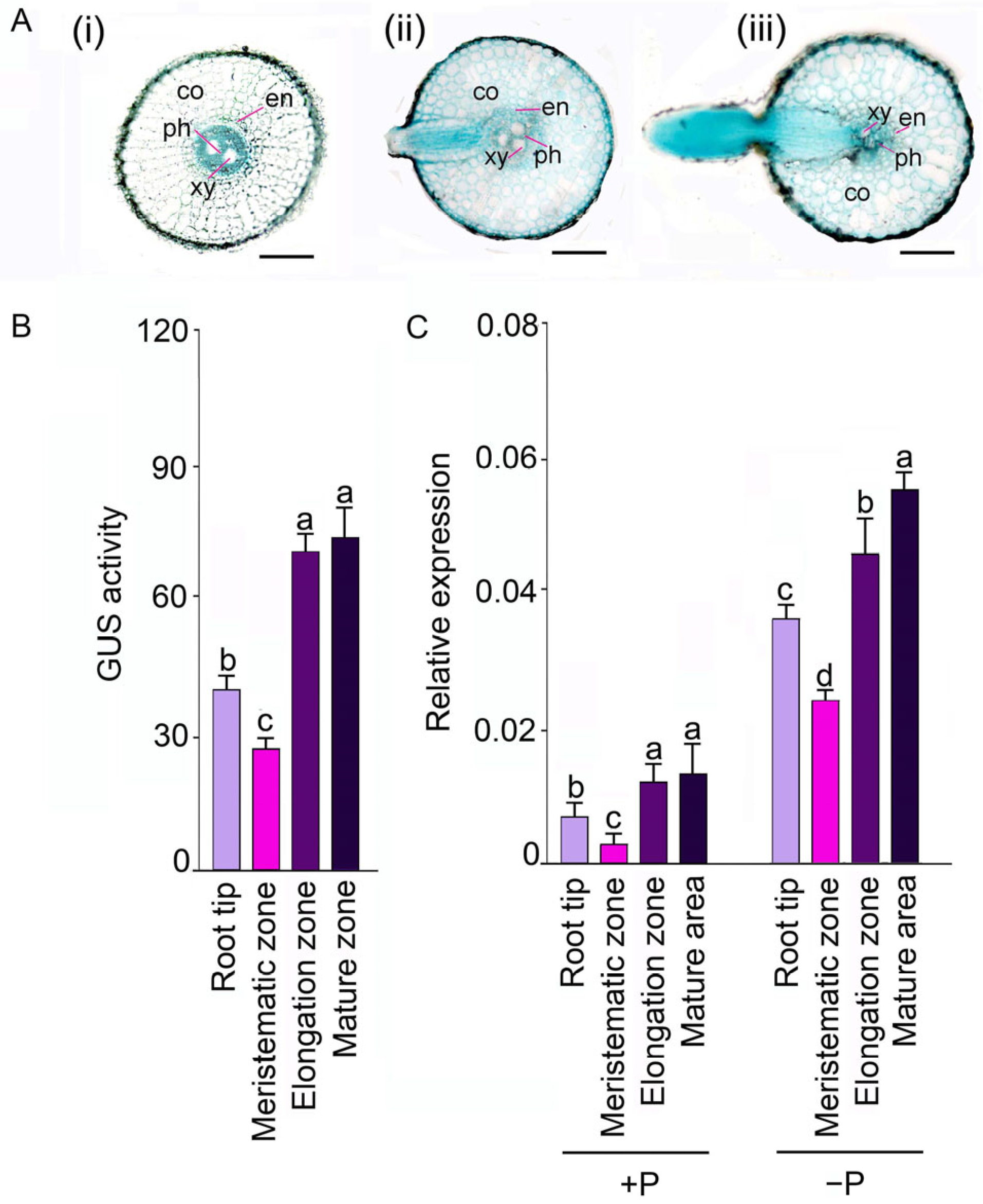
2.1. OsSQD1 Plays a Function in Formation and Development of Lateral Root

To understand the role of OsSQD1 in root development during the early growth stage, OsSQD1::GUS transgenic seedlings were grown under normal P conditions for 14 days. As shown in Figure 1A, the cross-section of the root revealed GUS expression in the xylem parenchyma, endodermis, phloem and cortex and in meristematic, elongation and mature zones. Deep GUS staining was also found in the lateral root primordium and lateral root (Figure 1A). We further examined the GUS activity in different parts of the root. The GUS activity of OsSQD1 for all four zones in roots was in the order of mature and elongation zones> meristematic zone > root tip (Figure 1B). Then, the steady-state transcript levels of OsSQD1 in different parts of roots under +P and −P conditions for another three days were measured by real-time qPCR (Figure 1C). Pi deficiency significantly induced the expression of OsSQD1 in all four zones compared with that under +P conditions. In addition, the expression pattern of OsSQD1 under −P supply was similar to the pattern under +P conditions, except for the relative expression in the mature and elongation zones. The relative expression of OsSQD1 in the mature zone was significantly higher than that in the elongation zone (Figure 1C). The expression pattern of OsSQD1 in roots revealed that OsSQD1 may play an important role in the development of roots, especially lateral roots, which form and develop in the elongation zone and mature zone.
For further study, a complete homozygous OsSQD1 knockout mutant (ossqd1) with a T-DNA insertion and 2 RNA interference (RNAi)-mediated knockdown lines were validated by quantitative RT-PCR and semiquantitative RT-PCR and used for root phenotype identification (Figure S1). The copy number of RNAi lines was detected by Southern blotting, and transgenic lines with a single copy were selected (Figure S1). The root phenotype of 14-d-old transgenic seedlings was observed under +P conditions (Figure S2). It was found that the knockout/down of OsSQD1 inhibited the growth of primary and adventitious roots. In particular, adventitious roots were severely affected by the downregulation of OsSQD1, resulting in almost no adventitious roots growing in the ossqd1 mutant (Figure S2A). Statistical analysis showed that the knockout/down of OsSQD1 reduced 59.3%/41.3% primary root length and 90.7%/62.5% adventitious root length compared with those in the WT (Figure S2B,C). Meanwhile, there was no significant difference in the root/shoot ratio between the OsSQD1 knockout/down lines and the WT (Figure S2D). To further study the effect of OsSQD1 on lateral roots under different Pi regimes, 7-d-old WT and OsSQD1 transgenic seedlings were transplanted under different Pi regimes for another 7 days. The lateral roots developing from primary roots were spread gently to reveal their architectural details (Figure S2E). There was a significant reduction in the lateral root density of the ossqd1 mutant (59.6 and 69.9%) and RNAi lines (30.9 and 30.6%) under +P and −P conditions compared with the WT, respectively (Figure S2F). Lateral root length was also significantly reduced by the mutation and silencing of OsSQD1 (Figure S2). These results suggested that the knockout/down of OsSQD1 inhibited root growth under different Pi conditions, especially the formation and development of lateral roots under −P conditions.

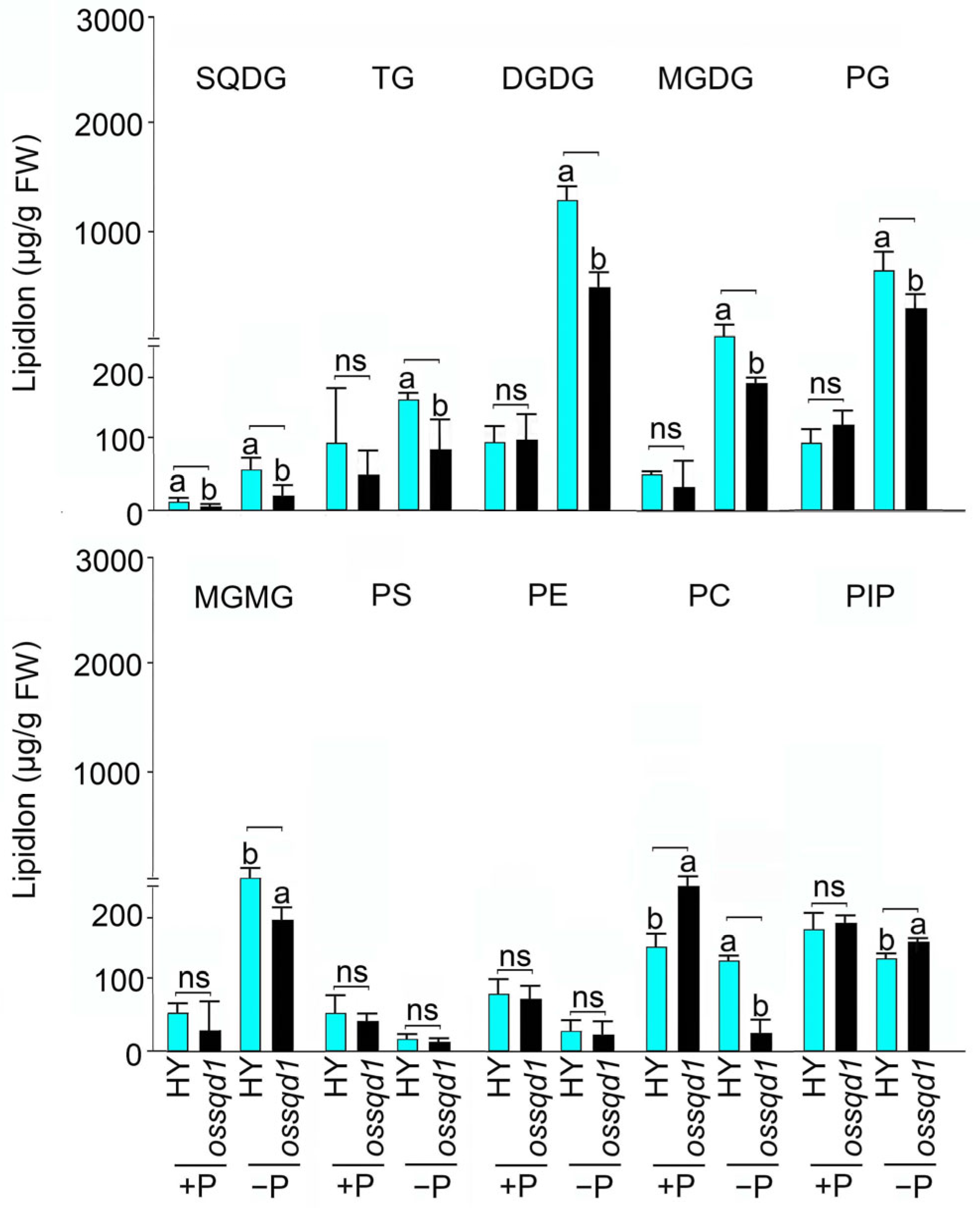
2.2. Mutation of OsSQD1 Decreased the Concentrations of Phospholipids and Glycolipids under −P Condition

The relative lipid intensity and lipid concentration of different lipid species in the ossqd1 mutant shoots were exhibited in our previous study [23]. A description of the different lipid species’ concentrations of the ossqd1 mutant roots under different Pi regimens is provided in this study. Under +P conditions, the mutation of OsSQD1 markedly induced the concentration of PC compared with that in the WT. However, the concentrations of TG, DGDG, MGDG, PG, MGMG, PS, PE and PIP showed no significant change (Figure 2). Under Pi deficiency, the concentrations of different lipid species showed that PS and PE were not changed in the ossqd1 mutant compared with WT rice. The analysis of TG (45.5%), DGDG (27.9%), MGDG (26.6%), PG (16%), MGMG (23%) and PC (78%) concentrations showed significant induction in ossqd1 mutant roots. In addition, the concentration of PIP was slightly induced by OsSQD1 mutation. The SQDG concentration was profoundly downregulated in ossqd1 roots under both +P and −P conditions. These data suggested that the mutation of OsSQD1 decreased the concentration of the majority of phospholipids and glycolipids under Pi deficiency.

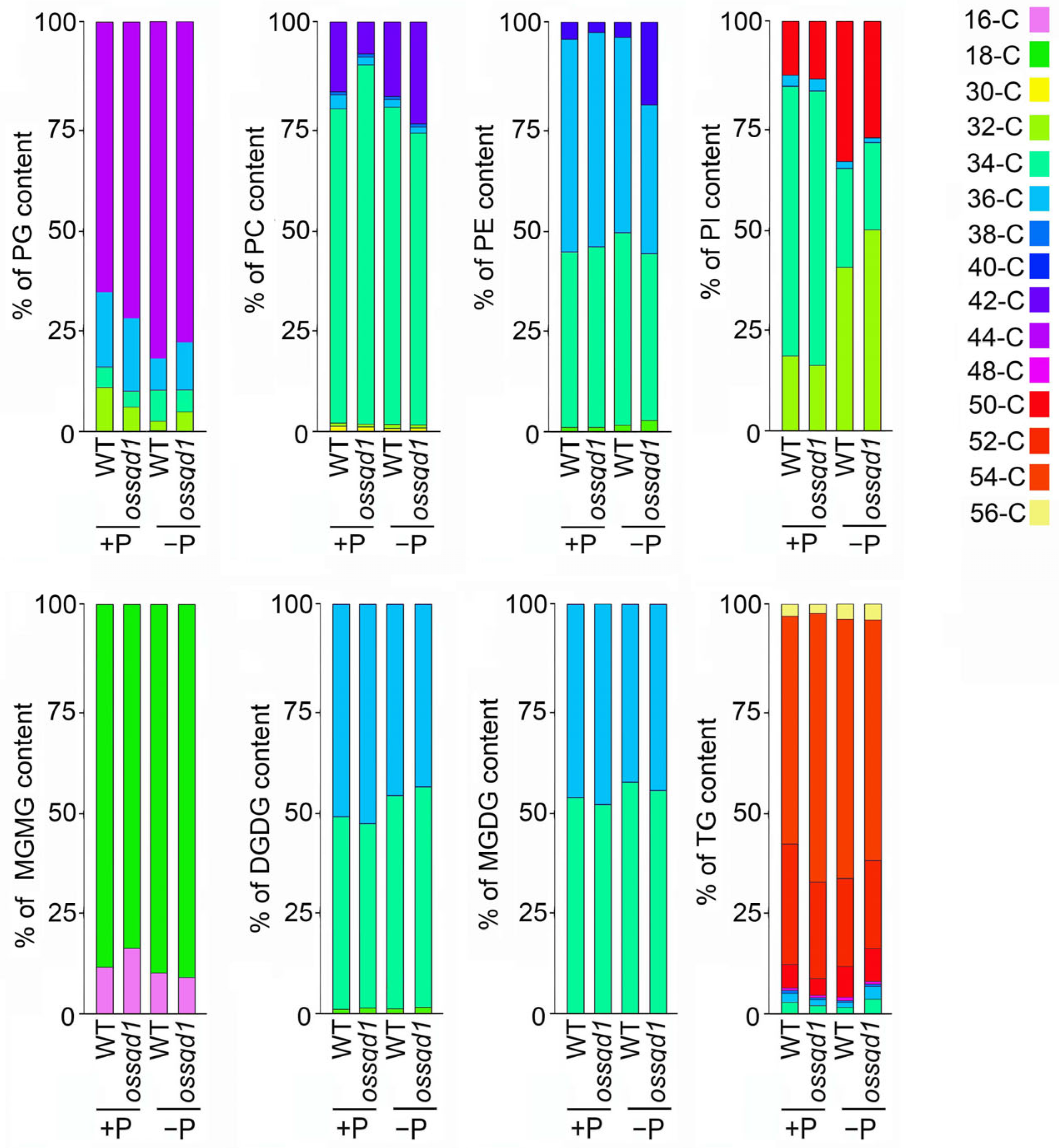
2.3. Knockout/Down of OsSQD1 Changed the Composition of Different Lipid Species under Pi Deficiency

To understand the effect of OsSQD1 on the composition of different lipid species, we examined the concentration of phospholipids and glycolipids with different acyl chain lengths under +P and −P conditions (Figure 3 and Figure S3). In contrast to the other phospholipids, the primary phospholipid in chloroplast inner membrane and thylakoid membrane is PG [48], which also showed the highest concentration in Pi-limited rice roots (Figure S3A). Mutation of OsSQD1 had no effect on the concentrations of PGs with different acyl chain lengths in Pi-deficient roots. However, the 34-C, 36-C and 44-C PG concentrations were much higher in the ossqd1 mutant roots than in the WT roots under −P conditions (Figure S3A). The effect of OsSQD1 on PC composition was more complex. Under +P conditions, the 34-C and 36-C PC concentrations were increased in ossqd1 mutant roots. The 34-C, 36-C and 42-C PC concentrations were decreased significantly in the ossqd1 mutant compared with the WT under Pi deficiency (Figure S3B). In addition, the 34-C and 36-C PE concentrations were reduced by the mutation of OsSQD1 under −P conditions (Figure S3C). The effect of OsSQD1 on the changes in PI in response to Pi limitation was quite different from that on the changes in other phospholipids. OsSQD1 mutation made the concentrations of 32-C and 34-C PI higher than those in WT roots under Pi deficiency (Figure S3D). There was no difference between the ossqd1 mutant and WT in the concentration of glycolipids with different chain lengths. Interestingly, the mutation of OsSQD1 under Pi deficiency had an impact on the glycolipid concentration of roots that could not be ignored (Figure S3E–G). Mutation of OsSQD1 resulted in a decrease in 34-C/36-C DGDG and MGDG under Pi deficiency (Figure S3E,F). Furthermore, the concentrations of 32-C and 34-C SQDG were profoundly reduced under −P conditions (Figure S3G). TG species with long acyl chains were found in rice roots. The concentration of 52-C was increased by the OsSQD1 mutation under Pi-sufficient conditions. Under −P conditions, the 50-C, 52-C and 54-C TG levels were considerably lower than those in the WT (Figure S3H).
To better visualize the changes in different lipid compositions, the proportion of different lipid species with various acyl chain lengths under +P and −P conditions in the ossqd1 mutant is shown in Figure 4. Under Pi sufficiency, the proportion of 32-C PG was slightly decreased while 44-C PG was increased in ossqd1 mutant roots compared with WT. Additionally, mutation of OsSQD1 caused an increase in the 34-C PC proportions together with a significant decrease in 42-C PC. Nevertheless, the composition of phospholipids in ossqd1 mutant roots was quite different from that in WT roots under Pi deficiency (Figure 3). The proportion of 36-C PG out of the total PG increased at the expense of 34-C PG in ossqd1 mutant roots. There was a slight decrease in the 34-C PC together with the increased proportion of 42-C PC. For PE, the proportion of 36-C PE was strongly decreased, while that of 40-C PE was induced in ossqd1 mutant roots under −P conditions. As observed for PG and PC, the proportion of 32-C PI was increased in Pi-deprived roots of the ossqd1 mutant. Meanwhile, mutation of OsSQD1 reduced the proportion of 50-C PI under −P conditions (Figure 3). Among glycolipids, the most obvious change in ossqd1 mutant roots was observed for the composition of MGDG and DGDG under −P conditions. The proportion of 32-C MGDG and DGDG increased together with a slight decrease in 34-C MGDG and 36-C DGDG in Pi-deprived roots of the ossqd1 mutant (Figure 3). In addition, the proportions of 34-C and 36-C TG were increased in the ossqd1 mutant compared with WT (Figure 3).

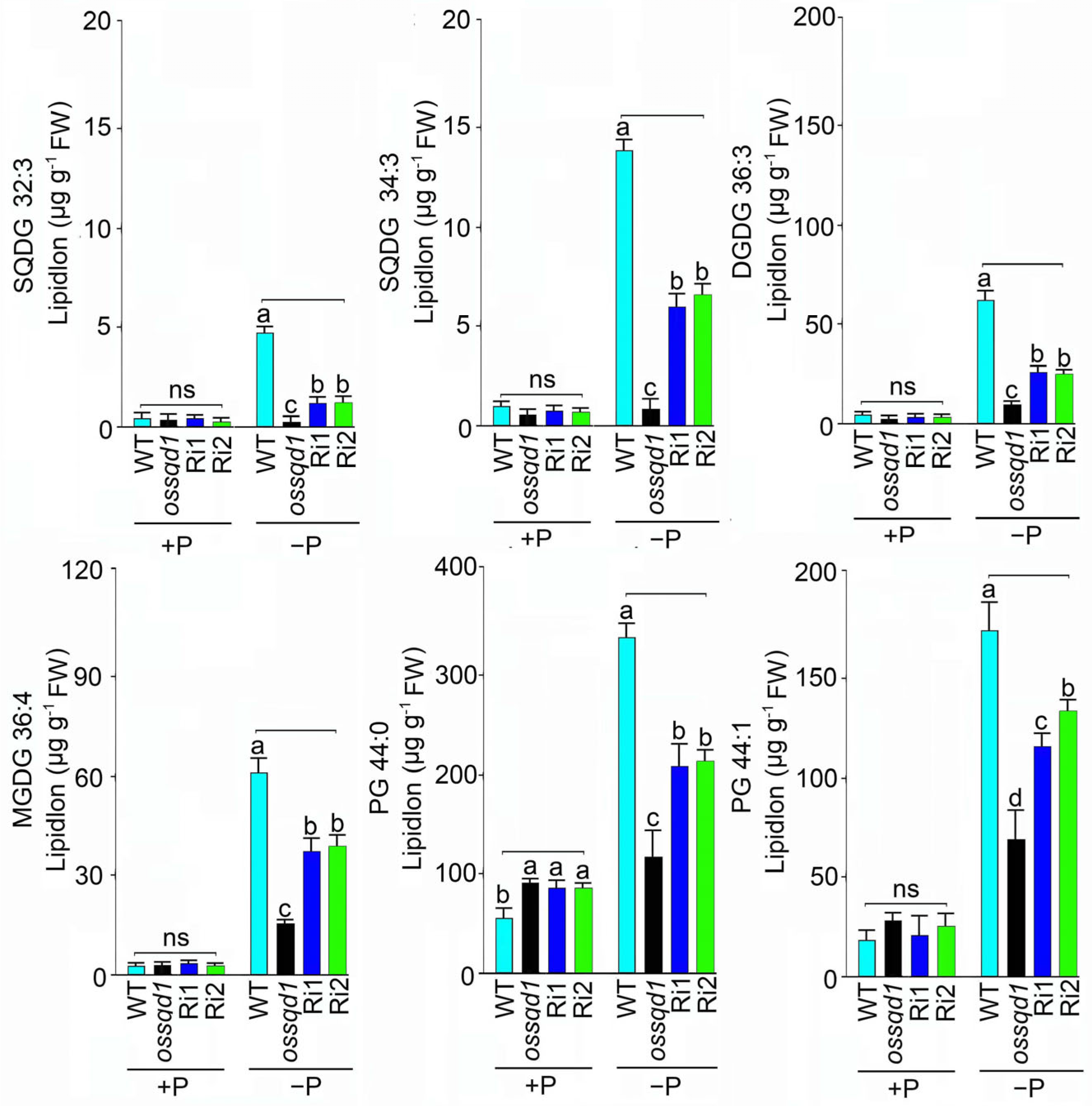
2.4. Knockout/Down of OsSQD1 Decreased the Unsaturated Double Bonds of Glycolipids and Phospholipids under Pi Deficiency

Previous studies have indicated that fatty acid saturation is involved in abiotic stress tolerance [49]. The fatty acid saturation in roots of the WT and ossqd1 mutant was employed and analyzed under different Pi regimes (Figure 4). Mutation of OsSQD1 significantly increased the saturated fatty acid content of PG under +P conditions. Under Pi deficiency, the number of PG with zero and one unsaturated double bonds was significantly decreased in ossqd1 roots (Figure 4A). Similarly, the number of fatty acids of PC with one, two, and four unsaturated double bonds increased, respectively, in ossqd1 roots compared with WT roots under +P conditions. However, these fatty acids decreased significantly in number in the Pi-deficient roots of ossqd1 (Figure 4B). No effect was observed in Pi-sufficient ossqd1 roots for PE fatty acids, but the fatty acids with one, two, three, and four unsaturated double bonds decreased markedly in number upon the mutation of OsSQD1 (Figure 4C). Due to the extremely low content of SQDG in roots under +P supply, the knockout of OsSQD1 did not result in a significant decline in SQDG in roots (Figure 4D). By comparison, SQDG with three unsaturated double bonds was observed to be extremely low in number under −P conditions (Figure 4D). The fatty acid concentrations of DGDG and MGDG showed a similar trend. The fatty acids with one, two, three, four, five and six unsaturated bonds of DGDG and MGDG were all decreased significantly in ossqd1 roots only under −P conditions (Figure 4E,F). These data indicated that the unsaturated double bonds of different lipid species were reduced by mutation of OsSQD1 in Pi-deprived roots.
To explore the changes in fatty acid concentrations affected by OsSQD1, knockout/down transgenic lines (ossqd1, Ri1 and Ri2) were used to measure fatty acid concentrations under different Pi conditions. The fatty acid concentrations of glycolipids MGDG, DGDG and SQDG showed that the polyunsaturated fatty acids SQDG (C32:3), SQDG (C34:3), DGDG (C36:3) and MGDG (C36:4) were markedly decreased by the mutation of OsSQD1 under −P conditions (Figure 5). As expected, all these fatty acids of glycolipids showed no change in ossqd1 roots compared with WT roots under Pi-sufficient conditions (Figure 5). Regarding the fatty acid concentrations of PG, the concentrations of C44:1 in Pi-sufficient roots of ossqd1 were similar to those in WT roots. Mutation of OsSQD1 resulted in a significant increase in C44:0 PG under +P conditions (Figure 5). In contrast, the knockout/down of OsSQD1 decreased C44:0 and C44:1 markedly compared with those in the WT (Figure 5). In summary, these results indicated that the mutation of OsSQD1 triggered responses in the composition and saturation of phospholipids and glycolipids to Pi supply. The concentrations of long-chain unsaturated fatty acids, phospholipids and glycolipids were generally decreased by the knockout/down of OsSQD1.

2.5. Knockout/Down of OsSQD1 Affects the Lipid Remodelling Regulatory Network in Roots under −P Conditions

To investigate the effect of knockout/down of OsSQD1 on the lipid remodeling regulatory network under +P and −P conditions, the expression of several genes related to lipid remodeling was measured by qRT-PCR in the ossqd1 mutant and RNAi lines (Ri1 and Ri2). The genes analyzed were OsSQD2.1 (LOC_Os07g01030), OsMGD2 (LOC_Os02g55910), OsDGD1β (LOC_Os04g34000), OsDGD1α (LOC_Os02g33580), and OsDGD2α (LOC_Os03g11560), which are involved in glycolipid synthesis; OsPLDβ1 (AK073012) and OsPLDα1 (AK065102), which are associated with phospholipid degradation; and OsNPC1 (AK067741) and OsNPC4 (AK243286), which function in hydrolyzing phospholipids and galactolipids (Figure 6). As shown in Figure 6, the relative expression of OsPLDα1, OsPLDβ1 and OsMGD2 was unaffected by the knockout/down of OsSQD1 under +P conditions (Figure 6B–D). Nevertheless, the relative expression of OsSQD2, OsNPC1, OsNPC4, OsDGD1α and OsDGD1β in roots was reduced by knockout/down of OsSQD1 under sufficient Pi (Figure 6A,E–H). The relative expression of OsDGD2α was downregulated only by the mutation of OsSQD1 (Figure 6I). These results indicated that the effect of OsSQD1 was much more obvious in Pi-deficient roots. The relative expression of all these genes was markedly decreased in the roots of the ossqd1 and RNAi lines (Figure 6A–I). This result suggested that the knockout/down of OsSQD1 caused the downregulation of phospholipid degradation and glycolipid synthesis genes under −P conditions.

3. Discussion

Lipids are the major components of the membranes of photosynthetic and nonphotosynthetic organs [6]. Pi-deprived plants utilize membrane phospholipids as an internal Pi source. To maintain membrane functionality, phospholipids are replaced by galactolipids [14]. Earlier, we demonstrated that OsSQD1 encodes UDP-sulfoquinovose synthase, which is the enzyme catalyzing the first step of SQDG synthesis in rice. It plays an essential function in maintaining the homeostasis of sulfur and P [50]. Additionally, glycolipids were reduced by the mutation of OsSQD1, causing the inhibition of photosynthesis in Pi-deficient shoots [23]. In this study, we highlight the function of OsSQD1 in the root phenotype under different Pi regimes (Figure S2). Knockout/down of OsSQD1 significantly reduced the formation and development of lateral roots and the growth of primary and adventitious roots, especially under −Pi conditions [50] (Figure S2E,F). Mutation of OsSQD1 significantly reduced PG, PC, SQDG, MGDG and DGDG concentrations under −P conditions (Figure 2). This finding highlights that the changes in different lipid species concentrations may have some effects on root development in response to external Pi supply. The overexpression of LPA2 (lysophosphatidic acid acyltransferase) increases the content of major phospholipid classes and root length under phosphate starvation [51]. Additionally, PPGP (phosphatidylglycerophosphate phosphatase) can enhance primary and lateral root development in Pi-deprived Arabidopsis [52]. This result suggested that PG and PG biosynthesis was required for root growth and development in Arabidopsis under −Pi conditions. As OsSQD1 encodes UDP-sulfoquinovose synthase, mutation of OsSQD1 should hinder the synthesis of SQDG [50] (Figure 2). Therefore, the SQDG content may also have a certain impact on root growth and development. The influence of MGDG and DGDG on root development was not clear. However, it has been reported that an increase in the DGDG: MGDG ratio may alter membrane permeability and fluidity under abiotic stress [53]. These results suggested that the knockout/down of OsSQD1 may inhibit root development by decreasing the contents of both phospholipids (PG) and glycolipids (SQDG, MGDG and DGDG) in Pi-deprived roots.
Mutation of OsSQD1 altered the composition of PG, PC, PE, PI, DGDG, MGDG and TG in Pi-deprived roots (Figure 3 and Figure S3). SQD2 is the sulfoquinovosyltransferase catalyzing the final step in the SQDG biosynthetic pathway [43,54]. The OsPLD (phospholipase D) family encodes rice phospholipases, hydrolyzing phospholipids to generate PA and a free radical [55]. NPC4 (a nonspecific phospholipase C) produces DAG that is converted to PA, affecting the C18/C16 of DAG in Arabidopsis and playing a positive role in root growth [56]. OsMGD2 is the MGDG synthase (MGD) that is significant for the accumulation of MGDG [57]. It can transfer a galactosyl residue from UDP-Gal to diacylglycerol [58,59,60]. In Arabidopsis, the enzymes MGD2 and MGD3 preferentially synthesize 16:0 and 18:2 MGDG, while DGD2 preferentially uses 16:0 [38,39]. Our data showed that the knockout/down of OsSQD1 downregulated the relative expression of OsSQD2, OsPLD1α, OsPLD1β, OsMGD2, OsNPC4, OsDGD1α, OsDGD2α and OsDGD1β upon Pi deprivation (Figure 6). This indicates that the lipid species’ composition alteration in ossqd1 mutant roots during Pi deficiency may be the result of changes in enzymatic reactions.
Several environmental parameters, especially temperature, have a significant impact on the physical properties of membranes and affect the fluidity of the membrane. It is generally believed that maintaining the integrity and fluidity of membranes is essential for plants to survive under extreme environmental conditions [61]. The unsaturation level and acyl chain length of different lipid species affect membrane fluidity [62]. A high level of unsaturated double bonds in the main membrane lipids may contribute to greater tolerance to low temperatures [63]. In Pi-deprived roots of the ossqd1 mutant, unsaturated double bonds were generally decreased in both phospholipids and glycolipids. This might affect the fluidity of the membrane, which may be a reason why the development of roots was inhibited more strongly in the ossqd1 mutant under −P conditions.
Compared with our previous research, this research revealed an obvious difference between the effect of OsSQD1 on the lipid concentration and composition of roots and shoots [23] (Figure 2, Figure 3, Figure 4 and Figure 5). In Pi-deprived shoots, the concentrations of PG, LPG, TG, and PC were induced by mutation of OsSQD1. Nevertheless, the concentrations of TG, PG and PC were greatly reduced in Pi-deprived roots of the ossqd1 mutant [23] (Figure 2 and Figure S3). These changes were accompanied by increases in 32-C PG, 34-C PG, 34-C PC and 36-PC in shoots under −P conditions. The concentrations of 32-C and 34-C PG and 34-C and 36-C PC were significantly decreased in roots upon Pi deficiency and were affected by the mutation of OsSQD1 [23] (Figure 3 and Figure S3). The glycolipid changes showed that 36-C MGDG, 34-C and 36-C DGDG were significantly reduced in Pi-deprived shoots of the ossqd1 mutant. These data were also slightly different from those on Pi-deprived roots [23] (Figure S3). According to previous studies, the concentration and composition of different lipid species in plant roots under conditions responding to Pi deficiency were different from those in shoots. Under −P conditions, the concentrations of PC, PE, PG, and PS were significantly lower than those under Pi sufficiency. However, the concentrations of PC and PS did not change, and PG was highly induced by Pi deficiency in both rice and Arabidopsis roots [20,23] (Figure 2). These results indicated that there may be distinctions in lipid metabolism in response to Pi deficiency between roots and shoots in rice, which caused the distinctions in the role of OsSQD1 in overall lipid homeostasis between roots and shoots.

4. Materials and Methods

4.1. Creation and Acquisition of Plant Materials

The ossqd1 mutant (PFG_2B-30,198.R) with T-DNA insertion in wild-type (WT; Oryza sativa L. ssp japonica ‘Hwayoung’) background was acquired from the rice T-DNA insertion mutant database RiceGE (http://signal.salk.edu/cgi-bin/RiceGE, accessed on 11 December 2022). The RNAi lines (Ri1 and Ri2) and OsSQD1 promoter-GUS fusion line were created as described in Sun et al., 2020. The primers used for confirming the insertion of T-DNA, and the transcript abundance of OsSQD1 were listed in Table S1. The promoter region of OsSQD1 was amplified from the genomic DNA using gene-specific primers with restriction sites KpnI and HindIII (Table S1).

4.2. Plant Growth and Treatment Conditions

Seeds of WT, ossqd1 mutant, Ri1 and Ri2 lines were germinated in the dark at 37 °C. WT and transgenic lines seeds were grown hydroponically for 10 d in the normal Pi solution making up with MgSO4 (1 mM), NH4NO3 (1.25 mM), Na2SiO3 (0.5 mM), KH2PO4 (0.2 mM), CaCl2 (1 mM), EDTA-Fe (20 µM), K2SO4 (0.35 mM), H3BO3 (20 µM), ZnSO4 (0.77 µM), (NH4)6Mo7O24 (0.39 µM), MnCl2 (9 µM), and CuSO4 (0.32 µM) with pH adjusted to 5.5. The 10-d-old seedlings were then transferred to the +P or –P (10 µM Pi) solutions for another 7 d (analysis of lipid-remodeling regulatory network) or 14 d (analysis of lipid and fatty acid concentration) as required. Rice was grown under controlled conditions in a light incubator (16 h light, 30 °C/8 h dark, 22 °C, relative humidity ~70%).

4.3. GUS Assays

Histochemical localization of GUS activity was detected by incubating 14-d-old roots with the substrate X-gluc (Research Organics, Cleveland, OH, USA), as described by Sun et al., 2020 [50]. Fluorometric assay of the bacterial β-glucuronidase (GUS) activity was detected as described in [64].

4.4. RNA Extraction and Expression Analyses

Total RNA (~1 μg) was extracted from roots of WT, ossqd1 mutant, Ri1 and Ri2 using TRIzol reagent (Invitrogen) for the synthesis of cDNA. First-strand cDNAs were synthesized from total RNA using the Goldenstar™ RT6 cDNA Synthesis Kit (TSINGKE, Shanghai, China). qRT-PCR was performed on LightCycler 480 II (Roche, Switzerland) using SYBR Green qPCR Mix (Tsingke, China) with 3 biological replicates, each with 3 technical repeats. The cycling conditions were as follows: 95 °C for 30 s, 35 cycles of 95 °C for 5 s and 60 °C for 30 s. Gene-specific primers used for qRT-PCR are listed in Table S1. ACTIN (OsRac1; LOC_Os03g50885) was used to normalize the reaction. Relative expression of the genes was computed by the 2 –ΔΔCT method as described in Livak and Schmittgen, 2001 [65].

4.5. Quantification of Different Lipid Species and Fatty Acids

The roots of WT, ossqd1 mutant, and RNAi lines grown in +P and −P conditions for 21 d were homogenized into powder in liquid nitrogen. 200 μL cold water, 20 μL lipid internal standard mixture and 800 μL cold methyl tert-butyl ether were added to the samples then vortexed for 30 s. Following this, 240 μL methanol was added to the samples and adequately vortexed, sonicated at 4 °C for 20 min and left to stand for 30 min, then centrifuged (14,000× g for 15 min at 10 °C) to extract lipids. The upper organic layer was dried in a vacuum centrifuge. The lipid extraction, seperation and quantification was detected as described in Sun et al., 2021. The Lipidsearch 4.0 software was used for peak detection and annotation of lipids or internal standards.

4.6. Statistical Analysis

Data were analyzed by ANOVA using SPSS 20 program (www.spss.com, accessed on 11 December 2022). Duncan’s multiple range test at p < 0.05 was carried out for all the experiments to determine the significance between the WT and knockout/down lines.

5. Conclusions

In this study, the function of OsSQD1 in root development and growth under different Pi regimes was identified. Knockout/down of OsSQD1 obviously inhibited lateral root formation and development under −P conditions. The analysis of the concentration and composition of different lipid species in Pi-deprived roots of the ossqd1 mutant showed that the mutation of OsSQD1 decreased the concentration and the unsaturated fatty acids of phospholipids and glycolipids in roots under −P conditions. Additionally, it also decreased the concentration of fatty acids with different acyl chain lengths, altering the composition of lipids under Pi deficiency. These results indicate that OsSQD1 influences root development under −P conditions by affecting lipid composition and saturation in rice.

Supplementary Materials

The following supporting information can be downloaded at: https://www.mdpi.com/article/10.3390/ijms24010114/s1.

Author Contributions

Y.S. and Q.Q. conducted the research work and prepared the manuscript. K.S. and L.S. helped in root phenotype observation and detection of gene expression. T.J., S.Y. and Z.L. helped technically to analyze lipids. G.X. and S.S. provide mutants and RNAi lines required for the experiment. Y.X. is the supervisor of the research work and also the corresponding author. All authors have read and agreed to the published version of the manuscript.

Funding

This study was supported by the Shanghai Agriculture Applied Technology Development Program, China (2022-02-08-00-12-F01152), Chinese National Natural Science Foundation (31902102), Shanghai Agriculture Applied Technology Development Program, China (2022-02-08-00-12-F01202). Shanghai Sailing Program (19YF1443200), Natural Science Foundation of Shanghai (21ZR1443300), National Agricultural Experimental Station for Agricultural Environment, Fengxian (grant number: NAES035AE03), Shanghai Agricultural Science and Technology Promotion Project ((2021) 2-19), and the Outstanding Team Program of Shanghai Academy of Agricultural Science (Hu-Nong-Ke-Zhuo 2022 (008)).

Institutional Review Board Statement

Not applicable.

Informed Consent Statement

Not applicable.

Data Availability Statement

Not applicable.

Conflicts of Interest

The authors declare no conflict of interest.

References

  1. Raghothama, K.G. PHOSPHATE ACQUISITION. Annu. Rev. Plant Physiol. Plant Mol. Biol. 1999, 50, 665–693. [Google Scholar] [CrossRef] [PubMed]
  2. Raghothama, K. Phosphate transport and signaling. Curr. Opin. Plant Biol. 2000, 3, 182–187. [Google Scholar] [CrossRef] [PubMed]
  3. Puga, M.I.; Rojas-Triana, M.; de Lorenzo, L.; Leyva, A.; Rubio, V.; Paz-Ares, J. Novel signals in the regulation of Pi starvation responses in plants: Facts and promises. Curr. Opin. Plant Biol. 2017, 39, 40–49. [Google Scholar] [CrossRef] [PubMed]
  4. López-Bucio, J.; Hernández-Abreu, E.; Sánchez-Calderón, L.; Nieto-Jacobo, M.F.; Simpson, J.; Herrera-Estrella, L.R. Phosphate Availability Alters Architecture and Causes Changes in Hormone Sensitivity in the Arabidopsis Root System. Plant Physiol. 2002, 129, 244–256. [Google Scholar] [CrossRef] [Green Version]
  5. Svistoonoff, S.; Creff, A.; Reymond, M.; Sigoillot-Claude, C.; Ricaud, L.; Blanchet, A.; Nussaume, L.; Desnos, T. Root tip contact with low-phosphate media reprograms plant root architecture. Nat. Genet. 2007, 39, 792–796. [Google Scholar] [CrossRef] [Green Version]
  6. Harayama, T.; Riezman, H. Understanding the diversity of membrane lipid composition. Nat. Rev. Mol. Cell Biol. 2018, 19, 281–296, Correction in Nat. Rev. Mol. Cell Biol. 2019, 20, 715. [Google Scholar] [CrossRef]
  7. Welti, R. Plant lipidomics: Discerning biological function by profiling plant complex lipids using mass spectrometry. Front. Biosci. J. Virtual Libr. 2007, 12, 2494–2506. [Google Scholar] [CrossRef] [Green Version]
  8. Sheffer, M.; Fried, A.; Gottlieb, H.E.; Tietz, A.; Avron, M. Lipid composition of the plasma-membrane of the halotolerant alga, Dunaliella salina. Biochim. Biophys. Acta (BBA)—Biomembr. 1986, 857, 165–172. [Google Scholar] [CrossRef]
  9. Yoshida, S.; Uemura, M. Lipid Composition of Plasma Membranes and Tonoplasts Isolated from Etiolated Seedlings of Mung Bean (Vigna radiata L.). Plant Physiol. 1986, 82, 807–812. [Google Scholar] [CrossRef] [Green Version]
  10. Rochester, C.P.; Kjellbom, P.; Andersson, B.; Larsson, C. Lipid composition of plasma membranes isolated from light-grown barley (Hordeum vulgare) leaves: Identification of cerebroside as a major component. Arch. Biochem. Biophys. 1987, 255, 385–391. [Google Scholar] [CrossRef]
  11. Brown, D.J.; DuPont, F.M. Lipid Composition of Plasma Membranes and Endomembranes Prepared from Roots of Barley (Hordeum vulgare L.). Plant Physiol. 1989, 90, 955–961. [Google Scholar] [CrossRef] [Green Version]
  12. Siebers, M.; Brands, M.; Wewer, V.; Duan, Y.; Hölzl, G.; Dörmann, P. Lipids in plant–microbe interactions. Biochim. Biophys. Acta (BBA)—Mol. Cell Biol. Lipids 2016, 1861, 1379–1395. [Google Scholar] [CrossRef]
  13. Gaude, N.; Nakamura, Y.; Scheible, W.R.; Ohta, H.; Dörmann, P. Phospholipase C5 (NPC5) is involved in galactolipid accumulation during phosphate limitation in leaves of Arabidopsis. Plant J. 2008, 56, 28–39. [Google Scholar] [CrossRef]
  14. Pfaff, J.; Denton, A.K.; Usadel, B.; Pfaff, C. Phosphate starvation causes different stress responses in the lipid metabolism of tomato leaves and roots. Biochim. Biophys. Acta (BBA)—Mol. Cell Biol. Lipids 2020, 1865, 158763. [Google Scholar] [CrossRef]
  15. Minnikin, D.E.; Abdolrahimzadeh, H.; Baddiley, J. Replacement of acidic phospholipids by acidic glycolipids in Pseudomonas diminuta. Nature 1974, 249, 268–269. [Google Scholar] [CrossRef]
  16. Benning, C.; Beatty, J.T.; Prince, R.C.; Somerville, C.R. The sulfolipid sulfoquinovosyldiacylglycerol is not required for photosynthetic electron transport in Rhodobacter sphaeroides but enhances growth under phosphate limitation. Proc. Natl. Acad. Sci. USA 1993, 90, 1561–1565. [Google Scholar] [CrossRef] [Green Version]
  17. Güler, S.; Seeliger, A.; Härtel, H.; Renger, G.; Benning, C. A Null Mutant of Synechococcus sp. PCC7942 Deficient in the Sulfolipid Sulfoquinovosyl Diacylglycerol. J. Biol. Chem. 1996, 271, 7501–7507. [Google Scholar] [CrossRef] [Green Version]
  18. Essigmann, B.; Güler, S.; Narang, R.A.; Linke, D.; Benning, C. Phosphate availability affects the thylakoid lipid composition and the expression of SQD1, a gene required for sulfolipid biosynthesis in Arabidopsis thaliana. Proc. Natl. Acad. Sci. USA 1998, 95, 1950–1955. [Google Scholar] [CrossRef] [Green Version]
  19. Okazaki, Y.; Otsuki, H.; Narisawa, T.; Kobayashi, M.; Sawai, S.; Kamide, Y.; Kusano, M.; Aoki, T.; Hirai, M.Y.; Saito, K. A new class of plant lipid is essential for protection against phosphorus depletion. Nat. Commun. 2013, 4, 1510. [Google Scholar] [CrossRef] [PubMed] [Green Version]
  20. Pant, B.D.; Burgos, A.; Pant, P.; Cuadros-Inostroza, A.; Willmitzer, L.; Scheible, W.-R. The transcription factor PHR1 regulates lipid remodeling and triacylglycerol accumulation in Arabidopsis thaliana during phosphorus starvation. J. Exp. Bot. 2015, 66, 1907–1918. [Google Scholar] [CrossRef]
  21. Bajhaiya, A.K.; Dean, A.P.; Zeef, L.A.; Webster, R.E.; Pittman, J.K. PSR1 Is a Global Transcriptional Regulator of Phosphorus Deficiency Responses and Carbon Storage Metabolism in Chlamydomonas reinhardtii. Plant Physiol. 2015, 170, 1216–1234. [Google Scholar] [CrossRef] [PubMed] [Green Version]
  22. Murakami, H.; Kakutani, N.; Kuroyanagi, Y.; Iwai, M.; Hori, K.; Shimojima, M.; Ohta, H. MYB-like transcription factor NoPSR1 is crucial for membrane lipid remodeling under phosphate starvation in the oleaginous microalga Nannochloropsis oceanica. FEBS Lett. 2020, 594, 3384–3394. [Google Scholar] [CrossRef] [PubMed]
  23. Sun, Y.; Song, K.; Liu, L.; Sun, L.; Qin, Q.; Jiang, T.; Zhou, B.; Zhu, C.; Xu, G.; Sun, S.; et al. Sulfoquinovosyl diacylglycerol synthase 1 impairs glycolipid accumulation and photosynthesis in phosphate-deprived rice. J. Exp. Bot. 2021, 72, 6510–6523. [Google Scholar] [CrossRef] [PubMed]
  24. Kuppusamy, T.; Giavalisco, P.; Arvidsson, S.; Sulpice, R.; Stitt, M.; Finnegan, P.M.; Scheible, W.-R.; Lambers, H.; Jost, R. Lipid Biosynthesis and Protein Concentration Respond Uniquely to Phosphate Supply during Leaf Development in Highly Phosphorus-Efficient Hakea prostrata. Plant Physiol. 2014, 166, 1891–1911. [Google Scholar] [CrossRef] [PubMed] [Green Version]
  25. Hammond, J.P.; Bennett, M.J.; Bowen, H.C.; Broadley, M.R.; Eastwood, D.C.; May, S.T.; Rahn, C.; Swarup, R.; Woolaway, K.E.; White, P.J. Changes in Gene Expression in Arabidopsis Shoot during Phosphate Starvation and the Potential for Developing Smart Plants. Society 2003, 132, 578–596. [Google Scholar] [CrossRef] [Green Version]
  26. Kobayashi, K.; Masuda, T.; Takamiya, K.-I.; Ohta, H. Membrane lipid alteration during phosphate starvation is regulated by phosphate signaling and auxin/cytokinin cross-talk. Plant J. 2006, 47, 238–248. [Google Scholar] [CrossRef]
  27. Misson, J.; Raghothama, K.G.; Jain, A.; Jouhet, J.; Block, M.A.; Bligny, R.; Ortet, P.; Creff, A.; Somerville, S.; Rolland, N.; et al. A genome-wide transcriptional analysis using Arabidopsis thaliana Affymetrix gene chips determined plant responses to phosphate deprivation. Proc. Natl. Acad. Sci. USA 2005, 102, 11934–11939. [Google Scholar] [CrossRef] [Green Version]
  28. Nakamura, Y.; Awai, K.; Masuda, T.; Yoshioka, Y.; Takamiya, K.-I.; Ohta, H. A Novel Phosphatidylcholine-hydrolyzing Phospholipase C Induced by Phosphate Starvation in Arabidopsis. J. Biol. Chem. 2005, 280, 7469–7476. [Google Scholar] [CrossRef] [Green Version]
  29. Cruz-Ramírez, A.; Oropeza-Aburto, A.; Razo-Hernandez, F.; Ramirez-Chavez, E.; Herrera-Estrella, L. Phospholipase DZ2 plays an important role in extraplastidic galactolipid biosynthesis and phosphate recycling in Arabidopsis roots. Proc. Natl. Acad. Sci. USA 2006, 103, 6765–6770. [Google Scholar] [CrossRef] [Green Version]
  30. Li, M.; Welti, R.; Wang, X. Quantitative Profiling of Arabidopsis Polar Glycerolipids in Response to Phosphorus Starvation. Roles of Phospholipases Dζ1 and Dζ2 in Phosphatidylcholine Hydrolysis and Digalactosyldiacylglycerol Accumulation in Phosphorus-Starved Plants. Plant Physiol. 2006, 142, 750–761. [Google Scholar] [CrossRef]
  31. Nakamura, Y.; Koizumi, R.; Shui, G.; Shimojima, M.; Wenk, M.R.; Ito, T.; Ohta, H. Arabidopsis lipins mediate eukaryotic pathway of lipid metabolism and cope critically with phosphate starvation. Proc. Natl. Acad. Sci. USA 2009, 106, 20978–20983. [Google Scholar] [CrossRef] [Green Version]
  32. Eastmond, P.J.; Quettier, A.-L.; Kroon, J.T.; Craddock, C.; Adams, N.; Slabas, A.R. PHOSPHATIDIC ACID PHOSPHOHYDROLASE1 and 2 Regulate Phospholipid Synthesis at the Endoplasmic Reticulum in Arabidopsis. Plant Cell 2010, 22, 2796–2811. [Google Scholar] [CrossRef] [Green Version]
  33. Nakamura, Y. Phosphate starvation and membrane lipid remodeling in seed plants. Prog. Lipid Res. 2013, 52, 43–50. [Google Scholar] [CrossRef]
  34. Heinz, E.; Roughan, P.G. Similarities and Differences in Lipid Metabolism of Chloroplasts Isolated from 18:3 and 16:3 Plants. Plant Physiol. 1983, 72, 273–279. [Google Scholar] [CrossRef] [Green Version]
  35. Härtel, H.; Essigmann, B.; Lokstein, H.; Hoffmann-Benning, S.; Peters-Kottig, M.; Benning, C. The phospholipid-deficient pho1 mutant of Arabidopsis thaliana is affected in the organization, but not in the light acclimation, of the thylakoid membrane. Biochim. Biophys. Acta (BBA)—Biomembr. 1998, 1415, 205–218. [Google Scholar] [CrossRef] [Green Version]
  36. Awai, K.; Maréchal, E.; Block, M.A.; Brun, D.; Masuda, T.; Shimada, H.; Takamiya, K.-I.; Ohta, H.; Joyard, J. Two types of MGDG synthase genes, found widely in both 16:3 and 18:3 plants, differentially mediate galactolipid syntheses in photosynthetic and nonphotosynthetic tissues in Arabidopsis thaliana. Proc. Natl. Acad. Sci. USA 2001, 98, 10960–10965. [Google Scholar] [CrossRef] [Green Version]
  37. Kobayashi, K.; Awai, K.; Takamiya, K.-I.; Ohta, H. Arabidopsis Type B Monogalactosyldiacylglycerol Synthase Genes Are Expressed during Pollen Tube Growth and Induced by Phosphate Starvation. Plant Physiol. 2004, 134, 640–648. [Google Scholar] [CrossRef] [Green Version]
  38. Kobayashi, K.; Awai, K.; Nakamura, M.; Nagatani, A.; Masuda, T.; Ohta, H. Type-B monogalactosyldiacylglycerol synthases are involved in phosphate starvation-induced lipid remodeling, and are crucial for low-phosphate adaptation. Plant J. 2009, 57, 322–331. [Google Scholar] [CrossRef]
  39. Kelly, A.A.; Dörmann, P. DGD2, an Arabidopsis Gene Encoding a UDP-Galactose-dependent Digalactosyldiacylglycerol Synthase Is Expressed during Growth under Phosphate-limiting Conditions. J. Biol. Chem. 2002, 277, 1166–1173. [Google Scholar] [CrossRef] [Green Version]
  40. Kelly, A.A.; Froehlich, J.E.; Dörmann, P. Disruption of the Two Digalactosyldiacylglycerol Synthase Genes DGD1 and DGD2 in Arabidopsis Reveals the Existence of an Additional Enzyme of Galactolipid Synthesis. Plant Cell 2003, 15, 2694–2706. [Google Scholar] [CrossRef]
  41. Okazaki, Y.; Shimojima, M.; Sawada, Y.; Toyooka, K.; Narisawa, T.; Mochida, K.; Tanaka, H.; Matsuda, F.; Hirai, A.; Hirai, M.Y.; et al. A Chloroplastic UDP-Glucose Pyrophosphorylase from Arabidopsis Is the Committed Enzyme for the First Step of Sulfolipid Biosynthesis. Plant Cell 2009, 21, 892–909. [Google Scholar] [CrossRef] [PubMed] [Green Version]
  42. Essigmann, B.; Hespenheide, B.M.; A Kuhn, L.; Benning, C. Prediction of the Active-Site Structure and NAD+ Binding in SQD1, a Protein Essential for Sulfolipid Biosynthesis in Arabidopsis. Arch. Biochem. Biophys. 1999, 369, 30–41. [Google Scholar] [CrossRef] [PubMed] [Green Version]
  43. Yu, B.; Xu, C.; Benning, C. Arabidopsis disrupted in SQD2 encoding sulfolipid synthase is impaired in phosphate-limited growth. Proc. Natl. Acad. Sci. USA 2002, 99, 5732–5737. [Google Scholar] [CrossRef] [PubMed] [Green Version]
  44. Moummou, H.; Kallberg, Y.; Tonfack, L.B.; Persson, B.; van der Rest, B. The Plant Short-Chain Dehydrogenase (SDR) superfamily: Genome-wide inventory and diversification patterns. BMC Plant Biol. 2012, 12, 219. [Google Scholar] [CrossRef] [PubMed] [Green Version]
  45. Mulichak, A.M.; Theisen, M.J.; Essigmann, B.; Benning, C.; Garavito, R.M. Crystal structure of SQD1, an enzyme involved in the biosynthesis of the plant sulfolipid headgroup donor UDP-sulfoquinovose. Proc. Natl. Acad. Sci. USA 1999, 96, 13097–13102. [Google Scholar] [CrossRef] [Green Version]
  46. Liu, Y.; Thoden, J.B.; Kim, J.; Berger, E.; Gulick, A.M.; Ruzicka, F.J.; Holden, A.H.M.; Frey, P.A. Mechanistic Roles of Tyrosine 149 and Serine 124 in UDP-galactose 4-Epimerase from Escherichia coli. Biochemistry 1997, 36, 10675–10684. [Google Scholar] [CrossRef]
  47. Thoden, J.B.; Frey, P.A.; Holden, H.M. Molecular Structure of the NADH/UDP-glucose Abortive Complex of UDP-galactose 4-Epimerase from Escherichia coli: Implications for the Catalytic Mechanism, Biochemistry 1996, 35, 5137–5144. [Google Scholar] [CrossRef]
  48. Marechal, V.; Elenbaas, B.; Taneyhill, L.; Piette, J.; Mechali, M.; Nicolas, J.C.; Levine, A.J.; Moreau, J. Conservation of structural domains and biochemical activities of the MDM2 protein from Xenopus laevis. Oncogene 1997, 14, 1427–1433. [Google Scholar]
  49. Nguyen, V.C.; Nakamura, Y.; Kanehara, K. Membrane lipid polyunsaturation mediated by FATTY ACID DESATURASE2 (FAD2) is involved in endoplasmic reticulum stress tolerance in Arabidopsis thaliana. Plant J. 2019, 99, 478–493. [Google Scholar] [CrossRef]
  50. Sun, Y.; Jain, A.; Xue, Y.; Wang, X.; Zhao, G.; Liu, L.; Hu, Z.; Hu, S.; Shen, X.; Liu, X.; et al. OsSQD1 at the crossroads of phosphate and sulfur metabolism affects plant morphology and lipid composition in response to phosphate deprivation. Plant Cell Environ. 2020, 43, 1669–1690. [Google Scholar] [CrossRef]
  51. Angkawijaya, A.E.; Nguyen, V.C.; Nakamura, Y. Enhanced root growth in phosphate-starved Arabidopsis by stimulating de novo phospholipid biosynthesis through the overexpression of LYSOPHOSPHATIDIC ACID ACYLTRANSFERASE 2 (LPAT2). Plant Cell Environ. 2017, 40, 1807–1818. [Google Scholar] [CrossRef]
  52. Lin, Y.-C.; Kobayashi, K.; Wada, H.; Nakamura, Y. Phosphatidylglycerophosphate phosphatase is required for root growth in Arabidopsis. Biochim. Biophys. Acta (BBA)—Mol. Cell Biol. Lipids 2018, 1863, 563–575. [Google Scholar] [CrossRef]
  53. Gigon, A.; Matos, A.-R.; Laffray, D.; Zuily-Fodil, Y.; Pham-Thi, A.-T. Effect of Drought Stress on Lipid Metabolism in the Leaves of Arabidopsis thaliana (Ecotype Columbia). Ann. Bot. 2004, 94, 345–351. [Google Scholar] [CrossRef] [Green Version]
  54. Yu, B.; Benning, C. Anionic lipids are required for chloroplast structure and function in Arabidopsis. Plant J. 2003, 36, 762–770. [Google Scholar] [CrossRef]
  55. Kaur, A.; Neelam, K.; Kaur, K.; Kitazumi, A.; de Los Reyes, B.G.; Singh, K. Novel allelic variation in the phospholipase D alpha1 gene (OsPLDα1) of wild Oryza species implies to its low expression in rice bran. Sci. Rep. 2020, 10, 6571. [Google Scholar]
  56. Yang, B.; Li, M.; Phillips, A.; Li, L.; Ali, U.; Li, Q.; Lu, S.; Hong, Y.; Wang, X.; Guo, L. Nonspecific phospholipase C4 hydrolyzes phosphosphingolipids and sustains plant root growth during phosphate deficiency. Plant Cell 2021, 33, 766–780. [Google Scholar] [CrossRef]
  57. Shimojima, M.; Ohta, H.; Iwamatsu, A.; Masuda, T.; Shioi, Y.; Takamiya, K.-I. Cloning of the gene for monogalactosyldiacylglycerol synthase and its evolutionary origin. Proc. Natl. Acad. Sci. USA 1997, 94, 333–337. [Google Scholar] [CrossRef] [Green Version]
  58. Dörmann, P.; Balbo, I.; Benning, C. Arabidopsis galactolipid biosynthesis and lipid trafficking mediated by DGD1. Science 1999, 284, 2181–2184. [Google Scholar] [CrossRef]
  59. Wang, S.; Uddin, M.I.; Tanaka, K.; Yin, L.; Shi, Z.; Qi, Y.; Mano, J.; Matsui, K.; Shimomura, N.; Sakaki, T.; et al. Maintenance of Chloroplast Structure and Function by Overexpression of the Rice MONOGALACTOSYLDIACYLGLYCEROL SYNTHASE Gene Leads to Enhanced Salt Tolerance in Tobacco. Plant Physiol. 2014, 165, 1144–1155. [Google Scholar] [CrossRef] [Green Version]
  60. Basnet, R.; Hussain, N.; Shu, Q. OsDGD2β is the Sole Digalactosyldiacylglycerol Synthase Gene Highly Expressed in Anther, and its Mutation Confers Male Sterility in Rice. Rice 2019, 12, 1–13. [Google Scholar] [CrossRef] [Green Version]
  61. Wallis, J.G.; Browse, J. Mutants of Arabidopsis reveal many roles for membrane lipids. Prog. Lipid Res. 2002, 41, 254–278. [Google Scholar] [CrossRef] [PubMed]
  62. Zheng, G.; Li, L.; Li, W. Glycerolipidome responses to freezing- and chilling-induced injuries: Examples in Arabidopsis and rice. BMC Plant Biol. 2016, 16, 1–15. [Google Scholar] [CrossRef] [PubMed]
  63. Zheng, Y.; Yang, Y.; Wang, M.; Hu, S.; Wu, J.; Yu, Z. Differences in lipid homeostasis and membrane lipid unsaturation confer differential tolerance to low temperatures in two Cycas species. BMC Plant Biol. 2021, 21, 1–15. [Google Scholar] [CrossRef] [PubMed]
  64. Zhang, S.; Lian, Y.; Liu, Y.; Wang, X.; Liu, Y.; Wang, G. Characterization of a Maize Wip1 Promoter in Transgenic Plants. Int. J. Mol. Sci. 2013, 14, 23872–23892. [Google Scholar] [CrossRef] [Green Version]
  65. Livak, K.J.; Schmittgen, T.D. Analysis of relative gene expression data using real-time quantitative PCR and the 2−ΔΔCT Method. Methods 2001, 25, 402–408. [Google Scholar] [CrossRef]
Figure 1. Expression pattern of OsSQD1 in rice roots. Histochemical analysis of the tissue-specific GUS staining (A) and activity (B) driven by OsSQD1 promoter (2028 bp upstream of its translation start codon). Cross-sections of OsSQD1 promoter-GUS fusion roots in (A) were meristematic zone (i), elongation zone (ii), and mature zone (iii) from left to right. co, en, xy, and ph refers to cortex, endodermis, xylem, and phloem, respectively. Bars represent 100 μm. (C) The relative expression of OsSQD1 in root tip, meristematic zone, elongation zone, and mature zone of roots under different +P and −P conditions. Actin (OsRac1; LOC_Os03g50885) was used as an internal control. Values (B,C) are means ± SE (n = 3) and different letters on the histograms indicate that the values differ significantly (p < 0.05, one-way analysis of variance).
Ijms 24 00114 g001
Figure 2. OsSQD1 altered concentration of different lipid species in roots under different Pi regimes. The concentrations of different species of lipids in WT and ossqd1 mutant roots under +P and −P conditions. SQDG, sulfoquinovosyldiacylglycerol; TG, triglyceride; DGDG, digalactosyldiacylglycerol; MGDG, monogalactosyldiacylglycerol; PG, phosphatidylglycerol; MGMG: monogalactosylmonoacylglycerol; PS, phosphatidylserine; PE, phosphatidylethanolamine; PC, phosphatidylcholine; PIP: phosphatidylinositol-4-monophosphate. Lipidlon replaces the concentration of lipid molecule. Values are means ± SE (n = 4) and different letters on the histograms indicate that the values differ significantly between WT and ossqd1 mutant. (p < 0.05, one-way analysis of variance, Duncan’s test). ns = not significant.
Ijms 24 00114 g002
Figure 3. Knockout of OsSQD1 changes the composition of phospholipids and glycolipids in roots under −P condition. Values show the proportion of the different acyl groups (groups of lipid species with the same number of acyl carbons) to the total class content, the sum of the normalized intensities of all the compounds belonging to the same class.
Ijms 24 00114 g003
Figure 4. Knockout of OsSQD1 affects the fatty acid saturation of different lipid species under different Pi regimes. The lipid saturation of PG (A), PC (B), PE (C), SQDG (D), DGDG (E) and MGDG (F). The abscissa represents the number of unsaturated bonds. Values are means ± SE (n = 4). Different letters indicate that the values differ significantly between WT and ossqd1 mutant (p < 0.05, one-way analysis of variance, Duncan’s test).
Ijms 24 00114 g004
Figure 5. Knockout/down of OsSQD1 decreased the fatty acid concentration of different phospholipids and glycolipids in roots under −P condition. Values are means ± SE (n = 4). Different letters indicate that the values differ significantly between WT, ossqd1 mutant, Ri1 and Ri2 (p < 0.05, one-way analysis of variance, Duncan’s test). ns = not significant.
Ijms 24 00114 g005
Figure 6. Knockout/down of OsSQD1 affects the relative expression of lipid biosynthesis-related genes in roots under different Pi regimes. (AI) Roots of WT, ossqd1 mutant, Ri1 and Ri2 were harvested for assaying the relative expression of the different lipid biosynthesis-related genes (AI, OsSQD2.1, OsPLDα1, OsPLDβ1, OsPLDβ2, OsMGD2, OsDGD1α, OsNPC1, OsNPC4, OsDGD2α and OsDGD1β) by qRT-PCR analysis. OsACTIN were used as internal controls. Values are means ± SE (n = 4). Different letters indicate that the values differ significantly between WT, ossqd1 mutant, Ri1 and Ri2 (p < 0.05, one-way analysis of variance, Duncan’s test).
Ijms 24 00114 g006
Disclaimer/Publisher’s Note: The statements, opinions and data contained in all publications are solely those of the individual author(s) and contributor(s) and not of MDPI and/or the editor(s). MDPI and/or the editor(s) disclaim responsibility for any injury to people or property resulting from any ideas, methods, instructions or products referred to in the content.

