Article

Identification of a DEAD-box RNA Helicase BnRH6 Reveals Its Involvement in Salt Stress Response in Rapeseed (Brassica napus)

1
Department of Biochemistry and Molecular Biology, College of Life Sciences, Nanjing Agricultural University, Nanjing 210095, China
2
Department of Biochemistry and Molecular Biology, College of Science, Jiangxi Agricultural University, Nanchang 330045, China
*
Authors to whom correspondence should be addressed.
Int. J. Mol. Sci. 2023, 24(1), 2; https://doi.org/10.3390/ijms24010002
Submission received: 13 November 2022 / Revised: 14 December 2022 / Accepted: 16 December 2022 / Published: 20 December 2022
(This article belongs to the Topic Plant Responses and Tolerance to Salinity Stress)

Abstract

:
Rapeseed (Brassica napus) is one of the most important vegetable oil crops worldwide. Abiotic stresses such as salinity are great challenges for its growth and productivity. DEAD-box RNA helicase 6 (RH6) is a subfamily member of superfamily 2 (SF2), which plays crucial roles in plant growth and development. However, no report is available on RH6 in regulating plant abiotic stress response. This study investigated the function and regulatory mechanism for BnRH6. BnRH6 was targeted to the nucleus and cytoplasmic processing body (P-body), constitutively expressed throughout the lifespan, and induced by salt stress. Transgenic overexpressing BnRH6 in Brassica and Arabidopsis displayed salt hypersensitivity, manifested by lagging seed germination (decreased to 55–85% of wild-type), growth stunt, leaf chlorosis, oxidative stress, and over-accumulation of Na ions with the K+/Na+ ratio being decreased by 18.3–28.6%. Given the undesirable quality of knockout Brassica plants, we utilized an Arabidopsis T-DNA insertion mutant rh6-1 to investigate downstream genes by transcriptomics. We constructed four libraries with three biological replicates to investigate global downstream genes by RNA sequencing. Genome-wide analysis of differentially expressed genes (DEGs) (2-fold, p < 0.05) showed that 41 genes were upregulated and 66 genes were downregulated in rh6-1 relative to wild-type under salt stress. Most of them are well-identified and involved in transcription factors, ABA-responsive genes, and detoxified components or antioxidants. Our research suggests that BnRH6 can regulate a group of salt-tolerance genes to negatively promote Brassica adaptation to salt stress.

1. Introduction

Agroecosystems are complex systems full of multi-environmental challenges, including biotic and abiotic stresses constantly encountered by sessile plants. Salinity stress is one of the most severe abiotic stresses for most terrestrial plants [1]. Plants grown in excess salt environments suffer from osmotic stress and ionic toxicity that disturb numerous metabolisms, antioxidative systems, and consequent growth and development [2,3,4]. To deal with the adverse environmental challenge, plants have developed diverse mechanisms, including the altered expression of massive stress responsive and tolerant genes [5,6]. The well-known transcription factors and kinases in signaling cascades, for example, are good cases in point [5,6].
RNA helicases are a class of ATPases functioning in RNA duplex unwinding and ribonucleoprotein (RNP) structure remodeling [7]. Based on the sequence, the RNA helicase can be classified into six superfamilies (SFs) [8]. SF2 is the major superfamily. Based on the amino acid sequence of conserved motif II, SF2 can be further divided into three subfamilies: (1) DEAD (Asp-Glu-Ala-Asp)-box; (2) DEAH (Asp-Glu-Ala-His)-box; and (3) DExD/H-box [9]. DEAD-box RNA helicases are the largest subgroup and participate in almost all RNA biological processes, such as RNA transcription, RNA transport, mRNA splicing, RNA silencing, and RNA decay [10]. They are also actively involved in plant growth, development, and plant–environment interactions [11,12]. To date, a bunch of DEAD-box RNA helicase genes has been shown to play roles in plant abiotic stress response regulation. For example, low expression of osmotically responsive genes 4 (LOS4, a DEAD-box RNA helicase gene) is required for the low temperature tolerance by promoting mRNA export in Arabidopsis [13]. OsBIRH1 is a DEAD-box RNA helicase gene in rice; heterologous overexpression of OsBIRH1 in Arabidopsis increased oxidative stress tolerance by elevating the expression of antioxidative genes [14]. In Arabidopsis, RNA helicase 3 (AtRH3) confers salt stress tolerance by splicing mRNA in chloroplast under salt stress [15]. Nevertheless, most DEAD-box RNA helicase members remain to be investigated, particularly the ones responding to abiotic stresses.
Brassica napus (Brassica) is an amphidiploid Brassica rapa and Brassica oleracea [16]. Brassica and Arabidopsis thaliana (Arabidopsis) belong to the same cruciferous family. As one of the main resources of vegetable oil, Brassica is cultivated on upland over the world and is vulnerable to a variety of adverse environmental stresses such as drought, salinity, and unfavorable temperature. Identifying the gene function related to abiotic stress tolerance would contribute to breeding varieties that ensure the vegetable plants are more productive under adverse environments. We previously isolated two DEAD-box RNA helicase genes in Brassica under environmental stress, both are homologs of DDX6 (DEAD-box RNA helicase 6) in animals and RH6 (RNA helicase 6) in Arabidopsis. Recent studies show that DDX6 and AtRH6 are involved in RNA turnover and miRNA biogenesis [17,18,19], but whether they function in plant salt stress response remains unknown. Here, we identified an uncharacterized locus encoding a DDX6 homolog gene BnRH6 in Brassica. We show that BnRH6 is localized to the nucleus and processing-body (P-body) in the cytoplasm, and can be upregulated under salt stress at the transcription level. Transgenic overexpressing BnRH6 in Brassica and Arabidopsis revealed hypersensitivity to salinity, suggesting a negative role of BnRH6 played in tolerance to salt stress. We took advantage of an AtRH6 T-DNA insertion mutant line rh6-1 to profile the genome-wide transcripts under salt stress and found significant changes in the expression of many salt-tolerant genes, including signaling components (phytohormones and transcription factors), and proteins or enzymes for metabolism and antioxidation. Our work supports the important role of BnRH6 in the negative regulation of the Brassica adaptation salinity environment and unveils its downstream genes with mechanistic pathways leading to the plant response to salt stress.

2. Results

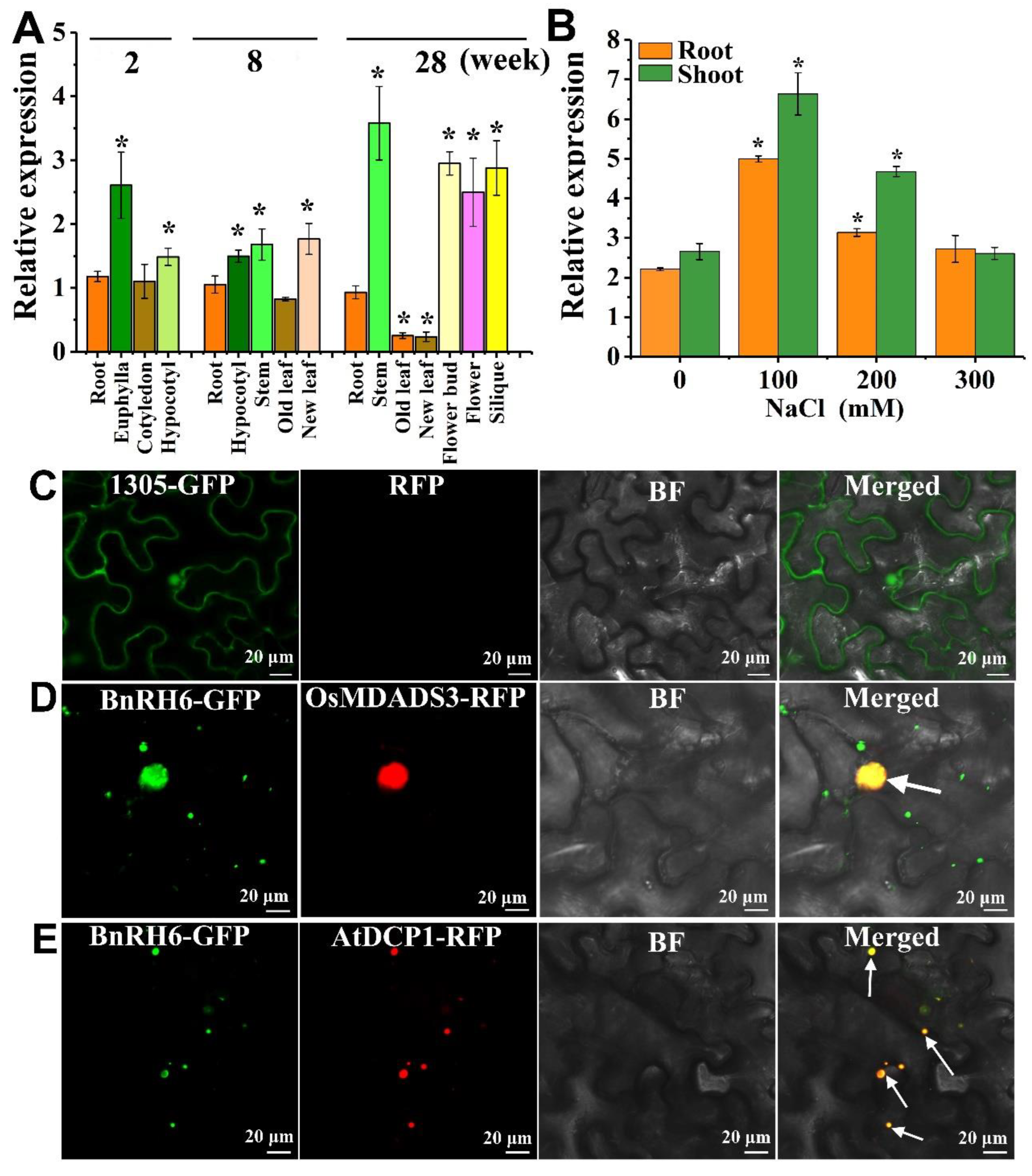
2.1. BnRH6 Is Transcriptionally Upregulated under Salt Stress

Two homologues, BnRH6-1 (DEAD-box RNA helicase 24, BnaA04g26450D, named BnRH6 in this article) and BnRH6-2 (DEAD-box RNA helicase 100, BnaC04g50480D), were identified in Brassica (Figure S1) [16,20]. Alignment of amino acids of BnRH6 revealed that there are typical DEAD-box RNA helicase domains with high similarity within Brassica and Arabidopsis (Figure S1B). RH6s have two homologs, RH8s and RH12s (Figure S1C), which is very similar to those of the Arabidopsis species, indicating that BnRH6s are highly conserved in Brassicaceae plants including Brassica and Arabidopsis.
BnRH6 was transcriptionally expressed in various tissues or organs throughout the lifespan. In young seedlings, BnRH6 is dominantly expressed in euphylla and hypocotyl and moderately in other tissues such as root and cotyledon (Figure 1A). At early developmental stages, BnRH6 is evenly expressed in all tissues. During flowering and seed developing stage, BnRH6 showed a higher expression in stems, flowers, and siliques but a relatively lower expression in leaves (Figure 1A). To examine whether BnRH6 was induced by salt stress, two-week-old seedlings were treated with NaCl at 100, 200, and 300 mM. When plants were exposed to 100 mM NaCl for 6 h, the BnRH6 transcript levels in roots and shoots were increased by 2.5- and 2.6-fold higher than those of the control, respectively (Figure 1B). At 200 mM NaCl, BnRH6 expression declined but remained at a significantly higher level over the control. These results indicated that BnRH6 is transcriptionally induced by salt stress.

2.2. BnRH6 Is Localized to the Nucleus and Cytoplasm

We further examined the subcellular localization of the BnRH6 protein. The BnRH6 coding sequence was amplified and fused to the green fluorescent protein (GFP) by linking the N- or C-terminal of BnRH6. The fusion and GFP alone were further ligated to a vector with a 35S promoter to drive its expression. In the meantime, two fluorescent marker proteins including MADS3 targeted to the nucleus and AtDCP1 to the cytoplasm were obtained. The vectors were transferred into the leaf cells of tobacco. GFP alone served as a technical control (Figure 1C). The BnRH6-GFP fusions with green signals and marker proteins with red signals were dominantly expressed in the same regions of the nucleus and cytoplasm. The overlapping colors merged to create yellow ones (Figure 1D,E), indicating that BnRH6-GFP can be targeted on both sides. Since AtDCP1 was reported as a P-body marker [21], there is a likelihood that BnRH6 is associated with P-body.

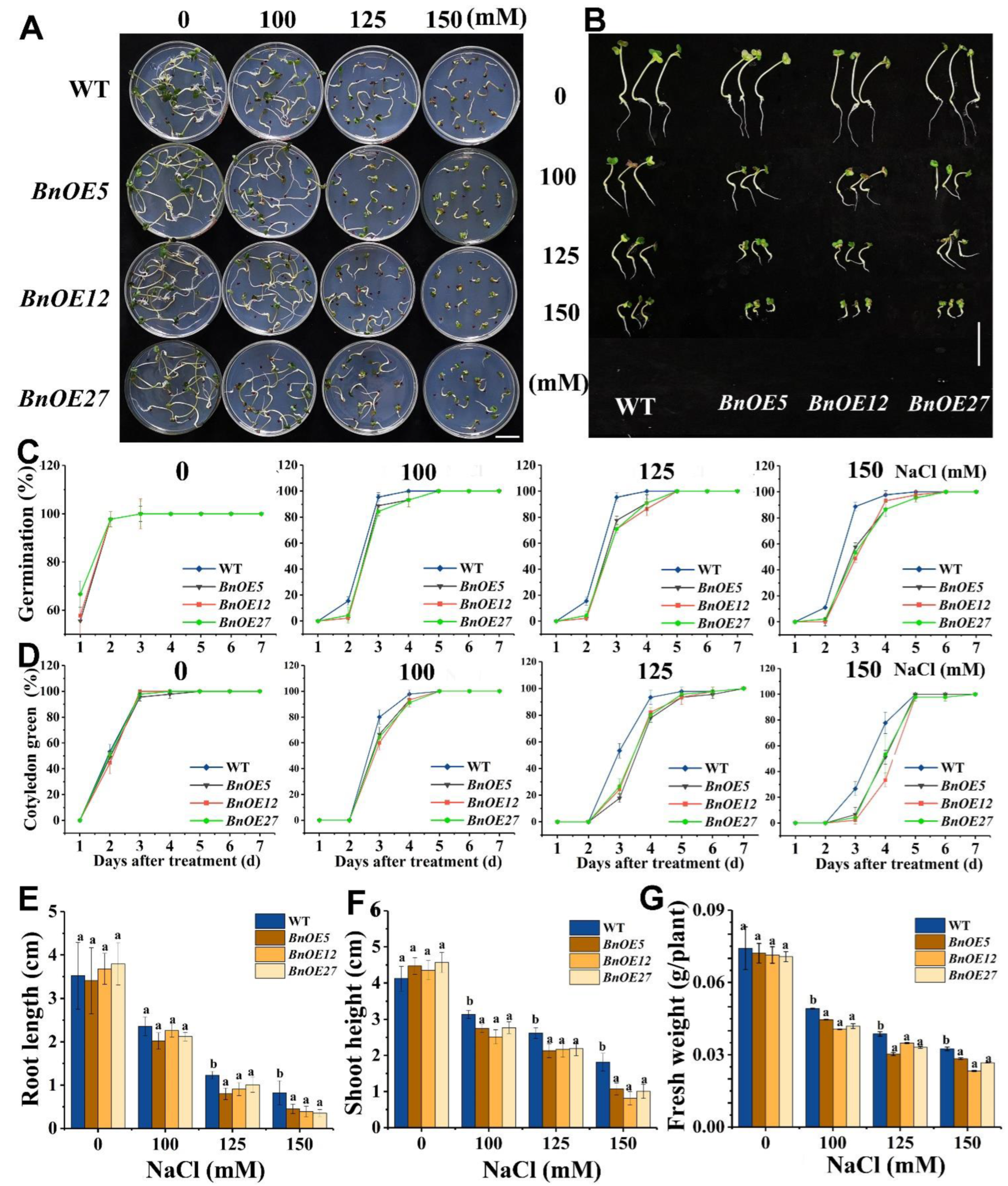
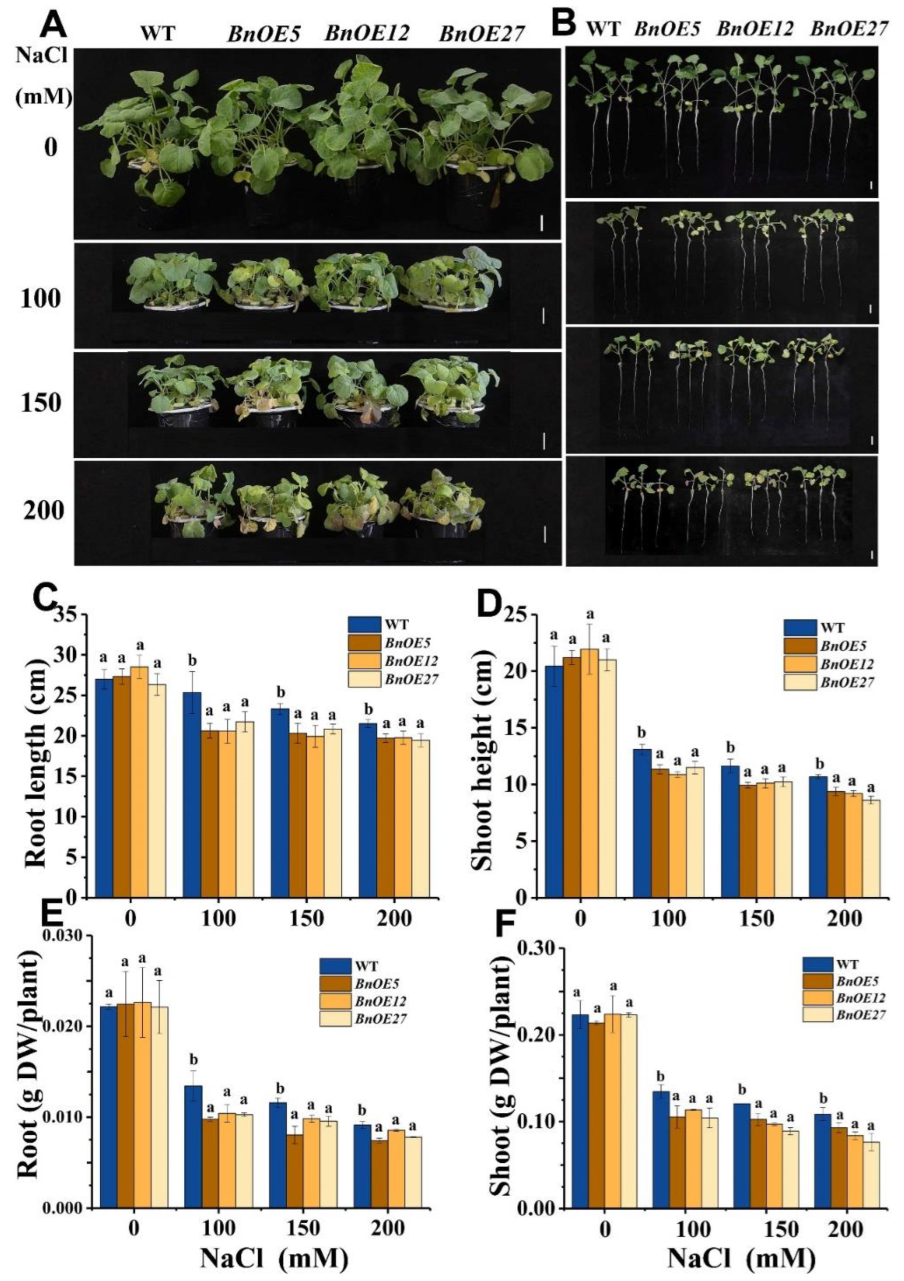
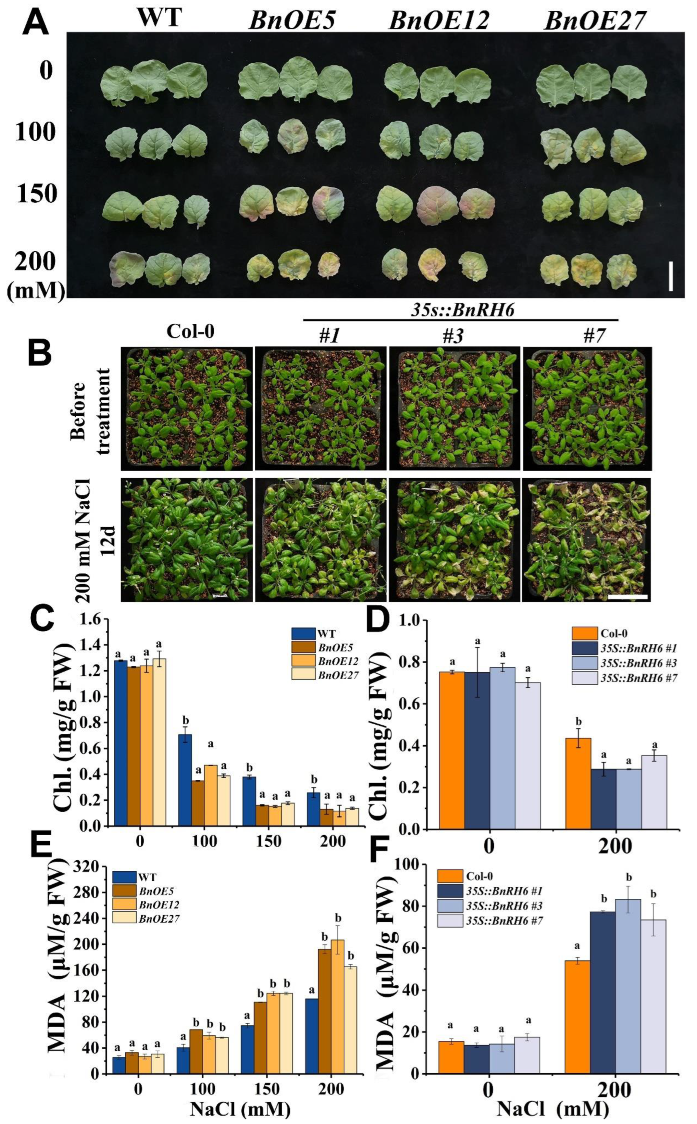
2.3. BnRH6 Overexpressed Brassica and Arabidopsis Display Salt Sensitivity

To evaluate the role of BnRH6 under salinity stress, we generated BnRH6-overexpressing Brassica (BnOE) and Arabidopsis (AtOE) lines (Figure S2A–D). Three transformants of each BnOE and AtOE plant (T3 Homozygotes) were randomly selected, genotyped at DNA levels (Figure S2A,C) and determined at transcriptional levels (Figure S2B,D). No obvious differences in phenotypes were observed between wild-type (WT) and BnOE lines without NaCl treatment (Figure 2A,B). When exposed to 100, 125, and 150 mM NaCl, however, the BnOE plants were more sensitive to salt stress than WT (Figure 2A,B). Consistently, the seed germination ratio between WT and BnOE plants remained unchanged in the absence of NaCl, while following a three-day exposure to 100, 125, and 150 mM NaCl, the seed germination ratio of BnOE lines decreased to 55–85% of the WT, respectively (Figure 2C). Similar responses were observed for the cotyledon greening rate (Figure 2D). Nearly all WT and BnOE seeds exhibited cotyledon greening on the 3rd day without NaCl, while more leaf chlorosis symptoms were observed in BnOE plants than in WT under NaCl exposure (Figure 2D). Furthermore, the growth of root and shoot and fresh biomass of BnOE seedlings was more severely inhibited under salt stress compared with that of WT (Figure 2E–G). With regard to AtOE, similar responses concerning seed germination, cotyledon greening, root elongation, and biomass were observed (Figures S3 and S4). These results suggest that BnRH6-overexpression repressed seed germination under salt stress.
We conducted an additional study with 10-day-old BnOE and its WT plants treated with NaCl. While both varieties showed no evident growth difference under normal conditions, the BnOE plants displayed compromised growth status relative to WT (Figure 3A,B). The root length and shoot height of BnOE plants were reduced by 8.14–18.79% and 11.97–19.34% under NaCl treatment, and the dry weight of root and shoot of BnOE seedlings dropped by 14.47–23.47% and 15.05–29.69%, respectively (Figure 3C–F). Likewise, the AtOE lines displayed more growth defects in root elongation and shoot growth than WT plants (Figure S4). These results pointed out that overexpression of BnRH6 could make the plant vulnerable to salt stress.
Chlorophyll and malondialdehyde (MDA) concentrations are typical physiological indicators reflecting plant growth status and stress-induced generation of peroxides under salinity stress, respectively [22,23]. As shown in (Figure 4A), when BnOE and WT were exposed to NaCl, the leaf of BnOE lines became much more chlorotic, particularly at higher degrees of salt stress (Figure 4A). The leaf phenotype in AtOE plants with salt treatment showed a similar chlorosis symptom (Figure 4B). Assessments of chlorophyll revealed that the chlorophyll concentrations in BnOE and AtOE plants significantly decreased as compared with those of wild-type (Figure 4C,D). Meanwhile, measurements of MDA showed that the MDA concentrations were significantly increased in the transgenic plants (Figure 4E,F). These results suggest that BnRH6 overexpression could also impair the physiological response under salt stress.

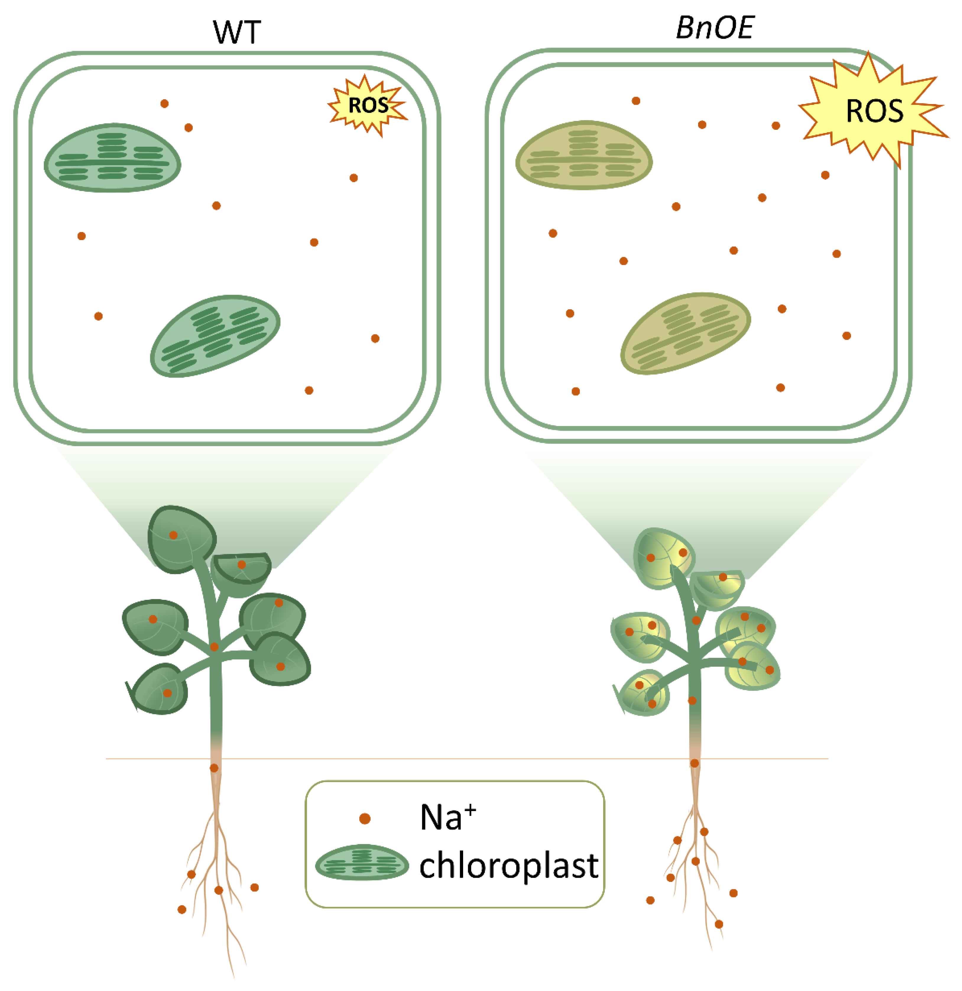
2.4. BnRH6 Overexpression Promotes Na+ Accumulation in Plants

To identify whether the BnRH6-mediated compromised growth and physiological response is associated with the accumulation of Na+ in plants, concentrations of Na ions in roots and shoots were separately determined in the transgenic and wild-type plants. An ICP-MS analysis showed that Na+ levels were significantly higher in both roots and shoots of the BnOE than in WT plants, especially at the high level of NaCl (200 mM) (Figure 5A,B). The enhanced Na+ levels were also detected in roots and shoots of the AtOE lines, where the Na+ concentrations increased by 61.5–73.1% and 14.3–33.3% compared with wild-type, respectively (Figure S5A,B). Since the ratio of K+/Na+ is commonly used as one of the important physiological responses for plant tolerance to salt stress [24], we simultaneously measured the K ions in the plants. The K+ concentrations were comparable between the overexpression lines and wild-type (Figure 5C,D and Figure S5C,D). However, calculations of the K+/Na+ ratio showed that the ratio declined in the BnOE and AtOE plants compared with the wild-type plants. The significant differences between the transgenic plants and wild-type occurred at 150–200 mM of NaCl (Figure 5E–G and Figure S5E,F). In the shoot of the BnOE plants, the K+/Na+ ratio decreased by 18.3–28.6% compared with wild-type (Figure 5G). The results suggest that overexpression of BnRH6 results in the over-accumulation of Na ions.

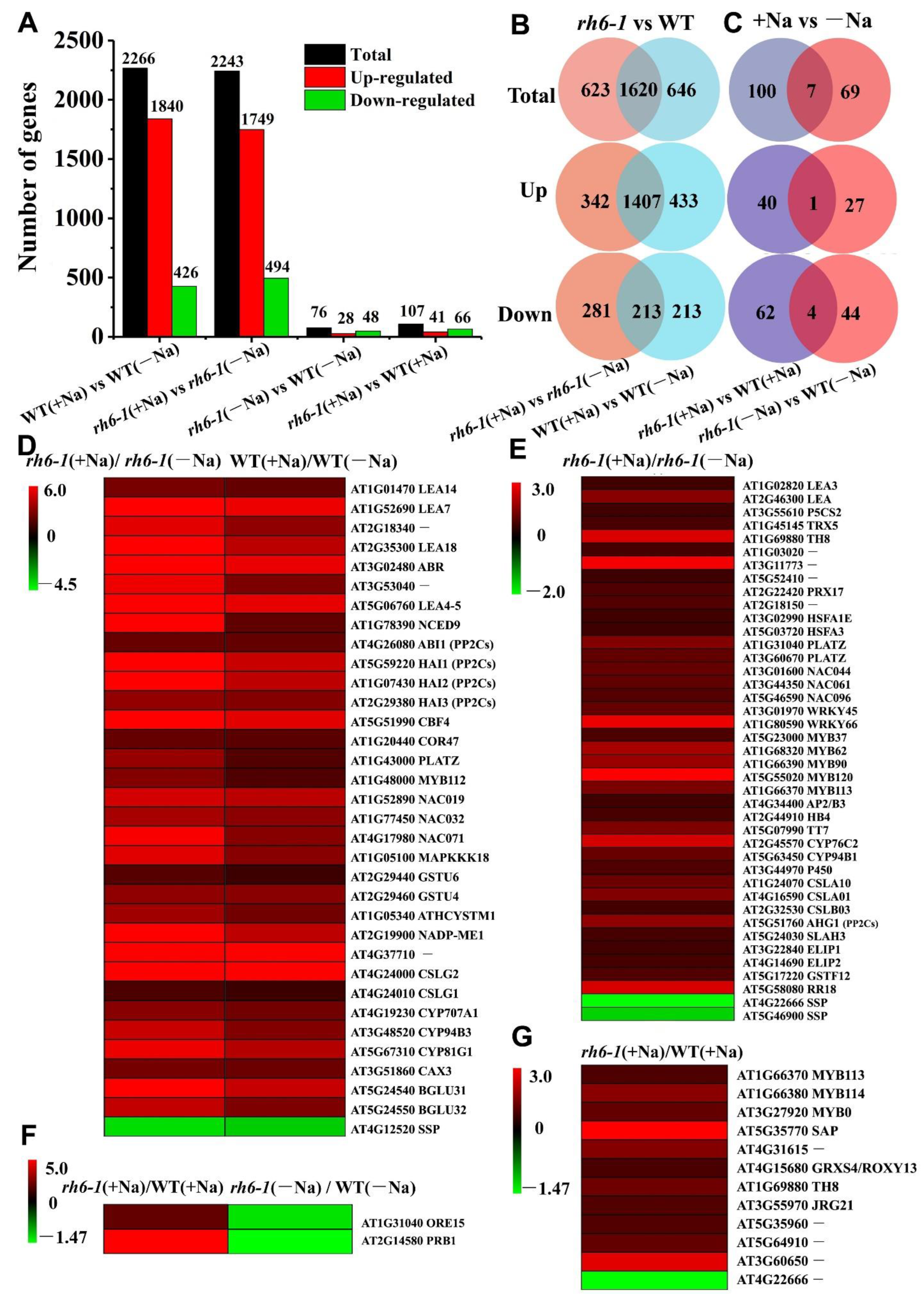
2.5. Suppression of RH6 Enhances Plant Tolerance to Salt Stress by Upregulating the Expression of Salt Stress-Tolerant Genes in Arabidopsis

To figure out the mechanisms or pathways responsible for plant tolerance to salinity regulated by RH6, we performed a genome-wide identification of transcriptomes by RNA sequencing. Due to the lack of desirable knockout lines of Brassica, we took an advantage of an Arabidopsis knockout mutant rh6-1. The rh6-1 mutant with a T-DNA insertion in an exon near 5′-UTR was identified by PCR (Figure S2E–G). We applied the atrh6-1 for the study because both BnRH6 and AtRH6 are highly conserved, by which the amino acid sequences between BnRH6 and AtRH6 share an 85.72% similarity, and the key domains, DEAD domain (ATPase domain including Q, I, Ia, Ib, II and III motif) and HELIC domain (RNA binding domain including III, IV and VI motif) between BnRH6 and AtRH6 share similarities of 98.98% and 97.56%, respectively (Figure S1B). Furthermore, the rh6-1 mutant displays an enhanced salt-tolerance phenotype with better growth (longer root length and greater biomass) compared with the wild-type (Col-0) (Figure S6), confirming that both AtRH6 and BnRH6 should be negative regulators of plant response to salt stress.
Four RNA libraries regarding rh6-1 and WT connecting -Na or +Na were constructed in terms of WT(−Na), WT(+Na), rh6-1(−Na), and rh6-1(+Na) with three biological replicates (4 × 3 samples) and sequenced by high-throughput RNA-sequencing using Illumina technology. The quality of the sequencing datasets was statistically evaluated, and it turned out to be reliable (Table S1 and Table S2; Figure S7). To validate the expression pattern of the genes determined by RNA-seq, a total of 21 genes were selected for qRT-PCR validation. Expression of all genes showed a pattern identical to the RNA-seq (Figure S8).
The differentially expressed genes (DEGs) (>2-fold change, p< 0.05) from the group pairs were identified, and the number of each pair was presented (Figure 6A,B, Table S3). The total number of DEGs (including up and down genes) between the two datasets WT(+Na)/WT(−Na) and rh6-1(+Na)/rh6-1(−Na) were comparable, while the number of DEGs between rh6-1(−Na)/WT(−Na) and rh6-1(+Na)/WT(+Na) were drastically changed (Figure 6A). Venn diagram analysis revealed that 342 DEGs were specifically upregulated and 281 DEGs were downregulated in rh6-1(+Na)/rh6-1(−Na), whereas 433 DEGs were specifically induced and 213 DEGs were repressed in WT(+Na)/WT(−Na) (Figure 6B). Further cross-analyses between rh6-1 and WT showed that 40 DEGs were specifically induced and 62 DEGs were suppressed in the rh6-1(+Na)/WT(+Na) dataset, while 27 DEGs were specifically upregulated and 44 DEGs were repressed in the rh6-1(−Na)/WT(−Na) pair (Figure 6C). Notably, compared to the minus sodium (−Na), treatment with plus sodium (+Na) induced 1.48-fold (40/27) more DEGs in rh6-1(+Na)/WT(+Na) than in rh6-1(−Na)/WT(−Na) datasets; on the other hand, the rh6-1(+Na)/WT(+Na) group contained 1.41-fold (62/44) more downregulated DEGs in rh6-1(+Na)/WT(+Na) than in rh6-1(−Na)/WT(−Na) (Figure 6C). These results point out that the mutation of RH6 would tend to alter the pattern of more gene expressions under salt stress.
We then performed Gene Ontology (GO) analysis and placed the DEGs into some functional categories related to salt stress response. While under the −Na condition, many DEGs in the rh6-1 mutant filled into the development of the biological process, and the DEGs of rh6-1(+Na) vs. rh6-1(−Na) were found with enrichment in the categories such as stimuli, chemical stress, and hormone response (Figure S9). It was important to find that in the dataset of rh6-1(+Na)/WT(+Na), the DEGs were also classified into stress responses and different kinds of metabolic processes (Figure S9). These results indicate that under salt stress the rh6-1 function most likely would shift from the developmental state to the stress responsive model by post-transcriptional regulation. The Kyoto Encyclopedia of Genes and Genomes (KEGG) refers to the genes categorized into specific pathways. The DEGs in the dataset rh6-1(−Na)/WT(−Na) were concentrated in the pathways of RNA degradation, MAPK and hormone-signaling pathway, metabolite biosynthesis, and plant–pathogen interaction (Figure S10). Similar results were found in the rh6-1(+Na)/WT(+Na) and rh6-1(+Na)/rh6-1(−Na) datasets, in which some DEGs participated in the stress signal, metabolite biogenesis, and RNA metabolism pathways (Figure S10). These results suggest that mutation of RH6 led to many stress-responsive genes being involved in the salt resilience process.
We further specified the DEGs in the datasets that were regulated in the rh6-1 mutant line under salt exposure. Four heatmaps were drawn based on the expression differences of the DEGs between the groups. In the datasets of WT(+Na)/WT(−Na) and rh6-1(+Na)/rh6-1(−Na), there were 34 salt-responsive genes in response to salt stress. Of these, 33 genes were upregulated more in rh6-1 compared with WT (Figure 6D, Table S4). Of those, six genes encoding transcription factors including MYB112, NAC019, NAC032, NAC071, PLATZ, and CBF4 were detected; four genes NCED9, ABR, ABI1, and HAIs (ABA pathway) were associated with phytohormone synthesis or signals; one gene encoding kinase involved MAPK cascades (MAPKKK18); two genes encoded antioxidative enzymes (GSTUs); one gene encoded an ion exchange protein (CAX3); and the rest of them were stress-related genes,including Late Embryogenesis-Abundant proteins (LEAs) (LEA14, LEA7, AT2G18340, LEA18, ABR, AT3G53040, COR47 and LEA4-5) and CYP450s (CYP707A1, CYP94B3 and CYP81G1) (Figure 6D, Table S4). One Seed Storage Protein (SSP) gene was downregulated more in rh6-1 relative to WT (Figure 6D, Table S4). In particular, 39 salt-related genes were uniquely upregulated in rh6-1(+Na)/rh6-1(−Na) but not in WT(+Na)/WT(−Na) (Figure 6E, Table S5), including 14 transcription factor genes (2 HSFAs, 2 PLATZs, 3 NACs, 2 WRKYs and 5 MYBs), an ABA responsive gene (AHG1), four CYP450s, seven encoding antioxidative enzymes (TH8, TRX5, AT1G03020, AT3G11773, AT5G52410, AT2G22420 and AT2G18150), one gene encoding enzyme in proline synthesis (P5CS2), one gene encoding ion channel (SLAH3), two LEA. Most of them have been reported to involve salt or abiotic stress responses. Two DEGs involved in salt stress response were also found in datasets of rh6-1(+Na)/WT(+Na) and rh6-1 (−Na)/WT(−Na) (Figure 6F, Table S6).

3. Discussion

3.1. Expression of BnRH6 Compromises Growth of Plants under Salt Stress

In this study, we report that BnRH6 is a new regulator in salt stress response by downregulating salt-tolerant genes. BnRH6 was transcriptionally expressed in various tissues or organs all lifelong, indicating a potential role in all stages (Figure 1A). This wide expression pattern is in agreement with its RH6 in Arabidopsis and other RNA helicases [25,26,27,28]. Moreover, BnRH6 was localized in the nucleus and cytoplasm (most likely in P-body) (Figure 1C–E), suggesting potential functions on both sides. This is reminiscent of RH6 in Arabidopsis, which plays roles in both the nucleus and cytoplasm [19,25]. There is a likelihood that BnRH6 is associated with P-body, similar to the report on AtRH6 [25].
The transcript levels were increased drastically in the shoot and root of Brassica with NaCl exposure, implying a possible role in salt stress response (Figure 1B). Several reports have indicated that the expression of RNA helicases correlated with salt stress response can be regulated by salinity stress in multiple species including Arabidopsis, rice, cabbage, tomato, barley, and so on [28,29,30,31,32]. However, not all salt stress response-related RNA helicases can be induced at the transcript level. For instance, Arabidopsis AtRH17 is a positive regulator to plant salt stress tolerance, whereas the AtRH17 transcript level was not affected under salinity stress [33]. Furthermore, a certain number of RHs participate in more than one kind of abiotic stress [28,29,30,32,34]. Whether BnRH6 expression can be altered by other stresses needs to be further investigated.
Genetic and functional investigation revealed that BnRH6 overexpression in both Brassica and Arabidopsis led to plant sensitivity to salt stress, in terms of a decreased germination ratio and cotyledon greening rate, weakened growing status, and more sensitive physiological reactions. This scenario is consistent with our RNA-seq data. BGLU31 and BGLU32 encode β-glucosidases as positive factors for seed germination [35] and were found to be upregulated in rh6-1 under salt stress (Figure 6D). Notably, the gene SSP negatively mediating seed germination was found to be downregulated in rh6-1 plants (Figure 6D,E) [36]. Several classes of growth-regulating genes, such as ELIP1 and ELIP2 encoding chlorophyll a/b binding family protein genes and ARR18 encoding response regulator genes, were all upregulated in the rh6-1 mutant lines (Figure 6E). Thus, BnRH6 affects the salt stress response likely via indirect control of the expression pattern of those genes.
Overload of NaCl into plants initially triggers osmotic stress and later ion imbalance and toxicity [37,38]. Proline is an osmotic regulator induced by salt stress. One gene encoding an enzyme (P5CS2) in the proline synthesis was specifically upregulated in salt-exposed rh6-1 plants (Figure 6E). Our data showed that the Na+ accumulation was increased in shoots and roots of BnRH6 overexpression lines. Even though the K+ accumulation was not altered in overexpression lines, a relatively lower K+/Na+ ratio was generated (Figure 5F,G). The Na+ accumulation and K+/Na+ ratio was barely discussed in RNA helicase gene mutants or overexpression plants involved in the salt-stress response. Similar changes have been evident in other salt-stress regulator mutants or overexpression lines. For instance, AtDIF1 overexpression Arabidopsis conferred plant tolerance to salt stress by reducing the content of Na+ and loss of K+ [39]. SERF1 is a positive regulator for salt stress tolerance in rice. Loss-of-function or the knock-down mutant of SERF1 showed an increased Na+ accumulation and a higher Na+/K+ ratio [40].
Our RNA seq data showed that one ion channel gene, CAX3 was induced in rh6-1 under salt stress (Figure 6D,E). A previous study showed that CAX3 can be induced by Na+ treatment. CAX3 is a Ca2+/H+ vacuolar antiporter for plant tolerance to salt under Na+ stress. The cax3 mutant confers decreased P-ATPase activity, which further decreased Na+ efflux [41]. On the contrary, an enhanced expression of CAX3 may promote Na+ efflux and benefit the maintenance of a high K+/Na+ ratio. Apart from the notorious detriment caused by Na+, the accompanied Cl poisoning is also fatal [42,43]. SLAH3 is a Cl efflux transporter [44]. Transcriptional SLAH3 was downregulated by NaCl treatment [45]. An increased expression of SLAH3 in rh6-1 under NaCl exposure was recovered from our RNA-seq data, which may decrease the Cl accumulation. These results suggest that CAX3 and SLAH3 could play critical roles in ion homeostasis under salt stress.

3.2. Mutation of rh6-1 Confers Plant Salt Tolerance by Regulating Salt-Resilient Genes

To identify the downstream genes and possible regulation pathways governed by RH6 under salt stress, we performed a genome-wide analysis of transcriptome across the rh6-1 and Col-0 plants. While a large number of DEGs were expressed in the rh6-1 and wild-type plants under salt stress, there were many DEGs across the two cultivars exposed to NaCl (>2.0-fold change, p < 0.05). The largest group of specific DEGs was associated with genes encoding transcription factors (TFs), whose expression patterns were altered in the rh6-1 mutant. Nine MYB genes were found to be upregulated in rh6-1 mutant after salt treatment. MYB proteins play a fundamental role in numerous aspects of plant growth, development, and stress responses [46,47]. MYB80, MYB113, and MYB114 were upregulated in rh6-1 plants relative to WT only under salt stress (Figure 6G, Table S7). MYB113 and MYB114 have been reported to promote anthocyanin synthesis in Arabidopsis and pear for defense against abiotic and biotic stresses [48,49]. MYB112 was strongly expressed in rh6-1 under salt stress (Figure 6D) and is also required for anthocyanin accumulation under salinity and high light stress [50]. Several other gene members including MYB37, MYB62, MYB90, and MYB120 were also induced in rh6-1 under NaCl stress (Figure 6E). Some of them are also involved in abiotic stress responses [51,52,53].
Several NAC (NAM, ATAF1/2 and CUC2) genes were detected in rh6-1 plants under salt stress including NAC019, NAC032 and NAC071 (Figure 6D) and NAC044, NAC061, and NAC096 (Figure 6E), and most of them have been reported to involve salt and other abiotic stress response [54,55]. Arabidopsis harboring an overexpressed GmNAC019 increased drought and salinity tolerance [56]. NAC061 is induced by AtHSFA7b (a heat shock transcription factor in Arabidopsis) and positively regulates salt tolerance [57]. In addition to MYB and NAC, several other TF family genes such as WRKY45 and CBF4 identified in the study are also reported to regulate plant response to salt stress [58,59]. Whether BnRH6 directly or indirectly interacts with TFs remains to be investigated.
Plant hormones are the major regulators of abiotic stress responses [60]. ABA has been long considered to be a crucial driver of plant abiotic stress, especially salinity and dehydration stress responses [61,62]. In this study, seven ABA signaling pathway genes for NCED9, ABI1, HAIs, AHG1, MAPKKK18, and CYP707A1 were identified as salt-responsive DEGs. Arabidopsis NCED9 (9-cis-epoxycarotenoid dioxygenase 9) is essential for ABA biosynthesis [63,64]. Expression of NCED9 is induced by salt, drought, and cold in Brassica oleracea [65]. Several DEGs encode ABI1, AHG1, HAI1, HAI2, and HAI3, and these proteins are phosphatases 2Cs (PP2Cs) responsible for ABA signal transduction [66,67]. CYP707A1 and MAPKKK18 were also involved in ABA-related stress responses [68,69,70]. This is reminiscent of some recent reports that other DEAD-box RNA helicases such as BnRH37 improved abiotic stresses of plants by inducing the expression of ABA biosynthesis-related genes or ABA-dependent stress-response genes [33,71,72], but no report was published for BnRH6.
It is well known that salinity stress is usually accompanied by oxidative stress [73,74]. Under salt stress, some anti-oxidative enzymes such as glutaredoxins and thioredoxins actively participate in the removal of salt stress-induced ROS and toxicity [75]. Five genes encoding ROS scavengers were upregulated in rh6-1 plants. For example, GRXS4/ROXY13 and TH8 were transcriptionally upregulated in rh6-1 mutants under NaCl stress. GRXS4 belongs to the CC-type glutaredoxin (ROXY) protein family, and was reported to be induced by salinity and drought stresses [76,77]. Both TRX5 (thioredoxin H-type 5) and TH8 (thioredoxin H-type 8) encode thioredoxins and can be induced by multiple biotic and abiotic stresses [78]. GSTU4 and GSTU6 are tau class glutathione S-transferases; GSTU6 was reported to contribute to Cd stress tolerance by regulating intracellular ROS homeostasis in rice [79]. Overexpressing GmGSTU4 displays salt tolerance in tobacco through peroxidase activity and detoxification mechanisms [80,81]. In addition, genes indirectly participating in ROS scavenging were screened. For example, PtrLEA7, a gene encoding a Late embryogenesis-abundant (LEA) protein, can be slightly induced by salt stress; overexpressing PtrLEA7 in tobacco and Poncirus trifoliata positively regulates drought tolerance through enhancing antioxidant capacity [82]. Similarly, an RNA helicase VviDEADRH25 in grapevine plays a role as a negative regulator to drought stress tolerance. Overexpressing VviDEADRH25 in Arabidopsis decreased the expression of genes encoding ROS scavenging enzymes [83]. These results suggest that rh6-1-mediated plant tolerance to salt stress would be likely through these homolog genes.
Apart from the subsets above, we also detected some other protein family genes involved in the RH6-mediated pathways under salt stress. LEA proteins belong to a large hydrophilic protein family with regulatory roles in plant stress responses [84]. We found that LEA4-5, LEA7, LEA14, and LEA18 were significantly upregulated in rh6 under salt stress. LEA14 was previously reported to confer salt stress tolerance by stabilizing an E3 ligase in Arabidopsis [85]. LEA3 was induced only in rh6 but not in WT under Na stress, suggesting that LEA3 was most likely involved in the salt stress response. Supportive evidence comes from the rice LEA3 because the OsLEA3-overexpression line endowed drought resistance in the plant [86]
These results suggest that the RH6 is likely a master regulator of RNA biosynthesis and metabolism under salt stress. Based on those results, we proposed a model of salt stress response in WT and BnOE Brassica (Figure 7). Our research found a new function of BnRH6, and it may be a promising molecular tool for engineering salt-tolerant Brassica and enriching Brassica varieties.

4. Materials and Methods

4.1. Plant Growth and Salt Treatments

Seeds of Brassica (genotype: Westar) were surface-sterilized in 75% ethanol alcohol for 5 min and 10% NaClO for 15 min with gentle shakes, followed by thoroughly rinse with sterile water. The clean seeds were placed on a plastic net floating on the 1/2 Hoagland solution. After germination at 25 °C in the dark for two days, seeds were grown under the condition of 22 °C, 200 μmol m−2s−1 light intensity and 14 h/10 h(light/dark) photoperiod [87]. Plant tissues at different stages were harvested for BnRH6 expression pattern analysis. Two-week-old Brassica seedlings were exposed to 1/2 Hoagland solution (pH = 5.8) with different NaCl concentrations (0, 100, 200, and 300 mM) for 6 h based on previous reports [88]. Shoots and roots were harvested separately for expression analysis.
Seeds of Arabidopsis (Col-0) and a rh6-1 T-DNA insertion mutant line (SALK_ 205997C, Columbia background, obtained from the Arabidopsis Biological Resource Center) were sterilized and sowed on ½ Murashige and Skoog (MS) medium supplemented with 3% sucrose and 0.9% agar. After two-day vernalization at 4 °C in darkness, seeds were grown in a growth chamber with 22 °C, 150 μmol m−2s−1 light intensity and 16/8 h (light/dark) photoperiod.

4.2. Vector Construction and Transgenic Plants

Full-length CDS sequence of BnRH6 (Bn04g26450D) was cloned using specific primers (Table S8) and inserted into the pCAMBIA1300 vector with CaMV35S as the promoter [89]. The sequence-confirmed construct was transformed into Agrobacterium tumefaciens strain EHA105 following the standard method of transformation [90]. The Agrobacterium-mediated plant transformation was performed with Brassica hypocotyl sections [87] for Brassica plants and floral dip method for Arabidopsis plants [88,91]. Transformants were selected with 100 and 50 mg/L kanamycin, respectively. More than fifteen independent transgenic lines (T1 generation) were obtained and genotyped by PCR. Three of them were randomly selected for propagation. The homozygous lines (T4 generation) were used in this study. For cloning of BnRH6-GFP, full-length CDS sequence of BnRH6 without stop codon was fused into pCAMBIA1305 backbone through Xba/IBamH I digestion and T4 ligation.

4.3. Analysis of Transcripts by RT-PCR and qRT-PCR

Total RNA was extracted with Trizol (Invitrogen). Total RNA was treated with DNase I (Transgen, Beijing, China). The first-strand cDNA was generated by reverse transcription with EasyScript First-Strand cDNA Synthesis SuperMix (Transgen, Beijing, China) by Oligo(dT) RTprimer (mRNAs) according to the manufacturer’s instructions. For regular PCR reactions, PCR products were fractioned in 1% agarose gels. For qRT-PCR, the amplification systems were processed in a 20 μL solution with 10 μL TransStart Green qPCR SuperMix (Transgen, Beijing, China), 4 ng template, and 0.8 μL (10 μmol) primers (Table S8). The reactions were run at 95 °C for 10 min, followed by 40 cycles of denaturation at 95 °C for 10 s and annealing at 60 °C for 1 min in the 7500 Real-Time PCR System (Applied Biosystems) [16]. Brassica Actin2 serves as an internal control. All primers used are listed in Table S8 (Table S8). Primers used for genotyping were designed on http://signal.salk.edu/tdnaprimers.2.html (accessed on 14 December 2022). AtRH6-qRT primers were synthesized according to [25]. AtActin2 primers were synthesized according to [19]. BnActin2 primers were synthesized according to [92]. Other primers were designed with Primer Premier6.

4.4. Subcellular Localization Analysis

The pCAMBIA1305 (negative control), pCAMBIA1305-BnRH6-GFP, OsMDADS3-RFP (nucleus marker), and AtDCP1-RFP (P-body marker) were constructed and transformed into Agrobacterium tumefaciens strain GV3101. Agrobacterium tumefaciens containing different vectors were cultivated overnight and then suspended in the inoculation buffer (150 mM acetosyringone, 10 mM MgCl2, and 10 mM MES, pH 5.7) when the value of OD600 reached 1.0. Combinations of A. tumefaciens with different constructs were injected into four-week-old tobacco (Nicotiana benthamiana) leaves with a needleless syringe [90]. After three-day agroinfiltration, transiently expressed leaves were photographed with a confocal laser scanning microscope (LSM780, Zeiss, Oberkochen, Germany).

4.5. Determination of Salt Stress Responses

For germination and cotyledon greening analysis, sterilized WT and BnOE Brassica seeds were placed on ½ MS medium containing 0, 100, 125, and 150 mM NaCl [93,94]. The germination proportion (emerged radicle) and cotyledon greening (expanded cotyledon turning green) were recorded from the 1st day (the day exposed to light) to the 7th day. Images were photographed on the 7th day. Sterilized WT and AtOE Arabidopsis seeds were placed on ½ MS medium containing 0, 50, 75, and 100 mM NaCl [95]. The germination proportion (emerged radicle) and cotyledon greening (expanded cotyledon turning green) were recorded and photographed on the 4th day and 7th day, respectively [96].
For root/shoot length and dry weight, ten-day-old Brassica plantlets were treated with ½ Hoagland solution containing 0, 100, 150, and 200 mM NaCl for 14 days. Sterilized WT and AtOE Arabidopsis were grown on ½ MS medium with 0, 50, 75, and 100 mM NaCl for 12 days. Sterilized WT and rh6-1 Arabidopsis seeds were placed on ½ MS medium containing 0, 45, and 70 mM NaCl for 14 days. The plates were vertically placed in a conditioned chamber. The root and shoot length were measured with a ruler, and the fresh mass and dry mass were weighted as described previously. [39,88,89,97].

4.6. Determination of Physiological Responses

For measuring seed germination, the surface-sterilized seeds of Brassica were placed on the solid 1/2 MS medium with the gradient NaCl concentrations at 0, 100, 125, and 150 mM. The germinated seeds (the seeds with emerged radicles) and the seeds with green cotyledons were counted every day from the 1st day to the 7th day. Surface-sterilized Arabidopsis seeds were sowed on solid 1/2 MS medium with the gradient NaCl concentrations at 0, 50, 75, and 100 mM. The germinated seeds (the seeds with emerged radicles) and the seeds with green cotyledons were counted on 4th day and 7th day, respectively. The calculation of seed germination ratio and cotyledon greening rate refers to [96].
For chlorophyll and MDA concentration measurements, Brassica seeds were hydroponically grown in ½ Hoagland solution for ten days and then grown in ½ Hoagland solution supplied with NaCl gradient from 0 to 100, 150, and 200 mM for 14 days. Arabidopsis seeds were potted in mixed soil (vermiculite and flower nutrient soil, 3:1) and placed in the chambers. Three-week-old Arabidopsis plants were irrigated with 0 and 200 mM NaCl solution for 12 days [98,99]. Total chlorophyll from Brassica or Arabidopsis was extracted with 80% (v/v) acetone and soaked for 36 h at room temperature in the dark until the leaves fully faded. The absorbance at 663 nm and 645 nm was recorded by a spectrophotometer [100]. The malondialdehyde (MDA) concentration was determined using a Lipid Peroxidation MDA Assay Kit (S0131, Beyotime, Shanghai, China) according to manufacturers’ instructions.

4.7. Determination of Na+ and K+ Concentrations

Plant shoot and root tissues were separately harvested after NaCl treatment (the same as materials for chlorophyll and MDA measurements). The harvested tissues were immersed in 4 mM CaSO4 solution for 5 min. After being washed with deionized water, the samples were exsiccated in an oven at 72 °C for 72 h. The samples were digested with nitric acid, and the Na+ and K+ concentrations were determined by ICP-MS (PerkinElmer, Waltham, MA, USA) as previously described [39].

4.8. Construction of Salt Treated cDNA Libraries of Arabidopsis rh6-1 and RNA-Sequencing

Arabidopsis seeds of rh6-1 and wild-type (WT, Col-0) were sowed on solid ½ MS medium for 14 days before being transferred to 1/2 Hoagland solution. The plants were then subjected to 200 mM NaCl treatment (+Na) with a 0 mM NaCl treatment (−Na) as a control (CK) for 6 h.
Four groups of samples were harvested and total RNA was isolated. The samples were prepared as RNA-seq libraries in terms of WT(−Na), WT(+Na), rh6-1(−Na), and rh6-1(+Na). For each library, three biological samples with a total of 12 samples were prepared for RNA sequencing according to the previous method [101]. The Illumina RNA sequencing platform (HiSeq 2500) was applied. Data analyses were performed as described previously [89].

4.9. Statistical Analysis

The study was set up in biological triplicate. Each result shown in the figures was the mean of three replicated treatments, and each treatment contained at least 15–18 seedlings.
With regard to the assessment of survival rates, 30 plants were employed. The significant difference between treatments was assessed through analyses of variance post hoc test (ANOVA, Tukey’s test). The values of each assay, followed by an asterisk, are significantly different at p < 0.05. The data were analyzed using the statistical software package SPSS 22.0.

5. Conclusions

This study functionally characterized a DEAD-box RNA helicase family gene BnRH6 from Brassica. BnRH6 can be transcriptionally induced by salt stress. Overexpression of BnOE revealed plant hypersensitivity to salinity stress, in terms of reduced germination rate, cotyledon greening, root and shoot elongation, chlorophyll concentration, and increased MDA concentrations in plants. These results were well-confirmed by similar studies with transgenic Arabidopsis (AtOEs) expressing BnRH6. Furthermore, both the BnOE and AtOE lines accumulated more Na+ than the wild-type. RNA-seq analysis revealed that a subset of downstream salt-tolerant genes was regulated by RH6, indicating that RH6 negatively regulates salt stress response by adjusting the downstream salt-tolerant genes. However, the detailed molecular mechanisms for how BnRH6 regulates its downstream genes or proteins remain to be investigated.

Supplementary Materials

The following supporting information can be downloaded at: https://www.mdpi.com/article/10.3390/ijms24010002/s1.

Author Contributions

Methodology, X.Z.; J.S. and L.W.; software, J.S.; validation, J.S.; Z.M.Y. and D.S.; investigation, X.Z.; formal analysis, X.Z. and J.S.; resources, Z.M.Y.; writing—original draft preparation, D.S.; writing—review and editing, Z.M.Y. and D.S.; visualization, X.Z.; supervision, Z.M.Y. and D.S.; project administration, D.S.; funding acquisition, Z.M.Y. and D.S. All authors have read and agreed to the published version of the manuscript.

Funding

This research was funded by National Natural Science Foundation of China (grant number: 21377055) and Jiangsu Natural Science Foundation (grant number: BK20221023). The APC was funded by Jiangsu Natural Science Foundation (grant number: BK20221023).

Institutional Review Board Statement

Not applicable.

Informed Consent Statement

Not applicable.

Data Availability Statement

Not applicable.

Conflicts of Interest

The authors declare that they have no conflict of interest.

References

  1. Joseph, B.; Jini, D. Proteomic analysis of salinity stress-responsive proteins in plants. Asian J. Plant Sci. 2010, 9, 307. [Google Scholar] [CrossRef]
  2. Yang, Y.; Guo, Y. Elucidating the molecular mechanisms mediating plant salt-stress responses. New Phytol. 2018, 217, 523–539. [Google Scholar] [CrossRef] [PubMed] [Green Version]
  3. Tavakkoli, E.; Rengasamy, P.; McDonald, G.K. High concentrations of Na+ and Cl ions in soil solution have simultaneous detrimental effects on growth of faba bean under salinity stress. J. Exp. Bot. 2010, 61, 4449–4459. [Google Scholar] [CrossRef] [PubMed]
  4. Ahmad, P.; Jaleel, C.A.; Salem, M.A.; Nabi, G.; Sharma, S. Roles of enzymatic and nonenzymatic antioxidants in plants during abiotic stress. Crit. Rev. Biotechnol. 2010, 30, 161–175. [Google Scholar] [CrossRef]
  5. Golldack, D.; Li, C.; Mohan, H.; Probst, N. Tolerance to drought and salt stress in plants: Unraveling the signaling networks. Front. Plant Sci. 2014, 5, 151. [Google Scholar] [CrossRef] [PubMed] [Green Version]
  6. Jamil, A.; Riaz, S.; Ashraf, M.; Foolad, M. Gene expression profiling of plants under salt stress. Crit. Rev. Plant Sci. 2011, 30, 435–458. [Google Scholar] [CrossRef]
  7. De la Cruz, J.; Kressler, D. Linder Unwinding RNA in Saccharomyces cerevisiae: DEAD-box proteins and related families. Trends Biochem. Sci. 1999, 24, 192–198. [Google Scholar] [CrossRef]
  8. Ranji, A.; Boris-Lawrie, K. RNA helicases: Emerging roles in viral replication and the host innate response. RNA Biol. 2010, 7, 775–787. [Google Scholar] [CrossRef] [Green Version]
  9. Tanner, N.K.; Linder, P. DExD/H box RNA helicases: From generic motors to specific dissociation functions. Mol. Cell 2001, 8, 251–262. [Google Scholar] [CrossRef]
  10. Linder, P.; Owttrim, G.W. Plant RNA helicases: Linking aberrant and silencing RNA. Trends Plant Sci. 2009, 14, 344–352. [Google Scholar] [CrossRef]
  11. Yadav, S.; Tuteja, N. Chapter 4-Evolution of RNA Helicases in Plants: Molecular and Functional Insights. In Helicases from All Domains of Life; Tuteja, R., Ed.; Academic Press: Cambridge, MA, USA, 2019; pp. 53–75. [Google Scholar]
  12. Pandey, S.; Prasad, A.; Sharma, N.; Prasad, M. Linking the plant stress responses with RNA helicases. Plant Sci. 2020, 299, 110607. [Google Scholar] [CrossRef] [PubMed]
  13. Gong, Z.; Dong, C.-H.; Lee, H.; Zhu, J.; Xiong, L.; Gong, D.; Stevenson, B.; Zhu, J.-K. A DEAD box RNA helicase is essential for mRNA export and important for development and stress responses in Arabidopsis. Plant Cell 2005, 17, 256–267. [Google Scholar] [CrossRef] [Green Version]
  14. Li, D.; Liu, H.; Zhang, H.; Wang, X.; Song, F. OsBIRH1, a DEAD-box RNA helicase with functions in modulating defence responses against pathogen infection and oxidative stress. J. Exp. Bot. 2008, 59, 2133–2146. [Google Scholar] [CrossRef] [PubMed]
  15. Gu, L.; Xu, T.; Lee, K.; Lee, K.H.; Kang, H. A chloroplast-localized DEAD-box RNA helicaseAtRH3 is essential for intron splicing and plays an important role in the growth and stress response in Arabidopsis thaliana. Plant Physiol. Biochem. 2014, 82, 309–318. [Google Scholar] [CrossRef]
  16. Zhang, X.D.; Sun, J.Y.; You, Y.Y.; Song, J.B.; Yang, Z.M. Identification of Cd-responsive RNA helicase genes and expression of a putative BnRH 24 mediated by miR158 in canola (Brassica napus). Ecotoxicol. Environ. Saf. 2018, 157, 159–168. [Google Scholar] [CrossRef]
  17. Mathys, H.; Basquin, J.; Ozgur, S.; Czarnocki-Cieciura, M.; Bonneau, F.; Aartse, A.; Dziembowski, A.; Nowotny, M.; Conti, E.; Filipowicz, W. Structural and biochemical insights to the role of the CCR4-NOT complex and DDX6 ATPase in microRNA repression. Mol. Cell 2014, 54, 751–765. [Google Scholar] [CrossRef] [PubMed] [Green Version]
  18. Ayache, J.; Bénard, M.; Ernoult-Lange, M.; Minshall, N.; Standart, N.; Kress, M.; Weil, D. P-body assembly requires DDX6 repression complexes rather than decay or Ataxin2/2L complexes. Mol. Biol. Cell 2015, 26, 2579–2595. [Google Scholar] [CrossRef]
  19. Li, Q.; Liu, N.; Liu, Q.; Zheng, X.; Lu, L.; Gao, W.; Liu, Y.; Liu, Y.; Zhang, S.; Wang, Q. DEAD-box helicases modulate dicing body formation in Arabidopsis. Sci. Adv. 2021, 7, eabc6266. [Google Scholar] [CrossRef]
  20. Zhou, Z.S.; Song, J.B.; Yang, Z.M. Genome-wide identification of Brassica napus microRNAs and their targets in response to cadmium. J. Exp. Bot. 2012, 63, 4597–4613. [Google Scholar] [CrossRef] [Green Version]
  21. Luo, Y.; Na, Z.; Slavoff, S.A. P-bodies: Composition, properties, and functions. Biochemistry 2018, 57, 2424–2431. [Google Scholar] [CrossRef]
  22. Tavakkoli, E.; Fatehi, F.; Coventry, S.; Rengasamy, P.; McDonald, G.K. Additive effects of Na+ and Clions on barley growth under salinity stress. J. Exp. Bot. 2011, 62, 2189–2203. [Google Scholar] [CrossRef] [PubMed] [Green Version]
  23. Munns, R. Comparative physiology of salt and water stress. Plant Cell Environ. 2002, 25, 239–250. [Google Scholar] [CrossRef] [Green Version]
  24. Shabala, S.; Pottosin, I. Regulation of potassium transport in plants under hostile conditions: Implications for abiotic and biotic stress tolerance. Physiol. Plant. 2014, 151, 257–279. [Google Scholar] [CrossRef] [PubMed]
  25. Chantarachot, T.; Sorenson, R.S.; Hummel, M.; Ke, H.; Kettenburg, A.T.; Chen, D.; Aiyetiwa, K.; Dehesh, K.; Eulgem, T.; Sieburth, L.E. DHH1/DDX6-like RNA helicases maintain ephemeral half-lives of stress-response mRNAs. Nat. Plants 2020, 6, 675–685. [Google Scholar] [CrossRef] [PubMed]
  26. Lu, C.-A.; Huang, C.-K.; Huang, W.-S.; Huang, T.-S.; Liu, H.-Y.; Chen, Y.-F. DEAD-box RNA helicase 42 plays a critical role in pre-mRNA splicing under cold stress. Plant Physiol. 2020, 182, 255–271. [Google Scholar] [CrossRef] [Green Version]
  27. Liu, Y.; Tabata, D.; Imai, R. A cold-inducible DEAD-box RNA helicase from Arabidopsis thaliana regulates plant growth and development under low temperature. PLoS ONE 2016, 11, e0154040. [Google Scholar] [CrossRef]
  28. Zhu, M.; Chen, G.; Dong, T.; Wang, L.; Zhang, J.; Zhao, Z.; Hu, Z. SlDEAD31, a putative DEAD-box RNA helicase gene, regulates salt and drought tolerance and stress-related genes in tomato. PLoS ONE 2015, 10, e0133849. [Google Scholar] [CrossRef] [Green Version]
  29. Kant, S.; Gordon, M.; Shaked, R.; Barak, S. STRESS RESPONSE SUPPRESSOR1 and STRESS RESPONSE SUPPRESSOR2, two DEAD-box RNA helicases that attenuate arabidopsis responses to multiple abiotic stresses. Plant Physiol. 2007, 145, 814–830. [Google Scholar] [CrossRef] [Green Version]
  30. Macovei, A.; Vaid, N.; Tula, S.; Tuteja, N. A new DEAD-box helicase ATP-binding protein (OsABP) from rice is responsive to abiotic stress. Plant Signal. Behav. 2012, 7, 1138–1143. [Google Scholar] [CrossRef] [Green Version]
  31. Nawaz, G.; Kang, H. Rice OsRH58, a chloroplast DEAD-box RNA helicase, improves salt or drought stress tolerance in Arabidopsis by affecting chloroplast translation. BMC Plant Biol. 2019, 19, 17. [Google Scholar] [CrossRef]
  32. Nawaz, G.; Lee, K.; Park, S.J.; Kim, Y.-O.; Kang, H. A chloroplast-targeted cabbage DEAD-box RNA helicase BrRH22 confers abiotic stress tolerance to transgenic Arabidopsis plants by affecting translation of chloroplast transcripts. Plant Physiol. Biochem. 2018, 127, 336–342. [Google Scholar] [CrossRef] [PubMed]
  33. Nguyen, L.V.; Seok, H.-Y.; Woo, D.-H.; Lee, S.-Y.; Moon, Y.-H. Overexpression of the DEAD-box RNA helicase gene AtRH17 confers tolerance to salt stress in Arabidopsis. Int. J. Mol. Sci. 2018, 19, 3777. [Google Scholar] [PubMed] [Green Version]
  34. Huang, C.-K.; Shen, Y.-L.; Huang, L.-F.; Wu, S.-J.; Yeh, C.-H.; Lu, C.-A. The DEAD-box RNA helicase AtRH7/PRH75 participates in pre-rRNA processing, plant development and cold tolerance in Arabidopsis. Plant Cell Physiol. 2016, 57, 174–191. [Google Scholar] [CrossRef] [Green Version]
  35. Manz, B.; Müller, K.; Kucera, B.; Volke, F.; Leubner-Metzger, G. Water uptake and distribution in germinating tobacco seeds investigated in vivo by nuclear magnetic resonance imaging. Plant Physiol. 2005, 138, 1538–1551. [Google Scholar] [CrossRef] [PubMed] [Green Version]
  36. Xu, J.; Chua, N.-H. Arabidopsis Decapping 5 Is Required for mRNA Decapping, P-Body Formation, and Translational Repression during Postembryonic Development. Plant Cell 2009, 21, 3270–3279. [Google Scholar] [CrossRef] [PubMed] [Green Version]
  37. Schubert, S. Salt resistance of crop plants: Physiological characterization of a multigenic trait. Mol. Physiol. Basis Nutr. Use Effic. Crops 2011, 13, 443–455. [Google Scholar]
  38. Hawkesford, M.J.; Barraclough, P. The Molecular and Physiological Basis of Nutrient Use Efficiency in Crops; John Wiley Sons: Oxford, UK, 2011. [Google Scholar]
  39. Gao, S.; Song, J.B.; Wang, Y.; Yang, Z.M. An F-box E3 ubiquitin ligase-coding gene AtDIF1 is involved in Arabidopsis salt and drought stress responses in an abscisic acid-dependent manner. Environ. Exp. Bot. 2017, 138, 21–35. [Google Scholar] [CrossRef]
  40. Schmidt, R.; Mieulet, D.; Hubberten, H.-M.; Obata, T.; Hoefgen, R.; Fernie, A.R.; Fisahn, J.; San Segundo, B.; Guiderdoni, E.; Schippers, J.H.M.; et al. SALT-RESPONSIVE ERF1 Regulates Reactive Oxygen Species–Dependent Signaling during the Initial Response to Salt Stress in Rice. Plant Cell 2013, 25, 2115–2131. [Google Scholar] [CrossRef] [Green Version]
  41. Zhao, J.; Barkla, B.J.; Marshall, J.; Pittman, J.K.; Hirschi, K.D. The Arabidopsiscax3 mutants display altered salt tolerance, pH sensitivity and reduced plasma membrane H+-ATPase activity. Planta 2008, 227, 659–669. [Google Scholar] [CrossRef]
  42. Li, B.; Tester, M.; Gilliham, M. Chloride on the move. Trends Plant Sci. 2017, 22, 236–248. [Google Scholar] [CrossRef] [PubMed]
  43. Wang, N.; Wang, X.; Zhang, H.; Liu, X.; Shi, J.; Dong, Q.; Xu, Q.; Gui, H.; Song, M.; Yan, G. Early ABA-stimulated maintenance of Cl homeostasis by mepiquat chloride priming confers salt tolerance in cotton seeds. Crop J. 2021, 9, 387–399. [Google Scholar] [CrossRef]
  44. Neang, S.; Goto, I.; Skoulding, N.S.; Cartagena, J.A.; Kano-Nakata, M.; Yamauchi, A.; Mitsuya, S. Tissue-specific expression analysis of Na+ and Cl transporter genes associated with salt removal ability in rice leaf sheath. BMC Plant Biol. 2020, 20, 502. [Google Scholar] [CrossRef] [PubMed]
  45. Cubero-Font, P.; Maierhofer, T.; Jaslan, J.; Rosales, M.A.; Espartero, J.; Díaz-Rueda, P.; Müller, H.M.; Hürter, A.-L.; Al-Rasheid, K.A.S.; Marten, I.; et al. Silent S-Type Anion Channel Subunit SLAH1 Gates SLAH3 Open for Chloride Root-to-Shoot Translocation. Curr. Biol. 2016, 26, 2213–2220. [Google Scholar] [CrossRef] [PubMed] [Green Version]
  46. Dubos, C.; Stracke, R.; Grotewold, E.; Weisshaar, B.; Martin, C.; Lepiniec, L. MYB transcription factors in Arabidopsis. Trends Plant Sci. 2010, 15, 573–581. [Google Scholar] [CrossRef] [PubMed]
  47. Ambawat, S.; Sharma, P.; Yadav, N.R.; Yadav, R.C. MYB transcription factor genes as regulators for plant responses: An overview. Physiol. Mol. Biol. Plants 2013, 19, 307–321. [Google Scholar] [CrossRef] [Green Version]
  48. Petroni, K.; Tonelli, C. Recent advances on the regulation of anthocyanin synthesis in reproductive organs. Plant Sci. 2011, 181, 219–229. [Google Scholar] [CrossRef]
  49. Li, C.; Wu, J.; Hu, K.-D.; Wei, S.-W.; Sun, H.-Y.; Hu, L.-Y.; Han, Z.; Yao, G.-F.; Zhang, H. PyWRKY26 and PybHLH3 cotargeted the PyMYB114 promoter to regulate anthocyanin biosynthesis and transport in red-skinned pears. Hortic. Res. 2020, 7, 37. [Google Scholar] [CrossRef] [Green Version]
  50. Lotkowska, M.E.; Tohge, T.; Fernie, A.R.; Xue, G.-P.; Balazadeh, S.; Mueller-Roeber, B. The Arabidopsis Transcription Factor MYB112 Promotes Anthocyanin Formation during Salinity and under High Light Stress. Plant Physiol. 2015, 169, 1862–1880. [Google Scholar] [CrossRef] [Green Version]
  51. Yu, Y.-T.; Wu, Z.; Lu, K.; Bi, C.; Liang, S.; Wang, X.-F.; Zhang, D.-P. Overexpression of the MYB37 transcription factor enhances abscisic acid sensitivity, and improves both drought tolerance and seed productivity in Arabidopsis thaliana. Plant Mol. Biol. 2016, 90, 267–279. [Google Scholar] [CrossRef] [Green Version]
  52. Devaiah, B.N.; Madhuvanthi, R.; Karthikeyan, A.S.; Raghothama, K.G. Phosphate starvation responses and gibberellic acid biosynthesis are regulated by the MYB62 transcription factor in Arabidopsis. Mol. Plant 2009, 2, 43–58. [Google Scholar] [CrossRef] [Green Version]
  53. Makki, R.M. Molecular networking of regulated transcription factors under salt stress in wild barley (H. spontaneum). Biosci. Biotechnol. Res. Asia 2020, 17, 543–557. [Google Scholar] [CrossRef]
  54. Vanderauwera, S.; Zimmermann, P.; Rombauts, S.P.; Vandenabeele, S.; Langebartels, C.; Gruissem, W.; Inzé, D.; Van Breusegem, F. Genome-wide analysis of hydrogen peroxide-regulated gene expression in arabidopsis reveals a high light-induced transcriptional cluster involved in anthocyanin biosynthesis. Plant Physiol. 2005, 139, 806–821. [Google Scholar] [CrossRef] [PubMed] [Green Version]
  55. Ghorbani, R.; Alemzadeh, A.; Razi, H. Microarray analysis of transcriptional responses to salt and drought stress in Arabidopsis thaliana. Heliyon 2019, 5, e02614. [Google Scholar] [CrossRef] [PubMed] [Green Version]
  56. Vy, T.H.; Nguyen, N.C.; Xuan, H.T.L.; Thao, N.P. Role of GmNAC019 transcription factor in salinity and drought tolerance of transgenic Arabidopsis thaliana. Vietnam. J. Biotechnol. 2018, 16, 611–619. [Google Scholar] [CrossRef]
  57. Zang, D.; Wang, J.; Zhang, X.; Liu, Z.; Wang, Y. Arabidopsis heat shock transcription factor HSFA7b positively mediates salt stress tolerance by binding to an E-box-like motif to regulate gene expression. J. Exp. Bot. 2019, 70, 5355–5374. [Google Scholar] [CrossRef]
  58. Qiu, Y.; Yu, D. Over-expression of the stress-induced OsWRKY45 enhances disease resistance and drought tolerance in Arabidopsis. Environ. Exp. Bot. 2009, 65, 35–47. [Google Scholar] [CrossRef]
  59. Li, D.; Zhang, Y.; Hu, X.; Shen, X.; Ma, L.; Su, Z.; Wang, T.; Dong, J. Transcriptional profiling of Medicago truncatula under salt stress identified a novel CBF transcription factor MtCBF4 that plays an important role in abiotic stress responses. BMC Plant Biol. 2011, 11, 109. [Google Scholar] [CrossRef] [Green Version]
  60. Waadt, R.; Seller, C.A.; Hsu, P.-K.; Takahashi, Y.; Munemasa, S.; Schroeder, J.I. Plant hormone regulation of abiotic stress responses. Nat. Rev. Mol. Cell Biol. 2022, 23, 680–694. [Google Scholar] [CrossRef]
  61. Zhu, J.-K. Abiotic stress signaling and responses in plants. Cell 2016, 167, 313–324. [Google Scholar] [CrossRef] [Green Version]
  62. Zhu, J.-K. Salt and drought stress signal transduction in plants. Annu. Rev. Plant Biol. 2002, 53, 247–273. [Google Scholar] [CrossRef] [Green Version]
  63. Lefebvre, V.; North, H.; Frey, A.; Sotta, B.; Seo, M.; Okamoto, M.; Nambara, E.; Marion-Poll, A. Functional analysis of Arabidopsis NCED6 and NCED9 genes indicates that ABA synthesized in the endosperm is involved in the induction of seed dormancy. Plant J. 2006, 45, 309–319. [Google Scholar] [CrossRef] [PubMed]
  64. Seo, M.; Kanno, Y.; Frey, A.; North, H.M.; Marion-Poll, A. Dissection of Arabidopsis NCED9 promoter regulatory regions reveals a role for ABA synthesized in embryos in the regulation of GA-dependent seed germination. Plant Sci. 2016, 246, 91–97. [Google Scholar] [CrossRef] [PubMed]
  65. Kim, Y.; Hwang, I.; Jung, H.-J.; Park, J.-I.; Kang, J.-G.; Nou, I.-S. Genome-wide classification and abiotic stress-responsive expression profiling of carotenoid oxygenase genes in Brassica rapa and Brassica oleracea. J. Plant Growth Regul. 2016, 35, 202–214. [Google Scholar] [CrossRef]
  66. Merlot, S.; Gosti, F.; Guerrier, D.; Vavasseur, A.; Giraudat, J. The ABI1 and ABI2 protein phosphatases 2C act in a negative feedback regulatory loop of the abscisic acid signalling pathway. Plant J. 2001, 25, 295–303. [Google Scholar] [CrossRef] [PubMed]
  67. Bhaskara, G.B.; Nguyen, T.T.; Verslues, E. Unique drought resistance functions of the highly ABA-induced clade A Protein Phosphatase 2Cs. Plant Physiol. 2012, 160, 379–395. [Google Scholar] [CrossRef] [PubMed] [Green Version]
  68. Saito, S.; Hirai, N.; Matsumoto, C.; Ohigashi, H.; Ohta, D.; Sakata, K.; Mizutani, M. Arabidopsis CYP707As encode (+)-Abscisic Acid 8'-Hydroxylase, a key enzyme in the oxidative catabolism of abscisic acid. Plant Physiol. 2004, 134, 1439–1449. [Google Scholar] [CrossRef] [PubMed] [Green Version]
  69. Li, Y.; Cai, H.; Liu, P.; Wang, C.; Gao, H.; Wu, C.; Yan, K.; Zhang, S.; Huang, J.; Zheng, C. Arabidopsis MAPKKK18 positively regulates drought stress resistance via downstream MAPKK3. Biochem. Biophys. Res. Commun. 2017, 484, 292–297. [Google Scholar] [CrossRef]
  70. Zhang, M.; Su, J.; Zhang, Y.; Xu, J.; Zhang, S. Conveying endogenous and exogenous signals: MAPK cascades in plant growth and defense. Curr. Opin. Plant Biol. 2018, 45, 1–10. [Google Scholar] [CrossRef]
  71. Nawaz, G.; Sai, T.Z.T.; Lee, K.; Park, S.J.; Dinh, S.N.; Kang, H. BrRH37, a cabbage (Brassica rapa) DEAD-Box RNA helicase, confers drought tolerance and ABA response in transgenic Arabidopsis plants. J. Plant Biol. 2021, 64, 327–336. [Google Scholar] [CrossRef]
  72. Nawaz, G.; Sai, T.Z.T.; Lee, K.; Kim, Y.-O.; Kang, H. Rice DEAD-box RNA helicase OsRH53 has negative impact on Arabidopsis response to abiotic stresses. Plant Growth Regul. 2018, 85, 153–163. [Google Scholar] [CrossRef]
  73. Parida, A.K.; Das, A.B. Salt tolerance and salinity effects on plants: A review. Ecotoxicol. Environ. Saf. 2005, 60, 324–349. [Google Scholar] [CrossRef] [PubMed]
  74. Ahmad, P.; Jaleel, C.A.; Sharma, S. Antioxidant defense system, lipid peroxidation, proline-metabolizing enzymes, and biochemical activities in two Morus alba genotypes subjected to NaCl stress. Russ. J. Plant Physiol. 2010, 57, 509–517. [Google Scholar] [CrossRef]
  75. Meyer, Y.; Siala, W.; Bashandy, T.; Riondet, C.; Vignols, F.; Reichheld, J.P. Glutaredoxins and thioredoxins in plants. Biochim. Et Biophys. Acta (BBA)-mol. Cell Res. 2008, 1783, 589–600. [Google Scholar] [CrossRef] [PubMed]
  76. Shariatipour, N.; Heidari, B. Investigation of Drought and Salinity Tolerance Related Genes and their Regulatory Mechanisms in Arabidopsis. Open Bioinform. J. 2018, 11, 12–28. [Google Scholar] [CrossRef]
  77. Zhou, X.F.; Jin, Y.H.; Yoo, C.Y.; Lin, X.-L.; Kim, W.-Y.; Yun, D.-J.; Bressan, R.A.; Hasegawa, P.M.; Jin, J.B. CYCLIN H; 1 regulates drought stress responses and blue light-induced stomatal opening by inhibiting reactive oxygen species accumulation in Arabidopsis. Plant Physiol. 2013, 162, 1030–1041. [Google Scholar] [CrossRef] [PubMed] [Green Version]
  78. Dos Santos, C.V.; Rey, P. Plant thioredoxins are key actors in the oxidative stress response. Trends Plant Sci. 2006, 11, 329–334. [Google Scholar] [CrossRef]
  79. Jing, X.-Q.; Zhou, M.-R.; Nie, X.-M.; Zhang, L.; Shi, P.-T.; Shalmani, A.; Miao, H.; Li, W.-Q.; Liu, W.-T.; Chen, K.-M. OsGSTU6 contributes to cadmium stress tolerance in rice by involving in intracellular ROS homeostasis. J. Plant Growth Regul. 2021, 40, 945–961. [Google Scholar] [CrossRef]
  80. Kissoudis, C.; Kalloniati, C.; Flemetakis, E.; Madesis, P.; Labrou, N.E.; Tsaftaris, A.; Nianiou-Obeidat, I. Stress-inducible GmGSTU4 shapes transgenic tobacco plants metabolome towards increased salinity tolerance. Acta Physiol. Plant. 2015, 37, 102. [Google Scholar] [CrossRef] [Green Version]
  81. Benekos, K.; Kissoudis, C.; Nianiou-Obeidat, I.; Labrou, N.; Madesis, P.; Kalamaki, M.; Makris, A.; Tsaftaris, A. Overexpression of a specific soybean GmGSTU4 isoenzyme improves diphenyl ether and chloroacetanilide herbicide tolerance of transgenic tobacco plants. J. Biotechnol. 2010, 150, 195–201. [Google Scholar] [CrossRef]
  82. Wei, T.; Guo, D.; Liu, J. Overexpression of PTRLEA7, a late embryogenesis abundant family gene from poncirus trifoliata, confers enhanced drought tolerance by enhancing antioxidant capacity. Front. Agric. Sci. Eng. 2021, 8, 236–246. [Google Scholar] [CrossRef]
  83. Yang, S.-D.; GUO, D.-L.; PEI, M.-S.; WEI, T.-L.; LIU, H.-N.; Lu, B.; YU, K.-K.; ZHANG, G.-H.; YU, Y.-H. Identification of the DEAD-box RNA helicase family members in grapevine reveals that VviDEADRH25a confers tolerance to drought stress. J. Integr. Agric. 2022, 21, 1357–1374. [Google Scholar] [CrossRef]
  84. Chen, Y.; Li, C.; Zhang, B.; Yi, J.; Yang, Y.; Kong, C.; Lei, C.; Gong, M. The Role of the Late Embryogenesis-Abundant (LEA) Protein Family in Development and the Abiotic Stress Response: A Comprehensive Expression Analysis of Potato (Solanum Tuberosum). Genes 2019, 10, 148. [Google Scholar] [CrossRef] [PubMed] [Green Version]
  85. Jia, F.; Qi, S.; Li, H.; Liu, P.; Li, P.; Wu, C.; Zheng, C.; Huang, J. Overexpression of Late Embryogenesis Abundant 14 enhances Arabidopsis salt stress tolerance. Biochem. Biophys. Res. Commun. 2014, 454, 505–511. [Google Scholar] [CrossRef] [PubMed]
  86. Xiao, B.; Huang, Y.; Tang, N.; Xiong, L. Over-expression of a LEA gene in rice improves drought resistance under the field conditions. Theor. Appl. Genet. 2007, 115, 35–46. [Google Scholar] [CrossRef] [PubMed]
  87. Shen, Q.; Jiang, M.; Li, H.; Che, L.L.; Yang, Z.M. Expression of a Brassica napus heme oxygenase confers plant tolerance to mercury toxicity. Plant Cell Environ. 2011, 34, 752–763. [Google Scholar] [CrossRef] [PubMed]
  88. Song, J.B.; Gao, S.; Sun, D.; Li, H.; Shu, X.X.; Yang, Z.M. miR394 and LCR are involved in Arabidopsis salt and drought stress responses in an abscisic acid-dependent manner. BMC Plant Biol. 2013, 13, 210. [Google Scholar] [CrossRef] [Green Version]
  89. Zhao, K.X.; Chu, S.S.; Zhang, X.D.; Wang, L.P.; Rono, J.K.; Yang, Z.M. AtWRKY21 negatively regulates tolerance to osmotic stress in Arabidopsis. Environ. Exp. Bot. 2020, 169, 103920. [Google Scholar] [CrossRef]
  90. Sun, J.Y.; Liu, X.S.; Khan, I.U.; Wu, X.C.; Yang, Z.M. OsPIP2;3 as an aquaporin contributes to rice resistance to water deficit but not to salt stress. Environ. Exp. Bot. 2021, 183, 104342. [Google Scholar] [CrossRef]
  91. Zhang, L.W.; Song, J.B.; Shu, X.X.; Zhang, Y.; Yang, Z.M. miR395 is involved in detoxification of cadmium in Brassica napus. J. Hazard. Mater. 2013, 250, 204–211. [Google Scholar] [CrossRef]
  92. Gill, R.A.; Ali, B.; Yang, S.; Tong, C.; Islam, F.; Gill, M.B.; Mwamba, T.M.; Ali, S.; Mao, B.; Liu, S.; et al. Reduced Glutathione Mediates Pheno-Ultrastructure, Kinome and Transportome in Chromium-Induced Brassica napus L. Front. Plant Sci. 2017, 8, 2037. [Google Scholar] [CrossRef] [Green Version]
  93. Falcinelli, B.; Sileoni, V.; Marconi, O.; Perretti, G.; Quinet, M.; Lutts, S.; Benincasa, P. Germination under moderate salinity increases phenolic content and antioxidant activity in rapeseed (Brassica napus var oleifera Del. ) sprouts. Molecules 2017, 22, 1377. [Google Scholar] [CrossRef] [PubMed]
  94. Wang, C.F.; Han, G.L.; Qiao, Z.Q.; Li, Y.X.; Yang, Z.R.; Wang, B.S. Root Na+ content negatively correlated to salt tolerance determines the salt tolerance of Brassica napus L. inbred seedlings. Plants 2022, 11, 906. [Google Scholar] [CrossRef] [PubMed]
  95. Gao, S.; Yang, L.; Zeng, H.Q.; Zhou, Z.S.; Yang, Z.M.; Li, H.; Sun, D.; Xie, F.; Zhang, B. A cotton miRNA is involved in regulation of plant response to salt stress. Sci. Rep. 2016, 6, 19736. [Google Scholar] [CrossRef] [PubMed] [Green Version]
  96. Wang, Y.; Wang, T.; Li, K.; Li, X. Genetic analysis of involvement of ETR1 in plant response to salt and osmotic stress. Plant Growth Regul. 2008, 54, 261–269. [Google Scholar] [CrossRef]
  97. Ma, N.; Hu, C.; Wan, L.; Hu, Q.; Xiong, J.; Zhang, C. Strigolactones improve plant growth, photosynthesis, and alleviate oxidative stress under salinity in rapeseed (Brassica napus L. ) by regulating gene expression. Front. Plant Sci. 2017, 8, 1671. [Google Scholar] [CrossRef] [Green Version]
  98. Zhao, W.T.; Feng, S.J.; Li, H.; Faust, F.; Kleine, T.; Li, L.N.; Yang, Z.M. Salt stress-induced FERROCHELATASE 1 improves resistance to salt stress by limiting sodium accumulation in Arabidopsis thaliana. Sci. Rep. 2017, 7, 14737. [Google Scholar] [CrossRef] [Green Version]
  99. Ma, Q.; Xia, Z.; Cai, Z.; Li, L.; Cheng, Y.; Liu, J.; Nian, H. GmWRKY16 enhances drought and salt tolerance through an ABA-mediated pathway in Arabidopsis thaliana. Front. Plant Sci. 2019, 9, 1979. [Google Scholar] [CrossRef] [Green Version]
  100. Song, J.; Feng, S.J.; Chen, J.; Zhao, W.T.; Yang, Z.M. A cadmium stress-responsive gene AtFC1 confers plant tolerance to cadmium toxicity. BMC Plant Biol. 2017, 17, 187. [Google Scholar] [CrossRef] [Green Version]
  101. Feng, S.J.; Liu, X.S.; Tao, H.; Tan, S.K.; Chu, S.S.; Oono, Y.; Zhang, X.D.; Chen, J.; Yang, Z.M. Variation of DNA methylation patterns associated with gene expression in rice (Oryza sativa) exposed to cadmium. Plant Cell Environ. 2016, 39, 2629–2649. [Google Scholar] [CrossRef]
Figure 1. Analysis of BnRH6 transcripts and subcellular localization in Brassica. (A) Transcript levels of BnRH6 at different tissues or organs of Brassica across the life circle were determined by qRT-PCR. (B) BnRH6 was transcriptionally induced under salt stress. Two-week-old seedlings were treated with 0, 100, 200, and 300 mM NaCl for 6 h. Vertical bars represent mean values ± SD (standard deviation) (*, p < 0.05, Student’s t-test). (CE): the subcellular localization assay was conducted with tobacco leaves. (C): BnRH6-GFP fluorescence only. (D): BnRH6-GFP fusion was co-expressed with the nuclear marker (OSMDADS3-RFP). (E): BnRH6-GFP fusion was co-expressed with P-body marker (DCP1-RFP). The arrows pointed to the merged fluorescent signals. BF: bright field. Scale bar = 20 μm.
Ijms 24 00002 g001
Figure 2. Seed germination and cotyledon greening of transgenic Brassica overexpressing BnRH6 (BnOE5, BnOE12 and BnOE27) lines and wild-type (westar) under salt stress. Seeds absorbing enough water were placed on the 1/2 MS medium with 0, 100, 125, and 150 mM NaCl for 7 days. (A,B): Phenotypes of BnOE lines. Scale bar = 3 cm. (C,D): Germination and cotyledon greening rates of BnOE lines. (EG): Root length, shoot height, and fresh weight of BnOE lines. WT: wild-type. BnOEs: transgenic Brassica overexpressing BnRH6. Vertical bars represent mean values ± SD (standard deviation). Different letters indicate significant differences between three independent experiments (analysis of variance; p < 0.05).
Ijms 24 00002 g002
Figure 3. BnRH6 overexpression reduces Brassica plant growth and dry weight under NaCl treatment. (A,B): Phenotypes of the transgenic BnRH6-overexpression Brassica (BnOE) lines. Scale bars = 3 cm. (C,D): Root length and shoot height of BnOE lines. (E,F): Root and shoot dry weight of BnOE lines. Ten-day-old wild-type and BnOE plants were treated with NaCl gradient from 75 to 100, 150, and 200 mM. After 14 days, the root length, shoot length, and dry weight were measured. DW: dry weight. Vertical bars represent mean values ± SD (n = 30). Different letters indicate significant differences between three independent experiments (analysis of variance; p < 0.05).
Ijms 24 00002 g003
Figure 4. Effects of NaCl on the concentrations of chlorophyll and MDA in the transgenic BnRH6-overexpression Brassica (BnOEs) and Arabidopsis (35S:BnRH6s or AtOE). For Brassica, 10-day-old BnOE and wild-type plants were hydroponically grown and treated with NaCl at 0 to 100, 150, and 200 mM for 14 days. Regarding Arabidopsis, plants were potted in soil in conditioned chambers. Three-week-old AtOE and wild-type plants were treated with NaCl at 0 to 75, 100, 150, and 200 mM for 14 days. (A): Phenotypes of Brassica leaves under salinity stress. (B): Phenotypes of AtOE plants following treatment with 200 mM NaCl for 12 days. (C): Chlorophyll concentrations in BnOE plants. (D): Chlorophyll concentrations in AtOE plants. (E): MDA concentrations in BnOE plants. (F): Chlorophyll concentrations in AtOE plants. Chl: Chlorophyll. MDA: malondialdehyde. FW: fresh weight. Vertical bars represent mean values ± SD (n = 30). Different letters indicate significant differences between three independent experiments (analysis of variance; p < 0.05). Sacle bar = 3 cm.
Ijms 24 00002 g004
Figure 5. Analysis of the concentration of Na+ and K+ and K+/Na+ ratio of root and shoot in WT and BnOE. (A,B): Na+ concentration of root and shoot in WT and BnOE. (C,D): K+ concentration of root and shoot in WT and BnOE. (EG): K+/Na+ ratio of root and shoot in WT and BnOE. Ten-day-old wild-type and BnOE plants were treated with NaCl gradient from 0 to 75, 100, 150, and 200 mM or without for 14 days. The root and shoot were harvested and dried at 80 °C for about 24 h. The concentration of Na+ and K+ of all samples was analyzed using ICP-AES. DW: dry weight. Vertical bars represent mean values ± SD (n = 30 seedlings). Different letters indicate significant differences between three independent experiments (analysis of variance; p < 0.05).
Ijms 24 00002 g005
Figure 6. Differential genes in expression of WT and rh6-1 under salt stress determined by RNA-seq. (A) The number of differential genes in expression in four datasets. (B) Venn diagram showing total, up- and downregulated genes in Col-0 under 200 mM NaCl stress (+NaCl) relative to normal condition (−NaCl), and in rh6-1 mutant seedlings under the same stress relative to normal condition. (C) Venn diagram showing total, up- and downregulated genes in rh6-1 seedlings relative to Col-0 grown under (+NaCl) and (−NaCl). (D): Heat map of 34 co-expressed genes in WT(+Na) vs. WT(−Na) and rh6-1(+Na) vs. rh6-1(-Na) datasets. (E) Heat map of 41 unique genes of rh6-1(+Na) vs. rh6-1(−Na) dataset. (F): Heat map of 2 co-expressed genes in rh6-1(−Na) vs. WT(−Na) and rh6-1(+Na) vs. WT(+Na) dataset. (G): Heat map of 12 unique genes of rh6-1(+Na) vs. WT(+Na) dataset.
Ijms 24 00002 g006
Figure 7. Overview of salt stress responses of WT and BnOE Brassica plants. ROS: reactive oxygen species.
Ijms 24 00002 g007
Disclaimer/Publisher’s Note: The statements, opinions and data contained in all publications are solely those of the individual author(s) and contributor(s) and not of MDPI and/or the editor(s). MDPI and/or the editor(s) disclaim responsibility for any injury to people or property resulting from any ideas, methods, instructions or products referred to in the content.

