Article

Changes in Growth, Ionic Status, Metabolites Content and Antioxidant Activity of Two Ferns Exposed to Shade, Full Sunlight, and Salinity

1
Department of Horticulture, West Pomeranian University of Technology in Szczecin, 71-459 Szczecin, Poland
2
Center of Bioimmobilisation and Innovative Packaging Materials, West Pomeranian University of Technology in Szczecin, 71-270 Szczecin, Poland
*
Authors to whom correspondence should be addressed.
Int. J. Mol. Sci. 2023, 24(1), 296; https://doi.org/10.3390/ijms24010296
Submission received: 25 October 2022 / Revised: 8 December 2022 / Accepted: 20 December 2022 / Published: 24 December 2022
(This article belongs to the Special Issue Environmental Stress and Plants 2.0)

Abstract

:
The interactions between ferns and the environment have been frequently researched. However, detailed data on how ferns respond to specific stresses and a combination of stress factors during cultivation are lacking. This study assessed the effects of salinity and full sunlight and the combination of both stresses on the growth and selected metabolic parameters of two hardy ferns (Athyrium nipponicum cv. Red Beauty and Dryopteris erythrosora) under production conditions. Hardy ferns are highly interesting ornamental plants that can serve as a potential source of antioxidants for the pharmaceutical, cosmetic, and food industries. The results showed that in both ferns, salinity and salinity combined with full sunlight lowered the dry weight of the aerial part and potassium/sodium and calcium/potassium ratio compared with control plants. Salinity, full sunlight, and multi-stress did not affect the total polyphenol content in both ferns but increased the total free amino acids and flavonoids in D. erythrosora. In A. nipponicum cv. Red Beauty, all stressors decreased the total free amino acids content and the antioxidant activities determined by ABTS, DPPH, FRAP, and reducing power assays. By contrast, plants of D. erythrosora grown under full sunlight are characterized by higher antioxidant activities determined by DPPH, FRAP, and reducing power assays. Overall, a greater adaptive potential to abiotic stresses was found in D. erythrosora than in A. nipponicum cv. Red Beauty. Our findings shed some light on the physiological mechanisms responsible for sensitivity/tolerance to salinity, full sunlight, and combined stresses in hardy ferns.

1. Introduction

The first species of ferns appeared about 360 million years ago at the end of the Paleozoic era. The presently known at least 10,000 fern species are probably only a tiny fraction of the very rich group of plants common at the time [1,2]. During their long evolution, ferns have adapted to various, highly diverse habitats. They can be found in almost all warm and cold climate zones. They grow in shady rainforests in equatorial and subtropical regions, along the banks of streams and rivers, and in sunny locations in the mountains, open meadows, and dry and desert areas [3]. Ferns have great economic importance. They are grown as ornamental and edible plants, are used in pharmacology and phytomedicine, and are effective in land reclamation [4,5]. Hardy ferns are becoming increasingly popular ornamental plants, including several species and varieties recommended for home gardens, rock gardens, ponds, and parks. Hardy ferns can be deciduous, winter-green, semi-evergreen, or evergreen, which makes them highly sought-after ornamental plants [6]. Some species of hardy ferns are rich sources of antioxidants with potential use in medicine, pharmacy, cosmetics, and the food industry [5,7]. Most ferns require moist and shady habitats, but there are also taxa tolerant of dry and sunny locations [6,8]. So far, no specific technologies have been developed for producing hardy ferns grown for both ornamental and medicinal purposes. There are also no detailed data on how ferns respond to specific stresses and a combination of stress factors during cultivation.
Plants are constantly exposed to abiotic stresses caused by chemical and physical factors. An example of a chemical factor evoking plant stress is excessive soil salinity [9]. Light stress is a type of stress caused by a physical factor in the form of excess light [10,11]. The consequence of salt and light stress is reduced plant growth, productivity, quality, and oxidative damage due to the overproduction of reactive oxygen species (ROS) [12]. The cellular level of ROS is controlled by a complex network of antioxidants that includes substances alleviating the effects of oxidative stress, such as low molecular weight antioxidants, polyphenols, flavonoids, carotenoids, and some free amino acids [13].
There are numerous studies on plant responses to single stress factors. However, the effects of combined stresses on plant physiological and metabolic processes have been much less studied [14,15]. The mechanisms of resistance to environmental stresses are known mainly in seed plants, and not much is known about how non-flowering plants, including hardy ferns, respond to abiotic stresses. Athyrium nipponicum (Mett.) Hance (family Athyriaceae) is a representative of deciduous ferns. Due to the colorful leaf innervation, the cultivars of this species are considered the most ornamental of all hardy ferns. A. nipponicum leaves contain sulfolipids with anti-HIV potential [16]. An example of hardy evergreen ferns is Dryopteris erythrosora (DC Eaton) Kuntze (family Dryopteridaceae). The species is distinguished by its orange leaves, rarely found in other ferns. Apart from its decorative value, D. erythrosora is a valuable source of flavonoids showing specific biological activity, including antioxidant and antineoplastic potential [17,18]. This study assessed the effects of salinity, full sunlight, and a combination of these stresses on the growth, mineral status, the content of selected primary and secondary metabolites, and antioxidant activity of A. nipponicum and D. erythrosora. We assumed that adverse environmental factors would affect plant growth, the content of metabolites, and the antioxidant potential of hardy ferns.

2. Results

2.1. Effect of Abiotic Stresses on Fern Growth Parameters

The studies showed changes in plant morphology depending on the treatment and fern taxon (Table 1). Plants of A. nipponicum cv. Red Beauty exposed to salinity stress in shade conditions were lower (by 19%) and had a reduced fresh weight of the aboveground part (by 16.3%) compared to control plants. Moreover, the salted plants had shorter fronds (by 18.9%). The exposure of plants to full sunlight (cultivation without shade) did not affect plant height or biomass. Under conditions of salinity and full sunlight, A. nipponicum cv. Red Beauty was lower (by 27.3%) and had a reduced fresh weight of the aboveground part (by 16.3%). In the case of D. erythrosora, salinity in shade conditions significantly reduced plant height (by 37.6%), frond length (by 27.7%), and fresh weight of the aboveground part (by 41.7%) compared to the control. When the plants were grown without shade in full sun, they were found to be taller (by 19.6%) and had increased biomass (by 36.4%) compared to untreated plants. Under salinity combined with no shading conditions, D. erythrosora plants had similar height and frond length, while the fresh weight of the aboveground part was reduced (by 31.8%) compared to the control.
In the case of D. erythrosora, no browning or drying of the leaf blades was observed in either fern taxa under the influence of salinity (Figure S1). Furthermore, the exposure of the plants to direct sunlight did not cause damage or discoloration of the leaf blades of the plants. Little visual leaf damage under conditions of salt stress was observed in A. nipponicum cv. Red Beauty (Figure S1).

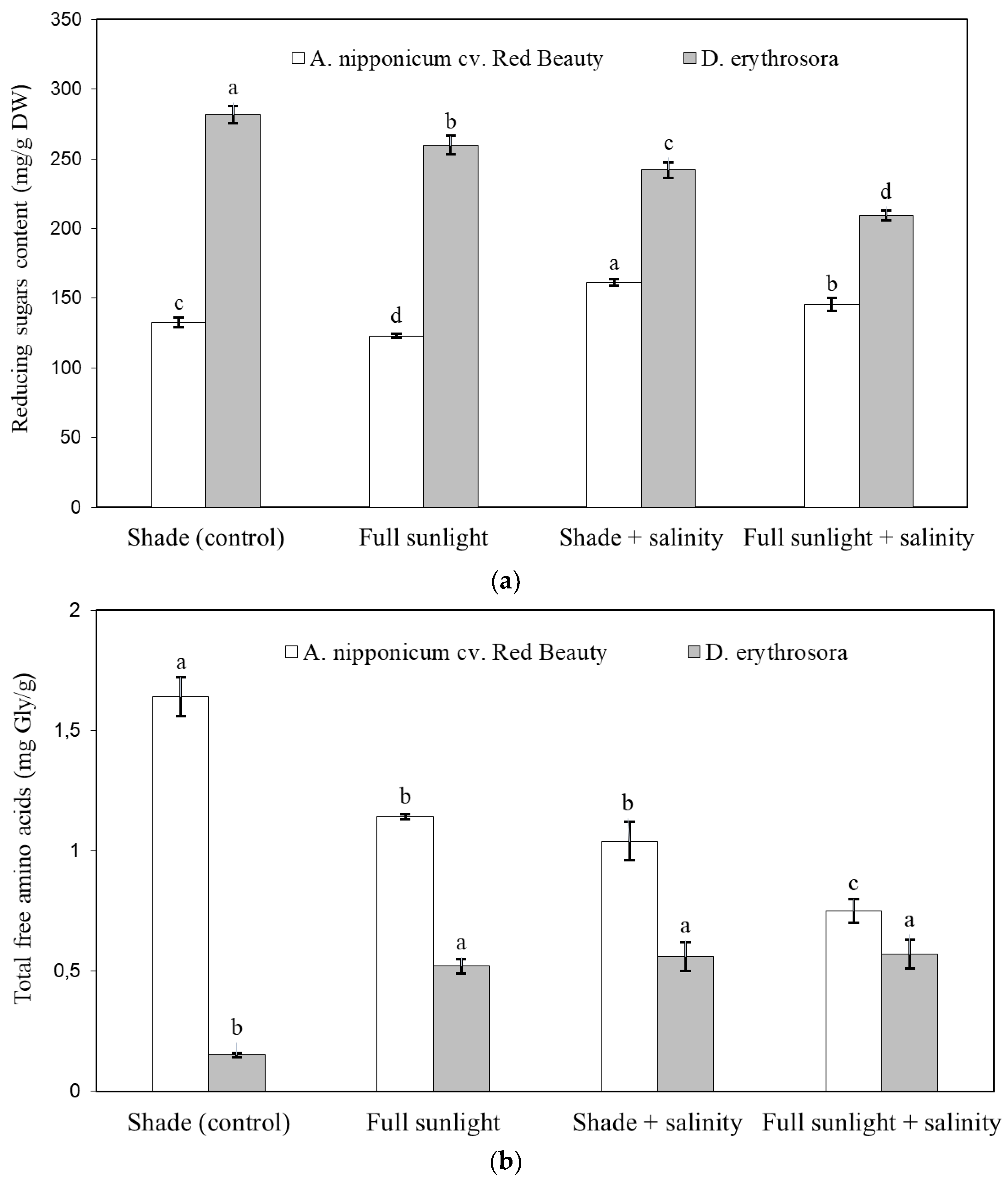
2.2. Effect of Abiotic Stresses on Fern Ion Balance, Chlorophyll Content, Carbohydrates, and Amino Acid Metabolism

The values of the K+/Na+ and Ca2+/Na+ coefficients decreased drastically in both fern taxa due to the applied stress factors (Table 2). The most significant decrease in the K+/Na+ and Ca2+/Na+ coefficients was noted in both ferns subjected to salinity stress in the shade and full sunlight conditions. In plants cultivated without screens, reduced K+/Na+ and Ca2+/Na+ coefficients were also noted compared with the control, with statistically confirmed differences for the K+/Na+ coefficient in A. nipponicum and the Ca2+/Na+ coefficient in D. erythrosora.
Because of stress factors, there were significant changes in the concentration of assimilation pigments depending on the fern genotype (Table 3). In A. nipponicum cv. Red Beauty, exposure of plants to salinity under shade conditions increased chlorophyll a, chlorophyll b, and total chlorophyll by 14.3%, 21.1%, and 17.0%, respectively, over the control. Alternatively, plants growing in full sunlight significantly decreased chlorophyll a, chlorophyll b, and total chlorophyll by 27.8%, 22.5%, and 26.0%, respectively, compared to the control. Similarly, decreases in the content of chlorophyll a, chlorophyll b, and total chlorophyll by 39.9%, 33.8%, and 37.9%, respectively, were shown in the salted plants growing without screens. In the case of D. erythrosora, chlorophyll a, chlorophyll b, and total chlorophyll decreased by 27.8%, 22.5%, and 26.0%, respectively, concerning unstressed plants. Salinity in the shade and full sunlight conditions did not affect the content of pigments in the leaves of D. erythrosora. The analysis of chlorophyll composition revealed significant differences in the Chl a/b ratio depending on the exposure of plants to stress. In A. nipponicum cv. Red Beauty, the Chl a/b ratio was lower than the control in all variants using stress factors. In D. erythrosora, salinity in shade conditions did not affect the value of the Chl a/b ratio, whereas the exposure of plants to full sunlight and total sun exposure combined with salinity resulted in a significant decrease in the Chl a/b ratio.
Significant changes were found in the content of reducing sugars and total free amino acids in fern leaves depending on the applied stress factors (Figure 1a,b). In A. nipponicum cv. Red Beauty, salinity stress in the shade and full sunlight conditions increased the synthesis of reducing sugars compared to the control. In turn, a decrease in the content of reducing sugars was shown in plants growing in full sunlight. In D. erythrosora, the amount of reducing sugars was significantly reduced under all applied stress variants, with the most significant decrease observed in plants exposed to salinity under full sunlight conditions. Regardless of the treatment, the leaves of D. erythrosora contained more reducing sugars than the leaves of A. nipponicum cv. Red Beauty.
In A. nipponicum cv. Red Beauty, the content of total free amino acids in the leaves, decreased due to the application of stresses and was the lowest in salted plants growing in full sunlight conditions. The opposite trend was shown in D. erythrosora under the influence of the tested stresses, for which the content of total free amino acids increased significantly, on average, by more than 2.7 times compared to the control (Figure 1b).

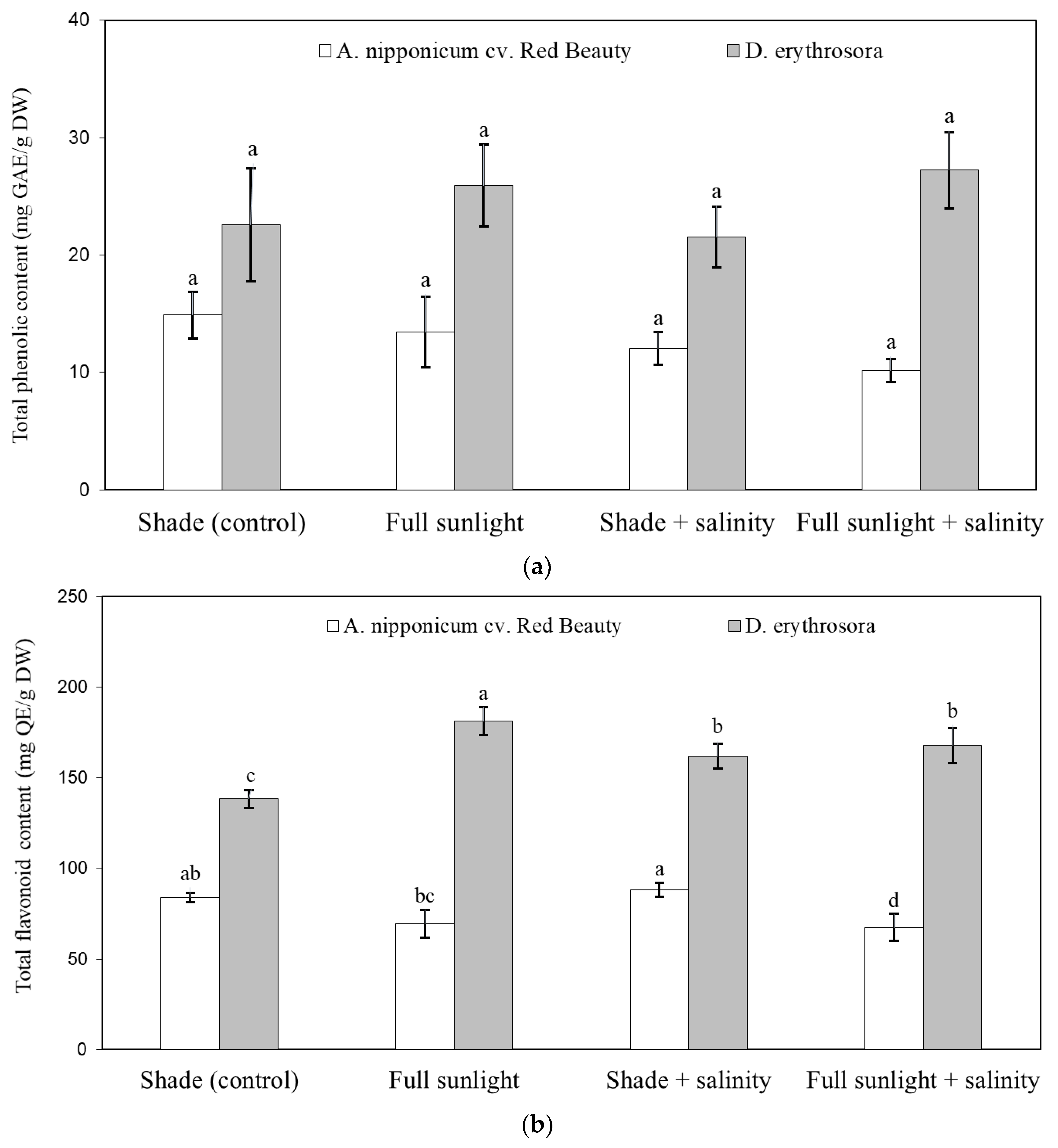
2.3. Effect of Abiotic Stresses on Antioxidant Compounds and Antioxidant Properties of Ferns

Figure 2a,b shows the influence of stress factors on the content of secondary metabolites in fern leaves. In both taxa, salinity, excess light, and a combination of these factors did not cause changes in the total polyphenol content but did affect the total flavonoid level. A. nipponicum cv. Red Beauty showed a high content of total flavonoids in salted plants growing in the shade, whereas a reduced content of these metabolites was found in salted plants growing in full sunlight. In the case of D. erythrosora, all stresses resulted in a significant increase in the total content of flavonoids, with the highest concentration of metabolites in plants exposed to full sunlight. Comparing both taxa, extracts from D. erythrosora leaves contained more total polyphenols and flavonoids than extracts from A. nipponicum cv. Red Beauty.
Analyses of the antioxidant potential of fern extracts grown under stress conditions are shown in Table 4. In A. nipponicum cv. Red Beauty, salinity, full sunlight, and the combined use of these stress factors resulted in a significant reduction in antioxidant activity measured by the ABTS, DPPH, FRAP, and reducing power tests. Deal stress impacted the neutralization of free radicals determined by the ABTS method. Multistress caused the most significant drop in ABTS, followed by full sunlight and saline. In D. erythrosora, the values of the ABTS radical were not affected by stress factors, whereas in the case of the second assessed radical, DPPH, significant differences were already demonstrated. Complete sunlight treatment gave the highest values for DPPH, followed by control, saline, and multi-stress. Similarly, the highest FRAP values were obtained in plants under full sunlight, followed by control, multistress, and saline. Moreover, extracts from plants exposed to full sunlight had the highest antioxidant activity measured by reducing power. The two investigated ferns demonstrated a significant correlation (Tables S1 and S2) between the total free amino acids content and the results of reducing power (r = 0.84 for A. nipponicum cv. Red Beauty and r = 0.69 for D. erythrosora). Moreover, in A. nipponicum cv. Red Beauty, the content of free amino acids also correlated with total phenolic content (r = 0.71) as well as the results of antioxidant activity determined by the ABTS (r = 0.75) and DPPH (r = 0.86) assays. In A. nipponicum cv. Red Beauty, equally, a significant correlation was found for total flavonoids and the results of ABTS (r = 0.80) and total polyphenols and the results of DPPH (r = 0.60).

3. Discussion

Plants, including ferns, are constantly exposed both in natural conditions and during cultivation to various stresses influencing changes at the morphological, physiological, and biochemical levels. Most studies on plant response to stress focus on assessing a single stress factor under strictly controlled conditions using model species [19]. Such studies may be of limited application value as many stress factors of variable intensity are usually imposed during plant production [20]. The reaction of plants to salinity in a phytotron differs from the natural variability of factors under cover or in the natural environment [21]. In these studies conducted in a polytunnel, salinity and various light conditions affected growth, ionic balance, and the content of pigments, metabolites, and antioxidant activity of two taxa of ornamental ferns rich in phytochemicals was assessed.

3.1. Morphology

Salinity caused by watering plants with 100 mM NaCl solution under shade conditions negatively modified the growth of both A. nipponicum cv. Red Beauty and D. erythrosora manifested by reduced plant height, shortened frond length, and reduced fresh leaf weight. The adverse effect of salt stress on the height of plants and the weight of the aboveground part is confirmed by previous studies on four ornamental species of ferns of the genus Dryopteris [8]. Plant growth limitation due to salinity could be related to disturbances in nutrient uptake, osmotic stress, and ion toxicity [22]. Furthermore, the high accumulation of Na+ ions in the tissues of glycophytes leads to reduced water absorption and, as a result, reduced plant growth [23]. Salt stress did not drastically reduce the aesthetic value of D. erythrosora, as no browning and drying of leaves were observed (Figure S1). No leaf chlorosis, despite salt stress, was found in ferns such as D. filix-mas and D. filix-mas cv. Linearis-Polydactylon [8]. It is crucial in the case of ornamental plants because the knowledge of taxa retaining decorative values despite salinity is of great practical importance [24]. There is an intensive search for ornamental plants for green areas that tolerate salinity [23], as well as species that can be watered with salty seawater without damaging their decorativeness [25].
Plants of A. nipponicum cv. Red Beauty grown in full sunlight did not differ in terms of morphological features from plants growing in the shade. A different reaction was observed in D. erythrosora, where plants were taller and produced more biomass under full sunlight. Both fern species under assessment grow under forests in the natural state, with A. nipponicum in the shade and D. erythrosora in half-shade conditions. The obtained results are interesting because, in both fern taxa, no symptoms of burns, whitening of leaves, or curling of leaf edges were observed when they were exposed to full sunlight (Figure S1). It should be emphasized that the research was carried out during the period when, in the climatic conditions of Poland, there is the highest insolation and the most significant number of sunny hours per year [26]. The morphological response of D. erythrosora to full sunlight leading to growth stimulation may result from a unique adaptive mechanism to high light intensity. Moreover, under saline conditions, D. erythrosora plants were taller and had longer fronds when grown without screens. However, further research is needed on which plants would be exposed to direct sunlight for a long time. Only then can it be determined whether D. erythrosora can be considered an ornamental plant that tolerates direct sunlight.

3.2. Physiological Responses

In both fern taxa, treatment with NaCl solution resulted in a significant increase in the content of Na+ ions and a decrease in K+ and Ca2+ ions in leaf tissues, which decreased the K+/Na+ and Ca2+/Na+ ratios. These changes have been demonstrated in both shaded and full-light plants. A high concentration of Na+ in leaves due to salinity, regardless of light conditions, was also demonstrated in ferns D. atrata, D. affinis, D. filix-mas, and D. filix-mas ‘Linearis-Polydactylon’ [8]. The reduced concentration of K+ and Ca2+ ions in salted plants, shown in the studies, is most likely related to the excessive accumulation of Na+ ions causing a cytotoxic effect [23,27]. An increase in the level of harmful Na+ ions in the plant increases osmotic pressure and can induce changes in the uptake of other ions, especially K+ and Ca2+, and their transport to the aboveground part. K+ and Ca2+ ions determine the activity of many enzymes and the processes of plant adaptation to stress [28]. The competition between ions leading to a decrease in K+/Na+ and Ca2+/Na+ ratios, as well as a deficit of K+ and Ca2+ in the cytoplasm, may result in the dysfunction of biological membranes and impairment of many metabolic pathways [20,23,29].
A pronounced decrease in chlorophyll content in leaf tissues distinguished plants of both taxa grown in full sunlight. As previously stated, both species prefer shaded habitats under the tree canopy, with A. nipponicum being more shade-loving than D. erythrosora. In very high light-intensity conditions, photosynthetic dyes absorb too much PAR energy, concerning the possibility of its conversion into chemical energy during photosynthesis [30]. It leads to photoinhibition and may cause oxidative stress, destroying photosynthetic pigments [31].
Analyzing the obtained results related to the effect of salinity on the concentration of assimilation pigments, it was shown that this stress factor stimulated chlorophyll biosynthesis in A. nipponicum. It has been confirmed by previous research that salinity in the initial stage and at low levels can lead to increased chlorophyll concentration in the leaves in some ferns, but at higher levels, it results in a drastic decrease in the content of dyes [32]. In the case of D. erythrosora, there were no changes in the concentration of assimilation dyes due to salinity. Moreover, in D. erythrosora, salinity did not decrease the Chl a/b ratio. Assessing the chlorophyll level in this species may be a valuable tool for assessing its adaptation to salt stress.
In A. nipponicum cv. Red Beauty plants, because of salinity and cultivation in full sun, the Chl a/b ratio decreased. Appropriate quantitative ratios between chlorophylls a and b determine the appropriate intensity of photosynthesis. A decrease in the Chl a/b ratio was also demonstrated in ferns Asplenium viridae, Ceterach officinarum, and Phyllitis scolopendrium due to exposure to high doses of NaCl (250–500 mM) [32]. The decrease in the Chl a/b ratio may be caused by a decrease in chlorophyll a’s content, which proves considerable damage to the photosystems and photoinhibition mentioned above [33,34].
Under salinity conditions, the amount of reducing sugars increased in A. nipponicum cv. Red Beauty, and in D. erythrosora, it decreased. Perhaps this relates to the different mechanisms of sensitivity/tolerance to stress in the taxa. Environmental stresses are the leading causes of changes in carbohydrate metabolism, and these changes can be multidirectional [35]. For example, in susceptible varieties of tomato, salt stress increased the total content of sucrose and other soluble sugars, while in moderately susceptible and tolerant varieties, it did not cause the accumulation of soluble carbohydrates [36].
The taxa differed toward changes in the total free amino acid content due to abiotic stresses. Whereas in A. nipponicum cv. Red Beauty, salinity, full sunlight, and a combination of both stresses reduced the content of amino acids, in D. erythrosora, the same treatments stimulated the biosynthesis of these low molecular weight organic compounds. It can be postulated that in D. erythrosora, total free amino acids play a role in the stress response such as proline, the accumulation of which increases under the influence of salinity in many plant species, including the water fern Azolla filiculoides [37]. The amino acid proline, as an osmoprotectant, reduces the water potential, preventing its outflow from the cell in response to osmotic stress caused by salinity or drought [38]. In A. nipponicum cv. Red Beauty, reduced total free amino acid content as a result of stress may indicate the sensitivity of this taxon to the applied stressors. A marked decrease in total essential amino acids, non-essential amino acids, and free amino acids was also shown in four edible species of ferns grown in conditions of intense sunlight [39].

3.3. Antioxidant Responses

On the whole, D. erythrosora exhibited higher total polyphenol and flavonoid content than A. nipponicum cv. Red Beauty. Similarly, Xia et al. [40] reported that extracts from D. erythrosora contained more total flavonoids than those of A. nipponicum. In this experiment, D. erythrosora plants exposed to stress were characterized by the increased production of total flavonoids, especially when exposed to full sunlight. The increase in total flavonoids in response to applied stresses appears to be an acclimation response of D. erythrosora to stress. Similarly, Wang et al. [7] reported increased total phenol and flavonoid content in leaves of the ferns Matteuccia struthiopteris, Athyrium multidentatum, and Osmunda cinnamomea var. asiatica exposed to the high light level. Flavanols protect plants against harmful UV-B radiation, and thus plants exposed to excess light contain much higher amounts of them than those shaded [41]. Generally, the plant limits the synthesis of secondary metabolites under favorable growth conditions due to the high energy expenditure associated with these processes. When plants are under stress, the secondary metabolism increases to balance the metabolic system [42]. Ecological factors such as light, habitat, altitude, and latitude, as well as stress factors, can activate the secondary metabolism pathways in ferns and increase the production of antioxidant compounds [7]. The confirmation of this hypothesis is the fact that extracts from the leaves of D. erythrosora plants grown in full sunlight had increased antioxidant activity measured by DPPH, FRAP, and reducing power tests.
Many studies have shown that the ability to eliminate free radicals strongly correlates with polyphenol and flavonoid content [43,44]. In these studies, positive linear correlations were found in A. nipponicum cv. Red Beauty between total polyphenol content and activity against DPPH and between flavonoid content and activity against ABTS. In D. erythrosora, no correlation was found between the level of flavonoids and the values of antioxidant tests, which may be influenced by the diverse profile of phenolic compounds found in ferns. In both taxa, it was shown that the content of total free amino acids is positively correlated with the antioxidant activity measured by reducing power. Moreover, A. nipponicum cv. Red Beauty showed positive linear correlations between total free amino acids and the content of total polyphenols, ABTS, and DPPH. Amino acids are lesser-known antioxidants with the ability to defend against free radicals [45]. However, their role in the antioxidant system of plants is still being explored.
Fern extracts are a very rich and promising source of polyphenolic compounds with potentially beneficial medical effects [4,7,21]. Natural polyphenols and flavonoids are particularly valuable in fern tissues with documented therapeutic antioxidant, anti-inflammatory, antimicrobial, and anticancer effects [46]. The development of technologies for cultivating ferns rich in bioactive compounds and finding ways to stimulate the biosynthesis of their secondary metabolites may help in the broader use of these plants and contribute to the protection of species occurring in the natural state. It is also essential to determine the level of stress at which the yield of plants does not decrease drastically, and simultaneously, the biosynthesis of valuable metabolites increases significantly.

4. Materials and Methods

4.1. Plant Material, Treatments, and Growth Characteristics

The study materials were in vitro propagated cuttings of Athyrium nipponicum cv. Red Beauty and Dryopteris erythrosora. They were planted on 17 March 2021 into pots (0.5 dm3) filled with peat substrate (pH 6.0) supplemented with fertilizer 14% N, 16% P, and 18% K (0.5 g dm−3) and acclimatized for eight weeks in a greenhouse (22/18 °C). Well-rooted plants were transferred into larger pots (1.7 dm3) filled with peat substrate (pH 6.0) and fertilizer 14% N, 16% P, and 18% K (1 g dm−3) and placed in a plastic tunnel located near the West Pomeranian University of Technology in Szczecin (53°25′ N, 14°32′ E; 25 m above mean sea level), covered with a double layer of inflatable plastic with anti-condensation properties. The temperature in the tunnel was regulated by automatically opening the top roof ventilation when it reached 18 °C inside. Half of the plants were placed under shade screens composed of fabric with aluminum stripes, and the other half were grown without shading. All plants were arranged at a density of nine pots per square meter. The mean photosynthetic photon flux density on cloudless days at midday between 1 May and 20 June 2021 reached a mean of 103.7–273.7 μmol m−2 s−1 under shade screens and 551.9–890.1 μmol m−2 s−1 without the screens. Since 17 May 2021, half of the shaded and non-shaded (excess light) plants of both taxa were watered every seven days with 100 mmol dm−3 NaCl. The stress was applied four times, with 100 mL solution per pot per treatment. NaCl concentration was established based on previous studies [8]. The control was watered with tap water of pH 6.5 and electrolytic conductivity of 0.66 mS cm−1. Each treatment for each taxon (shade (control), full sunlight, shade + salinity, and full sunlight + salinity) included a total of 27 plants, i.e., three replicates of nine plants each. The number of sunny hours per day was: 8.2 (May), 10.2 (June), and 8.2 (July). Accumulated sunshine hours were as follows: 221.7 (May), 296 (June), and 253.6 (July).
Two weeks after the last NaCl treatment (20 June 2021), 20 plants from each treatment were assessed for the height and length of the longest frond. Then, the above-ground parts were cut and dried at 75 °C in the shade [17], and their dry weight (DW) was determined. The two most developed leaves from 10 plants from each treatment were selected for analysis.

4.2. Determination of Ionic Status

To determine the content of sodium, potassium, and calcium, dried and ground leaf samples were mineralized in concentrated sulfuric acid. The ion content was determined by flame photometry on a flame photometer AFP-100 (Biotech Engineering Management, Nicosia, Cyprus), as described by Ostrowska et al. [47]. Each element was determined in three replicates. The potassium/sodium (K+/Na+) and calcium/potassium (Ca2+/K+) ratios were calculated based on the data obtained.

4.3. Determination of Photosynthetic Pigment Content

Dried fern leaves were ground in a laboratory mill, and 0.5 g samples were placed into Falcon tubes and mixed with H2O/acetone mixture (2:8 v/v). The mixtures were extracted for five minutes in an ultrasonic cleaner. The samples were then centrifuged at 6000 rpm for 10 min. The content of photosynthetic pigments (chlorophyll a, b, and total chlorophyll) was determined spectrophotometrically according to the modified method of Grzeszczuk et al. [48]. In short, 300 µL of the supernatant was pipetted into a single well of a 96-well TC plate. The microplate was put into a microplate reader, and the absorbance was read at 646, 652, and 663 nm. Each sample was read in three repetitions. The content of chlorophyll a and b and total chlorophyll was expressed in mg g−1 DW.

4.4. Preparation of Extracts

Extracts of fern leaves were obtained by solvent extraction, as described by Grzeszczuk et al. [48]. The solvent was a mixture of methanol (Merck, Darmstadt, Germany) with distilled H2O (7:3, v/v). Dried leaves were ground in a laboratory mill, then 0.5 g samples were transferred into 50 mL Falcon tubes (Bionovo, Legnica, Poland) and mixed with 40 mL of the mixture. The solutions were then extracted for 30 min in an ultrasonic cleaner (Elmasonic S30H, Elma Schmidbauer GmbH, Singen, Germany) and centrifuged for 5 min at 5000 rpm (Centrifuge 5418 Eppendorf, Warsaw, Poland). The extracts were filtered through 0.22 µm nylon membrane filters (Merck, Darmstadt, Germany). Three repetitions of each extract were prepared and stored in a freezer at −20 °C.

4.5. Determination of Total Reducing Sugars

Total reducing sugars were determined as described by Łopusiewicz et al. [49]. Briefly, 1 mL of the supernatant was combined with 1 mL of 0.05 M acetic buffer (pH 4.8) and 3 mL of DNS reagent. The mixture was shaken vigorously and then incubated for 5 min in hot water (~96 °C). The tubes were cooled to room temperature, and the absorbance was read at 540 nm.

4.6. Content of Total Free Amino Acids

The total content of free amino acids was analyzed spectrophotometrically according to Łopusiewicz et al. [50]. To this end, 1 mL of the supernatant was combined with 2 mL of ninhydrin-Cd reagent in test tubes. The samples were vortexed, heated at 84 °C (5 min), and then cooled on ice. The supernatant was transferred onto a plate, and the absorbance was measured at 507 nm. The results were calculated as mg of glycine (Gly) equivalent per 1 g sample (DW).

4.7. Total Polyphenols and Total Flavonoids

Total polyphenols in methanolic fern extracts were determined spectrophotometrically with a microplate reader (Synergy LX, Bio Tek, Vermont, VT, USA) using the Folin–Ciocalteu method described by Tong et al. [51] with modifications. The method consisted of mixing 20 µL of the supernatant with 150 µL of distilled water and 100 µL of Folin–Ciocalteu reagent. The solution was supplemented with 80 µL of saturated Na2CO3 (after 5 min). The mixture was then incubated in the dark at 40 °C for 30 min. The absorbance was measured at 765 nm, and the concentration of the polyphenols was expressed as mg gallic acid equivalents per g of DW (mg GAE g−1 DW).
The total content of flavonoids was measured according to the procedure proposed by Tong et al. [42], with modifications. To this end, 25 µL of the extract was combined with 100 µL of distilled H2O and 7.5 µL of 5% NaNO2. After 5 min, the mixture was supplemented with 7.5 µL of 10% AlCl3 solution. The mixture was kept for 6 min at room temperature before adding 25 µL of 1 M NaOH. Then, it was diluted with 135 µL of distilled water, and the absorbance was measured at 510 nm. The results were expressed as mg quercetin equivalent (QE) per g DW (mg QE g−1 DW).

4.8. 1,1-Diphenyl-2-Picryl-Hydrazyl (DPPH) and 2,2′-Azobis(3-Ethylbenzothiazoline-6-Sulfonate) (ABTS) Assays

The DPPH and ABTS assays were performed as described by Łopusiewicz et al. [50]. First, 0.5 mL of methanolic DPPH solution (0.01 mM) was mixed with 0.5 mL of the extract in a 1:1 ratio. The samples were incubated at 22 ± 2 °C for 30 min in darkness. Following the incubation, the absorbance was read at 517 nm. The total antioxidant potential of the analyzed extracts was determined spectrophotometrically based on the ABTS assay. Before the experiment, the ABTS radical solution was diluted with 96% ethanol to obtain an absorbance of 0.700 ± 0.02 measured at 734 nm. In our experiment, we combined 1.5 mL of ABTS solution with 25 µL of the extract in 1.5 mL Eppendorf tubes, and then the tubes were incubated at room temperature for 10 min in the dark. After incubation, the absorbance was read at 734 nm.

4.9. Ferric Reducing Antioxidant Power (FRAP)

The FRAP reagent was prepared by mixing 2.5 mL of TPTZ in 40 mM HCl, 2.5 mL of FeCl3, and 25 mL of acetate buffer at pH 3.6 (300 mM). Then, 20 µL of the supernatant in three repetitions was transferred into a 96-well TC plate by adding 280 µL of the FRAP reagent, and the plate was gently shaken for 10 s. The absorbance was read at 595 nm. The results were expressed as ascorbic acid equivalents (AAE) per g of sample (DW) using the standard curve for ascorbic acid.

4.10. Reducing Power

The reducing power of the methanolic extracts was measured according to Łopusiewicz et al. [50], with modifications. Briefly, 100 µL of the extract was added to 1.5 mL Eppendorf tubes and supplemented with 250 µL of phosphate buffer (0.2 M, pH 6.6) and 250 µL of 1% potassium hexacyanoferrate. The samples were incubated at 50 °C for 20 min before adding 250 µL of trichloroacetic acid solution. They were then centrifuged at 3000 rpm for 10 min. In the next step, 500 µL of the solution was transferred into Eppendorf tubes (1.5 mL), and 0.5 mL of H2O and 0.1 mL of iron chloride (1%) were added. Finally, the absorbance was measured at 700 nm.

4.11. Statistical Analysis

The obtained data with normal distribution were analyzed statistically using the analysis of variance (one-way ANOVA) for one-factor experiments using the Statistica 13.1 Statsoft package (Statsoft, Krakow, Poland). After checking the good fit of the model, the post hoc comparison was completed using Tukey’s HSD post hoc test at p ≤ 0.05. To assess the existence of correlations between the content of metabolites and antioxidant activity, the values of Pearson’s linear correlation coefficients were determined, and their significance was verified.

5. Conclusions

Considering the unfavorable climate change, understanding the physiological processes responsible for the adaptive capabilities of individual plant species from different systematic groups, including ferns, is crucial. This study demonstrated that plants exposed to salinity and salinity combined with excess light produced lower bio-mass and had an unfavorable proportion of Na+, K+, and Ca2+ ions. In both genotypes, excess light reduced the content of chlorophyll a, chlorophyll b, and total chlorophyll. The salinity did not affect the pigment level in D. erythrosora and A. nipponicum cv. Red Beauty and, surprisingly, even stimulated their biosynthesis. A greater adaptive potential to salinity and light stress was found in D. erythrosora plants than in A. nipponicum cv. Red Beauty. The response of D. erythrosora to abiotic stress was strongly associated with changes in reducing the sugar content, total free amino acids, and total flavonoid content. These biochemical parameters may consequently be indicators of the sensitivity/tolerance of D. erythrosora to salinity and light stress. The cultivation of D. erythrosora under abiotic stresses contributed to the increased bioaccumulation of flavonoids in leaf tissues, which may be a new strategy for obtaining these valuable secondary metabolites in this species.

Supplementary Materials

The following supporting information can be downloaded at: https://www.mdpi.com/article/10.3390/ijms24010296/s1.

Author Contributions

Conceptualization, A.P. and P.S.; methodology, A.P., P.S., and Ł.Ł.; formal analysis, A.P., P.S., and Ł.Ł.; investigation, A.P. and P.S.; data curation, A.P.; writing—original draft preparation, A.P. and P.S.; writing—review and editing, Ł.Ł. All authors have read and agreed to the published version of the manuscript.

Funding

This research received no external funding.

Institutional Review Board Statement

Not applicable.

Informed Consent Statement

Not applicable.

Data Availability Statement

Not applicable.

Conflicts of Interest

The authors declare no conflict of interest.

References

  1. Pittermann, J.; Brodersen, C.; Watkins, J. The Physiological resilience of fern sporophytes and gametophytes: Advances in water relations offer new insights into an old lineage. Front. Plant Sci. 2013, 4, 285. [Google Scholar] [CrossRef] [PubMed] [Green Version]
  2. Suissa, J.S.; Friedman, W.E. From cells to stems: The effects of primary vascular construction on drought-induced embolism in fern rhizomes. New Phytol. 2021, 232, 2238–2253. [Google Scholar] [CrossRef] [PubMed]
  3. Anderson, O.R. Physiological ecology of ferns: Biodiversity and conservation perspectives. Int. J. Biodivers. Conserv. 2021, 13, 49–63. [Google Scholar]
  4. Wang, Y.; Gao, S.; He, X.; Li, Y.; Li, P.; Zhang, Y.; Chen, W. Growth, Secondary Metabolites and Enzyme Activity Responses of Two Edible Fern Species to Drought Stress and Rehydration in Northeast China. Agronomy 2019, 9, 137. [Google Scholar] [CrossRef] [Green Version]
  5. Singh, A.P.; Johari, D. Scope of ferns in horticulture and economic development. In Current Advances in Fern Research; Fernández, H., Ed.; Springer International Publishing: Cham, Switzerland, 2018; pp. 153–175. ISBN 978-3-319-75103-0. [Google Scholar]
  6. Salachna, P.; Siemińska, I.; Pietrak, A.; Zawadzińska, A.; Piechocki, R.; Dymek, R. Response of hardy ferns to drought stress. Biol. Life Sci. Forum 2021, 3, 8. [Google Scholar]
  7. Wang, X.; Wang, M.; Cao, J.; Wu, Y.; Xiao, J.; Wang, Q. Analysis of flavonoids and antioxidants in extracts of ferns from Tianmu Mountain in Zhejiang Province (China). Ind. Crops Prod. 2017, 97, 137–145. [Google Scholar] [CrossRef]
  8. Salachna, P.; Piechocki, R. Salinity tolerance of four hardy ferns from the genus Dryopteris Adans. grown under different light conditions. Agronomy 2021, 11, 49. [Google Scholar] [CrossRef]
  9. Yadav, R.K.; Chatrath, A.; Tripathi, K.; Gerard, M.; Ahmad, A.; Mishra, V.; Abraham, G. Salinity tolerance mechanism in the aquatic nitrogen fixing pteridophyte Azolla: A Review. Symbiosis 2021, 83, 129–142. [Google Scholar] [CrossRef]
  10. Cramer, G.R.; Urano, K.; Delrot, S.; Pezzotti, M.; Shinozaki, K. Effects of abiotic stress on plants: A systems biology perspective. BMC Plant Biology 2011, 11, 163. [Google Scholar] [CrossRef] [Green Version]
  11. Mittler, R. Abiotic stress, the field environment and stress combination. Trends Plant Sci. 2006, 11, 15–19. [Google Scholar] [CrossRef]
  12. Das, K.; Roychoudhury, A. Reactive oxygen species (ROS) and response of antioxidants as ros-scavengers during environmental stress in plants. Front. Environ. Sci. 2014, 2, 53. [Google Scholar] [CrossRef] [Green Version]
  13. Hasanuzzaman, M.; Bhuyan, M.H.M.B.; Parvin, K.; Bhuiyan, T.F.; Anee, T.I.; Nahar, K.; Hossen, M.S.; Zulfiqar, F.; Alam, M.M.; Fujita, M. Regulation of ROS Metabolism in Plants under Environmental Stress: A Review of Recent Experimental Evidence. Int. J. Mol. Sci. 2020, 21, 8695. [Google Scholar] [CrossRef]
  14. Zhang, H.; Sonnewald, U. Differences and commonalities of plant responses to single and combined stresses. Plant J. 2017, 90, 839–855. [Google Scholar] [CrossRef] [PubMed] [Green Version]
  15. Sack, L.; Buckley, T.N. Trait multi-functionality in plant stress response. Integr. Comp. Biol. 2020, 60, 98–112. [Google Scholar] [CrossRef] [PubMed]
  16. Salehi, B.; Ezzat, S.M.; Tsouh Fokou, P.V.; Albayrak, S.; Vlaisavljevic, S.; Sharifi-Rad, M.; Bhatt, I.D.; Sharifi-Rad, M.; Belwal, T.; Ayatollahi, S.A.; et al. Athyrium plants—Review on phytopharmacy properties. J. Tradit. Complement. Med. 2019, 9, 201–205. [Google Scholar] [CrossRef] [PubMed]
  17. Zhang, X.; Wang, X.; Wang, M.; Cao, J.; Xiao, J.; Wang, Q. Effects of different pretreatments on flavonoids and antioxidant activity of Dryopteris erythrosora leave. PLoS ONE 2019, 14, e0200174. [Google Scholar] [CrossRef] [PubMed] [Green Version]
  18. Cao, J.; Xia, X.; Chen, X.; Xiao, J.; Wang, Q. Characterization of flavonoids from Dryopteris erythrosora and evaluation of their antioxidant, anticancer and acetylcholinesterase inhibition activities. Food Chem. Toxicol. 2013, 51, 242–250. [Google Scholar] [CrossRef]
  19. Møller, I.S.; Tester, M. Salinity tolerance of Arabidopsis: A good model for cereals? Trends Plant Sci. 2007, 12, 534–540. [Google Scholar] [CrossRef]
  20. Shannon, M.C. Adaptation of plants to salinity. Adv. Agron. 1997, 60, 75–120. [Google Scholar]
  21. Alkharabsheh, H.M.; Seleiman, M.F.; Hewedy, O.A.; Battaglia, M.L.; Jalal, R.S.; Alhammad, B.A.; Schillaci, C.; Ali, N.; Al-Doss, A. Field Crop Responses and Management Strategies to Mitigate Soil Salinity in Modern Agriculture: A Review. Agronomy 2021, 11, 2299. [Google Scholar] [CrossRef]
  22. Hernández, J.A. Salinity Tolerance in Plants: Trends and Perspectives. Int. J. Mol. Sci. 2019, 20, 2408. [Google Scholar] [CrossRef] [PubMed] [Green Version]
  23. Safdar, H.; Amin, A.; Shafiq, Y.; Ali, A.; Yasin, R.; Shoukat, A.; Hussan, M.U.; Sarwar, M.I. A review: Impact of salinity on plant growth. Nat. Sci. 2019, 17, 34–40. [Google Scholar]
  24. Salachna, P.; Zawadzińska, A.; Podsiadło, C. Response of Ornithogalum saundersiae Bak. to salinity stress. Acta Sci. Pol. Hortorum Cultus 2016, 15, 123–134. [Google Scholar]
  25. García-Caparrós, P.; Lao, M.T. The effects of salt stress on ornamental plants and integrative cultivation practices. Sci. Hortic. 2018, 240, 430–439. [Google Scholar] [CrossRef]
  26. Igliński, B.; Cichosz, M.; Kujawski, W.; Plaskacz-Dziuba, M.; Buczkowski, R. Helioenergy in Poland—Current state, surveys and prospects. Renew. Sustain. Energy Rev. 2016, 58, 862–870. [Google Scholar]
  27. Radić, S.; Prolić, M.; Pavlica, M.; Pevalek-Kozlina, B. Cytogenetic effects of osmotic stress on the root meristem cells of Centaurea ragusina L. Environ. Exp. Bot. 2005, 54, 213–218. [Google Scholar] [CrossRef]
  28. Porcelli, C.; Gutierrez, F.H.; Lavado, R. The K/Na and Ca/Na rations and rapeseed yield, under soil salinity or sodicity. Plant Soil 1995, 175, 251–255. [Google Scholar] [CrossRef]
  29. Muhammed, S.; Akbar, M.; Neue, H.U. Effect of Na/Ca and Na/K ratios in saline culture solution on the growth and mineral nutrition of rice (Oryza sativa L.). Plant Soil 1987, 104, 57–62. [Google Scholar] [CrossRef]
  30. Demmig-Adams, B.; Adams, W.W. Photoprotection and other responses of plants to high light stress. Annu. Rev. Plant Biol. 1992, 43, 599–626. [Google Scholar] [CrossRef]
  31. Krause, G.H. Photoinhibition of photosynthesis. An evaluation of damaging and protective mechanisms. Physiol. Plant. 1988, 74, 566–574. [Google Scholar] [CrossRef]
  32. Bogdanović, M.; Ilić, M.; Živković, S.; Sabovljević, A.; Grubišić, D.; Sabovljević, M. Comparative study on the effects of NaCl on selected moss and fern representatives. Aust. J. Bot. 2012, 59, 734–740. [Google Scholar] [CrossRef]
  33. Kura-Hotta, M.; Satoh, K.; Katoh, S. Relationship between photosynthesis and chlorophyll content during leaf senescence of rice seedlings. Plant Cell Physiol. 1987, 28, 1321–1329. [Google Scholar]
  34. Hayden, D.B.; Baker, N.R.; Percival, M.P.; Beckwith, P.B. Modification of the photosystem II light-harvesting chlorophyll ab protein complex in maize during chillinduced photoinhibition. Biochim. Biophys. Acta—Bioenerg. 1986, 851, 86–92. [Google Scholar] [CrossRef]
  35. de Moraes, M.G.; de Oliveira, A.A.Q.; Santos, M.G. Sugars in ferns and lycophytes growing on rocky outcrops from southeastern brazilian coast. Biosci. J. 2014, 30, 1882–1884. [Google Scholar]
  36. Balibrea, M.E.; Dell’Amico, J.; Bolarín, M.C.; Pérez-Alfocea, F. Carbon partitioning and sucrose metabolism in tomato plants growing under salinity. Physiol. Plant. 2000, 110, 503–511. [Google Scholar] [CrossRef]
  37. van Kempen, M.M.L.; Smolders, A.J.P.; Bögemann, G.M.; Lamers, L.L.M.; Visser, E.J.W.; Roelofs, J.G.M. Responses of the Azolla filiculoides Stras.–Anabaena azollae Lam. association to elevated sodium chloride concentrations: Amino acids as indicators for salt stress and tipping point. Aquat. Bot. 2013, 106, 20–28. [Google Scholar] [CrossRef]
  38. Batista-Silva, W.; Heinemann, B.; Rugen, N.; Nunes-Nesi, A.; Araújo, W.L.; Braun, H.-P.; Hildebrandt, T.M. The role of amino acid metabolism during abiotic stress release. Plant Cell Environ. 2019, 42, 1630–1644. [Google Scholar] [CrossRef] [Green Version]
  39. Wang, Y.; Gao, S.; He, X.; Li, Y.; Zhang, Y.; Chen, W. Response of total phenols, flavonoids, minerals, and amino acids of four edible fern species to four shading treatments. PeerJ 2020, 8, e8354. [Google Scholar] [CrossRef]
  40. Xia, X.; Cao, J.; Zheng, Y.; Wang, Q.; Xiao, J. Flavonoid concentrations and bioactivity of flavonoid extracts from 19 species of ferns from China. Ind. Crops Prod. 2014, 58, 91–98. [Google Scholar] [CrossRef]
  41. Tattini, M.; Galardi, C.; Pinelli, P.; Massai, R.; Remorini, D.; Agati, G. Differential accumulation of flavonoids and hydroxycinnamates in leaves of Ligustrum vulgare under excess light and drought stress. New Phytol. 2004, 163, 547–561. [Google Scholar] [CrossRef] [PubMed]
  42. Mahajan, M.; Kuiry, R.; Pal, P.K. Understanding the consequence of environmental stress for accumulation of secondary metabolites in medicinal and aromatic plants. J. Appl. Res. Med. Aromat. Pl. 2020, 18, 100255. [Google Scholar] [CrossRef]
  43. Piluzza, G.; Bullitta, S. Correlations between Phenolic Content and Antioxidant Properties in Twenty-Four Plant Species of Traditional Ethnoveterinary Use in the Mediterranean Area. Pharm. Biol. 2011, 49, 240–247. [Google Scholar] [CrossRef] [PubMed]
  44. Teixeira, T.; Vale, R.; Almeida, R.; Ferreira, T.; Guimarães, L. Antioxidant Potential and Its Correlation with the Contents of Phenolic Compounds and Flavonoids of Methanolic Extracts from Different Medicinal Plants. Rev. Virtual De Química 2017, 9, 1546–1559. [Google Scholar] [CrossRef]
  45. Stadtman, E.R.; Levine, R.L. Free radical-mediated oxidation of free amino acids and amino acid residues in proteins. Amino Acids 2003, 25, 207–218. [Google Scholar] [CrossRef]
  46. Visioli, F.; De La Lastra, C.A.; Andres-Lacueva, C.; Aviram, M.; Calhau, C.; Cassano, A.; D’Archivio, M.; Faria, A.; Favé, G.; Fogliano, V.; et al. Polyphenols and Human Health: A Prospectus. Crit. Rev. Food Sci. Nutr. 2011, 51, 524–546. [Google Scholar] [CrossRef]
  47. Ostrowska, A.; Gawliński, S.; Szczubiałka, Z. Methods for Analyzing and Assessing the Properties of Soil and Plants; Instytut Ochrony Środowiska: Warsaw, Poland, 1991; pp. 1–333. (In Polish) [Google Scholar]
  48. Grzeszczuk, M.; Salachna, P.; Meller, E. Changes in photosynthetic pigments, total phenolic content, and antioxidant activity of Salvia coccinea Buc’hoz ex Etl. Induced by exogenous salicylic acid and soil salinity. Molecules 2018, 23, 1296. [Google Scholar] [CrossRef] [Green Version]
  49. Łopusiewicz, Ł.; Drozłowska, E.; Siedlecka, P.; Mężyńska, M.; Bartkowiak, A.; Sienkiewicz, M.; Zielińska-Bliźniewska, H.; Kwiatkowski, P. Development, characterization, and bioactivity of non-dairy kefir-like fermented beverage based on flaxseed oil cake. Foods 2019, 8, 544. [Google Scholar] [CrossRef] [Green Version]
  50. Łopusiewicz, Ł.; Drozłowska, E.; Trocer, P.; Kwiatkowski, P.; Bartkowiak, A.; Gefrom, A.; Sienkiewicz, M. The Effect of fermentation with kefir grains on the physicochemical and antioxidant properties of beverages from blue lupin (Lupinus angustifolius L.) seeds. Molecules 2020, 25, 5791. [Google Scholar] [CrossRef]
  51. Tong, T.; Liu, Y.-J.; Kang, J.; Zhang, C.-M.; Kang, S.-G. Antioxidant activity and main chemical components of a novel fermented tea. Molecules 2019, 24, 2917. [Google Scholar] [CrossRef]
Figure 1. Effect of abiotic stresses on reducing sugars (a) and total free amino acid content (b) of two hardy ferns. Vertical error bars indicate the standard deviation of the mean. Different letters indicate a significant difference between treatments within given taxa at the p ≤ 0.05 (ANOVA, Tukey’s HSD post hoc test).
Ijms 24 00296 g001
Figure 2. Effect of abiotic stresses on total phenolic content (a) and total flavonoid content (b) of two hardy ferns. Vertical error bars indicate the standard deviation of the mean. Different letters indicate a significant difference between treatments within given taxa at p ≤ 0.05 (ANOVA, Tukey’s HSD post hoc test).
Ijms 24 00296 g002
Table 1. Effect of abiotic stresses on growth characteristics of two hardy ferns. Within a column means followed by the same lowercase letter are not significantly different at p ≤ 0.05 (ANOVA, Tukey’s HSD post hoc test).
TreatmentsPlant Height
(cm)
Frond Length
(cm)
Fresh Weight
(g)
A. nipponicum cv. Red Beauty
Shade (control)25.3 ± 1.07 a25.3 ± 1.06 ab8.90 ± 0.46 a
Full sunlight25.3 ± 0.96 a29.6 ± 3.92 a9.55 ± 0.42 a
Shade + salinity20.5 ± 1.50 b20.7 ± 1.47 c7.45 ± 0.18 b
Full sunlight + salinity18.8 ± 1.35 b24.1 ± 1.59 b7.47 ± 1.03 b
D. erythrosora
Shade (control)24.5 ± 0.50 b37.5 ± 1.81 a14.3 ± 0.75 b
Full sunlight29.3 ± 1.53 a36.9 ± 1.41 a19.5 ± 1.16 a
Shade + salinity15.3 ± 1.53 c27.1 ± 1.90 b8.34 ± 1.30 c
Full sunlight + salinity21.7 ± 2.52 b33.7 ± 1.02 a9.75 ± 0.99 c
Table 2. Effect of abiotic stresses excess on K+/Na+ and Ca2+/Na+ ratios of two hardy ferns. Within a column means followed by the same lowercase letter are not significantly different at p ≤ 0.05 (ANOVA, Tukey’s HSD post hoc test).
TreatmentsK+/Na+ RatioCa2+/Na+ Ratio
A. nipponicum cv. Red Beauty
Shade (control)9.00 ± 0.07 a2.97 ± 0.64 a
Full sunlight6.23 ± 0.92 b2.47 ± 0.47 a
Shade + salinity1.07 ± 0.06 c0.60 ± 0.01 b
Full sunlight + salinity1.93 ± 0.15 c0.60 ± 0.17 b
D. erythrosora
Shade (control)13.47 ± 2.36 a2.97 ± 0.64 a
Full sunlight11.57 ± 1.27 a2.47 ± 0.47 a
Shade + salinity2.80 ± 0.20 b0.60 ± 0.01 b
Full sunlight + salinity2.77 ± 0.06 b0.60 ± 0.17 b
Table 3. Effect of abiotic stresses on chlorophyll pigment content of two hardy ferns. Within a column means followed by the same lowercase letter are not significantly different at p ≤ 0.05 (ANOVA, Tukey’s HSD post hoc test).
TreatmentsChlorophyll a
mg/g DW
Chlorophyll b
mg/g DW
Total Chlorophyll
mg/g DW
Chlorophyll
a/b Ratio
A. nipponicum cv. Red Beauty
Shade (control)1.33 ± 0.05 b0.71 ± 0.03 b2.35 ± 0.09 b1.86 a ± 0.01 a
Full sunlight0.96 ± 0.03 c0.55 ± 0.02 c1.74 ± 0.06 c1.73 c ± 0.01 c
Shade + salinity1.52 ± 0.02 a0.86 ± 0.02 a2.75 ± 0.04 a1.78 b ± 0.01 b
Full sunlight + salinity0.80 ± 0.01 d0.47 ± 0.01 d1.46 ± 0.01 d1.71 c ± 0.03 c
D. erythrosora
Shade (control)0.38 ± 0.03 a0.19 ± 0.02 ab0.63 ± 0.06 a2.01 ± 0.02 b
Full sunlight0.23 ± 0.01 b0.11 ± 0.01 c0.38 ± 0.02 b2.16 ± 0.03 a
Shade + salinity0.35 ± 0.01 a0.17 ± 0.00 b0.58 ± 0.02 a2.06 ± 0.02 b
Full sunlight + salinity0.39 ± 0.01 a0.20 ± 0.01 a0.66 ± 0.03 ab1.93 ± 0.02 c
Table 4. Effect of abiotic stresses on antioxidant activity (as measured by ABTS, DPPH, FRAP, and reducing power methods) of two hardy ferns. Within a column means followed by the same lowercase letter are not significantly different at p ≤ 0.05 (ANOVA, Tukey’s HSD post hoc test).
TreatmentsABTS
(%)
DPPH
(%)
FRAP
(mg AAE/g DW)
Reducing Power
A. nipponicum cv. Red Beauty
Shade (control)80.77 ± 0.58 a28.44 ± 1.56 a12.60 ± 0.29 a1.55 ± 0.02 a
Full sunlight60.46 ± 0.62 c20.16 ± 0.16 b9.75 ± 0.57 b1.11 ± 0.01 c
Shade + salinity76.05 ± 1.34 b17.19 ± 1.88 b9.85 ± 0.24 b1.23 ± 0.02 b
Full sunlight + salinity56.41 ± 1.31 d17.97 ± 1.41 b10.50 ± 0.50 b1.17 ± 0.04 bc
D. erythrosora
Shade (control)98.62 ± 0.09 ab77.66 ± 0.47 b31.20 ± 1.77 b2.47 ± 0.03 b
Full sunlight98.15 ± 0.39 b90.16 ± 0.16 a34.91 ± 1.16 a2.55 ± 0.02 a
Shade + salinity98.97 ± 0.27 a66.72 ± 0.94 c22.43 ± 1.33 d2.50 ± 0.02 ab
Full sunlight + salinity98.92 ± 0.09 a76.88 ± 0.31 b26.09 ± 0.36 c2.53 ± 0.04 ab
Disclaimer/Publisher’s Note: The statements, opinions and data contained in all publications are solely those of the individual author(s) and contributor(s) and not of MDPI and/or the editor(s). MDPI and/or the editor(s) disclaim responsibility for any injury to people or property resulting from any ideas, methods, instructions or products referred to in the content.

