
Expression of the Selected Proteins of JAK/STAT Signaling Pathway in Diseases with Oral Mucosa Involvement
 ,
,
Abstract
:1. Introduction
2. Results
2.1. Immunohistochemistry
2.2. Immunoblotting
3. Discussion
4. Materials and Methods
4.1. Patients
4.2. Methods
4.2.1. Immunohistochemistry
4.2.2. Semiquantitative Analysis
4.2.3. Immunoblotting
4.2.4. Statistical Methods
5. Conclusions
Author Contributions
Funding
Institutional Review Board Statement
Informed Consent Statement
Data Availability Statement
Conflicts of Interest
References
- O’Shea, J.J.; Plenge, R. JAK and STAT signaling molecules in immunoregulation and immune-mediated disease. Immunity 2012, 36, 542–550. [Google Scholar] [CrossRef] [PubMed] [Green Version]
- Juczynska, K.; Wozniacka, A.; Waszczykowska, E.; Danilewicz, M.; Wagrowska-Danilewicz, M.; Wieczfinska, J.; Pawliczak, R.; Zebrowska, A. Expression of the JAK/STAT Signaling Pathway in Bullous Pemphigoid and Dermatitis Herpetiformis. Mediat. Inflamm. 2017, 2017, 6716419. [Google Scholar] [CrossRef] [PubMed] [Green Version]
- Nishio, H.; Matsui, K.; Tsuji, H.; Tamura, A.; Suzuki, K. Immunolocalisation of the janus kinases (JAK)--signal transducers and activators of transcription (STAT) pathway in human epidermis. J Anat. 2001, 198, 581–589. [Google Scholar] [CrossRef] [PubMed]
- Satyam, A.; Khandpur, S.; Sharma, V.K.; Sharma, A. Involvement of T(H)1/T(H)2 cytokines in the pathogenesis of autoimmune skin disease-Pemphigus vulgaris. Immunol. Investig. 2009, 38, 498–509. [Google Scholar] [CrossRef]
- Giordano, C.N.; Sinha, A.A. Cytokine networks in Pemphigus vulgaris: An integrated viewpoint. Autoimmunity 2012, 45, 427–439. [Google Scholar] [CrossRef]
- Alecu, M.; Alecu, S.; Coman, G.; Gălăţescu, E.; Ursaciuc, C. ICAM-1, ELAM-1, TNF-alpha and IL-6 in serum and blister liquid of pemphigus vulgaris patients. Roum. Arch. Microbiol. Immunol. 1999, 58, 121–130. [Google Scholar]
- Lee, S.H.; Hong, W.J.; Kim, S.C. Analysis of Serum Cytokine Profile in Pemphigus. Ann. Dermatol. 2017, 29, 438–445. [Google Scholar] [CrossRef] [Green Version]
- Blitstein-Willinger, E.; Stuttgen, G. Interleukin 2 deficiency in pemphigus. Iran J. Pharm. Res. 1986, 61, 82–85. [Google Scholar]
- Arakawa, M.; Dainichi, T.; Yasumoto, S.; Hashimoto, T. Lesional Th17 cells in pemphigus vulgaris and pemphigus foliaceus. J. Dermatol. Sci. 2009, 53, 228–231. [Google Scholar] [CrossRef]
- Feliciani, C.; Toto, P.; Amerio, P. In vitro C3 mRNA expression in Pemphigus vulgaris: Complement activation is increased by IL-1alpha and TNF-alpha. J. Cutan. Med. Surg. 1999, 3, 140–144. [Google Scholar] [CrossRef]
- Baroni, A.; Perfetto, B.; Ruocco, E.; Greco, R.; Criscuolo, D.; Ruocco, V. Cytokine pattern in blister fluid and sera of patients with pemphigus. Dermatology 2002, 205, 116–121. [Google Scholar] [CrossRef] [PubMed]
- Masjedi, M.; Esmaeil, N.; Saffaei, A.; Abtahi-Naeini, B.; Pourazizi, M.; Haghjooy Javanmard, S.; Asilian, A. Cytokine Indexes in Pemphigus Vulgaris: Perception of Its Immunpathogenesis and Hopes for Non-Steroidal Treatment. Iran J. Pharm. Res. 2017, 16, 1223–1229. [Google Scholar] [PubMed]
- Glimcher, L.H.; Murphy, K.M. Lineage commitment in the immune system: The T helper lymphocyte grows up. Genes Dev. 2000, 14, 1693–1711. [Google Scholar] [CrossRef] [PubMed]
- Rico, M.J.; Benning, C.; Weingart, E.S.; Streilein, R.D.; Hall, R.P. Characterization of skin cytokines in bullous pemphigoid and pemphigus vulgaris. Br. J. Dermatol. 1999, 140, 1079–1086. [Google Scholar] [CrossRef] [PubMed]
- Hertl, M.; Eming, R.; Veldman, C. T cell control in autoimmune bullous skin disorders. J. Clin. Investig. 2006, 116, 1159–1166. [Google Scholar] [CrossRef]
- Frezzolini, A.; Cianchini, G.; Ruffelli, M.; Cadoni, S.; Puddu, P.; De Pità, O. Interleukin-16 expression and release in bullous pemphigoid. Clin. Exp. Immunol. 2004, 137, 595–600. [Google Scholar] [CrossRef]
- Engmann, J.; Rüdrich, U.; Behrens, G.; Papakonstantinou, E.; Gehring, M.; Kapp, A.; Raap, U. Increased Activity and Apoptosis of Eosinophils in Blister Fluids, Skin and Peripheral Blood of Patients with Bullous Pemphigoid. Acta Derm. Venereol. 2017, 97, 464–471. [Google Scholar] [CrossRef] [Green Version]
- Di Napoli, A.; Jain, P.; Duranti, E.; Margolskee, E.; Arancio, W.; Facchetti, F.; Alobeid, B.; Santanelli di Pompeo, F.; Mansukhani, M.; Bhagat, G. Targeted next generation sequencing of breast implant-associated anaplastic large cell lymphoma reveals mutations in JAK/STAT signalling pathway genes, TP53 and DNMT3A. Br. J. Haematol. 2018, 180, 741–744. [Google Scholar] [CrossRef] [Green Version]
- Ameglio, F.; D’Auria, L.; Bonifati, C.; Ferraro, C.; Mastroianni, A.; Giacalone, B. Cytokine pattern in blister fluid and serum of patients with bullous pemphigoid: Relationships with disease intensity. Br. J. Dermatol. 1998, 138, 611–614. [Google Scholar] [CrossRef]
- Quelle, F.W.; Shimoda, K.; Thierfelder, W.; Fischer, C.; Kim, A.; Ruben, S.M.; Cleveland, J.L.; Pierce, J.H.; Keegan, A.D.; Nelms, K.; et al. Cloning of murine Stat6 and human Stat6, Stat proteins that are tyrosine phosphorylated in responses to IL-4 and IL-3 but are not required for mitogenesis. Mol. Cell Biol. 1995, 15, 3336–3343. [Google Scholar] [CrossRef] [Green Version]
- Terlou, A.; Santegoets, L.A.; van der Meijden, W.I.; Heijmans-Antonissen, C.; Swagemakers, S.M.; van der Spek, P.J.; Ewing, P.C.; van Beurden, M.; Helmerhorst, T.J.; Blok, L.J. An autoimmune phenotype in vulvar lichen sclerosus and lichen planus: A Th1 response and high levels of microRNA-155. J. Investig. Dermatol. 2012, 132, 658–666. [Google Scholar] [CrossRef] [PubMed] [Green Version]
- O’Shea, J.J.; Murray, P.J. Cytokine signaling modules in inflammatory responses. Immunity 2008, 28, 477–487. [Google Scholar] [CrossRef] [PubMed]
- Khan, A.; Farah, C.S.; Savage, N.W.; Walsh, L.J.; Harbrow, D.J.; Sugerman, P.B. Th1 cytokines in oral lichen planus. J. Oral Pathol. Med. 2003, 32, 77–83. [Google Scholar] [CrossRef] [PubMed]
- Saharinen, P.; Vihinen, M.; Silvennoinen, O. Autoinhibition of Jak2 tyrosine kinase is dependent on specific regions in its pseudokinase domain. Mol. Biol. Cell 2003, 14, 1448–1459. [Google Scholar] [CrossRef]
- Leung, S.; Li, X.; Stark, G.R. STATs Find That Hanging Together Can Be Stimulating. Science 1996, 273, 750–751. [Google Scholar] [CrossRef] [PubMed]
- Azzi, L.; Cerati, M.; Lombardo, M.; Pellilli, M.; Croveri, F.; Maurino, V.; Tagliabue, A.; Tettamanti, L.; Olszewska, M. Chronic ulcerative stomatitis: A comprehensive review and proposal for diagnostic criteria. Oral Dis. 2019, 25, 1465–1491. [Google Scholar] [CrossRef]


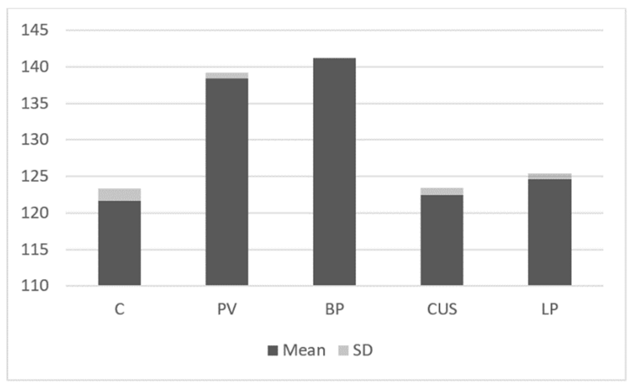
| C vs. PV p = 0.58 NS | C vs. BP p = 0.38 NS | C vs. CUS p < 0.04 | C vs. LP p < 0.02 |
| PV vs. BP p = 0.48, NS | PV vs. CUS p < 0.09 | PV vs. LP p < 0.02 | |
| BP vs. LP p = 0.23 NS | BP vs. CUS p = 0.13 NS | CUS vs. LP p = 0.05 NS |
| C vs. PV p < 0.006 | C vs. BP p < 0.001 | C vs. CUS p = 0.06 NS | C vs. LP p = 0.2 NS |
| PV vs. BP p = 0.45 NS | PV vs. CUS p < 0.05 | PV vs. LP p < 0.001 | |
| BP vs. LP p < 0.001 | BP vs. CUS p < 0.005 | CUS vs. LP p = 0.18 NS |
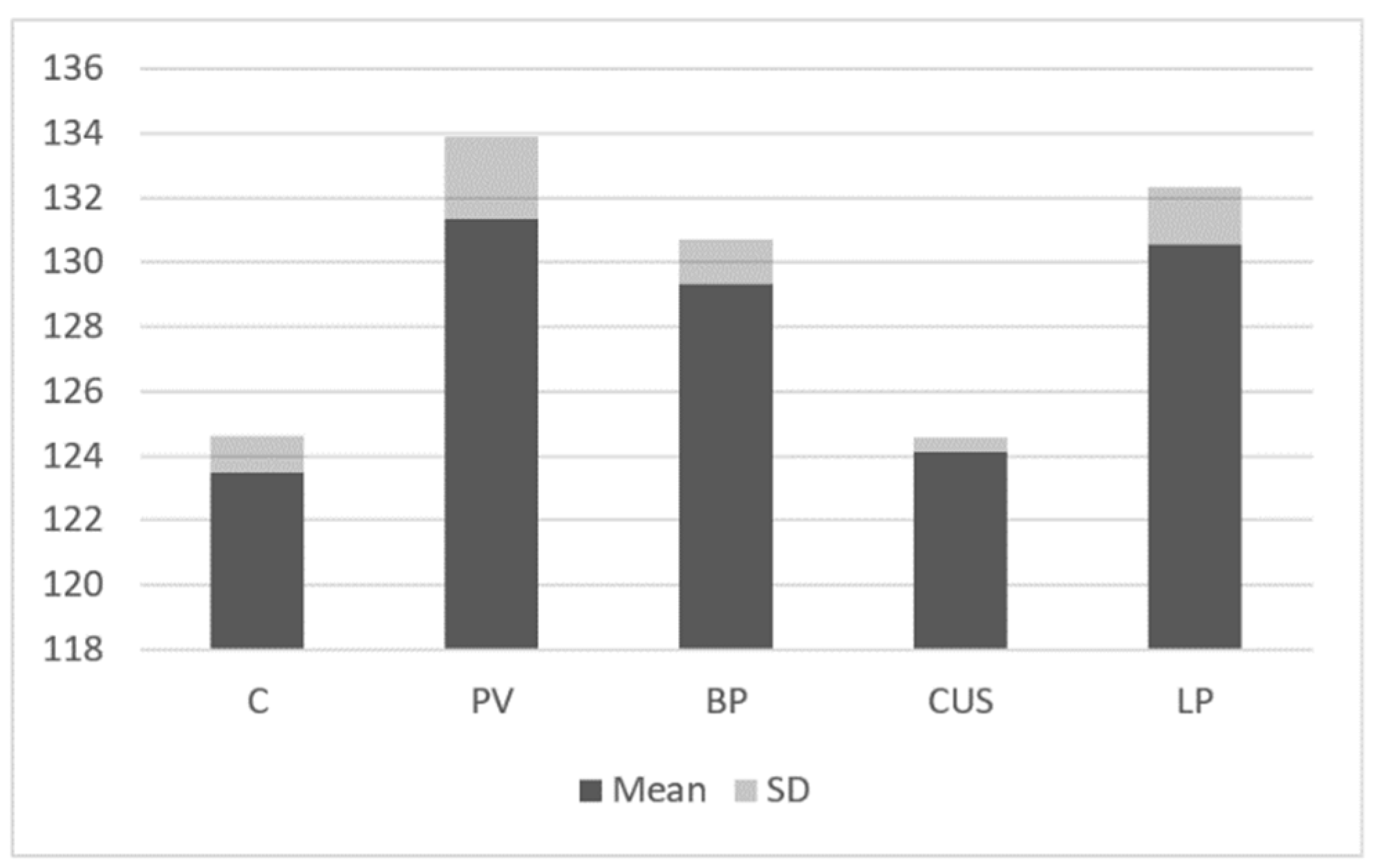
| C vs. PV p < 0.04 | C vs. BP p < 0.02 | C vs. CUS p = 0.49 NS | C vs. LP p < 0.05 |
| PV vs. BP p = 0.62 NS | PV vs. CUS p = 0.14 NS | PV vs. LP p = 0.78 NS | |
| BP vs. LP p = 0.45 NS | BP vs. CUS p = 0.14 NS | CUS vs. LP p = 0.13 NS |
| C vs. PV p < 0.02 | C vs. BP p < 0.007 | C vs. CUS p < 0.03 | C vs. LP p < 0.04 |
| PV vs. BP p = 0.7 NS | PV vs. CUS p < 0.04 | PV vs. LP p < 0.001 | |
| BP vs. LP p < 0.001 | BP vs. CUS p < 0.03 | CUS vs. LP p = 0.05 NS |
| Number of Patients | Age (Years) | Immunopathologic Results | STAT2 | STAT4 | STAT6 | JAK3 | |
|---|---|---|---|---|---|---|---|
| PV | 28 | 54.4 (45–64) | 28/28 anti Dsg3 (1:40–1:320) | - | + | + | + |
| BP | 31 | 67.3 (46–89) | 23/31 anti BMZ (1:80–1:160) 19/31 anti NC16a | - | + | + | + |
| LP | 38 | 64.6 (51–82) | Negative | + | - | + | + |
| CUS | 15 | 47.3 (22–61) | SES ANA (1:40–1:640) | + | - | - | + |
| C | 25 | 54.4 (45–64) | Negative | - | - | - | - |
Disclaimer/Publisher’s Note: The statements, opinions and data contained in all publications are solely those of the individual author(s) and contributor(s) and not of MDPI and/or the editor(s). MDPI and/or the editor(s) disclaim responsibility for any injury to people or property resulting from any ideas, methods, instructions or products referred to in the content. |
© 2022 by the authors. Licensee MDPI, Basel, Switzerland. This article is an open access article distributed under the terms and conditions of the Creative Commons Attribution (CC BY) license (https://creativecommons.org/licenses/by/4.0/).
Share and Cite
Ociepa, K.; Danilewicz, M.; Wągrowska-Danilewicz, M.; Peterson-Jęckowska, R.; Wójcicka-Rubin, A.; Lewkowicz, N.; Zajdel, R.; Żebrowska, A. Expression of the Selected Proteins of JAK/STAT Signaling Pathway in Diseases with Oral Mucosa Involvement. Int. J. Mol. Sci. 2023, 24, 323. https://doi.org/10.3390/ijms24010323
Ociepa K, Danilewicz M, Wągrowska-Danilewicz M, Peterson-Jęckowska R, Wójcicka-Rubin A, Lewkowicz N, Zajdel R, Żebrowska A. Expression of the Selected Proteins of JAK/STAT Signaling Pathway in Diseases with Oral Mucosa Involvement. International Journal of Molecular Sciences. 2023; 24(1):323. https://doi.org/10.3390/ijms24010323
Chicago/Turabian StyleOciepa, Kamila, Marian Danilewicz, Małgorzata Wągrowska-Danilewicz, Róża Peterson-Jęckowska, Angelika Wójcicka-Rubin, Natalia Lewkowicz, Radosław Zajdel, and Agnieszka Żebrowska. 2023. "Expression of the Selected Proteins of JAK/STAT Signaling Pathway in Diseases with Oral Mucosa Involvement" International Journal of Molecular Sciences 24, no. 1: 323. https://doi.org/10.3390/ijms24010323