Article

Next-Generation Sequencing in the Assessment of the Transcriptomic Landscape of DNA Damage Repair Genes in Abdominal Aortic Aneurysm, Chronic Venous Disease and Lower Extremity Artery Disease

by
Karol P. Ruszel
1,*,†,
Daniel P. Zalewski
2,†,
Andrzej Stępniewski
3,
Dariusz Gałkowski
4,
Jacek Bogucki
5,
Marcin Feldo
6,‡,
Bartosz J. Płachno
7,
Janusz Kocki
1,‡ and
Anna Bogucka-Kocka
2,‡
1
Department of Clinical Genetics, Chair of Medical Genetics, Medical University of Lublin, 11 Radziwiłłowska St., 20-080 Lublin, Poland
2
Chair and Department of Biology and Genetics, Medical University of Lublin, 4a Chodźki St., 20-093 Lublin, Poland
3
Ecotech Complex Analytical and Programme Centre for Advanced Environmentally Friendly Technologies, University of Marie Curie-Skłodowska, 39 Głęboka St., 20-612 Lublin, Poland
4
Department of Pathology and Laboratory Medicine, Rutgers-Robert Wood Johnson Medical School, One Robert Wood Johnson Place, New Brunswick, NJ 08903, USA
5
Chair and Department of Organic Chemistry, Medical University of Lublin, 4a Chodźki St., 20-093 Lublin, Poland
6
Chair and Department of Vascular Surgery and Angiology, Medical University of Lublin, 11 Staszica St., 20-081 Lublin, Poland
7
Department of Plant Cytology and Embryology, Institute of Botany, Faculty of Biology, Jagiellonian University in Kraków, 9 Gronostajowa St., 30-387 Kraków, Poland
*
Author to whom correspondence should be addressed.
These authors contributed equally to this work.
Int. J. Mol. Sci. 2023, 24(1), 551; https://doi.org/10.3390/ijms24010551
Submission received: 10 November 2022 / Revised: 17 December 2022 / Accepted: 21 December 2022 / Published: 29 December 2022

Abstract

:
Vascular diseases are one of the most common causes of death and morbidity. Lower extremity artery disease (LEAD), abdominal aortic aneurysm (AAA) and chronic venous disease (CVD) belong to this group of conditions and exhibit various presentations and courses; thus, there is an urgent need for revealing new biomarkers for monitoring and potential treatment. Next-generation sequencing of mRNA allows rapid and detailed transcriptome analysis, allowing us to pinpoint the most pronounced differences between the mRNA expression profiles of vascular disease patients. Comparison of expression data of 519 DNA-repair-related genes obtained from mRNA next-generation sequencing revealed significant transcriptomic marks characterizing AAA, CVD and LEAD. Statistical, gene set enrichment analysis (GSEA), gene ontology (GO) and literature analyses were applied and highlighted many DNA repair and accompanying processes, such as cohesin functions, oxidative stress, homologous recombination, ubiquitin turnover, chromatin remodelling and DNA double-strand break repair. Surprisingly, obtained data suggest the contribution of genes engaged in the regulatory function of DNA repair as a key component that could be used to distinguish between analyzed conditions. DNA repair–related genes depicted in the presented study as dysregulated in AAA, CVD and LEAD could be utilized in the design of new biomarkers or therapies associated with these diseases.

1. Introduction

1.1. Cardiovascular Diseases

The global burden of disease-related mortality is shaped mainly by the prevalence of noncommunicable diseases like cancer, diabetes mellitus, cardiovascular and chronic respiratory diseases. Within this set of conditions, those related to the cardiovascular system are one of the most common causes of death and morbidity. Moreover, they will exhibit a constant trend toward being more prevalent in future decades [1,2,3].
Peripheral arterial disease (PAD), along with coronary heart disease and stroke, is a pronounced cause of demise and debilitation due to atherosclerotic complications [4]. Lower extremities arterial disease (LEAD) is the most common manifestation of PAD, characterised by chronic degenerative changes in the lower limbs (like ulceration or gangrene), intermittent claudication and pain [5]. Symptoms emerge due to formation of atheromas at different aortic vessel locations. The overall global prevalence of LEAD is estimated at 5.56% of individuals aged 25 years and older and exceeds 10% in people older than 70 years [6].
Abdominal aortic aneurysm (AAA) is most commonly defined by the maximum diameter of the abdominal aorta in excess of 3 cm in either anterior–posterior or transverse planes or, alternatively, as a focal dilation at least 1.5 times larger than the diameter of the normal adjacent arterial segment [7]. The prevalence of AAA ranges between 4% and 8% in the general population of men aged 65–80 years [8]. Typical risk factors for the development of AAA include age over 60, tobacco smoking and use, male gender, Caucasian race and family history of AAA, while aneurysm growth and rupture risk appear to be associated with persistent tobacco use, female gender and chronic pulmonary disease [7].
The main characteristics of chronic venous disease (CVD) include chronic morphological and functional abnormalities of the venous system caused by venous hypertension, venous valve incompetency and reflux in veins of the lower limbs, leading to venous wall remodelling and vascular inflammation. The clinical presentation varies from telangiectasia and varicose veins to much more severe complications like leg edema, skin changes and ulcers [9,10,11,12].

1.2. Reactive Oxygen Species and DNA Repair in Cardiovascular Diseases

It is widely accepted that oxidative stress and resulting DNA damage is present in various cardiovascular conditions. In atherosclerosis, DNA damage and repair is an important element of disease onset and progression [13,14,15]. It was revealed that impaired base excision repair of oxidative DNA damage in vascular smooth muscle cells triggers atherosclerosis-related processes [16], and alterations of the expression levels of certain DNA damage genes are seen throughout the course of the disease, being present in early plaques and almost universal in advanced ones [17]. The importance of base excision repair (BER) was shown in carotid atherosclerosis, where enhanced transcriptional activity of BER genes may contribute to nuclear DNA stability [18]. Enzymes involved in oxidative DNA damage repair (like OGG1) as well as other components of DNA repair pathways (like PARP1 or ATM kinase) were shown to have an important role in both diseases reviewed in [19]. DNA repair machinery may have a somewhat ambiguous role in acute kidney disease following an ischemia-reperfusion injury event; on the one hand, it is benevolent to DNA integrity, but on the other, it could fail the completion of the DNA repair process [20].
BER could be utilized in therapies reducing ischemic injury, where enhanced mitochondrial DNA repair could have a benevolent effect on the state of the human heart muscle and cerebral vasculature in stroke models [20,21].
The presented research is a part of a larger project regarding the application of next-generation sequencing in the identification of the transcriptomic signatures of three vascular diseases: LEAD [22], CVD [23] and AAA [24]. Our previous studies focused on the principal dissimilarities and resemblances between patients with the mentioned conditions, leading to an indication of gene expression patterns being either specific or common for studied diseases [25]. This approach, utilizing application of strictly defined criteria for gene choice, allowed us to define only those genes with a very high impact on the conditions studied. Due to this fact, other genes with potential relevance in pathology may have been omitted. This prompted us to change our approach and focus on genes contributing to more specified biological processes. Our attention was attracted to DNA damage repair and metabolism. The results obtained revealed biological phenomena related to not only direct DNA repair, but also more general processes connected to regulation of DNA repair by phosphorylation or ubiquitination/deubiquitination, reactive oxygen species and chromatin remodeling, replication and transcription. Current work adds new knowledge to the area of DNA repair in vascular diseases which may provide new insights into their biomarker composition as well as into new therapeutic approaches utilizing DNA damage metabolism genes.

2. Results

2.1. Study Group Attributes

The study group consisted of seven patients with AAA, seven patients with CVD, eight patients with LEAD, and seven controls with no cardiovascular conditions. The clinical characteristics of the participants are presented in Table 1. Inclusion and exclusion criteria as well as a detailed demographical and clinical description of each study group and RNA sequencing quality data were provided in [22,23,24]. Although the statistical analysis revealed no significant differences in smoking habits and sex, there were significant (p < 0.05) dissimilarities in age and body mass index (BMI) (Table 1). While LEAD and AAA share common conspicuous risk factors, such as older age and higher BMI, the control group was devoid of any signs of vascular pathology due to the inclusion of younger, non–smoking subjects with a lower BMI. The influence of age, sex, BMI and smoking status on obtained results was further examined in the text.

2.2. The Differential Expression Analysis of DNA Damage Metabolism Genes in AAA, CVD, LEAD and Control Subjects

In order to select the set of genes engaged in DNA damage metabolism, the HUGO Gene Nomenclature Committee database (https://www.genenames.org/) (accessed on 15 June 2021) was queried with the term “DNA repair”, yielding 449 hits. To provide further details, the obtained gene list was compared with the Human DNA Repair Genes (https://www.mdanderson.org/documents/Labs/Wood-Laboratory/human-dna-repair-genes.html) (accessed on 17 June 2021), and all genes not presented in the HUGO set were added. Moreover, the gene list was supplemented with the remaining ALKB homologs (ALKBH1-8 and FTO) due to their established role in nucleic acid modification and repair [26]. After confronting gene names with names and synonyms indicated by Human Gene Database GeneCards v5.0 (https://www.genecards.org) (accessed on 18 June 2021), the final list of genes submitted to further analysis had 519 positions (Table S1).
DESeq2 and UVE-PLS (uninformative variable elimination by partial least squares) methods were used to assess the differential expression analysis of the set of 519 selected genes in pairwise comparisons between studied groups (LEAD vs. control, AAA vs. control, CVD vs. control, LEAD vs. AAA, LEAD vs. CVD and AAA vs. CVD). To supplement pairwise analysis, pooled group comparisons were also conducted. In the first instance, the transcriptomic landscape of genes in a pooled group including LEAD, AAA and CVD subjects was compared with gene expression in control group, to reveal a general scheme of DNA damage metabolism gene expression in vascular pathogenesis. The second association was performed between gene expression in a pooled group containing LEAD and AAA subjects, representing artery-related diseases and CVD patients, to pinpoint differences in DNA repair-associated pathological processes ongoing in arteries compared with venous systems.
The quality of the results was assessed and presented as Cook’s distances of genes expression across all samples (Figure S1) with accompanying MA plots and histograms of p values generated for each comparison (Figures S2–S9).
Distinct thresholds were set for further analysis; in the DESeq2 method, only genes with p < 0.05 (after adjustment using the Benjamini–Hochberg false discovery rate) and fold change > 1.2 (for upregulated genes) or fold change < 0.8 (for downregulated genes) were selected, while genes indicated by UVE-PLS as informative, with a minimum reliable score equal to 5, were considered. Both methods gave separate sets of genes in each of the comparisons (supplementary Tables S2 and S3). Genes universal for both sets were subjected to further analysis (59 genes in total). The differential expression parameters of these genes are presented in Table 2. Expression patterns of selected genes in the studied samples are presented on a heatmap with hierarchical clustering, as well as on a principal component analysis plot (Figure 1). Receiver operating characteristics (ROC) analysis was used to assess the specificity and sensitivity of selected genes as potential biomarkers for analyzed diseases. The results of the ROC analysis are presented in Table 2 (ROC-AUC values) and Table S4 (all results). Graphical representation of the relationships between sensitivity and specificity calculated for each gene (ROC plots) is presented in Figures S10–S17. Representative electropherograms of total RNA and corresponding libraries of randomly selected samples are presented in Figure S18.
Graphical representation of the results of UVE-PLS analysis is presented in Figures S19 and S20. Relationships between the categorical characteristics of study subjects (gender, smoking status) and expression of genes selected from comparisons, along with correlation analysis between characteristics (age and BMI) of studied groups and expression of genes selected from all comparisons performed, are presented on Table S6 and Table S7, respectively.
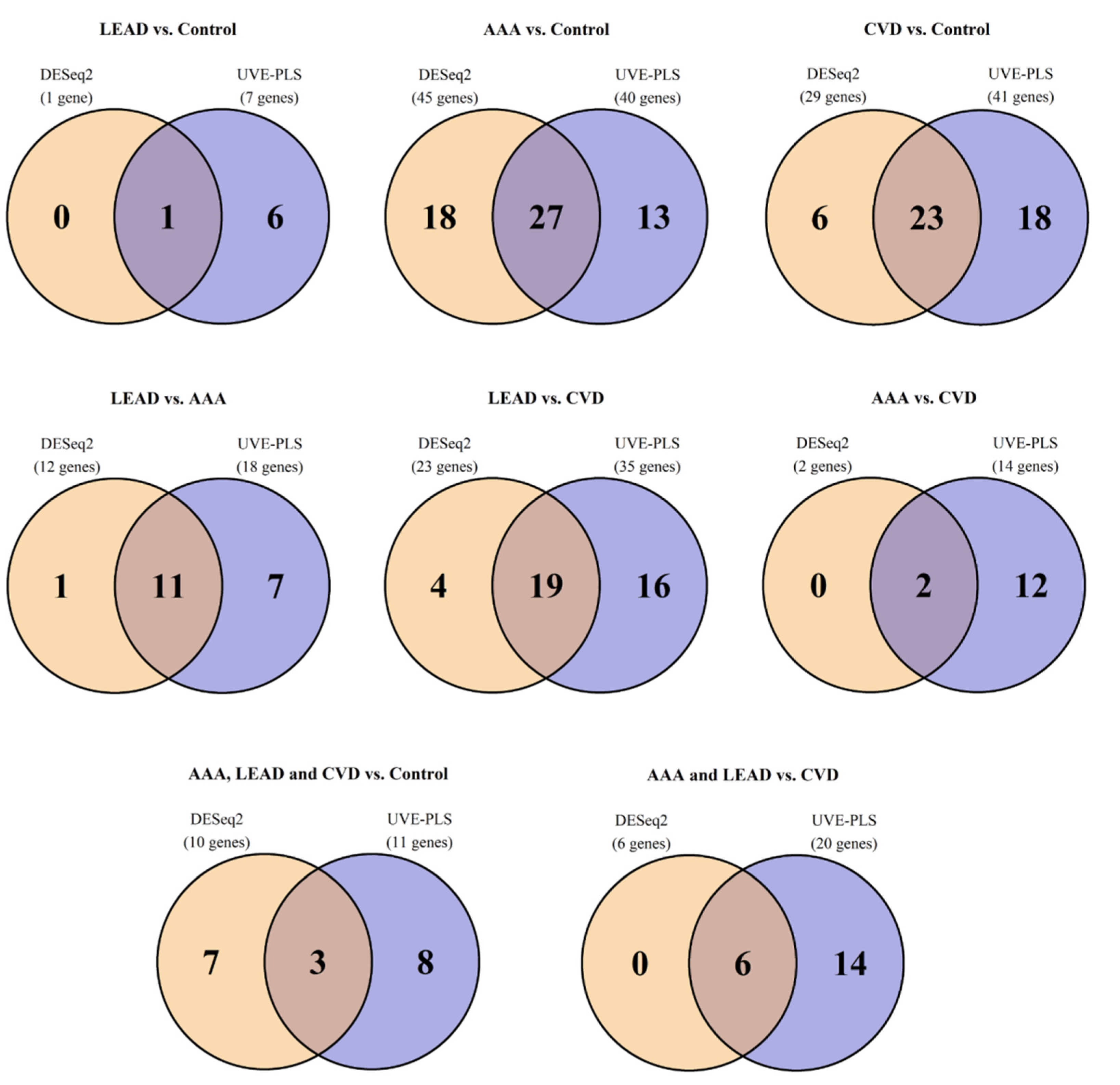
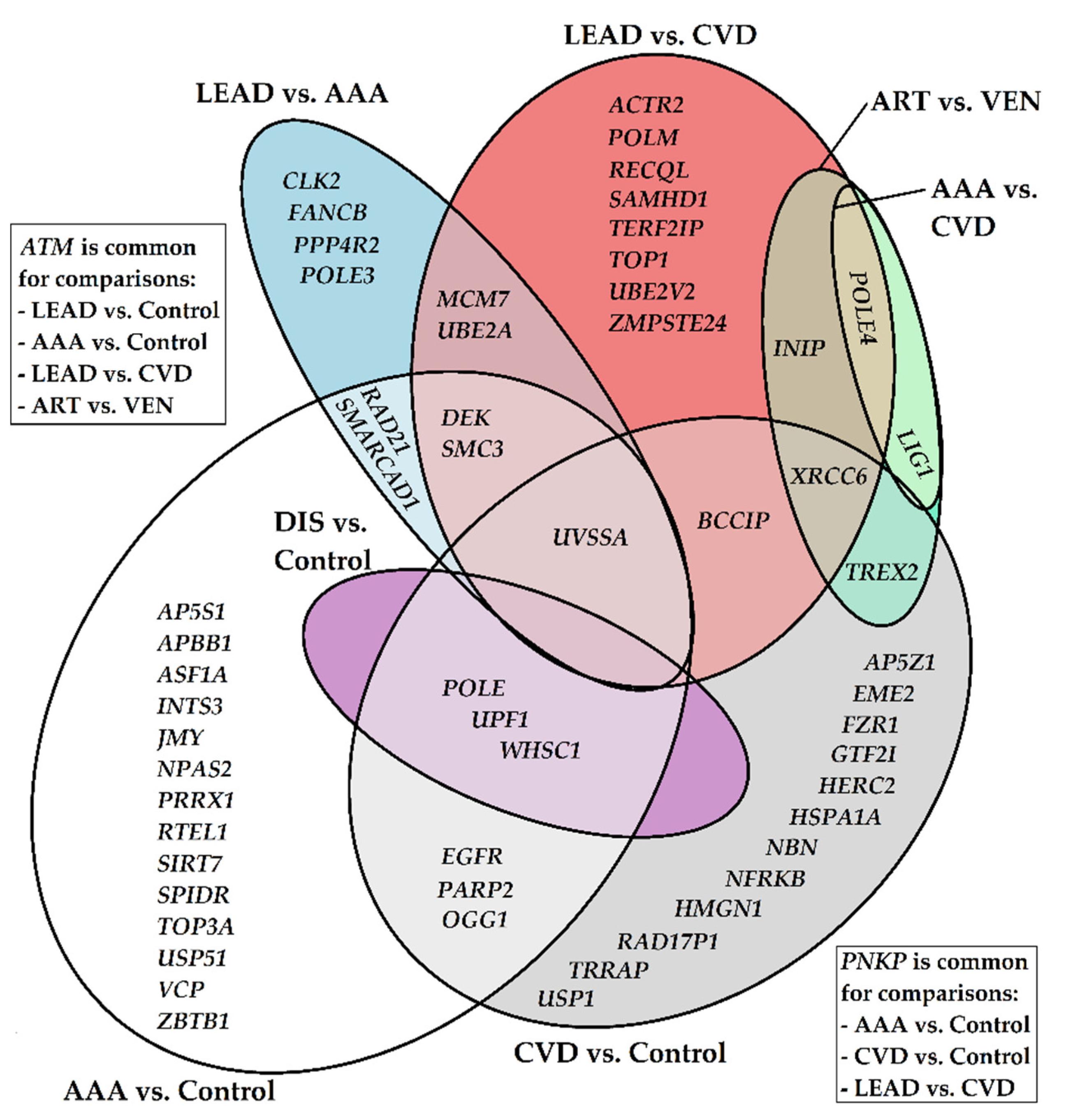
Sets of genes selected from performed comparisons are shown on a Venn Diagram to identify genes being unique or common for more than one comparison (Figure 2, Table S5).
Analysis revealed differentially expressed genes arranged in sets which are characteristic for more than two comparisons studied as well as unique ones for given comparisons (Figure 3). Throughout all comparisons, the most frequently occurring, differentially expressed genes were ATM and UVSSA (common for four comparisons). Correspondingly, there were groups of genes with less pronounced differential expression, common for three comparisons (gene names, and the most frequently occurring synonyms in brackets): PNKP, DEK, POLE, SMC3, UPF1, WHSC1 (NDS2), XRCC6 (Ku60), POLE4, and for two comparisons EGFR, RAD21, OGG1, PARP2, SMARCAD1, BCCIP, TREX2, MCM7, UBE2A, INIP (SSBIP1), LIG1.
Additionally, a large group of differentially expressed genes that are unique for performed comparisons was found as a result of analysis. Out of them, fourteen (AP5S1, APBB1 (FE65), ASF1A, INTS3, JMY, NPAS2, PRRX1, RTEL1, SIRT7, SPIDR, TOP3A, USP51, VCP (p97), ZBTB1) were differentiating the AAA and control groups; twelve (AP5Z1, EME2, FZR1, GTF2I (TFII–I), HERC2, HSPA1A, NBN (NBS1), NFRKB, HMGN1, RAD17P1, USP1, TRRAP) were exclusively characteristic of the CVD and control group comparison; and eight (ACTR2 (ARP2), POLM, RECQL, SAMHD1, TERF2IP (RAP1), TOP1, UBE2V2 (MMS2), ZMPSTE24 (STE24)) had a dissimilar pattern of expression between the LEAD and CVD groups, while only four (CLK2, FANCB, PPP4R2, POLE3) distinguished LEAD from AAA patients. Their gene products are mainly related to responsibility for various DNA repair-related and genetic information maintenance processes, such as non-homologous end joining (NHEJ), double-strand breaks (DSBs) repair, transcription, DNA replication and chromosome maintenance (Table S5). Additionally, analysis of the literature cited in the current publication has revealed a secondary, more general set of ascribed functions, such as ubiquitination and deubiquitination, kinase activity, circadian rhythm regulation, or histone modification and chromatin remodeling. Some of those genes have already been associated with various processes important in vascular physiology. Additional information regarding presented genes, including brief descriptions of related terms and processes, is placed in the Discussion section below and in Table S5.

2.3. Functional Analysis of Selected Genes

Functional analysis was performed for selected genes using two approaches: overrepresentation analysis and gene set enrichment analysis (GSEA). Overrepresentation analysis was performed to functionally annotate upregulated and downregulated gene sets selected from every comparison using the Database for Annotation, Visualization and Integrated Discovery (DAVID) v2021 [27,28] and the following categories for annotation: gene ontology (biological process, molecular function, cellular compartment), KEGG (Kyoto Encyclopedia of Genes and Genomes) and reactome. Functional terms with p value < 0.05 were taken into account (supplementary Figure S2).
The results showed that every comparison has been enriched with common terms like “DNA repair”, “DNA metabolic process”, “cellular response to DNA damage stimulus” and “cellular response to stress”, among others associated with nucleic acid metabolism. Differences in more specific terms were noticed between comparisons. Downregulated genes in the AAA vs. control group were primarily associated with “regulation of DNA metabolic process” (p = 7.9 × 10⁻¹⁰), “double-strand break repair” (p = 2.1 × 10⁻⁸), “regulation of response to DNA damage stimulus” (p = 3.3 × 10⁻⁸), “regulation of DNA repair” (p = 2.1 × 10⁻⁷) but also “chromosome organization” (p = 2.3 × 10⁻⁸), while upregulated genes were connected to “double-strand break repair” (p = 4.9 × 10⁻¹³), “regulation of double-strand break repair” (p = 7.9 × 10⁻⁷) and terms related to homologous recombination, such as “HDR through Homologous Recombination (HRR)” (p = 1.9 × 10⁻⁵) and the regulation of the DNA repair process, such as “regulation of DNA metabolic process” (p = 1 × 10⁻⁵) and “regulation of response to DNA damage stimulus” (p = 2.3 × 10⁻⁵). Downregulated genes in the CVD vs. control group were annotated to terms “double-strand break repair” (p = 3.4 × 10⁻³) and also terms connected to regulation, such as “regulation of DNA repair” (p = 3.4 × 10⁻³) and “regulation of DNA metabolic process” (p = 1.4 × 10⁻³), whereas upregulated genes represented terms like “double-strand break repair” (p = 1.7 × 10⁻⁵), “nucleotide excision repair” (p = 4.2 × 10⁻⁶) and various terms connected to “regulation of DNA repair” (p = 2.2 × 10⁻⁴). Downregulated genes in the LEAD vs. AAA group were primarily associated with “nucleoplasm” (p = 4.4 × 10⁻²), “double-strand break repair” (p = 0.0014) and “response to radiation” (p = 4.9 × 10⁻²), while upregulated ones, apart from terms connected to “double-strand break repair” (p = 1.9 × 10⁻⁶) and “regulation of double-strand break repair” (p = 0.001), were also associated with terms regarding “chromosome organization” (p = 3.2 × 10⁻⁵), “histone modification” (p = 6.2 × 10⁻⁴), and “chromatin remodeling” (p = 0.003). Upregulated genes in the LEAD vs. CVD group were ascribed to terms like “double-strand break repair” (p = 3.2 × 10⁻¹⁰), “DNA recombination” (p = 1.8 × 10⁻⁶) and “double-strand break repair via homologous recombination” (p = 1.5 × 10⁻⁴). Moreover, terms associated with various DNA repair regulatory processes, such as “regulation of DNA metabolic process” (p = 2.7 × 10⁻⁵) or “regulation of double-strand break repair” (p = 9.7 × 10⁻⁵) were present. Characteristic associations of downregulated genes from this comparison were, for example, “double-strand break repair via nonhomologous end joining” (p = 7.3 × 10⁻⁵) and “non-recombinational repair” (p = 2.55 × 10⁻⁴). Single genes from the LEAD vs. control and AAA vs. CVD comparisons were associated with terms like “histone mRNA catabolic process”, ATM; “Nucleotide Excision Repair”, “Base excision repair” and “Mismatch Repair”, LIG1; and “Nucleotide Excision Repair”, “HDR through Homologous Recombination (HRR) or Single Strand Annealing (SSA)”, “DNA Double-Strand Break Repair”, POLE4.
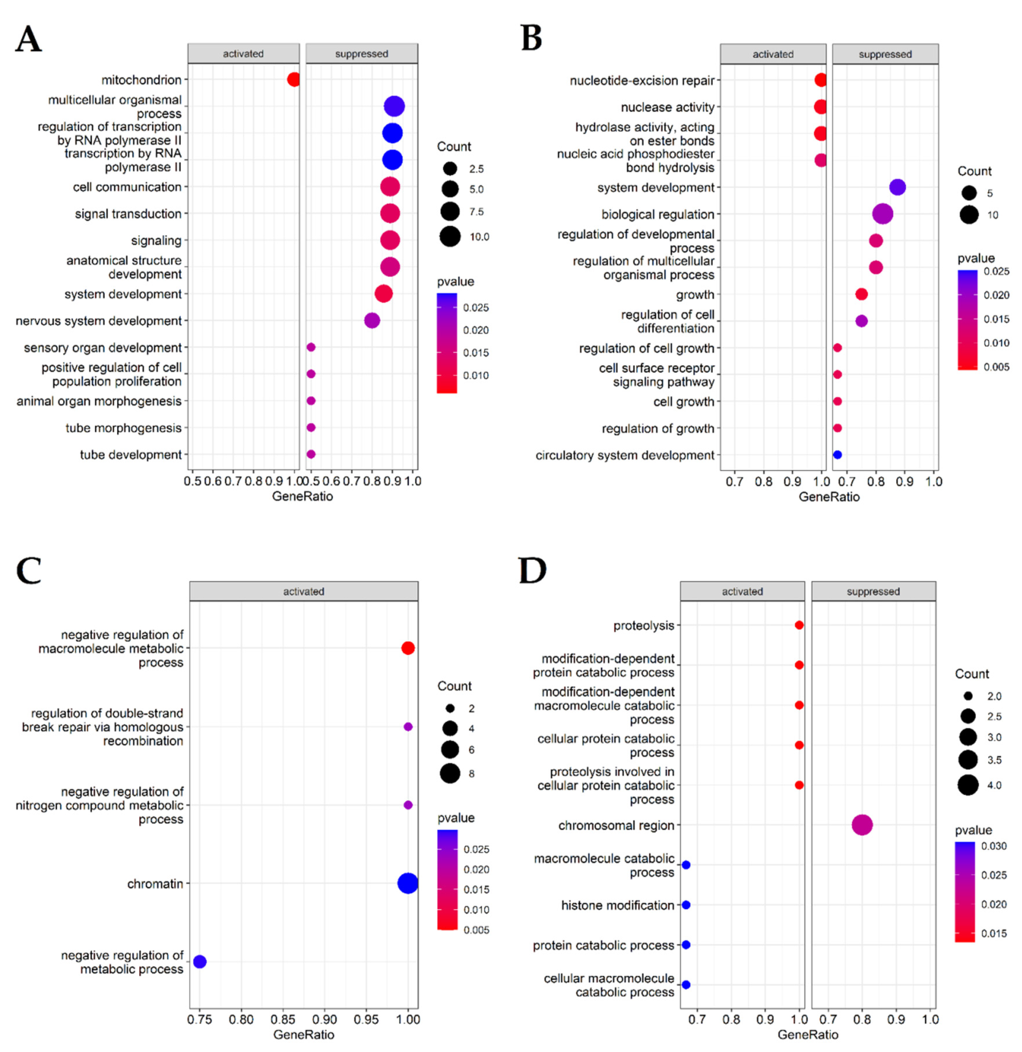
Gene set enrichment analysis (GSEA) was performed for gene ontology categories in R environment using package clusterProfiler v 4.4.4. (https://bioconductor.org/packages/release/bioc/html/clusterProfiler.html) (accessed on 12 July 2022) [29,30]. In order to obtain reliable data output, analysis was conducted for fold change-ordered gene sets selected from only those comparisons where more than ten genes were selected, namely AAA vs. control, CVD vs. control, LEAD vs. AAA and LEAD vs. CVD. Functional terms with p below 0.05 were harvested (supplementary Figure S3) and up to the top 15 functional terms with the lowest p value are presented on Figure 4.
In AAA vs. control, GO categories associated with transcription and regulation of transcription were downregulated 1.86-fold, whereas GO categories connected to “DNA–templated DNA replication” and “mitochondrion” were upregulated 1.64- and 1.68-fold, respectively. CVD vs. control categories describing nucleotide-excision repair were upregulated 1.77–1.98-fold, while those indicating various cell growth-related processes and their regulation were downregulated 1.91- to 1.98-fold. There were only upregulated GO categories in LEAD vs. AAA, including “regulation of double-strand break repair via homologous recombination” and “chromatin” with fold changes of 1.68 and 1.64, respectively. In LEAD vs. CVD, a cluster of more general categories emerged, such as “histone modification” (which increased 1.6-fold) or “proteolysis”; various references to catabolic processes and regulation were upregulated 1.81-fold.

2.4. Relationships between Gene Expression Patterns and Characteristics of the Study Groups of Patients from Performed Comparisons

Age, BMI, gender and smoking status (never and former smokers vs. current smokers) are all risk factors for cardiovascular diseases; thus, it is difficult to find a characteristics-matched group of control subjects. Due to differences in those traits between control and patient groups, statistical analysis regarding those traits was performed. Spearman rank correlation test was used in the analysis of continuous variables (age, BMI), while a two-sided Mann–Whitney U test was used in the analysis of categorical variables (gender and smoking status). A statistical significance of p < 0.05, corrected by the Benjamini–Hochberg false discovery rate, was set as a selection criterion for both tests. Genes selected from the correlation analysis are presented in Table 3 (the entire correlation results are provided in Table S6). PRRX1, USP51, VCP were correlated, albeit moderately, with both age and BMI in the AAA vs. control group. Apart from that, single genes were also moderately correlated either with BMI or age in different comparisons. Therefore, the differential character of their expression patterns may be determined to some extent by differences in age and BMI between studied groups (Table 1). There were only few statistically significant relationships regarding categorical characteristics. APBB1 (p = 0.036) expression levels were correlated with smoking status and POLM (p = 0.015) and XRCC6 (p = 0.023) with gender (Table S7).

3. Discussion

DNA has the most important biological function; it bears the information of a blueprint of the living entity. Despite that, this crucial biological polymer is under constant threat of damage through the action of a plethora of chemical and physical factors.
It was earlier established that some DNA repair-related genes and their protein products are also engaged in various vascular-related processes, such as vascular endothelial growth factor A (VEGFA) function (RAD21) [31], muscle stem cell activation (DEK) [32], atherosclerosis (MORF4L2 [33], FZR1 [34]), neointimal formation (SIRT7) [35] and foam cell formation (USP10) [36], and have been associated with cardiovascular risk factors (TRRAP [37], NPAS2 [38]). This prompted the present research focusing on similarities and differences in expression of 519 genes known to be related to DNA repair and other interconnected processes (Table S1). Out of them, 59 genes were identified as differentially expressed in at least one disease (Table 2). The transcriptional landscape of mentioned genes was assessed in PBMCs obtained from AAA, CVD and LEAD patients as well as from healthy volunteers included in the control group. Both pairwise and pooled groups comparisons were analyzed (AAA vs. control; CVD vs. control; LEAD vs. control; AAA vs. CVD; LEAD vs. AAA; LEAD vs. CVD and LEAD; AAA and CVD vs. control; LEAD and AAA vs. CVD). From each of those, genes resulting from DESeq2 (with Benjamini–Hochberg FDR p < 0.05) and indicated by UVE-PLS to be informative were selected. Relationships between the expression of selected genes and age, BMI, gender and smoking habits were also assessed.
All additional comments and information regarding genes described in the main text, along with an introductory description of DNA damage metabolism pathways and extended discussion, were placed in supplementary text section. Analysis pointed especially to genes contributing to reactive oxygen species’ response and DNA double-strand break repair as the most prominent in the mentioned diseases.

3.1. Gene Ontology (GO) and Gene Set Enrichment Analysis (GSEA) Reveals Terms and Categories Associated with Biological Process Regulation, DNA Repair Regulation, Double-Strand Break Repair and Homologous Recombination, Differentiating AAA, CVD and LEAD

Downregulated genes in the AAA vs. control group were primarily associated with various terms related to the regulation of DNA metabolism and repair, but also to transcription and the regulation of transcription, which may indicate decreased DNA repair activities associated with the transcription of genes required for DNA repair. On the other hand, terms describing the regulation of double-strand break repair, nucleotide-excision repair and various terms related to homologous recombination were overrepresented, leading to assumption that DSBs may be pronounced in AAA.
In CVD vs. control, genes ascribed to term “double-strand break repair” and “regulation of DNA repair” form two distinct groups, both upregulated (TRRAP, PNKP, OGG1, NFRKB for “regulation of DNA repair” and AP5Z1, TRRAP, EME2, PNKP, OGG1, ARID2 for “double-strand break repair”) and downregulated (USP1, EGFR, GTF2I for “regulation of DNA repair” and XRCC6, PARP2, RAD21, NBN for “double-strand break repair”) for each term, different in composition. This could be indicative of different modes of regulation of DNA repair present in CVD patients. Downregulated genes in the LEAD vs. AAA group were primarily associated with the replication of DNA, and “regulation of DNA metabolic process”. The term “double-strand break repair”, similarly to CVD, had ascribed both upregulated (PPP4R2, RAD21, SMARCAD1, DEK, FANCB) and downregulated (MCM7, PNKP) groups of genes. This again points to DSBs and regulation of their repair as prominent features in those two diseases. Upregulation of terms connected to histone modification and chromatin may reveal chromatin remodeling processes distinguishing LEAD and AAA. While upregulated genes in LEAD vs. CVD were ascribed to terms like double-strand break repair and homologous recombination, there was significant downregulation of genes responsible for repair of DSBs via the NHEJ pathway, indicating that HR is the main pathway of DSBs’ repair in LEAD, in comparison with CVD.
Gene set enrichment analysis (GSEA) was performed for comparisons where more than ten genes were shown as differentiating, namely: AAA vs. control, CVD vs. control, Lead vs. AAA and LEAD vs. CVD. Results are shown in Figure 4, and in supplementary Figure S3. In AAA vs. control group, pathways concerning various aspects of RNA and transcription and “positive regulation of cell population proliferation” were suppressed; whereas, telomere and mitochondrion processes were activated, which may indicate a decrease in metabolic activity caused by senescence. In CVD vs. control, the nucleotide excision repair pathway with accompanying enzymatic activities was activated, whereas general pathways were suppressed, suggesting elevated NER pathway activity affecting wide-scale regulation of biological processes. In the case of LEAD vs. AAA comparison, GSEA found terms ascribed to activated pathways, including “chromatin” and “regulation of double-strand break repair via homologous recombination”, which may indicate that those pathways distinguish LEAD and AAA. In the LEAD vs. CVD comparison, more general pathways were revealed, such as activated ones connected to proteolysis, and suppressed ones, such as the “chromosomal region”. This may be indicative of more general differences present in LEAD and CVD which require in-depth analysis. Such analysis is provided in the discussion below and in the supplementary text.

3.2. Expression of DNA Double Strand Break Repair Genes Is Altered in Vascular Diseases

Due to their detrimental effects and toxicity, double-strand breaks require rapid detection, safeguarding and prompt repair. This forces the mutual action of many proteins. The repair process is not only directed at enzymatic activities processing damaged DNA; it is also highly regulated by accessory proteins [39,40].
Three gene-encoding products possessing various enzymatic activities needed in HR were shown to be differentially expressed and characteristic for multiple comparisons, namely PNKP, POLE, UPF1 (Table S5).
Protein products of those genes contribute to the processing of free DNA ends (PNKP) [41], the sensing and resection of DNA in DSBs (POLE) [42,43] and the possessing DNA/RNA helicase function in the resection of DNA in DSBs (UPF1) [44]. REQL is a helicase preventing replication fork collapse during replication stress, which is needed for genomic stability [45,46]. Contrary to PNKP, POLE and UPF1, REQL has been found as upregulated only in LEAD vs. CVD.
The second group of genes involved in DSB repair is composed of gene-encoding proteins engaged mainly in DNA binding, with less consistent expression changes through the compared groups. It includes XRCC6 (Ku70), EGFR, INIP (SSBIP1) and INTS3. Proteins encoded by these genes play a critical role in efficient HR–dependent repair of DSBs and ATM-dependent signaling pathways [47,48].
It is tempting to assume that revealed dependencies in expression changes could be indicative of diverse regulation of the same process in various diseases, creating the possibility of targeted therapies and diagnosis.
The third group of genes consists of genes responsible for the regulation of the DSB repair process. It includes PARP2, SMARCAD1, ARID2 (Baf200) (in a set of genes common for at least two comparisons) and AP5S1, APBB1 (FE65), ASF1A, SIRT7, SPIDR, USP51 and VCP (p97) (unique for the AAA vs. control group comparison) and SAMHD1 (exclusive to the LEAD vs. CVD comparison).
The AAA vs. control group comparison was the richest in uniquely differentially expressed genes (eighteen positions). Among them, eight genes were ascribed to regulation of the DSBs repair process, on the basis of cited literature (AP5S1, APBB1, ASF1A, SIRT7, SPIDR, USP51, VCP).
An increase in the activity of the SIRT7 gene points to increased DSB repair [49], suggesting an alleviating action in AAA patients; meanwhile, a decrease in said gene expression may be responsible for impairing strand-break repair and inducing senescence in patients’ cells, potentially aggravating the course of AAA.
Elevated expression of SPIDR may indicate fine tuning and regulation of homologous recombination [50,51] in AAA patients.
DNA DSBs repair by homologous recombination is facilitated by SAMHD1 through promotion of DNA ends resection [52]. Its expression is upregulated in LEAD vs. CVD patients, again suggesting not only distinct regulation of homologous recombination, but also inclusion of inflammation [53] in LEAD and CVD.
There were other genes present in sets common for at least two comparisons (BCCIP, LIG1) and unique for only one comparison (RTEL1, TOP3A, EME2, FANCB) contributing to homologous recombination [54,55,56,57,58,59,60,61].
The composition of transcriptional patterns points to homologous repair as the main DSBs repair mechanism predominant in AAA, but occurring also in both LEAD and CVD.
To further support engagement of homologous repair of DSB in the presented diseases, non-homologous end joining-related gene (POLM, ASF1A, ERCC6L2, USP51) transcription was examined [62,63,64,65,66].
All NHEJ-related gene transcription was decreased, suggesting a shift to HR as the main mechanism of DNA DSBs in AAA patients.
Interestingly, genes identified by the current analysis were engaged more in different modes of DSBs repair regulation than in specific enzymatic activities. This allows us to speculate that the regulation of basic processes of DNA repair could be characteristic of given vascular disease, reflecting different onset, progression and specific presentations, while the main enzymatic activities proceeding HR remain unaltered. This could be a prerequisite for targeted treatment and/or discriminative diagnosis. Counterintuitive though may it seem, elevated activity of HR may promote excess DNA damage due to a rise in free DNA end resection which thus causes an increase in DNA damage intermediates known to cause DNA damage when unrepaired [67,68]. This is in line with the fact that intensified DNA damage may aggravate presentations of vascular diseases.

3.3. Specific Gene Expression Changes May Be Indicative for Altered Oxidative DNA Damage Responses in AAA, CVD and LEAD

Oxidative stress is both the crucial hallmark of cardiovascular diseases and their causative factor [69]. The MRGX protein was suggested to possess a role in defense against reactive oxygen species (ROS) [70]. MRGX, along with ERCC5 (XPG) gene, encodes protein taking part in NER and HR [71,72]. Downregulation of MRGX may indicate impaired response to elevated oxidative stress, especially in AAA and CVD patients. Analysis of transcriptional patterns of genes presented in this research is supported by the findings of other groups that oxidative stress is the prominent feature of many vascular diseases, including those assessed in the current paper.

3.4. Analysis Reveals Known Genes Being Involved in Vascular Diseases’ Initiation and Progression

Cardiovascular diseases have long been described as ‘multifactorial’ [73], involving a plethora of genetic and environmental factors contributing to the overall disease outcome.
Gene contribution revealed in our studies and according to the literature analysed varies from the regulation of a vascular-specific processes to commitment to a specific risk factor or cardiovascular system defects. The transcription pattern is also variable, ranging from four comparison occurrences (ATM and UVSSA) to exclusiveness to only one comparison.
Protein products of those genes take part in signal transduction from vascular endothelial growth factor (VEGF) receptors (RAD21, [39]), muscle stem cell activation (DEK [40]), the promotion of plaque instability through accelerated senescence (MORF4L2 [41]) and the regulation of inflammation and proliferation of cells (TREX2 [74]). The OGG glycosylase is the main glycosylase; it removes the most abundant DNA lesion resulting from oxidation, 8–oxoG, possessing not only enzymatic functions, but also contributing to cellular signaling and epigenetic-like pathways [75,76,77]. It has a dual action; on the one hand, it is an important modulatory factor restricting inflammatory responses and apoptosis in macrophages preventing atherosclerosis [78], while on the other it has the ability to upregulate pro-inflammatory gene expression [77]. Thus, elevated expression of OGG1 in AAA and CVD may have ambiguous effects: it may be benevolent through alleviation of DNA damage or promote inflammation through its ability to bind to DNA and upregulate pro-inflammatory gene expression [77].
Gene sets correlated with both vascular-related processes/terms and DNA repair, unique for one comparison, only had the following composition. In AAA patients (AAA vs. control group) there were six genes with differential expression: NPAS2 (MOP4), PRRX1, RTEL1, SIRT7, USP10, VCP (p97). In the LEAD vs. CVD group, there were three such genes: ACTR2 (ARP2), SAMHD1, TERF2IP (RAP1), HERC2. In CVD patients (CVD vs. control group), there were two characteristic genes: HSPA1A, TRRAP; only one appeared in the LEAD vs. AAA comparison (CLK2).
Interestingly, besides correlation with known vascular diseases and DNA repair, some genes point to the circadian oscillator being an important element of those conditions: NPAS2 [79], HERC2 [80] and CLK2 [81]. This may suggest some interesting therapeutic opportunities, such as application of chronotherapy, in vascular diseases. This approach takes into account the daily oscillation of various metabolic processes governing uptake, transport and activation of medicines in order to apply different treatment regimens for the best outcome [80].
  • AAA vs. control group: NPAS2 (MOP4), PRRX1, RTEL1, SIRT7, USP10, VCP (p97)
Pathological vasculogenesis and arteriolar remodeling are hallmarks of different vascular diseases; both PPRX and RTEL1 are known to be engaged in those processes [82,83], and their altered expression may influence AAA presentation in patients.
The SIRT7 gene product shapes VSMCs proliferation and migration in neointimal formation following vascular injury [84], while USP10 dysregulation may affect VSMC-derived foam cells formation also in AAA [85,86]. Moreover, elevated levels of VCP expression may facilitate the stability of the angiotensin type 1 receptor and enhance Ang II blood pressure elevating effects in AAA patients [87,88].
  • LEAD vs. CVD group: ACTR2 (ARP2), SAMHD1, TERF2IP (RAP1)
These genes are associated with various processes and correlated with various vasculopathies. They contribute to pathological cell motility, enhancing neointima formation after vascular injury (ACTR2 [89]), cerebral vasculopathy in Aicardi–Goutieres syndrome (SAMHD1 [90,91,92]) and cells’ activation, senescence and apoptosis (TERF2IP [93]).
  • CVD vs. control group: HSPA1A (HSP70), TRRAP
Decreased expression of HSPA1A may be an element of compromised tolerance for stresses present in the course of CVD, compromising defense mechanisms including DNA repair [94]. The TRRAP protein is engaged in the regulation of lipid metabolism through stimulation of lipid droplet formation [37], and positively regulates cholesterol metabolism and intake [95].

3.5. Chromatin Remodelling Could Be the Process Shaping and Regulating DNA Damage Responses in Vascular Diseases

DNA in eukaryotes tightly interacts with many specialized proteins, such as histones, building a highly organized entity called chromatin. Current NGS analysis did detect some of differentially expressed genes whose products are needed in various chromatin remodeling processes. In group of genes ascribed to more than one comparison, those were ATM, POLE and POLE4, WHSC1 (NDS2), SMARCAD1, ARID2 (Baf200).
The ATM kinase is the node factor in a vast biochemical network of reciprocal interactions, signaling the presence of DNA damage and controlling the repair process [96]. Recently, it was discovered that two of subunits (POLE3 and POLE4) are histone H3–H4 chaperones, maintaining chromatin integrity during DNA replication [97]. Another gene connected to posttranslational modifications of histones, encoding histone methyltransferase, is WHSC1; meanwhile, HMGN1 binds to 147-bp nucleosome core particle in purely structure-dependent manner [98] and facilitates UV light and ionizing irradiation-induced DNA damage [99,100]. Altered expression of genes encoding proteins belonging to chromatin-remodeling complexes, such as ARID2 [101] ACTR2 [102,103] and NRFKB [104], has also been observed in our transcriptomic data.
Differential expression of genes related to chromatin remodeling clearly indicates the important contribution of this process in the regulation of DNA damage in the presented vascular diseases. What is interesting, and could be assumed, is that similarly to DNA DSBs repair, the regulation of various chromatin–related processes is an important factor to distinguish between vascular diseases. Those differences could have a tremendous impact on disease course and presentation.

3.6. Ubiquitination and Deubiquitination Could Be a Prominent Mechanism Regulating Gene Activities in Vascular Diseases

One of potent mechanisms of protein function regulation is ubiquitination, defined as a covalent binding of ubiquitin protein to various other proteins. Current NGS analysis did detect some of differentially expressed genes, whose products are needed in various ubiquitin-related processes. Those genes were UBE2A, USP10, USP51, ZBTB1, HERC2, and SHPRH.
The UBE2A (RAD6A) gene encodes an enzyme whose function is monoubiquitination of PCNA and taking part in DNA damage response to ionizing radiation [105,106]. Upregulation of UBE2A was characteristic of LEAD in comparison with AAA and CVD.
USP10 and USP51 are deubiquitinases with increased expression in AAA patients. USP10 deubiquitinates and stabilizes MutS Homolog 2 (MSH2), which is the key DNA mismatch repair protein [107]. USP51 is a histone H2A deubiquitinase whose depletion results in the impairment of DNA damage response [44]. The ZBTB1 protein regulates monoubiquitinatination of proliferating cell nuclear antigen (PCNA) at DNA damage sites [108]. Expression of ZBTB1 was decreased in AAA patients.
The HERC2 protein is an E3 ubiquitin ligase negatively regulating both HR and NER [80,109]. Upregulation of this gene is present in CVD patients, suggesting that ubiquitination alterations may have a characteristic pattern in this disease.
Presented results show a connection between DNA damage responses and ubiquitin metabolism, showing possible different modes of regulation of DNA damage. Moreover, these seem to be specific for presented diseases, having potential value in both specific diagnosis and treatment.

3.7. Limitations of the Study and Accompanying Issues

Undoubtedly, the descriptive character of our study raised some limitations and additional questions. We are aware that a bias concerning age, BMI, sex and smoking exists in our data. The expression levels of the presented genes may be affected by those characteristics; thus, differential expression of genes could not be exclusive to the disease status, but their expression levels could be affected by the mentioned factors, and statistically significant differences between these variables could be observed (Table 1). Coherently, obtained results could be disrupted and undeniably, some relationships between picked genes and age and BMI can be found (Table 3). The main reason for their appearance is the groups’ construction. First and foremost, during control group construction, we have adhered to the selection of completely healthy persons with no cardiovascular conditions, which resulted in age, gender and smoking differences. It is clear that the characteristics of groups with different vascular diseases reflect their epidemiology; the disease mainly affects persons with predisposing traits. Consequently, age and BMI are well-known risk factors for all vascular diseases; therefore, selected genes could contribute to these diseases through age- and BMI-associated mechanisms [25]. There were only few correlations regarding categorical characteristics. APBB1 expression levels were correlated with smoking status, and POLM and XRCC6 with gender (Table S7).
The experimental design was initially focused on NGS analysis of the whole transcriptome and mirnome of patients [23,24,25], with subsequent determination of commonalities and dissimilarities present in the transcriptome of AAA, CVD and LEAD cases [25]. On this basis, our attention later converged on more specific processes such as DNA damage metabolism. PCR validation of our data was extremely difficult due to the fact that we were unable to predict the final number of genes resulting from analysis. That information was crucial for the design of the PCR experiment (e.g., the amount of material needed, the number of particular probes and the amount of master mix to purchase). For the same reasons, no protein expression validation, such as ELISA or Western blot, was conducted. Therefore, in order to recompensate the lack of qPCR validation and protein detection assays, broader and more advanced statistical analysis (DESeq2 with Benjamini–Hochberg false discovery rate correction, confirmed by UVE-PLS and ROC analysis) was applied, which substantially limited potential false positive results.
The number of screened patients could be considered low; however, in analogous research conducted so far, one can observe similar or even lower quantities of screened subjects. Moreover, there are a great variety of approaches to cardiovascular disease assessment. These issues are discussed in detail in the supplementary material in [22]. Interestingly, St. Laurent et al. discovered that most of statistical methods can detect reliable transcripts even when as few as three biological replicates are sequenced using single-molecule based RNAseq [110]. Despite all of the above, we are aware that a higher number of screened patients may be beneficial in our future research for discovering new disease-associated patterns in complicated processes such as DNA damage repair.
The typical subjectively chosen threshold for changes in gene expression is about 2–3 fold. St. Laurent et al. shed some light on this issue and drew the conclusion that even though a plethora of differentially expressed RNAs exhibit fold changes less than two, they still retain viable and biologically relevant information. This happens even at very low fold changes, and exclusion of those mRNAs may cause a loss of data and a needless drain on the discovery process [110]. Moreover, additive effects affecting the physiological response may be present in genes with low fold change in expression. Additionally, many of the indicated genes have regulatory function in DNA repair, so may exhibit big biologically relevant effects even with small expression changes. This is why it was finally decided that the terminal gene set for all analyses would contain genes with fold changes ranging from 0.8 (downregulated) to 1.2 (upregulated).
We are aware that the presented studies mainly have an initial and descriptive character, and we do not imply that the presented results and interpretations are definitive. Conclusions deduced from established gene sets need to be confirmed in the future by more detailed experiments. The explanatory character of transcriptomic landscape of DNA damage metabolism in vascular diseases and its role in pathology should be elucidated in detail in further studies. Presented results could be reassessed by other techniques like qPCR, flow cytometry, Western blot, gene expression modification in model systems by means of genetic engineering, and experiments with animal models and human cells or artificial tissues. These could be accompanied by techniques utilized by traditional DNA repair research, such as comet assays, micronuclei indexes, DNA modifications detection and oxidative stress assessment. Moreover, subsequent validation studies should encompass much larger and character-balanced populations. Similarly to preceding publications, we wished to share the findings of the current research to give the opportunity to start a discussion within the scientific community, to propose new, explorative paths for other research groups [25] and to broaden knowledge about DNA repair in the context of vascular diseases.

4. Materials and Methods

4.1. Study Groups Participants Characteristics

The study group consisted of four assemblages: LEAD (8 patients), AAA (7 patients), CVD (7 patients) and control group of healthy volunteers (7 persons). All subjects were assessed by a vascular surgeon in Independent Public Clinical Hospital No. 1 in Lublin. All persons gave informed and signed consent. All mentioned procedures were performed in accordance with the Declaration of Helsinki. The Ethics Committee of Medical University of Lublin approved the study design (resolution No. KE–0254/341/2015). The clinical characteristics regarding all participants are presented in Table 1. A detailed description of clinical features, as well as inclusion procedures and exclusion criteria were provided in previous papers [32,33,34].

4.2. Gene Expression Datasets Generation

Next-generation sequencing was the method of choice for the assessment of transcriptomic profiles of PBMCs from study participants. An entire detailed methodology with accompanying data regarding sequencing data quality was provided in previous papers [22,23,24].
Briefly, blood samples were subjected to PBMCs isolation using density gradient centrifugation with Gradisol L reagent (Aqua–Med, Łódź, Poland). Concisely, total RNA specimens (for transcriptome analysis) were isolated from PBMCs samples of vascular diseases patients and randomly selected controls using TRI Reagent Solution (Applied Biosystems, Foster City, CA, USA). The quality of total RNA samples was assessed using the Agilent 2100 Bioanalyzer instrument with the Agilent RNA 6000 Pico Kit for total RNA samples and the Agilent DNA 1000 Kit for libraries ([22] and supplementary Figure S20). Subsequently, total RNA preparations were submitted to ribosomal RNA removal with use of RiboMinus Eukaryote System v2 (Ambion, Austin, TX, USA). Transcriptome libraries were prepared using the Ion Total RNA-Seq Kit v2, Magnetic Bead Cleanup Module kit, Ion Xpress RNA-Seq Barcode 01-16 Kit and sequenced on Ion 540 chips (all Life Technologies, Carlsbad, CA, USA) using the Ion S5 XL System (Thermo Fisher Scientific, Waltham, MA, USA). Sequencing reaction of libraries was performed with Ion 540 chips (Life Technologies) using an Ion S5 XL System (Thermo Fisher Scientific, Waltham, MA, USA). An alignment of raw sequences of the hg19 version of the human genome was made with the help of Torrent Suite Software v5.0.4. and the Ion Torrent RNASeq Analysis plugin v.5.0.3.0 (Thermo Fisher Scientific). Raw sequences of transcriptomic libraries were aligned to 55,765 known genes, their splicing variants and the putative open reading frames of hg19 human genome.

4.3. Data Analysis Methods and Tools

R environment (R version 4.1.0, https://www.r-project.org) (accessed on 18 May 2021) and appropriate packages according to the provided reference manuals were used for statistical analyses and data visualizations. Kruskal–Wallis rank sum test (kruskal.test function in R) was applied to assess the statistical significance of differences in age and BMI between the LEAD, AAA, CVD and control groups. A two-sided Fisher’s exact test (fisher.test function in R) was applied to assess the statistical significance of differences in gender and smoking status between the studied groups.
Differential expression analysis was performed on data originating from biological replicates. DESeq2 and UVE-PLS (uninformative variable elimination by partial least squares) [111,112] methods were used, and the latter works especially well with partial least squares as a multivariate regression technique fitted for dealing with large sets of noisy and correlated variables as well as with rather small numbers of samples [113].
DESeq2 analysis was carried out using a DESeq2 1.26.0 package [111] (https://bioconductor.org/packages/release/bioc/html/DESeq2.html) (accessed on 18 May 2021) on expression data devoid of genes with mean number of reads lower than one. Differentially expressed genes were considered as statistically significant when the criterion of a p value below 0.05 after Benjamini–Hochberg false discovery rate correction was met.
A plsVarSel 0.9.6 package [114] (https://cran.r-project.org/web/packages/plsVarSel/index.html) (accessed on 18 May 2021) was used to perform UVE-PLS analysis. The initial data was transformed by regularized log normalization (rlog function in the DESeq2 package). 1000 iterations, a 0.75 ratio for splitting into trained and tested data subsets and a minimum reliable score equal to 5 cutoff threshold were the values used in UVE-PLS analysis. The number of PLS (partial least squares) components for UVE-PLS analysis (Table A1 in Appendix A) was established by inspecting the estimated root mean squared error of prediction (RMSEP) determined by PLS regression with leave-one-out (LOO) cross-validation (Figures S19 and S20).
The pROC package 1.16.2 [115] (https://cran.r-project.org/web/packages/pROC/index.html) (accessed on 19 May 2021) was used to conduct the ROC analysis.
Boxplots were generated using the ggplot2 3.3.0 package (https://ggplot2.tidyverse.org) (accessed on 29 August 2021).
The statistical significance of categorical characteristics (gender and smoking habits) was assessed with a two-sided Mann–Whitney U test implemented in wilcox.test function in R. Statistical significance of continuous clinical features (age and BMI) was performed using a Spearman rank correlation test implemented in the Hmisc package 4.4–0. (https://cran.r-project.org/web/packages/Hmisc/index.html) (accessed on 29 August 2021).
Overrepresentation functional analysis was performed by the Database for Annotation, Visualization and Integrated Discovery (DAVID) (https://david.ncifcrf.gov/home.jsp (accessed on 11 July 2022)) [27,28]. The list of genes resulting from each of comparisons was uploaded, and analysis was run on given settings: Select Identifier—OFFICIAL_GENE_SYMBOL; Select species—Homo sapiens; List Type—Gene List; Annotation Summary Results: No defaults; Gene_Ontology: GOTERM_BP_ALL, GOTERM_CC_ALL, GOTERM_MF_ALL; Pathways: KEGG_PATHWAY, REACTOME_PATHWAY. Selected annotations were combined by functional annotation clustering and placed in supplementary Figure S2.
Gene set enrichment analysis (GSEA) was performed in R environment with package clusterProfiler v4.4.4. (https://bioconductor.org/packages/release/bioc/html/clusterProfiler.html) (accessed on 12 July 2022) [29,30]. Analysis was performed for comparisons where more than 10 genes were selected, namely AAA vs. control, CVD vs. control, Lead vs. AAA and LEAD vs. CVD. Results are shown in supplementary Figure S3.

4.4. Advantages of Peripheral Blood Mononuclear Cells’ (PBMCs) Utilization for DNA Damage Metabolism and Vascular Conditions Assessment

Blood mononuclear cells were widely used in the assessment of not only various atherosclerotic risk factors like hypertension [116], type 2 diabetes [117], and hyperlipidemia [118], but also plaque vulnerability itself [119]. The contribution of PBMCs to vascular pathology is now widely accepted [120]. DNA damage is present both in the vascular bed in circulating cells and cells present in the lesions of vascular walls [15]. Exemplary patients with coronary artery disease possess a higher micronuclei index (a marker of genetic instability) than healthy controls, which correlates with disease severity [121]. PBMCs have been confirmed to be a good source of biological material exhibiting evidence of DNA damage in coronary artery disease. Moreover, DNA damage is confirmed in early atherosclerotic lesions and becomes overabundant in advanced plaques [16]. PBMCs encompass biological material which is easily accessible and appropriate for routine clinical practice without using expensive, labor- and time-consuming methods like cell sorting and flow cytometry. One of the goals of this study was to determine markers that could be applied in an easy test for the diagnosis and prediction of vascular diseases. Careful selection of possible tissues favored the use of blood circulating cells as an excellent biological material [22].

5. Conclusions

The presented research sheds light on DNA damage repair in patients with three vascular diseases: AAA, CVD and LEAD. In-depth data analysis revealed gene sets that could be ascribed to numerous DNA repair processes, pointing especially to the prominent homologous repair presence in all probed diseases. Data also indicate a significant crosstalk between diverse pathways interrelated with DNA repair, encompassing chromatin remodeling, cohesin metabolism and function and ubiquitin turnover, and oxidative stress presence and management. These characteristics show significant commonalities in processes contributing to the presented vascular diseases. Striking differences appear when the analysis takes into account the regulatory component present in the examined diseases. Data suggest that gene-encoding proteins responsible for control and fine–tuning of mentioned processes exhibit the most interesting dissimilarities in expression. This could be the key that describes and distinguishes one condition from the other, and may explain the relative richness of their clinical presentations and courses. This is also a prerequisite for the creation of new diagnostic methods and therapies utilizing DNA repair as the target of intervention.

Supplementary Materials

The following supporting information can be downloaded at: https://www.mdpi.com/article/10.3390/ijms24010551/s1.

Author Contributions

Conceptualization, M.F., J.K. and A.B.-K.; Data curation, K.P.R., D.P.Z., A.S. and M.F.; Formal analysis, K.P.R., D.P.Z. and A.B.-K.; Funding acquisition, A.B.-K.; Investigation, K.P.R. and D.P.Z.; Methodology, K.P.R., D.P.Z., M.F., J.K. and A.B.-K.; Project administration, A.S., J.K. and A.B.-K.; Resources, A.S., M.F., J.K. and A.B.-K.; Software, K.P.R., D.P.Z., A.S. and J.B.; Supervision, A.S., M.F., J.K. and A.B.-K.; Validation, K.P.R., D.P.Z., M.F. and A.B.-K.; Visualization, K.P.R. and D.P.Z.; Writing—original draft, K.P.R. and D.P.Z.; Writing—review and editing, D.G., M.F., B.J.P. and A.B.-K. All authors have read and agreed to the published version of the manuscript.

Funding

This research was funded by statutory funds of the Medical University of Lublin, number DS43 to A.B.-K.

Institutional Review Board Statement

The study was conducted according to the guidelines of the Declaration of Helsinki, and approved by the Ethics Committee of Medical University of Lublin (protocol code KE–0254/341/2015, 2015).

Informed Consent Statement

Informed consent was obtained from all subjects involved in the study.

Data Availability Statement

Acknowledgments

The research was performed using the equipment purchased within the project “The equipment of innovative laboratories doing research on new medicines used in the therapy of civilization and neoplastic diseases” within the Operational Program Development of Eastern Poland 2007–2013, Priority Axis I Modern Economy, Operations I.3 Innovation Promotion. Presented results were partially obtained using the apparatus located in the Center ECOTECH–COMPLEX in Lublin, purchased under the project “ECOTECH–COMPLEX Man, Environment, Production” project No POIG.02.01.00–06–212/09 funded from the European Regional Development Fund 2007–2013 by the National Center for Research and Development in Poland.

Conflicts of Interest

The authors declare no conflict of interest.

Abbreviations

8–oxoG8–oxo–7:8–dihydroguanine
AAAAbdominal aortic aneurysm
BAF200BAF Nuclear Assembly Factor 1
BERBase Excision Repair
BMIBody mass index
CVDChronic Venous Disease
DSBsDouble-strand breaks
FDRFalse Discovery Rate
HUGOHuman Genome Organization
LEADLower Extremities Arterial Disease
MA plotBland–Altman plot for visual representation of genomic data
NERNucleotide excision repair
NGSNext-Generation Sequencing
NHEJNon–Homologous End Joining
p53Tumor Protein P53
PADPeripheral Arterial Disease
PBMCsPeripheral blood mononuclear cells
ROCReceiver Operating Characteristics
ROSReactive oxygen species
Swi/Snf complexSWItch/Sucrose Non–Fermentable complex
UVUltraviolet
UVE–PLSUninformative Variable Elimination by Partial Least Squares
VEGFAVascular Endothelial Growth Factor A
VSMCsVascular Smooth Muscle Cells

Appendix A

Table A1. General results of differential gene expression analysis performed using DESeq2 and UVE-PLS methods obtained for comparisons.
ComparisonDESeq2UVE–PLSNumber of Genes Common for Sets of Genes Selected from DESeq2 (p < 0.05) and from UVE–PLS as Informative
Number of Differentially Expressed Genes with p < 0.05 and Fold Change > 1.2 (for Upregulated Genes) or Fold Change < 0.8 (for Downregulated Genes)Number of PLS Components/IterationsNumber of Informative Genes (Default Cutoff Threshold of Reliability Score)
LEAD vs. control14/100071
AAA vs. control452/10004027
CVD vs. control293/10004123
LEAD vs. AAA123/10001811
LEAD vs. CVD232/10003519
AAA vs. CVD22/1000142
LEAD, AAA and CVD vs. control103/1000113
LEAD and AAA vs. CVD64/1000206
p—statistical significance after correction by Benjamini–Hochberg false discovery rate, AAA—abdominal aortic aneurysm, CVD—chronic venous disease, LEAD—lower extremity artery disease, UVE–PLS—uninformative variable elimination by partial least squares.

References

  1. Fuster, V.; Kelly, B.B. Promoting Cardiovascular Health in the Developing World: A Critical Challenge to Acheive Global Health; The National Academies Press: Washington, DC, USA, 2010. [Google Scholar]
  2. Joseph, P.; Leong, D.; McKee, M.; Anand, S.S.; Schwalm, J.D.; Teo, K.; Mente, A.; Yusuf, S. Reducing the global burden of cardiovascular disease, part 1: The epidemiology and risk factors. Circ. Res. 2017, 121, 677–694. [Google Scholar] [CrossRef] [PubMed]
  3. Kaptoge, S.; Pennells, L.; De Bacquer, D.; Cooney, M.T.; Kavousi, M.; Stevens, G.; Riley, L.M.; Savin, S.; Khan, T.; Altay, S.; et al. World Health Organization cardiovascular disease risk charts: Revised models to estimate risk in 21 global regions. Lancet Glob. Health. 2019, 7, e1332–e1345. [Google Scholar] [CrossRef] [PubMed] [Green Version]
  4. Criqui, M.H.; Matsushita, K.; Aboyans, V.; Hess, C.N.; Hicks, C.W.; Kwan, T.W.; McDermott, M.M.; Misra, S.; Ujueta, F. American Heart Association Council on Epidemiology and Prevention; Council on Arteriosclerosis, Thrombosis and Vascular Biology; Council on Cardiovascular Radiology and Intervention; Council on Lifestyle and Cardiometabolic Health; Council on Peripheral Vascular Disease; and Stroke Council Lower Extremity Peripheral Artery Disease: Contemporary Epidemiology, Management Gaps, and Future Directions: A Scientific Statement From the American Heart Association. Circulation 2021, 144, e171–e191. [Google Scholar] [CrossRef] [PubMed]
  5. Aboyans, V.; Ricco, J.B.; Bartelink, M.L.E.L.; Björck, M.; Brodmann, M.; Cohnert, T.; Collet, J.P.; Czerny, M.; De Carlo, M.; Debusa, S.; et al. ESC Guidelines on the Diagnosis and Treatment of Peripheral Arterial Diseases, in collaboration with the European Society for Vascular Surgery (ESVS). Eur. Heart J. 2017, 39, 763–816. [Google Scholar] [CrossRef] [Green Version]
  6. Song, P.; Rudan, D.; Zhu, Y.; Fowkes, F.J.I.; Rahimi, K.; Fowkes, F.G.R.; Rudan, I. Global, regional, and national prevalence and risk factors for peripheral artery disease in 2015: An updated systematic review and analysis. Lancet Glob. Health 2019, 7, e1020–e1030. [Google Scholar] [CrossRef] [Green Version]
  7. Ullery, B.W.; Hallett, R.L.; Fleischmann, D. Epidemiology and contemporary management of abdominal aortic aneurysms. Abdom. Radiol. 2018, 43, 1032–1043. [Google Scholar] [CrossRef]
  8. Lattanzi, S. Abdominal aortic aneurysms: Pathophysiology and clinical issues. J. Intern. Med. 2020, 288, 376–378. [Google Scholar] [CrossRef]
  9. Eberhardt, R.T.; Raffetto, J.D. Chronic venous insufficiency. Circulation 2014, 130, 333–346. [Google Scholar] [CrossRef]
  10. Ligi, D.; Croce, L.; Mannello, F. Chronic venous disorders: The dangerous, the good, and the diverse. Int. J. Mol. Sci. 2018, 19, 2544. [Google Scholar] [CrossRef] [Green Version]
  11. Mansilha, A.; Sousa, J. Pathophysiological mechanisms of chronic venous disease and implications for venoactive drug therapy. Int. J. Mol. Sci. 2018, 19, 1669. [Google Scholar] [CrossRef]
  12. Lurie, F.; Passman, M.; Meisner, M.; Dalsing, M.; Masuda, E.; Welch, H.; Bush, R.L.; Blebea, J.; Carpentier, P.H.; De Maeseneer, M.; et al. The 2020 update of the CEAP classification system and reporting standards. J. Vasc. Surg. Venous Lymphat. Disord. 2020, 8, 342–352. [Google Scholar] [CrossRef] [PubMed]
  13. Cervelli, T.; Borghini, A.; Galli, A.; Andreassi, M.G. DNA damage and repair in atherosclerosis: Current insights and future perspectives. Int. J. Mol. Sci. 2012, 13, 16929–16944. [Google Scholar] [CrossRef] [PubMed] [Green Version]
  14. Martinet, W.; Knaapen, M.W.; De Meyer, G.R.; Herman, A.G.; Kockx, M.M. Elevated levels of oxidative DNA damage and DNA repair enzymes in human atherosclerotic plaques. Circulation 2002, 106, 927–932. [Google Scholar] [CrossRef] [PubMed] [Green Version]
  15. Mahmoudi, M.; Mercer, J.; Bennett, M. DNA damage and repair in atherosclerosis. Cardiovasc. Res. 2006, 71, 259–268. [Google Scholar] [CrossRef]
  16. Shah, A.; Gray, K.; Figg, N.; Finigan, A.; Starks, L.; Bennett, M. Defective Base Excision Repair of Oxidative DNA Damage in Vascular Smooth Muscle Cells Promotes Atherosclerosis. Circulation 2018, 138, 1446–1462. [Google Scholar] [CrossRef] [PubMed]
  17. Shah, N.; Meira, L.B.; Elliott, R.M.; Hoole, S.P.; West, N.E.; Brown, A.J.; Bennett, M.R.; Garcia-Garcia, H.M.; Kuku, K.O.; Dan, K.; et al. DNA Damage and Repair in Patients with Coronary Artery Disease: Correlation with Plaque Morphology Using Optical Coherence Tomography (DECODE Study). Cardiovasc. Revasc. Med. 2019, 20, 812–818. [Google Scholar] [CrossRef] [PubMed]
  18. Skarpengland, T.; Dahl, T.B.; Skjelland, M.; Scheffler, K.; de Sousa, M.; Gregersen, I.; Kuśnierczyk, A.; Sharma, A.; Slupphaug, G.; Eide, L.; et al. Enhanced base excision repair capacity in carotid atherosclerosis may protect nuclear DNA but not mitochondrial DNA. Free Radic. Biol. Med. 2016, 97, 386–397. [Google Scholar] [CrossRef]
  19. Hu, L.; Wang, Z.; Carmone, C.; Keijer, J.; Zhang, D. Role of Oxidative DNA Damage and Repair in Atrial Fibrillation and Ischemic Heart Disease. Int. J. Mol. Sci. 2021, 22, 3838. [Google Scholar] [CrossRef]
  20. Simon, R.; Meller, R.; Yang, T.; Pearson, A.; Wilson, G. Enhancing Base Excision Repair of Mitochondrial DNA to Reduce Ischemic Injury Following Reperfusion. Transl. Stroke Res. 2019, 10, 664–671. [Google Scholar] [CrossRef] [Green Version]
  21. Marín-García, J. Mitochondrial DNA repair: A novel therapeutic target for heart failure. Heart Fail. Rev. 2016, 21, 475–487. [Google Scholar] [CrossRef]
  22. Bogucka-Kocka, A.; Zalewski, D.P.; Ruszel, K.P.; Stępniewski, A.; Gałkowski, D.; Bogucki, J.; Komsta, Ł.; Kołodziej, P.; Zubilewicz, T.; Feldo, M.; et al. Dysregulation of MicroRNA Regulatory Network in Lower Extremities Arterial Disease. Front. Genet. 2019, 10, 1200. [Google Scholar] [CrossRef] [PubMed] [Green Version]
  23. Zalewski, D.P.; Ruszel, K.P.; Stępniewski, A.; Gałkowski, D.; Bogucki, J.; Komsta, Ł.; Kołodziej, P.; Chmiel, P.; Zubilewicz, T.; Feldo, M.; et al. Dysregulations of MicroRNA and Gene Expression in Chronic Venous Disease. J. Clin. Med. 2020, 9, 1251. [Google Scholar] [CrossRef] [PubMed]
  24. Zalewski, D.P.; Ruszel, K.P.; Stępniewski, A.; Gałkowski, D.; Bogucki, J.; Komsta, Ł.; Kołodziej, P.; Chmiel, P.; Zubilewicz, T.; Feldo, M.; et al. Dysregulation of microRNA Modulatory Network in Abdominal Aortic Aneurysm. J. Clin. Med. 2020, 9, 1974. [Google Scholar] [CrossRef] [PubMed]
  25. Zalewski, D.P.; Ruszel, K.P.; Stępniewski, A.; Gałkowski, D.; Bogucki, J.; Kołodziej, P.; Szymańska, J.; Płachno, B.J.; Zubilewicz, T.; Feldo, M.; et al. Identification of Transcriptomic Differences between Lower Extremities Arterial Disease, Abdominal Aortic Aneurysm and Chronic Venous Disease in Peripheral Blood Mononuclear Cells Specimens. Int. J. Mol. Sci. 2021, 22, 3200. [Google Scholar] [CrossRef] [PubMed]
  26. Ougland, R.; Rognes, T.; Klungland, A.; Larsen, E. Non—Homologous functions of the AlkB homologs. J. Mol. Cell Biol. 2015, 7, 494–504. [Google Scholar] [CrossRef] [Green Version]
  27. Sherman, B.T.; Hao, M.; Qiu, J.; Jiao, X.; Baseler, M.W.; Lane, H.C.; Imamichi, T.; Chang, W. DAVID: A web server for functional enrichment analysis and functional annotation of gene lists (2021 update). Nucleic Acids Res. 2022, 50, W216–W221. [Google Scholar] [CrossRef]
  28. Huang, D.W.; Sherman, B.T.; Lempicki, R.A. Systematic and integrative analysis of large gene lists using DAVID Bioinformatics Resources. Nat. Protoc. 2009, 4, 44–57. [Google Scholar] [CrossRef]
  29. Wu, T.; Hu, E.; Xu, S.; Chen, M.; Guo, P.; Dai, Z.; Feng, T.; Zhou, L.; Tang, W.; Zhan, L.; et al. clusterProfiler 4.0: A universal enrichment tool for interpreting omics data. Innovation 2021, 2, 100141. [Google Scholar] [CrossRef]
  30. Yu, G.; Wang, L.G.; Han, Y.; He, Q.Y. clusterProfiler: An R package for comparing biological themes among gene clusters. OMICS J. Integr. Biol. 2012, 16, 284–287. [Google Scholar] [CrossRef]
  31. Matsumoto, T.; Mugishima, H. Signal transduction via vascular endothelial growth factor (VEGF) receptors and their roles in atherogenesis. J. Atheroscler. Thromb. 2006, 13, 130–135. [Google Scholar] [CrossRef]
  32. Yue, L.; Wan, R.; Luan, S.; Zeng, W.; Cheung, T.H. Dek Modulates Global Intron Retention during Muscle Stem Cells Quiescence exit. Dev. Cell 2020, 53, 661–676.e6. [Google Scholar] [CrossRef]
  33. Zhang, R.; Ji, Z.; Yao, Y.; Zuo, W.; Yang, M.; Qu, Y.; Su, Y.; Ma, G.; Li, Y. Identification of hub genes in unstable atherosclerotic plaque by conjoint analysis of bioinformatics. Life Sci. 2020, 262, 118517. [Google Scholar] [CrossRef] [PubMed]
  34. Zhang, Y.M.; Meng, L.B.; Yu, S.J.; Ma, D.X. Identification of potential crucial genes in monocytes for atherosclerosis using bioinformatics analysis. Int. J. Med. Res. 2020, 48, 300060520909277. [Google Scholar] [CrossRef] [PubMed] [Green Version]
  35. Zheng, J.; Chen, K.; Wang, H.; Chen, Z.; Xi, Y.; Yin, H.; Lai, K.; Liu, Y. SIRT7 Regulates the Vascular Smooth Muscle Cells Proliferation and Migration via Wnt/β—Catenin Signaling Pathway. Biomed. Res. Int. 2018, 2018, 4769596. [Google Scholar] [CrossRef] [PubMed] [Green Version]
  36. Xia, X.; Hu, T.; He, J.; Xu, Q.; Yu, C.; Liu, X.; Shao, Z.; Liao, Y.; Huang, H.; Liu, N. USP10 deletion inhibits macrophage—Derived foam cell formation and cellular—Oxidized low density lipoprotein uptake by promoting the degradation of CD36. Aging 2020, 12, 22892–22905. [Google Scholar] [CrossRef]
  37. Abbey, D.; Conlon, D.; Rainville, C.; Elwyn, S.; Quiroz-Figueroa, K.; Billheimer, J.; Schultz, D.C.; Hand, N.J.; Cherry, S.; Rader, D.J. Lipid droplet screen in human hepatocytes identifies TRRAP as a regulator of cellular triglyceride metabolism. Clin. Transl. Sci. 2021, 14, 1369–1379. [Google Scholar] [CrossRef] [PubMed]
  38. Hoffman, A.E.; Zheng, T.; Ba, Y.; Zhu, Y. The circadian gene NPAS2, a putative tumor suppressor, is involved in DNA damage response. Mol. Cancer Res. MCR 2008, 6, 1461–1468. [Google Scholar] [CrossRef] [Green Version]
  39. Shrivastav, M.; De Haro, L.P.; Nickoloff, J.A. Regulation of DNA double-strand break repair pathway choice. Cell Res. 2008, 18, 134–147. [Google Scholar] [CrossRef] [Green Version]
  40. Marini, F.; Rawal, C.C.; Liberi, G.; Pellicioli, A. Regulation of DNA Double Strand Breaks Processing: Focus on Barriers. Front. Mol. Biosci. 2019, 6, 55. [Google Scholar] [CrossRef]
  41. Jilani, A.; Ramotar, D.; Slack, C.; Ong, C.; Yang, X.M.; Scherer, S.W.; Lasko, D.D. Molecular cloning of the human gene, PNKP, encoding a polynucleotide kinase 3’-phosphatase and evidence for its role in repair of DNA strand breaks caused by oxidative damage. J. Biol. Chem. 1999, 274, 24176–24186. [Google Scholar] [CrossRef]
  42. Casari, E.; Gobbini, E.; Gnugnoli, M.; Mangiagalli, M.; Clerici, M.; Longhese, M.P. Dpb4 promotes resection of DNA double—Strand breaks and checkpoint activation by acting in two different protein complexes. Nat. Commun. 2021, 12, 4750. [Google Scholar] [CrossRef] [PubMed]
  43. Pursell, Z.F.; Isoz, I.; Lundström, E.B.; Johansson, E.; Kunkel, T.A. Yeast DNA polymerase epsilon participates in leading—Strand DNA replication. Science 2007, 317, 127–130. [Google Scholar] [CrossRef] [PubMed] [Green Version]
  44. Ngo, G.; Grimstead, J.W.; Baird, D.M. UPF1 promotes the formation of R loops to stimulate DNA double—Strand break repair. Nat. Commun. 2021, 12, 3849. [Google Scholar] [CrossRef] [PubMed]
  45. Benedict, B.; van Bueren, M.A.; van Gemert, F.P.; Lieftink, C.; Guerrero Llobet, S.; van Vugt, M.A.; Beijersbergen, R.L.; Te Riele, H. The RECQL helicase prevents replication fork collapse during replication stress. Life Sci. Alliance 2020, 3, e202000668. [Google Scholar] [CrossRef]
  46. Debnath, S.; Sharma, S. RECQ1 Helicase in Genomic Stability and Cancer. Genes 2020, 11, 622. [Google Scholar] [CrossRef]
  47. Li, J.; Ma, X.; Banerjee, S.; Baruah, S.; Schnicker, N.J.; Roh, E.; Ma, W.; Liu, K.; Bode, A.M.; Dong, Z. Structural basis for multifunctional roles of human Ints3 C—Terminal domain. J. Biol. Chem. 2021, 296, 100112. [Google Scholar] [CrossRef]
  48. Jia, Y.; Cheng, Z.; Bharath, S.R.; Sun, Q.; Su, N.; Huang, J.; Song, H. Crystal structure of the INTS3/INTS6 complex reveals the functional importance of INTS3 dimerization in DSB repair. Cell Discov. 2021, 7, 66. [Google Scholar] [CrossRef]
  49. Vazquez, B.N.; Thackray, J.K.; Simonet, N.G.; Kane-Goldsmith, N.; Martinez-Redondo, P.; Nguyen, T.; Bunting, S.; Vaquero, A.; Tischfield, J.A.; Serrano, L. SIRT7 promotes genome integrity and modulates non-homologous end joining DNA repair. EMBO J. 2016, 35, 1488–1503. [Google Scholar] [CrossRef]
  50. Prakash, R.; Sandoval, T.; Morati, F.; Zagelbaum, J.A.; Lim, P.X.; White, T.; Taylor, B.; Wang, R.; Desclos, E.; Sullivan, M.R.; et al. Distinct pathways of homologous recombination controlled by the SWS1—SWSAP1—SPIDR complex. Nat. Commun. 2021, 12, 4255. [Google Scholar] [CrossRef]
  51. Martino, J.; Brunette, G.J.; Barroso-González, J.; Moiseeva, T.N.; Smith, C.M.; Bakkenist, C.J.; O’Sullivan, R.J.; Bernstein, K.A. The human Shu complex functions with PDS5B and SPIDR to promote homologous recombination. Nucleic Acids Res. 2019, 47, 10151–10165. [Google Scholar] [CrossRef]
  52. Daddacha, W.; Koyen, A.E.; Bastien, A.J.; Head, P.E.; Dhere, V.R.; Nabeta, G.N.; Connolly, E.C.; Werner, E.; Madden, M.Z.; Daly, M.B.; et al. SAMHD1 Promotes DNA End Resection to Facilitate DNA Repair by Homologous Recombination. Cell Rep. 2017, 20, 1921–1935. [Google Scholar] [CrossRef] [PubMed] [Green Version]
  53. Rice, G.I.; Bond, J.; Asipu, A.; Brunette, R.L.; Manfield, I.W.; Carr, I.M.; Fuller, J.C.; Jackson, R.M.; Lamb, T.; Briggs, T.A.; et al. Mutations involved in Aicardi-Goutières syndrome implicate SAMHD1 as regulator of the innate immune response. Nat. Genet. 2009, 41, 829–832. [Google Scholar] [CrossRef] [PubMed] [Green Version]
  54. Lu, H.; Guo, X.; Meng, X.; Liu, J.; Allen, C.; Wray, J.; Nickoloff, J.A.; Shen, Z. The BRCA2—Interacting protein BCCIP functions in RAD51 and BRCA2 focus formation and homologous recombinational repair. Mol. Cell. Biol. 2005, 25, 1949–1957. [Google Scholar] [CrossRef] [PubMed] [Green Version]
  55. Jurkiw, T.J.; Tumbale, P.P.; Schellenberg, M.J.; Cunningham-Rundles, C.; Williams, R.S.; O’Brien, P.J. LIG1 syndrome mutations remodel a cooperative network of ligand binding interactions to compromise ligation efficiency. Nucleic Acids Res. 2021, 49, 1619–1630. [Google Scholar] [CrossRef]
  56. Bellelli, R.; Youds, J.; Borel, V.; Svendsen, J.; Pavicic-Kaltenbrunner, V.; Boulton, S.J. Synthetic Lethality between DNA Polymerase Epsilon and RTEL1 in Metazoan DNA Replication. Cell Rep. 2020, 31, 107675. [Google Scholar] [CrossRef]
  57. Björkman, A.; Johansen, S.L.; Lin, L.; Schertzer, M.; Kanellis, D.C.; Katsori, A.M.; Christensen, S.T.; Luo, Y.; Andersen, J.S.; Elsässer, S.J.; et al. Human RTEL1 associates with Poldip3 to facilitate responses to replication stress and R—Loop resolution. Genes Dev. 2020, 34, 1065–1074. [Google Scholar] [CrossRef]
  58. Wyatt, H.D.; Sarbajna, S.; Matos, J.; West, S.C. Coordinated actions of SLX1-SLX4 and MUS81-EME1 for Holliday junction resolution in human cells. Mol. Cell 2013, 52, 234–247. [Google Scholar] [CrossRef] [Green Version]
  59. Amangyeld, T.; Shin, Y.K.; Lee, M.; Kwon, B.; Seo, Y.S. Human MUS81-EME2 can cleave a variety of DNA structures including intact Holliday junction and nicked duplex. Nucleic Acids Res. 2014, 42, 5846–5862. [Google Scholar] [CrossRef] [Green Version]
  60. Falquet, B.; Rass, U. Structure-Specific Endonucleases and the Resolution of Chromosome Underreplication. Genes 2019, 10, 232. [Google Scholar] [CrossRef] [Green Version]
  61. Nomura, Y.; Adachi, N.; Koyama, H. Human Mus81 and FANCB independently contribute to repair of DNA damage during replication. Genes Cells 2007, 12, 1111–1122. [Google Scholar] [CrossRef]
  62. Ghosh, D.; Raghavan, S.C. 20 years of DNA Polymerase μ, the polymerase that still surprises. FEBS J. 2021, 288, 7230–7242. [Google Scholar] [CrossRef] [PubMed]
  63. Lee, K.Y.; Im, J.S.; Shibata, E.; Dutta, A. ASF1a Promotes Non—Homologous End Joining Repair by Facilitating Phosphorylation of MDC1 by ATM at Double-Strand Breaks. Mol. Cell 2017, 68, 61–75.e5. [Google Scholar] [CrossRef] [Green Version]
  64. Francica, P.; Mutlu, M.; Blomen, V.A.; Oliveira, C.; Nowicka, Z.; Trenner, A.; Gerhards, N.M.; Bouwman, P.; Stickel, E.; Hekkelman, M.L.; et al. Functional Radiogenetic Profiling Implicates ERCC6L2 in Non-homologous End Joining. Cell Rep. 2020, 32, 108068. [Google Scholar] [CrossRef] [PubMed]
  65. Hang, Q.; Zeng, L.; Wang, L.; Nie, L.; Yao, F.; Teng, H.; Deng, Y.; Yap, S.; Sun, Y.; Frank, S.J.; et al. Non-canonical function of DGCR8 in DNA double-strand break repair signaling and tumor radioresistance. Nat. Commun. 2021, 12, 4033. [Google Scholar] [CrossRef]
  66. Wang, Z.; Zhang, H.; Liu, J.; Cheruiyot, A.; Lee, J.H.; Ordog, T.; Lou, Z.; You, Z.; Zhang, Z. USP51 deubiquitylates H2AK13,15ub and regulates DNA damage response. Genes Dev. 2016, 30, 946–959. [Google Scholar] [CrossRef] [Green Version]
  67. Aquila, L.; Atanassov, B.S. Regulation of Histone Ubiquitination in Response to DNA Double Strand Breaks. Cells 2020, 9, 1699. [Google Scholar] [CrossRef] [PubMed]
  68. Keyamura, K.; Arai, K.; Hishida, T. Srs2 and Mus81-Mms4 Prevent Accumulation of Toxic Inter-Homolog Recombination Intermediates. PLoS Genet. 2016, 12, e1006136. [Google Scholar] [CrossRef] [PubMed] [Green Version]
  69. Cervantes Gracia, K.; Llanas-Cornejo, D.; Husi, H. CVD and Oxidative Stress. J. Clin. Med. 2017, 6, 22. [Google Scholar] [CrossRef] [Green Version]
  70. Bomfim, M.M.; Andrade, G.M.; Del Collado, M.; Sangalli, J.R.; Fontes, P.K.; Nogueira, M.; Meirelles, F.V.; da Silveira, J.C.; Perecin, F. Antioxidant responses and deregulation of epigenetic writers and erasers link oxidative stress and DNA methylation in bovine blastocysts. Mol. Reprod. Dev. 2017, 84, 1296–1305. [Google Scholar] [CrossRef]
  71. Friedberg, E.C.; Wood, R.D. New insights into the combined Cockayne/xeroderma pigmentosum complex: Human XPG protein can function in transcription factor stability. Mol. Cell 2007, 26, 162–164. [Google Scholar] [CrossRef]
  72. Ferri, D.; Orioli, D.; Botta, E. Heterogeneity and overlaps in nucleotide excision repair disorders. Clin. Genet. 2020, 97, 12–24. [Google Scholar] [CrossRef] [Green Version]
  73. Schwartz, S.M.; Schwartz, H.T.; Horvath, S.; Schadt, E.; Lee, S.I. A systematic approach to multifactorial cardiovascular disease: Causal analysis. Arterioscler. Thromb. Vasc. Biol. 2012, 32, 2821–2835. [Google Scholar] [CrossRef] [PubMed] [Green Version]
  74. Manils, J.; Gómez, D.; Salla-Martret, M.; Fischer, H.; Fye, J.M.; Marzo, E.; Marruecos, L.; Serrano, I.; Salgado, R.; Rodrigo, J.P.; et al. Multifaceted role of TREX2 in the skin defense against UV—Induced skin carcinogenesis. Oncotarget 2015, 6, 22375–22396. [Google Scholar] [CrossRef] [PubMed] [Green Version]
  75. Aguilera-Aguirre, L.; Hosoki, K.; Bacsi, A.; Radák, Z.; Wood, T.G.; Widen, S.G.; Sur, S.; Ameredes, B.T.; Saavedra-Molina, A.; Brasier, A.R.; et al. Whole transcriptome analysis reveals an 8-oxoguanine DNA glycosylase-1—Driven DNA repair—Dependent gene expression linked to essential biological processes. Free Radic. Biol. Med. 2015, 81, 107–118. [Google Scholar] [CrossRef] [PubMed] [Green Version]
  76. Brasier, A.R.; Boldogh, I. Targeting inducible epigenetic reprogramming pathways in chronic airway remodeling. Drugs Context 2019, 8, 2019-8-3. [Google Scholar] [CrossRef] [PubMed]
  77. Hao, W.; Wang, J.; Zhang, Y.; Wang, C.; Xia, L.; Zhang, W.; Zafar, M.; Kang, J.Y.; Wang, R.; Ali Bohio, A.; et al. Enzymatically inactive OGG1 binds to DNA and steers base excision repair toward gene transcription. FASEB J. 2020, 34, 7427–7441. [Google Scholar] [CrossRef] [PubMed]
  78. Tumurkhuu, G.; Shimada, K.; Dagvadorj, J.; Crother, T.R.; Zhang, W.; Luthringer, D.; Gottlieb, R.A.; Chen, S.; Arditi, M. Ogg1 –Dependent DNA Repair Regulates NLRP3 Inflammasome and Prevents Atherosclerosis. Circ. Res. 2016, 119, e76–e90. [Google Scholar] [CrossRef] [PubMed] [Green Version]
  79. McNamara, P.; Seo, S.B.; Rudic, R.D.; Sehgal, A.; Chakravarti, D.; FitzGerald, G.A. Regulation of CLOCK and MOP4 by nuclear hormone receptors in the vasculature: A humoral mechanism to reset a peripheral clock. Cell 2001, 105, 877–889. [Google Scholar] [CrossRef] [Green Version]
  80. Kang, T.H.; Lindsey-Boltz, L.A.; Reardon, J.T.; Sancar, A. Circadian control of XPA and excision repair of cisplatin—DNA damage by cryptochrome and HERC2 ubiquitin ligase. Proc. Natl. Acad. Sci USA 2010, 107, 4890–4895. [Google Scholar] [CrossRef] [Green Version]
  81. Collis, S.J.; Barber, L.J.; Clark, A.J.; Martin, J.S.; Ward, J.D.; Boulton, S.J. HCLK2 is essential for the mammalian S-phase checkpoint and impacts on Chk1 stability. Nat. Cell Biol. 2007, 9, 391–401. [Google Scholar] [CrossRef]
  82. Higuchi, M.; Kato, T.; Yoshida, S.; Ueharu, H.; Nishimura, N.; Kato, Y. PRRX1—And PRRX2—Positive mesenchymal stem/progenitor cells are involved in vasculogenesis during rat embryonic pituitary development. Cell Tissue Res. 2015, 361, 557–565. [Google Scholar] [CrossRef]
  83. Mancini, M.; Petretto, E.; Kleinert, C.; Scavone, A.; De, T.; Cook, S.; Silhavy, J.; Zidek, V.; Pravenec, M.; d’Amati, G.; et al. Mapping genetic determinants of coronary microvascular remodeling in the spontaneously hypertensive rat. Basic Res. Cardiol. 2013, 108, 316. [Google Scholar] [CrossRef] [PubMed]
  84. Kimura, Y.; Izumiya, Y.; Araki, S.; Yamamura, S.; Hanatani, S.; Onoue, Y.; Ishida, T.; Arima, Y.; Nakamura, T.; Yamamoto, E.; et al. Sirt7 Deficiency Attenuates Neointimal Formation Following Vascular Injury by Modulating Vascular Smooth Muscle Cell Proliferation. Circ. J. 2021, 85, CJ-20. [Google Scholar] [CrossRef] [PubMed]
  85. Frösen, J.; Tulamo, R.; Heikura, T.; Sammalkorpi, S.; Niemelä, M.; Hernesniemi, J.; Levonen, A.L.; Hörkkö, S.; Ylä-Herttuala, S. Lipid accumulation, lipid oxidation, and low plasma levels of acquired antibodies against oxidized lipids associate with degeneration and rupture of the intracranial aneurysm wall. Acta Neuropathol. Commun. 2013, 1, 71. [Google Scholar] [CrossRef] [PubMed] [Green Version]
  86. Ollikainen, E.; Tulamo, R.; Lehti, S.; Lee-Rueckert, M.; Hernesniemi, J.; Niemelä, M.; Ylä-Herttuala, S.; Kovanen, P.T.; Frösen, J. Smooth Muscle Cell Foam Cell Formation, Apolipoproteins, and ABCA1 in Intracranial Aneurysms: Implications for Lipid Accumulation as a Promoter of Aneurysm Wall Rupture. J. Neuropathol. Exp. Neurol. 2016, 75, 689–699. [Google Scholar] [CrossRef] [PubMed] [Green Version]
  87. Schmitz, U.; Ishida, M.; Berk, B.C. Angiotensin II stimulates tyrosine phosphorylation of phospholipase C-gamma-associated proteins. Characterization of a c-Src-dependent 97-kD protein in vascular smooth muscle cells. Circ. Res. 1997, 81, 550–557. [Google Scholar] [CrossRef]
  88. Benigni, A.; Cassis, P.; Remuzzi, G. Angiotensin II revisited: New roles in inflammation, immunology and aging. EMBO Mol. Med. 2010, 2, 247–257. [Google Scholar] [CrossRef]
  89. Lv, P.; Zhang, F.; Yin, Y.J.; Wang, Y.C.; Gao, M.; Xie, X.L.; Zhao, L.L.; Dong, L.H.; Lin, Y.L.; Shu, Y.N.; et al. SM22α inhibits lamellipodium formation and migration via Ras—Arp2/3 signaling in synthetic VSMCs. Am. J. Physiol. Cell Physiol. 2016, 311, C758–C767. [Google Scholar] [CrossRef] [Green Version]
  90. Li, W.; Xin, B.; Yan, J.; Wu, Y.; Hu, B.; Liu, L.; Wang, Y.; Ahn, J.; Skowronski, J.; Zhang, Z.; et al. SAMHD1 Gene Mutations Are Associated with Cerebral Large-Artery Atherosclerosis. Biomed. Res. Int. 2015, 2015, 739586. [Google Scholar] [CrossRef] [Green Version]
  91. Wu, C.C.; Peng, S.S.; Lee, W.T. Intracerebral large artery disease in Aicardi-Goutières syndrome with TREX1 mutation: A case report. Neurol. Sci. 2020, 41, 3353–3356. [Google Scholar] [CrossRef]
  92. Xin, B.; Jones, S.; Puffenberger, E.G.; Hinze, C.; Bright, A.; Tan, H.; Zhou, A.; Wu, G.; Vargus-Adams, J.; Agamanolis, D.; et al. Homozygous mutation in SAMHD1 gene causes cerebral vasculopathy and early onset stroke. Proc. Natl. Acad. Sci. USA 2011, 108, 5372–5377. [Google Scholar] [CrossRef] [Green Version]
  93. Kotla, S.; Vu, H.T.; Ko, K.A.; Wang, Y.; Imanishi, M.; Heo, K.S.; Fujii, Y.; Thomas, T.N.; Gi, Y.J.; Mazhar, H.; et al. Endothelial senescence is induced by phosphorylation and nuclear export of telomeric repeat binding factor 2-interacting protein. JCI Insight 2019, 4, e124867. [Google Scholar] [CrossRef] [PubMed]
  94. Dulin, E.; García-Barreno, P.; Guisasola, M.C. Extracellular heat shock protein 70 (HSPA1A) and classical vascular risk factors in a general population. Cell Stress Chaperones 2010, 15, 929–937. [Google Scholar] [CrossRef] [PubMed] [Green Version]
  95. Zhang, D.D.; Song, Y.; Kong, P.; Xu, X.; Gao, Y.K.; Dou, Y.Q.; Weng, L.; Wang, X.W.; Lin, Y.L.; Zhang, F.; et al. Smooth muscle 22 alpha protein inhibits VSMC foam cell formation by supporting normal LXRα signaling, ameliorating atherosclerosis. Cell Death Dis. 2021, 12, 982. [Google Scholar] [CrossRef] [PubMed]
  96. Berger, N.D.; Stanley, F.; Moore, S.; Goodarzi, A.A. ATM—Dependent pathways of chromatin remodelling and oxidative DNA damage responses. Philos. Trans. R. Soc. B Biol. Sci. 2017, 372, 20160283. [Google Scholar] [CrossRef] [Green Version]
  97. Bellelli, R.; Belan, O.; Pye, V.E.; Clement, C.; Maslen, S.L.; Skehel, J.M.; Cherepanov, P.; Almouzni, G.; Boulton, S.J. POLE3-POLE4 Is a Histone H3-H4 Chaperone that Maintains Chromatin Integrity during DNA Replication. Mol. Cell 2018, 72, 112–126.e5. [Google Scholar] [CrossRef] [Green Version]
  98. Murphy, K.J.; Cutter, A.R.; Fang, H.; Postnikov, Y.V.; Bustin, M.; Hayes, J.J. HMGN1 and 2 remodel core and linker histone tail domains within chromatin. Nucleic Acids Res. 2017, 45, 9917–9930. [Google Scholar] [CrossRef]
  99. Subramanian, M.; Gonzalez, R.W.; Patil, H.; Ueda, T.; Lim, J.H.; Kraemer, K.H.; Bustin, M.; Bergel, M. The nucleosome—Binding protein HMGN2 modulates global genome repair. FEBS J. 2009, 276, 6646–6657. [Google Scholar] [CrossRef] [Green Version]
  100. Gerlitz, G. HMGNs, DNA repair and cancer. Biochim. Biophys. Acta 2010, 1799, 80–85. [Google Scholar] [CrossRef] [Green Version]
  101. De Castro, R.O.; Previato, L.; Goitea, V.; Felberg, A.; Guiraldelli, M.F.; Filiberti, A.; Pezza, R.J. The chromatin—Remodeling subunit Baf200 promotes homology—Directed DNA repair and regulates distinct chromatin—Remodeling complexes. J. Biol. Chem. 2017, 292, 8459–8471. [Google Scholar] [CrossRef]
  102. Caridi, C.P.; D’Agostino, C.; Ryu, T.; Zapotoczny, G.; Delabaere, L.; Li, X.; Khodaverdian, V.Y.; Amaral, N.; Lin, E.; Rau, A.R.; et al. Nuclear F-actin and myosins drive relocalization of heterochromatic breaks. Nature 2018, 559, 54–60. [Google Scholar] [CrossRef]
  103. Hurst, V.; Shimada, K.; Gasser, S.M. Nuclear Actin and Actin—Binding Proteins in DNA Repair. Trends Cell Biol. 2019, 29, 462–476. [Google Scholar] [CrossRef] [PubMed] [Green Version]
  104. Yao, T.; Song, L.; Jin, J.; Cai, Y.; Takahashi, H.; Swanson, S.K.; Washburn, M.P.; Florens, L.; Conaway, R.C.; Cohen, R.E.; et al. Distinct modes of regulation of the Uch37 deubiquitinating enzyme in the proteasome and in the Ino80 chromatin-remodeling complex. Mol. Cell 2008, 31, 909–917. [Google Scholar] [CrossRef] [PubMed] [Green Version]
  105. Masuda, Y.; Piao, J.; Kamiya, K. DNA replication-coupled PCNA mono-ubiquitination and polymerase switching in a human in vitro system. J. Mol. Biol. 2010, 396, 487–500. [Google Scholar] [CrossRef] [PubMed]
  106. Liu, C.; Wang, D.; Wu, J.; Keller, J.; Ma, T.; Yu, X. RNF168 forms a functional complex with RAD6 during the DNA damage response. J. Cell Sci. 2013, 126, 2042–2051. [Google Scholar] [CrossRef] [Green Version]
  107. Zhang, M.; Hu, C.; Tong, D.; Xiang, S.; Williams, K.; Bai, W.; Li, G.M.; Bepler, G.; Zhang, X. Ubiquitin—Specific Peptidase 10 (USP10) Deubiquitinates and Stabilizes MutS Homolog 2 (MSH2) to Regulate Cellular Sensitivity to DNA Damage. J. Biol. Chem. 2016, 291, 10783–10791. [Google Scholar] [CrossRef] [PubMed] [Green Version]
  108. Kim, H.; Dejsuphong, D.; Adelmant, G.; Ceccaldi, R.; Yang, K.; Marto, J.A.; D’Andrea, A.D. Transcriptional repressor ZBTB1 promotes chromatin remodeling and translesion DNA synthesis. Mol. Cell 2014, 54, 107–118. [Google Scholar] [CrossRef] [PubMed] [Green Version]
  109. Wu, W.; Sato, K.; Koike, A.; Nishikawa, H.; Koizumi, H.; Venkitaraman, A.R.; Ohta, T. HERC2 is an E3 ligase that targets BRCA1 for degradation. Cancer Res. 2010, 70, 6384–6392. [Google Scholar] [CrossRef] [Green Version]
  110. Laurent, G.; Shtokalo, D.; Tackett, M.R.; Yang, Z.; Vyatkin, Y.; Milos, P.M.; Seilheimer, B.; McCaffrey, T.A.; Kapranov, P. On the importance of small changes in RNA expression. Methods. 2013, 63, 18–24. [Google Scholar] [CrossRef]
  111. Love, M.I.; Huber, W.; Anders, S. Moderated Estimation of Fold Change and Dispersion for RNA-Seq Data with DESeq2. Genome Biol. 2014, 15, 550. [Google Scholar] [CrossRef]
  112. Centner, V.; Massart, D.-L.; de Noord, O.E.; de Jong, S.; Vandeginste, B.M.; Sterna, C. Elimination of Uninformative Variables for Multivariate Calibration. Anal. Chem. 1996, 68, 3851–3858. [Google Scholar] [CrossRef]
  113. Andries, J.P.M.; Vander Heyden, Y.; Buydens, L.M.C. Improved variable reduction in partial least squares modelling by Global-Minimum Error Uninformative-Variable Elimination. Anal. Chim Acta. 2017, 982, 37–47. [Google Scholar] [CrossRef] [PubMed]
  114. Mehmood, T.; Liland, K.H.; Snipen, L.; Sæbø, S. A Review of Variable Selection Methods in Partial Least Squares Regression. Chemom. Intell. Lab. Syst. 2012, 118, 62–69. [Google Scholar] [CrossRef]
  115. Robin, X.; Turck, N.; Hainard, A.; Tiberti, N.; Lisacek, F.; Sanchez, J.-C.; Müller, M. PROC: An Open-Source Package for R and S+ to Analyze and Compare ROC Curves. BMC Bioinform. 2011, 12, 77. [Google Scholar] [CrossRef] [PubMed]
  116. Ramasamy, R.; Maqbool, M.; Mohamed, A.L.; Noah, R.M. Elevated neutrophil respiratory burst activity in essential hypertensive patients. Cell. Immunol. 2010, 263, 230–234. [Google Scholar] [CrossRef]
  117. Hand, W.L.; Hand, D.L.; Vasquez, Y. Increased polymorphonuclear leukocyte respiratory burst function in type 2 diabetes. Diabetes Res. Clin. Pract. 2007, 76, 44–50. [Google Scholar] [CrossRef]
  118. Mazor, R.; Shurtz-Swirski, R.; Farah, R.; Kristal, B.; Shapiro, G.; Dorlechter, F.; Cohen-Mazor, M.; Meilin, E.; Tamara, S.; Sela, S. Primed polymorphonuclear leukocytes constitute a possible link between inflammation and oxidative stress in hyperlipidemic patients. Atherosclerosis 2008, 197, 937–943. [Google Scholar] [CrossRef]
  119. Leclercq, A.; Houard, X.; Philippe, M.; Ollivier, V.; Sebbag, U.; Meilhac, O.; Michel, J.B. Involvement of intraplaque hemorrhage in atherothrombosis evolution via neutrophil protease enrichment. J. Leukoc. Biol. 2007, 82, 1420–1429. [Google Scholar] [CrossRef]
  120. Dulin, E.; García-Barreno, P.; Guisasola, M.C. Genetic variations of HSPA1A, the heat shock protein levels, and risk of atherosclerosis. Cell Stress Chaperones 2012, 17, 507–516. [Google Scholar] [CrossRef] [Green Version]
  121. Botto, N.; Rizza, A.; Colombo, M.G.; Mazzone, A.M.; Manfredi, S.; Masetti, S.; Clerico, A.; Biagini, A.; Andreassi, M.G. Evidence for DNA damage in patients with coronary artery disease. Mutation Res. 2001, 493, 23–30. [Google Scholar] [CrossRef]
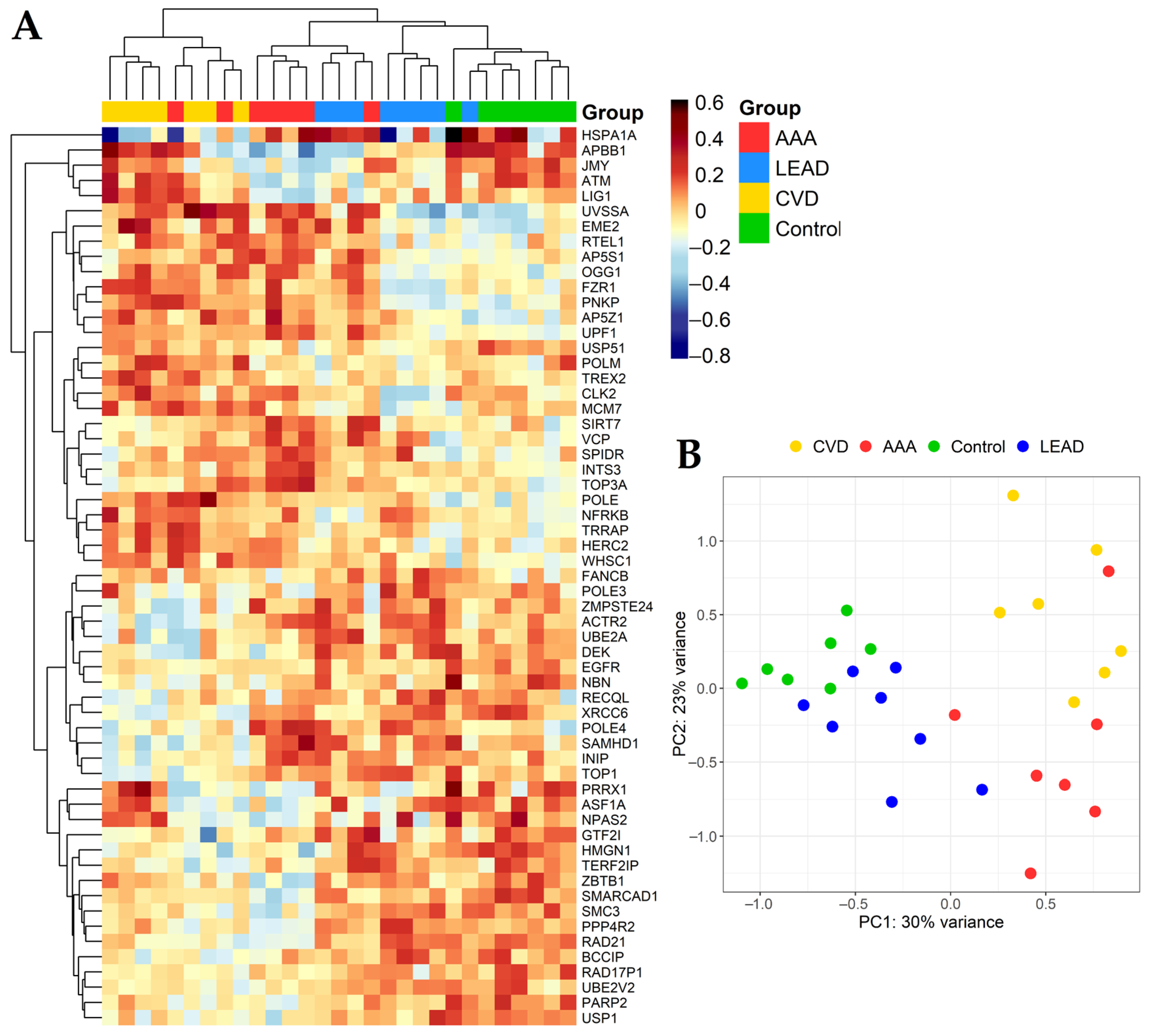
Figure 1. Differentiation in expression of genes selected from performed comparisons. (A) Heatmap with clustering of Euclidean distances using the complete method. (B) Principal component analysis (PCA) plot. LEAD—lower extremities arterial disease, AAA—abdominal aortic aneurysm, CVD—chronic venous disease, Control—healthy persons.
Ijms 24 00551 g001
Figure 2. Relations of the quantity of differentially expressed genes in the pairwise and pooled group comparisons. Gene sets were obtained by DESeq2 and UVE–PLS methods with unified selection criteria applied (p value adjusted by Benjamini—Hochberg false discovery rate < 0.05 and fold change > 1.2 (for upregulated genes) or <0.8 (for downregulated genes), as well as UVE-PLS reliability score ≥ 5). The numbers in overlapping fields in each Venn diagram represent the number of genes shared by both DESeq2 and UVE-PLS gene sets.
Ijms 24 00551 g002
Figure 3. Graphical representation of unique and common genes for gene sets selected from performed comparisons. AAA—abdominal aortic aneurysm, CVD—chronic venous disease, LEAD—lower extremity arterial disease, DIS—pooled group including AAA, CVD and LEAD patients, ART—pooled group of patients with diseases affecting arteries (AAA and LEAD), VEN—patients with disease affecting veins (CVD). Empty fields represent lack of common genes in given comparison(s).
Ijms 24 00551 g003
Figure 4. Results of gene set enrichment analysis (GSEA) analysis for genes selected from such comparisons, where more than ten genes were found to be indicative and differentially expressed. Up to the top 15 functional terms with the lowest p value are presented. (A)—AAA vs. control; (B)—CVD vs. control; (C)—LEAD vs. AAA; (D)—LEAD vs. CVD. AAA—abdominal aortic aneurysm, CVD—chronic venous disease, LEAD—lower extremity arterial disease.
Ijms 24 00551 g004
Table 1. Clinical attributes of the study subjects.
CharacteristicAAA
(n = 7)
CVD
(n = 7)
LEAD
(n = 8)
Control
(n = 7)
p1
Age66.3 ± 4.03 2
59–71 3
41.3 ± 4.03 2
35–47 3
62 ± 7.82 2
48–71 3
33.6 ± 9.64 2
24–54 3
6.290 × 10⁻⁵ 4
Body mass index (BMI)27.23 ± 2.76 2
23.66–30.85 3
23.36 ± 1.94 2
20.94–25.83 3
28.25 ± 2.07 2
25.5–31.2 3
21.73 ± 3.30 2
19.33–29.00 3
0.448 5
Gender: males/females6 (85.7%)/
1 (14.3%)
3 (42.9%)/
4 (57.1%)
6 (75%)/
2 (25%)
5 (71.4%)/
2 (28.6%)
1.564 × 10⁻³ 4
Smoking: never and former/current5 (71.4%)/
2 (28.6%)
5 (71.4%)/
2 (28.6%)
6 (75%)/
2 (25%)
7 (100%)/
0 (0%)
0.535 5
1 Statistical significance of differences between lower extremity arterial disease (LEAD), abdominal aortic aneurysm (AAA) and chronic venous disease (CVD) groups, 2 mean ± SD, 3 range, 4 p value calculated using Kruskal–Wallis rank sum test, 5 p value calculated using two-sided Fisher’s exact test.
Table 2. Differential expression parameters of genes selected from the pairwise and pooled group comparisons. The table presents p (FDR with Benjamini–Hochberg correction) and fold change values received from DESeq2 analysis, PLS coefficients received from UVE-PLS analysis, and areas under ROC curves (ROC–AUC) received from ROC analysis. Genes were ordered within each comparison according to increasing p value.
No.Gene Symbol 1Gene Name 1pFold ChangePLS CoefficientROC–AUC
LEAD vs. Control group
1.ATMATM serine/threonine kinase3.969 × 10−20.762−5.042 × 10−11.000
AAA vs. Control group
1. RAD21RAD21 cohesin complex component4.926 × 10−70.757−7.573 × 10−21.000
2. UPF1UPF1 RNA helicase and ATPase7.004 × 10−51.2867.567 × 10−21.000
3. SMARCAD1SWI/SNF-related, matrix-associated actin-dependent regulator of chromatin, subfamily a, containing DEAD/H box 12.260 × 10−40.725−8.092 × 10−20.959
4. WHSC1Nuclear receptor-binding SET
Domain protein 2
4.903 × 10−41.3007.990 × 10−21.000
5. TOP3ADNA topoisomerase III alpha2.659 × 10−31.3206.685 × 10−20.918
6. INTS3Integrator complex subunit 33.802 × 10−31.2275.369 × 10−20.918
7. PNKPPolynucleotide kinase 3’-phosphatase3.955 × 10−31.3118.177 × 10−20.959
8. PRRX1Paired related homeobox 13.955 × 10−30.208−1.010 × 10−10.918
9. UVSSAUV stimulated scaffold protein A3.955 × 10−31.4821.246 × 10−10.980
10. ZBTB1Zinc finger and BTB domain
containing 1
3.955 × 10−30.778−5.801 × 10−20.918
11. PARP2Poly(ADP-ribose) polymerase 24.303 × 10−30.692−5.700 × 10−20.918
12. SMC3Structural maintenance
of chromosomes 3
4.303 × 10−30.795−5.300 × 10−20.898
13. OGG18-oxoguanine DNA glycosylase8.026 × 10−31.3147.994 × 10−20.918
14. ATMATM serine/threonine kinase8.031× 10−30.761−6.130 × 10−20.918
15. EGFREpidermal growth factor receptor8.031× 10−30.250−4.767 × 10−20.959
16. ASF1AAnti-silencing function 1A histone chaperone8.587 × 10−30.681−1.029 × 10−10.959
17. RTEL1Regulator of telomere elongation helicase 19.642 × 10−31.8056.994 × 10−20.939
18. APBB1Amyloid beta precursor protein-binding family B member 11.132 × 10−20.638−9.918 × 10−20.878
19.NPAS2Neuronal PAS domain protein 21.145 × 10−20.384−6.987 × 10−21.000
20.DEKDEK proto-oncogene1.163 × 10−20.794−6.876 × 10−20.980
21. POLEDNA polymerase epsilon,
catalytic subunit
1.656 × 10−21.2356.854× 10−20.959
22. SIRT7Sirtuin 71.656 × 10−21.2366.185× 10−20.918
23.USP51Ubiquitin specific peptidase 511.656 × 10−20.541−5.382 × 10−20.959
24. AP5S1Adaptor-related protein complex 5 subunit sigma 12.422 × 10−21.3505.630 × 10−20.959
25. SPIDRScaffold protein involved in DNA repair2.655 × 10−21.2374.797 × 10−20.898
26. JMYJunction-mediating and regulatory protein, p53 cofactor2.874 × 10−20.767−5.160 × 10−20.898
27. VCPValosin-containing protein3.095 × 10−21.2095.048 × 10−20.898
CVD vs. Control group
1. POLEDNA polymerase epsilon,
catalytic subunit
2.750 × 10−41.389−1.007 × 10−11.000
2. UVSSAUV stimulated scaffold protein A5.268 × 10−41.608−1.571 × 10−11.000
3. PNKPPolynucleotide kinase 3’–phosphatase3.628 × 10−31.324−9.409 × 10−20.980
4. RAD17P1RAD17 pseudogene 13.628 × 10−30.2416.482 × 10−20.980
5. WHSC1Nuclear receptor-binding SET domain protein 23.628 × 10−31.267−7.529 × 10−20.980
6. XRCC6X-ray repair cross complementing 63.894 × 10−30.7737.724 × 10−21.000
7. TRRAPTransformation/transcription domain-associated protein3.937 × 10−31.247−8.331 × 10−21.000
8. AP5Z1Adaptor-related protein complex 5 subunit zeta 14.601 × 10−31.319−1.079 × 10−10.980
9. EME2Essential meiotic structure–specific endonuclease subunit 24.601 × 10−31.445−1.024 × 10−10.939
10. UPF1UPF1 RNA helicase and ATPase4.601 × 10−31.211−8.287 × 10−21.000
11. NBNNibrin7.122 × 10−30.7676.012 × 10−20.980
12. USP1Ubiquitin-specific peptidase 18.767 × 10−30.7866.183 × 10−21.000
13. GTF2IGeneral transcription factor IIi1.106 × 10−20.7481.004 × 10−10.980
14. HERC2HECT and RLD domain containing E3 ubiquitin protein ligase 21.291 × 10−21.229−5.896 × 10−20.959
15. TREX2Three prime repair exonuclease 21.291 × 10−22.039−5.716 × 10−20.959
16.HMGN1High-mobility group nucleosome-binding domain 11.303 × 10−20.7601.011 × 10−11.000
17. EGFREpidermal growth factor receptor1.396 × 10−20.2444.115 × 10−20.918
18. OGG18-oxoguanine DNA glycosylase2.368 × 10−21.282−7.221 × 10−20.898
19. PARP2Poly(ADP-ribose) polymerase 22.490 × 10−20.7255.284 × 10−20.918
20. NFRKBNuclear factor related to kappaB-binding protein2.618 × 10−21.249−9.573 × 10−20.980
21. BCCIPBRCA2 and CDKN1A interacting protein3.014 × 10−20.7886.583 × 10−20.939
22. FZR1Fizzy and cell division cycle 20 related 13.418 × 10−21.250−9.201 × 10−20.980
23. HSPA1AHeat shock protein family A (Hsp70) member 1A3.418 × 10−20.5331.376 × 10−10.898
LEAD vs. AAA
1. PPP4R2Protein phosphatase 4 regulatory subunit 22.732 × 10−61.3351.229 × 10−11.000
2. RAD21RAD21 cohesin complex component9.034 × 10−61.2721.049× 10−11.000
3. SMC3Structural maintenance of chromosomes 37.431 × 10−41.2941.195 × 10−10.964
4. POLE3DNA polymerase epsilon 3,
accessory subunit
5.547 × 10−31.3261.340 × 10−10.946
5. MCM7Minichromosome maintenance complex component 79.869 × 10−30.786−1.065 × 10−10.911
6. CLK2CDC like kinase 21.350 × 10−20.774−1.063 × 10−10.946
7. DEKDEK proto–oncogene1.350 × 10−21.2741.133 × 10−10.929
8. SMARCAD1SWI/SNF–related, matrix–associated actin–dependent regulator of chromatin, subfamily a, containing DEAD/H box 11.350 × 10−21.2651.110 × 10−10.964
9. UVSSAUV stimulated scaffold protein A1.350 × 10−20.703−1.890× 10−10.875
10. FANCBFA complementation group B2.563 × 10−21.5989.226 × 10−20.929
11. UBE2AUbiquitin conjugating enzyme E2 A3.975 × 10−21.2911.428 × 10−10.911
LEAD vs. CVD
1. UVSSAUV-stimulated scaffold protein A2.430 × 10−30.648−1.885 × 10−10.893
2. MCM7Minichromosome maintenance complex component 75.487 × 10−30.771−9.060 × 10−20.964
3. ACTR2Actin-related protein 27.278 × 10−31.2906.713 × 10−20.946
4. POLMDNA polymerase mu7.278 × 10−30.774−8.552 × 10−20.946
5. PNKPPolynucleotide kinase 3′–phosphatase1.780 × 10−20.793−6.935 × 10−20.946
6. UBE2AUbiquitin conjugating enzyme E2 A1.780 × 10−21.3276.779 × 10−20.893
7. DEKDEK proto–oncogene1.795 × 10−21.2565.554 × 10−20.875
8. INIPINTS3 and NABP interacting protein1.795 × 10−21.2836674 × 10−20.929
9. XRCC6X–ray repair cross complementing 61.795 × 10−21.2397.014 × 10−20.911
10. UBE2V2Ubiquitin conjugating enzyme E2 V21.820 × 10−21.2507.772 × 10−21.000
11. BCCIPBRCA2 and CDKN1A
interacting protein
1.992 × 10−21.2718.439 × 10−20.964
12. ATMATM serine/threonine kinase2.223 × 10−20.785−6.956 × 10−20.911
13. SMC3Structural maintenance
of chromosomes 3
2.226 × 10−21.2047.135 × 10−20.982
14. RECQLRecQ like helicase2.592 × 10−21.2465.847 × 10−20.857
15. TOP1DNA topoisomerase I2.677 × 10−21.2196.918 × 10−20.893
16.ZMPSTE24Zinc metallopeptidase STE242.677 × 10−21.3145.716 × 10−20.929
17. POLE4DNA polymerase epsilon 4,
accessory subunit
3.143 × 10−21.2976.662 × 10−20.929
18. TERF2IPTERF2 interacting protein4.007 × 10−21.2318.871 × 10−20.946
19. SAMHD1SAM and HD domain containing deoxynucleoside triphosphate triphosphohydrolase 14.624 × 10−21.2706.332 × 10−20.893
AAA vs. CVD
1. LIG1DNA ligase 11.405 × 10−20.734−2.215 × 10−10.878
2. POLE4DNA polymerase epsilon 4.
accessory subunit
1.405 × 10−21.3812.107 × 10−10.918
LEAD, AAA and CVD vs. Control group
3. WHSC1Nuclear receptor binding SET
domain protein 2
2.248 × 10−31.2451.269 × 10−10.981
4.UPF1UPF1 RNA helicase and ATPase3.682 × 10−31.2049.414 × 10−20.955
5. POLEDNA polymerase epsilon,
catalytic subunit
3.016 × 10−21.2429.073 × 10−20.909
LEAD and AAA vs. CVD
1. ATMATM serine/threonine kinase2.116 × 10−20.791−1.108 × 10−10.867
2. INIPINTS3 and NABP interacting protein2.116 × 10−21.2821.131 × 10−10.914
3. LIG1DNA ligase 12.116 × 10−20.764−1.165 × 10−10.895
4. POLE4DNA polymerase epsilon,
4 accessory subunit
2.116 × 10−21.3341.197 × 10−10.924
5. TREX2Three prime repair exonuclease 22.116 × 10−20.569−1.122 × 10−10.914
6. XRCC6X–ray repair cross complementing 62.116 × 10−21.2228.096 × 10−20.914
AAA—abdominal aortic aneurysm. CVD—chronic venous disease. LEAD—lower extremity arterial disease, ROC—receiver operating characteristics, UVE-PLS—uninformative variable elimination by partial least squares. 1 Gene name and symbol according to HUGO Gene Nomenclature Committee.
Table 3. Correlation analysis between characteristics of studied groups: age and body mass index (BMI) and expression of genes selected from comparisons performed. The table presents genes correlated with statistical significance p < 0.05 (adjusted by Benjamini–Hochberg FDR).
ComparisonGene SymbolRp
Age
AAA vs. controlAPBB1−0.5907.116 × 10−3
NPAS2−0.4564.393 × 10−2
PRRX1−0.5102.300 × 10−2
SIRT70.4992.630 × 10−2
TOP3A0.5798.596 × 10−3
USP51−0.6721.536 × 10−3
VCP0.6194.216 × 10−3
JMY−0.4902.959 × 10−2
LEAD vs. control,
AAA vs. control,
LEAD vs. CVD,
ART vs. CVD
ATM−0.6532.262 × 10−3
AAA vs. CVD,
ART vs. CVD
LIG1−0.4614.177 × 10−2
LEAD vs. CVD,
AAA vs. CVD,
ART vs. CVD
POLE40.5808.546 × 10−3
AAA vs. control,
CVD vs. control,
Disease vs. control
UPF10.5581.200 × 10−2
WHSC10.4793.370 × 10−2
BMI
AAA vs. controlPRRX1−0.5192.061 × 10−2
USP51−0.5331.726 × 10−2
VCP0.4673.826 × 10−2
LEAD vs. CVDRECQL0.4504.744 × 10−2
LEAD vs. CVDPOLM−0.4693.729 × 10−2
AAA—abdominal aortic aneurysm, CVD—chronic venous disease, LEAD—lower extremity arterial disease, BMI—body mass index, R—correlation coefficient.
Disclaimer/Publisher’s Note: The statements, opinions and data contained in all publications are solely those of the individual author(s) and contributor(s) and not of MDPI and/or the editor(s). MDPI and/or the editor(s) disclaim responsibility for any injury to people or property resulting from any ideas, methods, instructions or products referred to in the content.