Article

Genomic Assessment of the Contribution of the Wolbachia Endosymbiont of Eurosta solidaginis to Gall Induction

1
Department of Biology, San Francisco State University, San Francisco, CA 94112, USA
2
Department of Thoracic Surgery, Brigham and Women’s Hospital, Harvard Medical School, Boston, MA 02115, USA
3
Iridian Genomes Inc., Bethesda, MD 20817, USA
*
Author to whom correspondence should be addressed.
Int. J. Mol. Sci. 2023, 24(11), 9613; https://doi.org/10.3390/ijms24119613
Submission received: 31 January 2023 / Revised: 25 May 2023 / Accepted: 25 May 2023 / Published: 1 June 2023
(This article belongs to the Special Issue Plant-Insect Interactions 2022)

Abstract

:
We explored the genome of the Wolbachia strain, wEsol, symbiotic with the plant-gall-inducing fly Eurosta solidaginis with the goal of determining if wEsol contributes to gall induction by its insect host. Gall induction by insects has been hypothesized to involve the secretion of the phytohormones cytokinin and auxin and/or proteinaceous effectors to stimulate cell division and growth in the host plant. We sequenced the metagenome of E. solidaginis and wEsol and assembled and annotated the genome of wEsol. The wEsol genome has an assembled length of 1.66 Mbp and contains 1878 protein-coding genes. The wEsol genome is replete with proteins encoded by mobile genetic elements and shows evidence of seven different prophages. We also detected evidence of multiple small insertions of wEsol genes into the genome of the host insect. Our characterization of the genome of wEsol indicates that it is compromised in the synthesis of dimethylallyl pyrophosphate (DMAPP) and S-adenosyl L-methionine (SAM), which are precursors required for the synthesis of cytokinins and methylthiolated cytokinins. wEsol is also incapable of synthesizing tryptophan, and its genome contains no enzymes in any of the known pathways for the synthesis of indole-3-acetic acid (IAA) from tryptophan. wEsol must steal DMAPP and L-methionine from its host and therefore is unlikely to provide cytokinin and auxin to its insect host for use in gall induction. Furthermore, in spite of its large repertoire of predicted Type IV secreted effector proteins, these effectors are more likely to contribute to the acquisition of nutrients and the manipulation of the host’s cellular environment to contribute to growth and reproduction of wEsol than to aid E. solidaginis in manipulating its host plant. Combined with earlier work that shows that wEsol is absent from the salivary glands of E. solidaginis, our results suggest that wEsol does not contribute to gall induction by its host.

1. Introduction

Bacteria are commonly associated with insect hosts, but the nature of these associations is quite variable. Some bacteria establish mutually symbiotic relationships with their hosts in which, in exchange for shelter and nutrition, the bacteria provide some benefit to the host such as provisioning amino acids or riboflavin or providing immunity against natural enemies [1,2,3]. However, other bacteria may not only use the host for shelter and nutrition but also manipulate the host to enhance bacterial fitness [4,5,6,7,8].
One species of bacterial symbiont that has both mutually symbiotic and manipulative strains is Wolbachia pipientis. Wolbachia are a group of obligatory intracellular and maternally transmitted bacterial strains which can be found in a range of eukaryotic species, including crustaceans, nematodes, and a high proportion of insect species [5,9,10,11,12]. Most notably, many strains of Wolbachia are considered reproductive parasites of their hosts that distort sex ratio and sexual reproduction to favor maternal transmission [4,5,6]. Other strains of Wolbachia have evolved to be mutualistic. For example, the Wolbachia strain wBm in the nematode Brugia malayi has been shown to contribute essential compounds such as heme, nucleotides, and riboflavin to the host [1], while Wolbachia strains associated with Drosophila confer protection from RNA viruses [3].
A gall is a mass of plant tissue that is formed as a response to a gall inducer, and in which the gall inducer or their offspring resides while feeding on the host plant. A variety of organisms, including insects, mites, fungi, bacteria, protists, and nematodes, are known to induce plant galls [13,14,15,16,17,18,19,20,21]. While the mechanism of gall induction has been established for bacteria, how insects induce plant galls remains an open question.
The fly Eurosta solidaginis (Diptera: Tephritidae) induces galls in the apical meristems of Solidago altissima L. (Asteraceae) and hosts a strain of Wolbachia whose interaction with its host is yet uncharacterized [22]. Herein, we explore the interaction of Wolbachia and E. solidaginis specifically to determine if the strain of Wolbachia associated with E. solidaginis, which we name wEsol, contributes to gall induction by this species of fly.
The hypothesis proposed by Giron et al. [23] and Bartlett and Connor [24] that symbiotic bacteria might be completely or partially responsible for gall induction by insects was motivated by two observations: several species of bacteria commonly found associated with plant surfaces and in the rhizosphere (Agrobacterium tumefaciens, Allorhizobium vitis, Pantoea agglomerans, Rhodococcus fascians, Pseudomonas savastanoi, Streptomyces turgidiscabies, and others) have strains that themselves induce plant galls [22,25,26]. While none of the gall-inducing strains of these bacterial species have been demonstrated to be symbiotic with insects, strains of some of these genera have been found in association with insects [27,28,29]. Secondly, in a parallel phenomenon known as “green islands”, a Wolbachia strain associated with the leaf-mining moth Phyllonorycter blancardella (Lepidoptera: Gracillariidae) has been implicated as providing the inducing agent responsible for green island formation [30,31,32,33].
Gall formation by bacteria has been shown to be triggered by the phytohormones indole-3-acetic acid (IAA, an auxin) and cytokinin (CK), which are either secreted by the bacteria into the host plant or genes responsible for their biosynthesis are inserted by the bacteria into the host plant, where they are expressed, leading to localized CK and IAA production [14,15,34,35,36,37]. CK and IAA are the primary hormones involved in cell division and growth in plants, and their synthesis has been demonstrated in gall-inducing bacteria [15,37,38,39].
Cytokinins and auxin (IAA) are widespread in insects [40,41,42,43,44,45,46,47,48,49,50,51,52,53], and they are also found in other gall-inducing organisms [18,20,21,54,55]. Some of the highest concentrations of CKs and IAA have been reported in larvae of Eurosta solidaginis [40,41,50].
“Green island” formation by the leaf-mining moth P. blancardella has also been attributed to the secretion of CKs by the inducing insect [30,33]. The high concentration of secreted CKs prevents leaf tissues surrounding the feeding site from senescing, thus forming “green islands” in otherwise senescing leaves. P. blancardella has been shown to have high whole-body concentrations of CKs and is associated with high concentrations of CKs in the surrounding leaf tissues [30,33]. This moth harbors a strain of Wolbachia, which has been implicated as the source of the cytokinins secreted by its host [31,32]. When the adult moths were cured of Wolbachia, the offspring were unable to induce “green islands”, suggesting that the Wolbachia symbiont is providing the CKs to induce green islands [31].
It is also hypothesized that the secretion of proteinaceous effectors by a gall-inducing insect species might alter gene expression in the plant host, leading to gall induction [56,57,58,59]. However, thus far, secreted effector proteins that have been reported in gall-inducing insects appear to modify the gall structure or color but are not primarily responsible for gall induction [58,59]. Still, some gall-inducing bacteria and fungi do secrete effector proteins that contribute to gall induction [36,60,61].
Hammer et al. [22] examined several gall-inducing and non-gall-inducing insect species using bacterial 16S ribosomal RNA gene amplicon analysis. They conclude that there was no evidence to support the idea that a common bacterial species or a community of bacteria are, in general, associated with gall-inducing insects and absent from non-gall inducing insects. However, they admit that in specific instances, gall induction could potentially be symbiont-dependent.
As an obligate, maternally transmitted symbiont, Wolbachia could be stably transmitted and would appear to be a potential candidate bacterium to participate in gall induction in an insect host that is an obligate gall-inducing species. However, no Wolbachia have yet been reported to be associated with gall induction, and as an obligate endosymbiont, Wolbachia has a much smaller genome with reduced metabolic capacities relative to free-living, gall-inducing bacteria and depends on its host for many metabolic processes [62]. So, while overcoming the hurdle of host-specific transmission, Wolbachia may not have the metabolic pathways to provide its insect host with the requisite means to induce a plant gall.
To investigate the potential role of a bacterial symbiont in gall induction, we sequenced the metagenome of E. solidaginis and its Wolbachia symbiont wEsol, and we assembled and annotated the genome of wEsol. Annotation of the wEsol genome should reveal whether genes in the IAA or CK pathways are present in wEsol and potentially identify effector proteins secreted by wEsol as well.

2. Results

2.1. Parsing wEsol and E. solidaginis Contigs

The E. solidaginis holobiont metagenome comprised 49,193 contigs and was approximately 1.44 Gbp in length (Eurosta solidaginis genome NCBI accession JARSSU01). Using BLASTn and default settings, we detected 1567 metagenomic contigs that had alignments to our database of Wolbachia genomes. However, most of these alignments were short (<100 bp) with percent identities less than 70%. We selected a subset of 577 of these contigs with bitscore values ≥ 60, e-values ≤ 1 × 10⁻⁵, and length ≥ 55 bp for further consideration as candidate wEsol contigs. Bootstrap binning of these 577 contigs identified a group of 147 contigs that were provisionally considered Wolbachia contigs. One additional contig of 1031 bp that had a BLAST alignment that was 100% identical to a member of our Wolbachia database was added to the group of putative wEsol contigs. Coverage patterns were consistent with the interpretation that 146 of the initially identified 148 contigs were indeed wEsol contigs (wEsol had coverage > 6 times higher than Eurosta, see below), while the consistent coverage and read mapping of two contigs lead us to classify them as wEsol inserts into the host genome. Three additional contigs not identified using BusyBee Web had all CDSs annotated as alphaproteobacterial and showed coverage patterns consistent with wEsol contigs so were added to the set of wEsol contigs. An additional 58 contigs showed evidence of abrupt coverage changes within contigs, including 4 contigs identified in the initial group by BusyBee Web. Based on mapping of Illumina read pairs and PacBio reads to contigs, we determined that those 4 contigs along with 13 more were chimeras and we added the parts annotated as alphaproteobacterial to our draft genome of wEsol (Supplemental Table S1).

2.2. Features of the wEsol Genome

Tentatively, the genome of wEsol (NCBI accession JAQZAU01) consists of 162 contigs with a total length of 1.66 Mbp (Table 1 and Supplemental File S1—annotated genome of wEsol). The wEsol assembly N50 was 19,308 with an L50 of 23. Overall, there was little difference between putative Wolbachia contigs and putative Eurosta contigs in GC content or CAI values. GC content for wEsol was slightly lower than for the Eurosta genome (35.27% versus 37.76%), while CAI values (CAI [mean ± sd]) for wEsol [0.612 ± 0.049] were slightly higher than for Eurosta [0.572 ± 0.061] or mixed contigs [0.601 ± 0.036]). Overall, Eurosta coverage (12–20×) appeared to be substantially lower than wEsol coverage (128× Illumina and 189× PacBio), and this was consistent across all three Illumina libraries. Therefore, contig coverage appears to be a reliable means to sort between Eurosta and wEsol contigs.
For all instances where BLAST identified reading frames within these 162 wEsol contigs as putatively eukaryotic, we detected regions of significant similarity of the genome of the predicted eukaryote to Wolbachia based on aligning the genomes of these eukaryotes to our large database of Wolbachia genomes. A total of 49 CDSs, out of 1878, had best BLAST alignments on 11 arthropod species, with the spider Trichonephila clavata (Arachnida: Araneidae) accounting for almost half of those alignments. Therefore, even those reading frames with best BLAST alignments identified as eukaryotic “hypothetical proteins” may arise because of contamination of reference eukaryotic host genomes with proteins from an as yet unreported strain of a Wolbachia symbiont.

2.3. Horizontal Gene Transfer of wEsol DNA into Eurosta Contigs

Examining the annotations of the eukaryotic contigs with some CDSs showing BLAST alignments to Wolbachia, we detected 88 contigs with evidence of insertions of wEsol DNA into the host E. solidaginis genome (Supplemental Table S2). We defined a contig that had potentially experienced horizontal transfer as one possessing CDSs that were classified by BLAST alignments as a mixture of both alphaproteobacterial and eukaryotic CDSs, with no evidence of discontinuity in read coverage across the contig, and with Illumina read pairs and PacBio reads that spanned the alphaproteobacterial and eukaryotic CDSs. In most instances, the putative wEsol insertion into the host genome consisted of 4 or fewer CDSs (83%), but in one instance the insertion consisted of 19 CDSs. These 88 contigs were on average 59,322 ± 6302 bp in length (mean ± standard error) and possessed on average 2.77 ± 0.35 alphaproteobacterial CDSs that were 1700 ± 248 bp in total length. The longest insertion was 9733 bp. Many other contigs had BLAST alignments to our database of Wolbachia genomes and may represent inserts that had degraded to the extent that recognizable CDSs are no longer present.

2.4. Phylogenetic Placement of wEsol and Strain Origin

Phylogenetic inference based on the GTDB markers for our set of Wolbachia genomes selected HIVb + F + R5 as the best model with high bootstrap support, particularly for Supergroups A and B (Figure 1). wEsol was placed solidly in Supergroup A. Interestingly, wEsol was placed in a clade with Wolbachia strains from several distantly related hosts, Carposina sasaki (Lepidoptera: Carposinidae), Cardiocondyla obscurior (Hymenoptera: Formicidae), Drosophila simulans (Diptera: Drosophilidae), and Megacopta cribraria (Hemiptera: Platispidae), rather than with Wolbachia strains from hosts more closely related to E. solidaginis (Rhagoletis pomonella and R. zephyria, Diptera: Tephritidae) or with strains from other gall-inducing hosts, Pediapsis aceris and Biorhiza pallida (Hymenoptera: Cynipidae). This pattern suggests that Wolbachia is not co-radiating with its hosts (Figure 2).

2.5. Comparative Genomics

The predicted proteome of wEsol included 1878 proteins, of which 74.9% had predicted functions. The genome of wEsol is somewhat larger than its closest relatives and contained an abundance of mobile genetic elements including proteins encoded by the remnants of seven different phages that are likely related to WO, P2, Lambda, phi-C31, HK97, T4, and Mu (Table 2).
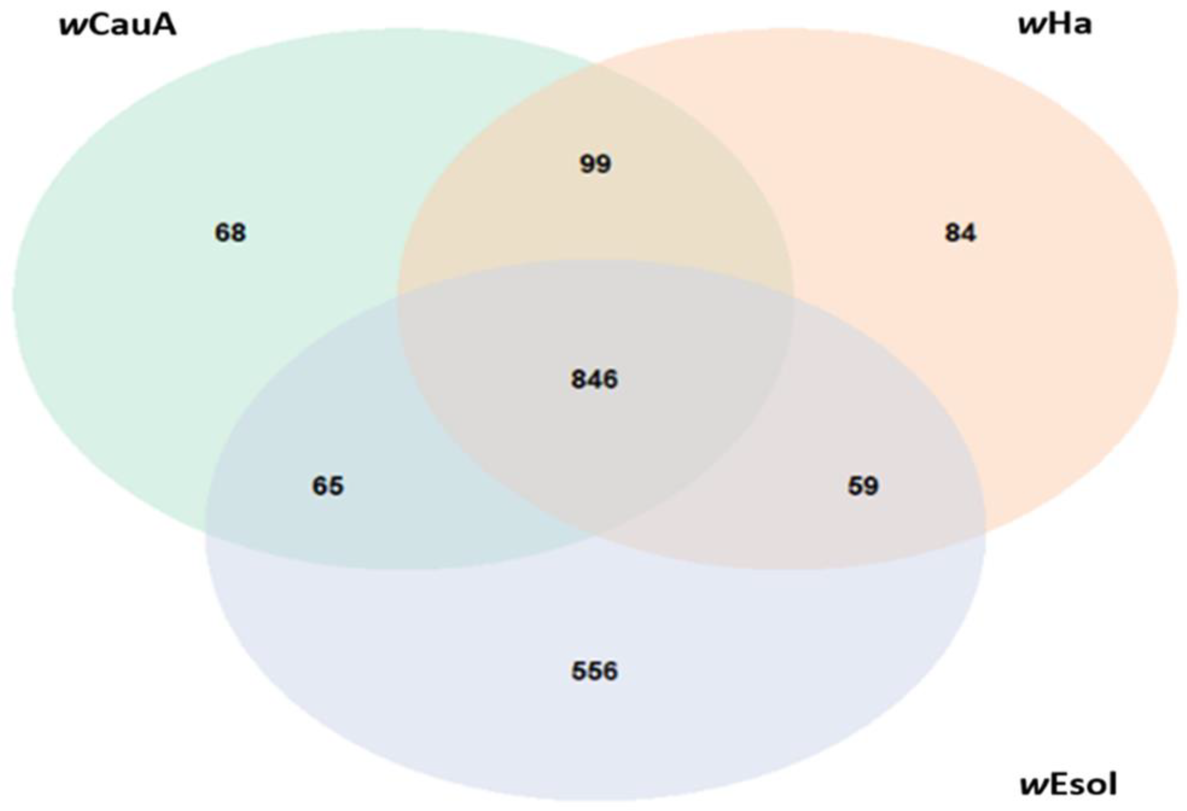
Pangenome analysis suggested that the Wolbachia strains wEsol, wCauA, and wHa share a core genome of 846 proteins, with approximately equal numbers of proteins shared among each pair of strains and unique to each strain, except for wEsol (Figure 3). wEsol possesses 556 proteins (30% of the total number of predicted proteins in wEsol) that are not shared with wCauA or wHa. Approximately 20% of these wEsol specific proteins are from putative mobile genetic elements, 15% are ankyrin-repeat-domain-containing proteins, and another 20% comprise multi-species conserved proteins.
Visualization of regions of sequence similarity (Figure 4) indicates that there are broad regions of similarity within the contigs of wEsol and chromosomes of the closely related strains wCauA and wHa. However, due to the fragmented assembly of wEsol, we can reach no conclusions about the level of synteny above the contig level. Furthermore, since we are ignorant of the true orientation of the contigs of wEsol relative to each other, we can only make inferences about the orientation of the regions of synteny within a contig as being direct or inverted relative to wCauA or wHa, not for whole contigs.

2.6. Potential Contribution of wEsol to Gall Induction by E. solidaginis

2.6.1. Phytohormone Biosynthesis Proteins

Examination of the protein annotations indicates that wEsol does not possess an adenylate dimethylallyltransferase gene (EC: 2.5.1.27 or EC: 2.5.1.112) so is incapable of de novo synthesis of cytokinins. Yet, wEsol, like other Wolbachia strains, possess genes encoding both the MiaA (tRNA dimethylallyltransferase, EC: 2.5.1.75) and MiaB (tRNA-2-methylthio-N6-dimethylallyladenosine synthase, EC: 2.8.4.3) enzymes, suggesting that wEsol could synthesize tRNA-bound cytokinins and tRNA-bound methylthiolated cytokinins. However, wEsol appears to be incapable of synthesizing either of the prenyl group donors (DMAPP or HMBDP) from the MEP pathway. wEsol is lacking the dxs (EC: 2.2.1.7) gene to initiate the MEP pathway, and its ispG/gcpE gene (EC: 1.17.7.1 and EC: 1.17.7.3) is rendered non-functional due to an insertion leading to a frameshift mutation. Our examination of a broader set of Wolbachia strains (Supplemental Table S3) indicates that the majority are not predicted to be able to synthesize DMAPP or HMBDP, with only approximately 20% of strains capable of doing so only if supplied 1-deoxy-d-xylulose-5-phosphate by the host (Figure 5). wEsol and the other 63 strains of Wolbachia we examined lack the dxs gene, as do apparently most of the larger Rickettsiales (KEGG). ispG/gcpE is also missing from 66% of the 63 strains we examined, and ispG/gcpE is responsible for the penultimate step in the MEP pathway, leading to the synthesis of HMBDP, which is converted to DMAPP by ispH.
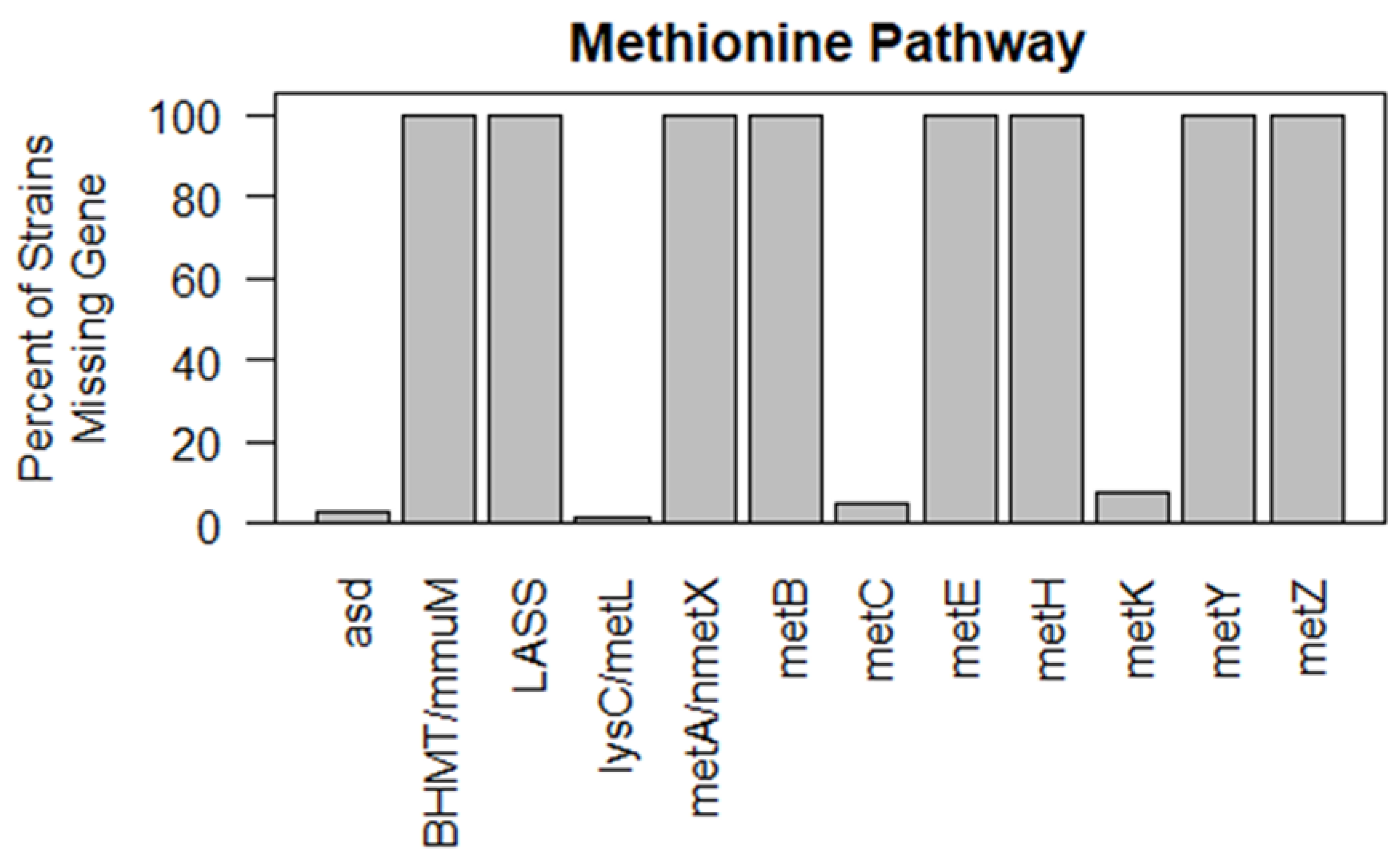
wEsol also appears incapable of synthesizing S-adenosyl-L-methionine (SAM), the methyl group donor required for the production of methylthiolated cytokinins, unless provided L-methionine by its host. While the wEsol genome encodes the enzyme to convert L-methionine to SAM (metK, EC: 2.5.1.6), it lacks several others involved in methionine synthesis and is incapable of synthesizing L-methionine from either homoserine or aspartate. Our examination of other Wolbachia strains found that most strains lacked several enzymes involved in methionine synthesis and none were capable of synthesizing L-methionine (Figure 6 and Supplemental Table S4).
wEsol possesses a homolog of the enzyme 5′-nucleotidase (EC: 3.1.3.5), which converts AMP to adenosine, so could potentially contribute to conversion of CK nucleotides to nucleosides [68].
We found no evidence in the genome of wEsol or other Wolbachia strains for any enzymes for the synthesis of IAA from tryptophan. Furthermore, Wolbachia strains and bacteria in the Rickettsiales, in general, are unable to synthesize tryptophan [69], so wEsol would also need to acquire this amino acid from its insect host.
Examination of the multiple BLASTP alignments of the “hypothetical” and “uncharacterized” proteins revealed no hidden alignments to enzymes annotating as part of the IAA, CK, MEP, SAM, or adenine salvage pathways.

2.6.2. Secreted Effector Proteins

PSORTb predicted only seven proteins as being extracellularly secreted. However, PSORTb tends to predict the location of proteins secreted by wEsol as being the host cytoplasm [70]. When using a threshold of 50% probability to classify proteins as Type IV secreted proteins, T4SEfinder predicted that 539 of the 1878 proteins in the wEsol genome were Type IV secreted proteins. Approximately half of these predicted Type IV secreted proteins had no functional annotation, and of the remaining proteins, half were encoded by mobile genetic elements, VIR complex proteins, or ankyrin-repeat-domain-containing proteins.

3. Discussion

3.1. General Features of the wEsol Genome

The wEsol genome is larger than the average Wolbachia genome but falls within the range of previously determined Wolbachia genome sizes (0.86 to 3.1 Mbp) [71,72]. Wu et al. [73] report only three prophages and 138 insertion sequences (e.g., transposases, terminases, maturases) from wMel yet describe its genome as being “overrun” with mobile elements. However, wEsol appears to have an even greater abundance of proteins encoded by mobile elements, with evidence of seven different phages and over 300 insertion sequences (Table 1). wEsol shared 846 proteins (45% of total predicted proteins) with its closest relatives wCauA and wHa but had a large number of unique proteins, many of which were encoded by mobile genetic elements or were ankyrin repeat domain proteins.
Our phylogenetic analysis placed wEsol centrally within Supergroup A strains of W. pipientis. wEsol was placed in a clade, which is also supported by the phylogenetic analysis of over 1000 Wolbachia strains performed by [74], with strains from distantly related host species (Drosophila simulans wHa, Carposina sasakii, Megacopta cribaria, and Cardiocondyla obscurior). wEsol was not grouped with other Wolbachia strains associated with species in the Tephritidae. This pattern of related Wolbachia strains not occurring on related hosts is common throughout Supergroups A and B and is illustrated by the crossing lines linking hosts to Wolbachia strains in the tanglegram (Figure 2). Furthermore, this pattern suggests that Wolbachia strains are not strictly co-radiating with their insect hosts, which is similar to conclusions reached in other studies and suggests that Wolbachia strains can be readily transmitted horizontally between hosts [74,75,76,77,78].
wEsol also does not group with other strains of Wolbachia hosted by gall-inducing insects. We interpret this to indicate that there is no clade of Wolbachia strains that possess traits unique to gall-inducing insect hosts. This result is consistent with the interpretation that Wolbachia infections of gall-inducing insect hosts are not associated with the acquisition of the ability to induce plant galls, but represent opportunistic colonization.
We detected evidence of multiple instances of horizontal gene transfer from the wEsol genome to the genome of E. solidaginis, as previously reported for other Wolbachia strains and their hosts [79,80,81,82,83,84]. For wEsol and E. solidaginis, these insertions usually involve a single or very few CDSs, and slightly over half are annotated as being phage-encoded, tetratricopeptide, or ankyrin repeat domain proteins. Of the remainder, approximately half have functional annotations, and half are hypothetical proteins. As obligate intracellular, maternally inherited endosymbionts, Wolbachia has had the opportunity to engage in such transfers. With evidence of wEsol having seven phages, although some possibly degraded, perhaps phage transfer of flanking regions may account for some of these insertions [85]. We believe that the existence of the transfer events certainly made assembly of the metagenome more difficult, but without further study and experimentation, we cannot explore the ramifications of these apparent insertions.

3.2. Does wEsol Contribute to Gall Induction by Eurosta solidaginis?

Gall induction by insects has been hypothesized to result from the production and secretion of the phytohormones IAA and cytokinin by the insect, or from the secretion of proteinaceous effectors that alter gene expression in the host plant. Both mechanisms are then proposed to lead the cell division and growth in the host plant that constitutes the gall. Eurosta solidaginis larvae are known to contain high concentrations of CKs and IAA that are localized to the salivary glands [40,41,50,86]. However, in the absence of an annotated genome for E. solidaginis, no examination of secreted effector proteins has yet been conducted.

3.2.1. wEsol and Phytohormone Synthesis

wEsol is capable of synthesizing CKs. We detected both the miaA and miaB genes in wEsol, which are involved in the prenylation of adenine in tRNA at position 37 of the anticodon loop and methylthiolation of the prenyl group, resulting in these two types of modified tRNA bound adenine molecules. Upon tRNA degradation, CKs and methylthiolated CKs are released, respectively. However, we found wEsol and many other strains of Wolbachia to be compromised in the synthesis of DMAPP, HMBDP, and SAM, the precursors necessary for such tRNA modifications (Figure 5 and Figure 6, and Supplemental Tables S3 and S4). As an obligate intracellular symbiont, Wolbachia has a genome that is smaller than free-living bacteria that induce galls in plants. Like other strains of Wolbachia and Rickettsia, in wEsol the metabolic pathways for the synthesis of DMAPP and SAM are not functional, and wEsol must steal these compounds, or in the case of SAM, its precursor L-methionine, from its host to complete these metabolic functions [62,87,88,89]. Therefore, we believe it is highly unlikely that wEsol contributes to CK production and secretion by its host.
Andreas et al. [50] report that CKs and methylthiolated CKs are widespread and abundant in insects, not just in gall- and green-island-inducing species, and more extensive work by Tokuda et al. [52] reached similar conclusions for both CKs and IAA. Andreas et al. [50] proposed that insects obtain CKs and methylthiolated CKs from their own biosynthesis of these compounds, although they suggested that symbiotic acquisition was possible in specific instances. A bioinformatic analysis of the transcriptomes of 670 species of Hexapoda and Insecta found that transcripts that encode proteins that are homologs of the enzymes involved in tRNA modifications at position A37 and eventually lead to the release of CK and methylthiolated CKs after tRNA degradation (EC: 2.5.1.75 and EC: 2.8.4.3, respectively) are widespread among insects [68]. An examination of the BmIPT1 gene from Bombyx mori (Lepidoptera: Bombycidae) showed that it is a functional tRNA-dimethylallyltransferase gene since it could complement a mutant yeast strain lacking tRNA-dimethylallytransferase [90]. Furthermore, isopentenyladenosine (a form of cytokinin) was detected by HPLC in the recombinant yeast strain complemented by BmIPT1 but was absent from the mutant yeast [90]. Finally, the MVA pathway for DMAPP biosynthesis and the enzymes for biosynthesis of SAM from dietary methionine (EC: 2.5.1.6) are also widespread in insects [91,92]. Zhang et al. [33] imply that methylthiolated CKs are largely if not exclusively produced by bacteria. However, Schweizer et al. [93] outline that enzymes for both prenylation of adenine at position 37 of tRNA and its subsequent methylthiolation are widespread in all domains of life. Reiter et al. [94] show that the gene CDK5RAP1 in animals is a homolog of the miaB gene. Andreas et al. [50] found methylthiolated CKs in insects, and they have been detected in bacteria, protists, fungi, plants, and humans and other mammalian species [95]. Mooi et al. [68] report that transcripts that encode proteins that are homologs of the CDK5RAP1 gene are widespread in Hexapoda and Insecta. Hence, E. solidaginis is capable of synthesizing CKs and methylthiolated CKs without assistance from its bacterial symbiont, wEsol.
wEsol appears unable to synthesize IAA. We found no evidence in the genome of wEsol or other Wolbachia strains for any enzymes for the synthesis of IAA from tryptophan. Furthermore, Wolbachia strains and bacteria in the Rickettsiales, in general, are unable to synthesize tryptophan [69], so wEsol would also need to acquire this amino acid from its insect host, which, in turn, would acquire tryptophan via consumption of its host plant. While many species of free-living rhizosphere bacteria synthesize IAA, no Wolbachia strains have been reported to do so [96].
Recent work suggests that insects themselves can synthesize IAA from tryptophan [47,97,98,99]. Tokuda et al. [52] found IAA to be ubiquitous in a wide variety of terrestrial arthropods including insects. In a search for the enzymes responsible for IAA biosynthesis in B. mori, Miyata et al. [99] reported that two enzymes that constitute a functional pathway for the synthesis of IAA from tryptophan were encoded in the B. mori genome and not in the genome of any bacterial associate.

3.2.2. Bacterial Effectors and Gall Induction

Hypotheses that extracellularly secreted effector proteins are responsible for gall induction focus on production and secretion of effectors by the gall-inducing organism, not by its microbial symbionts [56,57,58,59], the idea being that these effectors are secreted into the host plant where they affect expression of host-plant genes, presumably genes that are involved in the cell cycle, leading to increased cell division and growth, hence the plant-tissue growth that becomes the gall. Secretion into the host plant most likely involves secretions associated with feeding—salivary secretions—or secretions associated with oviposition—accessory gland fluids [45,86].
While we cannot rule out the possibility that proteinaceous effectors secreted by wEsol or small RNAs play some role in gall induction, we think it unlikely [100]. The bacterial symbiont of E. solidaginis, wEsol, is predicted to secrete over 500 Type IV secretion system proteins. Both effector proteins and small RNAs are secreted into its host cell and are most likely exclusively involved in the interactions between wEsol and its insect host. Most strains of Wolbachia are hosted by insects and other arthropod species that do not induce galls in their food plants or are not herbivorous. While many of these predicted Type IV effectors have unknown function, many are associated with mobile genetic elements and VIR complex genes involved in the secretion system. A large number of the predicted effectors are ankyrin repeat domain proteins, which are thought to be involved in protein–protein interactions, rather than interacting with gene expression in the insect host [101,102]. On the other hand, we have not sought to determine the range of small RNAs that wEsol may secrete into the host cell, so we are unable to evaluate their potential effects. As an obligate intracellular symbiont, all strains of Wolbachia have small genomes that are at most one-third the size of those of free-living, gall-inducing bacteria. Wolbachia strains are deficient in a variety of metabolic pathways and must obtain metabolites from their insect hosts [1,62,89]. Wolbachia effectors most likely are involved in manipulating the cellular environment of the host to suppress host immune response and to gather metabolites required for growth and reproduction by wEsol. Furthermore, many strains of Wolbachia distort the sex ratio of their host to facilitate maternal transmission, and effector proteins are likely to be involved in this process. However, in several years of rearing thousands of E. solidaginis infected with wEsol, we found no evidence of sex ratio distortion, although cytoplasmic incompatibility leading to sex ratio distortion has been detected in other species in Tephritidae with Wolbachia symbionts [103].
In this matryoshka-doll-like system of effector protein or small RNA inside a bacterial cell, inside an insect host cell, to be directly responsible for gall induction, effectors of any type produced by a bacterial symbiont must traverse not only the cell membrane of the bacteria but also that of its host, as well as be either produced in or transported to salivary or accessory glands in order to be secreted directly into the host plant of its gall-inducing host. Alternatively, secreted bacterial effectors could conceivably have a knock-on effect by altering gene expression in the insect host, leading the host to produce and secrete effector proteins into the host plant, which in turn lead to gall induction. However, in either instance, the bacterial effectors must have an effect that ultimately results in the secretion of effector proteins by the host insect from either the salivary or accessory glands to achieve an effect on the host plant.
For E. solidaginis, which induces the gall via larval feeding, histochemical staining did not reveal any bacterial chromosomes in its salivary glands, which we interpret to indicate that wEsol is not present in the salivary glands [86]. The absence of wEsol from the salivary glands makes it all the less likely that wEsol contributes to gall induction. Bing et al. [104] report that a Wolbachia strain alters expression of genes encoding salivary proteins in the plant-eating spider mite, Tetranychus urticae (Trombidiformes: Tetranychidae). However, Zhao et al. [105] had previously shown using fluorescence in situ hybridization (FISH) that, unlike in E. solidaginis, the Wolbachia strain associated with T. urticae is associated with the gnathosoma (mouthparts) of its host.
Other gall-inducing species of insects also have associations with strains of Wolbachia. Among gall-inducing Cynipidae, some species host Wolbachia symbionts, and others do not, yet all induce plant galls [106]. In the gall-inducing Dryocosmus kuriphilus (Hymenoptera: Cynipidae), some populations are Wolbachia-free, while others have a Wolbachia symbiont, yet all populations of D. kuriphilus induce galls [107]. A Wolbachia symbiont was recently detected in the gall-inducing mite Fragariocoptes setiger (Eriophyoidea); however, the authors suggest it is unlikely to be involved in gall induction [108].

4. Materials and Methods

4.1. Genome Assembly

We sequenced and assembled the metagenome of E. solidaginis and its Wolbachia symbiont wEsol using an Illumina 150 bp-paired end DNA library from a single adult male (NCBI accessions SRR5487544, SRR12231018) and a PacBio Sequel library using 5 SMRT cells based on DNA pooled from the ovaries of 12 adult females (NCBI accession SRR23272044). Metagenome assembly was accomplished using FLYE with default settings for PacBio libraries [109]. Two additional Illumina genomic libraries, one from a pool of first instar larvae (NCBI accession SRR23959424) and one from a pool of adult females (NCBI accession SRR23984855), were also obtained and used to parse contigs but not used for genome assembly.

4.2. Identification of wEsol Contigs and Putative Instances of HGT

Identification of wEsol contigs from among the more common host-insect contigs consisted of a multi-step process. We initially established a database consisting of at least one assembly for every strain of Wolbachia in NCBI (as of 6/1/2020) and the 51 additional Wolbachia genomes assembled by Pascar and Chandler [66] from material deposited in the NCBI-Sequence Read Archive. We used BLASTn to search for alignments of our genomic contigs against this database. We then used BusyBee Web and bootstrap binning based on penta-nucleotide frequencies to classify contigs that had alignments to our database of Wolbachia genomes [110].
We calculated and plotted the average GC content of each of our candidate contigs using a sliding window (not shown). We computed the Codon Adaptation Index (CAI) for all candidate contigs, using a subset of 72 of the putative wEsol contigs that had high similarity to members of our Wolbachia database and were at least 5000 bp long to serve as a control group [111]. We also chose approximately 200 contigs with no BLAST alignment to our Wolbachia database to serve as a positive control for pure-Eurosta contigs. NCBI screening of the Eurosta genome and our own BLAST alignments of Eurosta contigs against NR yielded no evidence of contamination.
To determine if there was evidence of Wolbachia DNA integrated into eukaryotic contigs, we examined the remaining annotated contigs that had significant BLAST alignments to Wolbachia yet consisted largely of DNA coding sequences (CDSs) with alignments to Eukaryota.

4.3. Genome Annotation

We used an in-house annotation pipeline that predicted DNA coding sequences from the assembled contigs using Prodigal [112]. tRNA and rRNA genes were predicted using tRNAscan-SE, ssu-align, and meta-rna [113,114,115]. Functional annotations were inferred by comparing predicted protein sequences against the NCBI NR database using DIAMOND [116] and against Pfam, COG and TIGRFAM databases using HMMscan [117,118,119,120].
Reading frames in contigs classified as alphaproteobacterial whose best BLAST alignment was to a species of eukaryote were checked by aligning the genomes of the BLAST identified eukaryote species to our large database of Wolbachia genomes. High similarity of large sections of the eukaryotic genome to Wolbachia was taken as evidence that these species of eukaryotes were potential hosts of an undescribed strain of Wolbachia.

4.4. Coverage Patterns

We examined all candidate contigs for coverage patterns in all three Illumina libraries and the PacBio library, combined with the coordinates of the BLASTn alignments on our Wolbachia database and the coordinates of regions annotated as either Alphaproteobacteria or Eukaryota. Some contigs showed evidence of abrupt changes in coverage within the contig when plots of read coverage were generated for each contig (not shown). For contigs with abrupt coverage changes, we performed BLASTn alignments of the male and female Illumina libraries against these contigs to determine if read pairs spanned these regions of abrupt changes in coverage. We also examined the alignment of our PacBio SMRT cell sequence reads for these contigs using BWA-mem [121,122]. For contigs that had no Illumina read pairs and 2 or fewer PacBio reads spanning these regions, we classified the contig as chimeric and split the contig at the midpoint of the region. The section of contig identified as Wolbachia was added to our set of contigs to comprise the wEsol genome and the remainder of the contig was classified as part of the Eurosta genome. For those contigs where changes in coverage were spanned by read pairs, we classified the contig as either a wEsol or a Eurosta contig based on the overall coverage pattern. Some contigs had regions that were annotated as alphaproteobacterial and regions annotated as eukaryotic yet showed no abrupt change in coverage between these regions and had Illumina read pairs and PacBio reads that spanned their boundaries. We treated the alphaproteobacterial regions as instances of horizontal gene transfer from Wolbachia into the host genome.

4.5. Phylogenetic Placement of wEsol and Host Relationships

To place the wEsol strain within the species and determine supergroup membership, we constructed a phylogenetic tree for a subset of the strains of Wolbachia for which genomes are available. We selected strains to cover the major Wolbachia supergroups and to represent the overall structure of the phylogeny of Wolbachia (Table 2). We selected 29 strains from Supergroup A (including wEsol), 27 from Supergroup B, and 6 strains from other supergroups, including the Supergroup E strain from Folsomia candida Berlin, which we used to root the tree (Collembola: Isotomidae [123]).
To infer phylogenetic relationships among Wolbachia strains, we used the 120 bacterial marker proteins, all single-copy genes, from the Genome Taxonomy Database (GTDB) [124,125,126] and employed the GTDB tool kit (GTDB-tk) to extract, determine completeness, and align these proteins from our selected set of Wolbachia genomes [127]. We used IQ-TREE 1.6.11 to infer a tree using maximum likelihood (ML) based on a concatenated alignment of the 120 marker proteins [63]. The amino acid substitution model was inferred using jModelTest 2 based on the Bayesian Information Criteria (BIC) [64]. Bootstrap support was estimated using the Ultrafast bootstrap option in IQ -TREE with 1000 replicates [128,129]. Visualization of the tree was accomplished using the R package ape [65,130].
We used the phylogenetic information contained in the Open Tree of Life project (OTL) to produce a cladogram for the insect taxa in our sample that host the Supergroup A strains of Wolbachia [131,132,133]. We used the R packages ape and rotl to extract the phylogenetic information and produce an ultrametric, Newick tree [65,67]. To compare the phylogeny of the Wolbachia strains to their insect hosts, we generated a tanglegram using the function “cophylo” from the R package phytools [134].

4.6. Genome Comparisons

We used the two most closely related strains of Wolbachia, wCauA from Carposina sasakii (Lepidoptera: Carposinidae) and wHa from Drosophila simulans (Diptera: Drosophilidae), as a basis for examining the core and pangenome of wEsol. We used the OrthoMCL algorithm [135] and default settings from the package get_homologues to classify orthologous proteins from wEsol and the two closely related Wolbachia strains [136].
To compare the synteny of the wEsol genome to that of wCauA and wHa, we used Easyfig to visualize the similarity in nucleotide composition of these three genomes [137]. The assembly of the wEsol genome is fragmented into multiple contigs; however, the assemblies of both wCauA and wHa are represented by a single chromosome. We arbitrarily ordered the contigs of wEsol to be as similar in order to the wCauA genome as possible. BLASTn alignments of both wCauA and wHa to wEsol were then visualized. Although the visualization depicts regions of direct and inverted matches, because of the fragmented nature of the wEsol genome, regions of similarity can only indicate matches, not their orientation as direct or inverted matches.

4.7. Potential Contribution of wEsol to Gall Induction by E. solidaginis

4.7.1. Phytohormone Biosynthesis Proteins

We used the functional annotation of the wEsol genome to search for evidence that wEsol could contribute to phytohormone production by E. solidaginis. Our search of the genome of wEsol was guided by the comprehensive set of annotations from KEGG (Kyoto Encyclopedia of Genes and Genomes [138,139,140]) of orthologs for all enzymes with EC and KO numbers from cytokinin and IAA biosynthesis pathways, as well as from pathways for the synthesis of substrates necessary for CK biosynthesis and metabolism (CK, IAA, and substrate synthesis enzymes, Supplemental Tables S5–S8). We used a similar process to examine the annotated genomes of 63 other strains of Wolbachia for enzymes in CK and IAA pathways and from pathways for the synthesis of substrates necessary for CK synthesis (Wolbachia strains, Supplemental Tables S3 and S4).
Specifically, we examined all enzymes involved in the zeatin biosynthesis pathway for CK biosynthesis and metabolism (KEGG pathway map 00908), the tryptophan metabolism pathway for the synthesis of IAA (KEGG pathway map 00380), the adenine salvage pathway for the inter-conversion of adenosine monophosphate (AMP) and adenine (KEGG pathway map 00230), the MEP pathway for the production of 2-C-methyl-D-erythritol 4-phosphate (dimethylallyl pyrophosphate, DMAPP) and 4-hydroxy-3-methyl-2-but-3-enyl-pyrophosphate (HMBDP) (KEGG pathway map 00900, Supplemental Figure S1), and the synthesis of S-adenosyl-L-methionine (SAM) from homoserine or aspartate (KEGG pathway map 00270, Supplemental Figure S2). Enzymes in the adenine salvage pathway are capable of converting cytokinin nucleotides into cytokinin nucleosides and freebase cytokinins, which are proposed to be the active forms of CKs [141,142,143,144,145,146]. DMAPP and HBMPP are prenyl group donors responsible for prenylation of adenine at position 37 of the anticodon in tRNA that leads to the release of cytokinins after tRNA degradation [141,146,147]. SAM is the methyl group donor for the production of methylthiolated CKs [94,95]. Although tryptophan-independent pathways for the biosynthesis of IAA have been proposed for plants [148,149,150], support for such a pathway is equivocal [151]. Furthermore, no tryptophan-independent IAA pathway has been proposed for bacteria. All confirmed pathways for IAA biosynthesis in plants and bacteria that have enzymes identified involve metabolism of tryptophan.
To determine if predicted proteins in the wEsol annotation that had the best BLAST alignments that were identified as “hypothetical” or “uncharacterized” proteins had substantial similarity to enzymes in IAA, CK, MEP, SAM, or adenine salvage pathways, we used Diamond BLASTp to align these proteins against NR, saving the top 25 hits. We then examined these results to determine if, among the best BLAST alignments, the annotations indicated any functional similarity to enzymes in these pathways.

4.7.2. Effector Proteins

To determine which members of the wEsol proteome might be extracellularly secreted and potentially interact with host proteins, we used both PSORTb 3.0 to predict sub-cellular and extracellular localization of proteins and T4SE finder to predict Type IV secretion system proteins [70,152].

5. Conclusions

Here, we present the first draft genome of the Wolbachia endosymbiont from Eurosta solidaginis. Our findings suggest that Wolbachia is not involved in gall induction in this system. Our characterization of the genome of wEsol indicates that it is unlikely to augment E. solidaginis in producing CKs and IAA to induce plant galls. Furthermore, in spite of its large repertoire of predicted Type IV secreted effector proteins, these effectors are more likely to contribute to the acquisition of nutrients and the manipulation of the host’s cellular environment to contribute to growth and reproduction of wEsol than to aid E. solidaginis in manipulating its host plant. Combined with the observations that wEsol is absent from the salivary glands of E. solidaginis [86], that other gall-inducing insects and mites induce galls without a Wolbachia symbiont [106,107,108], and that no microbial strain or community has been found to characterize gall-inducing insects [22], we conclude gall induction by E. solidaginis is independent of its symbiosis with wEsol and that, in general, gall-induction is symbiont independent. However, we echo Hammer et al. [22] in stating that it remains possible that in specific instances, bacterial symbionts could contribute to gall induction by other insect species. More studies annotating the genomes of bacterial symbionts associated with gall-inducing insects, examining the localization of the bacterial symbiont within the insect host, and functional analysis of secreted effectors could provide critical evidence pertinent to understanding the contribution of microbial symbionts to gall induction by insects.

Supplementary Materials

The supporting information can be downloaded at: https://www.mdpi.com/article/10.3390/ijms24119613/s1.

Author Contributions

Conceptualization, J.R.d.l.T. and E.F.C.; Methodology, M.B.C., J.R.d.l.T. and E.F.C.; Software, J.R.d.l.T. and E.F.C.; Validation, J.R.d.l.T. and E.F.C.; Formal analysis, N.F.; Investigation, N.F., M.B.C. and E.F.C.; Resources, J.R.d.l.T.; Data curation, M.B.C.; Writing—original draft, N.F. and E.F.C.; Writing—review & editing, N.F., M.B.C., S.P., S.W.R., J.R.d.l.T. and E.F.C.; Visualization, N.F. and E.F.C.; Supervision, S.W.R., J.R.d.l.T. and E.F.C.; Funding acquisition, S.P. and E.F.C. All authors have read and agreed to the published version of the manuscript.

Funding

This research was supported in part by NSF Grant DEB 0943263, an SFSU Office of Research and Sponsored Program MiniGrant to EFC, and by a grant from Iridian Genomes, grant# IRGEN_RG_2021-1345 Genomic Studies of Eukaryotic Taxa.

Institutional Review Board Statement

Not applicable.

Informed Consent Statement

Not applicable.

Data Availability Statement

All date are available from NCBI or in the Supplemental Material.

Acknowledgments

We would like to thank Mark McKone and Nancy Braker for permission to collect Eurosta solidaginis in the Carleton College arboretum and continuing guidance from Tim Craig on the natural history of E. solidaginis.

Conflicts of Interest

The authors declare no conflict of interest.

References

  1. Foster, J.; Ganatra, M.; Kamal, I.; Ware, J.; Makarova, K.; Ivanova, N.; Bhattacharyya, A.; Kapatral, V.; Kumar, S.; Posfai, J.; et al. The Wolbachia genome of Brugia malayi: Endosymbiont evolution within a human pathogenic nematode. PLoS Biol. 2005, 3, e121. [Google Scholar] [CrossRef] [PubMed]
  2. Moran, N. Symbiosis as an adaptive process and source of phenotypic complexity. Proc. Natl. Acad. Sci. USA 2007, 104, 8627–8633. [Google Scholar] [CrossRef] [PubMed]
  3. Pimentel, A.C.; Cesar, C.S.; Martins, M.; Cogni, R. The antiviral effects of the symbiont bacteria Wolbachia in insects. Front. Immunol. 2021, 11, 626329. [Google Scholar] [CrossRef] [PubMed]
  4. Yen, J.H.; Barr, A.R. New hypothesis of the cause of cytoplasmic incompatibility in Culex pipiens L. Nature 1971, 232, 657–658. [Google Scholar] [CrossRef] [PubMed]
  5. Werren, J.H. Biology of Wolbachia. Annu. Rev. Entomol. 1997, 42, 587–609. [Google Scholar] [CrossRef]
  6. Stouthamer, R.; Breeuwer, J.A.; Hurst, G.D. Wolbachia pipientis: Microbial manipulator of arthropod reproduction. Annu. Rev. Microbiol. 1999, 53, 71–102. [Google Scholar] [CrossRef]
  7. Hussain, M.; Frentiu, F.D.; Moreira, L.A.; O’Neill, S.L.; Asgari, S. Wolbachia uses host microRNAs to manipulate host gene expression and facilitate colonization of the dengue vector Aedes aegypti. Proc. Natl. Acad. Sci. USA 2011, 108, 9250–9255. [Google Scholar] [CrossRef]
  8. Goodacre, S.L.; Martin, O.Y. Modification of insect and arachnid behaviours by vertically transmitted endosymbionts: Infections as drivers of behavioral change and evolutionary novelty. Insects 2012, 3, 246–261. [Google Scholar] [CrossRef]
  9. Kozek, W.; Rao, R.U. The discovery of Wolbachia in arthropods and nematodes: A historical perspective. In Wolbachia: Issues in Infective Diseases; Hoerauf, A., Rao, R.U., Eds.; Karger: Basel, Switzerland, 2007; Volume 5, pp. 1–14. [Google Scholar] [CrossRef]
  10. Werren, J.; Baldo, L.; Clark, M.E. Wolbachia: Master manipulators of invertebrate biology. Nat. Rev. Microbiol. 2008, 6, 741–751. [Google Scholar] [CrossRef]
  11. Zug, R.; Hammerstein, P. Still a host of hosts for Wolbachia: Analysis of recent data suggests that 40% of terrestrial arthropod species are infected. PLoS ONE 2012, 7, e38544. [Google Scholar] [CrossRef]
  12. Weinert, L.A.; Araujo-Jnr, E.V.; Ahmed, M.Z.; Welch, J.J. The incidence of bacterial endosymbionts in terrestrial arthropods. Proc. R. Soc. B Biol. Sci. 2015, 282, 20150249. [Google Scholar] [CrossRef]
  13. Mani, M.S. Ecology of Plant Galls; Springer: Dordrecht, The Netherlands, 1964. [Google Scholar] [CrossRef]
  14. MacDonald, E.M.S.; Powell, G.K.; Regier, D.A.; Glass, N.L.; Roberto, F.; Kosuge, T.; Morris, R.O. Secretion of zeatin, ribosylzeatin, and ribosyl-1”-methylzeatin by Pseudomonas savastanoi. Plant Physiol. 1986, 82, 742–747. [Google Scholar] [CrossRef]
  15. Zhu, J.; Oger, P.M.; Schrammeijer, B.; Hooykaas, P.J.J.; Farrand, S.K.; Winans, S.C. The bases of crown gall tumorigenesis. J. Bacteriol. 2000, 182, 3885–3895. [Google Scholar] [CrossRef]
  16. Stone, G.; Schönrogge, K. The adaptive significance of insect gall morphology. Trends Ecol. Evol. 2003, 18, 512–522. [Google Scholar] [CrossRef]
  17. De Lillo, E.; Montfreda, R. ‘Salivary secretions’ of eriophyoids (Acari: Eriophyoidea): First results of an experimental model. Exp. Appl. Acarol. 2004, 34, 291–306. [Google Scholar] [CrossRef]
  18. Reineke, G.; Heinze, B.; Schiraski, J.; Buettner, H.; Kahmann, R.; Basse, C.W. Indole-3-acetic acid (IAA) biosynthesis in the smut fungus Ustilago maydis and its relevance for increased IAA levels in infected tissue and host tumor formation. Mol. Plant Pathol. 2008, 9, 339–355. [Google Scholar] [CrossRef]
  19. Ludwig-Müller, J.; Prinsen, E.; Rolfe, S.A.; Scholes, J.D. Metabolism and plant hormone action during clubroot disease. J. Plant Growth Regul. 2009, 28, 229–244. [Google Scholar] [CrossRef]
  20. Bruce, S.A.; Saville, B.J.; Emery, R.J.N. Ustilago maydis produces cytokinins and abscisic acid for potential regulation of tumor formation in maize. J. Plant Growth Regul. 2011, 30, 51–63. [Google Scholar] [CrossRef]
  21. Dodueva, I.E.; Lebedeva, M.A.; Kuznetsova, K.A.; Gancheva, M.S.; Paponova, S.S.; Lutova, L.L. Plant tumors: A hundred years of study. Planta 2020, 251, 82. [Google Scholar] [CrossRef]
  22. Hammer, T.J.; De Clerck-Floate, R.; Tooker, J.F.; Price, P.W.; Miller, D.G., III; Connor, E.F. Are bacterial symbionts associated with gall induction in insects? Arthropod-Plant Interact. 2021, 15, 1–12. [Google Scholar] [CrossRef]
  23. Giron, D.; Frago, E.; Glevarec, G.; Pieterse, C.M.J.; Dicke, M. Cytokinins as key regulators in plant–microbe–insect interactions: Connecting plant growth and defence. Funct. Ecol. 2013, 27, 599–609. [Google Scholar] [CrossRef]
  24. Bartlett, L.; Connor, E.F. Exogenous phytohormones and the induction of plant galls by insects. Arthropod-Plant Interact. 2014, 8, 339–348. [Google Scholar] [CrossRef]
  25. Best, V.M.; Vasanthakumar, A.; McManus, P.S. Anatomy of cranberry stem gall and localization of bacteria in galls. Phytopathology 2004, 94, 1172–1177. [Google Scholar] [CrossRef] [PubMed]
  26. Sanchez-Contreras, M.; Vlisidou, I. The diversity of insect-bacteria interactions and its applications for disease control. Biotechnol. Genet. Eng. Rev. 2008, 25, 203–244. [Google Scholar] [CrossRef]
  27. Zeidan, M.; Crosnek, H. Acquisition and transmission of Agrobacterium by the whitefly Bemisia tabaci. Mol. Plant-Microbe Interact. 1994, 7, 792–798. [Google Scholar] [CrossRef]
  28. Wells, M.L.; Gitaitis, R.D.; Sanders, F.H. Association of tobacco thrips, Frankliniella fusca (Thysanoptera: Thripidae) with two species of bacteria of the genus Pantoea. Ann. Entomol. Soc. Am. 2002, 95, 719–723. [Google Scholar] [CrossRef]
  29. Dillon, R.; Charnley, K. Mutualism between the desert locust Schistocerca gregaria and its gut microbiota. Res. Microbiol. 2002, 153, 503–509. [Google Scholar] [CrossRef]
  30. Giron, D.; Kaiser, W.; Imbault, N.; Casas, J. Cytokinin-mediated leaf manipulation by a leafminer caterpillar. Biol. Lett. 2007, 3, 340–343. [Google Scholar] [CrossRef]
  31. Kaiser, W.; Huguet, E.; Casas, J.; Commin, C.; Giron, D. Plant green-island phenotype induced by leaf-miners is mediated by bacterial symbionts. Proc. R. Soc. B Biol. Sci. 2010, 277, 2311–2319. [Google Scholar] [CrossRef]
  32. Body, M.; Kaiser, W.; Dubreuil, G.; Casas, J.; Giron, D. Leaf-miners co-opt microorganisms to enhance their nutritional environment. J. Chem. Ecol. 2013, 39, 969–977. [Google Scholar] [CrossRef]
  33. Zhang, H.; Guiguet, A.; Dubreil, G.; Kisiala, A.; Andreas, P.; Emery, R.J.N.; Body, M.; Giron, D. Dynamics and origin of cytokinins involved in plant manipulation by a leaf-mining insect. Insect Sci. 2017, 24, 1065–1078. [Google Scholar] [CrossRef]
  34. Lichter, A.; Barash, I.; Valinsky, L.; Manulis, S. The genes involved in cytokinin biosynthesis in Erwinia herbicola pv. gypsophilae: Characterization and role in gall formation. J. Bacteriol. 1995, 177, 4457–4465. [Google Scholar] [CrossRef]
  35. Jameson, P. Cytokinins and auxins in plant pathogen interactions—An overview. Plant Growth Regul. 2000, 32, 369–380. [Google Scholar] [CrossRef]
  36. Barash, I.; Manulis-Sasson, S. Virulence mechanisms and host-specificity of gall forming Pantoea agglomerans. Trends Microbiol. 2007, 15, 538–545. [Google Scholar] [CrossRef]
  37. Barash, I.; Manulis-Sasson, S. Recent evolution of bacterial pathogens: The gall-forming Pantoea agglomerans case. Annu. Rev. Phytopath. 2009, 47, 133–152. [Google Scholar] [CrossRef]
  38. Joshi, M.V.; Loria, R. Streptomyces turgidiscabies possesses a functional cytokinin biosynthetic pathway and produces leafy galls. Mol. Plant-Microbe Interact. 2007, 20, 751–758. [Google Scholar] [CrossRef]
  39. Jameson, P.E.; Dhandapani, P.; Song, J.; Zatloukal, M.; Strnad, M.; Remus-Emsermann, M.N.P.; Schlechter, R.O.; Novák, O. The cytokinin complex associated with Rhodococcus fascians: Which compounds are critical for virulence? Front. Plant Sci. 2019, 10, 674. [Google Scholar] [CrossRef]
  40. Mapes, C.C.; Davies, P.J. Indole-3-acetic acid and ball gall development on Solidago altissima. New Phytol. 2001, 151, 195–202. [Google Scholar] [CrossRef]
  41. Mapes, C.C.; Davies, P.J. Cytokinins in the ball gall of Solidago altissima and the gall forming larvae of Eurosta solidaginis. New Phytol. 2001, 151, 203–212. [Google Scholar] [CrossRef]
  42. Dorchin, N.; Hoffman, J.; Stirk, W.; Novák, O.; Strnad, M.; van Staden, J. Sexually dimorphic gall structures correspond to differential phytohormone contents in male and female wasps. Physiol. Entomol. 2009, 34, 359–369. [Google Scholar] [CrossRef]
  43. Straka, J.; Hayward, A.; Emery, R. Gall-inducing Pachypsylla celtidis (Psyllidae) infiltrate hackberry trees with high concentrations of phytohormones. J. Plant Interact. 2010, 5, 197–203. [Google Scholar] [CrossRef]
  44. Tooker, J.F.; De Moraes, C.M. Feeding by a gall-inducing caterpillar species alters levels of indole-3-acetic acid and abscisic acid in Solidago altissima (Asteraceae) stems. Arthropod-Plant Interact. 2011, 5, 115–124. [Google Scholar] [CrossRef]
  45. Yamaguchi, H.; Tanaka, H.; Hasegawa, M.; Tokuda, M.; Asami, T.; Suzuki, Y. Phytohormones and willow gall induction by a gall-inducing sawfly. New Phytol. 2012, 196, 586–595. [Google Scholar] [CrossRef] [PubMed]
  46. Tanaka, Y.; Okada, K.; Asami, T.; Suzuki, Y. Phytohormones in Japanese mugwort gall induction by a gall-inducing gall midge. Biosci. Biotechnol. Biochem. 2013, 9, 1942–1948. [Google Scholar] [CrossRef] [PubMed]
  47. Suzuki, H.; Yokokaura, J.; Ito, T.; Arai, R.; Yokoyama, C.; Toshima, H.; Nagat, S.; Asami, T.; Suzuki, Y. Biosynthetic pathway of the phytohormone auxin in insects and screening of its inhibitors. Insect Biochem. Mol. Biol. 2014, 53, 66–72. [Google Scholar] [CrossRef] [PubMed]
  48. Takei, M.; Yoshida, S.; Kawai, T.; Hasegawa, M.; Suzuki, Y. Adaptive significance of gall formation for a gall-inducing aphids on Japanese elm trees. J. Insect Physiol. 2015, 72, 43–51. [Google Scholar] [CrossRef]
  49. Kai, S.; Kumashiro, S.; Adachi, S.; Suzuki, Y.; Shiomi, Y.; Matsunaga, K.; Gyoutoku, N.; Asami, T.; Tokuda, M. Life history of Stenopsylla nigricornis (Hemiptera: Psylloidea: Triozidae) and phytohormones involved in gall induction. Arthropod-Plant Interact. 2017, 11, 99–108. [Google Scholar] [CrossRef]
  50. Andreas, P.; Kisiala, A.; Emery, R.J.N.; De Clerck-Floate, R.; Tooker, J.F.; Price, P.W.; Miller, D.G., III; Chen, M.S.; Connor, E.F. Cytokinins are abundant and widespread among insect species. Plants 2020, 9, 208. [Google Scholar] [CrossRef]
  51. Jia, M.; Li, Q.; Hua, J.; Liu, J.; Zhou, W.; Qu, B.; Luo, S. Phytohormones regulate both “fish scale” galls and cones on Picea koraiensis. Front. Plant Sci. 2020, 11, 580155. [Google Scholar] [CrossRef]
  52. Tokuda, M.; Suzuki, Y.; Fujita, S.; Matsuda, H.; Adachi-Fukunaga, S.; Elsayed, A.K. Terrestrial arthropods broadly possess endogenous phytohormones auxin and cytokinins. Sci. Rep. 2022, 12, 4750. [Google Scholar] [CrossRef]
  53. Wang, W.; Guo, W.; Tang, J.; Li, X. Phytohormones in galls on eucalypt trees and in the gall-forming wasp Leptocybe invasa (Hymenoptera: Eulophidae). Agric. For. Entomol. 2022, 24, 609–617. [Google Scholar] [CrossRef]
  54. Siddique, S.; Radakovic, Z.S.; De La Torre, C.M.; Chronis, D.; Novák, O.; Ramireddy, E.; Holbein, J.; Matera, C.; Hütten, M.; Gutbrod, P.; et al. A parasitic nematode releases cytokinin that controls cell division and orchestrates feeding site formation in host plants. Proc. Natl. Acad. Sci. USA 2015, 112, 12669–12674. [Google Scholar] [CrossRef] [PubMed]
  55. Chanclud, E.; Kisiala, A.; Emery, R.J.N.; Chalvon, V.; Ducasse, A.; Romiti-Michel, C.; Gravot, A.; Kroj, T.; Morel, J. Cytokinin production by the rice blast fungus is a pivotal requirement for full virulence. PLoS Pathog. 2016, 12, e1005457. [Google Scholar] [CrossRef]
  56. Wu, J.; Baldwin, I.T. New insights into plant responses to the attack from insect herbivores. Annu. Rev. Genet. 2010, 44, 1–24. [Google Scholar] [CrossRef] [PubMed]
  57. Zhao, C.; Navarro Escalante, L.; Chen, H.; Benatti, T.R.; Qu, J.; Chellapilla, S.; Waterhouse, R.M.; Wheeler, D.; Andersson, M.N.; Bao, R.; et al. A massive expansion of effector genes underlies gall-formation in the wheat pest Mayetiola destructor. Curr. Biol. 2015, 25, 613–620. [Google Scholar] [CrossRef]
  58. Cambier, S.; Ginis, O.; Moreau, S.J.M.; Gayral, P.; Hearn, J.; Stone, G.N.; Giron, D.; Huguet, E.; Drezen, J.M. Gall wasp transcriptomes unravel potential effectors involved in molecular dialogues with oak and rose. Front. Physiol. 2019, 10, 926. [Google Scholar] [CrossRef]
  59. Korgaonkar, A.; Han, C.; Lemire, A.L.; Siwanowicz, I.; Bennouna, D.; Kopec, R.E.; Andolfatto, P.; Shigenobu, S.; Stern, D.L. A novel family of secreted insect proteins linked to plant gall development. Curr. Biol. 2021, 31, 1836–1849. [Google Scholar] [CrossRef]
  60. Doehlemann, G.; Wahl, R.; Horst, R.J.; Voll, L.M.; Usadel, B.; Poree, F.; Stitt, M.; Pons-Kühnemann, J.; Sonnewald, U.; Kahmann, R.; et al. Reprogramming a maize plant: Transcriptional and metabolic changes induced by the fungal biotroph Ustilago maydis. Plant J. 2008, 56, 181–195. [Google Scholar] [CrossRef]
  61. Redkar, A.; Hoser, R.; Schilling, L.; Zechmann, B.; Krzymowska, M.; Walbot, V.; Doehlemann, G.A. secreted effector protein of Ustilago maydis guides maize leaf cells to form tumors. Plant Cell 2015, 27, 1332–1351. [Google Scholar] [CrossRef]
  62. Jimenez, N.E.; Gerdtzen, Z.P.; Olivera-Nappa, A.; Salgado, J.C.; Conca, C. A systems biology approach for studying Wolbachia metabolism reveals points of interaction with its host in the context of arboviral infection. PLoS Negl. Trop. Dis. 2019, 13, e0007678. [Google Scholar] [CrossRef]
  63. Trifinopoulos, J.; Nguyen, L.T.; von Haeseler, A.; Minh, B.Q. W-IQ-TREE: A fast online phylogenetic tool for maximum likelihood analysis. Nucleic Acids Res. 2016, 44, W232–W235. [Google Scholar] [CrossRef]
  64. Darriba, D.; Taboada, G.L.; Doallo, R.; Posada, D. jModelTest 2: More models, new heuristics and parallel computing. Nat. Methods 2012, 9, 772. [Google Scholar] [CrossRef]
  65. Paradis, E.; Claude, J.; Strimmer, K. APE: Analyses of phylogenetics and evolution in R language. Bioinformatics 2004, 20, 289–290. [Google Scholar] [CrossRef]
  66. Pascar, J.; Chandler, C.H. A bioinformatics approach to identifying Wolbachia infections in arthropods. PeerJ 2018, 6, e5486. [Google Scholar] [CrossRef]
  67. Michonneau, F.; Brown, J.W.; Winter, D.J. rotl: An R package to interact with the Open Tree of Life data. Methods Ecol. Evol. 2016, 7, 1476–1481. [Google Scholar] [CrossRef]
  68. Mooi, N.; Roy, S.; Connor, E.F. A bioinformatic examination of cytokinin biosynthesis in insects. Arthropod-Plant Interact. 2022; in prep. [Google Scholar]
  69. Xie, G.; Keyhani, N.O.; Bonner, C.A.; Jensen, R.A. Ancient origin of the tryptophan operon and the dynamics of evolutionary change. Microbiol. Mol. Biol. Rev. 2003, 67, 303–342. [Google Scholar] [CrossRef]
  70. Yu, N.Y.; Wagner, J.R.; Laird, M.R.; Melli, G.; Rey, S.; Lo, R.; Dao, P.; Sahinalp, S.C.; Ester, M.; Foster, L.J.; et al. PSORTb 3.0: Improved protein subcellular localization prediction with refined localization subcategories and predictive capabilities for all prokaryotes. Bioinformatics 2010, 26, 1608–1615. [Google Scholar] [CrossRef]
  71. Sun, L.V.; Foster, J.M.; Tzertzinis, G.; Ono, M.; Bandi, C.; Slatko, B.E.; O’Neill, S.L. Determination of Wolbachia genome size by pulsed-field gel electrophoresis. J. Bacteriol. 2001, 183, 2219–2225. [Google Scholar] [CrossRef]
  72. National Center for Biotechnology Information. Genome Table. Available online: https://www.ncbi.nlm.nih.gov/genome/browse/#!/prokaryotes/Wolbachia (accessed on 10 May 2020).
  73. Wu, M.; Sun, L.V.; Vamathevan, J.; Riegler, M.; Deboy, R.; Brownlie, J.C.; McGraw, E.A.; Martin, W.; Esser, C.; Ahmadinejad, N.; et al. Phylogenomics of the reproductive parasite Wolbachia pipientis wMel: A streamlined genome overrun by mobile genetic elements. PLoS Biol. 2004, 2, E69. [Google Scholar] [CrossRef]
  74. Scholz, M.; Albanese, D.; Tuohy, K.; Donati, C.; Segata, N.; Rota-Stabelli, O. Large scale genome reconstruction illuminate Wolbachia evolution. Nat. Commun. 2020, 11, 5235. [Google Scholar] [CrossRef]
  75. Haine, E.R.; Cook, J.M. Convergent incidences of Wolbachia infection in fig wasp communities from two continents. Proc. R. Soc. B Biol. Sci. 2005, 272, 421–429. [Google Scholar] [CrossRef] [PubMed]
  76. Yang, X.-H.; Zhu, D.-H.; Liu, Z.; Zhao, L.; Su, C.-Y. High levels of multiple infections, recombination and horizontal transmission of Wolbachia in the Andricus mukaigawae (Hymenoptera; Cynipidae) Communities. PLoS ONE 2013, 8, e78970. [Google Scholar] [CrossRef] [PubMed]
  77. Ahmed, M.Z.; Breinholt, J.W.; Kawahara, A.Y. Evidence for common horizontal transmission of Wolbachia among butterflies and moths. BMC Evol. Biol. 2016, 16, 118. [Google Scholar] [CrossRef] [PubMed]
  78. Tolley, S.J.A.; Nonacs, P.; Sapountzis, P. Wolbachia horizontal transmission events in ants: What do we know and what can we learn? Front. Microbiol. 2019, 10, 296. [Google Scholar] [CrossRef] [PubMed]
  79. Kondo, N.; Nikoh, N.; Ijichi, N.; Shimada, M.; Fukatsu, T. Genome fragment of Wolbachia endosymbiont transferred to X chromosome of host insect. Proc. Natl. Acad. Sci. USA 2002, 99, 14280–14285. [Google Scholar] [CrossRef]
  80. Nikoh, N.; Tanaka, K.; Shibata, F.; Kondo, N.; Hizume, M.; Shimada, M.; Fukatsu, T. Wolbachia genome integrated in an insect chromosome: Evolution and fate of laterally transferred endosymbiont genes. Genome Res. 2008, 8, 272–280. [Google Scholar] [CrossRef]
  81. Klasson, L.; Kambris, Z.; Cook, P.E.; Walker, T.; Sinkins, S.P. Horizontal gene transfer between Wolbachia and the mosquito Aedes aegypti. BMC Genomics 2009, 10, 33. [Google Scholar] [CrossRef]
  82. Brelsfoard, C.; Tsiamis, G.; Falchetto, M.; Gomulski, L.M.; Telleria, E.; Alam, U.; Doudoumis, V.; Scolari, F.; Benoit, J.B.; Swain, M.; et al. Presence of extensive Wolbachia symbiont insertions discovered in the genome of its host Glossina morsitans morsitans. PLoS. Negl. Trop. Dis. 2014, 8, e2728. [Google Scholar] [CrossRef]
  83. Hotopp, J.C.D.; Clark, M.E.; Oliveira, D.C.S.G.; Foster, J.M.; Fischer, P.; Muñoz Torres, M.C.; Giebel, J.D.; Kumar, N.; Ishmael, N.; Wang, S.; et al. Widespread lateral gene transfer from intracellular bacteria to multicellular eukaryotes. Science 2007, 317, 1753–1756. [Google Scholar] [CrossRef]
  84. Hotopp, J.C.D.; Slatko, B.E.; Foster, J.M. Targeted enrichment and sequencing of recent endosymbiont-host lateral gene transfers. Sci. Rep. 2017, 7, 857. [Google Scholar] [CrossRef]
  85. Bordenstein, S.R.; Bordenstein, S.R. Eukaryotic association module in phage WO genomes from Wolbachia. Nat. Commun. 2016, 7, 13155. [Google Scholar] [CrossRef]
  86. Ponce, G.E.; Fuse, M.; Chan, A.; Connor, E.F. The localization of phytohormones within the gall-inducing insect Eurosta solidaginis (Diptera: Tephritidae). Arthropod-Plant Interact. 2021, 15, 375–385. [Google Scholar] [CrossRef]
  87. Ferla, M.P.; Patrick, W.M. Bacterial methionine synthesis. Microbiology 2014, 160, 1571–1584. [Google Scholar] [CrossRef]
  88. Driscoll, T.P.; Verhoeve, V.I.; Guillotte, M.L.; Lehman, S.S.; Rennoll, S.A.; Beier-Sexton, M.; Rahman, M.S.; Azad, M.F.; Gillespie, J.J. Wholly Rickettsia! Reconstructed metabolic profile of the quintessential bacterial parasite of eukaryotic cells. mBio 2017, 8, e00859-17. [Google Scholar] [CrossRef]
  89. Ahyong, V.; Berdan, C.A.; Burke, T.P.; Nomura, D.K.; Welch, M.D. A metabolic dependency for host isoprenoids in the obligate intracellular pathogen Rickettsia parkeri underlies a sensitivity to the statin class of host-targeted therapeutics. mSphere 2019, 4, e00536-19. [Google Scholar] [CrossRef]
  90. Chen, Y.; Bai, B.; Yan, H.; Wen, F.; Qin, D.; Jander, G.; Xia, Q.; Wang, G. Systemic disruption of the homeostasis of transfer RNA isopentenyltransferase causes growth and development abnormalities in Bombyx mori. Insect Mol. Biol. 2019, 28, 380–391. [Google Scholar] [CrossRef]
  91. Bellés, X.; Martín, D.; Piulachs, M.-D. The mevalonate pathway and the synthesis of juvenile hormone in insects. Annu. Rev. Entomol. 2005, 50, 181–199. [Google Scholar] [CrossRef]
  92. Hayashi, Y.; Kashio, S.; Murotomi, K.; Hino, S.; Kang, W.; Miyado, K.; Nakao, M.; Miura, M.; Kobayashi, S.; Namihira, M. Biosynthesis of S-adenosyl-methionine enhances aging-related defects in Drosophila oogenesis. Sci. Rep. 2022, 12, 5593. [Google Scholar] [CrossRef]
  93. Schweizer, U.; Bohleber, S.; Fradejas-Villar, N. The modified base isopentenyladenosine and its derivatives in tRNA. RNA Biol. 2017, 14, 1197–1208. [Google Scholar] [CrossRef]
  94. Reiter, V.; Matschal, D.M.S.; Wagner, M.; Globisch, D.; Kneuttinger, A.C.; Muller, M.; Carell, T. The CDK5 repressor CDK5RAP1 is a methylthiotransferase acting on nuclear and mitochondrial RNA. Nucleic Acids Res. 2012, 40, 6235–6240. [Google Scholar] [CrossRef]
  95. Gibb, M.; Kisiala, A.B.; Morrison, E.N.; Emery, R.J.N. The origins and roles of methylthiolated cytokinins: Evidence from among life kingdoms. Front. Cell Dev. Biol. 2020, 8, 605672. [Google Scholar] [CrossRef] [PubMed]
  96. Zhang, P.; Jin, T.; Sahi, S.K.; Xu, J.; Shi, Q.; Liu, H.; Wang, Y. The distribution of tryptophan-dependent indole-3-acetic acid synthesis pathways in bacteria unraveled by large-scale genomic analysis. Molecules 2019, 24, 1411. [Google Scholar] [CrossRef] [PubMed]
  97. Yokoyama, C.; Takei, M.; Kozuma, Y.; Nagata, S.; Suzuki, Y. Novel tryptophan metabolic pathways in auxin biosynthesis in silkworm. J. Insect Physiol. 2017, 101, 91–96. [Google Scholar] [CrossRef] [PubMed]
  98. Takei, M.; Kogure, S.; Yokoyama, C.; Kouzuma, Y.; Suzuki, Y. Identification of an aldehyde oxidase involved in indole-3-acetic acid synthesis in Bombyx mori silk gland. Biosci. Biotech. Biochem. 2019, 83, 129–136. [Google Scholar] [CrossRef] [PubMed]
  99. Miyata, U.; Arakawa, K.; Takei, M.; Asami, T.; Asanbou, K.; Oshima, H.; Suzuki, Y. Identification of an aromatic aldehyde synthase involved in indole-3-acetic acid biosynthesis in the galling sawfly (Pontania sp.) and screening of an inhibitor. Insect Biochem. Mol. Biol. 2021, 137, 103639. [Google Scholar] [CrossRef] [PubMed]
  100. Mayoral, J.G.; Hussain, M.; Joubert, D.A.; Iturbe-Ormaetxeb, I.; O’Neill, S.L.; Asgari, S. Wolbachia small noncoding RNAs and their role in cross-kingdom communications. Proc. Natl. Acad. Sci. USA 2014, 111, 18721–18726. [Google Scholar] [CrossRef] [PubMed]
  101. Jernigan, K.K.; Bordenstein, S.R. Ankyrin domains across the tree of life. PeerJ 2014, 2, e264. [Google Scholar] [CrossRef]
  102. Rice, D.W.; Sheehan, K.B.; Newton, I.L.G. Large-scale identification of Wolbachia pipientis effectors. Genome Biol. Evol. 2017, 9, 1925–1937. [Google Scholar] [CrossRef]
  103. Mateos, M.; Martinez Montoya, H.; Lanzavecchia, S.B.; Conte, C.; Guillén, K.; Morán-Aceves, B.M.; Toledo, J.; Liedo, P.; Asimakis, E.D.; Doudoumis, V.; et al. Wolbachia pipientis associated with Tephritid fruit fly pests: From basic research to applications. Front. Microbiol. 2020, 11, 1080. [Google Scholar] [CrossRef]
  104. Bing, X.L.; Xia, C.B.; Ye, Q.T.; Gong, X.; Cui, J.R.; Peng, C.W.; Hong, X.Y. Wolbachia manipulates reproduction of spider mites by influencing herbivore salivary proteins. Pest Manag. Sci. 2023, 79, 315–323. [Google Scholar] [CrossRef]
  105. Zhao, D.X.; Zhang, X.F.; Chen, D.S.; Zhang, Y.K.; Hong, X.Y. Wolbachia-host interactions: Host mating patterns affect Wolbachia density dynamics. PLoS ONE 2013, 8, e66373. [Google Scholar] [CrossRef]
  106. Hearn, J.; Blaxter, M.; Schönrogge, K.; Nieves-Aldrey, J.L.; Pujade-Villar, J.; Huguet, E.; Drezen, J.M.; Shorthouse, J.D.; Stone, G.N. Genomic dissection of an extended phenotype: Oak galling by a cynipid gall wasp. PLoS Genet. 2019, 15, e1008398. [Google Scholar] [CrossRef]
  107. Hou, H.Q.; Zhao, G.Z.; Su, C.Y.; Zhu, D.H. Wolbachia prevalence patterns: Horizontal transmission, recombination, and multiple infections in chestnut gall wasp-parasitoid communities. Entomol. Exp. Appl. 2020, 168, 752–765. [Google Scholar] [CrossRef]
  108. Klimov, P.B.; Chetverikov, P.E.; Dodueva, I.E.; Vishnyakov, A.E.; Bolton, S.J.; Poponova, S.S.; Lutova, L.A.; Tolstikov, A.V. Symbiotic bacteria of the gall-inducing mite Fragariocoptes setiger (Eriophyoidea) and phylogenomic resolution of the eriophyoid position among Acari. Sci. Rep. 2022, 12, 3811. [Google Scholar] [CrossRef]
  109. Kolmogorov, M.; Yuan, J.; Lin, Y.; Pevzner, P. Assembly of long error-prone reads using repeat graphs. Nat. Biotechnol 2019, 37, 540–546. [Google Scholar] [CrossRef]
  110. Laczny, C.C.; Kiefer, C.; Galata, V.; Fehlmann, T.; Backes, C.; Keller, A. BusyBee WeB: Metagenomic data analysis by bootstrapped supervised binning and annotation. Nucleic Acids Res. 2017, 3, W171–W179. [Google Scholar] [CrossRef]
  111. Sharp, P.M.; Li, W.H. The codon adaptation index—A measure of directional synonymous codon usage bias, and its potential applications. Nucleic Acids Res. 1987, 15, 1281–1295. [Google Scholar] [CrossRef]
  112. Hyatt, D.; Chen, G.L.; LoCascio, P.F.; Land, M.L.; Larimer, F.W.; Hauser, L.J. Prodigal: Prokaryotic gene recognition and translation initiation site identification. BMC Bioinform. 2010, 11, 119. [Google Scholar] [CrossRef]
  113. Lowe, T.M.; Eddy, S.R. tRNAscan-SE: A program for improved detection of transfer RNA genes in genomic sequence. Nucleic Acids Res. 1997, 25, 955–964. [Google Scholar] [CrossRef]
  114. Nawrocki, E.P. Structural RNA Homology Search and Alignment Using Covariance Models. Ph.D. Dissertation, Washington University, St. Louis, MO, USA, 2009. [Google Scholar]
  115. Huang, Y.; Gilna, P.; Li, W. Identification of ribosomal RNA genes in metagenomic fragments. Bioinformatics 2009, 25, 1338–1340. [Google Scholar] [CrossRef]
  116. Buchfink, B.; Xie, C.; Huson, D.H. Fast and sensitive protein alignment using Diamond. Nat. Methods 2015, 12, 59–60. [Google Scholar] [CrossRef] [PubMed]
  117. Tatusov, R.L.; Natale, D.A.; Garkavtsev, I.V.; Tatusova, T.A.; Shankavaram, U.T.; Rao, B.S.; Kiryutin, B.; Galperin, M.Y.; Fedorova, N.D.; Koonin, E.V. The COG database: New developments in phylogenetic classification of proteins from complete genomes. Nucleic Acids Res. 2001, 29, 22–28. [Google Scholar] [CrossRef] [PubMed]
  118. Haft, D.H.; Loftus, B.J.; Richardson, D.L.; Yang, F.; Eisen, J.A.; Paulsen, I.T.; White, O. TIGRFAMs: A protein family resource for the functional identification of proteins. Nucleic Acids Res. 2001, 29, 41–43. [Google Scholar] [CrossRef] [PubMed]
  119. Eddy, S.R. Accelerated profile HMM searches. PLoS Comput. Biol. 2011, 7, e1002195. [Google Scholar] [CrossRef]
  120. Mistry, J.; Chuguransky, S.; Williams, L.; Qureshi, M.; Salazar, G.A.; Sonnhammer, E.L.L.; Tosatto, S.C.E.; Paladin, L.; Raj, S.; Richardson, L.J.; et al. Pfam: The protein families database in 2021. Nucleic Acids Res. 2021, 49, D412–D419. [Google Scholar] [CrossRef]
  121. Li, H.; Durban, R. Fast and accurate short read alignment with Burrows-Wheeler algorithm. Bioinformatics 2009, 25, 1754–1760. [Google Scholar] [CrossRef]
  122. Li, H. Aligning sequence reads, clone sequences and assembly contigs with BWA-MEM. arXiv 2013, arXiv:1303.3997. [Google Scholar] [CrossRef]
  123. Faddeeva-Vakhrusheva, A.; Kraaijeveld, K.; Derks, M.F.L.; Anvar, S.Y.; Agamennone, V.; Suring, W.; Kampfraath, A.A.; Ellers, J.; Ngoc, G.L.; van Gestel, C.A.M.; et al. Coping with living in the soil: The genome of the parthenogenetic springtail Folsomia candida. BMC Genom. 2017, 18, 493. [Google Scholar] [CrossRef]
  124. Parks, D.H.; Rinke, C.; Chuvochina, M.; Chaumeil, P.; Woodcroft, B.J.; Evans, P.N.; Waite, D.W.; Hugenholtz, P.; Tyson, G.W. Recovery of nearly 8,000 metagenome-assembled genomes substantially expands the tree of life. Nat. Microbiol. 2017, 2, 1533–1542. [Google Scholar] [CrossRef]
  125. Parks, D.H.; Chuvochina, M.; Waite, D.W.; Rinke, C.; Skarshewski, A.; Chaumeil, P.; Hugenholtz, P. A standardized bacterial taxonomy based on genome phylogeny substantially revises the tree of life. Nat. Biotechnol. 2018, 36, 996–1004. [Google Scholar] [CrossRef]
  126. Parks, D.H.; Chuvochina, M.; Waite, D.W.; Rinke, C.; Mussig, A.J.; Hugenholtz, P. A complete domain-to-species taxonomy for Bacteria and Archaea. Nat. Biotechnol. 2020, 38, 1079–1086. [Google Scholar] [CrossRef]
  127. Chaumeil, P.A.; Mussig, A.J.; Hugenholtz, P.; Parks, D.H. GTDB-Tk: A toolkit to classify genomes with the Genome Taxonomy Database. Bioinformatics 2019, 36, 1925–1927. [Google Scholar] [CrossRef]
  128. Minh, B.Q.; Nguyen, M.A.T.; von Haeseler, A. Ultrafast approximation for phylogenetic bootstrap. Mol. Biol. Evol. 2013, 30, 1188–1195. [Google Scholar] [CrossRef]
  129. Hoang, D.T.; Chernomor, O.; von Haeseler, A.; Minh, B.Q.; Vinh, L.S. UFBoot2: Improving the ultrafast bootstrap approximation. Mol. Biol. Evol. 2018, 35, 518–522. [Google Scholar] [CrossRef]
  130. R Core Team. R: A Language and Environment for Statistical Computing; R Foundation for Statistical Computing: Vienna, Austria, 2020; Available online: https://www.R-project.org/ (accessed on 30 January 2023).
  131. McTavish, E.J.; Hinchliff, C.E.; Allman, J.F.; Brown, J.W.; Cranston, K.A.; Holder, M.T.; Rees, J.A.; Smith, S.A. Phylesystem: A git-based data store for community curated phylogenetic estimates. Bioinformatics 2015, 31, 2794–2800. [Google Scholar] [CrossRef]
  132. Rees, J.; Cranston, K. Automated assembly of a reference taxonomy for phylogenetic data synthesis. Biodivers. Data J. 2017, 5, e12581. [Google Scholar] [CrossRef]
  133. Redelings, B.D.; Holder, M.T. A supertree pipeline for summarizing phylogenetic and taxonomic information for millions of species. PeerJ 2017, 5, e3058. [Google Scholar] [CrossRef]
  134. Revell, L.J. phytools: An R package for phylogenetic comparative biology (and other things). Methods Ecol. Evol. 2012, 3, 217–223. [Google Scholar] [CrossRef]
  135. Li, L.; Stoeckert, C.J., Jr.; Roos, D.S. OrthoMCL: Identification of ortholog groups for eukaryotic genomes. Genome Res. 2003, 13, 2178–2189. [Google Scholar] [CrossRef]
  136. Contreras-Moreira, B.; Vinuesa, P. Get_homologues, a versatile software packages for scalable and robust microbial pangenome analysis. Appl. Environ. Microbiol. 2013, 79, 7696–7701. [Google Scholar] [CrossRef]
  137. Sullivan, M.J.; Petty, N.K.; Beatson, S.A. Easyfig: A genome comparison visualizer. Bioinformatics 2011, 27, 1009–1010. [Google Scholar] [CrossRef] [PubMed]
  138. Kanehisa, M.; Goto, S. KEGG: Kyoto encyclopedia of genes and genomes. Nucleic Acids Res. 2000, 28, 27–30. [Google Scholar] [CrossRef] [PubMed]
  139. Kanehisa, M. Toward understanding the origin and evolution of cellular organisms. Protein Sci. 2019, 28, 1947–1951. [Google Scholar] [CrossRef] [PubMed]
  140. Kanehisa, M.; Furumichi, M.; Sato, Y.; Ishiguro-Watanabe, M.; Tanabe, M. KEGG: Integrating viruses and cellular organisms. Nucleic Acids Res. 2021, 49, D545–D551. [Google Scholar] [CrossRef] [PubMed]
  141. Sakakibara, H. Cytokinins: Activity, biosynthesis, and translocation. Annu. Rev. Plant Biol. 2006, 57, 431–449. [Google Scholar] [CrossRef]
  142. Galuszka, P.; Spíchal, L.; Kopečný, D.; Tarkowski, P.; Frébortová, J.; Šebela, M.; Frébort, I. Metabolism of plant hormones cytokinins and their functions signaling, cell differentiation and plant development. St. Nat. Prod. Chem. 2008, 34, 203–263. [Google Scholar] [CrossRef]
  143. Frébort, I.; Kowalska, M.; Hluska, T.; Frébortová, J.; Galuszka, T. Evolution of cytokinin biosynthesis and degradation. J. Exp. Bot. 2011, 62, 2431–2452. [Google Scholar] [CrossRef]
  144. Morrison, E.N.; Knowles, S.; Thorn, R.G.; Saville, B.J.; Emery, R.J.N. Detection of phytohormones in temperate forest fungi predicts consistent abscisic acid production and a common pathway for cytokinin biosynthesis. Mycologia 2015, 107, 245–257. [Google Scholar] [CrossRef]
  145. Nguyen, H.N.; Nguyen, T.Q.; Kisiala, A.B.; Emery, R.J.N. Beyond transport: Cytokinin ribosides are translocated and active in regulating the development and environmental responses of plants. Planta 2021, 254, 45. [Google Scholar] [CrossRef]
  146. Mok, D.W.S.; Mok, M.C. Cytokinin metabolism and action. Annu. Rev. Plant Physiol. Plant Mol. Biol. 2001, 52, 89–118. [Google Scholar] [CrossRef]
  147. Frébortová, J.; Frébort, I. Biochemical and structural aspects of cytokinin biosynthesis and degradation in bacteria. Microorganisms 2021, 9, 1314. [Google Scholar] [CrossRef]
  148. Bartel, B. Auxin biosynthesis. Annu. Rev. Plant Physiol. Plant Mol. Biol. 1997, 48, 51–66. [Google Scholar] [CrossRef]
  149. Woodward, A.; Bartel, B. Auxin: Regulation, action, and interaction. Ann. Bot. 2005, 95, 707–735. [Google Scholar] [CrossRef]
  150. Wang, B.; Chu, J.; Tianying, Y.; Xu, Q.; Sun, X.; Yuan, J.; Xiong, G.; Wang, G.; Wang, Y.; Jiayang, L. Tryptophan-independent auxin biosynthesis contributes to early embryogenesis in Arabidopsis. Proc. Natl. Acad. Sci. USA 2015, 122, 4821–4826. [Google Scholar] [CrossRef]
  151. Nonhebel, H.M. Tryptophan-independent indole-3-acetic acid synthesis: Critical evaluation of the evidence. Plant Physiol. 2015, 169, 1001–1005. [Google Scholar] [CrossRef]
  152. Zhang, Y.; Zhang, Y.; Xiong, Y.; Wang, H.; Deng, Z.; Song, J. T4SEfinder: A bioinformatics tool for genome-scale prediction of bacterial type IV secreted effectors using pre-trained protein language model. Brief. Bioinform. 2022, 23, bbab420. [Google Scholar] [CrossRef]
Figure 1. Phylogenetic relationship of 62 Wolbachia strains. The tree suggests that wEsol is a member of Supergroup A, and since it does not group with other strains from the insect family Tephritidae, that it is not part of a clade co-radiating with other tephritid flies. The tree was constructed using maximum likelihood from a concatenated amino acid alignment of 120 ubiquitous single-copy marker genes (bac120 marker set from GTDB). Numbers on the branches represent the bootstrap support based on 1000 replicates. Tree construction was performed using IQ-TREE 1.6.1 [63]. The final model (HIVb + F + R5) was inferred in jModelTest2 [64] and visualized using ape [65] Supergroups (A–E) are color-coded. The strains assembled by Pascar and Chandler [66] are designated by the suffix PC.
Ijms 24 09613 g001
Figure 2. Tanglegram comparing the phylogeny of insect hosts (left) to associated Wolbachia strains from Supergroup A (right). Bold lines indicate links between host and endosymbiont. The insect tree was constructed using the R implementation of the Open Tree of Life project [67]. This tree shows wEsol does not group with other Wolbachia strains from the Tephritidae family, indicating that wEsol was likely horizontally transferred from a non-tephritid host.
Ijms 24 09613 g002
Figure 3. Venn diagram showing the size of the unique and shared components of the proteomes of wEsol from E. solidaginis and its two closest relatives, wCauA from Carposina sasakii and wHa from Drosophila simulans.
Ijms 24 09613 g003
Figure 4. Visualization of regions of nucleotide sequence similarity between Wolbachia strains wEsol and wCauA and wHa. Extensive areas of dark blue and red indicate high synteny within contigs. Because of the fragmented nature of the wEsol assembly, no inferences can be made about whether regions of similarity represent direct matches or inverted sequences.
Ijms 24 09613 g004
Figure 5. The MEP pathway in Wolbachia. (A) Enzymes missing from the MEP pathway for DMAPP and HBMPP synthesis in 63 Wolbachia strains. (B) Extent of enzymatic loss in 63 Wolbachia strains.
Ijms 24 09613 g005
Figure 6. Compromised S-Adenosyl-l-methionine synthesis from aspartate and L-homoserine in 63 Wolbachia strains. Most Wolbachia strains possess only the asd, LASS, metC, and metK genes and are thus compromised in synthesis of S-adenosyl-l-methionine.
Ijms 24 09613 g006
Table 1. wEsol genome features.
Genome size1,664,961 bp
Predicted CDSs1878
Average gene length723 bp
Percent coding 81.5
Assigned function1406
Multispecies hypothetical184
Unknown function472
Hypothetical245
Transfer RNAs35
Ribosomal RNAs1 each of 5S, 16S, and 23S
Prophage7
GC content35.3%
Ankyrin-repeat-domain-containing proteins63
Proteins encoded by mobile genetic elements (transposase, recombinase, integrase)307
Table 2. Accession numbers and supergroups for 62 Wolbachia genomes, including wEsol, used in the phylogenetic analysis. Host species with “PC” indicate that they were assembled from reads deposited in the GENBANK Sequence Read Archive by Pascar and Chandler [66]. The assemblies generated by [66] are available in their supplemental file.
SpeciesStrainSuper GroupGenBank Accession
Pseudomyrmex sp. PSW-54 PC-ASRR1742977
Acromyrmex echinatior PC-AERR034186, ERR034187
Formica exsectawFexAGCA_003704235.1
Cardiocondyla obscuriorwCobsAGCA_902713635.1
Nomada panzeriwNpaAGCA_001675775.1
Nomada ferruginatawNfeAGCA_001675785.1
Pediaspis aceris 1 PC-AERR1355090
Biorhiza pallida 1 PC-AERR233308
Nasonia vitripenniswVitAAGCA_001983615.1
Mucidifurax uniraptorwUniAGCA_000174095.1
Diachasma alloeum PC-ASRR2042503, SRR2046752
Drosophila ananassaewRiAGCA_002907405.1
Drosophila triauraria 1 PC-ADRR061000
Drosophila simulans 1 PC -AERR1597896
Drosophila simulanswHaAGCA_000376605.1
Drosophila simulanswAuAGCA_018690095.1
Eurosta solidaginiswEsolAGCA_029238795.1
Drosophila melanogasterwMelAGCA_000008025.1
Drosophila yakuba 1 PC-ASRR2318687
Drosophila santomeawSanAGCA_005862095.1
Drosophila recenswRecAGCA_000742435.1
Rhagoletis zephyria PC-ASRR3670117, SRR3670118, SRR3670120
Rhagoletis pomonella PC-ASRR3900841, SRR3901027
Drosophila incomptawInc cuAGCA_001758565.1
Drosophila sturtevantiwStvAGCA_014107475.1
Dactylopius coccuswDacAAGCA_001648025.1
Megacopta cribraria PC-ASRR1145746
Diabrotica virgifera virgifera 1 PC-ASRR1106544, SRR1106897, SRR1106898
Carposina sasakiiwCauAAGCA_006542295.1
Nasonia vitripenniswVitBBGCA_000204545.1
Isocolus centaureae 1 PC-BERR1359249
Diplolepis spinosa 1 PC-BERR1359308
Leptopilina clavipeswLclaBGCA_006334525.1
Trichogramma pretiosumwTpreBGCA_001439985.1
Gerris buenoi 1 PC-BSRR1197265
Mycopsylla fici 1 PC-BSRR2954433
Nilaparvata lugenswLugBGCA_007115045.1
Homalodisca vitripennis 1 PC-BSRR941995, SRR941996, SRR9419967
Laodelphax striatelluswStriBGCA_001637495.1
Diaphorina citri 1 PC-BSRR183690, SRR189238
Maconellicoccus hirsutus PC-BERR1189167
Bemesia tabaci China 1 -BGCA_003999585.1
Dactylopius coccuswDacBBGCA_001648015.1
Ecitophya simulans PC-BSRR4301374
Diploeciton nevermanni PC-BSRR4342174
Callosobruchus chinensis PC-BSRR949786, SRR952345
Bembidion lapponicum PC-BSRR2939026
Operothoptera brumatawBaBGCA_001266585.1
Plutella australianawAusBGCA_002318985.1
Delias oraia PC-BSRR4341246
Drosophila simulanswNoBGCA_000376585.1
Aedes albopictuswAlbBBGCA_000242415.3
Culex quinquefasciatuswPipBBGCA_000156735.1
Culex pipiens molestuswPip MolBGCA_000208785.1
Anopheles gambiae PC-BERR1554834, ERR1554870, ERR1554906
Onchocerca volvuluswOvCGCA_000338375.1
Onchocerca ochingiwOoCGCA_000306885.1
Cimex lectulariuswCleFGCA_000829315.1
Brugia malayiwBmDGCA_000008385.1
Wuchereria bancroftiwWbDGCA_002204235.2
Folsomia candida Berlin-EGCA_001931755.2
Disclaimer/Publisher’s Note: The statements, opinions and data contained in all publications are solely those of the individual author(s) and contributor(s) and not of MDPI and/or the editor(s). MDPI and/or the editor(s) disclaim responsibility for any injury to people or property resulting from any ideas, methods, instructions or products referred to in the content.