Article

Thermochemical Study of the Interaction of Cytosine and Uracil with Peptides in a Buffered Saline: Complex Formation with beta-Endorphin 30-31 (Human), L-Glutathion (Reduced) and α-L-Alanyl-L-Tyrosine

by
Vladimir P. Barannikov
*,
Valeriy I. Smirnov
,
Igor N. Mezhevoi
and
Damir R. Koltyshev
G.A. Krestov Institute of Solution Chemistry, Russian Academy of Science, 153045 Ivanovo, Russia
*
Author to whom correspondence should be addressed.
Int. J. Mol. Sci. 2023, 24(11), 9764; https://doi.org/10.3390/ijms24119764
Submission received: 15 May 2023 / Revised: 30 May 2023 / Accepted: 2 June 2023 / Published: 5 June 2023
(This article belongs to the Section Physical Chemistry and Chemical Physics)

Abstract

:
The complex formation of uracil and cytosine with glycyl-L-glutamic acid (β-endorphin 30-31), γ-L-glutamyl-L-cysteinyl-glycine (glutathione reduced), α-L-alanyl-L-tyrosine, and α-L-alanyl-α-L-alanine in a buffered saline has been studied using dissolution calorimetry. The values of the reaction constant, the change in Gibbs energy, enthalpy, and entropy were obtained. It is shown that the ratio of the enthalpy and entropy factors depends on the charge of the peptide ion, and the number of H-bond acceptors in the peptide structure. The contributions of interaction between charged groups and polar fragments, hydrogen bonding, and stacking interaction are discussed, taking into account the effect of solvent reorganization around the reactant molecules.

1. Introduction

The interaction of proteins with various ligands, which include enzymes, hormones, and nucleic acids, contributes to the structural and regulatory functions of many biological objects. Unsurprisingly, a lot of biochemical and physicochemical research is aimed at a comprehensive study of these interactions. Recently, the complexation between proteins and DNA, which underlie the effect of viruses on a living organism, has attracted the attention of researchers [1,2,3,4,5,6,7]. Due to the compact structure of the viral particle, proteins and nucleic bases are spatially close, due to which a large number of bonds and interactions arise (hydrophobic, hydrogen bonding, salt bridges, etc.). Elucidating the fine molecular inhibitory mechanisms of these processes requires the study of factors that provide the stability of the formed complexes of proteins with nucleic acids. The complexity of the structure of macromolecules makes it difficult to solve this problem. Therefore, the study of model compounds of proteins and RNA has become widespread, which makes it possible to analyze the thermodynamic and structural aspects of their interactions under conditions of physiological pH values in aqueous solutions. It is relevant to study the mechanism and forces motivating the interaction between heterocyclic nucleic acid base molecules as RNA fragments and peptides as protein fragments. This work aims to study the influence of the structure and ionic state of peptides on the patterns of their interaction with nucleic acid bases in a buffered saline medium. To achieve this, the following was necessary:
(a)
To obtain thermochemical characteristics of the interaction of uracil and cytosine with a number of peptides of various structures by calorimetry of dissolution;
(b)
To calculate the values of the complexation constant, and the change in Gibbs energy, enthalpy, and entropy from calorimetric data;
(c)
To study the effect of the charge and the number of donors and acceptors of the H-bond in the structure of peptide on the patterns of binding to nucleic bases.
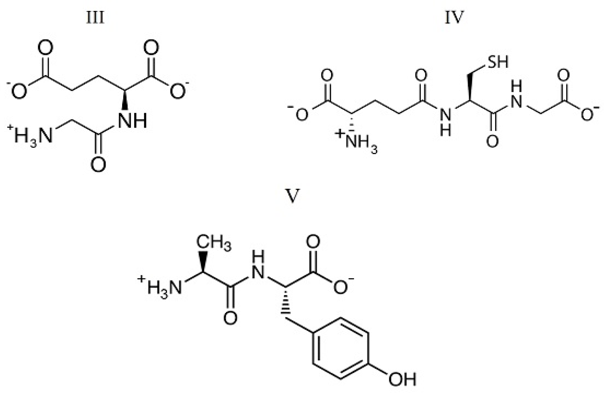
The objects of this study were uracil and cytosine as nucleic bases of the pyrimidine series, the structures of which are shown in Figure 1. In a neutral solution at pH < 9.5, uracil and cytosine take a lactam molecular form [8]. Peptides with different compositions and structures were chosen for the study. Among them, beta-endorphin 30-31 (human) is a C-terminal dipeptide of human beta-endorphin, whose chemical name is glycyl-L-glutamic acid (GlyGlu). Further, glutathione (reduced) is a tripeptide, the chain composition is described as gamma-L-glutamyl-L-cysteinyl-glycine (GluCysGly), and L-α-Alanyl-L-tyrosine is a dipeptide (AlaTyr) containing an aromatic phenolic group in the side chain. These peptides are involved in many metabolic processes, such as the biosynthesis, thiol protection, and redox regulation of cellular thiol proteins under oxidative stress, and conjugation to lipophilic xenobiotics. The peptides take a multicharged ionic form in an aqueous solution due to the processes of hydrolysis and acid dissociation. The structures of the ionic forms of the peptides that dominate in a neutral aqueous solution are shown in Figure 2. In the structure of these di- and tripeptides, oppositely charged terminal groups are distant from each other, unlike amino acids. Such a structure produces favorable steric conditions for the interaction of a bidentate peptide with polar nucleobase groups located on opposite sides of the aromatic ring. The selected peptides differ from each other in chain length, charge distribution, and the number of H-bond donors and acceptors in the molecule.

2. Results and Discussion

2.1. Distribution of Ionic Forms of Peptides

The mole fractions of ionic forms coexisting in an aqueous solution of the studied peptides at various pH values were determined based on the known acid dissociation constants by using the RRSU software (the creators are Vasiliev, V.P.; Borodin, V.A.; Kozlovsky, E.V.; Moscow, Russia) [9]. We used the following data: pKº1 = 2.91, pKº2 = 4.50, pKº3 = 8.21 for GlyGlu at T = 298.15 K [10]; pKº1 = 2.05, pKº2 = 3.49, pKº3 = 8.65, pKº4 = 9.60 for γ-GluCysGly at T = 298.15 K [11]. By extrapolating the results [12] for AlaTyr in a mixed (1,4-dioxane + water) solvent, the following values were obtained for an aqueous solution: pKº1 = 2.81; pKº2 = 7.92. The influence of the ionic strength of the solution on the dissociation constants of peptides is insignificant. As shown earlier for the GlyGlu peptide [10], the change in pK values for the stepwise dissociation is 0.2 in magnitude when the ionic strength varies from 0 to 1.0 mol dm−3. Therefore, we can assume that varying the ionic strength does not significantly affect the distribution of ionic forms of peptides in solution. In all experiments performed in this work, the ionic strength was constant, equal to the ionic strength of saline, 0.15 mol dm−3. The results of calculating the distribution of ionic forms in solution at various pH values are shown in Figure 3. As follows from the diagram, two anionic forms of the peptides dominate in a neutral solution at pH = 7.4: the mole fractions of +GlyGlu2− (marked as +HL2− in Figure 3) and GlyGlu2− (L2−) anions are 0.86 and 0.13, and similar values for +/−GluCysGly (+H2L2−) and GluCysGly (HL2−) are equal to 0.95 and 0.05, respectively. The +AlaTyr (+HL) zwitter ion is the dominant species (mole fraction is 0.77) in an aqueous solution of L-α-alanyl-L-tyrosine at pH = 7.4. The mole fraction of each ionic form of the peptides remained constant in a buffered saline medium with pH = 7.4 during the experiments. The chemical structures of the +GlyGlu2− and +/−GluCysGly dominant anions and +AlaTyr zwitter ion are presented in Figure 2.

2.2. Enthalpies of Dissolution of Peptides

All peptide dissolution enthalpies measured in this work are presented in Table 1. The enthalpies of dissolution in a phosphate-buffered saline differ from those for dissolution in pure water: ΔsolH = 10.84 kJ·mol−1 for GlyGlu; ΔsolH = 10.72 kJ·mol−1 for AlaTyr (measured in this work); ΔsolH = 20.85 kJ·mol−1 for γGluCysGly [13]. The dissolution of GlyGlu and AlaTyr in buffered saline is accompanied by a greater endothermic effect than the process in water. The dissolution effect of the γGluCysGly tripeptide in buffered saline is less endothermic than in water. The observed differences may be associated with a change in the ionic state of the peptides and the contribution of their interaction with electrolytes in a buffered saline.
The enthalpies of dissolution of peptides noticeably change when nucleic bases are added to the buffer solution. Changes in enthalpy depend on the concentration of the nucleic base. The difference between the enthalpies of dissolution in a buffered saline solution ΔsolHm(buffer) and in the same solution with additions of a nucleic base ΔsolHm(buffer+NB) can be considered as a thermochemical characteristic of the interaction between a peptide and a nucleic base ΔtrHm.
ΔtrHm = ΔsolHm(buffer+NB) − ΔsolHm(buffer)
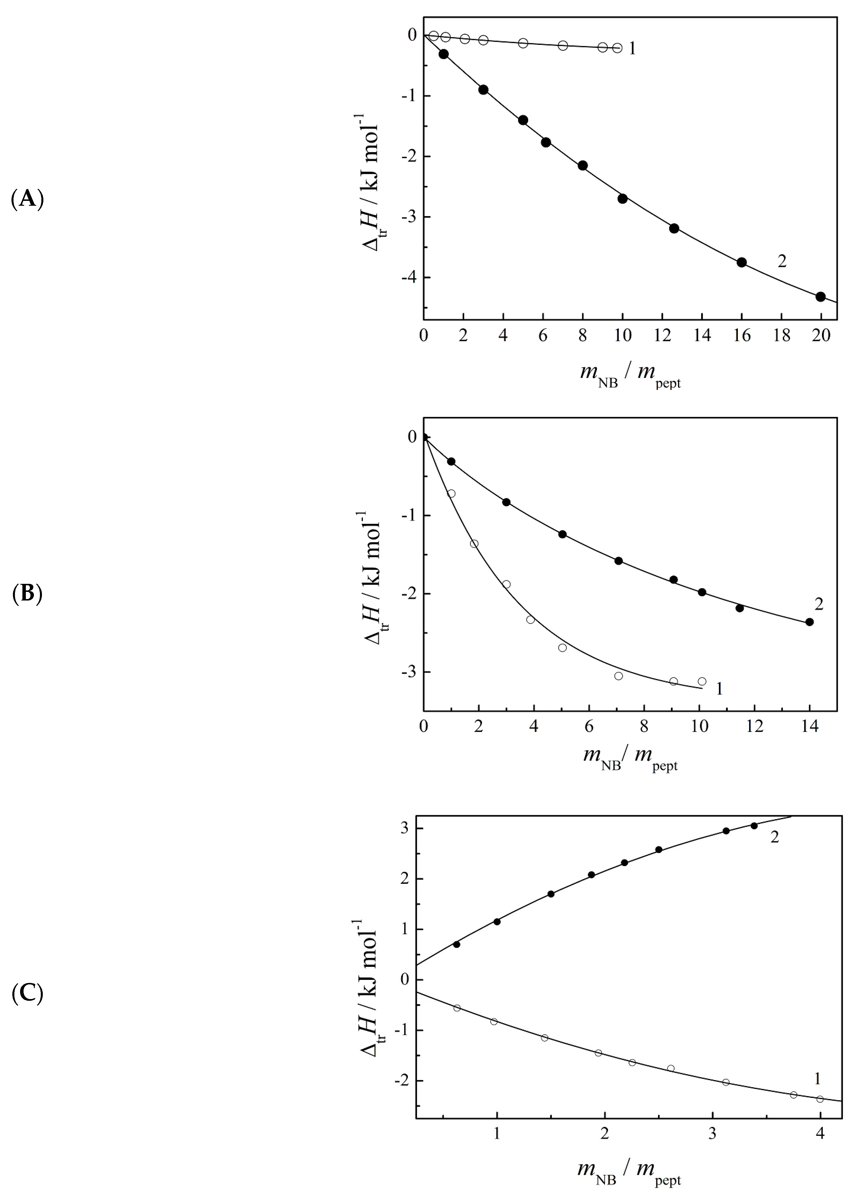
The dependences of ΔtrHm values on the molar ratio of the nucleic base and peptide at a constant mpept = 0.003 mol·kg−1 are shown in Figure 4. As can be seen, the interaction of peptides with uracil and cytosine is usually accompanied by an exothermic effect, with the exception of the interaction of AlaTyr with cytosine, for which an endothermic effect is observed. The shape of the curves in Figure 4 demonstrates the nonlinear dependence of ΔtrHm on the concentration of the nucleic base. The ΔtrHm values change most sharply with small additions of a nucleic base, and the changes are fading when mNB/mpept > 4. In cases of interaction of peptides with uracil and interaction of γGluCysGly with cytosine, the ΔtrHm values change until almost constant values are reached. Such behavior suggests complex formation in the studied systems. Now there is information about the structure of a number of protein-DNA and protein-RNA complexes [14]. The binding of proteins to DNA was established based on the results of UV spectroscopy, mass spectrometry, and phase transition temperatures. However, the mechanism and driving forces of complex formation have not been sufficiently disclosed even for the interaction between the structural units of these biomacromolecules, i.e., between amino acids (or short peptides) and nucleobases. The formation of complexes between the protein model and nucleic acid bases has been theoretically proven by the B3LYP method [15,16], and a significant contribution of hydrogen bonds and stacking interactions to the binding of amino acids to nucleobases has been established [16,17]. It is known that the NH fragment at the 3rd position of the uracil cycle and the NH2 group at the 4th position of cytosine cycle exhibit the ability of an H-bond donor. The O-4 atom of uracil and N-3 and O-2 atoms of cytosine can act as H-bond acceptors.

2.3. Thermodynamic Parameters of Complexation of Peptides with Nucleic Bases

The ΔtrHm values formed a basis for calculating the thermodynamic functions of complex formation. Apparent complexation constants and enthalpy changes (lgKr and ΔrH) were calculated using the initial total concentrations of reagents and experimental values of ΔtrHm by means of the computer program HEAT [18,19]. For peptides, total concentrations were used, which include all ionic forms coexisting in solution. The search for unknown parameters (lgKr, ∆rH) is reduced to the minimization of the F-functional given by
F = i = 1 N w i ( Δ tr H i m exp Δ tr H i m calc ) 2
where ΔtrHi is the enthalpy effect from the i-th reaction, N is the number of experiments, and wi is a weighted factor. Thus, mathematical treatment of the ΔtrHm = f(mNB) dependences using the minimization computer program HEAT allows the enthalpy and apparent constant of complex formation to be simultaneously found. The binding stoichiometry was also a fitting parameter when processing the results. The best agreement between the experimental and calculated ΔtrHmi values was achieved at 1:1 stoichiometry of nucleic base–peptide complexes. All the calculated thermodynamic parameters of complex formation are summarized in Table 2.
The constants of complexation of the studied peptides with uracil and cytosine in buffered saline are small; the values of lgKr take values in the range of 0.95 to 2.0 in magnitude, which is typical for many reversible biochemical processes. The ability of nucleic bases to reverse complexation reactions is a necessary and important condition to perform their biological functions. Complexes of uracil exhibit a greater stability compared to cytosine complexes. Among the uracil complexes, the smallest constant appears for the interaction with the aliphatic peptide AlaAla. As can be seen from Table 1, the enthalpies of dissolution of the peptide change insignificantly with varying concentration, which leads to the lowest positive values of the enthalpies of transfer. Small values of ΔtrHm make it difficult to study the complexation with this peptide, and reliable data could not be obtained in the case of cytosine.
The effect of the structure of the peptide and the nucleobase on the thermodynamic parameters of complexation is manifested in the ratio of the enthalpy and entropy factors of the process. A correct interpretation of the patterns of complex formation in solution is possible only if the effect of solvent reorganization around the reactant molecules is taken into account. For this purpose, the model of overlapping hydrate co-spheres [20,21] has proved to be suitable. The penetration of charged groups of peptides into the hydration shells of nucleic bases can lead to partial dehydration of the NH group and two O-atoms in the case of uracil, or O, N-atoms and NH2-group in the case of cytosine. The NH-fragment in the uracil cycle forms a more stable hydration shell structured by H-bonds with water molecules, unlike the shell around the N-heteroatom in the cytosine cycle. Thus, the set of reaction centers capable of forming complexes with peptides means we can expect that their dehydration will be accompanied by a greater expenditure of energy and a greater increase in disorder in the case of uracil compared to cytosine. From the data in Table 3, it follows that the complexation of +GlyGlu2− and +/−GluCysGly anions, and +AlaAla zwitter-ions with uracil is controlled by the entropy factor, −ΔrH < TΔrS. During complexation, water molecules move from the hydration shells of the polar groups of uracil into the solvent bulk. A significant increase in entropy during the formation of complexes proves the predominance of the contribution of the dehydration of polar groups in this case. The formation of complexes with cytosine is controlled by the enthalpy factor, −ΔrH > TΔrS, which indicates the predominance of the exothermic contribution from the interactions between polar groups and the formation of H-bonds between the peptide and cytosine.
In the case of the +AlaTyr zwitter-ion, the side chain of which is an aromatic ring with a hydroxo group, the complexation exhibits inverse ratios of ΔrH and TΔrS factors. This may be connected with the fact that tyrosine residue is capable of stacking interaction with the nucleic base ring. Other peptides of the studied series do not possess such an ability. The complexation with +AlaTyr is an exothermic process when interacting with uracil and, conversely, an endothermic process when interacting with cytosine. The endothermic effect in the latter case can be attributed to the contribution of stacking interactions of AlaTyr with Cyt in an aqueous solution, accompanied by a dominant endothermic contribution of the partial dehydration of aromatic rings of the reactants. The greatest favorable entropy effect TΔrS = 17.15 kJ·mol−1 confirms this assumption. The interaction of AlaTyr with uracil, in contrast, proceeds due to a favorable enthalpy factor, −ΔrH > TΔrS. It is likely that the stacking interaction does not significantly contribute to the complexation with uracil.
A comparison of the results for the studied complexes shows the effect of the charge of peptide ions on the enthalpies of binding to neutral nucleic bases. The interaction with tripolar anions (+GlyGlu2−, +/−GluCysGly) having two charged carboxylate groups in the structure is an exothermic process. Moreover, the exothermic effects of interaction with cytosine are greater than the effects characteristic of uracil. The greatest exothermic effect, ΔrH = −12.7 kJ·mol−1, manifested during the interaction of Cyt with +GlyGlu2−, is accompanied by the most unfavorable entropy effect, TΔrS = −7.3 kJ·mol−1, and this results in the minimum value of the reaction constant (lgKr = 0.95).
The influence of the number of H-bond acceptors in the peptide structure can be noted. Thus, complexation with the +/−GluCysGly anion having six acceptor centers (four O-atoms in the carboxylate groups and two O-atoms in the amide fragments) proceeds with the highest reaction constant (lgKr values are equal to 2.01 and 1.39) compared to other peptides having from three to five centers.
Summing up the results obtained, we can highlight the trend of compensation of enthalpy and entropy factors in the process of complexation of peptides with nucleic bases, which is described by the following relation:
T ΔrS = (7.0 ± 0.5) + (1.0 ± 0.07) × Δr H,
where the standard deviation is 1.2 kJ·mol−1 and the correlation coefficient is 0.99016 at a confidence level of 0.95.

3. Materials and Methods

The list of chemicals used is presented in Table 3. The chemicals were dried under a vacuum at T = 343.15 K for 24 h. The water content was determined using the Karl Fischer method (Volumetric KF Titrator V30, Mettler Toledo) and amounted to 0.001 mass fraction for Ura and Cyt, and for GlyGlu, GluCysGly, and GlyTyr, the water content was 0.003, 0.002, and 0.002, respectively. All measurements were performed in an aqueous phosphate-buffered saline. A buffered saline, pH = 7.4, was prepared by dissolving five tablets (Sigma, USA) in 1 dm3 of purified water at T = 298 K. The solution contained 0.0073 mol dm−3 Na2HPO4; 0.0046 mol dm−3 KH2PO4; 0.137 mol dm−3 NaCl and 0.0027 mol dm−3 KCl. The ionic strength of the solution was 0.15 mol dm−3. The peptide and nucleic base samples were weighed with an uncertainty of 1 × 10⁻⁵ g. The pH index of the solutions was checked using a digital Five-Easy pH-meter (Mettler Toledo), with a standard uncertainty of 0.02 at T = 298.15 K. The buffered saline was used as solvent for the preparation of sample solutions by mass (with an accuracy of ±1 × 10⁻⁵ g) using a Sartorius-ME21 analytical balance. The peptide concentration remained constant at the level mpept = 0.003 mol∙kg−1. The molalities of nucleic bases varied within the range of 0.0015 to 0.06 mol∙kg−1. The standard uncertainty in the molality of the solutions was estimated within ±2 × 10⁻⁴ mol·kg−1.
Calorimetric measurements were made with an ampoule-type “isoperibol” calorimeter with a 60 cm3 reaction vessel and electrical calibration at T = 298.15 ± 0.01 K and P = 100.5 ± 0.7 kPa, which has been described in detail previously [22]. Electrical calibration of the calorimeter was performed after each experiment. The heat effect from sample dissolution was compared with the effect from the calibrated Joule heating using the digital Standard Temperature Measuring Instrument (BIC, Minsk, Belarus). The calorimetric device’s accuracy was checked prior to the experiments by measuring the enthalpy of the KCl dissolution in water. The experimentally observed value ΔsolH° = (17.52 ± 0.06) kJ·mol−1 agrees well with the ICTAC working group suggestion (17.47 ± 0.07) kJ·mol−1 [23]. In each experiment, a constant amount of the peptide was dissolved in the buffer solution of a nucleic base with a concentration varying from 0.003 to 0.06 mol kg−1. The heat effects of dissolution ranged from 1.5 to 3.5 J. The relative error in the dissolution enthalpy measurements did not exceed 0.5%. Each experiment was repeated at least two times, achieving reproducibility of the enthalpy of dissolution within the standard uncertainty of u(ΔsolHm) = ±0.005 × ΔsolHm.

4. Conclusions

The resulting set of thermodynamic characteristics of the complexation of nucleic bases (uracil and cytosine) with a number of peptides allows us to note some important patterns of this process. Complexes of uracil and cytosine with peptides show low stability in a buffered saline medium, which is typical for many reversible biochemical reactions. The values of the logarithm of the complexation constant vary from 0.9 to 2.0 in magnitude.
The effect of the structure of the peptide and the nucleobase on the thermodynamic parameters of complexation is manifested in the ratio of the enthalpy and entropy factors. The complexation with uracil in most cases is controlled by the entropy factor, −ΔrH < TΔrS, and the enthalpy factor dominates when interacting with cytosine, −ΔrH > TΔrS. The inverse ratios of the factors ΔrH and TΔrS are observed in the case of +AlaTyr zwitter ions, which are capable of stacking interaction with nucleic bases.
The charge of the peptide ion affects the enthalpies of binding with neutral nucleic bases. The complexation with tripolar anions of peptides having two charged carboxylate groups in the structure is accompanied by a greater exothermic effect than the interaction with zwitterions. This trend proves that contact between charged groups of peptides and polar groups of nucleic bases in solution produce an exothermic contribution to the reaction enthalpy.
The dependence of the complexation constants on the number of H-bond acceptors in the peptide structure shows a significant contribution of H-bonding to the stabilization of complexes of peptides with nucleic bases. The highest reaction constants were found for the tripeptide with six acceptor centers in the structure.

Author Contributions

Conceptualization, V.P.B.; Validation, V.P.B., V.I.S. and I.N.M.; Investigation, V.I.S., I.N.M. and D.R.K.; Writing—review & editing, V.P.B. and V.I.S.; Supervision, V.P.B. All authors have read and agreed to the published version of the manuscript.

Funding

The study was supported financially by the Russian Science Foundation, grant No. 22-23-01118.

Institutional Review Board Statement

Not applicable.

Informed Consent Statement

Not applicable.

Data Availability Statement

All new data obtained in this work are contained in the tables presented in the article.

Conflicts of Interest

The authors declare no conflict of interest.

References

  1. Kruse, M.; Möser, C.; Smith, D.M.; Müller-Landau, H.; Rant, U.; Hölzel, R.; Bier, F.F. Measuring Influenza A Virus and Peptide Interaction Using Electrically Controllable DNA Nanolevers. Adv. Mater. Technol. 2022, 7, 2101141. [Google Scholar] [CrossRef]
  2. Kruse, M.; Altattan, B.; Laux, E.-M.; Grasse, N.; Heinig, L.; Möser, C.; Smith, D.M.; Hölzel, R. Characterization of binding interactions of SARS-CoV–2 spike protein and DNA—Peptide nanostructures. Sci. Rep. 2022, 12, 12828. [Google Scholar] [CrossRef] [PubMed]
  3. Kolchina, N.; Khavinson, V.; Linkova, N.; Yakimov, A.; Baitin, D.; Afanasyeva, A.; Petukhov, M. Systematic search for structural motifs of peptide binding to double-stranded DNA. Nucleic Acids Res. 2019, 47, 10553–10563. [Google Scholar] [CrossRef] [PubMed]
  4. Costa, D.; Albuquerque, T.; Queiroz, J.A.; Valente, A.J.M. A co-delivery platform based on plasmid DNA peptide-surfactant complexes: Formation, characterization and release behavior. Colloids Surf. B Biointerfaces 2019, 178, 430–438. [Google Scholar] [CrossRef] [PubMed]
  5. Uittenbogaard, J.P.; Zomer, B.; Hoogerhout, P.; Metz, B. Reactions of Propiolactone with Nucleobase Analogues, Nucleosides, and Peptides. J. Biol. Chem. 2011, 286, 36198–36214. [Google Scholar] [CrossRef] [PubMed] [Green Version]
  6. Chen, Y.-C.; Huang, J.T.B.; Jeng, K.-C.G.; Yang, R.C.K.; Liao, M.-K.; Chen, C.-S.; Chien, W.-J.; Wey, M.-T.; Kan, L.-S.; Sheh, L. Determination of Allosteric Effects and Interstrand Bidentate Interactions in DNA-peptide Molecular Recognition. J. Chin. Chem. Soc. 2010, 57, 266–274. [Google Scholar] [CrossRef]
  7. Bajpayee, N.S.; McGrath, W.J.; Mangel, W.F. Interaction of the adenovirus proteinase with protein cofactors with high negative charge densities. Biochemistry 2005, 44, 8721–8729. [Google Scholar] [CrossRef] [PubMed]
  8. Balodis, E.; Madekufamba, M.; Trevani, L.N.; Tremaine, P.R. Ionization constants and thermal stabilities of uracil and adenine under hydrothermal conditions as measured by in situ UV-visible spectroscopy. Geochim. Cosmochim. Acta 2012, 93, 182–204. [Google Scholar] [CrossRef]
  9. Vasiliev, V.P.; Borodin, V.A.; Kozlovsky, E.V. The Use of Computers in Chemical-Analytical Calculations; Higher School: Moscow, Russia, 1993; p. 110. Available online: https://chemequ.ru/online-progs/rrsu-online/ (accessed on 1 January 2023).
  10. Badelin, V.G.; Barannikov, V.P.; Katrovtseva, A.V.; Tarasova, G.N. Dissociation Constants of Protolytic Dissociation of Glutamyl-Glutamic and Glycyl-Glutamic Acids in Aqueous Solution at 298 K. Russ. J. Gen. Chem. 2013, 83, 945–948. [Google Scholar] [CrossRef]
  11. Lytkin, A.I.; Krutova, O.N.; Chernikov, V.V.; Mokhova, Y.V.; Krutova, E.D.; Tyunina, E.Y. Thermochemical study of reactions of acid–base interaction in an L-glutathione aqueous solution. Russ. J. Phys. Chem. A 2021, 95, 2051–2054. [Google Scholar] [CrossRef]
  12. Aslan, N.; Erden, P.E.; Doğan, A.; Canel, E.; Kılıç, E. Protonation Constants of Some Alanyl Dipeptides in Mixed Aqueous Organic Solvents. J. Solut. Chem. 2016, 45, 299–312. [Google Scholar] [CrossRef]
  13. Lytkin, I.; Chernikov, V.V.; Krutova, O.N.; Golubev, A.A.; Romanov, R.A. Standard Enthalpies of Formation of L-Glutathion and Products of Its Dissociation in Aqueous Solutions. Russ. J. Phys. Chem. A 2021, 95, 1831–1834. [Google Scholar] [CrossRef]
  14. Solovyev, A.Y.; Tarnovskaya, S.I.; Chernova, I.A.; Shataeva Larisa, K.; Skorik, Y.A. The interaction of amino acids, peptides, and proteins with DNA. Int. J. Biol. Macromol. 2015, 78, 39–45. [Google Scholar] [CrossRef] [PubMed]
  15. Alkorta, I.; Elguero, J. Interaction of Protein Backbone with Nucleic Acid Bases. J. Phys. Chem. B 2003, 107, 5306–5310. [Google Scholar] [CrossRef]
  16. Hunter, K.C.; Millen, A.L.; Wetmore, S.D. Effects of Hydrogen-Bonding and Stacking Interactions with Amino Acids on the Acidity of Uracil. J. Phys. Chem. B 2007, 111, 1858–1871. [Google Scholar] [CrossRef] [PubMed]
  17. Rutledge, L.R.; Campbell-Verduyn, L.S.; Hunter, K.C.; Wetmore, S.D. Characterization of Nucleobase-Amino Acid Stacking Interactions Utilized by a DNA Repair Enzyme. J. Phys. Chem. B 2006, 110, 19652–19663. [Google Scholar] [CrossRef] [PubMed]
  18. Borodin, V.A.; Vasil’ev, V.P.; Kozlovsky, E.V. Universal program package for treatment experimental data on the complex equilibria in solutions. In Mathematical Problems of Chemical Thermodynamics; Nauka: Novosibirsk, Russia, 1985; pp. 219–226. (In Russian) [Google Scholar]
  19. Borodin, V.A.; Kozlovsky, E.V.; Vasil’ev, V.P. Treatment of the results of calorimetric measurements on digital computer in the study of complex equilibriums in solutions. Russ. J. Inorg. Chem. 1982, 27, 2169–2172. [Google Scholar]
  20. Gurney, R.W. Ionic Processes in Solution; McGraw Hill: New York, NY, USA, 1953. [Google Scholar]
  21. Kaur, N.; Banipal, P.K.; Banipal, T.S. Physico-chemical effects of caffeine on aqueous solutions of pyrimidine based model compounda of nucleic acids. J. Mol. Liq. 2016, 221, 721–732. [Google Scholar] [CrossRef]
  22. Barannikov, V.P.; Kurbatova, M.S.; Mezhevoi, I.N. The influence of structure of isomolecular dipeptides of α-Ala-α-Ala and β-Ala-β-Ala on their behavior in aqueous micellar solution of SDS. Thermochim. Acta 2020, 689, 178647. [Google Scholar] [CrossRef]
  23. Gunther, C.; Pfestorf, R.; Rother, M.; Seidel, J.; Zimmermann, R.; Wolf, G.; Schreder, V. An interlaboratory test for certification of potassium chloride as a certified reference material (CRM) for solution calorimetry. J. Therm. Anal. 1988, 33, 359–363. [Google Scholar] [CrossRef]
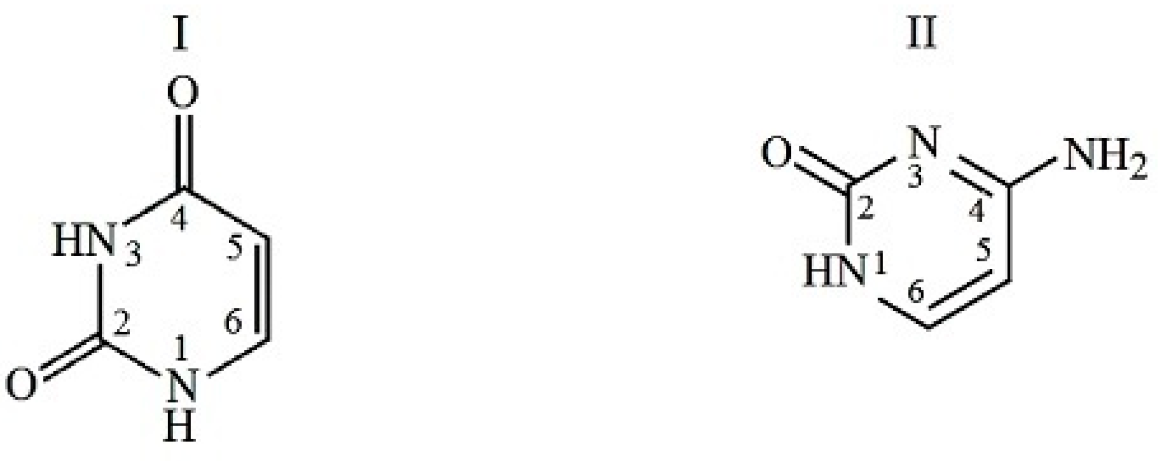
Figure 1. Chemical structure of uracyl (I) and cytosine (II).
Ijms 24 09764 g001
Figure 2. Dominant anionic forms of glycyl-L-glutamic acid (III) and L-γ-glutamyl-L-cysteinyl-glycine (IV) and zwitter ion of L-alanyl-L-tyrosine (V).
Ijms 24 09764 g002
Figure 3. A diagram of the distribution of ionic forms of glycyl-L-glutamic acid, L-γ-glutamyl-L-cysteinyl-glycine, and alanyl-L-tyrosine depending on pH at T = 298.15 K; the dotted line marks the value of pH = 7.4.
Ijms 24 09764 g003
Figure 4. The transfer enthalpies of peptides ((A)—GlyGlu; (B)—γGluCysGly; (C)—AlaTyr) from buffered saline to buffer solutions of (1) uracil and (2) cytosine at different ratios of nucleic base and peptide, mpept = 0.003 mol·kg−1, T = 298.15 K, pH = 7.4.
Ijms 24 09764 g004
Table 1. The enthalpies of peptide dissolution at constant peptide molality (m = 0.003 mol·kg−1) in phosphate-buffered saline with additives of uracil and cytosine (ΔsolHm, kJ·mol−1) at T = 298.15 K; p = 100.5 kPa; pH = 7.4; and molality of nucleic bases mNB, mol·kg−1.
mNBΔsolHmmNBΔsolHm
GlyGlu + uracilGlyGlu + cytosine
011.430.003011.12
0.001511.420.009110.53
0.003311.390.015310.03
0.006211.370.01859.66
0.009011.350.02429.28
0.015111.300.03038.73
0.021211.250.03788.24
0.027211.200.04807.68
0.029211.210.05997.11
γGluCysGly + uracilγGluCysGly + cytosine
020.360.003020.05
0.003019.640.009019.53
0.005519.000.015019.12
0.009018.480.021018.78
0.011618.030.024018.64
0.015117.670.030018.38
0.021217.240.034418.18
0.027217.210.042017.98
0.030317.28
AlaTyr + uracilAlaTyr + cytosine
010.890.005011.59
0.005010.330.008012.04
0.007810.060.011512.59
0.01159.740.015512.97
0.01559.440.017513.21
0.01809.270.020113.47
0.02019.130.025013.84
0.02508.860.027113.94
0.03008.61
0.03208.53
AlaAla + uracil
0−7.46
0.0050−7.44
0.0078−7.41
0.0115−7.36
0.0155−7.32
0.0201−7.27
0.0250−7.25
0.0300−7.24
0.0320−7.23
Note. Standard deviation (u) of experimental parameters: u(m) = 0.00005 mol·kg−1; temperatures u(T) = 0.01 K and u(p) = 0.7 kPa; u(ΔsolHm) = ± 0.005 × ΔsolHm. The molalities of peptides and nucleic bases are calculated per kilogram of buffer saline.
Table 2. Thermodynamic parameters of complexation of peptides with uracil and cytosine in buffered saline at pH = 7.4 and T = 298.15 K.
lgKr *ΔrGΔrHT ΔrS
kJ·mol−1
Ur + GlyGlu1.16 ± 0.001−6.64 ± 0.01−0.7 ± 0.15.9 ± 0.1
Ur + γ-GluCysGly2.01 ± 0.04−11.4 ± 0.2−4.4 ± 0.17.0 ± 0.3
Ur + AlaTyr1.45 ± 0.001−8.3 ± 0.01−5.30 ± 0.053.0 ± 0.06
Ur + AlaAla1.03 ± 0.05−5.9 ± 0.31.0 ± 0.046.9 ± 0.3
Cyt + GlyGlu0.95 ± 0.001−5.40 ± 0.01−12.7 ± 0.08−7.3 ± 0.1
Cyt + γ-GluCysGly1.39 ± 0.002−7.89 ± 0.01−4.79 ± 0.073.1 ± 0.1
Cyt + AlaTyr1.29 ± 0.002−7.35 ± 0.019.8 ± 0.117.15 ± 0.1
* unit of Kr is kg·mol−1.
Table 3. The list of chemicals, their origin, and purity values.
Chemical aM bCAS No. cOriginPurity d
Uracil112.0966-22-8Termo Ficher Scientific0.996
Cytosine111.1070-30-7Appolo Scientific0.996
Glycyl-L-glutamic acid
(beta-endorphin 30-31 human)
204.187412-78-4Bachem>0.99
L-γ-Glutamyl-L-cysteinyl-glycine (glutathion reduced)307.32108457-42-7Tokyo Chemical Industry>0.99
L-α-Alanyl-L-tyrosine252.273061-88-9Abcr GmbH>0.98
a All reagents were dried in vacuum; b Molar mass of chemical, kg·kmol−1; c Chemical Abstract Service registry number; d Mass fraction (as stated by the supplier).
Disclaimer/Publisher’s Note: The statements, opinions and data contained in all publications are solely those of the individual author(s) and contributor(s) and not of MDPI and/or the editor(s). MDPI and/or the editor(s) disclaim responsibility for any injury to people or property resulting from any ideas, methods, instructions or products referred to in the content.