Article

Superparamagnetic Spinel-Ferrite Nano-Adsorbents Adapted for Hg2+, Dy3+, Tb3+ Removal/Recycling: Synthesis, Characterization, and Assessment of Toxicity

by
A. F. P. Allwin Mabes Raj
1,2,3,
Maja Bauman
3,
Nena Dimitrušev
3,4,
Lamiaa M. A. Ali
5,
Mélanie Onofre
5,
Magali Gary-Bobo
5,
Jean-Olivier Durand
6,
Aleksandra Lobnik
3,4 and
Aljoša Košak
4,*
1
Jožef Stefan International Postgraduate School, Jamova 39, 1000 Ljubljana, Slovenia
2
Department of Environmental Science, Jožef Stefan Institute, Jamova 39, 1000 Ljubljana, Slovenia
3
Institute for Environmental Protection and Sensors (IOS) Ltd., Beloruska 7, 2000 Maribor, Slovenia
4
Faculty for Mechanical Engineering, University of Maribor, Smetanova 17, 2000 Maribor, Slovenia
5
IBMM, Univ Montpellier, CNRS, ENSCM, 34293 Montpellier, France
6
ICGM, Univ Montpellier, CNRS, ENSCM, 34293 Montpellier, France
*
Author to whom correspondence should be addressed.
Int. J. Mol. Sci. 2023, 24(12), 10072; https://doi.org/10.3390/ijms241210072
Submission received: 4 April 2023 / Revised: 22 May 2023 / Accepted: 26 May 2023 / Published: 13 June 2023
(This article belongs to the Special Issue Recent Advances in Nanomaterials Science)

Abstract

:
In the present work, superparamagnetic adsorbents based on 3-aminopropyltrimethoxy silane (APTMS)-coated maghemite (γFe2O3@SiO2–NH2) and cobalt ferrite (CoFe2O4@SiO2–NH2) nanoparticles were prepared and characterized using transmission-electron microscopy (TEM/HRTEM/EDXS), Fourier-transform infrared spectroscopy (FTIR), specific surface-area measurements (BET), zeta potential (ζ) measurements, thermogravimetric analysis (TGA), and magnetometry (VSM). The adsorption of Dy3+, Tb3+, and Hg2+ ions onto adsorbent surfaces in model salt solutions was tested. The adsorption was evaluated in terms of adsorption efficiency (%), adsorption capacity (mg/g), and desorption efficiency (%) based on the results of inductively coupled plasma optical emission spectrometry (ICP-OES). Both adsorbents, γFe2O3@SiO2–NH2 and CoFe2O4@SiO2–NH2, showed high adsorption efficiency toward Dy3+, Tb3+, and Hg2+ ions, ranging from 83% to 98%, while the adsorption capacity reached the following values of Dy3+, Tb3+, and Hg2+, in descending order: Tb (4.7 mg/g) > Dy (4.0 mg/g) > Hg (2.1 mg/g) for γFe2O3@SiO2–NH2; and Tb (6.2 mg/g) > Dy (4.7 mg/g) > Hg (1.2 mg/g) for CoFe2O4@SiO2–NH2. The results of the desorption with 100% of the desorbed Dy3+, Tb3+, and Hg2+ ions in an acidic medium indicated the reusability of both adsorbents. A cytotoxicity assessment of the adsorbents on human-skeletal-muscle derived cells (SKMDCs), human fibroblasts, murine macrophage cells (RAW264.7), and human-umbilical-vein endothelial cells (HUVECs) was conducted. The survival, mortality, and hatching percentages of zebrafish embryos were monitored. All the nanoparticles showed no toxicity in the zebrafish embryos until 96 hpf, even at a high concentration of 500 mg/L.

1. Introduction

Transition metals (TM) and internal transition metals (ITM), often referred to as d- and f-block elements, respectively, are key raw materials for the European economy, forming a strong industrial base that produces a wide range of products and applications used in everyday life and modern technologies. Many of these metals are considered to be highly toxic and have negative environmental and human health effects due to anthropogenic factors (e.g., mercury, lead, chromium, etc.). Furthermore, both groups also include strategic metals, such as rare-earth metals (e.g., dysprosium, terbium, samarium, neodymium, etc.), which are in increasing demand and subject to supply risks. Therefore, reliable and unhindered access to these raw materials is a growing concern in the EU and globally [1,2,3,4,5].
Of these metals, mercury (Hg) is particularly noteworthy. It can be found in many common devices, from thermometers, barometers, thermostats, and pressure gauges to fluorescent lamps, etc., and it is considered to be the most toxic heavy metal in the environment, particularly due to its high bioaccumulation and biomagnification capacity [6]. This is because spilled elemental mercury (Hg0) is converted by microbial processes in the environment, particularly in water, into an organic form called methylmercury (MeHg), which is the most toxic form of mercury. Methylmercury is then transferred to fish and other wildlife and, eventually, it can be ingested, causing adverse health effects [6,7].
The removal of toxic metals, such as mercury, from the environment plays a significant role in minimizing their environmental and human health effects, while the recycling of essential and precious transition metals, such as platinum, palladium, gold, silver, rhodium, iridium, ruthenium, cobalt, niobium, tungsten, etc., and rare-earth metals, such as terbium, dysprosium, neodymium, lanthanum, samarium, cerium, etc., from e-waste and other raw waste materials, is of major importance to increase the availability of secondary resources, as well as improving the knowledge base that provides prerequisites for a circular economy on a larger scale than today [1,6,8,9,10]. Generally, current processing/removal technologies include, but are not limited to, hydrometallurgy (solvent extraction, ion exchange, precipitation, and crystallization), pyrometallurgy, electrometallurgy (electrorefining, and electrowinning), and aeriometallurgy (supercritical fluid extraction) [11,12].
All these technologies have many disadvantages. Pyrometallurgy is energy-intensive and generates greenhouse gas (GHG) emissions, while hydrometallurgy relies on large volumes of acids and organic solvents, thus generating hazardous wastes [13,14,15]. The primary disadvantage of aeriometallurgy is that the extraction must be operated at the high pressure (1000–5000 psi) required to maintain the solvent in a supercritical state using supercritical CO2 [16]. In electrometallurgy, an inert atmosphere is usually required for recycling related to operational and maintenance drawbacks, while the recycling of raw metals can generate a small volume of waste, which is not yet developed, qualified, certified, or accepted. However, the electrometallurgy process also features the drawbacks of huge energy consumption for heating and electrolytic reduction and potential chlorine-gas emission [17,18].
An attractive alternative to these technologies is the solid-phase extraction (SPE) of metal ions from the solution using nanostructured materials as adsorbents, characterized by surface functionality, high surface-to-volume ratio, and/or porosity [19,20]. The use of SPE involves the adsorption of the target-metal ions from the solution onto the adsorbent surface followed by the subsequent recycling of the metals and the regeneration of the adsorbents [21,22]. The advantages of SPE include its low solvent consumption, ease of use, efficient removal of metal ions, even at low concentrations, and automation capabilities.
In the last decade, ferrimagnetic iron-oxide nanoparticles (NPs) have received special attention in the field of adsorbents, as reported in many reviews [23,24,25,26]. Magnetite (Fe3O4) and maghemite (γFe2O3) are the main types of ferrimagnetic NP, and so far, they have received considerable attention due to their nontoxicity and biocompatibility [27,28,29], as well as their ability to be easily dispersed and collected using an external magnetic field [30,31,32]. These ferrimagnetic materials, when reduced to particle dimensions smaller than a certain domain, exhibit superparamagnetic behavior, which means that when an external magnetic field is applied, they magnetize to saturation magnetization (σs), but when the magnetic field is removed, they no longer exhibit either residual magnetism (Mr) or coercivity (Hc) [33]. Hence, superparamagnetic iron-oxide NPs can be easily guided in the magnetic field [25,27,34,35]. One prominent example is the introduction of cobalt (Co2+) ions into an iron-oxide spinel crystal lattice. Cobalt-doped iron oxides, also known as cobalt ferrites (CoFe2O4), arouse interest in adsorption applications as their magneto-crystalline anisotropy, which affects the magnetization, coercivity, reversal, and relaxation of nanoparticles, can be tuned by the substitution of cobalt for iron [36,37,38,39,40].
Due to the increasing use of iron-oxide NPs as adsorbents in the recycling of strategic transition metals, there is a high likelihood that these NPs may ultimately enter aquatic ecosystems through effluent discharge and leaching during or after recycling activities, thereby affecting the environment and human health. Unfortunately, there is a serious lack of accurate and sufficient information on their toxic effects. Therefore, toxicity assessment has become increasingly important to understand the impact of these NPs on human health and the environment [41].
In many studies, it was found that uncoated NPs usually tend to be more toxic than coated particles; therefore, the surface modification of uncoated NPs can significantly reduce their toxicity [41,42,43,44,45,46,47,48,49]. Other studies revealed significant cytotoxic effects of these NPs, such as inflammation, the formation of apoptotic bodies, impaired mitochondrial function (MTT), the leakage of membrane lactate dehydrogenase (LDH assay), the generation of reactive oxygen species (ROS), increases in the number of micronuclei as indicators of gross chromosomal damage (a measure of genotoxicity), and chromosome condensation [44,48,50,51,52,53,54].
Furthermore, little is known about the toxicity of the metal dopants of iron-oxide NPs such as cobalt (Co2+). The potential toxicity of cobalt-doped iron oxide (CoFe2O4) is therefore the subject of many debates concerning environmental, health, and safety issues, particularly their use in the environment and their effects on human health [28,44,55,56,57,58,59,60,61,62].
One of the simplest ways to modify the surfaces of NPs is to use alkoxysilanes, which are considered among the preferred coating materials due to their chemical stability, biocompatibility [63,64,65], and versatility, to achieve the functionality [65,66] required in the end-use applications of NPs.
To ensure the functionality of iron-oxide NPs, various alkoxysilane ligands can be grafted directly to their surfaces in one step, avoiding an intermediate multistage reaction mechanism. The grafting principle of alkoxysilanes is based on the sol-gel hydrolysis of alkoxide groups in the structures of alkoxysilane precursors, producing silanol groups (Si-OH), which undergo condensation reactions to form siloxane bonds (Si–O–Si) on the surface of the iron oxide, resulting in the formation of a protective silica surface layer (SiO2). Many alkoxysilanes may contain various functional groups in their aliphatic chains, such as hydroxyl (–OH), amine (–NH2), mercapto (–SH), carboxylic (–COOH), sulphonic (–SO3H), phosphonate (–PO(OH)2), phosphate (–PO2(OH)2), etc., which contain electron-donor atoms (O, N, P, S) and allow the formation of relatively strong complexes with the target transition-metal ions to be recycled [31,41,42,45,67,68,69,70,71].
Although many studies report that alkoxysilanes are non-toxic [18,19,20,71,72,73], their toxicity in terms of reactivity, stability, and degradation effects has not yet been thoroughly investigated [74,75,76,77].
In the present work, we attempted to fabricate efficient superparamagnetic adsorbents based on two different spinel-type iron oxides, both maghemite (γFe2O3) and cobalt ferrite (CoFe2O4), which were surface-functionalized using a (3-aminopropyl)trimethoxy silane (APTMS) precursor. The functionalized superparamagnetic adsorbents were characterized to test their adsorption efficiency and adsorption capacity towards Dy3+, Tb3+, and Hg2+ ions in aqueous solutions and their desorption efficiency when an acidic medium was used. The assessment of the cytotoxicity of both types of NP with and without an amino-propyl (–(CH2)3NH2) surface coating was conducted on four different types of healthy cell: human-skeletal-muscle-derived cells, human fibroblasts, murine macrophages cells, and human-umbilical-vein endothelial cells. Further, their toxic effects on zebrafish embryos were also evaluated by recording the survival, mortality, and hatching percentages during embryo development.

2. Results and Discussion

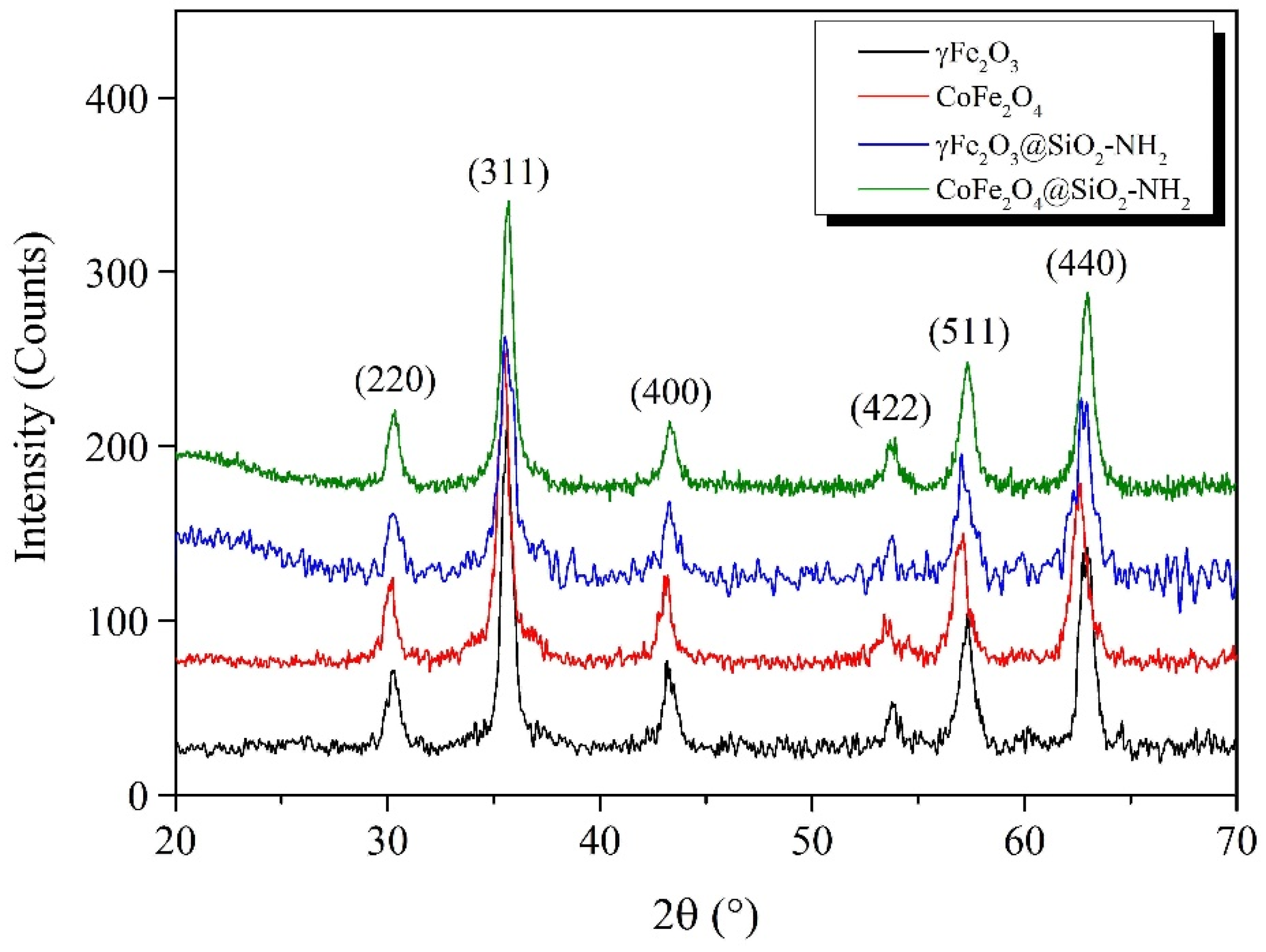
2.1. Synthesis and Characterization of MNPs

The magnetic γFe2O3 and CoFe2O4 NPs prepared by the coprecipitation method were characterized using XRD (Figure 1). All the diffraction peaks of the prepared samples were consistent with the cubic spinel crystal structure (JCPDS Card 39-1346). It can be seen from the XRD pattern that the presence of diffraction lines at 2θ of 30.5°, 35.5°, 43.2°, 53.6°, 57.1°, and 62.9° for both samples corresponded to the cubic crystal planes of (220), (311), (400), (422), (511), and (440), respectively. The particle sizes of the γFe2O3 and CoFe2O4 were calculated from the broadening of the most intensive diffraction peak corresponding to the (311) crystal plane using the Deby–Scherrer equation [78,79]. The calculated average particle sizes of the γFe2O3 and CoFe2O4 were 10.2 nm and 11.5 nm, respectively, and the crystalline-lattice parameters corresponding to the cubic spinel crystal structure obtained based on Bragg’s law were 0.8358 nm and 0.8345 nm, respectively. The presence of broad amorphous diffraction peaks for the functionalized γFe2O3@SiO2–NH2 and CoFe2O4@SiO2–NH2 NPs, which appeared at a low diffraction angle 2θ of 20°, was due to the presence of the amorphous SiO2 surface layer, indicating that the crystalline cubic spinel γ-Fe2O3 and CoFe2O4 NPs were successfully surface-functionalized with APTMS [80].
The positions of the diffraction peaks for the γFe2O3@SiO2–NH2 and CoFe2O4@SiO2-NH2 NPs were at the same positions 2θ as those of the γFe2O3 and CoFe2O4, indicating that the crystalline cubic spinel structures remained unchanged after their functionalization with APTMS.
The transmission-electron micrographs in Figure 2 represent the morphological properties of the as-prepared γFe2O3 and CoFe2O4 NPs and functionalized Fe2O3@SiO2–NH2 and CoFe2O4@SiO2–NH2 core@shell nanostructures. It can be seen that the obtained γFe2O3 and CoFe2O4 NPs were relatively spherical in shape, with average particle sizes of (9.9 ± 0.9) nm and (11.5 ± 1.0) nm, respectively, while the particle-size distributions of the functionalized γFe2O3@SiO2–NH2 and CoFe2O4@SiO2–NH2 NPs were (14.5 ± 1.1) nm and (17.7 ± 1.2) nm, respectively. The electron-diffraction patterns of the γFe2O3 and CoFe2O4 NPs indicated the crystalline nature of the prepared powders, with each of the concentric diffraction rings belonging to the spinel crystal structure.
The EDXS patterns of the γFe2O3 and CoFe2O4 NPs in Figure 3a,b confirm the presence of Co, Fe, and O elements, indicating the formation of γFe2O3 and CoFe2O4 nanostructures, while on the EDXS spectrum of the γFe2O3@SiO2–NH2 and CoFe2O4@SiO2–NH2 in Figure 3c,d, respectively, the presence of C, O(N), Co, Fe, and Si confirmed the success of the surface functionalization of the γFe2O3 and CoFe2O4 NPs with APTMS and, thus, the formation of the core@shell nanostructures. Small proportions of Cu and C elements belong to the TEM copper-grid supported transparent carbon foil.
The Brunauer–Emmet–Teller (BET) analysis showed specific surface areas of 94.9 m2/g for the γFe2O3 and 62.5 m2/g for the CoFe2O4. According to the BET-specific surface area at a relative pressure of 0.3, average particle sizes (dbet) of 15.1 nm and 18.5 nm were calculated for the γFe2O3 and CoFe2O4, respectively, assuming the sphericity of the NPs using the equation Sbet = 6/(dbetρ), where ρ is a theoretical density of 4.9 g/cm3 for γFe2O3 [81] and 5.2 g/cm3 for CoFe2O4 [82]. The average size calculated from the surface area was a little higher than that determined using the XRD, most probably due to the agglomeration of the particles [83]. For the Barrett–Joyner–Halenda (BJH) adsorption, the average pore size was 7.0 nm, with a total pore volume of 0.2335 cm3/g, for the γFe2O3 NPs, and 5.8 nm, with a total pore volume of 0.1275 cm3/g, for the CoFe2O4 NPs. Furthermore, for the BJH desorption, the average pore size was 8.4 nm, with a total pore volume of 0.3152 cm3/g, for the γFe2O3, and 6.1 nm, with a total pore volume of 0.1353 cm3/g, for the CoFe2O4 NPs.
Due to the surface functionalization of the γFe2O3 and CoFe2O4 NPs with APTMS, the obtained specific surface area decreased to 40.5 m2/g for the γFe2O3@SiO2–NH2 and 44.7 m2/g for the CoFe2O4@SiO2–NH2. It is known that the larger the surface area, the smaller the particle size, and a smaller BET surface area means a larger particle size. According to the specific surface areas, the average particle sizes of the prepared γFe2O3@SiO2–NH2 and CoFe2O4@SiO2–NH2 samples were calculated as 30.2 nm and 25.8 nm, respectively.
A FTIR analysis (Figure 4) was performed to obtain additional information on the coverage of the NPs with the APTMS.
In the FTIR spectra (Figure 4a), the two peaks near 3400 cm−1 and 1630 cm−1 were assigned to the hydroxyl group OH for all the synthesized NPs. The functionalization process of the γFe2O3 and CoFe2O4 NPs with alkoxysilanes was verified by the asymmetric stretching vibrations of the Si-O-Si bonds at 1050 cm−1 and the bending of the Si-H bonds at 796 cm−1 and at 988 cm−1, indicating the formation of silica (SiO2) shells.
The presence of amino-propyl groups in the γFe2O3@SiO2–NH2 and CoFe2O4@SiO2–NH2 samples was confirmed by the peaks at 2934 cm−1, 1615 cm−1, 1336 cm−1, and 781 cm−1, which were assigned to the stretching vibrations of the –CH2–NH2 bonds, the bending of N–H and NH2, the wagging and twisting of –CH2–NH2, and the wagging and twisting of primary amino groups (–NH2), respectively. These peaks in the source spectra were not sufficiently visible, but enlarged individual peak areas confirmed their presence (Figure 4b).
A thermogravimetric analysis (Figure S1) was used to determine the thermal stability and the percentage of amino-propyl ligands grafted onto the surface of the magnetic γFe2O3 and CoFe2O4 NPs. According to the literature, the estimation of mass-loss values of 1.1% and 1.3% (not shown in Figure 5) while heating as-prepared magnetic γFe2O3 and CoFe2O4 NPs up to 200 °C usually corresponds to the evaporation of physically and chemically absorbed moisture. Further heating of the γFe2O3 and CoFe2O4 magnetic NPs up to 900 °C, respectively, resulted in additional mass losses of 2.7% and 3.4%, respectively, which were most likely due to phase and surface changes, the reduction in porosity, and the degradation of the remaining surface species.
The mass losses of the prepared γFe2O3@SiO2–NH2 and CoFe2O4@SiO2–NH2 samples of 3.1% and 1.8%, respectively (not shown in Figure S1), began at the initial 30 °C mark and continued evolving up to 150 °C. These changes were related to the evaporation of the absorbed moisture from their structures. Further heating of the γFe2O3@SiO2–NH2 and CoFe2O4@SiO2–NH2 samples up to 900 °C caused more remarkable mass losses of 21.5% and 20.1%, respectively, which corresponded to the decomposition of the SiO2 shell and the removal of amino-propyl groups from the NP surfaces, followed by the reduction in the porosity and the cracking of the residual siloxane species, Si–O–Si.
To establish the stability of the prepared γFe2O3, CoFe2O4, γFe2O3@SiO2–NH2, and CoFe2O4@SiO2–NH2 NPs in an aqueous medium and to determine their surface potential and isoelectric points (IEP), the electrokinetic (ζ) potential as a function of pH media was measured (Figure 5). The pH values of the IEPs for the γFe2O3 and CoFe2O4 NPs were about 6.3 and 6.6, respectively, while the silica-coated NPs showed pH dependencies similar to that of pure silica, i.e., at pH 2–3 [69,84].
The values of the ζ-potential for the γFe2O3 and CoFe2O4 NPs were higher than +30 mV at pH < 5.2 and lower than −30 mV at pH > 7.9, which means that the γFe2O3 and CoFe2O4 NPs were stable in the aqueous media at pH smaller than 5.2 and higher than 7.9. In that pH range, the electrostatic repulsions between the NPs dispersed in an aqueous medium are stronger than the random thermal Brownian motion and, therefore, prevent them from accidental collision and agglomeration and, subsequently, from settling out.
The observed IEP at pH 2.4 for the γFe2O3@SiO2 NPs confirmed that the silica-coating process of the γFe2O3 NPs was effective, since the charged surface properties were close to those of pure silica (i.e., at pH 2–3) [84]. The silica shell at the maghemite cores caused an increase in their chemical stability at pH values > 4.1, where the surface potential was lower than −30 mV, thus rendering their performance suitable for environmental applications. Moreover, the silica coating prevented the dissolution of the γFe2O3 and CoFe2O4 NPs and, thus the leaching of potentially toxic Co2+ ions from the spinel crystalline structure and the Fe2+/Fe3+ oxidation, which otherwise occurs in an acidic medium at values of pH < 3.
It can be seen that an APTMS precursor may be employed to functionalize γFe2O3 and CoFe2O4 NPs to form functional γFe2O3@SiO2–NH2 and CoFe2O4@SiO2–NH2 core@shell nanostructures. The presence of an amine layer on the surface of the γFe2O3@SiO2–NH2 and CoFe2O4@SiO2–NH2 NPs makes them positive in a broad range of pH due to the protonation and deprotonation of the amine groups, which depend on the solution’s pH values.
The zeta (ζ) potential measurement of the γFe2O3@SiO2–NH2 and CoFe2O4@SiO2–NH2 NPs in an aqueous solution showed an isoelectric point at about pH 9.0 and stability of the NPs at pH < 6.6 for the γFe2O3@SiO2-NH2 and at pH < 7.9 for the CoFe2O4@SiO2–NH2, where the ζ–potential was higher than +30 mV, and at pH > 10.6, where the ζ–potential was lower than −30 mV.
We used pH potentiometric titrations for the determination of the total charge of the aqueous colloidal dispersions of the γFe2O3@SiO2–NH2 and CoFe2O4@SiO2–NH2 NPs.
The results of the potentiometric titration isotherms for the γFe2O3@SiO2–NH2 and CoFe2O4@SiO2–NH2 NPs are presented in Figure S2. The data exhibited protonation and deprotonation progress for both samples at an alkaline pH of around 10, exhibiting a pKa value of 10.1 for the γFe2O3@SiO2–NH2 with a maximum charge of 0.0485 mmol/g and a pKa value of 9.9 for the CoFe2O4@SiO2–NH2 with a maximum charge of 0.0155 mmol/g. This can be attributed to the contribution of the total primary amine groups, which is in agreement with data published elsewhere [85,86], and indicates the successful surface functionalization of the MNPs with APTMS.
Figure 6a shows the hydrodynamic size distribution of the aqueous colloidal γFe2O3, CoFe2O4, γFe2O3@SiO2–NH2, and CoFe2O4@SiO2–NH2 NPs at 21 °C, according to the intensity-distribution pattern, showing a narrow distribution with homogeneous sizes. The γFe2O3 and CoFe2O4 NPs, which were an average diameters of 9.9 nm and 11.5 nm by the TEM, in fact exhibited slightly larger hydrodynamic sizes of approximately 11.7 nm and approximately 14.5 nm, respectively (Figure 6a). On the other hand, after the surface functionalization with the APTMS, the γFe2O3@SiO2–NH2 and CoFe2O4@SiO2–NH2 NPs showed larger hydrodynamic sizes, of about 16.6 nm and about 19.1 nm, respectively (Figure 6a), compared to the previous primary particle sizes of the same nanoparticles observed by TEM.
It is worth noting that these hydrodynamic sizes were maintained over the applied time range of 1 h (Figure 6a), indicating that both the uncoated γFe2O3 and CoFe2O4 and the surface-functionalized γFe2O3@SiO2–NH2 and CoFe2O4@SiO2–NH2 NPs retained colloidal stability.
The time-dependent hydrodynamic diameters of uncoated γFe2O3 and CoFe2O4, and coated γFe2O3@SiO2–NH2 and CoFe2O4@SiO2–NH2 NPs, are shown in Figure 6b.
Figure 6b shows the samples showed a trend towards colloidal stability. The hydrodynamic diameter of the colloidal CoFe2O4 NPs did not change significantly with time. The average hydrodynamic size of the colloidal CoFe2O4 NPs was maintained at approximately (11.7 ± 1.1) nm over the entire time range. The colloidal γFe2O3 NPs also had a similar behavioral pattern in terms of hydrodynamic size, with a slightly larger fluctuation in the values around the average diameter of the NPs of (14.8 ± 1.5) nm. Although the overall maximum average hydrodynamic sizes of the γFe2O3 and CoFe2O4 NPs increased with respect to the particle-size values estimated from the XRD and TEM images, no agglomeration or aggregation of NPs was observed.
After the surface functionalization of the γFe2O3 and CoFe2O4 NPs by the APTMS, a fluctuation and an increase in the hydrodynamic diameters of the γFe2O3@SiO2–NH2 and CoFe2O4@SiO2–NH2 NPs by about 42% and 30%, respectively, were observed in relation to the uncoated γFe2O3 and CoFe2O4 NPs. The final hydrodynamic sizes of the γFe2O3@SiO2–NH2 and CoFe2O4@SiO2–NH2 NPs were 16.6 ± 1.5 nm and 19.1 ± 2.0 nm, respectively, compared to the particle-size values estimated from the XRD and TEM images. Despite the fluctuation and increase in the total maximum hydrodynamic sizes of the γFe2O3@SiO2–NH2 and CoFe2O4@SiO2–NH2 NPs, no agglomeration or aggregation of NPs was observed. The samples showed a trend toward colloidal stability.
It is obvious that the average particle sizes measured by the DLS technique were slightly larger than the average particle sizes estimated on the basis of the XRD and TEM. It is known that the hydrodynamic sizes of particles dispersed in liquids are usually larger than the primary particle sizes, as reported by many other studies [87,88,89,90]. The hydrodynamic sizes of particles measured by DLS depend on many factors, particularly the concentration of the dispersion, temperature, pH, etc., due to which dispersed nanoparticles may tend to aggregate; therefore, the measured hydrodynamic diameters in such cases are usually much larger than the actual sizes [88,90].
Figure 7 shows the comparison of the magnetic properties of the uncoated γFe2O3 and CoFe2O4 NPs and the functionalized γFe2O3@SiO2–NH2 and CoFe2O4@SiO2–NH2 NPs, which were carried out using VSM analysis.
At the maximum magnetic field (H) strength in the magnetization phase, the specific mass magnetization (Ms) of the samples γFe2O3 and CoFe2O4 reached values of 54.98 emu/g and 52.67 emu/g, respectively. The remanent magnetization (Mr) and coercivity (Hci) values for the samples γFe2O3 and CoFe2O4 can be determined from the shape of the hysteresis curves in the vicinity of the zero-magnetic-field strength. We determined that the remanent magnetization and coercivity of the γFe2O3 sample were 1.17 emu/g and 12.55 Oe, respectively, and that the remanent magnetization and coercivity of the CoFe2O4 sample were 6.27 emu/g and 163.76 Oe, respectively.
As can be seen, the γFe2O3 NPs showed very low coercivity due to the small particle sizes. In this case, the γFe2O3 NPs exhibited a superparamagnetic character; in other words, they were monodomain. In contrast, when the iron in the spinel crystal structure of the maghemite (γFe2O3) replaced the cobalt (CoFe2O4), the coercivity was non-zero, and the samples did not show superparamagnetic behavior.
A comparison of the magnetic characteristics of these two samples showed that with the integration of the cobalt into the γFe2O3 spinel crystal structure, the coercivity increased by thirteen times, and the remanent magnetization increased by almost five times, while the specific mass magnetization did not change significantly.
The increase in coercivity was the cause of the increase in the magnetocrystalline anisotropy due to the cobalt substitution [39]. Magnetocrystalline anisotropy is a key factor that determines the superparamagnetic behavior of nanocrystalline particles and serves as an energy barrier to block spin relaxation, which changes the magnetic state from ferrimagnetic to superparamagnetic [37,38].
The average particle sizes of samples γFe2O3 and CoFe2O4 were approximately similar (10.2 nm for γFe2O3 and 11.5 nm for CoFe2O4), so the loss of superparamagnetic properties when replacing the iron with the cobalt in the γFe2O3 spinel crystal structure may have been due to an increase in the magnetocrystalline anisotropy of the CoFe2O4 [37].
In the magnetization step, under the maximum magnetic field strength (Hci), the specific mass magnetization (Ms) of the synthesized γFe2O3@SiO2–NH2 decreased from 54.98 emu/g for the γFe2O3 NPs to 39.32 emu/g, and from 52.67 emu/g for the CoFe2O4 NPs to 33.24 emu/g for the CoFe2O4@SiO2–NH2 NPs. This was due to the presence of a non-magnetic SiO2-NH2 coating on the surfaces of the γFe2O3 and CoFe2O4 cores, which, due to its diamagnetic quality, contributed to the reduction in the net-specific magnetization of the γFe2O3@SiO2–NH2 and CoFe2O4@SiO2–NH2 NPs. After the surface functionalization of the samples γFe2O3 and CoFe2O4, a pronounced decrease in the remanence (Mr) and coercivity (Hci) of the samples was noticed because of the increase in the average particle size at the expense of the SiO2-NH2 surface coating. The remanence (Mr) thus decreased from 1.17 emu/g for the sample γFe2O3 to 0.77 emu/g for the sample γFe2O3@SiO2–NH2, and from 6.27 emu/g for the sample CoFe2O4 to 4.43 emu/g for the sample CoFe2O4@SiO2-NH2, while the coercivity of the sample γFe2O3@SiO2–NH2 decreased to 11.32 Oe from 12.55 Oe for the γFe2O3, and for the sample CoFe2O4@SiO2-NH2 m it decreased to 122.02 Oe from 163.76 Oe for the CoFe2O4 NPs. The decrease in saturation magnetization (Ms), coercivity (Hci), and remanent magnetization (Mr) in the γFe2O3@SiO2–NH2 and CoFe2O4@SiO2–NH2 NPs compared to the γFe2O3 and CoFe2O4 NPs was expected due to the larger NP sizes, corresponding to the SiO2 shell and the functionalization with amino (–NH2) groups.

2.2. Adsorption and Desorption Tests

To evaluate the performances and basic adsorption affinity of the γ-Fe2O3@SiO2–NH2 and CoFe2O4@SiO2–NH2 adsorbents toward Dy3+, Tb3+, and Hg2+ ions in aqueous solutions, batch adsorption experiments at pH 4.5 and with a contact time of 2 h were performed. The graphical representations of the adsorption efficiency and adsorption capacity of the prepared samples are depicted in Figure 8. The numerical results are presented in Table 1.
The adsorption results of the Dy3+, Tb3+, and Hg2+ ions are presented only for the functionalized NPs, both γFe2O3@SiO2–NH2 and CoFe2O4@SiO2–NH2, as adsorption by functionalized NPs is usually much more efficient than adsorption by non-functionalized NPs [46].
The adsorption results showed that both the γFe2O3@SiO2–NH2 and the CoFe2O4@SiO2–NH2 samples had high adsorption affinity towards Dy3+ and Tb3+ ions, with relatively high adsorption efficiencies of 83.1% and 89.3%, respectively, but low adsorption capacities of 4.0 mg/g and 4.7 mg/g, respectively.
The adsorption affinity of the γFe2O3@SiO2–NH2 and CoFe2O4@SiO2–NH2 toward the Dy3+, Tb3+, and Hg2+ ions in the aqueous medium can be explained by Pearson’s hard-and-soft acid-base (HSAB) theory [91]. This concept is based on Lewis’ definition of acids as electron acceptors and bases as electron donors, and it states that soft acids prefer to coordinate and form stronger bonds and more stable complexes with soft bases, whereas hard acids prefer to coordinate and form stronger bonds and more stable complexes with hard bases.
According to the HSAB concept, Dy3+ and Tb3+ ions are classified as hard Lewis acids, and functional amino (–NH2) groups are classified as hard Lewis bases, so Dy3+ and Tb3+ ions have a high affinity for NH2 groups. On the other hand, as a soft Lewis acid, Hg2+ is a relatively large (1.02 Å) and polarizable atom, which, in practice, prefers to associate with soft bases. Since Hg2+ is larger than Tb3+ (0.923 Å) and Dy3+ (0.912 Å) and, thus, more polarised, it has a weaker preference for interactions with hard NH2 groups. This resulted in its significantly lower adsorption capacity of 2.1 mg/g for the γFe2O3@SiO2–NH2 and 1.2 mg/g for the CoFe2O4@SiO2–NH2 NPs.
The lower adsorption capacity of Hg2+ compared to Tb3+ and Dy3+, in the context of the HSAB concept, can be explained by the use of the absolute-hardness parameter (ηs). Parr et al. [92] defined the absolute-hardness parameter as ηs = (IpAs)/2, where Ip (eV) is the ionization potential and As (eV) is the electron affinity. Pearson, in a 1988 paper [93], conveniently provided cumulative experimental values for ionization potential and electron affinity, referring to earlier work by Moore [94]. From these values, it is possible to calculate the absolute-hardness parameter (ηs) values, which for Hg2+, Tb3+, and Dy3+ are 5.4 eV, 3.1 eV, and 3.1 eV, respectively. Pearson [93] also defined softness (σs) as the inverse of hardness, σs = 1/ηs, with zero as the maximum softness. The values of the softness parameter for Hg2+, Tb3+, and Dy3+ are thus 0.19, 0.32, and 0.32, respectively, indicating the softer nature of Hg2+ compared to Tb3+ and Dy3+. Therefore, in this case, a higher complexation affinity of the hard NH2 groups for the hard Tb3+ and Dy3+ ions and a lower complexation affinity for the softer Hg2+ is expected, which also agreed with the results of our work.
A possible explanation for the low adsorption capacity of Hg2+ is its unusual chemical properties and its character as a soft Lewis acid. The soft nature of Hg2+ is related to its ground-state electronic configuration ([Xe]4f145d106s2) with filled electron subshells up to 6s, which, due to its stability, strongly resists electron removal, resulting in the very high ionization potential (Ip 10.434 eV) and moderately high electronegativity (χP 2.00 by Pauling) [95,96] of Hg, which is reflected in its low chemical reactivity [97].
Because all the main energy levels of the Hg atom are filled, and because of the unusually stable 6s2 electron pair, Hg can form only very weak hard–soft chemical bonds with amino groups, with a covalent character [96]. Therefore, the interactions of soft Hg with the hard electron-donating N-atom in the NH2 group, which is a small (0.16 Å), low-polarizable atom with high electronegativity (χP 3.04 Pauling) and high ionization potential (Ip 14.534 eV), are not favored [95,98].
In contrast, Tb3+ and Dy3+, which are hard Lewis acids, according to the HSAB concept, tend to have chemical interactions with the electron-donor N-atoms in the NH2 groups, which have the character of hard Lewis bases. This is due to differences in the stability of their electron configurations and electron-density distributions. Compared to Hg, which has a stable electron configuration, Tb ([Xe]6s24f9) and Dy ([Xe]6s24f10) have stabilized 4f electrons that do not contribute to the formation of chemical bonds, and their chemical behavior is dictated by the 6 =s valence electrons [97]. Despite the presence of 6s2 electrons in addition to the 4f and [Xe] nuclei in Tb and Dy, their most stable oxidation state in aqueous media is +3, which makes them more reactive than divalent Hg. Their chemical reactivity gradually decreases from terbium towards mercury in the sequence of Tb > Dy > Hg [97], which is also reflected in their higher affinity for the formation of complexes with NH2 groups of APTMS compared to Hg. This is consistent with our finding, in this study, that the adsorption capacities of Tb3+, Dy3+, and Hg2+ decrease in the order of Tb3+ > Dy3+ > Hg2+.
On the other hand, the unexpectedly low adsorption capacity of Tb3+ and Dy3+ ions, which have the character of hard Lewis acids, is probably also due to the strong hydration of these cations in aqueous solutions and the formation of aqua complexes ([Ln(OH2)9]3+, Ln = Tb, Dy), which is also reflected in their high hydration-enthalpy values for Tb3+Hhydr −3540 kcal/mol) and Dy3+Hhydr −3570 kcal/mol) [99,100,101]. Such aqua complexes are easily formed because of excess water, and they are prone to substitution reactions, in which water molecules are successively replaced by amino ligands and vice versa [102].
Bjerrum [103] determined that a metal complex in an aqueous solution is formed by the exchange of a coordinated water molecule directly bound to the central lanthanide ion (Ln3+) with other ligands, provided that the ligand has a sufficiently strong affinity for the lanthanide ion to compete with the affinity of the coordinated water. Such exchanges result in the formation of strong complexes with inner hydration shells [104]. When the ligand replaces the water molecule of the aqua-complex ion, a new metal complex is formed and equilibrium is established. It is assumed that this formation does not take place in one step, but in several steps, involving the following: (i) the joint diffusion of the hydrated cation and anion, (ii) the partial loss of solvent to form the ion pair, (iii) the loss of water from the first coordination sphere of the cation, and (iv) the formation of the complex species. The rate-determining step is the loss of the water molecule from the coordination sphere of the Ln3+ ion and, thus, depends only on the hydration properties of the Ln3+ ion [105,106].
In general, a maximum number of water molecules are distributed around Ln3+ ions during the hydration process, depending on the size of the Ln3+ ion and its electronic properties. It is known that the ionic radii of Ln3+ ions in the Ln species decrease as the atomic number increases due to lanthanide contraction, which is a consequence of the incomplete mutual protection of the valence f-orbitals.
According to the ratio of the radii of the Ln3+ ions to the radii of the oxygen atoms (1.34 Å) in coordinated water molecules (rion/r0), all the hydrated Ln3+-ions in aqueous solutions occupy the configuration of a tricapped trigonal prismatic [107] geometry with six nearly identical water molecules at the vertices of the trigonal prism and the remaining three water molecules capping the prism faces [108].
Hydrates of the lighter Ln3+ ions (La3+-Nd3+) have a regular tricapped trigonal prismatic configuration, with slightly longer bond distances from the Ln3+ ion to the capping water molecules (Ln-O) than to the water molecules forming the prism. The decrease in the radii of the Ln3+-ions with the increase in the atomic number of the Ln-species starting from Nd3+ does not, in principle, affect the structure of the prism, but has a strong effect on the more weakly bound capping positions of the water molecules in the prismatic structure, resulting in a partial loss of the water molecules in the capping positions for the heaviest Ln3+-ions (Ho3+-Lu3+). In fact, studies have shown that the bond strength of the three-capping water molecules is strong at the beginning of the Ln series for nonahydrates (e.g., La3+-Sm3+), while in the Ln series starting from (Sm3+-Lu3+), the Ln–O capping bonds become weaker and shorter at the same time. The Ln–O change in the [Ln(OH2)9]3+ for Ln3+-ions (e.g., La3+, Sm3+, Tb3+, Dy3+, Ho3+, and Lu3+) occurs in the following sequence: La-O (2.52 Å) > Sm-O (2.46 Å) > Tb-O (2.39 Å) > Dy–O (2.37 Å) > Ho–O (2.36 Å) > Lu–O (2.31 Å) [109]. Consequently, in the lanthanide series starting from Sm3+ onward, the three water molecules in the structure are not equally strongly bound and one of the water molecules is at a shorter distance from the Ln-center than the other two. In the series from Ho3+ to Lu3+, the water deficit and the differences between the strongly bound water molecules and the more weakly-bound molecules are even greater [108,109].
Therefore, since Ln-O bonds are more easily broken, in the whole Ln series, the lighter lanthanide Ln3+ -ions (La3+–Nd3+) form stable nonahydrates, while the heavier Ln3+-ions (Ho3+–Lu3+) form octahydrates. Intermediate Ln3+-ions (Sm3+–Dy3+) favor the formation of complex forms of nona- and octahydrates with non-integer coordination numbers (CN) between 8 and 9. Therefore, the CN of these intermediate lanthanides should be average with respect to the ratio of nona- to octahydrate forms [110,111].
Such Ln-O bond behavior determines the hydration behavior of Ln3+ ions and explains their very unusual and complex ligand exchange kinetics throughout the Ln series [110,111]. The peculiarity is that the exchange rate of water molecules between the first hydration shell and the bulk solvent increases in the direction from La3+ to Gd3+, reaches its maximum in the central region of the Ln series (Tb3+, Dy3+), and then decreases up to Lu3+ [112,113]. The physical reasons for these phenomena are still not well understood and are the subject of many investigations [110,111].
On the other hand, H2O is also known to be a hard base [114], which, in accordance with the HSAB concept and the above values of absolute hardness, associates with hard ligands rather than soft ligands, because, in this case, hard–hard interactions are more favorable. Since the amino (–NH2) group is a hard Lewis base, the substitution of the amino ligand and H2O is relatively favorable from this point of view [100,114], which makes lanthanide-ion complexes of Ln3+ (Ln = Tb, Dy) extremely labile [115]. In contrast, for transition metals, the lability of complexes generally varies with their electronic configuration. Some complexes are labile, while others are kinetically very inert, such as d3 species and low-spin d6 species, with high stabilities and high activation energies for ligand substitution [115].
The formation of amino complexes is largely related to the availability of active sites on the adsorbent surfaces and the coordination number. The two key factors influencing the coordination number and complex formation, as well as their chemical stability, areas follows: (i) the influence of the donor N-atoms in the immediate vicinity of the metal ion (Tb3+, Dy3+, Hg2+), which, due to interatomic tension, prevent more N-atoms from making contact with the metal ion; and (ii) the steric repulsions between the larger substituent groups in the APTMS, which are bonded to the donor N-atom (i.e., H2N(CH2)3-), which determine how much of the ligand can be surrounded by the metal ion [115]. The lack of available active sites thus reduces the ability to form complexes, resulting in a lower adsorption capacity.
The electron-donating N-atoms of the amino groups of APTMS in the surface coating of the adsorbent possess a free-electron pair that can be donated to form a coordination bond with the Tb3+, Dy3+, and Hg2+ metal ions in the aqueous medium. Depending on the coordination number, which is 9 for Tb3+ and Dy3+ [98,116] and 6 for Hg2+ [117,118], these metal ions can coordinate linearly with one or two amino groups, with the remaining coordination sites occupied by water molecules. Thus, the coordination mechanism of Tb3+, Dy3+, and Hg2+ with the amino groups of APTMS on the surfaces of γFe2O3@SiO2–NH2 and CoFe2O4@SiO2–NH2 adsorbents can be represented as [101,119,120]:
RNH2 + Ln3+ ⇔ [Ln(RNH2)(H2O)8]3+ (Ln = Tb, Dy)
RNH2 + Hg2+ ⇔ [Hg(RNH2)(H2O)5]2+
After the adsorption of Tb3+, Dy3+, and Hg2+, the possibility of recovering both adsorbents, γFe2O3@SiO2–NH2 and CoFe2O4@SiO2–NH2, for the reuse and recycling of Tb3+, Dy3+, and Hg2+ ions were verified by a desorption process. The choice of agent for desorption is based on the type of adsorbate–adsorbent system [121] and depends mainly on the compatibility between adsorbate and adsorbent, the pH and ionic strength of the medium, the complexation ability, the desorption-agent content, and the exposure time, as these variables can modify the desorption behavior or destroy the adsorbent structure [121]. In addition, desorption phenomena are related to a series of surface interactions and diffusion into micropores or the intraparticle spaces of the adsorbents [99].
In our previous research [122,123], we studied some desorption conditions and the use of different acidic desorption agents, such as hydrochloric acid (HCl), nitric acid (HNO3), and citric acid, and we found that HNO3 gave better results than the other two desorption agents. Therefore, in the present study, a desorption procedure for Tb3+, Dy3+, and Hg2+ was carried out using a 1-M aqueous solution of HNO3 for 1 h at room temperature and with pH 4.5 [124]. The desorption was carried out in one cycle only due to the loss of material during the desorption process. The results of the desorption efficiency are shown graphically in Figure 9. After 1 h, the Tb3+, Dy3+, and Hg2+ ions were completely desorbed from the surfaces of the γFe2O3@SiO2–NH2 and CoFe2O4@SiO2–NH2 adsorbents, indicating the potential for the stable reusability of the prepared adsorbents and the high potential of these adsorbents for the recycling and removal of heavy metals.
Table 2 shows the comparison of the adsorption of the Dy3+, Tb3+, and Hg2+ ions onto the prepared γFe2O3@SiO2–NH2 and CoFe2O4@SiO2–NH2 NPs with the adsorption of Dy3+, Tb3+, and Hg2+ on different adsorbents. It can be observed that the adsorption conditions, such as the initial adsorbate concentration (cads,0), adsorbent dosage (γads), time of adsorption (tads), adsorption temperature (Tads), and solution pH, were very different, making the comparison of the adsorption performances a difficult and a complex task.
Su et al. [125] studied the adsorption capacity of Dy3+ with the adsorbate Fe3O4@SiO2@polyaniline-graphene oxide, and they demonstrated an adsorption capacity of 16.0 mg/g, an adsorption efficiency of 98%, and a desorption efficiency of 95% at pH 4. Liu et al. [126] reported an adsorption capacity of 28.3 mg/g, an adsorption efficiency of >80%, and a desorption efficiency of >95% for Dy3+ at pH 7 using Fe3O4-C18–chitosan–DETA. With the same pH of 7, using NH4OH@SiO2 (APTMS), Kegl et al. [84] reported an adsorption capacity of 23.2 mg/g and an adsorption efficiency of 94% for Dy3+. Javadian et al. [127] reported the adsorption characteristics of synthetic-polymer-based magnetic adsorbent (M-PPTA) toward Dy3+ at pH 5.5, and found an adsorption capacity of 24.0 mg/g, an adsorption efficiency of 98.4%, and a desorption efficiency of >84%. Shinozaki et al. [128] noted 18.4 mg/g of adsorption capacity and 30% of adsorption efficiency for Dy3+ at pH 1 using an adsorbate of polymeric adsorbents modified with ethylenediamine (EDA) and diglycolamic acid (DGA).
Alcaraz et al. [129] reported the use of chemically and physically activated carbons from spent-coffee waste to adsorbed Dy3+ ions. They showed an adsorption capacity of chemically activated carbons toward Dy3+ of 31.26 mg/g and an adsorption efficiency of Dy3+ of 96% at pH 4. When using physically activated carbons from spent-coffee waste, an adsorption capacity of 33.52 mg/g and an adsorption efficiency of 99% were obtained. Further, Viana et al. [130] reported an adsorption capacity of 0.570 mg/g and an adsorption efficiency of 89% for Dy3+ using ulva lactuca—Chlorophyta (green), while an adsorption capacity of 0.526 mg/g and an adsorption efficiency of 84% were obtained for Dy3+ by using Gracilaria sp.—Rhodophyta (red). According to Zhang et al. [131], an adsorption capacity of 1.85 mg/g and an adsorption efficiency of 84% of Dy3+ at pH 3 were obtained using an Fe0–SiO2–PA/SiO2–DTPA adsorbent.
Su et al. [125], Zhang et al. [131], Barros et al. [132], Tong et al. [133], and Kegl et al. [134] studied the adsorption capacity of Tb3+ using various adsorbent materials under different adsorption conditions. Using graphene oxide at pH 4, Su et al. [125] obtained an adsorption capacity of 11.8 mg/g, an adsorption efficiency of 98%, and a desorption efficiency of 95% for Tb3+. Zhang et al. [131] used an Fe0–SiO2–PA/SiO2–DTPA adsorbate at a pH of 3 and obtained an adsorption capacity of 1.4 mg/g for Tb3+. Barros et al. [132] reported the use of molecular-sieve zeolite for the adsorption of Tb3+ ions at a pH of 5, and they noted an adsorption capacity of 2.59 mg/g alongside an adsorption efficiency of 80%, and a desorption efficiency of >60% for Tb3+; furthermore, they obtained an adsorption capacity of 5.07 mg/g for the Tb3+ using B. cereus biomass-supported zeolite adsorbate. Tong et al. [133] used multi-walled carbon nanotubes with tannic acid (TA-MWCNTs) for the adsorption of Tb3+ at pH 5 and obtained an adsorption capacity of 8.55 mg/g, while Kegl et al. [134] used a superparamagnetic γ-Fe2O3-NH4OH@SiO2 (APTMS) adsorbent and obtained an adsorption capacity of 0.204 mg/g for the adsorption of Tb3+ ions from water at a pH of 7.
Adsorption studies on Hg2+ ions with CoFe2O4–chitosan–graphene, polypyrrole-functionalized magnetic kaolin (Ppy-Fe3O4/kaolin), and CoFe2O4@SiO2–NH2 adsorbents at a pH of 7 showed adsorption capacities of 361.0 mg/g, 255.2 mg/g, and 149.3 mg/g, respectively. These studies were performed by Zhang et al. [135], Lin et al. [136], and Wang et al. [137], respectively.
In recent works, Xia et al. [138], Allwin Mabes Raj et al. [122], and Inglezakis et al. [139] studied the adsorption of Hg2+ using CoFe2O4@SiO2–EDTA, γ–Fe2O3@NH2, and Fe3O4 as adsorbates, and they noted adsorption capacities of 103.3 mg/g, 85.6 mg/g, and 28.0 mg/g for Hg2+, respectively. Liu et al. [140] studied the adsorption capacity of Hg2+ with the adsorbate rice-husk-activated carbon (RHAC) and demonstrated an adsorption capacity of 55.87 mg/g at a pH of 5. Denizli et al. [141] studied the adsorption of Hg2+ with magnetic poly (vinyl alcohol)—procion blue MX-3G as the adsorbent and noted an adsorption capacity of 69.2 mg/g, an adsorption efficiency of >94%, and a desorption capacity of 95% for Hg2+. An adsorption study of Hg2+ with amino-functionalized SiO2 particles (NH2@SiO2) conducted by Raj et al. [123] and a study with activated carbon performed by Solis et al. [142] showed adsorption capacities of 3.75 mg/g and 2.5 mg/g, respectively.
Nevertheless, it can be observed that the adsorption of Dy3+, Tb3+, and Hg2+ ions is tested mostly in acidic or neutral aqueous media, and that the adsorption capacity is higher in surface-functionalized adsorbent nanomaterials. In addition, some adsorbents have significantly higher adsorption properties for Hg2+ than for Dy3+ and Tb3+ ions compared to our prepared adsorbent materials. However, the main advantages of our adsorbents are their relatively fast kinetics of adsorption, which are associated with their nano-size and functionality, their high desorption efficiency, and the convenient and highly efficient sustainable recovery of the used adsorbate material at the end of the adsorption process by magnetic attraction.

2.3. Cytotoxicity Study

The cytotoxicity of the nanoparticles was tested in different healthy cell lines after 3 days of incubation. In the SKMDCs, the γFe2O3 nanoparticles showed lower toxicity than the γFe2O3@SiO2–NH2, as shown in Figure 10a. The cell viability reached 69 ± 0.72% after 3 days of incubation with 5 µg/mL of γFe2O3@SiO2–NH2, which decreased to 57 ± 0.47% at a concentration of 500 µg/mL. Similarly, the CoFe2O4 nanoparticles showed lower toxicity than the CoFe2O4@SiO2–NH2. The incubation of the SKMDCs with CoFe2O4@SiO2–NH2 up to a concentration of 125 µg/mL for 3 days showed lower toxicity with a cell viability above 80%; however, the cell viability decreased to 70 ± 3.41% at 500 µg/mL.
In the fibroblasts, all the nanoparticles showed lower toxicity when the cell viability was above 80%, with concentrations of up to 500 µg/mL (Figure 10b).
In the macrophage RAW264.7 cell line, both the γFe2O3 and the CoFe2O4 showed lower toxicity than the γFe2O3@SiO2–NH2 and CoFe2O4@SiO2–NH2 (Figure 10c). At 50 µg/mL, the cell viability reached 52 ± 0.61% and 74 ± 7% for the γFe2O3@SiO2–NH2 and CoFe2O4@SiO2–NH2, respectively. However, at 500 µg/mL, the cell-viability values were 59 ± 0.10%, 13 ± 0.51%, 66 ± 0.31%, and 7 ± 0.23% for the γFe2O3, γFe2O3@SiO2–NH2, CoFe2O4, and CoFe2O4@SiO2–NH2, respectively. It is worth mentioning that the macrophage RAW264.7 cells were more sensitive to the γFe2O3@SiO2–NH2 and CoFe2O4@SiO2–NH2 than the other cell lines.
The toxicity of the nanoparticles was also tested in the HUVECs (Figure 10d). The results showed the low toxicity of the γFe2O3, γFe2O3@SiO2–NH2, and CoFe2O4 compared with the CoFe2O4@SiO2–NH2, which showed a decrease in cell viability (69 ± 1.26%) at 25 µg/mL, reaching 25 ± 0.86% at 500 µg/mL.
The results presented in Figure 11a show that none of the nanoparticles had hemolytic effects, except the nanoparticles of γFe2O3@SiO2–NH2, which showed a dose-dependent hemolytic effect. A change in the supernatant color was observed by the naked eye at concentrations of 50 µg/mL and above, indicating the lysis of red blood cells and hemoglobin release (Figure 11b) [5].
A toxicity study on zebrafish embryos exposed to different concentrations of nanoparticles as shown in Figure S3 revealed that none of the nanoparticles had toxic effects on the zebrafish embryos until 96 hpf compared to the control, even at a high concentration of 500 mg/L (Figure 12).
Table S1 depicts the list of various adsorbent materials with their toxicological assessment in different biological systems. Many studies investigating the toxicities of different materials using in vivo and in vitro studies, such as a toxicity study of human kidneys (HEK293) using magnetic, SiO2-coated nanoparticles with an exposure of up to 1.0 μg/μL for 12 h, which was reported by Shim et al. [143]. In their SiO2 in vitro study, Pisani et al. [144] used A549 (human) lung cells with 0.1–6-μg/cm2 dosages and exposed them for 24 h. A toxicity study was reported by Ellinger-Ziegelbauer and Pauluhn [145] on rat-lung cells with MWCNT at a 11-mg/m3 concentration for a 6-h (aerosol) 90-day post-exposure period. Jovanović et al. [146] studied the TiO2 (anatase) and hydroxylated fullerene toxicity of 40-μg/mL fullerenes and 170 ng/mL in Danio rerio (embryo). With 2.5 μg/mL and 25 μg/mL of Ag in human colon cells, toxicity was studied by Böhmert et al. [147]. Conde et al. [148] studied toxicity using Au, functionalized with anti-sense cDNAs at dosages of 30 nM in the HCT-116 (human) colon.
Our present study investigating the cytotoxicity of iron-oxide NPs generally showed that both these NPs, γFe2O3@SiO2–NH2 and CoFe2O4@SiO2–NH2, are non-toxic.

3. Materials and Methods

All the chemicals used in this study were generally of reagent grade, obtained from commercial sources without further purification: iron (II) chloride tetrahydrate (FeCl2·4H2O, 98%, 198.81 g/mol, CAS no. 13478-10-9, Sigma-Aldrich, Merck Group KGaA, Darmstadt, Germany), iron (III) chloride hexahydrate (FeCl3·6H2O, ≥98%, 270.3 g/mol, CAS no. 10025-77-1, Sigma-Aldrich, Merck Group KGaA, Darmstadt, Germany), cobalt (II) chloride hexahydrate (CoCl2·6H2O, 98%, 237.93 g/mol, CAS no. 7791-13-1, Sigma-Aldrich, Merck Group KGaA, Darmstadt, Germany), ammonium hydroxide aqueous solution (NH4OH, 25%, 35.05 g/mol, 0.91 g/mL, CAS no. 1336-21-6, GramMol, Zagreb, Croatia), sodium hydroxide (NaOH, ≥98% (anhydrous), 40 g/mol, CAS no. 1310-73-2, Sigma-Aldrich, Merck Group KGaA, Darmstadt, Germany), potassium hydroxide (KOH, 1 mol/L (1-N), Titripur®, 56.11 g/mol, 1.05 g/mL, CAS no. 1310-58-3, Sigma-Aldrich, Merck Group KgaA, Darmstadt, Germany), potassium chloride (KCl, ACS reagent, 99.0–100.5%, 74.55 g/mol, CAS no. 7447-40-7, Sigma-Aldrich, Merck Group KgaA, Darmstadt, Germany), hydrochloric acid (HCl, for 1000 mL, 1 mol/L (1-N), Titrisol®, 36.46 g/mol, 1.09 g/ml, CAS no. 7647-01-0, Sigma-Aldrich, Merck Group KgaA, Darmstadt, Germany), nitric acid (HNO3, ACS reagent, 70%, 63.01 g/mol, 1.413 g/mL, CAS no. 7697-37-2, Sigma-Aldrich, Merck Group KgaA, Darmstadt, Germany), 2-propanol (C3H8O, 99.8%, 60.1 g/mol, 0.785 g/mL, CAS no. 67-63-0, GramMol, Zagreb, Croatia), ethanol (C2H5OH, 96%, 46.07 g/mol, 0.810 g/mL, CAS no. 64-17-5, GramMol, Zagreb, Croatia), tetraethyl orthosilicate TEOS (C6H20O4Si, 99%, 208.33 g/mol, 0.94 g/mL, CAS no. 78-10-4, Sigma-Aldrich, Merck Group KGaA, Darmstadt, Germany), 3-aminopropyltrimethoxysilane APTMS (C6H17NO3Si, 97%, 179.29 g/mol, 1.027 g/mL, CAS no. 13822-56-5, Sigma-Aldrich, Merck Group KGaA, Darmstadt, Germany), terbium (III) chloride hexahydrate (TbCl3∙6H2O, 99.9%, 373.38 g/mol, CAS no. 13798-24-8, Sigma-Aldrich, Merck Group KGaA, Darmstadt, Germany), dysprosium (III) nitrate pentahydrate (Dy(NO3)3∙5H2O, 99.9%, 438.59 g/mol, CAS no. 10031-49-9, Sigma-Aldrich, Merck Group KGaA, Darmstadt, Germany), and mercury (II) nitrate monohydrate (Hg(NO3)2∙H2O, ≥98.5%, 342.62 g/mol, CAS no. 7783-34-8, Sigma-Aldrich, Merck Group KGaA, Darmstadt, Germany). For the preparation of all suspensions and solutions, deionized water (dH2O) was used.

3.1. Synthesis of Magnetic Nanoparticles (MNPs)

Spinel-type MNPs of maghemite (γ-Fe2O3) and Co-ferrite (CoFe2O4) were obtained by co-precipitation of M2+ (M = Fe, Co) and Fe3+ salts at slightly elevated temperature in an alkaline aqueous medium according to Schikorr reaction [149]:
M2+ + 2Fe3+ + 4OH + O2 = MO∙Fe2O3 (s)↓ + 2H2O

3.1.1. γ-Fe2O3 NPs

For the synthesis of γFe2O3 NPs, 50 mL of 25% NH4OH in a round-bottomed reaction flask was heated up to (87 ± 2) °C by reflux and stirred at 400 rpm. A 0.5-M aqueous solution of Fe2+ and Fe3+ in a molar ratio of 1:2 was added to the ammonia solution until pH 10 was reached. The reaction was carried out for 1 h at (87 ± 2) °C. Instantly, when the two solutions were mixed, a dark-brown precipitate of magnetic phase was formed. After the reaction, the dark-brown γFe2O3 precipitate was separated from the supernatant by settling on the permanent magnet and rinsed with deionized water several times. Finally, the rinsed precipitate was dried in a laboratory oven at 90 °C for 24 h.

3.1.2. CoFe2O4 NPs

For the synthesis of CoFe2O4 NPs, stock solutions containing Co2+ and Fe3+ ions were prepared using CoCl2·6H2O and FeCl3·6H2O as source materials. Stoichiometric amounts of the appropriate chlorides were dissolved in deionized water. The concentration of the solution was 0.5 M in all experiments, referred to as chloride concentration. The solution was then hydrolyzed in a 0.5-M aqueous solution of sodium hydroxide preheated to (87 ± 2) °C by reflux and stirred at 400 rpm. The reaction was carried out for 1 h at pH 10. After the reaction was completed, the dark-brown CoFe2O4 precipitate was separated from the supernatant by settling on the permanent magnet and rinsed with deionized water. The rinsing procedure was repeated several times, and the rinsed precipitate was finally dried in a laboratory oven at 90 °C for 24 h.

3.1.3. γ-Fe2O3@SiO2-NH2 and CoFe2O4@SiO2-NH2 NPs

For the in situ preparation of γ-Fe2O3@SiO2–NH2 and CoFe2O4@SiO2–NH2 core@shell NPs, 21.6 mol% of 2-propanol, 15.1 mol% of distilled water, 2.2 mol% of 25% NH4OH solution, 4 mL of the prepared γ-Fe2O3 or CoFe2O4 colloidal suspension with a mass concentration (γi) 1.035 ± 0.005 g/mL, 0.25 mol% of TEOS, and 0.36 mol% of APTMS were mixed under magnetic stirring at 500 rpm in a closed vessel for 24 h at room temperature. After the reaction finished, the sediment was rinsed several times with ethanol (96 wt.%) and distilled water. The obtained core@shell superparamagnetic NPs were separated from the supernatant by using the permanent external magnet and dried overnight in the laboratory oven at 90 °C.
The experimental procedure for this study is schematically presented in Figure 13.

3.2. Characterization of MNPs

Prepared samples were characterized using X-ray diffractometry and transmission-electron microscopy in combination with energy-dispersive X-ray spectroscopy, Brunauer–Emmet–Teller specific-surface-area technique, Fourier-transform infrared spectroscopy, thermogravimetric analysis, electro-kinetic (ξ) potential measurements, inductively coupled plasma atomic emission spectroscopy, and vibrating-sample magnetometry.

3.2.1. X-ray Diffractometry (XRD)

We used X-ray diffractometry (XRD) for structural analysis with a Brucker D4 Endeavor X-ray diffractometer coupled with CuKα radiation (Bruker D4 Endeavor, Bruker, Billerica, MA, USA). The measurements were performed at room temperature with a time step of 30 s within the range of Bragg’s angle 2 θ from 20° to 80°, with an angle step of 0.036°. The XRD utilized a Cu anode with a wavelength of 0.154 nm.

3.2.2. Transmission-Electron Microscopy (TEM) with Energy-Dispersive X-ray Spectroscopy (EDXS)

The TEM images were taken using a JEOL JEM-2100 microscope, operated by drop-casting the nanoparticle suspensions on the thin carbon-coated copper grid (200 mesh, holly carbon) and drying under ambient conditions. The EDXS analyses were performed at 200 kV using a JEOL JEM-2010 microscope (JEM 2100 JEOL, JEOL Ltd., Musashino Akishima, Tokyo, Japan).

3.2.3. Fourier-Transform Infrared Spectroscopy (FT-IR)

The FT-IR data were collected using a Spectrum Two FT-IR Spectrometer (PerkinElmer, Waltham, MA, USA) utilizing a KBr window for data collection over a spectral range of 400 cm−1 to 4000 cm−1 at a resolution of 0.5 cm−1. The FT-IR spectra were recorded with PerkinElmer’s Spectrum 10™ software at room temperature in the transmittance mode.

3.2.4. Brunauer–Emmet–Teller Method (BET)

The BET was used to determine the specific surface areas of NPs by using Micromeritics, Flow Prep 060, with Tristar II 3020 (Micromeritics Instrument Corporation, Norcross, GA, USA). All samples were degassed at 110 °C for 24 h prior to each measurement. The specific surface area was measured in the 0.05–0.3 range of relative pressure in nitrogen gas at a temperature of 77.35 K after 24 h.

3.2.5. Thermogravimetric Analysis (TGA)

To predict the thermal stability and chemical degradation of the functional groups grafted to the surfaces of the NPs, a TGA analysis was performed using a PerkinElmer TGA4000 thermogravimetric analyzer (PerkinElmer, Waltham, MA, USA) calibrated with nickel and iron as Curie-point-reference materials.
For the experiments, prepared powdered sample specimens were placed in a corundum ceramic sample pan, and the weights of these specimens ranged between 2 mg and 50 mg. The experiments were conducted by continuously monitoring the mass of a sample in nitrogen purge gas at a flow rate of 20 mL/min and a heating rate of 10 °C/min over a temperature range of 30 °C to 900 °C, and were controlled by PerkinElmer’s thermal software Pyris Software™ version 10.1.

3.2.6. Electro-Kinetic (ξ)-Potential Measurements

Dynamic light scattering was used to determine electro-kinetic phenomena, which involve the interrelation between mechanical and electrical effects at a moving interface. Electro-kinetic results were expressed in terms of ζ-potential, determined from electrophoretic mobility of particles through a field with known strength, and the term of isoelectric point (IEP), referring to the conditions under which the ζ-potential is zero. When pH is equal to or close to the isoelectric point, NPs tend to be unstable, form clusters, and precipitate. The ζ-potential was measured by ZetaSizer Nanoseries Malvern Instruments (Malvern Panalytical Ltd., Spectris Group, London, UK). Aqueous solutions of NaOH and HCl were employed to adjust the pH values of suspensions. All measurements were performed at room temperature.

3.2.7. Potentiometric Titration

The pH potentiometric titrations were used for the determination of the total charge of aqueous colloidal dispersions of MNPs. The titrations were carried out in forward (acidic-to-alkaline) and backward (alkaline-to-acidic) directions at 2.5 < pH < 11.0 using 0.1-M-HCl and 0.1-M-KOH aqueous solutions as titrants. A two-burette instrument, Mettler T-70 (Mettler Toledo, Columbus, OH, USA), was used. It was equipped with a combined glass-electrode Mettler T DG 117. The burettes were filled with 0.1 M HCl and 0.1 M KOH. All the solutions were prepared with distilled H2O with a carbonate content <10−6 mol/L, which was achieved through boiling and consequent cooling in a nitrogen atmosphere.
Prior to the titration, the ionic strength was adjusted to an approximate value of 0.1 mol/L by the addition of a 3-M-KCl aqueous solution and then maintained constantly within 2% of the initial value upon the addition of HCl and KOH solutions.
The samples were titrated in forward and backward runs between pH 2 and pH 11. After each addition, the volume of the titration reagent was read when the equilibrium condition <0.1 mV/min was reached or the condition of the maximum waiting time of 3 min was satisfied. The blank HCl–KOH titrations were performed under the same conditions as mentioned above [150].
The titrant volume was normalized to the mass of the titrated samples and expressed as a charge per mass Q/m (in mmol/g) vs. pH curve. The amounts of charged NH2 surface groups in the products were expressed in mmol/g sample. The determination of the amount of charged functional groups is described in detail elsewhere [90,150,151].

3.2.8. Vibrating-Sample Magnetometry (VSM)

For magnetization measurements, a Lake Shore 7400 vibrating-sample magnetometer was used (Lake Shore Cryotronics, Inc, Westerville, OH, USA). The mass magnetization M (emu/g) as a function of the applied magnetic field H (Oe) was measured at room temperature for all the prepared samples.

3.3. Adsorption and Desorption Tests for Dy3+, Tb3+ and Hg2+ Ions

To evaluate the affinity of Dy3+, Tb3+, and Hg2+ ions to the surfaces of the prepared γFe2O3@SiO2–NH2 and CoFe2O4@SiO2–NH2 adsorbent NPs, 20 mL of 10-mM standard aqueous solutions was prepared from TbCl3∙6H2O, Dy(NO3)3∙5H2O, and Hg(NO3)2∙H2O at pH 4 and temperature of 25 °C.
The adsorption study was performed by separate mixing of 50 mg γFe2O3@SiO2-NH2 and CoFe2O4@SiO2–NH2 adsorbents with the prepared aqueous solutions of Dy3+, Tb3+, and Hg2+ ions with a concentration of 10 mM at a temperature of 25 ºC, for an adsorption time of 2 h. After the adsorption of Dy3+, Tb3+, and Hg2+ ions, the magnetic adsorbents were removed from aqueous solutions with an external permanent magnet. To determine the adsorption efficiency and capacity, the ICP-OES method was used (ICP-OES, SPECTRO CITROS VISION, SPECTRO Analytical Instruments GmbH, Kleve, Germany).
The adsorption capacity qads, mass (mg) of adsorbed Dy3+, Tb3+, and Hg2+ ions per mass (g) of γFe2O3@SiO2–NH2 or CoFe2O4@SiO2–NH2 adsorbents and adsorption efficiency qads,% were calculated by the following equations:
q a d s = ( c a d s , 0   c a d s , e )   ·   M a d s   ·   V m a d s     ( m g g )
q a d s , % = c a d s , 0 c a d s , e c a d s , 0   ( % )
where cads,0 (mol/L) and cads,e (mol/L) relate to the initial and equilibrium concentrations of Dy3+, Tb3+, and Hg2+ ions, respectively, V (L) denotes the solution volume, Mads (g/mol) is the molar mass of adsorbate, and mads (g) is the mass of adsorbent NPs.
Furthermore, desorption of adsorbed Dy3+, Tb3+, and Hg2+ ions from γFe2O3@SiO2–NH2 and CoFe2O4@SiO2–NH2 adsorbent surfaces was performed by mixing adsorbents with the prepared 1-M aqueous solution of HNO3 at 25 °C for 1 h. Desorption capacity was determined using the ICP-OES method and calculated by Equation (6):
q d e s , % = c d e s c a d s   · 100   ( % )
where cdes (mg/g) is the concentration of adsorbate desorbed and cads (mg/g) is the concentration of adsorbate adsorbed. Results for adsorption and desorption of Dy3+, Tb3+, and Hg2+ ions are shown in Table 1.

3.4. Toxicity Study of MNPs

3.4.1. Cell Cultures

Four different types of healthy cell were used; human-skeletal-muscle-derived cells (SKMDCs), human fibroblasts, murine macrophage cells (RAW264.7), and human-umbilical-vein endothelial cells (HUVECs).
The SKMDCs were maintained in an F-10 nutrient medium supplemented with 25% fetal bovine serum (FBS), 1% penicillin/streptomycin (P/S), 0.1% insulin, 0.01% fibroblast-growth factor (FGF), and 0.01% epidermal growth factor (EGF). Fibroblasts were maintained in RPMI medium supplemented with 10% FBS and 1% P/S. The RAW264.7 cells were maintained in Dulbecco’s Modified Eagle’s Medium (DMEM) supplemented with 10% FBS and 1% P/S. The HUVECs were maintained in Endothelial cell Growth Medium 2 supplemented with FBS (2%), EGF (5 ng/mL), basic FGF (10 ng/mL), insulin-like growth factor (ILGF) (20 ng/mL), vascular endothelial growth factor (VEGF) (0.5 ng/mL), ascorbic acid (1 µg/mL), heparin (22.5 µg/mL), and hydrocortisone (0.2 µg/mL), 1% P/S. All cell types were allowed to grow in a humidified atmosphere at 37 °C under 5% CO2.

3.4.2. Cytotoxicity Study

For cell-viability experiments, cells were seeded in a 96-well plate in 200 µL of their respective culture media; 24 h after cell growth, cells were treated with different concentrations of NPs and incubated for 3 days. Control cells were treated with the vehicle. After the incubation time, cells were incubated for 4 h with 0.5 mg mL−1 of 3-(4,5-dimethylthiazol-2-yl)-2,5-diphenyltetrazoliumbromide (MTT). After MTT incubation, MTT/medium was removed, and the precipitated violet crystals were dissolved in ethanol/DMSO (1:1, v:v) solution with shaking for 20 min. The absorbance was measured at 540 nm. The percentage (%) of live cells was calculated as Abtest/Abcontrol 100. The experiment was performed three times.

3.4.3. In Vitro Hemolytic Studies

Human-blood samples were obtained from a local blood bank (Établissement Français du Sang, Occitanie, France). Blood samples were collected in lithium heparin and stored at 4 °C until use. In total, 10 mL of blood were centrifuged at 1500 rpm for 5 min, after which the obtained platelet-poor plasma (PPP) was removed (~5 mL). The blood pellet was washed with 5 mL of phosphate-buffered saline (PBS) and mixed by inversion followed by centrifugation at 1500 rpm for 5 min, a process that was repeated 5 times.
The obtained red blood cells (RBCs) were diluted with PBS (1:10, v/v), and then treated with nanoparticles at concentrations ranging from 0 to 500 µg/mL, after which they were incubated at 37 °C for 1 h. The positive controls were RBCs treated with 1% Triton X-100, and the negative control was PBS (diluent). After incubation, samples were centrifuged at 1500 rpm for 5 min and the obtained supernatant was transferred to a polystyrene 96-well plate for reading at 540 nm, corresponding to the free hemoglobin band, using Thermo Scientific™ Multiskan SkyHigh Microplate Spectrophotometer. The hemolysis percentage was calculated as = (ODtest − ODPBS/ODpositive control − ODPBS) × 100, where OD is the optical density.

3.4.4. Toxicity in Zebrafish Embryos

Fertilized wild-type AB zebrafish embryos were obtained from the laboratory facility of molecular mechanisms in neurodegenerative dementia (MMDN), Inserm U1198, Montpellier University, collected and maintained at 28 °C and 14-h-light/10-h-dark cycle.
At 7 h post-fertilization (hpf), embryos were examined under the microscope, and only embryos that developed normally were selected for the study. The 7 hpf embryos (15 per group) were placed in 12-well plates and exposed to 4 mL of water containing 0, 10, 50, 125, and 500 mg/L NPs. The exposure to NPs started at 7 hpf and ended at 96 hpf. The percentages of survival, mortality, and hatching of embryos were recorded using ZEISS Stemi 508 stereo microscope (ZEISS International, Oberkochen, Germany) at 24, 48, 52, 56, 72, and 96 hpf. The experiment was performed twice.

4. Conclusions

Superparamagnetic γFe2O3@SiO2–NH2 and CoFe2O4@SiO2–NH2 core@shell crystalline NPs were synthesized in a simple manner using two different approaches the classical coprecipitation method and the sol-gel technique, to obtain functionalized nano-sized superparamagnetic adsorbents designed for the binding and recycling of Dy3+, Tb3+, and Hg2+ ions from aqueous solutions. The synthesized spherical superparamagnetic γFe2O3@SiO2–NH2 and CoFe2O4@SiO2–NH2 NPs have excellent characteristics related to their structural, morphological, and surface properties, as well as their thermal stability, functionality, electrokinetic charge, and magnetic responsiveness. These properties were confirmed by TEM/HRTEM/EDXS, FT-IR, XRD, TGA, BET, DLS, and VSM.
Each individual material phase in the prepared γFe2O3@SiO2–NH2 and CoFe2O4@SiO2–NH2 adsorbent NPs plays a significant role in the adsorption processes. The superparamagnetic γFe2O3 and CoFe2O4 monodomain cores give the adsorbents the necessary magnetic properties and magnetic response in an external magnetic field, while the SiO2 amorphous shell allows the magnetic cores to be chemically and thermally stable and, due to the high content of hydroxyl groups on its surfaces, allows the high-density grafting of amino functional groups, which is necessary for interactions with metal ions. As Dy3+ and Tb3+ preferentially react with amino groups, unlike Hg2+, APTMS allows a higher capacity for their adsorption. The maximum adsorption capacities of Dy3+, Tb3+, and Hg2+ ions by the γFe2O3@SiO2–NH2 are 4.0 mg/g, 4.7 mg/g, and 2.1 mg/g, respectively, and 4.7 mg/g, 6.2 mg/g, and 1.2 mg/g by the CoFe2O4@SiO2–NH2. These values were obtained with a mass adsorbate of 50 mg, a contact time of 120 min, an initial concentration of Dy3+, Tb3+, and Hg2+ ions of 2 × 10⁻⁶ mol/L, and a temperature of 25 °C. The adsorption efficiency toward the Dy3+, Tb3+, and Hg2+ ions ranged from 83% to 98% for both the γFe2O3@SiO2–NH2 and the CoFe2O4@SiO2–NH2. In the post-adsorption treatment of the γFe2O3@SiO2–NH2 and CoFe2O4@SiO2–NH2 adsorbents in an acidic medium at pH 4.5, the Dy3+, Tb3+, and Hg2+ ions were completely desorbed from their surfaces. The desorption efficiency was 100%.
The toxicity assessment of the prepared adsorbents provided information on the relationship between their minimum dose and their responses to adverse effects on SKMDCs, fibroblasts, RAW264.7, and HUVECs, under the expected exposure conditions. The results showed the low toxicity of all the nanoparticles in the fibroblasts; however, higher toxicity was observed in macrophage RAW264.7 cells treated with γFe2O3@SiO2–NH2 and CoFe2O4@SiO2–NH2. The recording of the survival, mortality, and hatching percentages of zebrafish embryos exposed to different concentrations of the nanoparticles showed no toxicity compared to the control until 96 hpf, even at a high adsorbent concentration of 500 mg/L.

Supplementary Materials

The following supporting information can be downloaded at: https://www.mdpi.com/article/10.3390/ijms241210072/s1.

Author Contributions

Conceptualization, A.L., J.-O.D. and A.K.; methodology: A.F.P.A.M.R., M.B., J.-O.D. and A.K.; formal analysis, A.F.P.A.M.R., L.M.A.A., M.O., M.G.-B. and A.K.; investigation, A.F.P.A.M.R., M.B., L.M.A.A., M.O., M.G.-B., J.-O.D. and A.K.; writing—original draft preparation, A.F.P.A.M.R. and A.K.; writing—review and editing, A.F.P.A.M.R., J.-O.D., L.M.A.A. and A.K.; visualization, A.F.P.A.M.R. and A.K.; supervision, A.K.; project administration, A.L.; funding acquisition, N.D. and A.L. All authors have read and agreed to the published version of the manuscript.

Funding

The project HMRecycle (E! 113543) received funding from the Eurostars-2 joint program. We express our gratitude to the national funding organization, Slovenian Ministry of Economic Development and Technology (MEDT), for co-financing the project HMRecycle. The work was also supported by the international research project, the Marie Skłodowska-Curie Action “Global Mercury Observation and Training Network in Support to the Minamata Convention,” financed under the funding line “excellent science” of the Horizon 2020 research-and-innovation program of the European Commission, under the Marie Sklodowska-Curie grant agreement no. 860497. The results were created within the Research program Design of new properties of (nano)materials and applications, no. P2-0424, and the authors acknowledge the financial support from the Slovenian Research Agency. This research was also funded by the Ministry of Education, Science and Sport of the Republic of Slovenia and the European Union, the European, t Regional Development Fund (ERDF), and Early Research Career 2.1 (contract no. C3330-19-952032).

Institutional Review Board Statement

Not applicable.

Informed Consent Statement

Not applicable.

Data Availability Statement

No such data that were created with the scope to share it publically.

Acknowledgments

All authors would like to thank Nicolas Cubedo for providing zebrafish embryos. The author sincerely acknowledges the efforts of Matej Bračič for performing potentiometric titrations, Tjaša Kraševec Glaser B.Sc. for DLS measurements, and Nadir Bettache for the Hemolysis measurement.

Conflicts of Interest

The authors declare that there is no conflict of interest.

References

  1. European Commission. COM(2020) 474 COM(2020) 474: Critical Raw Materials Resilience: Charting a Path towards Greater Security and Sustainability. 2020. Available online: https://eur-lex.europa.eu/legal-content/EN/TXT/?uri=CELEX:52020DC0474 (accessed on 31 March 2023).
  2. Valko, M.; Morris, H.; Cronin, M.T.D. Metals, Toxicity and Oxidative Stress. Curr. Med. Chem. 2005, 12, 1161–1208. [Google Scholar] [CrossRef] [PubMed] [Green Version]
  3. Crans, D.C.; Kostenkova, K. Open questions on the biological roles of first-row transition metals. Commun. Chem. 2020, 3, 1–4. [Google Scholar] [CrossRef] [PubMed]
  4. De Granda-Orive, J.I.; García-Quero, C. E-waste: Rare earth elements, new toxic substances in cigarettes and electronic cigarettes. Arch. Bronconeumol. 2020, 56, 477–478. [Google Scholar] [CrossRef] [PubMed]
  5. Tchounwou, P.B.; Yedjou, C.G.; Patlolla, A.K.; Sutton, D.J. Heavy Metals Toxicity and the Environment. EXS 2012, 101, 133–164. [Google Scholar] [CrossRef] [PubMed] [Green Version]
  6. Jaishankar, M.; Tseten, T.; Anbalagan, N.; Mathew, B.B.; Beeregowda, K.N. Toxicity, mechanism and health effects of some heavy metals. Interdiscip. Toxicol. 2014, 7, 60–72. [Google Scholar] [CrossRef] [Green Version]
  7. Baughman, T.A. Elemental mercury spills. Environ. Health Perspect. 2006, 114, 147–152. [Google Scholar] [CrossRef] [Green Version]
  8. Blengini, G.A.; Nuss, P.; Dewulf, J.; Nita, V.; Talens Peiró, L.; Vidal-Legaz, B.; Latunussa, C.; Mancini, L.; Blagoeva, D.; Pennington, D.; et al. EU methodology for critical raw materials assessment: Policy needs and proposed solutions for incremental improvements. Resour. Policy 2017, 53, 12–19. [Google Scholar] [CrossRef] [Green Version]
  9. Gislev, M.; Grohol, M. European Commission: Report on Critical Raw Materials and the Circular Economy; Publications Office of the European Union: Luxembourg, 2018. [Google Scholar] [CrossRef]
  10. Azimi, G.; Forsberg, K.; Ouchi, T.; Kim, H.; Alam, S.; Baba, A.A. Rare Metal Technology 2020; Springer International Publishing: Cham, Switzerland, 2020. [Google Scholar] [CrossRef]
  11. Cui, J.; Forssberg, E. Mechanical recycling of waste electric and electronic equipment: A review. J. Hazard. Mater. 2003, 99, 243–263. [Google Scholar] [CrossRef]
  12. Cui, J.; Zhang, L. Metallurgical recovery of metals from electronic waste: A review. J. Hazard. Mater. 2008, 158, 228–256. [Google Scholar] [CrossRef]
  13. Birloaga, I.; Veglió, F. Simulation and economic analysis of a hydrometallurgical approach developed for the treatment of waste printed circuit boards (WPCB). Glob. Nest J. 2018, 20, 695–699. [Google Scholar] [CrossRef] [Green Version]
  14. Cui, J.; Roven, H.J. Electronic Waste; Elsevier Inc.: Amsterdam, The Netherlands, 2011. [Google Scholar] [CrossRef]
  15. Ebin, B.; Isik, M.I. Pyrometallurgical Processes for the Recovery of Metals from WEEE; Elsevier Inc.: Amsterdam, The Netherlands, 2016. [Google Scholar] [CrossRef]
  16. Zhang, J.; Anawati, J.; Yao, Y.; Azimi, G. Aeriometallurgical Extraction of Rare Earth Elements from a NdFeB Magnet Utilizing Supercritical Fluids. ACS Sustain. Chem. Eng. 2018, 6, 16713–16725. [Google Scholar] [CrossRef]
  17. Gupta, C.K. Chemical Metallurgy: Principles and Practice; Wiley-VCH: Weinheim, Germany, 2003. [Google Scholar]
  18. Tan, Q.; Li, J. Rare earth metal recovery from typical e-waste. In Waste Electrical and Electronic Equipment (WEEE) Handbook; Elsevier: Amsterdam, The Netherlands, 2019; pp. 393–421. [Google Scholar] [CrossRef]
  19. Jowitt, S.M.; Werner, T.T.; Weng, Z.; Mudd, G.M. Recycling of the rare earth elements. Curr. Opin. Green Sustain. Chem. 2018, 13, 1–7. [Google Scholar] [CrossRef]
  20. Mezy, A.; Vardanyan, A.; Garcia, A.; Schmitt, C.; Lakić, M.; Krajnc, S.; Daniel, G.; Košak, A.; Lobnik, A.; Seisenbaeva, G.A. Long-chain ligand design in creating magnetic nano adsorbents for separation of REE from LTM. Sep. Purif. Technol. 2021, 276, 119340. [Google Scholar] [CrossRef]
  21. Eldridge, D.S.; Crawford, R.J.; Harding, I.H. The role of metal ion-ligand interactions during divalent metal ion adsorption. J. Colloid Interface Sci. 2015, 454, 20–26. [Google Scholar] [CrossRef] [PubMed]
  22. Kampalanonwat, P.; Supaphol, P. The study of competitive adsorption of heavy metal ions from aqueous solution by aminated polyacrylonitrile nanofiber mats. In Energy Procedia; Elsevier Ltd.: Amsterdam, The Netherlands, 2014; pp. 142–151. [Google Scholar] [CrossRef] [Green Version]
  23. Das, R.; Vecitis, C.D.; Schulze, A.; Cao, B.; Ismail, A.F.; Lu, X.; Chen, J.; Ramakrishna, S. Recent advances in nanomaterials for water protection and monitoring. Chem. Soc. Rev. 2017, 46, 6946–7020. [Google Scholar] [CrossRef]
  24. Simeonidis, K.; Mourdikoudis, S.; Kaprara, E.; Mitrakas, M.; Polavarapu, L. Inorganic engineered nanoparticles in drinking water treatment: A critical review. Environ. Sci. Water Res. Technol. 2016, 2, 43–70. [Google Scholar] [CrossRef] [Green Version]
  25. Leonel, A.G.; Mansur, A.A.P.; Mansur, H.S. Advanced Functional Nanostructures based on Magnetic Iron Oxide Nanomaterials for Water Remediation: A Review. Water Res. 2021, 190. [Google Scholar] [CrossRef]
  26. Hao, L.; Liu, M.; Wang, N.; Li, G. A critical review on arsenic removal from water using iron-based adsorbents. RSC Adv. 2018, 8, 39545–39560. [Google Scholar] [CrossRef]
  27. Reddy, L.H.; Arias, J.L.; Nicolas, J.; Couvreur, P. Magnetic nanoparticles: Design and characterization, toxicity and biocompatibility, pharmaceutical and biomedical applications. Chem. Rev. 2012, 112, 5818–5878. [Google Scholar] [CrossRef]
  28. Laurent, S.; Forge, D.; Port, M.; Roch, A.; Robic, C.; Vander Elst, L.; Muller, R.N. Magnetic iron oxide nanoparticles: Synthesis, stabilization, vectorization, physicochemical characterizations and biological applications. Chem. Rev. 2008, 108, 2064–2110. [Google Scholar] [CrossRef]
  29. Durán, J.D.G.; Arias, J.L.; Gallardo, V.; Delgado, A.V. Magnetic colloids as drug vehicles. J. Pharm. Sci. 2008, 97, 2948–2983. [Google Scholar] [CrossRef]
  30. Rehman, A.U.; Nazir, S.; Irshad, R.; Tahir, K.; ur Rehman, K.; Islam, R.U.; Wahab, Z. Toxicity of heavy metals in plants and animals and their uptake by magnetic iron oxide nanoparticles. J. Mol. Liq. 2021, 321, 114455. [Google Scholar] [CrossRef]
  31. Hadela, A.; Lakić, M.; Potočnik, M.; Košak, A.; Gutmaher, A.; Lobnik, A. Novel reusable functionalized magnetic cobalt ferrite nanoparticles as oil adsorbents. Adsorpt. Sci. Technol. 2020, 38, 168–190. [Google Scholar] [CrossRef]
  32. Li, F.; Gong, A.; Qiu, L.; Zhang, W.; Li, J.; Liu, Z. Diglycolamide-grafted Fe3O4/polydopamine nanomaterial as a novel magnetic adsorbent for preconcentration of rare earth elements in water samples prior to inductively coupled plasma optical emission spectrometry determination. Chem. Eng. J. 2019, 361, 1098–1109. [Google Scholar] [CrossRef]
  33. Sumit, W.A. Superparamagnetic iron oxide nanoparticles: Magnetic nanoplatforms as drug carriers. Int. J. Nanomed. 2012, 7, 3445–3471. [Google Scholar] [CrossRef] [Green Version]
  34. Liu, X.M.; Yang, G.; Fu, S.Y. Mass synthesis of nanocrystalline spinel ferrites by a polymer-pyrolysis route. Mater. Sci. Eng. C 2007, 27, 750–755. [Google Scholar] [CrossRef]
  35. Koroteev, P.S.; Dobrokhotova, Z.V.; Grechnikov, F.V.; Novotortsev, V.M. Heterometallic Carboxylate Complexes as Precursors for Mixed Oxides: II. d–d Carboxylates. Russ. J. Gen. Chem. 2018, 88, 1290–1305. [Google Scholar] [CrossRef]
  36. Sajjia, M.; Baroutaji, A.; Olabi, A.G. The Introduction of Cobalt Ferrite Nanoparticles as a Solution for Magnetostrictive Applications. In Reference Module in Materials Science and Materials Engineering; Elsevier: Amsterdam, The Netherlands, 2017. [Google Scholar] [CrossRef]
  37. Saddeler, S.; Bendt, G.; Salamon, S.; Haase, F.T.; Landers, J.; Timoshenko, J.; Rettenmaier, C.; Jeon, H.S.; Bergmann, A.; Wende, H.; et al. Influence of the cobalt content in cobalt iron oxides on the electrocatalytic OER activity. J. Mater. Chem. A 2021, 9, 25381–25390. [Google Scholar] [CrossRef]
  38. Mozaffari, M.; Hadadian, Y.; Aftabi, A.; Oveisy Moakhar, M. The effect of cobalt substitution on magnetic hardening of magnetite. J. Magn. Magn. Mater. 2014, 354, 119–124. [Google Scholar] [CrossRef]
  39. Slonczewski, J.C. Origin of Magnetic Anisotropy in Cobalt-Substituted Magnetite*. Phys. Rev. 1958, 110, 1341–1348. [Google Scholar] [CrossRef]
  40. Prado, Y.; Daffé, N.; Michel, A.; Georgelin, T.; Yaacoub, N.; Grenèche, J.M.; Choueikani, F.; Otero, E.; Ohresser, P.; Arrio, M.A.; et al. Enhancing the magnetic anisotropy of maghemite nanoparticles via the surface coordination of molecular complexes. Nat. Commun. 2015, 6, 10139. [Google Scholar] [CrossRef] [PubMed] [Green Version]
  41. Cypriyana, P.J.J.; Saigeetha, S.; Angalene, J.L.A.; Samrot, A.V.; Kumar, S.S.; Ponniah, P.; Chakravarthi, S. Overview on toxicity of nanoparticles, it’s mechanism, models used in toxicity studies and disposal methods—A review. Biocatal. Agric. Biotechnol. 2021, 36, 102117. [Google Scholar] [CrossRef]
  42. Khanna, R.; Mukherjee, P.S.; Park, M. A critical assessment on resource recovery from electronic waste: Impact of mechanical pre-treatment. J. Clean. Prod. 2020, 268, 122319. [Google Scholar] [CrossRef]
  43. Devasena, T.; Iffath, B.; Renjith Kumar, R.; Muninathan, N.; Baskaran, K.; Srinivasan, T.; John, S.T. Insights on the Dynamics and Toxicity of Nanoparticles in Environmental Matrices. Bioinorg. Chem. Appl. 2022, 2022, 1–21. [Google Scholar] [CrossRef] [PubMed]
  44. Singh, N.; Jenkins, G.J.S.; Asadi, R.; Doak, S.H. Potential toxicity of superparamagnetic iron oxide nanoparticles (SPION). Nano Rev. 2010, 1, 5358. [Google Scholar] [CrossRef] [Green Version]
  45. Zhu, X.; Tian, S.; Cai, Z. Toxicity Assessment of Iron Oxide Nanoparticles in Zebrafish (Danio rerio) Early Life Stages. PLoS ONE 2012, 7, e46286. [Google Scholar] [CrossRef] [Green Version]
  46. Seisenbaeva, G.A.; Ali, L.M.A.; Vardanyan, A.; Gary-Bobo, M.; Budnyak, T.M.; Kessler, V.G.; Durand, J.O. Mesoporous silica adsorbents modified with amino polycarboxylate ligands—Functional characteristics, health and environmental effects. J. Hazard. Mater. 2021, 406, 124698. [Google Scholar] [CrossRef]
  47. Kim, J.S.; Yoon, T.J.; Yu, K.N.; Kim, B.G.; Park, S.J.; Kim, H.W.; Lee, K.H.; Park, S.B.; Lee, J.K.; Cho, M.H. Toxicity and tissue distribution of magnetic nanoparticles in mice. Toxicol. Sci. 2006, 89, 338–347. [Google Scholar] [CrossRef]
  48. Hussain, S.M.; Hess, K.L.; Gearhart, J.M.; Geiss, K.T.; Schlager, J.J. In vitro toxicity of nanoparticles in BRL 3A rat liver cells. Toxicol. In Vitro 2005, 19, 975–983. [Google Scholar] [CrossRef]
  49. Karlsson, H.L.; Gustafsson, J.; Cronholm, P.; Möller, L. Size-dependent toxicity of metal oxide particles-A comparison between nano- and micrometer size. Toxicol. Lett. 2009, 188, 112–118. [Google Scholar] [CrossRef]
  50. Veranth, J.M.; Kaser, E.G.; Veranth, M.M.; Koch, M.; Yost, G.S. Cytokine responses of human lung cells (BEAS-2B) treated with micron-sized and nanoparticles of metal oxides compared to soil dusts. Part. Fibre Toxicol. 2007, 4, 2. [Google Scholar] [CrossRef] [PubMed] [Green Version]
  51. Häfeli, U.O.; Riffle, J.S.; Harris-Shekhawat, L.; Carmichael-Baranauskas, A.; Mark, F.; Dailey, J.P.; Bardenstein, D. Cell uptake and in vitro toxicity of magnetic nanoparticles suitable for drug delivery. Mol. Pharm. 2009, 6, 1417–1428. [Google Scholar] [CrossRef] [PubMed]
  52. Jeng, H.A.; Swanson, J. Toxicity of metal oxide nanoparticles in mammalian cells. J. Environ. Sci. Health Part A Toxic/Hazardous Subst. Environ. Eng. 2006, 41, 2699–2711. [Google Scholar] [CrossRef] [PubMed]
  53. Stroh, A.; Zimmer, C.; Gutzeit, C.; Jakstadt, M.; Marschinke, F.; Jung, T.; Pilgrimm, H.; Grune, T. Iron oxide particles for molecular magnetic resonance imaging cause transient oxidative stress in rat macrophages. Free Radic. Biol. Med. 2004, 36, 976–984. [Google Scholar] [CrossRef] [PubMed]
  54. Sadeghiani, N.; Barbosa, L.S.; Silva, L.P.; Azevedo, R.B.; Morais, P.C.; Lacava, Z.G.M. Genotoxicity and inflammatory investigation in mice treated with magnetite nanoparticles surface coated with polyaspartic acid. J. Magn. Magn. Mater. 2005, 289, 466–468. [Google Scholar] [CrossRef]
  55. Volatron, J.; Kolosnjaj-Tabi, J.; Javed, Y.; Vuong, Q.L.; Gossuin, Y.; Neveu, S.; Luciani, N.; Hémadi, M.; Carn, F.; Alloyeau, D.; et al. Physiological Remediation of Cobalt Ferrite Nanoparticles by Ferritin. Sci. Rep. 2017, 7, 40075. [Google Scholar] [CrossRef] [PubMed] [Green Version]
  56. Ahmad, F.; Liu, X.; Zhou, Y.; Yao, H. An in vivo evaluation of acute toxicity of cobalt ferrite (CoFe2O4) nanoparticles in larval-embryo Zebrafish (Danio rerio). Aquat. Toxicol. 2015, 166, 21–28. [Google Scholar] [CrossRef]
  57. Ahmad, F.; Yao, H.; Zhou, Y.; Liu, X. Toxicity of cobalt ferrite (CoFe2O4) nanobeads in Chlorella vulgaris: Interaction, adaptation and oxidative stress. Chemosphere 2015, 139, 479–485. [Google Scholar] [CrossRef]
  58. Horev-Azaria, L.; Baldi, G.; Beno, D.; Bonacchi, D.; Golla-Schindler, U.; Kirkpatrick, J.C.; Kolle, S.; Landsiedel, R.; Maimon, O.; Marche, P.N.; et al. Predictive Toxicology of cobalt ferrite nanoparticles: Comparative in-vitro study of different cellular models using methods of knowledge discovery from data. Part. Fibre Toxicol. 2013, 10, 32. [Google Scholar] [CrossRef] [Green Version]
  59. Romih, T.; Drašler, B.; Jemec, A.; Drobne, D.; Novak, S.; Golobič, M.; Makovec, D.; Susič, R.; Kogej, K. Bioavailability of cobalt and iron from citric-acid-adsorbed CoFe2O4 nanoparticles in the terrestrial isopod Porcellio scaber. Sci. Total Environ. 2015, 508, 76–84. [Google Scholar] [CrossRef]
  60. López-Moreno, M.L.; Avilés, L.L.; Pérez, N.G.; Irizarry, B.Á.; Perales, O.; Cedeno-Mattei, Y.; Román, F. Effect of cobalt ferrite (CoFe2O4) nanoparticles on the growth and development of Lycopersicon lycopersicum (tomato plants). Sci. Total Environ. 2016, 550, 45–52. [Google Scholar] [CrossRef] [PubMed] [Green Version]
  61. Haneda, K.; Morrish, A.; Morrish, A.H. Magnetite to Maghemite Transformation in Ultrafine Particles. J. Phys. Colloq. 1977, 38, 321–323. [Google Scholar] [CrossRef] [Green Version]
  62. Li, Z.; Chanéac, C.; Berger, G.; Delaunay, S.; Graff, A.; Lefèvre, G. Mechanism and kinetics of magnetite oxidation under hydrothermal conditions. RSC Adv. 2019, 9, 33633–33642. [Google Scholar] [CrossRef] [Green Version]
  63. Malvindi, M.A.; Brunetti, V.; Vecchio, G.; Galeone, A.; Cingolani, R.; Pompa, P.P. SiO2 nanoparticles biocompatibility and their potential for gene delivery and silencing. Nanoscale 2012, 4, 486–495. [Google Scholar] [CrossRef] [PubMed]
  64. Bardi, G.; Malvindi, M.A.; Gherardini, L.; Costa, M.; Pompa, P.P.; Cingolani, R.; Pizzorusso, T. The biocompatibility of amino functionalized CdSe/ZnS quantum-dot-Doped SiO2 nanoparticles with primary neural cells and their gene carrying performance. Biomaterials 2010, 31, 6555–6566. [Google Scholar] [CrossRef]
  65. Malvindi, M.A.; De Matteis, V.; Galeone, A.; Brunetti, V.; Anyfantis, G.C.; Athanassiou, A.; Cingolani, R.; Pompa, P.P. Toxicity assessment of silica coated iron oxide nanoparticles and biocompatibility improvement by surface engineering. PLoS ONE 2014, 9, e85835. [Google Scholar] [CrossRef] [Green Version]
  66. Juang, J.H.; Wang, J.J.; Shen, C.R.; Kuo, C.H.; Chien, Y.W.; Kuo, H.Y.; Tsai, Z.T.; Yen, T.C. Magnetic resonance imaging of transplanted mouse islets labeled with chitosan-coated superparamagnetic iron oxide nanoparticles. Transplant. Proc. 2010, 42, 2104–2108. [Google Scholar] [CrossRef]
  67. Kyle, P.B. Toxicology: GCMS. In Mass Spectrometry for the Clinical Laboratory; Elsevier Inc.: Amsterdam, The Netherlands, 2017; pp. 131–163. [Google Scholar] [CrossRef]
  68. Dupont, D.; Brullot, W.; Bloemen, M.; Verbiest, T.; Binnemans, K. Selective uptake of rare earths from aqueous solutions by EDTA-functionalized magnetic and nonmagnetic nanoparticles. ACS Appl. Mater. Interfaces 2014, 6, 4980–4988. [Google Scholar] [CrossRef] [Green Version]
  69. Košak, A.; Bauman, M.; Padežnik-Gomilšek, J.; Lobnik, A. Lead (II) complexation with 3-mercaptopropyl-groups in the surface layer of silica nanoparticles: Sorption, kinetics and EXAFS/XANES study. J. Mol. Liq. 2017, 229, 371–379. [Google Scholar] [CrossRef]
  70. Topel, S.D.; Legaria, E.P.; Tiseanu, C.; Rocha, J.; Nedelec, J.M.; Kessler, V.G.; Seisenbaeva, G.A. Hybrid silica nanoparticles for sequestration and luminescence detection of trivalent rare-earth ions (Dy3+ and Nd3+) in solution. J. Nanoparticle Res. 2014, 16, 1–17. [Google Scholar] [CrossRef]
  71. Fröhlich, E. The role of surface charge in cellular uptake and cytotoxicity of medical nanoparticles. Int. J. Nanomed. 2012, 7, 5577–5591. [Google Scholar] [CrossRef] [Green Version]
  72. Zhu, X.M.; Wang, Y.X.J.; Leung, K.C.F.; Lee, S.F.; Zhao, F.; Wang, D.W.; Lai, J.M.Y.; Wan, C.; Cheng, C.H.K.; Ahuja, A.T. Enhanced cellular uptake of aminosilane-coated superparamagnetic iron oxide nanoparticles in mammalian cell lines. Int. J. Nanomed. 2012, 7, 953–964. [Google Scholar] [CrossRef] [Green Version]
  73. Sun, Z.; Yathindranath, V.; Worden, M.; Thliveris, J.A.; Chu, S.; Parkinson, F.E.; Hegmann, T.; Miller, D.W. Characterization of cellular uptake and toxicity of aminosilane-coated iron oxide nanoparticles with different charges in central nervous system-relevant cell culture models. Int. J. Nanomed. 2013, 8, 961–970. [Google Scholar] [CrossRef] [PubMed] [Green Version]
  74. Berry, C.C.; Wells, S.; Charles, S.; Curtis, A.S.G. Dextran and albumin derivatised iron oxide nanoparticles: Influence on fibroblasts in vitro. Biomaterials 2003, 24, 4551–4557. [Google Scholar] [CrossRef] [PubMed]
  75. Berry, C.C.; Wells, S.; Charles, S.; Aitchison, G.; Curtis, A.S.G. Cell response to dextran-derivatised iron oxide nanoparticles post internalisation. Biomaterials 2004, 25, 5405–5413. [Google Scholar] [CrossRef] [PubMed]
  76. Wu, X.; Tan, Y.; Mao, H.; Zhang, M. Toxic effects of iron oxide nanoparticles on human umbilical vein endothelial cells. Int. J. Nanomed. 2010, 5, 385–399. [Google Scholar] [CrossRef] [Green Version]
  77. Chen, B.A.; Jin, N.; Wang, J.; Ding, J.; Gao, C.; Cheng, J.; Xia, G.; Gao, F.; Zhou, Y.; Chen, Y.; et al. The effect of magnetic nanoparticles of Fe3O4 on immune function in normal ICR mice. Int. J. Nanomed. 2010, 5, 593–599. [Google Scholar] [CrossRef] [Green Version]
  78. Patterson, A.L. The scherrer formula for X-ray particle size determination. Phys. Rev. 1939, 56, 978–982. [Google Scholar] [CrossRef]
  79. Holzwarth, U.; Gibson, N. The Scherrer equation versus the “Debye-Scherrer equation”. Nat. Nanotechnol. 2011, 6, 534. [Google Scholar] [CrossRef]
  80. Viltužnik, B.; Lobnik, A.; Košak, A. The removal of Hg(II) ions from aqueous solutions by using thiol-functionalized cobalt ferrite magnetic nanoparticles. J. Sol-Gel Sci. Technol. 2015, 74, 199–207. [Google Scholar] [CrossRef]
  81. Spencer, M.P.; Lee, W.; Alsaati, A.; Breznak, C.M.; Braga Nogueira Branco, R.; Dai, J.; Gomez, E.D.; Marconnet, A.; Lockette, P.; Yamamoto, N. Cold sintering to form bulk maghemite for characterization beyond magnetic properties. Int. J. Ceram. Eng. Sci. 2019, 1, 119–124. [Google Scholar] [CrossRef] [Green Version]
  82. Rajput, A.B.; Hazra, S.; Ghosh, N.N. Synthesis and characterisation of pure single-phase CoFe2O4 nanopowder via a simple aqueous solution-based EDTA-precursor route. J. Exp. Nanosci. 2013, 8, 629–639. [Google Scholar] [CrossRef]
  83. Košak, A.; Makovec, D.; Drofenik, M. Microemulsion synthesis of MnZn-ferrite nanoparticles. Mater. Sci. Forum 2004, 453–454, 219–224. [Google Scholar] [CrossRef]
  84. Kegl, T.; Ban, I.; Lobnik, A.; Košak, A. Synthesis and characterization of novel Γ-Fe2O3-NH4OH@SiO2(APTMS) nanoparticles for dysprosium adsorption. J. Hazard. Mater. 2019, 378, 120764. [Google Scholar] [CrossRef]
  85. Wang, Q.Z.; Chen, X.G.; Liu, N.; Wang, S.X.; Liu, C.S.; Meng, X.H.; Liu, C.G. Protonation constants of chitosan with different molecular weight and degree of deacetylation. Carbohydr. Polym. 2006, 65, 194–201. [Google Scholar] [CrossRef]
  86. Al-Zebari, N.; Best, S.M.; Cameron, R.E. Effects of reaction pH on self-crosslinked chitosan-carrageenan polyelectrolyte complex gels and sponges. J. Phys Mater. 2019, 2, 15003. [Google Scholar] [CrossRef]
  87. Jiang, J.; Oberdörster, G.; Biswas, P. Characterization of size, surface charge, and agglomeration state of nanoparticle dispersions for toxicological studies. J. Nanoparticle Res. 2009, 11, 77–89. [Google Scholar] [CrossRef]
  88. Amornkitbamrung, L.; Mohan, T.; Hribernik, S.; Reichel, V.; Faivre, D.; Gregorova, A.; Engel, P.; Kargl, R.; Ribitsch, V. Polysaccharide stabilized nanoparticles for deacidification and strengthening of paper. RSC Adv. 2015, 5, 32950–32961. [Google Scholar] [CrossRef] [Green Version]
  89. Elzey, S.; Grassian, V.H. Agglomeration, isolation and dissolution of commercially manufactured silver nanoparticles in aqueous environments. J. Nanoparticle Res. 2010, 12, 1945–1958. [Google Scholar] [CrossRef]
  90. Bračič, M.; Mohan, T.; Griesser, T.; Stana-Kleinschek, K.; Strnad, S.; Fras-Zemljič, L. One-Step Noncovalent Surface Functionalization of PDMS with Chitosan-Based Bioparticles and Their Protein-Repellent Properties. Adv. Mater. Interfaces 2017, 4, 1700416. [Google Scholar] [CrossRef]
  91. Pearson, R.G. The HSAB Principle—More quantitative aspects. Inorg. Chim. Acta 1995, 240, 93–98. [Google Scholar] [CrossRef]
  92. Parr, R.G.; Pearson, R.G. Absolute Hardness: Companion Parameter to Absolute Electronegativity. J. Am. Chem. Soc. 1983, 105, 7512–7516. [Google Scholar] [CrossRef]
  93. Pearson, R.G. Absolute Electronegativity and Hardness: Application to Inorganic Chemistry. Inorg. Chem. 1988, 27, 734–740. [Google Scholar] [CrossRef]
  94. Moore, C.E. Ionization Potentials and Ionization Limits Derived from the Analyses of Optical Spectra; U.S. Government Printing Office: Washington, DC, USA, 1970. [Google Scholar] [CrossRef]
  95. Allred, A.L.; Rochow, E.G. A Scale of Electronegativity Based on Electrostatic Force. J. Lnorg. Nucl. Chem. 1958, 5, 246–268. [Google Scholar] [CrossRef]
  96. Gaffney, J.; Marley, N. In-depth review of atmospheric mercury: Sources, transformations, and potential sinks. Energy Emiss. Control Technol. 2014, 2, 1–21. [Google Scholar] [CrossRef] [Green Version]
  97. Huang, C. Rare Earth Coordination Chemistry: Fundamentals and Applications; John Wiley & Sons: Singapore, 2010. [Google Scholar]
  98. Li, K.; Xue, D. Estimation of electronegativity values of elements in different valence states. J. Phys. Chem. A 2006, 110, 11332–11337. [Google Scholar] [CrossRef]
  99. Košak, A.; Lobnik, A.; Bauman, M. Adsorption of mercury(II), lead(II), cadmium(II) and zinc(II) from aqueous solutions using mercapto-modified silica particles. Int. J. Appl. Ceram. Technol. 2015, 12, 461–472. [Google Scholar] [CrossRef]
  100. Zhao, F.; Repo, E.; Song, Y.; Yin, D.; Hammouda, S.B.; Chen, L.; Kalliola, S.; Tang, J.; Tam, K.C.; Sillanpää, M. Polyethylenimine-cross-linked cellulose nanocrystals for highly efficient recovery of rare earth elements from water and a mechanism study. Green Chem. 2017, 19, 4816–4828. [Google Scholar] [CrossRef]
  101. Smith, D.W. Ionic Hydration Enthalpies. J. Chem. Educ. 1977, 54, 540–542. [Google Scholar] [CrossRef]
  102. Sparks, D.L. Environmental Soil Chemistry, 2nd ed.; Academic Press Inc.: San Diego, CA, USA, 2003. [Google Scholar]
  103. Bjerrum, J. Metal Ammine Formation in Aqueous Solution: Theory of the Reversiblestep Reactions; P. Haase and Son: Copenhagen, Denmark, 1941. [Google Scholar]
  104. Hübener, S. Actinide Elements, Encyclopedia of Physical Science and Technology, 3rd ed.; Academic Press: Cambridge, MA, USA, 2003. [Google Scholar] [CrossRef]
  105. Muthaiah, S.; Bhatia, A.; Kannan, M. Stability of Metal Complexes. In Stability and Applications of Coordination Compounds; Intechopen: London, UK, 2020; pp. 1–18. [Google Scholar] [CrossRef] [Green Version]
  106. Karroker, D.G. Coordination of Trivalent Lanthanide Ions. J. Chem. Educ. 1970, 47, 424–430. [Google Scholar] [CrossRef]
  107. Beattie, J.K.; Best, S.P.; Skelton, B.W.; White, A.H. Structural studies on the caesium alums, CsMIII[SO4]2·12H2O. J. Chem. Soc. Dalt. Trans. 1981, 2105–2111. [Google Scholar] [CrossRef]
  108. Buzko, V.; Sukhno, I.; Buzko, M. Ab initio and DFT study of Lu3+ hydration. J. Mol. Struct. Theocem. 2009, 894, 75–79. [Google Scholar] [CrossRef]
  109. Persson, I. Hydrated metal ions in aqueous solution: How regular are their structures? Pure Appl. Chem. 2010, 82, 1901–1917. [Google Scholar] [CrossRef]
  110. Rudolph, W.W.; Irmer, G. On the Hydration of the Rare Earth Ions in Aqueous Solution. J. Solut. Chem. 2020, 49, 316–331. [Google Scholar] [CrossRef] [Green Version]
  111. Zhang, J.; Heinz, N.; Dolg, M. Understanding lanthanoid(III) hydration structure and kinetics by insights from energies and wave functions. Inorg. Chem. 2014, 53, 7700–7708. [Google Scholar] [CrossRef]
  112. Helm, L.; Merbach, A.E. Inorganic and bioinorganic solvent exchange mechanisms. Chem. Rev. 2005, 105, 1923–1959. [Google Scholar] [CrossRef]
  113. Dangelo, P.; Zitolo, A.; Migliorati, V.; Chillemi, G.; Duvail, M.; Vitorge, P.; Abadie, S.; Spezia, R. Revised ionic radii of lanthanoid(III) ions in aqueous solution. Inorg. Chem. 2011, 50, 4572–4579. [Google Scholar] [CrossRef]
  114. Housecroft, C.E.; Sharpe, A.G. Inorganic Chemistry, 4th ed.; Pearson Education Limited: London, UK, 2012. [Google Scholar]
  115. Atwood, D.A. The Rare Earth Elements: Fundamentals and Applications; John Wiley & Sons Ltd.: Chichester, UK, 2012. [Google Scholar]
  116. Huheey, J.E. Inorganic Chemistry, Principles of Structure and Reactivity, 2nd ed.; Harper & Row: New York, NY, USA, 1978. [Google Scholar]
  117. Partana, C.F.; Suwardi; Salim, A. Structure and dynamics of Hg2+ in aqueous solution: An Ab Initio QM/MM molecular dynamics study. J. Phys. Conf. Ser. 2019, 1156, 12012. [Google Scholar] [CrossRef]
  118. Persson, I. Structures of Hydrated Metal Ions in Solid State and Aqueous Solution. Liquids 2022, 2, 210–242. [Google Scholar] [CrossRef]
  119. Meyer, G.; Nockemann, P. Affinity of divalent mercury towards nitrogen donor ligands. Zeitschrift Anorg. Allg. Chem. 2003, 629, 1447–1461. [Google Scholar] [CrossRef]
  120. Chen, H.; Shi, R.; Ow, H. Predicting Stability Constants for Terbium(III) Complexes with Dipicolinic Acid and 4-Substituted Dipicolinic Acid Analogues using Density Functional Theory. ACS Omega 2019, 4, 20665–20671. [Google Scholar] [CrossRef] [PubMed] [Green Version]
  121. Patel, H. Review on solvent desorption study from exhausted adsorbent. J. Saudi Chem. Soc. 2021, 25, 101302–101313. [Google Scholar] [CrossRef]
  122. Allwin Mabes Raj, A.F.P.; Bauman, M.; Lakić, M.; Dimitrušev, N.; Lobnik, A.; Košak, A. Removal of Pb2+, CrT, and Hg2+ Ions from Aqueous Solutions Using Amino-Functionalized Magnetic Nanoparticles. Int. J. Mol. Sci. 2022, 23, 16186. [Google Scholar] [CrossRef]
  123. Raj, A.F.P.A.M.; Krajnc, S.; Bauman, M.; Lakić, M.; Gutmaher, A.; Lobnik, A.; Košak, A. Removal of Pb2+, Cr3+ and Hg2+ ions from aqueous solutions using SiO2 and amino-functionalized SiO2 particles. J. Sol-Gel Sci. Technol. 2022, 103, 290–308. [Google Scholar] [CrossRef]
  124. Liu, C.; Liang, X.; Liu, J.; Yuan, W. Desorption of copper ions from the polyamine-functionalized adsorbents: Behaviors and mechanisms. Adsorpt. Sci. Technol. 2016, 34, 455–468. [Google Scholar] [CrossRef]
  125. Su, S.; Chen, B.; He, M.; Hu, B.; Xiao, Z. Determination of trace/ultratrace rare earth elements in environmental samples by ICP-MS after magnetic solid phase extraction with Fe3O4@SiO2@polyaniline-graphene oxide composite. Talanta 2014, 119, 458–466. [Google Scholar] [CrossRef]
  126. Liu, E.; Zheng, X.; Xu, X.; Zhang, F.; Liu, E.; Wang, Y.; Li, C.; Yan, Y. Preparation of diethylenetriamine-modified magnetic chitosan nanoparticles for adsorption of rare-earth metal ions. New J. Chem. 2017, 41, 7739–7750. [Google Scholar] [CrossRef]
  127. Javadian, H.; Taghavi, M.; Ruiz, M.; Tyagi, I.; Farsadrooh, M.; Sastre, A.M. Adsorption of neodymium, terbium and dysprosium using a synthetic polymer-based magnetic adsorbent. J. Rare Earths, 2022; in press. [Google Scholar] [CrossRef]
  128. Shinozaki, T.; Ogata, T.; Kakinuma, R.; Narita, H.; Tokoro, C.; Tanaka, M. Preparation of Polymeric Adsorbents Bearing Diglycolamic Acid Ligands for Rare Earth Elements. Ind. Eng. Chem. Res. 2018, 57, 11424–11430. [Google Scholar] [CrossRef]
  129. Alcaraz, L.; Escudero, M.E.; Alguacil, F.J.; Llorente, I.; Urbieta, A.; Fernández, P.; López, F.A. Dysprosium removal fromwater using active carbons obtained from spent coffee ground. Nanomaterials 2019, 9, 1372. [Google Scholar] [CrossRef] [Green Version]
  130. Viana, T.; Henriques, B.; Ferreira, N.; Lopes, C.; Tavares, D.; Fabre, E.; Carvalho, L.; Pinheiro-Torres, J.; Pereira, E. Sustainable recovery of neodymium and dysprosium from waters through seaweeds: Influence of operational parameters. Chemosphere 2021, 280, 130600–130612. [Google Scholar] [CrossRef] [PubMed]
  131. Zhang, H.; Mcdowell, R.G.; Martin, L.R.; Qiang, Y. Selective Extraction of Heavy and Light Lanthanides from Aqueous Solution by Advanced Magnetic Nanosorbents. ACS Appl. Mater. Interfaces 2016, 8, 9523–9531. [Google Scholar] [CrossRef] [PubMed]
  132. Barros, Ó.; Costa, L.; Costa, F.; Lago, A.; Rocha, V.; Vipotnik, Z.; Silva, B.; Tavares, T. Recovery of rare earth elements from wastewater towards a circular economy. Molecules 2019, 24, 1005. [Google Scholar] [CrossRef] [Green Version]
  133. Tong, S.; Zhao, S.; Zhou, W.; Li, R.; Jia, Q. Modification of multi-walled carbon nanotubes with tannic acid for the adsorption of La, Tb and Lu ions. Microchim. Acta 2011, 174, 257–264. [Google Scholar] [CrossRef]
  134. Kegl, T.; Košak, A.; Lobnik, A.; Ban, I. Terbium ion adsorption from aqueous solution by using magneticγ-Fe2O3-NH4OH@SiO2 nanoparticles functionalized with amino groups. Materials 2019, 12, 1294. [Google Scholar] [CrossRef] [PubMed] [Green Version]
  135. Zhang, Y.; Yan, T.; Yan, L.; Guo, X.; Cui, L.; Wei, Q.; Du, B. Preparation of novel cobalt ferrite/chitosan grafted with graphene composite as effective adsorbents for mercury ions. J. Mol. Liq. 2014, 198, 381–387. [Google Scholar] [CrossRef]
  136. Lin, Z.; Pan, Z.; Zhao, Y.; Qian, L.; Shen, J.; Xia, K.; Guo, Y.; Qu, Z. Removal of Hg2+ with polypyrrole-functionalized Fe3O4/kaolin: Synthesis, performance and optimization with response surface methodology. Nanomaterials 2020, 10, 1370. [Google Scholar] [CrossRef]
  137. Wang, X.; Zhang, Z.; Zhao, Y.; Xia, K.; Guo, Y.; Qu, Z.; Bai, R. A mild and facile synthesis of amino functionalized CoFe2O4 @SiO2 for Hg(II) removal. Nanomaterials 2018, 8, 673. [Google Scholar] [CrossRef] [Green Version]
  138. Xia, K.; Guo, Y.; Shao, Q.; Zan, Q.; Bai, R. Removal of mercury (II) by EDTA-functionalized magnetic CoFe2O4@SiO2 nanomaterial with core-shell structure. Nanomaterials 2019, 9, 1532. [Google Scholar] [CrossRef] [Green Version]
  139. Inglezakis, V.J.; Kurbanova, A.; Molkenova, A.; Zorpas, A.A.; Atabaev, T.S. Magnetic Fe3O4-Ag0 nanocomposites for effective mercury removal from water. Sustainability 2020, 12, 5489. [Google Scholar] [CrossRef]
  140. Liu, Z.; Sun, Y.; Xu, X.; Qu, J.; Qu, B. Adsorption of Hg(II) in an Aqueous Solution by Activated Carbon Prepared from Rice Husk Using KOH Activation. ACS Omega 2020, 5, 29231–29242. [Google Scholar] [CrossRef] [PubMed]
  141. Denizli, A.; Dem Arpa, I.V.; Bektas, S.; Genç, Ö. Adsorption of Mercury(II) Ions on Procion Blue MX-3G-attached Magnetic Poly(vinyl alcohol) Gel Beads. Adsorpt. Sci. Technol. 2002, 20, 91–106. [Google Scholar] [CrossRef] [Green Version]
  142. Solis, K.L.; Nam, G.U.; Hong, Y. Effectiveness of gold nanoparticle-coated silica in the removal of inorganic mercury in aqueous systems: Equilibrium and kinetic studies. Environ. Eng. Res. 2016, 21, 99–107. [Google Scholar] [CrossRef] [Green Version]
  143. Shim, W.; Paik, M.J.; Nguyen, D.T.; Lee, J.K.; Lee, Y.; Kim, J.H.; Shin, E.H.; Kang, J.S.; Jung, H.S.; Choi, S.; et al. Analysis of changes in gene expression and metabolic profiles induced by silica-coated magnetic nanoparticles. ACS Nano 2012, 6, 7665–7680. [Google Scholar] [CrossRef] [PubMed]
  144. Pisani, C.; Gaillard, J.C.; Nouvel, V.; Odorico, M.; Armengaud, J.; Prat, O. High-throughput, quantitative assessment of the effects of low-dose silica nanoparticles on lung cells: Grasping complex toxicity with a great depth of field. BMC Genom. 2015, 16, 315. [Google Scholar] [CrossRef] [Green Version]
  145. Ellinger-Ziegelbauer, H.; Pauluhn, J. Pulmonary toxicity of multi-walled carbon nanotubes (Baytubes®) relative to α-quartz following a single 6 h inhalation exposure of rats and a 3 months post-exposure period. Toxicology 2009, 266, 16–29. [Google Scholar] [CrossRef]
  146. Jovanović, B.; Ji, T.; Palić, D. Gene expression of zebrafish embryos exposed to titanium dioxide nanoparticles and hydroxylated fullerenes. Ecotoxicol. Environ. Saf. 2011, 74, 1518–1525. [Google Scholar] [CrossRef]
  147. Böhmert, L.; Niemann, B.; Lichtenstein, D.; Juling, S.; Lampen, A. Molecular mechanism of silver nanoparticles in human intestinal cells. Nanotoxicology 2015, 9, 852–860. [Google Scholar] [CrossRef]
  148. Conde, J.; Larguinho, M.; Cordeiro, A.; Raposo, L.R.; Costa, P.M.; Santos, S.; Diniz, M.S.; Fernandes, A.R.; Baptista, P.V. Gold-nanobeacons for gene therapy: Evaluation of genotoxicity, cell toxicity and proteome profiling analysis. Nanotoxicology 2014, 8, 521–532. [Google Scholar] [CrossRef]
  149. Schikorr, V.G. Uber Eisen(l1)-hydroxyd und ein lferromagnetisches Eisen(ll1)-hydroxyd. Z. Anorg. Allg. Chem. 1931, 35, 33–39. [Google Scholar]
  150. Čakara, D.; Fras, L.; Bračič, M.; Kleinschek, K.S. Protonation behavior of cotton fabric with irreversibly adsorbed chitosan: A potentiometric titration study. Carbohydr. Polym. 2009, 78, 36–40. [Google Scholar] [CrossRef]
  151. Dobaj Štiglic, A.; Kargl, R.; Beaumont, M.; Strauss, C.; Makuc, D.; Egger, D.; Plavec, J.; Rojas, O.J.; Stana Kleinschek, K.; Mohan, T. Influence of Charge and Heat on the Mechanical Properties of Scaffolds from Ionic Complexation of Chitosan and Carboxymethyl Cellulose. ACS Biomater. Sci. Eng. 2021, 7, 3618–3632. [Google Scholar] [CrossRef] [PubMed]
Figure 1. XRD patterns of γFe2O3 and CoFe2O4 NPs.
Ijms 24 10072 g001
Figure 2. TEM micrographs of (a) γFe2O3, (b) CoFe2O4, (c) γFe2O3@SiO2–NH2, (d) CoFe2O4@SiO2–NH2, (e) high-resolution image (HRTEM), and (f) electron-diffraction pattern of spinel γFe2O3 and CoFe2O4 NPs.
Ijms 24 10072 g002
Figure 3. EDXS spectra of (a) γFe2O3, (b) CoFe2O4, (c) γFe2O3@SiO2–NH2, and (d) CoFe2O4@SiO2–NH2 NPs.
Ijms 24 10072 g003
Figure 4. (a) FTIR spectra of as-prepared γFe2O3@SiO2–NH2 and CoFe2O4@SiO2–NH2 NPs and pure alkoxide precursors TEOS and APTMS, and (b) enlarged area corresponding to vibrations of amino groups.
Ijms 24 10072 g004
Figure 5. The electrokinetic (ζ) potential of the prepared NPs.
Ijms 24 10072 g005
Figure 6. (a) Hydrodynamic particle-size distribution for γFe2O3, CoFe2O4, γFe2O3@SiO2–NH2, and CoFe2O4@SiO2–NH2 NPs, and (b) the time-dependent hydrodynamic diameters of uncoated γFe2O3 and CoFe2O4, and coated γFe2O3@SiO2-NH2 and CoFe2O4@SiO2-NH2 NPs.
Ijms 24 10072 g006
Figure 7. M-H curves of the prepared samples of (a) γ-Fe2O3 and γ-Fe2O3@SiO2–NH2 NPs, and (b) CoFe2O4 and CoFe2O4@SiO2–NH2 NPs.
Ijms 24 10072 g007aIjms 24 10072 g007b
Figure 8. (a) Adsorption efficiency (%) and (b) adsorption capacity (mg/g) of γFe2O3@SiO2–NH2 and CoFe2O4@SiO2–NH2 NPs toward Dy3+, Tb3+, and Hg2+ ions.
Ijms 24 10072 g008
Figure 9. Desorption efficiency of γFe2O3@SiO2–NH2 and CoFe2O4@SiO2–NH2 NPs.
Ijms 24 10072 g009
Figure 10. Cell viability (%) of (a) SKMDCs, (b) fibroblasts, (c) RAW264.7, and (d) HUVECs treated with different concentrations of nanoparticles for 3 days. Results are presented as mean ± SEM, n = 3.
Ijms 24 10072 g010
Figure 11. In vitro hemolytic studies. (a) Hemolytic effect of different nanoparticles on human blood at different concentrations (a), representative image of the hemolytic effect (red-colored supernatant) of nanoparticles (b).
Ijms 24 10072 g011
Figure 12. Zebrafish-embryo development expressed as percentages of dead, chorionated, and hatched, in water containing concentration of 500 mg/L of nanoparticles. Control group (a) is of growth without the use of any NPs (b) γFe2O3 (c) γFe2O3@SiO2–NH2 (d) CoFe2O4, and (e) CoFe2O4@SiO2–NH2 for 24, 48, 52, 56, 72, and 96 h post-fertilization (hpf). Data are presented as mean ± SEM of two independent experiments.
Ijms 24 10072 g012
Figure 13. Schematic representation of the experimental procedure.
Ijms 24 10072 g013
Table 1. Results of the adsorption and desorption tests for Dy3+, Tb3+, and Hg2+ ions.
NPsAdsorption Efficiency qads,% (%)Adsorption Capacity qads (mg/g)Desorption Efficiency qdes (%)
Dy3+Tb3+Hg2+Dy3+Tb3+Hg2+Dy3+Tb3+Hg2+
γ-Fe2O3@SiO2–NH283.189.394.34.04.72.1100100100
CoFe2O4@SiO2–NH297.998.492.14.76.21.2100100100
Table 2. The adsorption capacity of Dy3+, Tb3+, and Hg2+ with various adsorbent materials.
Adsorbent
(NPs)
AdsorbateAdsorption
Conditions
Adsorption/Desorption CharacteristicsRef.
cads,0 (mg/L)γads,NPs (g/L)tads (min)Tads (°C)pHqads
(mg/g)
qads,%
(%)
qdes,%
(%)
Dysprosium (Dy3+)
Fe3O4@SiO2@polyaniline–graphene oxideDy3+0.010.4225416.09895[125]
Fe3O4–C18–chitosan–DETADy3+501.072025728.3>80>95[126]
γ-Fe2O3-NH4OH@SiO2 (APTMS)Dy3+8.1253.012025723.294N/A[84]
Synthetic-polymer-based magnetic adsorbent (M-PPTA)Dy3+503.0130255.524.098.4>84[127]
Polymeric adsorbents modified with ethylenediamine (EDA) and diglycolamic acid (DGA)Dy3+162.510.0432025118.430N/A[128]
Chemically activated carbons from spent-coffee wasteDy3+5.00.312025431.2696N/A[129]
Physically activated carbons from spent-coffee waste33.5299
Ulva lactuca—Chlorophyta (green)Dy3+0.53.0432025N/A (1)0.57089N/A[130]
Gracilaria sp.—Rhodophyta (red)0.52684
Fe0–SiO2–PA/SiO2–DTPADy3+1.50.5302131.85N/AN/A[131]
γFe2O3@SiO2–NH2Dy3+322.51202544.083.1100This work
CoFe2O4@SiO2–NH2Dy3+322.51202544.797.9100This work
Terbium (Tb3+)
Fe3O4@SiO2@polyaniline-graphene oxideTb3+0.010.4225411.89895[125]
Fe0–SiO2–PA/SiO2–DTPATb3+1.50.5302131.4N/AN/A[131]
Molecular-sieve zeoliteTb3+200.528802552.5980>60[132]
B. cereus biomass-supported zeolite5.07
Multi-walled carbon nanotubes with tannic acid (TA-MWCNTs)Tb3+405602058.55N/A>95[133]
γ-Fe2O3–NH4OH@SiO2 (APTMS)Tb3+0.321.51202570.20493N/A[134]
γFe2O3@SiO2–NH2Tb3+322.51202544.789.3100This
work
CoFe2O4@SiO2–NH2Tb3+322.51202546.298.4100This
work
Mercury (Hg2+)
CoFe2O4–chitosan–grapheneHg2+200.12230507361.090<5[135]
Polypyrrole-functionalized magnetic Kaolin (Ppy-Fe3O4/kaolin)Hg2+500.05420427.2317.1N/A>90[136]
CoFe2O4@SiO2–NH2Hg2+200.1720257149.3N/A75[137]
CoFe2O4@SiO2–EDTAHg2+200.1720257103.3>90>90[138]
γ-Fe2O3@NH2Hg2+2002.253025785.684100[122]
Fe3O4Hg2+1002.572023N/A (2)28.0<40N/A[139]
Fe3O4–Ag071.3>80
Rice-husk-activated carbon (RHAC)Hg2+200.26025555.87N/AN/A[140]
Magnetic poly(vinyl alcohol)—procion blue MX-3GHg2+4005.01020669.2>9495[141]
Magnetic poly(vinyl alcohol) (mPVAL)0.57
Amino-functionalized SiO2 particles (NH2@SiO2)Hg2+1002.25602543.7588100[123]
Activated carbonHg2+0.1–3002.31440227.42.595N/A[142]
Gold-NP-coated silica1.496
γFe2O3@SiO2–NH2Hg2+402.51202542.194.3100This work
CoFe2O4@SiO2–NH2Hg2+402.51202541.292.1100This work
(1) Seawater (salinity 0.175 mol/L–0.525 mol/L); (2) pH not adjusted.
Disclaimer/Publisher’s Note: The statements, opinions and data contained in all publications are solely those of the individual author(s) and contributor(s) and not of MDPI and/or the editor(s). MDPI and/or the editor(s) disclaim responsibility for any injury to people or property resulting from any ideas, methods, instructions or products referred to in the content.

