Article

Molecular and Functional Relevance of NaV1.8-Induced Atrial Arrhythmogenic Triggers in a Human SCN10A Knock-Out Stem Cell Model

by
Nico Hartmann
1,2,†,
Maria Knierim
2,3,†,
Wiebke Maurer
1,2,
Nataliya Dybkova
1,2,
Gerd Hasenfuß
1,2,
Samuel Sossalla
1,2,4,‡ and
Katrin Streckfuss-Bömeke
1,2,5,*,‡
1
Clinic for Cardiology and Pneumology, University Medical Center, 37075 Göttingen, Germany
2
DZHK (German Center for Cardiovascular Research), Partner Site Göttingen and Rhein Main, 61231 Bad Nauheim, Germany
3
Clinic for Cardio-Thoracic and Vascular Surgery, University Medical Center, 37075 Göttingen, Germany
4
Departments of Cardiology at Kerckhoff Heart and Lung Center, Bad Nauheim and University of Giessen, 61231 Bad Nauheim, Germany
5
Institute of Pharmacology and Toxicology, University of Würzburg, 97078 Würzburg, Germany
*
Author to whom correspondence should be addressed.
These authors contributed equally to this work.
Int. J. Mol. Sci. 2023, 24(12), 10189; https://doi.org/10.3390/ijms241210189
Submission received: 2 May 2023 / Revised: 26 May 2023 / Accepted: 13 June 2023 / Published: 15 June 2023
(This article belongs to the Special Issue Molecular Study of Cardiac Death)

Abstract

:
In heart failure and atrial fibrillation, a persistent Na+ current (INaL) exerts detrimental effects on cellular electrophysiology and can induce arrhythmias. We have recently shown that NaV1.8 contributes to arrhythmogenesis by inducing a INaL. Genome-wide association studies indicate that mutations in the SCN10A gene (NaV1.8) are associated with increased risk for arrhythmias, Brugada syndrome, and sudden cardiac death. However, the mediation of these NaV1.8-related effects, whether through cardiac ganglia or cardiomyocytes, is still a subject of controversial discussion. We used CRISPR/Cas9 technology to generate homozygous atrial SCN10A-KO-iPSC-CMs. Ruptured-patch whole-cell patch-clamp was used to measure the INaL and action potential duration. Ca2+ measurements (Fluo 4-AM) were performed to analyze proarrhythmogenic diastolic SR Ca2+ leak. The INaL was significantly reduced in atrial SCN10A KO CMs as well as after specific pharmacological inhibition of NaV1.8. No effects on atrial APD90 were detected in any groups. Both SCN10A KO and specific blockers of NaV1.8 led to decreased Ca2+ spark frequency and a significant reduction of arrhythmogenic Ca2+ waves. Our experiments demonstrate that NaV1.8 contributes to INaL formation in human atrial CMs and that NaV1.8 inhibition modulates proarrhythmogenic triggers in human atrial CMs and therefore NaV1.8 could be a new target for antiarrhythmic strategies.

1. Introduction

Voltage-gated sodium channels (NaV) trigger the fast upstroke of the action potential (AP), making them important for the physiological conduction of electrical impulses in the heart. Under physiological conditions, NaV channels (predominantly NaV1.5) quickly become inactive after activation. However, in some cardiac pathologies such as ischemia and heart failure (HF), NaV channels were described to remain persistently open or reopen, thus creating the late sodium current (INaL) as a persistent inward current [1,2,3,4]. It has been demonstrated that this pathologically enhanced INaL has detrimental effects on cellular electrophysiology and can induce arrhythmias [3,5,6,7,8]. Previous reports have been published on the existence of non-cardiac NaV isoforms in the heart including NaV1.8. NaV1.8 is encoded by the SCN10A gene and described as a voltage-gated sodium channel like the predominant cardiac isoform NaV1.5. NaV1.8 has been shown to be predominantly expressed in neuronal tissues with mainly nociception functions and in human and rat spinal cord ganglia and cranial sensory ganglia [9,10,11,12]. Recent work has demonstrated that NaV1.8 mRNA is expressed in murine and human myocardia [13,14]. In situ hybridization experiments displayed that NaV1.8 has comparable cellular localizations to NaV1.5 in murine cardiomyocytes [15]. Genome-wide association studies reported that variants in the SCN10A gene (coding for NaV1.8) are associated with cardiac arrhythmias such as atrial fibrillation, sudden cardiac death [16,17], impaired conduction in the form of alterations in the PQ and QRS intervals, heart rate and increased arrhythmogenic risk [16], and with J-wave syndromes, specifically Brugada syndrome (BrS) and early repolarization syndrome (ERS) [18]. However, it remains controversial whether these NaV1.8-associated effects are mechanistically mediated by NaV1.8 and, if so, if they occur in cardiac ganglia or cardiomyocytes (CMs). NaV1.8 mRNA and protein were found to be significantly more abundant in human atrial myocardium compared to the ventricular myocardium. The expression levels of NaV1.8 and NaV1.5 did not show any differences between myocardial samples obtained from patients with atrial fibrillation and those with sinus rhythm [19,20,21,22]. Functional single-cell experiments of atrial and ventricular human and murine CMs demonstrated direct effects of pharmacological NaV1.8 inhibition on the INaL and cellular arrhythmogenesis [19,22]. However, these studies were limited by utilizing respective ion channel blockers that could theoretically have unspecific effects. The currently available drugs, such as amiodarone, have limited efficacy, poor tolerability, and notable adverse side effects, including life-threatening ventricular arrhythmias. Clinical guidelines recommend amiodarone treatment for most patients with severe structural heart disease, especially heart failure (HF). However, chronic use of amiodarone can lead to severe extra-cardiac side effects and organ toxicity despite its relative effectiveness against arrhythmias. There is a demand for new and safer innovative compounds to address this issue. Therefore, we aimed to investigate the electrophysiological contribution of NaV1.8 using CRISPR/Cas9-generated homozygous atrial SCN10A knock out (KO) induced-pluripotent stem cell CMs (iPSC-CMs). We ultimately describe the influence of the NaV1.8 channel on the electrophysiological and molecular properties of human atrial CMs and further demonstrate that NaV1.8 is a potential new target for atrial antiarrhythmic strategies.

2. Results

2.1. CRISPR/Cas9 Based Homozygous Knock-Out of SCN10A in Human Atrial iPSC-Cardiomyocytes

Homozygous SCN10A/NaV1.8-deficient (SCN10A KO) human atrial iPSC-CMs were generated using CRISPR/Cas9 genome editing as previously described [22,23]. Full pluripotency, genome integrity and spontaneous differentiation capacity into all three germ layers were confirmed in SCN10A KO iPSCs [23]. Homozygous KO of the gene was confirmed by Sanger sequencing in two different SCN10A-KO iPSC cell lines [22,23] as well as in atrial differentiated SCN10A KO iPSC-CMs by showing premature stop codons on both alleles (A1: delC/insCAC → premature stop in Ex1 and A2: delCT → premature stop in Ex1) (Figure 1a). Successful differentiation of control and SCN10A KO iPSCs into atrial iPSC-CMs was demonstrated by immunostaining of atrial myosin light chain 2 isoform (MLC2a) (Figure 1b) and mRNA expression of the atrial marker PITX2 (Figure 1c).

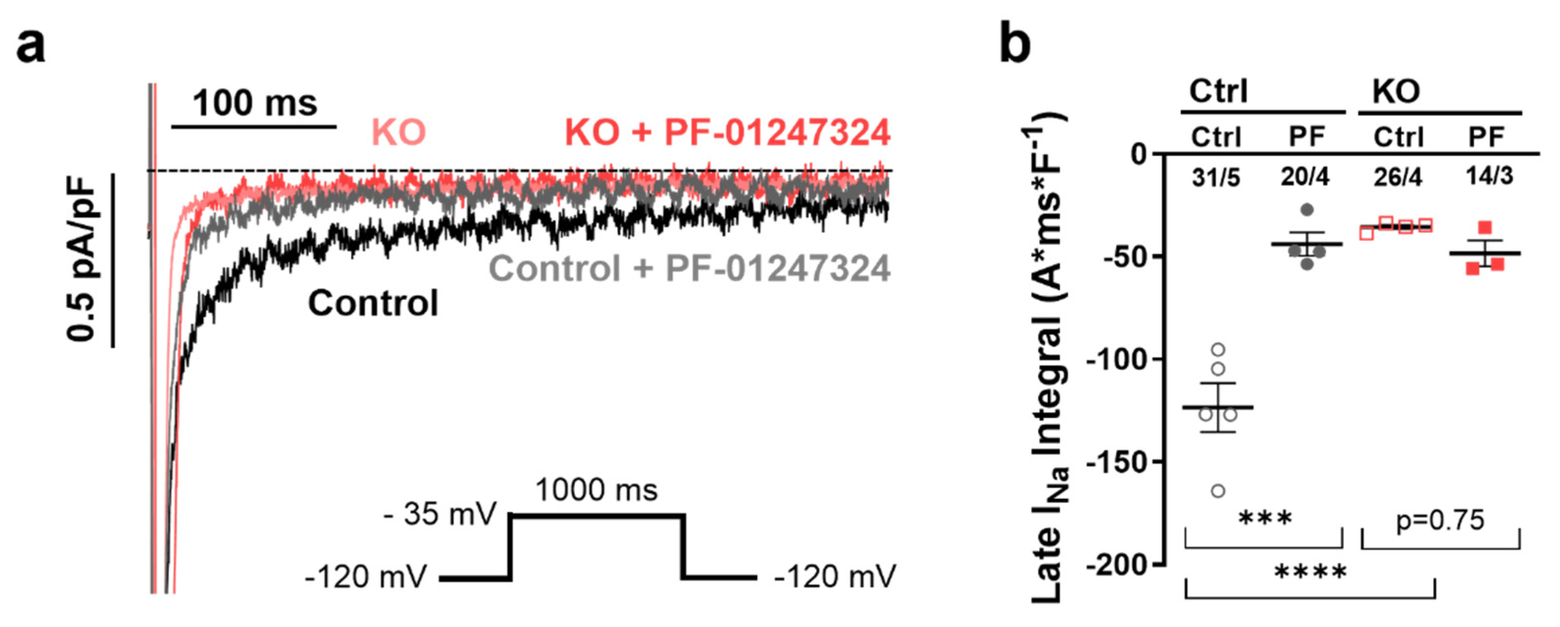
2.2. Influence of NaV1.8 on INaL in Human Atrial iPSC-Cardiomyocytes

We hypothesized that the KO of SCN10A in atrial iPSC-CMs would reduce the proarrhythmogenic INaL. Therefore, whole-cell voltage clamp experiments were performed to direct measure the INaL integral in human atrial SCN10A KO and control iPSC-CMs.
Since the amplitude of the INaL is relatively small in healthy hiPSC-CMs under physiological conditions [24], we used isoproterenol (Iso, 50 nmol/L) for slight beta-adrenergic stimulation in control and experimental groups during all functional experiments as described previously [20]. To further compare SCN10A KO with pharmacological inhibition of NaV1.8 and test for potential side effects of either KO or pharmacological intervention, we used the specific NaV1.8 blocker PF-01247324 (1 µmol/L) [19,22]. Voltage-clamp experiments demonstrated that the INaL was significantly reduced by genetical KO of NaV1.8 as well as by pharmacological inhibition. The Iso-induce increase in the INaL in control iPSC-CMs (−125.5 ± 8.4 A*ms*F−1) was significantly reduced in KO iPSC-CMs (−34.4 ± 4.8 A*ms*F−1, p < 0.0001, Figure 2). Moreover, the INaL was reduced to the level of KO iPSC-CMs by application of the specific NaV1.8 inhibitor [PF-01247324, 1 µmol/L, atrial control iPSC-CMs vs. PF-01247324 (−44.9 ± 3.8 A*ms*F−1, p < 0.001, Figure 2)]. Notably, we observed no additional effects on the INaL in KO iPSC-CMs after application of PF-01247324.

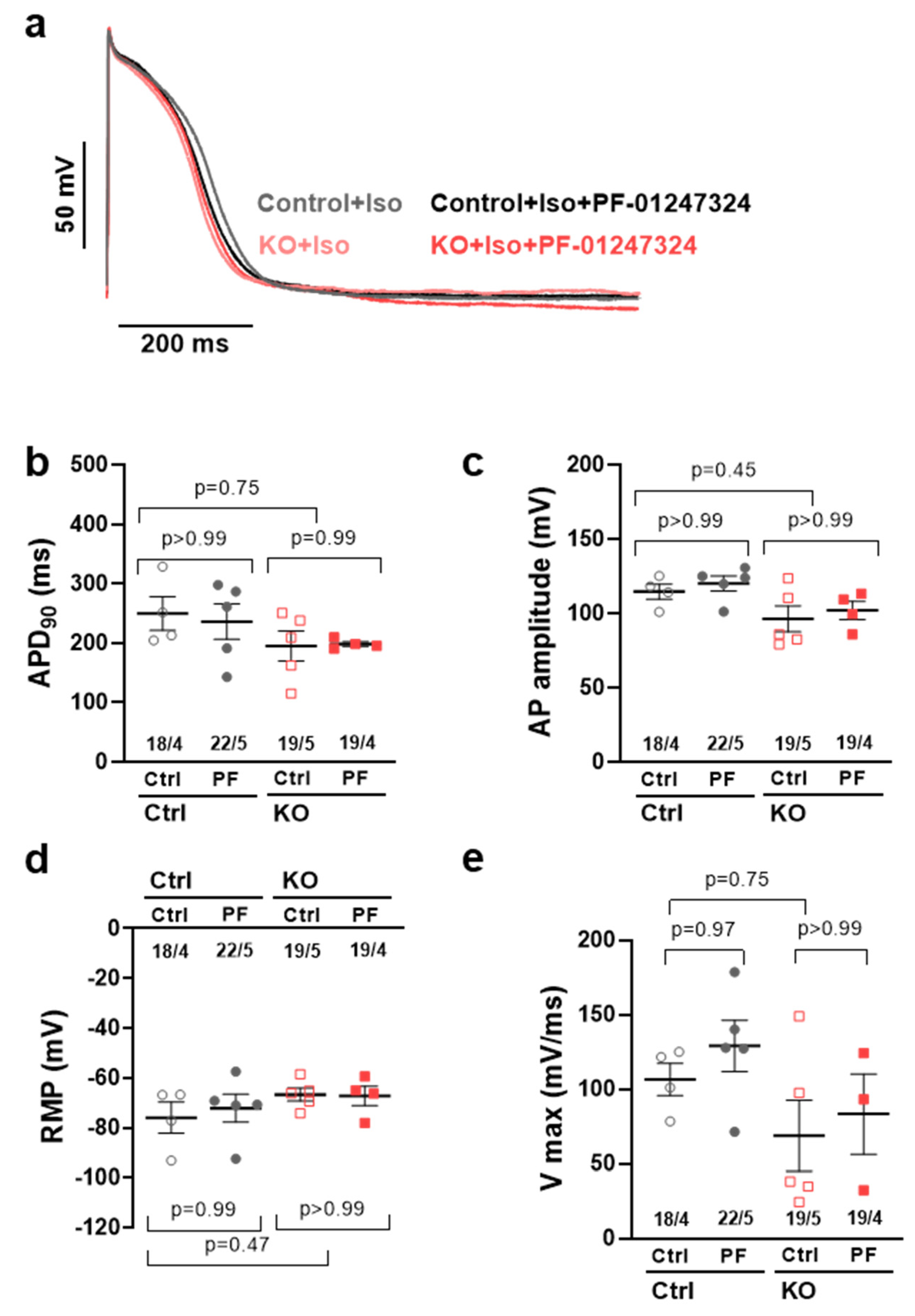
2.3. Effects of NaV1.8 on the Atrial Action Potential

To assess the potential influence of KO and pharmacological NaV1.8 inhibition on the action potential characteristics in human atrial iPSC-CMs, we performed whole-cell current-clamp experiments. The data presented herein are representative of measurements conducted at a frequency of 1 Hz. No effects on atrial action potential duration at 90% repolarization (APD90) were observed in KO iPSC-CMs as well as after the additional application of the specific NaV1.8 blocker PF-01247324 (Figure 3a,b; control at 1.0 Hz, 243.0 ± 30.5 ms vs. control + PF 229.3 ± 19.0 ms,−5.8%; KO control 204.4 ± 24.2 ms, −16%, vs. KO + PF 198.9 ± 30.1 ms, −3%). Furthermore, no discernible impacts were observed on the duration of atrial action potential at 20% repolarization (APD20), action potential duration at 50% repolarization (APD50), and action potential duration at 70% repolarization (APD70). The available data, including Table S1 and Figure S1, were included in the Supplemental Materials. To rule out potential side effects of KO or pharmacological inhibition of NaV1.8, we compared the resting membrane potential and action potential amplitude in all groups. No significant effects of KO or pharmacological inhibition of NaV1.8 on either AP amplitude (APA, Figure 3c, 113.7 ± 4.4 ms vs. control + PF 118.7 ± 3.4 ms, KO control 105.7 ± 5.1 ms, KO + PF 102.1 ± 4.2 ms ), resting membrane potential (RMP, Figure 3d; −76.0 ± 6.2 ms vs. control + PF −72.2 ± 5.7 ms; KO control −66.7 ± 2.6 ms vs. KO + PF −67.3 ± 3.9 ms), or upstroke velocity (Vmax, Figure 3e; 106.2 ± 11.2 vs. control + PF 127.5 ± 11.9 mV/ms; KO control 92.7 ± 13.8 vs. KO + PF 82.8 ± 12.4 mV/ms) could be observed.

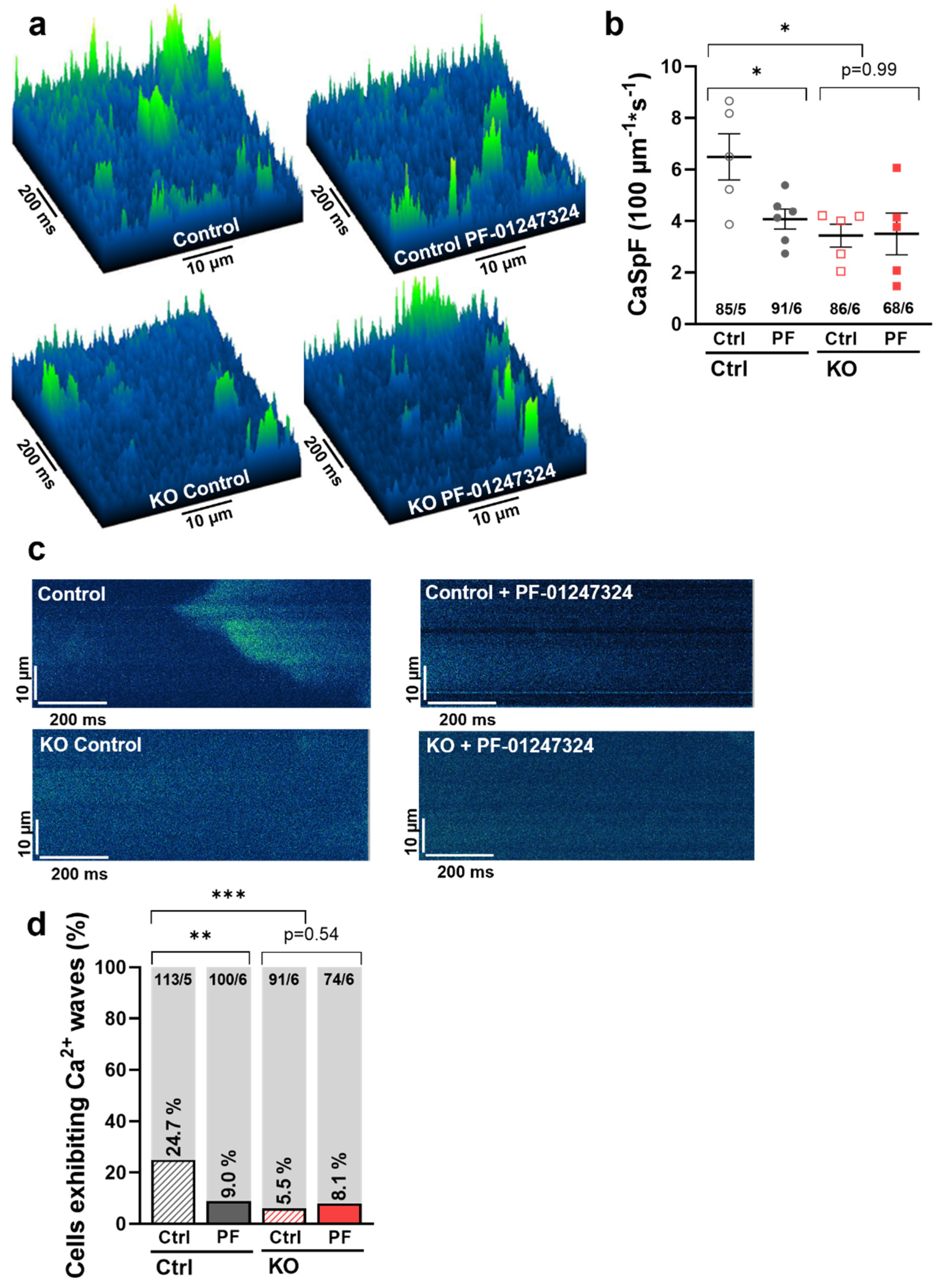
2.4. Effects of NaV1.8 on Atrial Sarcoplasmic Reticulum Ca2+ Leak and Arrhythmogenesis

As we previously demonstrated, NaV1.8 exerts its arrhythmogenic potential in the atria via enhancement of the INaL [19]. To investigate the functional cellular effects of the NaV1.8-dependent INaL on Ca2+ homeostasis and cellular arrhythmogenesis in atrial iPSC-CMs, we recorded line scans in confocal microscopy experiments using Fluo 4-AM in human atrial SCN10A KO and control iPSC-CMs. Diastolic confocal line scans (Fluo 4-AM) showed that KO of SCN10A in atrial iPSC-CMs massively decreased the frequency of spontaneous arrhythmogenic Ca2+ sparks compared to the respective control cells (KO: 3.25 ± 0.23 sparks/100 µm/s vs. control: 6.34 ± 0.43, p = 0.0142). Similarly, pharmacological inhibition of NaV1.8 by PF-01247324 led to a significant reduction of diastolic Ca2+ sparks in atrial control iPSC CMs (control + PF-01247324: 3.96 ± 0.21, p = 0.0469), while having no further effect on SCN10A KO cells (KO + PF-01247324: 3.47 ± 0.34, p = 0.9998) (Figure 4a,b). Furthermore, we investigated the incidence of spontaneous diastolic Ca2+ waves as major arrhythmogenic events. The proportion of cells exhibiting diastolic Ca2+ waves was significantly reduced from 24.7% in atrial control -iPSC-CMs to 5.5% in the SCN10A KO group. After pharmacological inhibition of NaV1.8, we observed a comparable reduction of cells displaying Ca2+ waves compared to control (9.0%). There was no significant additional effect of NaV1.8 inhibition in SCN10A KO cells (8.7%) (Figure 4c,d).

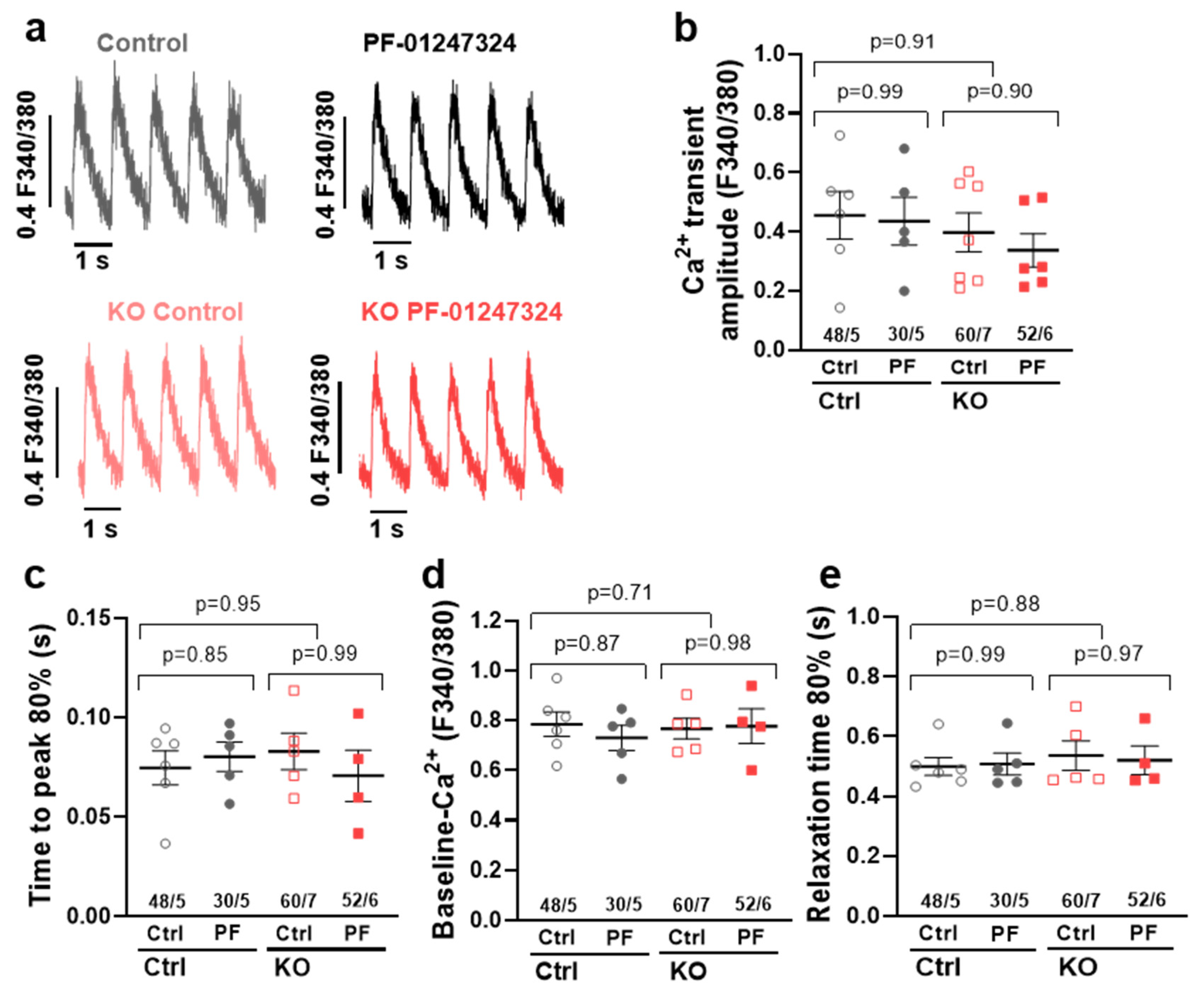
2.5. Influence of SCN10A KO on Intracellular Ca2+ Transients

Since KO of SCN10A was shown to reduce the arrhythmogenic potential of the increased INaL in atrial human iPSC-CMs by reduction of spontaneous SR Ca2+ release events, we further sought to rule out any potential adverse effects on cellular Ca2+ handling.
We therefore performed epifluorescence microscopy (Fura 2-AM) in atrial iPSC-CMs with and without KO of SCN10A and/or pharmacological inhibition of NaV1.8. SCN10A KO did not show any significant effects on Ca2+ transient amplitude, diastolic Ca2+ levels, time to peak, or relaxation time (RT 80%), demonstrating intact Ca2+ handling in both KO and WT cells. Of note, specific inhibition of NaV1.8 by PF-01247324 also did not exert any additional effects on Ca2+ transient parameters in either control or KO atrial iPSC-CMs (Figure 5).

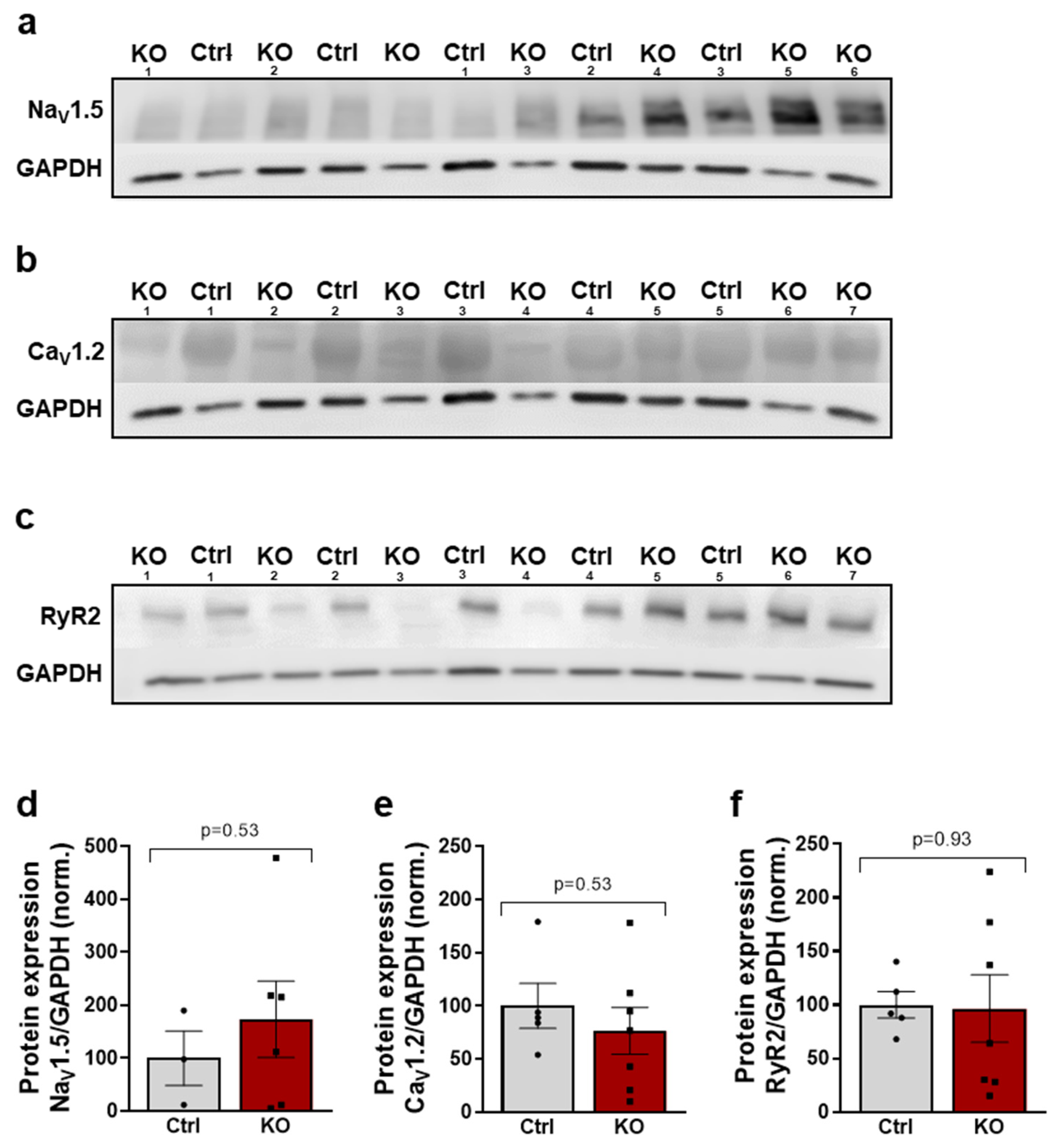
2.6. The Expression of Key Proteins of Excitation–Contraction Coupling Is Not Altered by a SCN10A KO

Since we demonstrated a reduction in the arrhythmogenic potential in atrial human SCN10A KO iPSC-CMs, we wanted to analyze the potential underlying effects on a molecular level. Therefore, we investigated the expression of key proteins of excitation–contraction coupling (voltage-gated sodium channel isoform NaV1.5; L-type Ca2+ channel CaV1.2; cardiac ryanodine receptor 2, RyR2) using Western blot experiments.
In atrial control iPSC-CMs, we found a lower expression of NaV1.5 compared to SCN10A KO iPSC-CMs, but it did not reach statistical significance (Figure 6a,d). Furthermore, RyR2 and CaV1.2 were not regulated in atrial SCN10A KO iPSC-CMs compared to control atrial iPSC-CMs according to the Western blot results (Figure 6b,c,e,f). Thus, SCN10A KO seems to exert no significant side effects on the expression of the other main proteins relevant to excitation–contraction coupling in atrial iPSC-CMs compared with their respective control cells.

3. Discussion

Atrial fibrillation (AF) is the most prevalent clinically significant arrhythmia. It represents a major risk factor for embolic stroke and exacerbation of heart failure (HF), consequently contributing to heightened morbidity and mortality rates [25]. The current prevalence of atrial fibrillation (AF) in adults ranges between 2% and 4%, with an anticipated 2.3-fold increase due to the extended longevity of the general population. For patients with atrial fibrillation (AF), first-line therapies for rhythm control include anti-arrhythmic drugs and/or left atrial pulmonary vein ablation [25]. However, pharmacological rhythm control is notably restricted in patients with underlying structural heart disease. The currently available drugs for these patients have limitations, poor tolerability, and adverse side effects [26]. Therefore, there is a demand for new and safer innovative compounds to address the treatment of AF in patients with structural heart disease. Sodium currents are effective therapeutic targets for the treatment of AF. In this context, the INaL has been increasingly identified as a potential target to inhibit cellular arrhythmogenic triggers in AF and the first hopeful results have been shown in clinical trials [26,27,28,29,30].
However, the mechanism of INaL regulation with respect to cellular arrhythmogenic triggers is not yet well understood. Besides NaV1.5, other NaV isoforms have been reported to be present in the heart. We have shown that the expression of the Na+ channel NaV1.8 in left ventricular CMs is upregulated in human HF myocardium [20], and that NaV1.8 contributes to arrhythmogenesis by inducing the INaL [19,20,22,31]. Variants in the SCN10A gene (NaV1.8) were shown to be associated with cardiac arrhythmias such as atrial fibrillation and sudden cardiac death [32]. Whether these NaV1.8-related effects are mediated by cardiac ganglia or cardiomyocytes is still under debate. In the present study, we used human atrial SCN10A KO iPSC-CMs and demonstrated that NaV1.8 is responsible for the generation of the INaL. Both inhibition and KO of NaV1.8 potently suppressed the INaL and diastolic SR-Ca2+ leak as proarrhythmogenic triggers in atrial CMs. These findings suggest that targeting NaV1.8 constitutes a novel therapeutic antiarrhythmic strategy for the treatment of atrial rhythm disorders.

3.1. NaV1.8 and Atrial INaL

Under pathological conditions, the enhanced persistent Na+ influx, known as enhanced INaL, has been demonstrated to play an important role throughout the action potential [33]. The prolongation of the action potential duration caused by an INaL increases the likelihood of early afterdepolarizations (EADs), which serve as triggers for the occurrence of arrhythmias. The specific NaV isoforms involved in the generation of an INaL, particularly in clinically relevant conditions like atrial fibrillation (AF) and heart failure (HF), remain unclear. This information is of translational relevance because selectively targeting the inhibition of the INaL would be a desirable antiarrhythmic approach.
Genome-wide association studies have identified SCN10A as a regulator of cardiac conduction. By employing various methodologies in both human and mouse cardiomyocytes, we have demonstrated the significance of NaV1.8 in the generation of the late sodium current (INaL). We found that NaV1.8 is upregulated under conditions of HF and cardiac hypertrophy [20,22,31]. Recent studies have provided evidence for the involvement of NaV1.8 in atrial cellular electrophysiology and have successfully linked SCN10A variants to AF [32,34]. However, some of the preliminary studies are limited by the use of appropriate ion channel blockers, which theoretically could have nonspecific effects.
Therefore, in the present study we used homozygous atrial SCN10A-KO iPSC-CMs to show that the NaV1.8-associated effects are mechanistically mediated by NaV1.8. Since under healthy conditions the INaL is very low, we applied isoproterenol in order to enhance the INaL for a better comparison between the control and KO iPSC CMs. Casini et al. did not detect any NaV1.8-based INaL in non-diseased human atrial and rabbit ventricular CMs without beta-adrenergic stimulation [24]. Most importantly, the incidence of an enhanced INaL depends on pharmacological (beta-adrenergic activation) or pathological stimulation and explain the absence of NaV1.8 effects in this study. Here, we show that NaV1.8 contributes to an enhanced INaL in atrial control iPSC-CMs by reducing the INaL by simultaneous treatment with isoproterenol and PF-01247324. Moreover, the specific blocker PF-01247324, when used to inhibit NaV1.8, did not induce any additional effects on the INaL in NaV1.8 KO atrial cells compared to untreated NaV1.8 KO atrial cells. This finding highlights the specificity of the drug in targeting NaV1.8 [35]. Pabel et al. demonstrated that both pharmacological inhibition and genetic ablation of NaV1.8 resulted in a reduction of the late sodium current (INaL) in human and murine atrial CMs [19]. In line with this, patch-clamp recordings of isolated human atrial CMs obtained from patients in sinus rhythm revealed that following mild beta-adrenergic stimulation with isoproterenol, the inhibition of NaV1.8 using PF-01247324 and A-803467 led to a significant reduction in the late sodium current (INaL) [19]. Isolated atrial CMs from SCN10A-/- mice revealed a significantly lower INaL compared to WT while pharmacological inhibition by PF-01247324 exerted no additional effect on the INaL in SCN10A-/- mice [19]. Therefore, the results of the present study are in line with previous findings in atrial human and mice atrial KO [19] and ventricular KO mice and human iPSC-CMs and isolated CMs [22,36]. Moreover, the impact of SCN10A variants associated with AF on the modulation of the INaL was demonstrated through transfection experiments in ND7/23 cells. This additional evidence further strengthens the notion that NaV1.8 plays a significant role in the development of INaL-related arrhythmias [37].

3.2. NaV1.8 and Atrial Action Potential Duration

Previous studies have provided evidence that the INaL plays a significant role in determining the APD in both atrial and ventricular CMs [2,3,8,27,29]. Having demonstrated the upregulation of NaV1.8 expression in human AF and HF, we proceeded to investigate the impact of NaV1.8-induced INaL on various action potential parameters. We also used isoproterenol to enhance the INaL. In line with previous data from our group in atrial human and mice CMs [19], the present study showed that in atrial control or SCN10A KO iPSC CMs, NaV1.8 has negligible effects on the atrial action potential parameters. In AF, the APD becomes shorter and a further shortening of APD may lead to shorter refractory periods, thereby further facilitating reentry. Therefore, negligible effects on APD point towards a positive therapeutic profile of targeting NaV1.8 in AF. Since dv/dt is a surrogate for the fast Na+ influx and peak Na+ current, these data show that there is no involvement of NaV1.8 in the peak Na+ current in atrial iPSC CMs. A negligible effect of Nav1.8 inhibition on cardiac conduction peak Na+ current blockade would be desirable in order to treat patients with structural heart disease and AF.

3.3. NaV1.8 and Atrial Ca2+ Handling

In our previous studies, we demonstrated that the INaL-mediated Na+ influx has the ability to induce Ca2+ influx through reverse-mode NCX, resulting in elevated cytosolic [Ca2+] levels and an increased occurrence of Ca2+ sparks in the human atrium [21,28]. Furthermore, the inhibition of the INaL through specific targeting of NaV1.8 has the capability to reduce the reverse mode NCX, thereby also mitigating diastolic proarrhythmogenic SR-Ca2+ leak [20,21,31]. The relationship between enhanced INaL and an increased risk of arrhythmias is indeed complex. This complexity arises from the fact that the increased leak of Ca2+ from the SR can induce a transient inward current, which, in turn, leads to arrhythmogenic delayed afterdepolarizations. Additionally, it can also result in significant spontaneous proarrhythmic Ca2+ release from the SR [8,28].
In the present study, we show a reduction in spontaneous SR Ca2+ spark frequency as well as a decreased frequency of spontaneous Ca2+ waves in human atrial SCN10A KO CMs and in control CMs after pharmacological inhibition. As Ca2+ waves represent a major proarrhythmic trigger, we hereby establish the principle of NaV1.8-induced INaL and its triggering role in cellular arrhythmogenesis that is independent of neuronal influence in isolated human atrial CMs. Interestingly, we found no effects on intracellular Ca2+ transients in either SCN10A KO or following NaV1.8 inhibition in control CMs. Thus, we propose that the intracellular Ca2+ handling and likely contractile function of CMs remain mostly unaffected by NaV1.8. In summary, our results and current evidence indicate that the discussed NaV1.8-induced INaL mainly influences arrhythmogenesis on a subcellular level while leaving cellular Ca2+ release and contractile function unaffected.

3.4. Clinical Relevance

The currently available anti-arrhythmic drugs, particularly for patients with structural heart disease, are limited in their effectiveness. Drugs like flecainide or amiodarone, which are commonly used, demonstrate suboptimal efficacy and are associated with significant adverse side effects, including life-threatening ventricular arrhythmias and organ toxicity. Therefore, new, safer, and more precise compounds for the treatment of atrial arrhythmias are highly desirable. NaV1.8 was detected in atria, and human hypertrophied and failing ventricles [19,22,31]. The results of the present study demonstrate that either genetic ablation of NaV1.8 using SCN10A KO iPSC-CMs or pharmacological inhibition can reverse cellular proarrhythmic effects in the atria. Both inhibition and KO of NaV1.8 potently suppressed proarrhythmogenic triggers (e.g., INaL and diastolic SR-Ca2+ leak) while leaving the peak Na+ current unaffected. These findings suggest targeting NaV1.8-dependent INaL constitutes a novel therapeutic anti-arhythmic strategy for the treatment of atrial rhythm disorders.

4. Materials and Methods

4.1. Generation of Homozygous Knockout iPSCs Using CRISPR/Cas9 and Directed Differentiation into Atrial iPSC-Cardiomyocytes

All procedures conducted in this study adhered to the principles outlined in the Declaration of Helsinki and received approval from the local ethics committee of the University Medicine of Göttingen (Az-10/9/15). Informed consent was signed by all tissue donors. A homozygous SCN10A KO iPSC line was generated from a control iPSC line by CRISPR/Cas9 genome editing as described in detail in previous studies [22,23]. The generated SCN10A KO iPSCs were differentiated into functionally beating, atrial iPSC-derived cardiomyocytes as described in [38].
In order to achieve directed atrial cardiac differentiation of the induced pluripotent stem cells (iPSCs), manipulation of the Wnt signaling pathway was employed, as previously described [38]. The cells were cultured for 60 days and then passaged onto glass-bottom Fluoro Dishes (WPI, 30 K/dish) by subjecting them to trypsinization at 37  °C for 3 min. The cells were allowed to settle for 7 days prior to further measurements, with medium changes performed every 2 days. iPSC-derived cardiac myocytes (iPSC-CMs) were analyzed 8–10 weeks after the initiation of differentiation, unless otherwise specified. The purity of the iPSC-CMs was determined by flow analysis, with a focus on cardiac troponin T positivity (>90% cardiac TNT+), as well as through qPCR and immunofluorescence analysis of atrial-specific markers (PITX2, MLC2a). Four to five differentiation experiments were performed to generate atrial iPSC-CMs from two NaV1.8 knockout lines and their corresponding healthy isogenic control line.

4.2. Pharmacological Intervention

For selective inhibition of NaV1.8-induced sodium currents, a specific NaV1.8 blocker PF-01247324 (1 µmol/L, Sigma-Aldrich, Taufkirchen, Germany)) was used. Cellular electrophysiological measurements were performed under slight beta-adrenergic stimulation (isoproterenol (Iso), 50 nmol/L, Sigma-Aldrich, Taufkirchen, Germany)) [20]. Prior to the start of experiments, the CMs were incubated for 15 min with both substances or isoproterenol alone as a control.

4.3. Patch-Clamp Experiments

The patch-clamp experiments was performed as previously described [19,22]. Briefly, 35,000 atrial iPSC-CMs were plated on glass-bottom Fluoro Dishes and incubated with either isoproterenol (50 nmol/L, Sigma-Aldrich, Taufkirchen, Germany)) or isoproterenol + PF01247324 (1 µmol/L, Sigma-Aldrich, Taufkirchen, Germany)) for 15 min before starting the measurements. The experiments were conducted at room temperature.
Action potential recordings were performed using the whole-cell patch-clamp technique. To elicit action potentials, square current pulses with amplitudes of 0.5–1 nA and durations of 1–5 ms were applied. The stimulation frequency was increased gradually from 0.5 to 2 Hz.
The late sodium current (INaL) was measured using the ruptured-patch whole-cell patch-clamp technique. The pipette used had a resistance ranging from 2 to 3 mega-ohms (MΩ). INaL recordings were performed exclusively in CMs where a seal with a resistance of over 1 giga-ohm (GΩ) was achieved, and the access resistance remained below 7 MΩ. After a stabilization period of 3 min, the iPSC-derived CMs were held at a holding potential of −120 mV and then depolarized to −35 mV for 1000 ms with 10 pulses and a basic cycle length of 2 s. The INaL was quantified as the integral current amplitude between 100 and 500 ms and was normalized to the membrane capacitance.

4.4. Confocal Ca2+ Imaging

A total of 35.000 atrial iPSC-CMs plated on glass-bottom FluoroDishes were incubated with the Ca2+ indicator Fluo 4-AM (10 µmol/L, Invitrogen, Darmstadt, Germany) for 15 min at RT for de-esterification of the dye. The solution was substituted with Tyrode’s solution (as described in [19]) and the respective pharmacological agents and left to incubate for 15 min. Confocal line scans were obtained with a laser scanning confocal microscope (LSM 5 Pascal, Zeiss, Jena, Germany). Scans were conducted after continuous electrical field stimulation at 1 Hz during pausing of stimulation. Ca2+ release events were analyzed using the SparkMaster plugin for ImageJ. The mean Ca2+ spark frequency was calculated from the number of sparks normalized to scan width, duration, and scan rate (100 µm/s). Cells exhibiting major Ca2+ release events (Ca2+ wavelets or waves) were excluded from the calculation of Ca2+ spark frequency and separately classified as proarrhythmic cells as a proportion of all cells.

4.5. Epifluorescence Microscopy for Ca2+ Transient Measurements

A total of 35.000 atrial CMs were dissociated and plated as described above and loaded with the radiometric Ca2+ indicator Fura 2-AM (5 µmol/L, Invitrogen) for 15 min at RT. Subsequently, the cells were washed with Tyrode’s solution for de-esterification and incubated with pharmacological agents as described above. The measurements were performed using a fluorescence detection system (IonOptix, Amsterdam, Netherlands) connected to an inverted microscope with oil immersion lens (40×). Cardiomyocytes were subjected to electrical field stimulation at 1 Hz for the duration of the experiment to ensure steady intracellular Ca2+ concentrations. Recording of Ca2+ transients for analysis was performed at 1 Hz at steady state. For each cell, the stimulation was paused for 30 s to detect spontaneous Ca2+ release events and evaluate the spontaneous beating frequency of the iPSC-CMs. Ca2+ transients were analyzed using the software IonWizard (IonOptix).

4.6. Statistical Analysis

The data are reported as mean ± SEM, unless otherwise stated. Analysis was carried out with Prism 9 software (Graphpad, San Diego, CA, USA). For comparisons of two groups, unpaired Student’s t test was used in the case of parametric distribution of the data. Three or more groups including more than one differentiation experiment were compared using nested one-way ANOVA. The results were corrected for multiple comparisons by Sidak’s correction. Fisher’s exact test was used to statistically compare proportions. p values are two-sided and considered statistically significant if p < 0.05.

5. Conclusions

In conclusion, we showed that the neuronal sodium channel NaV1.8, which contributes to the INaL in the heart, is down-regulated in atrial SCN10A-KO iPSC-CMs and, importantly, contributes to INaL formation in human atrial CMs. NaV1.8 KO or the inhibition of NaV1.8 modulates proarrhythmogenic triggers such as INaL and diastolic SR-Ca2+ leak in human atrial CMs. Therefore, NaV1.8 might represent a novel treatment target for antiarrhythmic strategies.

Supplementary Materials

The following supporting information can be downloaded at: https://www.mdpi.com/article/10.3390/ijms241210189/s1.

Author Contributions

Conceptualization, N.H. and S.S.; Data curation, N.H. and M.K.; Formal analysis, N.H., M.K. and N.D.; Funding acquisition, N.H. and G.H.; Investigation, N.H. and M.K.; Methodology, N.H., M.K., W.M., N.D., S.S. and K.S.-B.; Project administration, S.S.; Resources, N.D., G.H. and S.S.; Software, M.K. and N.D.; Supervision, G.H. and K.S.-B.; Validation, N.H., M.K. and S.S.; Visualization, N.H., M.K., W.M. and N.D.; Writing—original draft preparation, N.H. and M.K. All authors have read and agreed to the published version of the manuscript.

Funding

This work was supported by the Else-Kröner-Fresenius Foundation to N.H. (2020_EKEA.56); German Heart Foundation/German Foundation of Heart Research (to S.S.) and SFB 1002 (to G.H.); and grants from the Deutsche Forschungsgemeinschaft (DFG) through the International Research Training Group Award (IRTG) 1816 (to K.S.-B.; W.M. is a fellow under IRTG 1816), and to K.S.-B. and S.S. (471241922)).

Institutional Review Board Statement

The study was conducted in accordance with the Declaration of Helsinki, and approved by the local Ethics Committee of the University Medical Center Göttingen (Az-10/9/15). Informed consent was obtained from all subjects involved in the study.

Informed Consent Statement

Not applicable.

Data Availability Statement

Not applicable.

Acknowledgments

We thank Yvonne Metz and Johanna Heine for their excellent technical assistance.

Conflicts of Interest

N.H. and M.K. declare no conflicts of interest. K.S.-B. has no competing interest directly related to this work, but has grants from Novartis. S.S. received speaker’s/consultancy honoraria from Boehringer Ingelheim, AstraZeneca, Berlin-Chemie, Novartis, and Lilly.

References

  1. Valdivia, C.R.; Chu, W.W.; Pu, J.; Foell, J.D.; Haworth, R.A.; Wolff, M.R.; Kamp, T.J.; Makielski, J.C. Increased late sodium current in myocytes from a canine heart failure model and from failing human heart. J. Mol. Cell. Cardiol. 2005, 38, 475–483. [Google Scholar] [CrossRef] [PubMed]
  2. Maltsev, V.A.; Silverman, N.; Sabbah, H.N.; Undrovinas, A.I. Chronic heart failure slows late sodium current in human and canine ventricular myocytes: Implications for repolarization variability. Eur. J. Heart Fail. 2007, 9, 219–227. [Google Scholar] [CrossRef] [PubMed] [Green Version]
  3. Song, Y.; Shryock, J.C.; Belardinelli, L. An increase of late sodium current induces delayed afterdepolarizations and sustained triggered activity in atrial myocytes. Am. J. Physiol. Heart Circ. Physiol. 2008, 294, H2031–H2039. [Google Scholar] [CrossRef] [PubMed] [Green Version]
  4. Sossalla, S.; Wagner, S.; Rasenack, E.C.; Ruff, H.; Weber, S.L.; Schondube, F.A.; Tirilomis, T.; Tenderich, G.; Hasenfuss, G.; Belardinelli, L.; et al. Ranolazine improves diastolic dysfunction in isolated myocardium from failing human hearts—role of late sodium current and intracellular ion accumulation. J. Mol. Cell. Cardiol. 2008, 45, 32–43. [Google Scholar] [CrossRef]
  5. Maltsev, V.A.; Sabbah, H.N.; Higgins, R.S.; Silverman, N.; Lesch, M.; Undrovinas, A.I. Novel, ultraslow inactivating sodium current in human ventricular cardiomyocytes. Circulation 1998, 98, 2545–2552. [Google Scholar] [CrossRef]
  6. Undrovinas, A.I.; Maltsev, V.A.; Sabbah, H.N. Repolarization abnormalities in cardiomyocytes of dogs with chronic heart failure: Role of sustained inward current. Cell Mol. Life Sci. 1999, 55, 494–505. [Google Scholar] [CrossRef]
  7. Despa, S.; Islam, M.A.; Weber, C.R.; Pogwizd, S.M.; Bers, D.M. Intracellular Na(+) concentration is elevated in heart failure but Na/K pump function is unchanged. Circulation 2002, 105, 2543–2548. [Google Scholar] [CrossRef] [Green Version]
  8. Toischer, K.; Hartmann, N.; Wagner, S.; Fischer, T.H.; Herting, J.; Danner, B.C.; Sag, C.M.; Hund, T.J.; Mohler, P.J.; Belardinelli, L.; et al. Role of late sodium current as a potential arrhythmogenic mechanism in the progression of pressure-induced heart disease. J. Mol. Cell. Cardiol. 2013, 61, 111–122. [Google Scholar] [CrossRef] [Green Version]
  9. Djouhri, L.; Fang, X.; Okuse, K.; Wood, J.N.; Berry, C.M.; Lawson, S.N. The TTX-resistant sodium channel Nav1.8 (SNS/PN3): Expression and correlation with membrane properties in rat nociceptive primary afferent neurons. J. Physiol. 2003, 550, 739–752. [Google Scholar] [CrossRef]
  10. Dib-Hajj, S.D.; Binshtok, A.M.; Cummins, T.R.; Jarvis, M.F.; Samad, T.; Zimmermann, K. Voltage-gated sodium channels in pain states: Role in pathophysiology and targets for treatment. Brain Res. Rev. 2009, 60, 65–83. [Google Scholar] [CrossRef]
  11. Akopian, A.N.; Sivilotti, L.; Wood, J.N. A tetrodotoxin-resistant voltage-gated sodium channel expressed by sensory neurons. Nature 1996, 379, 257–262. [Google Scholar] [CrossRef]
  12. Liu, M.; Wood, J.N. The roles of sodium channels in nociception: Implications for mechanisms of neuropathic pain. Pain Med. 2011, 12 (Suppl. S3), S93–S99. [Google Scholar] [CrossRef] [Green Version]
  13. Sotoodehnia, N.; Isaacs, A.; de Bakker, P.I.; Dorr, M.; Newton-Cheh, C.; Nolte, I.M.; van der Harst, P.; Muller, M.; Eijgelsheim, M.; Alonso, A.; et al. Common variants in 22 loci are associated with QRS duration and cardiac ventricular conduction. Nat. Genet. 2010, 42, 1068–1076. [Google Scholar] [CrossRef] [Green Version]
  14. Chambers, J.C.; Zhao, J.; Terracciano, C.M.; Bezzina, C.R.; Zhang, W.; Kaba, R.; Navaratnarajah, M.; Lotlikar, A.; Sehmi, J.S.; Kooner, M.K.; et al. Genetic variation in SCN10A influences cardiac conduction. Nat. Genet. 2010, 42, 149–152. [Google Scholar] [CrossRef]
  15. van den Boogaard, M.; Wong, L.Y.; Tessadori, F.; Bakker, M.L.; Dreizehnter, L.K.; Wakker, V.; Bezzina, C.R.; ’t Hoen, P.A.; Bakkers, J.; Barnett, P.; et al. Genetic variation in T-box binding element functionally affects SCN5A/SCN10A enhancer. J. Clin. Investig. 2012, 122, 2519–2530. [Google Scholar] [CrossRef] [Green Version]
  16. Ritchie, M.D.; Denny, J.C.; Zuvich, R.L.; Crawford, D.C.; Schildcrout, J.S.; Bastarache, L.; Ramirez, A.H.; Mosley, J.D.; Pulley, J.M.; Basford, M.A.; et al. Genome- and phenome-wide analyses of cardiac conduction identifies markers of arrhythmia risk. Circulation 2013, 127, 1377–1385. [Google Scholar] [CrossRef] [Green Version]
  17. Bezzina, C.R.; Barc, J.; Mizusawa, Y.; Remme, C.A.; Gourraud, J.B.; Simonet, F.; Verkerk, A.O.; Schwartz, P.J.; Crotti, L.; Dagradi, F.; et al. Common variants at SCN5A-SCN10A and HEY2 are associated with Brugada syndrome, a rare disease with high risk of sudden cardiac death. Nat. Genet. 2013, 45, 1044–1049. [Google Scholar] [CrossRef]
  18. Hu, D.; Barajas-Martinez, H.; Pfeiffer, R.; Dezi, F.; Pfeiffer, J.; Buch, T.; Betzenhauser, M.J.; Belardinelli, L.; Kahlig, K.M.; Rajamani, S.; et al. Mutations in SCN10A are responsible for a large fraction of cases of Brugada syndrome. J. Am. Coll. Cardiol. 2014, 64, 66–79. [Google Scholar] [CrossRef] [Green Version]
  19. Pabel, S.; Ahmad, S.; Tirilomis, P.; Stehle, T.; Mustroph, J.; Knierim, M.; Dybkova, N.; Bengel, P.; Holzamer, A.; Hilker, M.; et al. Inhibition of NaV1.8 prevents atrial arrhythmogenesis in human and mice. Basic Res. Cardiol. 2020, 115, 20. [Google Scholar] [CrossRef]
  20. Dybkova, N.; Ahmad, S.; Pabel, S.; Tirilomis, P.; Hartmann, N.; Fischer, T.H.; Bengel, P.; Tirilomis, T.; Ljubojevic, S.; Renner, A.; et al. Differential regulation of sodium channels as a novel proarrhythmic mechanism in the human failing heart. Cardiovasc. Res. 2018, 114, 1728–1737. [Google Scholar] [CrossRef]
  21. Bengel, P.; Ahmad, S.; Tirilomis, P.; Trum, M.; Dybkova, N.; Wagner, S.; Maier, L.S.; Hasenfuss, G.; Sossalla, S. Contribution of the neuronal sodium channel NaV1.8 to sodium- and calcium-dependent cellular proarrhythmia. J. Mol. Cell. Cardiol. 2020, 144, 35–46. [Google Scholar] [CrossRef] [PubMed]
  22. Bengel, P.; Dybkova, N.; Tirilomis, P.; Ahmad, S.; Hartmann, N.; Mohamed, B.A.; Krekeler, M.C.; Maurer, W.; Pabel, S.; Trum, M.; et al. Detrimental proarrhythmogenic interaction of Ca(2+)/calmodulin-dependent protein kinase II and NaV1.8 in heart failure. Nat. Commun. 2021, 12, 6586. [Google Scholar] [CrossRef] [PubMed]
  23. Maurer, W.; Hartmann, N.; Argyriou, L.; Sossalla, S.; Streckfuss-Bomeke, K. Generation of homozygous Na(v)1.8 knock-out iPSC lines by CRISPR Cas9 genome editing to investigate a potential new antiarrhythmic strategy. Stem Cell Res. 2022, 60, 102677. [Google Scholar] [CrossRef] [PubMed]
  24. Casini, S.; Marchal, G.A.; Kawasaki, M.; Nariswari, F.A.; Portero, V.; van den Berg, N.W.E.; Guan, K.; Driessen, A.H.G.; Veldkamp, M.W.; Mengarelli, I.; et al. Absence of Functional Nav1.8 Channels in Non-diseased Atrial and Ventricular Cardiomyocytes. Cardiovasc. Drugs Ther. 2019, 33, 649–660. [Google Scholar] [CrossRef] [PubMed] [Green Version]
  25. Hindricks, G.; Potpara, T.; Dagres, N.; Arbelo, E.; Bax, J.J.; Blomstrom-Lundqvist, C.; Boriani, G.; Castella, M.; Dan, G.A.; Dilaveris, P.E.; et al. 2020 ESC Guidelines for the diagnosis and management of atrial fibrillation developed in collaboration with the European Association for Cardio-Thoracic Surgery (EACTS): The Task Force for the diagnosis and management of atrial fibrillation of the European Society of Cardiology (ESC) Developed with the special contribution of the European Heart Rhythm Association (EHRA) of the ESC. Eur. Heart J. 2021, 42, 373–498. [Google Scholar] [CrossRef]
  26. Reiffel, J.A.; Camm, A.J.; Belardinelli, L.; Zeng, D.; Karwatowska-Prokopczuk, E.; Olmsted, A.; Zareba, W.; Rosero, S.; Kowey, P. The HARMONY Trial: Combined Ranolazine and Dronedarone in the Management of Paroxysmal Atrial Fibrillation: Mechanistic and Therapeutic Synergism. Circ. Arrhythmia Electrophysiol. 2015, 8, 1048–1056. [Google Scholar] [CrossRef]
  27. Sossalla, S.; Kallmeyer, B.; Wagner, S.; Mazur, M.; Maurer, U.; Toischer, K.; Schmitto, J.D.; Seipelt, R.; Schondube, F.A.; Hasenfuss, G.; et al. Altered Na(+) currents in atrial fibrillation effects of ranolazine on arrhythmias and contractility in human atrial myocardium. J. Am. Coll. Cardiol. 2010, 55, 2330–2342. [Google Scholar] [CrossRef] [Green Version]
  28. Fischer, T.H.; Herting, J.; Mason, F.E.; Hartmann, N.; Watanabe, S.; Nikolaev, V.O.; Sprenger, J.U.; Fan, P.; Yao, L.; Popov, A.F.; et al. Late INa increases diastolic SR-Ca2+-leak in atrial myocardium by activating PKA and CaMKII. Cardiovasc. Res. 2015, 107, 184–196. [Google Scholar] [CrossRef] [Green Version]
  29. Hartmann, N.; Mason, F.E.; Braun, I.; Pabel, S.; Voigt, N.; Schotola, H.; Fischer, T.H.; Dobrev, D.; Danner, B.C.; Renner, A.; et al. The combined effects of ranolazine and dronedarone on human atrial and ventricular electrophysiology. J. Mol. Cell. Cardiol. 2016, 94, 95–106. [Google Scholar] [CrossRef]
  30. White, C.M.; Nguyen, E. Novel Use of Ranolazine as an Antiarrhythmic Agent in Atrial Fibrillation. Ann. Pharmacother. 2017, 51, 245–252. [Google Scholar] [CrossRef]
  31. Ahmad, S.; Tirilomis, P.; Pabel, S.; Dybkova, N.; Hartmann, N.; Molina, C.E.; Tirilomis, T.; Kutschka, I.; Frey, N.; Maier, L.S.; et al. The functional consequences of sodium channel Na(V) 1.8 in human left ventricular hypertrophy. ESC Heart Fail. 2019, 6, 154–163. [Google Scholar] [CrossRef] [Green Version]
  32. Jabbari, J.; Olesen, M.S.; Yuan, L.; Nielsen, J.B.; Liang, B.; Macri, V.; Christophersen, I.E.; Nielsen, N.; Sajadieh, A.; Ellinor, P.T.; et al. Common and rare variants in SCN10A modulate the risk of atrial fibrillation. Circ. Cardiovasc. Genet. 2015, 8, 64–73. [Google Scholar] [CrossRef] [Green Version]
  33. Maier, L.S.; Sossalla, S. The late Na current as a therapeutic target: Where are we? J. Mol. Cell. Cardiol. 2013, 61, 44–50. [Google Scholar] [CrossRef] [Green Version]
  34. Pfeufer, A.; van Noord, C.; Marciante, K.D.; Arking, D.E.; Larson, M.G.; Smith, A.V.; Tarasov, K.V.; Muller, M.; Sotoodehnia, N.; Sinner, M.F.; et al. Genome-wide association study of PR interval. Nat. Genet. 2010, 42, 153–159. [Google Scholar] [CrossRef]
  35. Dybkova, N.; Wagner, S.; Backs, J.; Hund, T.J.; Mohler, P.J.; Sowa, T.; Nikolaev, V.O.; Maier, L.S. Tubulin polymerization disrupts cardiac beta-adrenergic regulation of late INa. Cardiovasc. Res. 2014, 103, 168–177. [Google Scholar] [CrossRef] [Green Version]
  36. Yang, T.; Atack, T.C.; Stroud, D.M.; Zhang, W.; Hall, L.; Roden, D.M. Blocking Scn10a channels in heart reduces late sodium current and is antiarrhythmic. Circ. Res. 2012, 111, 322–332. [Google Scholar] [CrossRef] [Green Version]
  37. Savio-Galimberti, E.; Weeke, P.; Muhammad, R.; Blair, M.; Ansari, S.; Short, L.; Atack, T.C.; Kor, K.; Vanoye, C.G.; Olesen, M.S.; et al. SCN10A/Nav1.8 modulation of peak and late sodium currents in patients with early onset atrial fibrillation. Cardiovasc. Res. 2014, 104, 355–363. [Google Scholar] [CrossRef]
  38. Kleinsorge, M.; Cyganek, L. Subtype-Directed Differentiation of Human iPSCs into Atrial and Ventricular Cardiomyocytes. STAR Protoc. 2020, 1, 100026. [Google Scholar] [CrossRef]
Figure 1. CRISPR/Cas9-based knock-out of SCN10A/NaV1.8 in atrial iPSC-CMs. (a) Sanger sequencing of control iPSCs and SCN10A iPSC-KO CMs demonstrating frameshifts in both alleles leading to a premature stop in exon 1 (A1: delC/insCAC and A2: delCT). (b) Atrial control and SCN10A KO iPSC-CMs were stained for MLC2a (green) and MLC2v (red) demonstrating atrial differentiation. Nuclei were stained with DAPI. (c) mRNA expression level of atrial marker PITX2 normalized to house-keeping gene HPRT in atrial control and SCN10A KO iPSC-CMs (n = 6/3 differentiations) compared to ventricular control and SCN10A KO iPSC-CMs (n = 7/4 differentiations). Student’s t-test was applied for normally distributed data. *: p < 0.05; **: p < 0.01.
Ijms 24 10189 g001
Figure 2. (a) Original traces of INaL in atrial control iPSC vs. SCN10A KO-iPSC cells according to the inserted protocol. (b) Mean values per differentiation ± SEM of INaL (atrial control n = 31 cells/5 differentiations; atrial control + PF n = 20 cells/4 differentiations; SCN10A KO iPSC−CM control n = 26 cells/4 differentiations, SCN10A KO-iPSC-CMs+ PF n = 14 cells/3 differentiations). Mean values per differentiation were compared using one-way ANOVA with Sidak’s test for multiple comparisons to calculate p values (*** = p < 0.001; **** = p < 0.0001).
Ijms 24 10189 g002
Figure 3. (a) Original traces of APD90 in atrial control iPSC−CMs vs SCN10A KO-iPSC-CMs at 1 Hz. (b) Mean (nested) ± SEM of APD90 (atrial control n = 18 cells/4 differentiations; atrial control + PF n = 22 cells/5 differentiations; SCN10A KO control n = 19 cells/5 differentiations; SCN10A KO + PF n = 19 cells/4 differentiations); statistics calculated using nested oneway ANOVA. (c) Mean ± SEM of amplitude (atrial control n = 18 cells/4 differentiations; atrial control + PF n = 22 cells/5 differentiations; SCN10A KO control n = 19 cells/5 differentiations; SCN10A KO + PF n = 19 cells/4 differentiations). (d) Mean ± SEM of RMP (atrial control n = 18 cells/4 differentiations; atrial control + PF n = 22 cells/5 differentiations; SCN10A KO control n = 19 cells/5 differentiations; SCN10A KO + PF n = 19 cells/4 differentiations); statistics calculated using nested one−way ANOVA. (e) Mean ± SEM of Vmax (atrial control n = 18 cells/4 differentiations; atrial control + PF n = 22 cells/5 differentiations; SCN10A KO control n = 19 cells/5 differentiations; SCN10A KO + PF n = 19 cells/4 differentiations).
Ijms 24 10189 g003
Figure 4. Contribution of NaV1.8 to spontaneous diastolic sarcoplasmic reticulum Ca2+ release in atrial iPSC-CMs. (a) Representative surface plots showing spontaneous diastolic Ca2+ sparks (green) in atrial iPSC-CMs. (b) Mean values of Ca2+ spark frequency (CaSpF) normalized to scan width and duration. Numbers indicate total cell count of control CMs (atrial control, n = 85 cells/5 differentiations), control cells treated with NaV1.8 inhibitor PF01247324 (atrial control + PF-01247324, n = 91/6), and atrial CMs with KO of NaV1.8 after control treatment and treatment with PF-01247324 (n = 86/6 vs. 68/6). Symbols indicate the mean values of different differentiation experiments. (c) Original representative line scans of atrial iPSC-CMs illustrating a spontaneous proarrhythmogenic diastolic Ca2+ wave (green). (d) Percentage of cells exhibiting diastolic Ca2+ waves in relation to cells without Ca2+ waves (grey bars) in atrial control CMs (24.7%, n = 28 of 113 cells from 5 differentiations) compared to atrial control CMs + PF-01247324 (9%, n= 9/100 cells/6 diff.) and to atrial SCN10A KO CMs with inhibition of NaV1.8 by PF-01247324 (8.1%, n = 6/74cells/6 diff.) or without (5.5%, n = 5/91 cells/6 diff.). Values are presented as mean ± SEM or absolute numbers. Mean values per differentiation were compared using one-way ANOVA with Sidak’s test for multiple comparisons to calculate p values. Proportions were compared using Fisher’s exact test (* = p < 0.05, ** = p < 0.01; *** = p < 0.001).
Ijms 24 10189 g004
Figure 5. (a) Representative original recordings of stimulated systolic Ca2+ transients (epifluorescence microscopy, Fura 2-AM, 1 Hz) of human atrial SCN10A or control iPSC-CMs and after additional NaV1.8 inhibition by PF-01247324. Mean values ± SEM of (b) systolic Ca2+ transient amplitude, (c) time to peak 80%, (d) diastolic Ca2+ level, and (e) relaxation time 80% in control CMs (n = 48 cells/5 differentiations), SCN10A KO CMs (n = 60/7), and each after treatment with PF-01247324 (control + PF-01247324 n = 30/5, KO + PF-01247324 n = 52/6). Values are presented as mean ± SEM. Mean values per differentiation were compared using one-way ANOVA with Sidak’s test for multiple comparisons to calculate p values.
Ijms 24 10189 g005
Figure 6. Original Western blots of NaV1.5 (a), CaV1.2 (b), and RyR2 (c) in atrial control and SCN10A KO iPSC-CMs. Normalized values of NaV1.5 (d) (n = 3 control/6 KO differentiations), CaV1.2 (e) (n = 5/7 differentiations), and RyR2 (f) (n = 5/7 differentiations) in atrial control and SCN10A KO iPSC-CMs normalized to GAPDH (n = 5/7 differentiations). Student’s t-test was used for statistical analysis.
Ijms 24 10189 g006
Disclaimer/Publisher’s Note: The statements, opinions and data contained in all publications are solely those of the individual author(s) and contributor(s) and not of MDPI and/or the editor(s). MDPI and/or the editor(s) disclaim responsibility for any injury to people or property resulting from any ideas, methods, instructions or products referred to in the content.

