Article

Epidermal Loss of RORα Enhances Skin Inflammation in a MC903-Induced Mouse Model of Atopic Dermatitis

1
School of Pharmacy, University of Wisconsin, Madison, WI 53705, USA
2
Biomedical Research Model Services, University of Wisconsin, Madison, WI 53705, USA
3
UW Carbone Cancer Center, University of Wisconsin, Madison, WI 53705, USA
*
Author to whom correspondence should be addressed.
Int. J. Mol. Sci. 2023, 24(12), 10241; https://doi.org/10.3390/ijms241210241
Submission received: 19 April 2023 / Revised: 14 June 2023 / Accepted: 15 June 2023 / Published: 16 June 2023
(This article belongs to the Special Issue Functional Defects of Keratinocytes in Inflammatory Skin Diseases 2.0)

Abstract

:
Atopic dermatitis (AD) is a chronic inflammatory skin disease featuring skin barrier dysfunction and immune dysregulation. Previously, we reported that the retinoid-related orphan nuclear receptor RORα was highly expressed in the epidermis of normal skin. We also found that it positively regulated the expression of differentiation markers and skin barrier-related genes in human keratinocytes. In contrast, epidermal RORα expression was downregulated in the skin lesions of several inflammatory skin diseases, including AD. In this study, we generated mouse strains with epidermis-specific Rora ablation to understand the roles of epidermal RORα in regulating AD pathogenesis. Although Rora deficiency did not cause overt macroscopic skin abnormalities at the steady state, it greatly amplified MC903-elicited AD-like symptoms by intensifying skin scaliness, increasing epidermal hyperproliferation and barrier impairment, and elevating dermal immune infiltrates, proinflammatory cytokines, and chemokines. Despite the normal appearance at the steady state, Rora-deficient skin showed microscopic abnormalities, including mild epidermal hyperplasia, increased TEWL, and elevated mRNA expression of Krt16, Sprr2a, and Tslp genes, indicating subclinical impairment of epidermal barrier functions. Our results substantiate the importance of epidermal RORα in partially suppressing AD development by maintaining normal keratinocyte differentiation and skin barrier function.

1. Introduction

Atopic dermatitis (AD) is a chronic, pruritic, and inflammatory skin disease [1]. AD has been attributed to two major factors: (a) T helper 2 (Th2)-mediated immune responses featuring high serum IgE levels and hyperactivation of mast cells and eosinophils, and (b) epidermal barrier dysfunctions [2]. Recent genetic studies with human subjects and mice have strongly connected AD etiology with the loss-of-function (null) mutations of the FLG gene, which encodes the cross-linking protein filaggrin, critical for cornified envelope (CE) assembly and epidermal barrier functions [3,4]. Consequently, barrier dysfunction has become widely recognized as a leading cause of AD development [5,6].
Normal barrier formation depends on a progressive keratinocyte differentiation process, accompanied by stage-dependent expression of signature structural proteins, such as keratin 5/14 in the basal layer, keratin 1/10 and involucrin in the spinous layer, and loricrin and profilaggrin in the granular layer [7]. This stepwise gene expression relies on specific transcription factors and signaling pathways. In addition to forming a physical barrier, keratinocytes have important innate immune functions by secreting diverse antimicrobial peptides, cytokines, and chemokines in response to various external stimuli. Keratinocyte-derived Th2-promoting cytokines, including thymic stromal lymphopoietin (TSLP), IL-25, and IL-33, are frequently elevated in AD lesions due to a disrupted barrier [8]. TSLP, an IL-7-like cytokine, allows dendritic cells to prime naïve T-cells toward Th2 polarization and promotes pruritus by activating cutaneous sensory neurons [9,10,11]. Therefore, keratinocyte-deregulated immune activities contribute significantly to AD pathogenesis [5,12,13].
The retinoid-related orphan receptors (RORα, RORb, and RORg) belong to the ligand-gated nuclear hormone receptor superfamily and function as transcription factors by binding to ROR response elements, ROREs, within the regulatory regions of their target genes [14,15,16]. RORα is expressed in a temporal and spatial-dependent manner during embryonic development and is critical for Purkinje cell maturation [17,18], bone formation [19], lymphocyte development [20,21], lipid metabolism [22], and inflammation [23,24,25]. In addition, RORα is a well-established component of the circadian clock, with Bmal1 as one of its best-studied targets [26,27]. Recent studies have identified RORα as a regulator of the development of multiple types of immune cells, including type 2 innate lymphoid cells (ILC2) [28] and T help 17 (Th17) cells [29,30]. Malfunctions in these cells are associated with the pathogenesis of allergic inflammation and autoimmune diseases [30,31].
RORα is highly expressed in human and mouse skin [32,33,34,35], specifically in the suprabasal layers of the epidermis, sebaceous glands, and hair follicles [35]. Our earlier studies have demonstrated that among the four RORα isoforms, RORα4 is prominently expressed by human keratinocytes in a manner that increases with the progression of differentiation [32]. While increasing the levels of RORα4 in HKCs enhanced the expression of structural proteins associated with early and late differentiation and genes involved in lipid barrier formation, RORA gene silencing suppressed the expression of these genes. Furthermore, the pro-differentiation function of RORα is mediated at least in part by FOXN1, a well-known pro-differentiation transcription factor that we have established as a novel direct target of RORα in keratinocytes [32]. A recent study using human keratinocytes also revealed RORα’s direct regulation of the expression of CXCL14, an epithelial chemokine involved in the clearance of the skin pathogen Staphylococcus aureus. In summary, there is compelling evidence that RORα may be critical for both the differentiation and innate immune activities of keratinocytes [36].
Despite these findings, the in vivo functions of epidermal RORα in skin homeostasis remain largely unknown. Recent reports have shown that epidermal RORα levels are significantly down-regulated in clinical samples of several inflammatory skin diseases, including allergic contact dermatitis, lichen simplex chronicus, sarcoidosis, and AD [37,38]. Our current study addresses the impact of reduced epidermal RORα in cutaneous inflammation. For this purpose, we generated mice with an epidermis-specific knockout of Rora to determine their reactions to the topical application of MC903, a low-calcemic vitamin D analog for stimulating keratinocyte TSLP production. We found that epidermal Rora deletion was insufficient to trigger spontaneous dermatitis, but it significantly intensified the AD-like symptoms elicited by the MC903 application. In this report, we present our findings.

2. Results

2.1. Epidermal Rora Expression in Mouse Skin in Response to MC903

To determine the essential roles of RORα in the epidermis, we obtained Roratm1a(EUCOMM)Wtsi (Roratm1a/+) C57BL/6 embryos with a “knockout first” allele [39,40]. This allele contains a splice acceptor–LacZ cassette inserted upstream of the floxed Rora “exon 3” (Figure 1A). We first generated the heterozygous Roratm1b/+ strain, a LacZ reporter strain with heterozygous Rora gene deletion (Figure 1A). Consistent with a previous report [35], Rora mRNA was predominantly expressed in the suprabasal layers of the epidermis and sebaceous glands in unchallenged mouse ear skin, as determined by the intensity of the X-gal signal (Figure 1B). At the same time, only a limited signal was detected in the dermis (Figure 1B). In response to the MC903 treatment, the X-gal intensity was substantially reduced in the hyperproliferative epidermis on day 5 and partially restored in the upper layer after day 8. However, no significant change was observed in the dermis (Figure 1B). These results revealed the importance of epidermal RORα for its skin functions under physiological and AD-like pathophysiological conditions.

2.2. Epidermal Rora Ablation Exaggerates MC903-Induced Ear Thickening

To verify the essential roles of epidermal RORa in MC903-induced skin inflammation, we generated a Roraflox/flox strain and a RoraLacZΔ/+; K14-CRE strain from the Roratm1a/+ mice (Figure 1A). Crossing of the two strains resulted in mice with wildtype (WT, Rora+/fl), heterozygous Rora null (RoraLacZΔ/fl), or an epidermal-specific Rora knockout (RoraLacZΔ/fl;K14-CRE), referred to as RoraLacZΔ/EKO. The epidermal Rora gene deletion efficiency was analyzed via genotyping using short-range end-point PCR assays (Figure 1C; Supplementary Figure S1) and through semi-quantitative RT-PCR analysis of Rora mRNA in adult ear skin (Figure 1D). Both young and adult mice with epidermal Rora deletion displayed normal hair development and skin appearance at the macroscopic level. However, starting from day 4, the MC903-induced ear thickening was profoundly enhanced in RoraLacZΔ/EKO mice compared to their WT and heterozygous RoraLacZΔ/fl littermates (Figure 1E). On day 11, the epidermal Rora-deficient mice also displayed severe ear swelling and skin scaliness (Figure 1F), along with greater epidermal hyperplasia and heavy immune infiltration in the dermis, as shown in the hematoxylin and eosin (H&E) staining (Figure 1G). The heterozygous RoraLacZΔ/fl mice also showed more severe ear swelling than the WT controls starting from day 9 (Figure 1E). The histological analysis also revealed the epidermal and dermal expansions in Rora heterozygous mice at a level higher than the WT but lower than the epidermal Rora deficient RoraLacZΔ/EKO mice in the MC903-treated group (Figure 1G). These results substantiate the important roles of epidermal RORα in suppressing MC903-induced skin inflammation in mice.

2.3. Epidermal Rora Deletion Aggravates MC903-Induced AD-like Symptoms

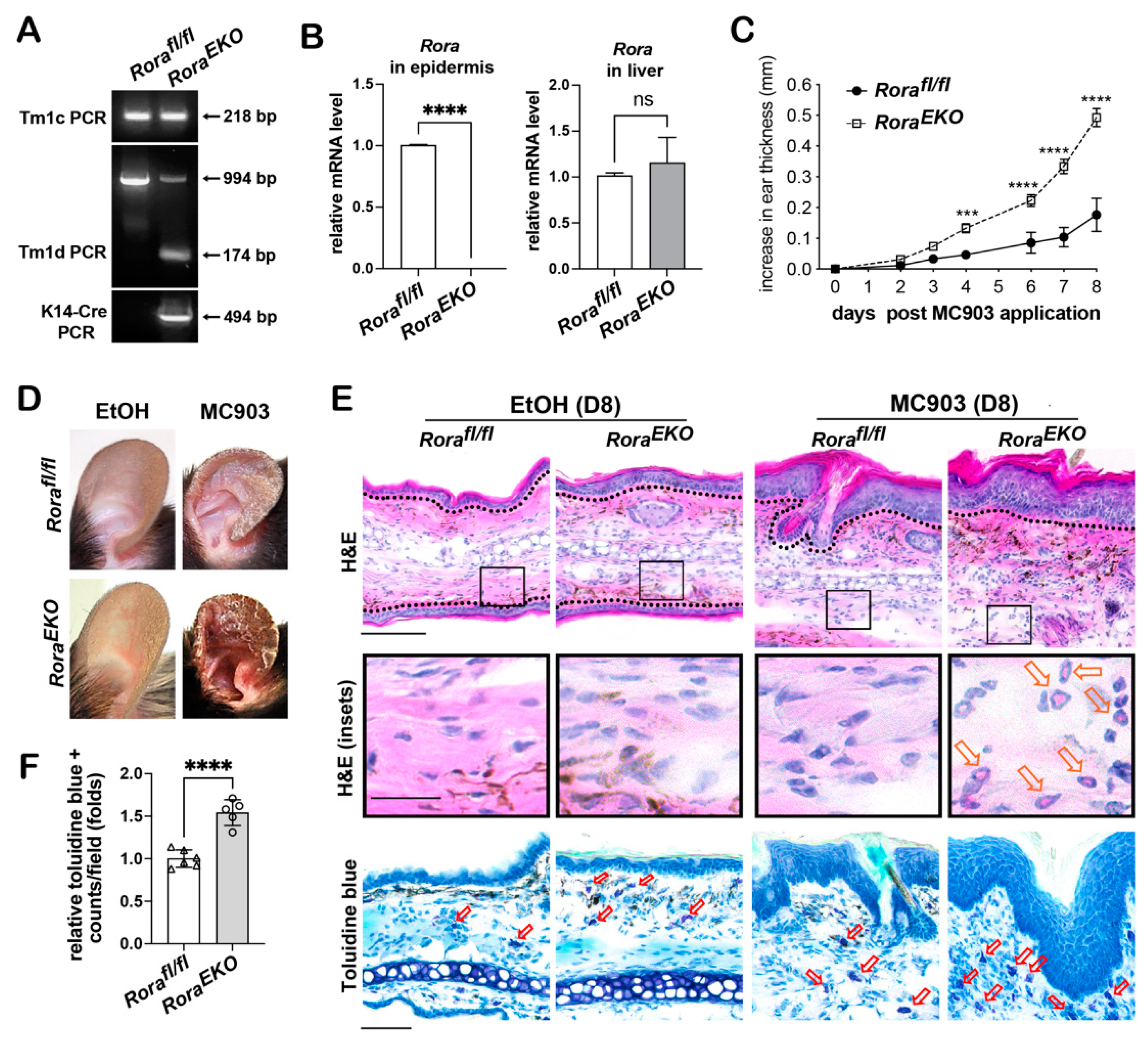
Previous reports have identified RORα expression in dermal immune cells, including regulatory T cells (Treg) and group II lymphoid cells (ILC2s) [25,30]. To rule out the contribution of reduced RORα expression from other cell types to the enhanced inflammation shown in RoraLacZΔ/EKO mice carrying a global heterozygous Rora deletion, we established the Rorafl/fl; K14-CRE strain (referred to as RoraEKO) to confirm the previous findings. The efficiency of epidermal Rora gene deletion was validated through genotyping (Figure 2A; Supplementary Figure S2) and semi-quantitative RT-PCR analysis of Rora mRNA in the back skin epidermis of postnatal day 4 (P4) pups, using the liver tissue as a negative control (Figure 2B). Compared to Rorafl/fl littermates, MC903-treated RoraEKO mice exhibited enhanced ear swelling (Figure 2C) that led to severe skin scaliness (Figure 2D), greater epidermal hyperplasia, and heavier dermal infiltrates, including eosinophils and mast cells, as shown in the histological analysis (Figure 2E,F).
Immunostaining further revealed the prominent dermal recruitment of other immune cells in MC903-challenged RoraEKO skin, including CD11c+ positive dendritic cells, CD4+ T cells, and F4/80+ macrophages, contrasting with the moderate infiltration of these cell types in the Rorafl/fl control skin (Figure 3A). The hyperproliferative epidermis in RoraEKO skin also showed higher induction of keratin 6, the marker of inflamed or hyperproliferative keratinocytes, and lower loricrin expression than the control (Figure 3A). Consistent with the heavier dermal infiltration, MC903-stimulated expression of the proinflammatory cytokine Il1b and chemokines Cxcl2 and Cxcl10 [41] was markedly enhanced by epidermal loss of RORα (Figure 3B). In contrast to the robust local skin inflammation in RoraEKO mice, MC903-induced systemic Th2 responses, measured by the serum IgE elevation, were slightly increased compared to the control (Figure 3C). These results support the primary contribution of epidermal RORα in suppressing MC903-initiated cutaneous inflammation in mice.

2.4. Epidermal Loss of RORα Accelerates MC903-Induced Dermal Activation of Dendritic Cells and CD4+ T Cells

To understand the origin of robust skin inflammation in RoraEKO mice, we further analyzed AD features at an earlier time point post-MC903 application before the robust takeoff of ear swelling. The level of CD11c+ dendritic cells in Rorafl/fl mice reached 2-fold and 3.6-fold of the original level at day 2 and day 8 post-MC903 application, respectively. In comparison, RoraEKO mice exhibited higher elevations, with a 4.5-fold and 7.5-fold elevations of CD11c+ cells (Figure 4A). In contrast to the rapid activation of dermal dendritic cells, MC903-initiated dermal recruitment of CD4+ T cells did not occur until day 8 in Rorafl/fl mice. This infiltration occurred at least three days in advance, with a distinct enhancement in RoraEKO mice (Figure 4B). Therefore, epidermal loss of RORα accelerated the MC903-initiated dermal activation of dendritic cells and CD4+ T cells.

2.5. Epidermal Rora Deletion Increases Tslp Gene Transcription

MC903-initiated AD-like inflammation relies on keratinocyte production of TSLP [42,43]. An immediate effect of TSLP is to activate skin residential dendritic cells, which could migrate into the ear-draining lymph nodes and prime naïve T cells into Th2 effector cells [11,44,45]. Thus, we studied whether the accelerated activation of CD11c+ dendritic cells in MC903-challenged RoraEKO skin was due to a higher TSLP production. RT-PCR analysis revealed higher levels of Tslp mRNA compared to the Rorafl/fl controls in both the vehicle- and MC903-treated groups on day 2 (Figure 5A). On the other hand, epidermal Rora deletion did not affect the mRNA level of Cyp24a1, a direct target gene of VDR’s transcription activity stimulated by MC903 [42,46] (Figure 5A). These results suggest that RORα could regulate Tslp gene transcription in keratinocytes via a VDR-independent mechanism.
Despite the higher Tslp mRNA expression in RoraEKO mice (Figure 5A), MC903-induced epidermal and serum production of TSLP protein from day 2 to day 5 were comparable between these mice and the Rorafl/fl controls (Figure 5B–D). Furthermore, the levels of MC903-stimulated IL13 secretion from ear-draining lymph nodes were comparable between the two genotypes (Figure 5E), which was expected because they should depend on TSLP production from the ear skin in this model [42]. These findings suggest that the enhanced activation of dermal dendritic cells in RoraEKO mice at the early stages post-MC903 treatment (Figure 4A) was likely due to a TSLP-independent mechanism.

2.6. Epidermal Loss of RORα Deregulates Keratinocyte Differentiation and Accelerates Skin Barrier Disruption in Response to MC903

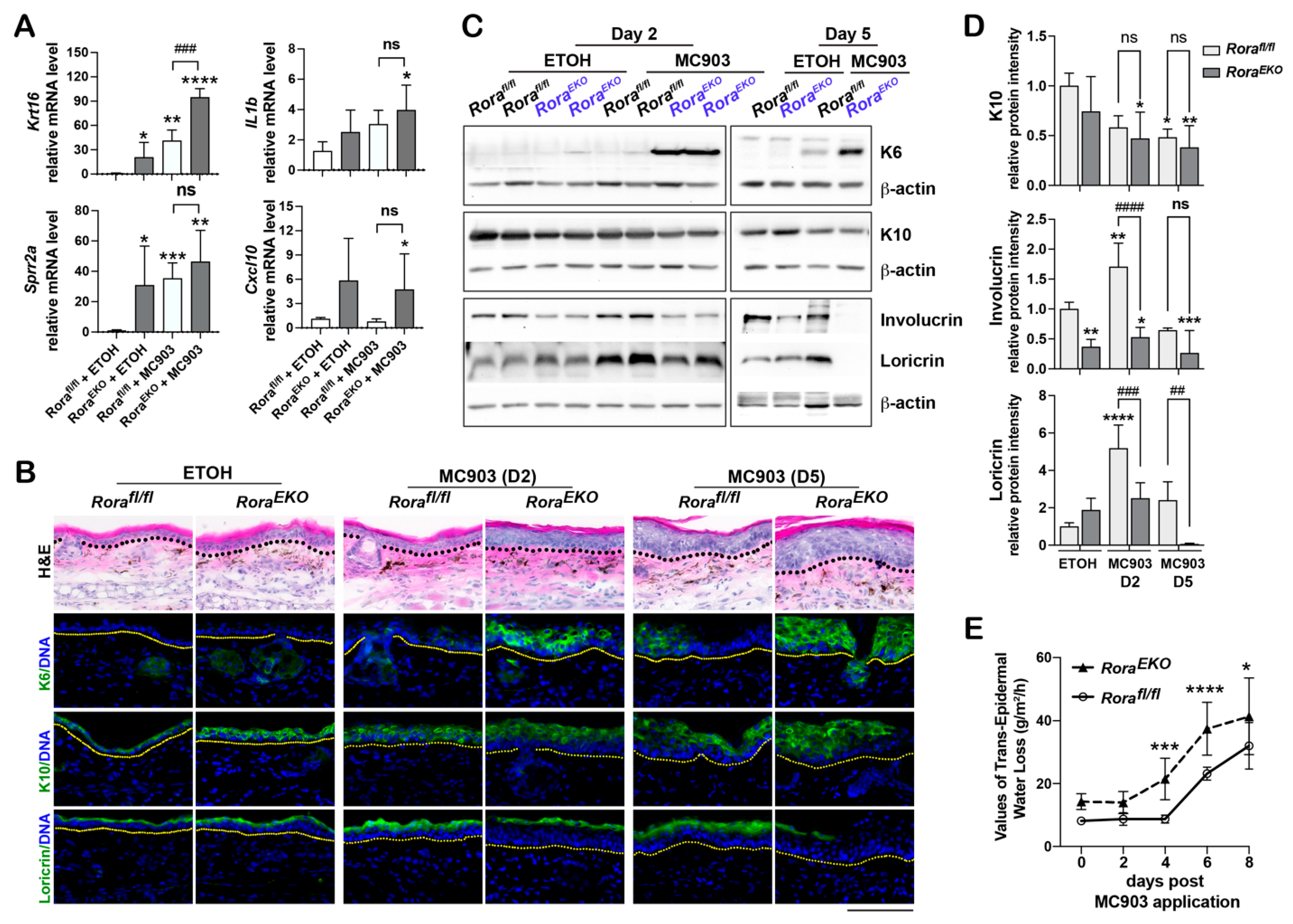
In addition to increased Tslp mRNA expression, vehicle-treated RoraEKO mice also expressed higher mRNA levels of Krt16, which encodes keratin 16: the interacting partner of keratin 6 (Figure 6A). Another highly increased gene in unchallenged RoraEKO skin was Sprr2a, which encodes the proline-rich protein 2 (SPRR2), a cross-linking protein in the cornified envelope [47] (Figure 6A). In contrast to its strong effects on Krt16 and Sprr2a expression, epidermal Rora deletion had a marginal enhancing effect on IL1b and Cxcl10 expression in both the vehicle-treated and MC903-treated groups (Figure 6A), indicating that these proinflammatory immune factors were more involved in the enhanced inflammatory responses in the RoraEKO mice at later stages (Figure 3B).
In addition to the elevated expression of Krt16, Sprr2a, and Tslp, vehicle-treated RoraEKO ear skin showed mild epidermal hyperplasia, which was rapidly enhanced between day 2 and day 5 after MC903 application, along with a robust keratin 6 induction (Figure 6B,C). Although keratin 10 levels were comparable between the two genotypes over this period, involucrin and loricrin levels were markedly lower in the RoraEKO epidermis than in the Rorafl/fl control on day 2 (Figure 6B–D; Supplementary Figure S3). On day 5, while loricrin largely disappeared from the RoraEKO epidermis, its signal remained intact across the control skin (Figure 6B–D; Supplementary Figure S3). We also noticed that the involucrin levels in RoraEKO epidermis was significantly lower than the control levels in the vehicle treatment group (Figure 6C,D; Supplementary Figure S3). These results substantiate the importance of epidermal RORa in maintaining the expression of late differentiation markers at the steady state and during inflammation. Consistent with the deregulated expression of differentiation markers, RoraEKO mice also exhibited higher transepidermal water loss (TEWL) rates than the controls before and after the MC903 application, suggesting an accelerated epidermal barrier disruption (Figure 6E). Overall, the overt inflammation in MC903-challenged RoraEKO mice can be partially attributed to keratinocyte deregulation at the steady state, followed by acceleration in epidermal barrier impairment.

3. Discussion

Despite the prevalent RORα expression in the epidermal compartment of normal skin [32,33], the pathophysiological functions of epidermal RORα in AD development have remained underexplored. Adult mice with epidermal Rora ablation displayed overall normal skin phenotypes but exhibited severe AD-like symptoms after exposure to MC903. Our data uncovered epidermal RORα’s crucial role in suppressing AD-like cutaneous inflammation.
Genetic studies with human subjects have revealed a strong link between AD etiology and loss-of-function (null) mutations of the Flg gene [3]. It has been shown that the Flg-/- null mutation alone was insufficient to trigger spontaneous inflammation in mice. Still, this mutation can lower the threshold for cutaneous sensitization to haptens in mouse models with allergic contact dermatitis [48,49]. These investigations confirm barrier dysfunction as a primary cause of allergic cutaneous inflammation. In our study, despite its normal appearance at the steady state, epidermal Rora-deficient skin exhibited mild epidermal hyperplasia, greater TEWL rate, and higher mRNA levels of Krt16, Sprr2a, and Tslp compared to the skin of wildtype littermates (Figure 6). Expressions of keratin 16/17 and its partner, keratin 6, are similarly elevated in the hyperproliferative keratinocytes under pathological conditions such as injury and AD [50,51,52]. It is also noteworthy that SPRR2 proteins have been found to be upregulated in mice with impaired barrier functions [53,54,55,56]. More importantly, these barrier dysfunction-related phenotypes in Rora deficient skin partially resembled the subclinical conditions found in nonlesional skin of AD patients [52,57]. Therefore, we anticipate that the amplified cutaneous inflammation in MC903-challenged RoraEKO mice can be partly attributed to the pre-existing epidermal barrier dysfunctions.
Despite the comparable TSLP production between RoraEKO and Rorafl/fl mice upon MC903 stimulation (Figure 5), RoraEKO skin displayed a faster and stronger activation of CD11c+ dendritic cells (Figure 4A). In fact, CD11c+ cells were already detected in Rora-deficient skin treated with the vehicle (Figure 4A). A similarly elevated CD11c+ population has also been observed in human skin epidermis carrying FLG-null mutations [58]. The increased baseline activation of antigen presentation cells can be attributed to barrier dysfunction and increased exposure to dangerous signals [58]. We postulate that the pre-existing CD11c+ cells or altered microenvironment in the RoraEKO skin may amplify the TSLP activation of dermal dendritic cells, which can enhance T cell activation at the later stage (Figure 4B).
A recent study demonstrated that epidermal RORα protein expression is significantly lower in human skin lesions of several inflammatory skin diseases than in normal skin [37]. Consistent with this report, using the Rora lacZ-reporter mice, we found that Rora gene transcription in the granular layers of the epidermis was markedly reduced on day 5 after the MC903 application (Figure 1B), simultaneous with the TSLP peak production (Figure 5B–D). These results implicate epidermal Rora as a negative target of the TSLP-initiated inflammatory pathway in the skin. Downregulation of epidermal RORα can further contribute to AD development by accelerating skin barrier impairment, thus perpetuating a vicious cycle. It has been found that elevated Th2 cytokines (IL4 and IL13), a hallmark of AD, broadly suppress the expression of barrier-related genes, such as filaggrin, involucrin, and loricrin, through IL-4Rα-STAT6 signaling [59,60,61]. Along with Staphylococcus aureus lipoteichoic acid, Th2 cytokines also repress genes related to early keratinocyte differentiation by modulating the activities of p63 and Notch, the key transcription factors for skin development and keratinocyte differentiation [62]. We believe that epidermal RORα expression is also under the negative control of certain proinflammatory cytokines and downstream signaling pathways.
We have previously reported the effect of SR1001, an inverse agonist of RORα/γ, in suppressing MC903-induced AD-like inflammation [63]. Surprisingly, epidermal loss of RORα had the opposite effect in the same mouse model. This contrast can be attributed to the opposite effects of SR1001 and epidermal Rora gene deletion on TSLP gene expression. While SR1001 blocked MC903-induced Tslp expression at both the mRNA and protein levels [63], epidermal Rora deletion enhanced Tslp transcription (Figure 5A). The latter evidence indicates that RORα may function as a transcription repressor of Tslp in keratinocytes. As a RORα/γ inverse agonist, binding of SR1001 to RORα can increase the recruitment of co-repressors [64,65]. While this recruitment can downregulate the positive target genes of RORα, it may also enhance the repression of the negative targets, such as Tslp. An examination of the anti-inflammatory effects of SR1001 in RoraEKO mice would further validate the dependency of these effects on epidermal RORα.
A recent report showed that epidermal Rora deletion alleviated imiquimod-induced psoriatic-like skin inflammations by attenuating the activation of nuclear factor-κB (NF-κB and STAT3 in keratinocytes) [66]. The opposite effects of epidermal RORα loss on cutaneous inflammation in the two disease models suggest it has context-dependent regulatory functions. While MC903-initiated AD-like responses rely on epithelial production of TSLP, imiquimod-caused psoriatic-like inflammations depend on the activations of Toll-like receptor 7 and subsequent pathways in numerous cell types in mice [67]. Furthermore, while epidermal RORα expression is downregulated in skin lesions of multiple inflammatory skin disorders, including AD, its expression is unaltered in psoriatic skin lesions [37]. We believe that RORα prevents AD development by maintaining normal barrier functions while potentiating psoriatic symptoms through positive regulation of STAT3 activity in imiquimod-treated keratinocytes [66].
Overall, we identified the protective effects of epidermal RORα against MC903-induced AD development. Our findings also revealed the beneficial potentials of RORα agonists to restore barrier functions and alleviate inflammation in treating AD and other inflammatory skin diseases [68]. A strength of this study is that tissue-specific Rora knockout mice were used to uncover epidermal RORα’s functions in regulating AD development. Our work differs from most previous studies using the staggerer Rorasg/sg mice with a global Rora-null mutation. Thus far, the underlying molecular mechanisms of epidermal RORα’s pathophysiological functions have not been elucidated. Therefore, identifying keratinocyte-specific targets and upstream regulators of RORα will lead to mechanistic insight into cutaneous inflammation and the discovery of novel therapeutic targets.

4. Materials and Methods

4.1. Mice

All animal studies were conducted under approved protocols from Institutional Animal Care and Use Committee. Roratm1a(EUCOMM)Wtsi (Roratm1a/+) embryonic stem cells were obtained from INFRAFRONTIER/EMMA (European Mouse Mutant Archive, Munich, Germany) and subjected to in vitro injection into the C57BL/6 female mice to generate the Roratm1a/+ mice (performed by Animal Models of Biotechnology at the University of Wisconsin, Madison, WI, USA). A male Roratm1a/+ mouse was crossed with a female Sox2 (SRY-box containing gene 2)-Cre transgenic mouse (B6.Cg-Edil3Tg(Sox2-cre)1Amc/J; The Jackson Laboratory, Bar Harbor, ME, USA) [69] to generate the Roratm1b/+ strain (RoraLacZΔ/+), which contains a LacZ-cassette upstream the deleted Rora exon 3 (Figure 1A). To generate the Roratm1c/+ (Roraflox/+) mice, the Roratm1a/+ mice were crossed with ACTB-FLPe transgenic mice (B6.Cg-Tg(ACTFLPe)9205Dym/J; The Jackson Laboratory) to delete the FRT-flanked section. The Roraflox/+; FLP mice were backcrossed with wildtype C57BL/6 mice to remove FLP [39]. The RoraLacZΔ/EKO mice were generated by crossing the Roraflox/flox strain with the RoraLacZΔ/+;K14-Cre strain (B6N.Cg-Tg(KRT14-cre)1Amc/J, The Jackson Laboratory). The RoraEKO mice were generated by crossing the Roraflox/flox strain with the K14-Cre strain.

4.2. Genotyping

Genotyping was performed using short-range end-point Polymerase Chain Reaction (PCR) assays with specific primers according to the EUCOMM/KOMP-CSD allele conversion guide. Genomic DNA was extracted from tail tips or punched ear tissues, followed by PCR analysis using the GoTaq Green master mix (Promega, Madison, WI, USA). The PCR products were separated on 2% agarose gels dissolved in Tris/Acetic Acid/EDTA (TAE) buffer and stained with ethidium bromide (Millipore-Sigma, St. Louis, MO, USA). Primer sequences are listed in Supplementary Table S1.

4.3. MC903-Induced AD-like Model

MC903 (calcipotriol hydrate) was purchased from Millipore-Sigma (Saint Louis, MO, USA). Both ears of 8–10-week-old mice were topically painted with 1 nmol of MC903 (in 20 μL ethanol; 10 μL on each side) on days 0, 1, 2, 3, 9, and 11. Ear thickness was measured with a digital caliper (Mitutoyo Corp., Tokyo, Japan).

4.4. X-Gal Staining

According to standard procedures, β-galactosidase activities encoded by the Rora promoter-driven LacZ gene in Roratm1b/+ mice were evaluated for the frozen ear sections. Frozen sections (8 μM) were fixed with 2% PFA/Phosphate Buffer Saline (PBS) containing 0.02% NP-40 at 4 ℃ for 30 min. After washing with the PBS buffer containing 0.02% NP-40 and 0.01% deoxycholate, the samples were incubated in X-gal reaction buffer (5 mM K3Fe(CN)6, 5 mM K4Fe(CN)6, 0.02% NP-40, 0.01% deoxycholate, 2 mM MgCl2, 5mM EGTA, and 1 mg/mL X-gal) at room temperature for at least 2 h. The sections were counterstained with Nuclear Fast Red. All reagents in this assay were purchased from Millipore-Sigma (St. Louis, MO, USA).

4.5. Histology and Immunofluorescence

Frozen sections (8 μM) of ear samples were fixed with 10% formalin for Hematoxylin and Eosin Y (H&E) staining and toluidine blue staining (Electron Microscopy Sciences, Hatfield, PA, USA). For immunostaining, frozen sections were fixed with 4% paraformaldehyde/PBS, permeabilized with 0.1% NP-40/PBS, and incubated with the appropriate primary antibody at 4 °C overnight. The slides were then incubated with Alexa 488- or Alexa 594-conjugated secondary antibodies (Invitrogen-Thermo Fisher Scientific, Waltham, MA, USA) and Hoechst (Invitrogen-Thermo Fisher Scientific) for DNA detection. H&E and fluorescence images were acquired using a Lionheart FX microscope (Biotek, Winooski, VT, USA). The number of dermal infiltrates (mast cells, CD11c+ positive cells, and CD4+ T cells) and the intensity of the epidermal TSLP signal were quantified using the ImageJ 1.53k software (National Institute of Health). A list of the primary antibodies is provided in Supplementary Table S2.

4.6. Quantitative Real-Time (RT)-PCR

According to the manufacturer’s instructions, total RNA from ear tissues was isolated using Trizol (Invitrogen-Thermo Fisher Scientific). Reverse transcription of isolated RNA was done using the High-Capacity cDNA RT Kit (Applied Biosystems-Thermo Fisher Scientific, Foster City, CA, USA). Amplification reactions were performed using PowerSYBR Green PCR Master Mix and a QuantStudio 3 Real-Time PCR system (Applied Biosystems-Thermo Fisher Scientific). Each sample was tested in duplicates. A comparative method for relative quantification (2XΔΔ cycle threshold) was used to calculate the expression levels of the target genes, which were normalized to the level of 18S rRNA. The primer sequences are listed in Supplementary Table S1.

4.7. ELISA

Peripheral blood was collected to measure the serum IgE and TSLP levels at specific time points and analyzed using ELISA with commercial kits from eBioscience-Thermo Fisher Scientific according to the manufacturer’s instructions. For measuring TSLP protein levels from the ear skin, ear tissue was processed with a homogenizer (Fisherbrand Bead Mill 4, Fisher Scientific, Hanover Park, IL, USA) in protein lysis buffer (100 mM Tris, pH 7.4, 2 mM Na3VO4, 100 mM NaCl, 1% Triton X-100, 1 mM EDTA, 10% glycerol, 1 mM EGTA, 0.1% SDS, and protease inhibitors). For measuring IL13 secretion from ear-draining lymph nodes, single-cell suspension from the isolated lymph nodes was plated at a density of 5 × 10⁵ cells/well in 96-well plates. After 24 h of re-stimulation with 50 ng/mL TPA and 1 μM ionomycin, the media were collected to measure cytokine production using ELISA.

4.8. Transepidermal Water Loss Measurements

Transepidermal water loss (TEWL) was measured on ear skin before and after MC903 application using a noninvasive Tewameter TM Hex probe (Courage and Khazaka Electronic GmbH, Cologne, Germany) according to the manufacturer’s instructions.

4.9. Western Blot Analysis

The ear tissue was processed in protein lysis buffer using a tissue homogenizer, as described in the ELISA assay. Protein concentration was determined using the BCA Protein Assay Kit (Pierce-Thermo Fisher Scientific). Proteins were separated using SDS-polyacrylamide gel electrophoresis and transferred onto 0.45 μm polyvinylidene difluoride (PVDF) membranes (Thermo Fisher Scientific). After blocking with 5% non-fat milk, the membranes were incubated with specific antibodies (see Supplementary Table S2) overnight at 4 °C, followed by incubation with horseradish peroxidase (HRP)-conjugated secondary antibodies. The membranes were incubated with the enhanced chemiluminescence (ECL) western blotting substrate (Pierce Biotechnology-Thermo Fisher Scientific). Chemiluminescent images were acquired using an iBright CL1000 (Invitrogen—Thermo Fisher Scientific, Pittsburgh, PA, USA).

4.10. Statistical Analysis

All statistical evaluations used Prism 9.5 (GraphPad Software, La Jolla, CA, USA). Unpaired Student’s t-tests were used to compare the statistical difference between the two groups. The statistical differences between multiple groups were analyzed using a one-way ANOVA or two-way ANOVA, as indicated in the figure legends, and p values < 0.05 were considered significant.

Supplementary Materials

The following supporting information can be downloaded at: https://www.mdpi.com/article/10.3390/ijms241210241/s1.

Author Contributions

Conceptualization, J.D.; methodology, J.D. and X.H.; validation, X.H., H.D., M.K.F. and N.L.W.; formal analysis, J.D. and X.H.; investigation, X.H.; resources, C.D.B.; data curation, J.D. and X.H.; writing—original draft preparation, J.D.; writing—review and editing, J.D., R.P.H. and X.H.; visualization, J.D. and X.H.; supervision, J.D.; project administration, J.D.; funding acquisition, J.D. and R.P.H. All authors have read and agreed to the published version of the manuscript.

Funding

Support for this project includes the Wisconsin Alumni Research Foundation, the School of Pharmacy of the University of Wisconsin-Madison, the Fall Research Competition at the University of Wisconsin–Madison, and the UW SDRC grant P30 AR066524, funded by the National Institute of Arthritis and Musculoskeletal and Skin Diseases (NIAMS). N. Wallace was supported by the Summer Undergraduate Research Grant (URG) from Northwestern University. R. P. Hsung would like to thank the Laura and Edward Kremers Family Foundation for an endowed chair in natural products chemistry, the University of Wisconsin, and the Vilas Trust for a distinguished achievement professorship.

Institutional Review Board Statement

The animal study protocol was approved by the School of Medicine and Public Health Institutional Animal Care and Use Committee of University of Wisconsin-Madison (Protocol ID: M006246; date of approval: 22 October 2019).

Informed Consent Statement

Not applicable.

Data Availability Statement

Data sharing is not applicable as no datasets have been generated or analyzed in this study.

Acknowledgments

J. Dai would like to thank Hao Chang from the Department of Dermatology at the University of Wisconsin–Madison for his technical support on mouse breeding strategies. The authors would like to thank the Biomedical Research Model Service and The Animal Model Core at the University of Wisconsin Biotechnology Center for assisting with mouse colony generation and maintenance.

Conflicts of Interest

The authors state no conflicts of interest.

References

  1. Weidinger, S.; Beck, L.A.; Bieber, T.; Kabashima, K.; Irvine, A.D. Atopic dermatitis. Nat. Rev. Dis. Prim. 2018, 4, 1. [Google Scholar] [CrossRef] [PubMed]
  2. Brunner, P.M.; Guttman-Yassky, E.; Leung, D.Y. The immunology of atopic dermatitis and its reversibility with broad-spectrum and targeted therapies. J. Allergy Clin. Immunol. 2017, 139, S65–S76. [Google Scholar] [CrossRef] [PubMed] [Green Version]
  3. Drislane, C.; Irvine, A.D. The role of filaggrin in atopic dermatitis and allergic disease. Ann. Allergy Asthma Immunol. 2020, 124, 36–43. [Google Scholar] [CrossRef] [PubMed] [Green Version]
  4. Moosbrugger-Martinz, V.; Leprince, C.; Méchin, M.C.; Simon, M.; Blunder, S.; Gruber, R.; Dubrac, S. Revisiting the Roles of Filaggrin in Atopic Dermatitis. Int. J. Mol. Sci. 2022, 23, 5318. [Google Scholar] [CrossRef]
  5. Leung, D.Y.M.; Berdyshev, E.; Goleva, E. Cutaneous barrier dysfunction in allergic diseases. J. Allergy Clin. Immunol. 2020, 145, 1485–1497. [Google Scholar] [CrossRef]
  6. Elias, P.M.; Hatano, Y.; Williams, M.L. Basis for the barrier abnormality in atopic dermatitis: Outside-inside-outside pathogenic mechanisms. J. Allergy Clin. Immunol. 2008, 121, 1337–1343. [Google Scholar] [CrossRef] [Green Version]
  7. Blanpain, C.; Fuchs, E. Epidermal homeostasis: A balancing act of stem cells in the skin. Nat. Rev. Mol. Cell Biol. 2009, 10, 207–217. [Google Scholar] [CrossRef] [Green Version]
  8. Hasegawa, T.; Oka, T.; Demehri, S. Alarmin Cytokines as Central Regulators of Cutaneous Immunity. Front. Immunol. 2022, 13, 876515. [Google Scholar] [CrossRef]
  9. Pandey, A.; Ozaki, K.; Baumann, H.; Levin, S.D.; Puel, A.; Farr, A.G.; Ziegler, S.F.; Leonard, W.J.; Lodish, H.F. Cloning of a receptor subunit required for signaling by thymic stromal lymphopoietin. Nat. Immunol. 2000, 1, 59–64. [Google Scholar] [CrossRef]
  10. Ziegler, S.F.; Artis, D. Sensing the outside world: TSLP regulates barrier immunity. Nat. Immunol. 2010, 11, 289–293. [Google Scholar] [CrossRef] [Green Version]
  11. Soumelis, V.; Reche, P.A.; Kanzler, H.; Yuan, W.; Edward, G.; Homey, B.; Gilliet, M.; Ho, S.; Antonenko, S.; Lauerma, A.; et al. Human epithelial cells trigger dendritic cell mediated allergic inflammation by producing TSLP. Nat. Immunol. 2002, 3, 673–680. [Google Scholar] [CrossRef] [PubMed] [Green Version]
  12. Nestle, F.O.; Di Meglio, P.; Qin, J.Z.; Nickoloff, B.J. Skin immune sentinels in health and disease. Nat. Rev. Immunol. 2009, 9, 679–691. [Google Scholar] [CrossRef] [PubMed] [Green Version]
  13. Chieosilapatham, P.; Kiatsurayanon, C.; Umehara, Y.; Trujillo-Paez, J.V.; Peng, G.; Yue, H.; Nguyen, L.T.H.; Niyonsaba, F. Keratinocytes: Innate immune cells in atopic dermatitis. Clin. Exp. Immunol. 2021, 204, 296–309. [Google Scholar] [CrossRef] [PubMed]
  14. Jetten, A.M. Retinoid-related orphan receptors (RORs): Critical roles in development, immunity, circadian rhythm, and cellular metabolism. Nucl. Recept. Signal 2009, 7, e003. [Google Scholar] [CrossRef] [Green Version]
  15. Jetten, A.M.; Joo, J.H. Retinoid-related Orphan Receptors (RORs): Roles in Cellular Differentiation and Development. Adv. Dev. Biol. 2006, 16, 313–355. [Google Scholar]
  16. Giguere, V.; Tini, M.; Flock, G.; Ong, E.; Evans, R.M.; Otulakowski, G. Isoform-specific amino-terminal domains dictate DNA-binding properties of ROR alpha, a novel family of orphan hormone nuclear receptors. Genes Dev. 1994, 8, 538–553. [Google Scholar] [CrossRef] [Green Version]
  17. Hadj-Sahraoui, N.; Frederic, F.; Zanjani, H.; Delhaye-Bouchaud, N.; Herrup, K.; Mariani, J. Progressive atrophy of cerebellar Purkinje cell dendrites during aging of the heterozygous staggerer mouse (Rora(+/sg)). Brain Res. Dev. Brain Res. 2001, 126, 201–209. [Google Scholar] [CrossRef]
  18. Hamilton, B.A.; Frankel, W.N.; Kerrebrock, A.W.; Hawkins, T.L.; FitzHugh, W.; Kusumi, K.; Russell, L.B.; Mueller, K.L.; van Berkel, V.; Birren, B.W.; et al. Disruption of the nuclear hormone receptor RORalpha in staggerer mice. Nature 1996, 379, 736–739. [Google Scholar] [CrossRef]
  19. Meyer, T.; Kneissel, M.; Mariani, J.; Fournier, B. In vitro and in vivo evidence for orphan nuclear receptor RORalpha function in bone metabolism. Proc. Natl. Acad. Sci. USA 2000, 97, 9197–9202. [Google Scholar] [CrossRef] [Green Version]
  20. Dzhagalov, I.; Giguere, V.; He, Y.W. Lymphocyte development and function in the absence of retinoic acid-related orphan receptor alpha. J. Immunol. 2004, 173, 2952–2959. [Google Scholar] [CrossRef] [Green Version]
  21. Ferreira, A.C.F.; Szeto, A.C.H.; Heycock, M.W.D.; Clark, P.A.; Walker, J.A.; Crisp, A.; Barlow, J.L.; Kitching, S.; Lim, A.; Gogoi, M.; et al. RORalpha is a critical checkpoint for T cell and ILC2 commitment in the embryonic thymus. Nat. Immunol. 2021, 22, 166–178. [Google Scholar] [CrossRef] [PubMed]
  22. Lau, P.; Fitzsimmons, R.L.; Raichur, S.; Wang, S.C.; Lechtken, A.; Muscat, G.E. The orphan nuclear receptor, RORalpha, regulates gene expression that controls lipid metabolism: Staggerer (SG/SG) mice are resistant to diet-induced obesity. J. Biol. Chem. 2008, 283, 18411–18421. [Google Scholar] [CrossRef] [PubMed] [Green Version]
  23. Chi, X.; Jin, W.; Bai, X.; Zhao, X.; Shao, J.; Li, J.; Sun, Q.; Su, B.; Wang, X.; Yang, X.O.; et al. RORalpha is critical for mTORC1 activity in T cell-mediated colitis. Cell Rep. 2021, 36, 109682. [Google Scholar] [CrossRef] [PubMed]
  24. Oh, S.K.; Kim, D.; Kim, K.; Boo, K.; Yu, Y.S.; Kim, I.S.; Jeon, Y.; Im, S.K.; Lee, S.H.; Lee, J.M.; et al. RORα is crucial for attenuated inflammatory response to maintain intestinal homeostasis. Proc. Natl. Acad. Sci. USA 2019, 116, 21140–21149. [Google Scholar] [CrossRef] [Green Version]
  25. Malhotra, N.; Leyva-Castillo, J.M.; Jadhav, U.; Barreiro, O.; Kam, C.; O’Neill, N.K.; Meylan, F.; Chambon, P.; von Andrian, U.H.; Siegel, R.M.; et al. RORα-expressing T regulatory cells restrain allergic skin inflammation. Sci. Immunol. 2018, 3, eaao6923. [Google Scholar] [CrossRef] [Green Version]
  26. Akashi, M.; Takumi, T. The orphan nuclear receptor RORalpha regulates circadian transcription of the mammalian core-clock Bmal1. Nat. Struct. Mol. Biol. 2005, 12, 441–448. [Google Scholar] [CrossRef]
  27. Sato, T.K.; Panda, S.; Miraglia, L.J.; Reyes, T.M.; Rudic, R.D.; McNamara, P.; Naik, K.A.; FitzGerald, G.A.; Kay, S.A.; Hogenesch, J.B. A functional genomics strategy reveals Rora as a component of the mammalian circadian clock. Neuron 2004, 43, 527–537. [Google Scholar] [CrossRef] [Green Version]
  28. Wong, S.H.; Walker, J.A.; Jolin, H.E.; Drynan, L.F.; Hams, E.; Camelo, A.; Barlow, J.L.; Neill, D.R.; Panova, V.; Koch, U.; et al. Transcription factor RORalpha is critical for nuocyte development. Nat. Immunol. 2012, 13, 229–236. [Google Scholar] [CrossRef] [Green Version]
  29. Yang, X.O.; Pappu, B.P.; Nurieva, R.; Akimzhanov, A.; Kang, H.S.; Chung, Y.; Ma, L.; Shah, B.; Panopoulos, A.D.; Schluns, K.S.; et al. T helper 17 lineage differentiation is programmed by orphan nuclear receptors ROR alpha and ROR gamma. Immunity 2008, 28, 29–39. [Google Scholar] [CrossRef] [Green Version]
  30. Salimi, M.; Barlow, J.L.; Saunders, S.P.; Xue, L.; Gutowska-Owsiak, D.; Wang, X.; Huang, L.C.; Johnson, D.; Scanlon, S.T.; McKenzie, A.N.; et al. A role for IL-25 and IL-33-driven type-2 innate lymphoid cells in atopic dermatitis. J. Exp. Med. 2013, 210, 2939–2950. [Google Scholar] [CrossRef]
  31. Patel, D.D.; Kuchroo, V.K. Th17 Cell Pathway in Human Immunity: Lessons from Genetics and Therapeutic Interventions. Immunity 2015, 43, 1040–1051. [Google Scholar] [CrossRef] [PubMed] [Green Version]
  32. Dai, J.; Brooks, Y.; Lefort, K.; Getsios, S.; Dotto, G.P. The retinoid-related orphan receptor RORalpha promotes keratinocyte differentiation via FOXN1. PLoS ONE 2013, 8, e70392. [Google Scholar] [CrossRef] [PubMed] [Green Version]
  33. Slominski, A.; Fischer, T.W.; Zmijewski, M.A.; Wortsman, J.; Semak, I.; Zbytek, B.; Slominski, R.M.; Tobin, D.J. On the role of melatonin in skin physiology and pathology. Endocrine 2005, 27, 137–148. [Google Scholar] [CrossRef] [Green Version]
  34. Hanyu, O.; Nakae, H.; Miida, T.; Higashi, Y.; Fuda, H.; Endo, M.; Kohjitani, A.; Sone, H.; Strott, C.A. Cholesterol sulfate induces expression of the skin barrier protein filaggrin in normal human epidermal keratinocytes through induction of RORalpha. Biochem. Biophys. Res. Commun. 2012, 428, 99–104. [Google Scholar] [CrossRef] [PubMed]
  35. Steinmayr, M.; Andre, E.; Conquet, F.; Rondi-Reig, L.; Delhaye-Bouchaud, N.; Auclair, N.; Daniel, H.; Crepel, F.; Mariani, J.; Sotelo, C.; et al. Staggerer phenotype in retinoid-related orphan receptor alpha-deficient mice. Proc. Natl. Acad. Sci. USA 1998, 95, 3960–3965. [Google Scholar] [CrossRef] [Green Version]
  36. Tsujihana, K.; Tanegashima, K.; Santo, Y.; Yamada, H.; Akazawa, S.; Nakao, R.; Tominaga, K.; Saito, R.; Nishito, Y.; Hata, R.I.; et al. Circadian protection against bacterial skin infection by epidermal CXCL14-mediated innate immunity. Proc. Natl. Acad. Sci. USA 2022, 119, e2116027119. [Google Scholar] [CrossRef]
  37. Brożyna, A.A.; Żmijewski, M.A.; Linowiecka, K.; Kim, T.K.; Slominski, R.M.; Slominski, A.T. Disturbed expression of vitamin D and retinoic acid-related orphan receptors α and γ and of megalin in inflammatory skin diseases. Exp. Dermatol. 2022, 31, 781–788. [Google Scholar] [CrossRef]
  38. Guttman-Yassky, E.; Suárez-Fariñas, M.; Chiricozzi, A.; Nograles, K.E.; Shemer, A.; Fuentes-Duculan, J.; Cardinale, I.; Lin, P.; Bergman, R.; Bowcock, A.M.; et al. Broad defects in epidermal cornification in atopic dermatitis identified through genomic analysis. J. Allergy Clin. Immunol. 2009, 124, 1235–1244.e58. [Google Scholar] [CrossRef]
  39. Coleman, J.L.; Brennan, K.; Ngo, T.; Balaji, P.; Graham, R.M.; Smith, N.J. Rapid Knockout and Reporter Mouse Line Generation and Breeding Colony Establishment Using EUCOMM Conditional-Ready Embryonic Stem Cells: A Case Study. Front. Endocrinol. 2015, 6, 105. [Google Scholar] [CrossRef] [Green Version]
  40. Oliphant, C.J.; Hwang, Y.Y.; Walker, J.A.; Salimi, M.; Wong, S.H.; Brewer, J.M.; Englezakis, A.; Barlow, J.L.; Hams, E.; Scanlon, S.T.; et al. MHCII-mediated dialog between group 2 innate lymphoid cells and CD4(+) T cells potentiates type 2 immunity and promotes parasitic helminth expulsion. Immunity 2014, 41, 283–295. [Google Scholar] [CrossRef] [Green Version]
  41. Walsh, C.M.; Hill, R.Z.; Schwendinger-Schreck, J.; Deguine, J.; Brock, E.C.; Kucirek, N.; Rifi, Z.; Wei, J.; Gronert, K.; Brem, R.B.; et al. Neutrophils promote CXCR3-dependent itch in the development of atopic dermatitis. eLife 2019, 8, e48448. [Google Scholar] [CrossRef] [PubMed]
  42. Li, M.; Hener, P.; Zhang, Z.; Kato, S.; Metzger, D.; Chambon, P. Topical vitamin D3 and low-calcemic analogs induce thymic stromal lymphopoietin in mouse keratinocytes and trigger an atopic dermatitis. Proc. Natl. Acad. Sci. USA 2006, 103, 11736–11741. [Google Scholar] [CrossRef] [Green Version]
  43. Li, M.; Hener, P.; Zhang, Z.; Ganti, K.P.; Metzger, D.; Chambon, P. Induction of thymic stromal lymphopoietin expression in keratinocytes is necessary for generating an atopic dermatitis upon application of the active vitamin D3 analogue MC903 on mouse skin. J. Investig. Dermatol. 2009, 129, 498–502. [Google Scholar] [CrossRef]
  44. Liu, Y.J. TSLP in epithelial cell and dendritic cell cross talk. Adv. Immunol. 2009, 101, 1–25. [Google Scholar] [PubMed] [Green Version]
  45. Liu, Y.J.; Soumelis, V.; Watanabe, N.; Ito, T.; Wang, Y.H.; Malefyt Rde, W.; Omori, M.; Zhou, B.; Ziegler, S.F. TSLP: An epithelial cell cytokine that regulates T cell differentiation by conditioning dendritic cell maturation. Annu. Rev. Immunol. 2007, 25, 193–219. [Google Scholar] [CrossRef] [PubMed]
  46. Jones, G.; Strugnell, S.A.; DeLuca, H.F. Current understanding of the molecular actions of vitamin D. Physiol. Rev. 1998, 78, 1193–1231. [Google Scholar] [CrossRef] [Green Version]
  47. Carregaro, F.; Stefanini, A.C.; Henrique, T.; Tajara, E.H. Study of small proline-rich proteins (SPRRs) in health and disease: A review of the literature. Arch. Dermatol. Res. 2013, 305, 857–866. [Google Scholar] [CrossRef]
  48. Scharschmidt, T.C.; Man, M.Q.; Hatano, Y.; Crumrine, D.; Gunathilake, R.; Sundberg, J.P.; Silva, K.A.; Mauro, T.M.; Hupe, M.; Cho, S.; et al. Filaggrin deficiency confers a paracellular barrier abnormality that reduces inflammatory thresholds to irritants and haptens. J. Allergy Clin. Immunol. 2009, 124, 496–506.e6. [Google Scholar] [CrossRef] [Green Version]
  49. Muhandes, L.; Chapsa, M.; Pippel, M.; Behrendt, R.; Ge, Y.; Dahl, A.; Yi, B.; Dalpke, A.; Winkler, S.; Hiller, M.; et al. Low Threshold for Cutaneous Allergen Sensitization but No Spontaneous Dermatitis or Atopy in FLG-Deficient Mice. J. Investig. Dermatol. 2021, 141, 2611–2619.e2. [Google Scholar] [CrossRef]
  50. Wang, F.; Chen, S.; Liu, H.B.; Parent, C.A.; Coulombe, P.A. Keratin 6 regulates collective keratinocyte migration by altering cell-cell and cell-matrix adhesion. J. Cell Biol. 2018, 217, 4314–4330. [Google Scholar] [CrossRef] [Green Version]
  51. Zhang, X.; Yin, M.; Zhang, L.J. Keratin 6, 16 and 17-Critical Barrier Alarmin Molecules in Skin Wounds and Psoriasis. Cells 2019, 8, 807. [Google Scholar] [CrossRef] [Green Version]
  52. Jensen, J.M.; Fölster-Holst, R.; Baranowsky, A.; Schunck, M.; Winoto-Morbach, S.; Neumann, C.; Schütze, S.; Proksch, E. Impaired sphingomyelinase activity and epidermal differentiation in atopic dermatitis. J. Investig. Dermatol. 2004, 122, 1423–1431. [Google Scholar] [CrossRef] [PubMed] [Green Version]
  53. Kypriotou, M.; Boéchat, C.; Huber, M.; Hohl, D. Spontaneous atopic dermatitis-like symptoms in a/a ma ft/ma ft/J flaky tail mice appear early after birth. PLoS ONE 2013, 8, e67869. [Google Scholar] [CrossRef] [PubMed] [Green Version]
  54. Fallon, P.G.; Sasaki, T.; Sandilands, A.; Campbell, L.E.; Saunders, S.P.; Mangan, N.E.; Callanan, J.J.; Kawasaki, H.; Shiohama, A.; Kubo, A.; et al. A homozygous frameshift mutation in the mouse Flg gene facilitates enhanced percutaneous allergen priming. Nat. Genet. 2009, 41, 602–608. [Google Scholar] [CrossRef] [Green Version]
  55. Koch, P.J.; de Viragh, P.A.; Scharer, E.; Bundman, D.; Longley, M.A.; Bickenbach, J.; Kawachi, Y.; Suga, Y.; Zhou, Z.; Huber, M.; et al. Lessons from loricrin-deficient mice: Compensatory mechanisms maintaining skin barrier function in the absence of a major cornified envelope protein. J. Cell Biol. 2000, 151, 389–400. [Google Scholar] [CrossRef]
  56. Segre, J.A.; Bauer, C.; Fuchs, E. Klf4 is a transcription factor required for establishing the barrier function of the skin. Nat. Genet. 1999, 22, 356–360. [Google Scholar] [CrossRef]
  57. Suárez-Fariñas, M.; Tintle, S.J.; Shemer, A.; Chiricozzi, A.; Nograles, K.; Cardinale, I.; Duan, S.; Bowcock, A.M.; Krueger, J.G.; Guttman-Yassky, E. Nonlesional atopic dermatitis skin is characterized by broad terminal differentiation defects and variable immune abnormalities. J. Allergy Clin. Immunol. 2011, 127, 954–964.e4. [Google Scholar] [CrossRef] [Green Version]
  58. Leitch, C.S.; Natafji, E.; Yu, C.; Abdul-Ghaffar, S.; Madarasingha, N.; Venables, Z.C.; Chu, R.; Fitch, P.M.; Muinonen-Martin, A.J.; Campbell, L.E.; et al. Filaggrin-null mutations are associated with increased maturation markers on Langerhans cells. J. Allergy Clin. Immunol. 2016, 138, 482–490.e7. [Google Scholar] [CrossRef] [PubMed] [Green Version]
  59. Howell, M.D.; Kim, B.E.; Gao, P.; Grant, A.V.; Boguniewicz, M.; Debenedetto, A.; Schneider, L.; Beck, L.A.; Barnes, K.C.; Leung, D.Y. Cytokine modulation of atopic dermatitis filaggrin skin expression. J. Allergy Clin. Immunol. 2007, 120, 150–155. [Google Scholar] [CrossRef] [Green Version]
  60. Kim, B.E.; Leung, D.Y.; Boguniewicz, M.; Howell, M.D. Loricrin and involucrin expression is down-regulated by Th2 cytokines through STAT-6. Clin. Immunol. 2008, 126, 332–337. [Google Scholar] [CrossRef] [Green Version]
  61. Kim, K.; Kim, H.; Sung, G.Y. An Interleukin-4 and Interleukin-13 Induced Atopic Dermatitis Human Skin Equivalent Model by a Skin-On-A-Chip. Int. J. Mol. Sci. 2022, 23, 2116. [Google Scholar] [CrossRef] [PubMed]
  62. Brauweiler, A.M.; Leung, D.Y.M.; Goleva, E. The Transcription Factor p63 Is a Direct Effector of IL-4- and IL-13-Mediated Repression of Keratinocyte Differentiation. J. Investig. Dermatol. 2021, 141, 770–778. [Google Scholar] [CrossRef] [PubMed]
  63. Dai, J.; Choo, M.K.; Park, J.M.; Fisher, D.E. Topical ROR Inverse Agonists Suppress Inflammation in Mouse Models of Atopic Dermatitis and Acute Irritant Dermatitis. J. Investig. Dermatol. 2017, 137, 2523–2531. [Google Scholar] [CrossRef] [PubMed] [Green Version]
  64. Solt, L.A.; Kumar, N.; Nuhant, P.; Wang, Y.; Lauer, J.L.; Liu, J.; Istrate, M.A.; Kamenecka, T.M.; Roush, W.R.; Vidovic, D.; et al. Suppression of TH17 differentiation and autoimmunity by a synthetic ROR ligand. Nature 2011, 472, 491–494. [Google Scholar] [CrossRef] [PubMed] [Green Version]
  65. Solt, L.A.; Burris, T.P. Action of RORs and their ligands in (patho)physiology. Trends Endocrinol. Metab. 2012, 23, 619–627. [Google Scholar] [CrossRef] [Green Version]
  66. Park, K.C.; Kim, J.; Lee, A.; Lim, J.S.; Kim, K.I. Alleviation of imiquimod-induced psoriasis-like symptoms in Rorα-deficient mouse skin. BMB Rep. 2023, 56, 296. [Google Scholar] [CrossRef] [PubMed]
  67. Flutter, B.; Nestle, F.O. TLRs to cytokines: Mechanistic insights from the imiquimod mouse model of psoriasis. Eur. J. Immunol. 2013, 43, 3138–3146. [Google Scholar] [CrossRef]
  68. Weidinger, S.; Novak, N. Atopic dermatitis. Lancet 2016, 387, 1109–1122. [Google Scholar] [CrossRef]
  69. Hayashi, S.; Lewis, P.; Pevny, L.; McMahon, A.P. Efficient gene modulation in mouse epiblast using a Sox2Cre transgenic mouse strain. Gene Expr. Patterns 2002, 2, 93–97. [Google Scholar] [CrossRef]
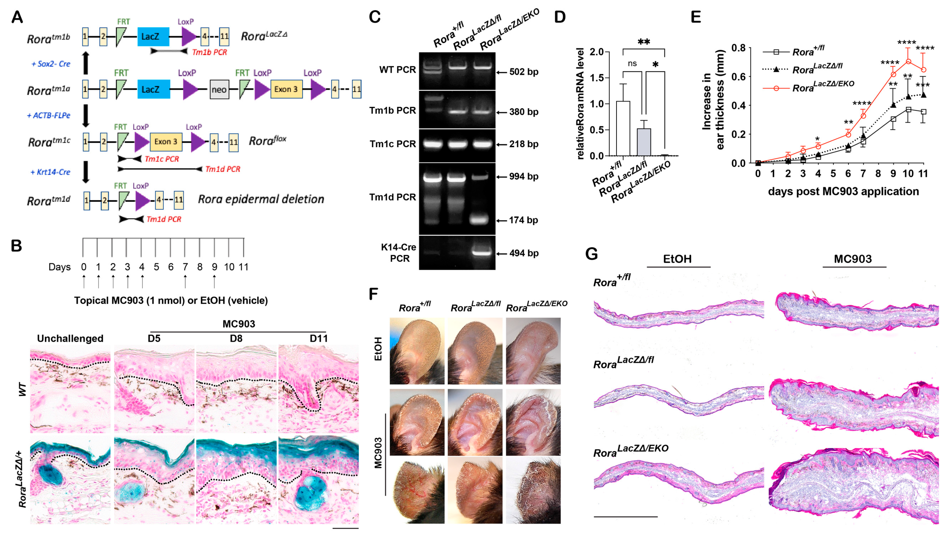
Figure 1. Epidermal Rora expression in response to MC903 and the effect of epidermal Rora deletion on MC903-induced ear swelling in RoraLacZΔ/EKO mice. (A) Scheme of the targeted strategies for generating Rora mutant strains from the Roratm1a/+ strain. (B) Top: scheme of the MC903 treatment regimen. Arrows indicate the days when both ears were topically treated with 1 nmol of MC903 or 100% ethanol (EtOH). Bottom: X-gal staining (blue) of frozen ear skin sections from wildtype (WT) or the RoraLacZΔ/+ reporter mice collected at the indicated time points after topical MC903 application. DNA was counterstained with Nuclear Fast Red (pink). Black dotted lines mark the dermal–epidermal junctures; scale bar = 100 μm. (C) Validation of epidermal Rora depletion in Rora LacZΔ/EKO mice using short-range end-point PCR assays. The PCR products were separated using a 2% agarose gel and stained with ethidium bromide. (D) Total RNA was isolated from the adult ear skin of each genotype, followed by RT-PCR analysis of Rora mRNA normalized to 18S rRNA. Values from MC903-treated ears were normalized to ethanol (EtOH)-treated control ears (set as 1) and presented as mean fold ± SD, n = 5/genotype. (E) Mouse ear thickness was measured at the indicated time points and plotted as the increase in ear thickness after subtracting the value of day 0; n = 8–10/genotype from 3 independent experiments. (F) Representative photos of mouse ears collected on day 11. (G) Representative images of the hematoxylin and eosin (H&E) staining on frozen sections of ear samples collected on day 11; scale bar = 1000 μm. *, p < 0.05, **, p < 0.01, ***, p < 0.001, ****, p < 0.0001, or ns (not significant), were determined through a one-way ANOVA (D) or a two-way ANOVA (E).
Ijms 24 10241 g001
Figure 2. Epidermal Rora deletion exaggerates MC903-induced ear swelling in RoraEKO mice. (A) Validation of epidermal Rora depletion in Rorafl/fl;K14-CRE (RoraEKO) mice using short-range end-point PCR assays. (B) Total RNA was isolated from the back skin epidermis or the liver of postnatal day 4 (P4) mice, followed by RT-PCR analysis of Rora mRNA, as described in Figure 1D. (C) Mouse ears were topically treated with EtOH or MC903, as described in Figure 1B. Mouse ear thickness was measured and plotted, as described in Figure 1E; n = 8/genotype from 3 independent experiments. (D) Representative photos of mouse ears collected on day 11. (E) Top two panels show representative H&E images of frozen ear sections collected on day 8. The insets show eosinophils (arrows) in the dermis. The lower panel shows the representative images of toluidine blue (TB) staining of the frozen ear sections. Individual mast cells with intensive purple–blue signals are denoted by red arrows; scale bar = 100 μm (top and bottom panels); scale bar = 25 μm (insets). (F) The number of mast cells/field of vision under 20× magnification was quantified in MC903-treated ears (day 8). The average value was obtained for each ear section from 9–10 fields. Values from RoraEKO mice were normalized to the Rorafl/fl group and presented as mean fold ± SD, n = 5–6 from 3 mice/group. ***, p < 0.001, ****, p < 0.0001, or ns (not significant), were determined by an unpaired t-test.
Ijms 24 10241 g002
Figure 3. Epidermal Rora deletion aggravates MC903-induced AD-like symptoms in RoraEKO mice. Ear samples were collected on day 8 post-treatment. (A) Representative images of ear sections stained with antibodies against keratin 6 (K6, green) + CD11c (red), keratin 10 (K10, green) + CD4 (red), and Loricrin (green) + F4/80 (red). DNA was counterstained with Hoechst (blue). Dotted lines mark the dermal–epidermal junctures; scale bar = 200 μm. (B) mRNA expression of the indicated inflammatory factors was measured using quantitative RT-PCR and normalized to 18S for each sample. Values were normalized to EtOH-treated Rorafl/fl ears and presented as mean fold ± SD, n ≥ 4/group. (C) Serum IgE levels of EtOH- or MC903-treated mice were measured using ELISA. *, p < 0.05, **, p < 0.01, ***, p < 0.001, or ****, p < 0.0001, was determined by one-way ANOVA; #, p < 0.05, ##, p < 0.01, or ns (not significant), indicate the difference between Rorafl/fl and RoraEKO mice within the MC903 treatment group.
Ijms 24 10241 g003
Figure 4. Epidermal loss of RORα accelerates MC903-induced dermal activation of dendritic cells and CD4+ T cells. Mouse ears were challenged with EtOH or MC903 and collected on day 2 or 5. (A,B) Representative immunostaining images with antibodies against CD11c (red) (A) or CD4 (red) (B); DNA was counterstained with Hoechst (blue); scale bar = 100 μm. Dotted lines mark the dermal–epidermal junctures. The number of CD11c or CD4-positive counts/field of vision under 20× magnification was quantified for 9–10 fields per ear section to obtain the average value. All values were normalized to an average value of the EtOH-treated Rorafl/fl group and presented as mean fold ± SD, n ≥ 3 for 3 mice/group. *, p < 0.05, **, p < 0.01, or ***, p < 0.001, were determined by two-way ANOVA.
Ijms 24 10241 g004
Figure 5. Epidermal Rora deletion on Tslp gene expression in the skin. (A) Tslp and Cyp24a1 mRNA expression in ear samples collected on day 2 after EtOH- or MC903-treatment was measured using RT-PCR analysis. Values were normalized to EtOH-treated control ears and presented as mean fold ± SD, n = 4/group. (B) Representative images of frozen ear sections stained with an antibody against TSLP (red); DNA was counterstained with Hoechst (blue); scale bar = 100 μm. Dotted lines mark the dermal–epidermal junctures. (C) Epidermal TSLP Fluorescence intensity was quantified using ImageJ. The average value for each skin section was obtained from ≥5 fields of vision under 10× magnification, normalized to the EtOH-treated Rorafl/fl group, and presented as mean ± SD, n = 5–6 from 3 mice/group. (D) TSLP protein levels of the skin lysates and the serum were measured using ELISA. (E) IL13 secreted by the ear-draining lymph nodes was measured using ELISA. Single-cell suspension was prepared from the ear skin-draining lymph nodes and re-stimulated with PMA/ionomycin for 24 h. The supernatant was collected for IL13 analysis. Values are presented as mean ± SD, n ≥ 4/group. *, p < 0.05, **, p < 0.01, ***, p < 0.001, or ****, p < 0.0001, was determined by one-way ANOVA; ##, p < 0.01, or ns (not significant), indicate the difference between Rorafl/fl and RoraEKO mice within each of the MC903 treatment groups.
Ijms 24 10241 g005
Figure 6. Epidermal loss of RORα deregulates keratinocyte differentiation and accelerates skin barrier disruption in response to MC903. (A) mRNA expression of indicated genes in the ear samples collected on day 2 after EtOH- or MC903-treatment; n = 4/group. (B) Representative images of H&E staining or immunostaining of frozen ear sections with antibodies against keratin 10 (K10, green), keratin 6 (K6, green), or loricrin (green). DNA was counterstained with Hoechst (blue); scale bar = 100 μm. Dotted lines mark the dermal–epidermal junctures. (C) Western blot analysis of indicated proteins in ear samples collected on day 2 after EtOH- or MC903-treatment. (D) The protein levels of indicated genes were quantified by densitometry scanning and normalized to b-tubulin. Values are presented as mean-fold over EtOH-treated Rorafl/fl control ± SD, n = 3–5/group. (E) Trans-epidermal water loss (TEWL) was measured on ear skin before and at different time points after MC903 application. *, p < 0.05, **, p < 0.01, ***, p < 0.001, or ****, p < 0.0001, was determined by one-way ANOVA (A) or two-way ANOVA (D,E); ##, p < 0.01, ###, p < 0.001, ####, p < 0.0001, or ns (not significant), indicate the difference between Rorafl/fl and RoraEKO mice within each of the MC903 treatment groups.
Ijms 24 10241 g006
Disclaimer/Publisher’s Note: The statements, opinions and data contained in all publications are solely those of the individual author(s) and contributor(s) and not of MDPI and/or the editor(s). MDPI and/or the editor(s) disclaim responsibility for any injury to people or property resulting from any ideas, methods, instructions or products referred to in the content.