Article

Characterization of Two Na+(K+, Li+)/H+ Antiporters from Natronorubrum daqingense

School of Life Science and Technology, Harbin Institute of Technology, Harbin 150080, China
*
Author to whom correspondence should be addressed.
Int. J. Mol. Sci. 2023, 24(13), 10786; https://doi.org/10.3390/ijms241310786
Submission received: 16 May 2023 / Revised: 25 June 2023 / Accepted: 25 June 2023 / Published: 28 June 2023

Abstract

:
The Na+/H+ antiporter NhaC family protein is a kind of Na+/H+ exchanger from the ion transporter (IT) superfamily, which has mainly been identified in the halophilic bacteria of Bacillus. However, little is known about the Na+/H+ antiporter NhaC family of proteins in the extremely halophilic archaea. In this study, two Na+/H+ antiporter genes, nhaC1 and nhaC2, were screened from the genome of Natronorubrum daqingense based on the gene library and complementation of salt-sensitive Escherichia coli KNabc. A clone vector pUC18 containing nhaC1 or nhaC2 could make KNabc tolerate 0.6 M/0.7 M NaCl or 30 mM/40 mM LiCl and a pH of up to 8.5/9.5, respectively. Functional analysis shows that the Na+(K+, Li+)/H+ antiport activities of NhaC1 and NhaC2 are both pH-dependent in the range of pH 7.0–10.0, and the optimal pH is 9.5. Phylogenetic analysis shows that both NhaC1 and NhaC2 belong to the Na+/H+ antiporter NhaC family of proteins and are significantly distant from the identified NhaC proteins from Bacillus. In summary, we have identified two Na+(K+, Li+)/H+ antiporters from N. daqingense.

1. Introduction

In environments with a high salt concentration, the damage of Na+ stress to organisms is mainly manifested as water loss, ion toxicity, and imbalance in osmotic pressure. In order to adapt to the toxicity, microorganisms have formed three major salt tolerance mechanisms: (1) microorganisms change the permeability of their cell membranes to adapt to high-salinity environments by changing the proportion of membrane lipid components in the cell membrane [1,2]; (2) microorganisms accumulate a high concentration of compatible substances within cells as osmotic regulators to maintain osmotic balance [3]; (3) microorganisms extrude Na+ from cells while accumulating KCl to maintain an osmotic balance within the cells [4]. These proteins are named Na+/H+ exchangers (NHEs) [5], can extrude Na+, and can be divided into primary Na+ pumps [6] and secondary Na+/H+ pumps [7]. The primary Na+ pumps mainly include four types [8]: Na+/K+-ATPase [9,10], decarboxylase [11,12], NADH-ubiquinone oxidoreductase [13,14], and N5-methyltetrahydromethanopterin: coenzyme M methyltransferase [15,16,17]. The secondary Na+/H+ pumps are the main Na+ efflux system in halophilic bacteria, namely, the Na+/H+ antiporter, and are the main adaptive response system of halophilic bacteria to Na+ stress [18]. At present, the reported Na+/H+ antiporters can be divided into cation/proton antiporters (CPAs) superfamilies [5], IT superfamilies [19,20], and Na+-translocating multiple resistance and pH transporter (Mrp) superfamilies (TC identifier: 2.A.63.1.4) [21]. NHEs have also been widely reported in plants [22] and animals [23].
The NhaC family protein is a Na+/H+ exchanger belong to the IT superfamily, mainly identified in Bacillus. As early as 1997, a Na+/H+ antiporter encoded by nhaC was found, and the gene sequence from Alkaliphilic Bacillus firmus OF4 was predicted to encode a membrane protein with 462 residues and 12 transmembrane segments (TMSs). Bf-NhaC was highly homologous to the unknown function products encoded by homologous genes from Bacillus subtilis and Haemophilus influenzae [24]. The g1-nhaC isolated from Bacillus sp. G1 could restore the growth of E. coli BL21 (DE3) when grown in a medium containing 0.2–2.0 M NaCl, and the Na+ efflux activity of g1-NhaC was detected in the pH range 8.0–10.0 [25]. Haja D. K. et al. [26] also reported gene sequences that could encode NhaC proteins from the hyperthermophilic archaeon Pyrococcus furiosus.
In our previous study, an extremely halophilic archaeon was isolated from the salinized soil of Daqing City, Heilongjiang Province, China [27]. The strain JX313T was once named Haloterrigena daqingense until de la Haba R. R. et al. [28] reclassified it into the genus Natronorubrum, and it was renamed N. daqingense. N. daqingense has extremely strong salt tolerance, and its growth salinity ranges from 10% to saturated (w/v) (the optimum growth salinity is 17.5%), with a pH range 8.0–11.0 (the optimum pH is 10.0) [27]. Therefore, N. daqingense has great potential to screen genes that can encode Na+/H+ exchangers. In 2022, Wang S. et al. [29] disclosed more detailed data when analyzing the whole genome sequencing of Natronorubrum daqingense, which will also be conducive to the identification of Na+/H+ antiporters. In this study, the salt-sensitive E. coli KNabc (E. coli with the knockout of three major Na+/H+ antiporter genes: nhaA, nhaB, and chaA) was used to screen Na+/H+ antiporter genes from the genome of N. daqingense. Two NhaC-type Na+/H+ antiporter genes were found, which allow E. coli Knabc to tolerate 0.6 M/0.7 M NaCl or 30 mM/40 mM LiCl, respectively, with a maximum resistance of pH 8.5/9.5. In summary, we cloned nhaC1 and nhaC2 from the N. daqingense genome, expressed and functionally validated their encoded proteins, and finally identified two novel NhaC-type Na+/H+ antiporters from extremely halophilic archaea.

2. Results

2.1. Cloning and Sequence Analysis of Na+(K+, Li+)/H+ Antiporter Genes

To screen for genes that exert an Na+/H+ antiport function from N. daqingense JX313T, the genomic library and the salt-sensitive E. coli KNabc functional complementary method [30] were combined to select positive clones that could enable KNabc to restore the growth in the medium containing 0.2 M NaCl. Positive clones were grouped based on plasmid electrophoresis and double enzyme digestion results, and two different positive clones [pUC1028-1,9 (pUC18 carrying a 3.5 kb or 4.9 kb fragment)] were obtained, as shown in Figure 1A,B, which enabled E. coli KNabc to grow on the LBK medium under the intolerable 0.2 M NaCl concentration.
Sequence analysis revealed that: (1) a pUC1028-1 DNA fragment (3.5 kb) containing two 5′end-truncated ORFs (ORF1, 2) and two complete ORFs (ORF3, 4), of which only ORF3 (1701 bp) contained the promoter-predicted sequence and Shine-Dalgarno (SD) sequence; (2) a pUC1028-9 DNA fragment (4.9 kb) containing one 5′end-truncated ORF (ORF1) and three complete ORFs (ORF2, 3, 4), with only ORF2 (1575 bp) containing the promoter-predicted sequence and SD sequence.
In order to screen out ORFs that exert a Na+/H+ antiport function from positive clones, complete ORFs containing predicted promoter-like sequences were subcloned into pUC18, and complete ORFs without a predicted promoter-like sequence were subcloned into pET28AK (pET28(a) inserted ampicillin resistance gene at BglII;, as shown in Figure 1C). During the subcloning process, the transformed mixture was coated on the LBK medium (containing 50 mg∙mL−1 of ampicillin) plates to obtain positive subclones. Subclones were lined on the LBK medium (containing 0.2 M NaCl and 50 mg∙mL−1 of ampicillin) plates to screen for the ORF exerts the Na+/H+ antiport function, as shown in Figure 1D,E.
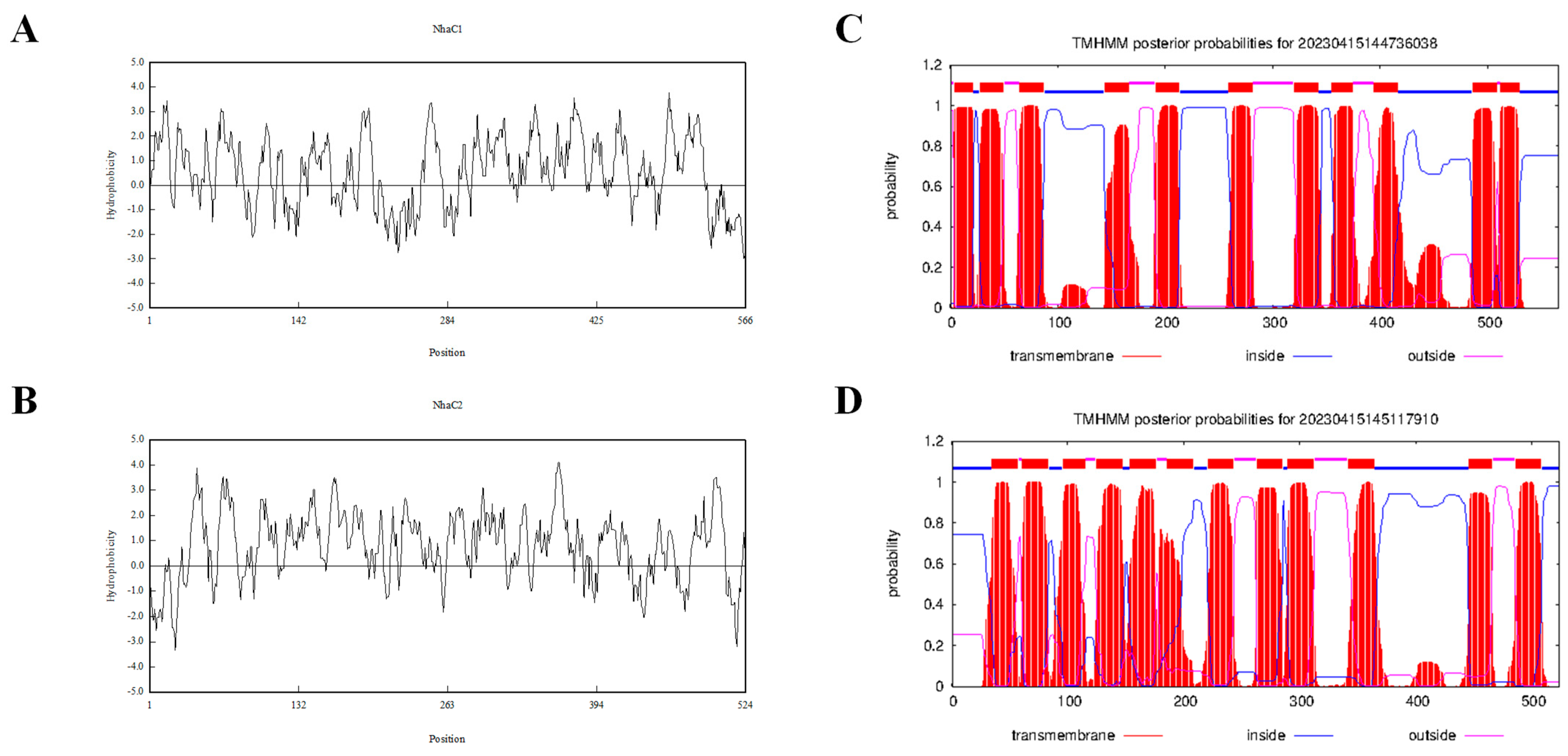
As shown in Figure 1D,E, three ORFs could enable KNabc to restore salt tolerance function, that is, two ORFs exhibit Na+/H+ antiport activity. The gene names of 1028-1-ORF3 and 1028-9-ORF2 will be abbreviated as nhaC1 and nhaC2, and the proteins they may encode will be abbreviated as NhaC1 and NhaC2 in subsequent studies. The characteristics of the hypothetical proteins corresponding to the identified ORFs are listed in Table 1. The protein encoded by 1028-1-ORF3 is predicted to be a membrane protein composed of eleven transmembrane segments (TMSs) with a calculated molecular weight of 60.7 kD. A total of 302 out of 566 amino acids that make up this protein are hydrophobic, resulting in a lower polarity of the entire protein sequence. As a predicted membrane protein, its TMSs include TMS I (4–21), TMS II (28–50), TMS III (65–87), TMS IV (144–166), TMS V (191–213), TMS VI (259–281), TMS VII (320–342), TMS VIII (355–374), TMS IX (399–416), TMS X (486–508), and TMS XI (512–529) (Figure 2C). Another ORF capable of restoring the salt tolerance function of KNabc is 1028-9-ORF2, which is predicted to be a membrane protein with twelve TMSs (Figure 2D), including TMS I (35–57), TMS II (61–83), TMS III (96–115), TMS IV (125–147), TMS X (154–176), TMS VI (186–208), TMS VII (221–243), TMS VIII (263–285), TMS IX (290–312), TMS X (342–364), TMS XI (446–466), and TMS XII (486–508). The translation of the DNA sequence into the amino acid sequence revealed that 301 of the 524 amino acids are hydrophobic, with a calculated molecular weight of 54,785.66 Dalton, resulting in a low polarity of the entire sequence. Based on the characteristics of the Na+/H+ antiporter, the above two ORFs are likely to possess Na+/H+ antiport activity [31,32,33,34,35]. The protein encoded by 1028-7-ORF2 could also restore the growth of salt-sensitive E. coli KNabc. However, our identification results in subsequent studies showed that it belongs to different types of Na+/H+ antiporters from the two ORFs in this article.

2.2. Phylogenetic Analysis Based on the Neighbour-Joining Algorithm

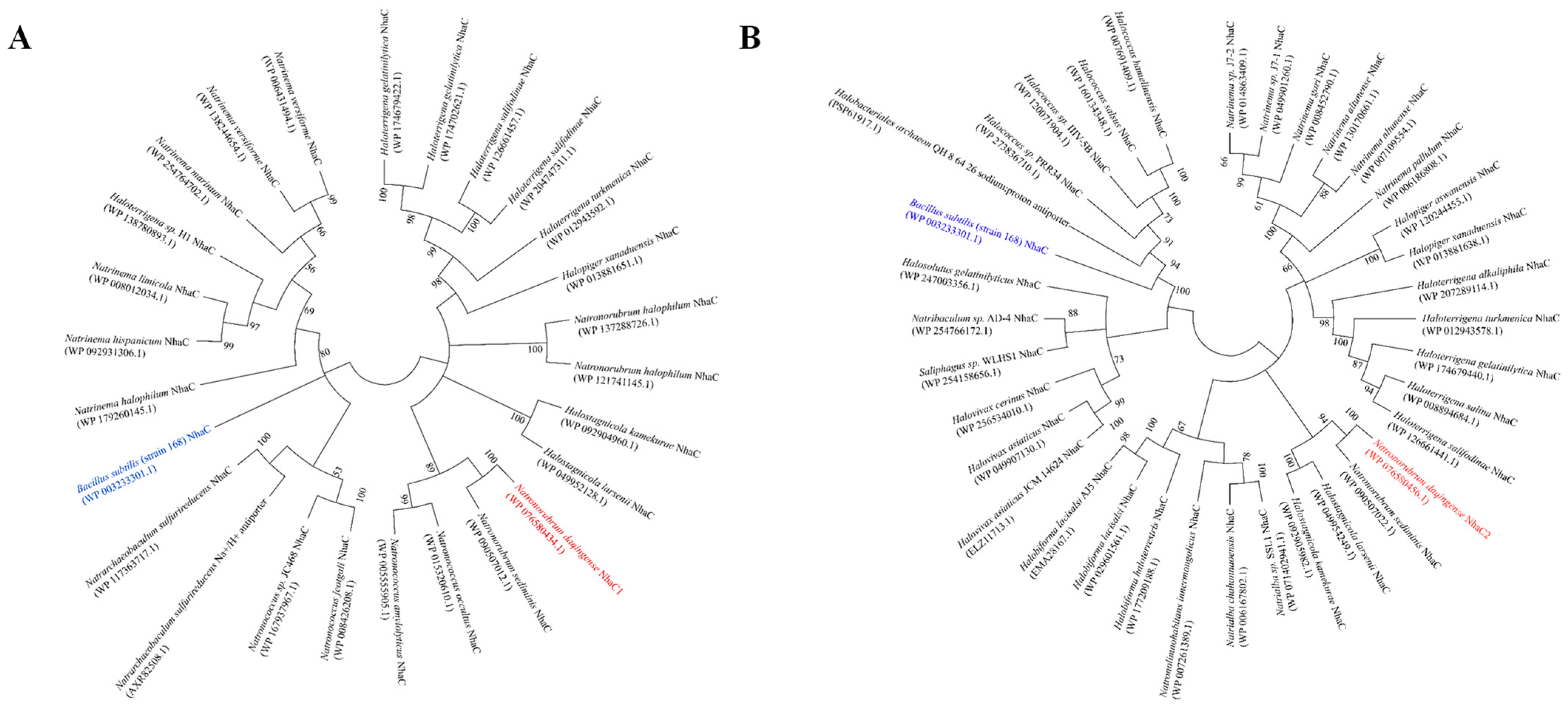
According to the BlastX comparison results on NCBI [36], both 1028-1-ORF3 and 1028-9-ORF2 were assumed to be Na+/H+ antiporter NhaC family proteins based on the genome sequencing results; two sequences showed 100% similarity to the published gene sequences BB347_08225 and BB347_08285, respectively, and 1028-1-ORF3 and 1028-9-ORF2 were speculated to be proteins of the Na+/H+ antiporter NhaC family. To verify this hypothesis and their evolutionary relationships with the identified/predicted proteins with Na+/H+ antiport activity, a phylogenetic analysis was conducted based on the neighbour-joining algorithm, as shown in Figure 3. Two ORFs were translated into amino acid sequences and then compared using BlastP to select homologs on NCBI [36].
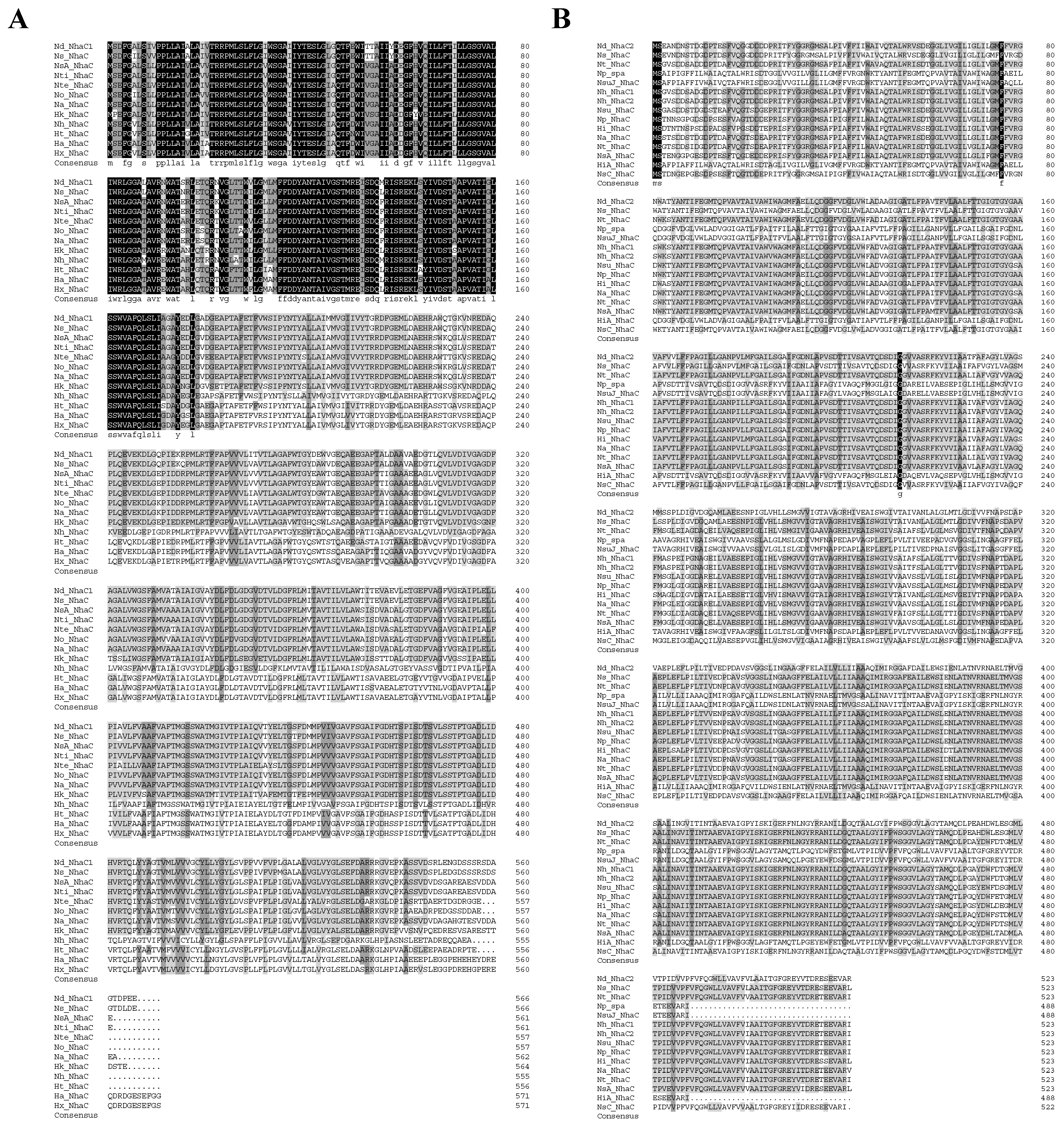
1028-1-ORF3 and 1028-9-ORF2 were also aligned with their 11 and 14 sequences producing significant alignments with a percent identity from 80.43% to 96.47% and from 85.11% to 98.28%, respectively, and the similarities and differences between ORFs and their homologs in the composition of amino acid sequences are as shown in Figure 4A,B. 1028-1-ORF3 has the respective identities of from 87% to 100% with closely related homologs from Natronorubrum sediminis, Natronococcus occultus, and Natronococcus amylolyticus, and the Na+/H+ antiporter NhaC family protein of Natronorubrum sediminis shares the bootstap of 100 with 1028-1-ORF3, which means they are sisterly in phylogeny. 1028-9-ORF2 has the respective identities of from 55% to 100% with 21 homologs from Haloterrigena, Halopiger, Natrinema, Natronorubrum, Halostagnicola, Euryarchaeota, Halakalicoccus, and Halovivax. The Na+/H+ antiporter NhaC family protein of Natronorubrum sediminis shares the bootstrap of 100 with 1028-1-ORF3, which means they are sisterly in phylogeny. 1028-1-ORF3 clustered with some homologs; the 1028-9-ORF2 clustered with all homologs belonging to the Na+/H+ antiporter NhaC family. 1028-1-ORF3 is relatively close to the identified protein in terms of Na+/H+ antiport activity, but both 1028-1-ORF3 and 1028-9-ORF2 are significantly distant from it.
According to sequence alignment and phylogenetic analysis, the proteins encoded by 1028-1-ORF3 and 1028-9-ORF2 were Na+/H+ antiporter NhaC family proteins.

2.3. Test for Salt Tolerance and Alkaline pH Resistance

In order to test the salt tolerance of E. coli KNabc containing nhaC1 or nhaC2, growth tests were performed in LBK medium (containing 50 mg∙mL−1 of ampicillin, pH 7.5) with varying concentrations of NaCl (0–0.8 M) or LiCl (0–50 mM), respectively (KNabc containing pUC18 as a negative control). As shown in Figure 5A,B, KNabc/nhaC1 could grow up to the presence of 0.6 M NaCl or 30 mM LiCl, and KNabc/nhaC2 could grow up to the presence of 0.7 M NaCl or 40 mM LiCl, but KNabc/pUC18 could not grow in the presence of 0.2 M NaCl or 5 mM LiCl. KNabc/nhaC1, KNabc/nhaC2, and KNabc/pUC18 were cultured in an LBK medium (containing 50 mM NaCl, pH 7.0–10.0) to analyze the alkaline pH resistance of nhaC1 and nhaC2. With the gradual increase in pH, the growth of all groups was inhibited to varying degrees, as shown in Figure 5C. Compared with KNabc/pUC18, which was significantly inhibited and almost unable to grow at pH 8.0, both KNabc/nhaC1 and KNabc/nhaC2 exhibited strong alkaline pH resistance. Although KNabc/nhaC1 could still grow at pH 8.5, nhaC2 could confer higher alkaline pH resistance on KNabc, resisting up to pH 9.5.

2.4. SDS-PAGE and Western Blot Analysis of NhaC1 and NhaC2

In order to study the protein localization and function of NhaC1 and NhaC2, two genes were respectively inserted into pET28AK containing a T7 promoter using homologous recombination for protein expression, and the inserted gene sequence would be co-expressed with the 6×His tag. To determine the approximate position of the protein NhaC1 and NhaC2’s SDS-PAGE bands, the everted membrane vesicles from E. coli KNabc containing recombinant plasmids were purified by 6×His tag affinity chromatography and used for SDS-PAGE. Combined with the prediction of protein molecular weights in the UniProt database and the peptide molecular weight after translation sites on the pET28AK, the theoretical molecular weights of NhaC1 fusion 6×His protein and NhaC2 fusion 6×His protein should be 64 kD and 58 kD, respectively. As shown in Figure 6A,B, the molecular weights of the fusion proteins after SDS-PAGE were approximately 64 kD and 58 kD, respectively.
Total protein, cytoplasmic protein, and membrane protein were extracted from E. coli KNabc containing recombinant plasmids (KNabc containing pET28AK was used as negative control) and used for the Western blot analysis. As shown in Figure 6C,D, both NhaC1 and NhaC2 were only detected from total protein and membrane protein. Combined with previous bioinformatics predictions, it was proven that both of them were located on membranes in cells and were membrane proteins.

2.5. Detection of Na+(K+, Li+)/H+ Antiport Activity

The Na+(K+, Li+)/H+ antiport activities were detected by adding D-lactic acid to the reaction buffer A (pH 7.0–10.0) containing everted membrane vesicles to quench fluorescence (everted membrane vesicles prepared by KNabc/pET28AK-nhaC1 and KNabc/pET28AK-nhaC2 as treatment groups, everted membrane vesicles prepared by KNabc/pET28AK as negative control), and then measuring the ratio of dequenching fluorescence after adding various concentrations of NaCl, KCl (Na free), and LiCl. As shown in Figure 7A, Na+(K+, Li+)/H+ antiport activities were detected in everted membrane vesicles from KNabc containing pET28AK/nhaC1 and pET28AK/nhaC2 when pH 7.5, while no Na+(K+, Li+)/H+ antiport activity was detected in everted membrane vesicles from E. coli KNabc containing pET28AK. NhaC1 exhibited Na+(K+, Li+)/H+ antiport activity when pH ranged from 7.0 to 10.0, with optimal antiport activities at pH 9.5. NhaC2 also exhibited Na+(K+, Li+)/H+ antiport activity when pH ranged from 7.0 to 10.0; notably, the Li+/H+ antiport activity of NhaC2 was lower than that of NhaC1 most of the time, but its Li+/H+ antiport activity reached its maximum, and was higher than that of NhaC1, at pH 9.5.

2.6. Calculation of K0.5 Values for Monovalent Cations

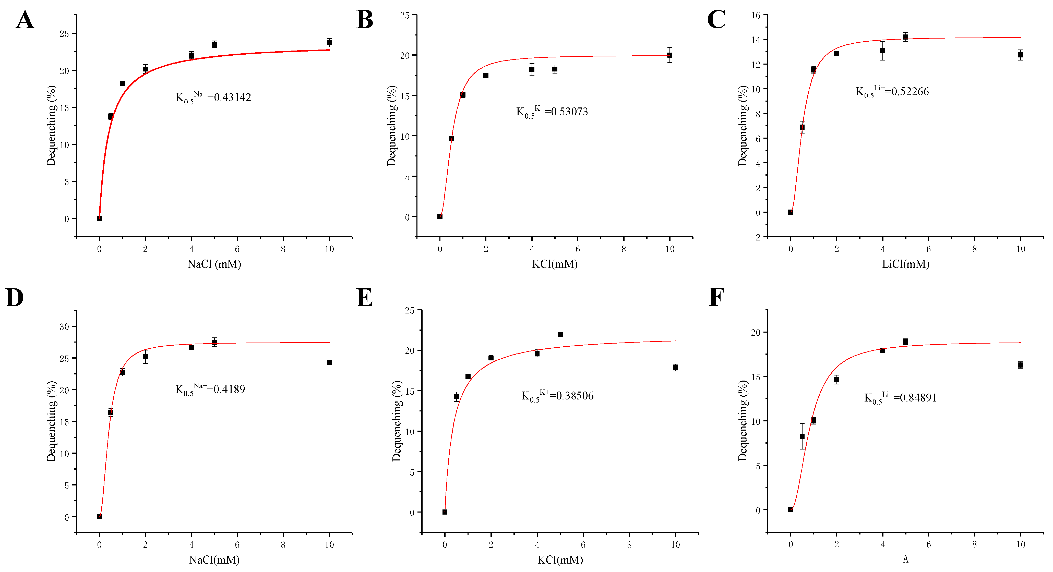
To evaluate the substrate affinity activity of NhaC1 and NhaC2 to monovalent cations (Na+, K+ and Li+), the fluorescence-dequenching rates of the everted membrane vesicles were measured at pH 9.5 after adding NaCl, Na-free KCl, or LiCl (final concentration 0.5–10 mM), respectively. According to the data, nonlinear regression analysis was carried out to determine the concentration of monovalent cations that were added when reaching the half-maximum deactivation rate to calculate the value of K0.5. After analysis and calculation, it was found that the K0.5 values of NhaC1 for Na+, K+, and Li+ were 0.43 mM (Figure 8A), 0.53 mM (Figure 8B), and 0.52 mM (Figure 8C), respectively, indicating that the substrate affinity activity for monovalent cations was Na+ > Li+ > K+. Additionally, the K0.5 values of NhaC2 for Na+, K+, and Li+ were 0.42 mM (Figure 8D), 0.39 mM (Figure 8E), and 0.85 mM (Figure 8F), indicating that the substrate affinity activity for monovalent cations was K+ > Na+ > Li+.

3. Discussion

In this study, Na+(K+, Li+)/H+ antiporter NhaC1 and NhaC2 were identified from N. daqingense, an extremely halophilic archaeonisolated from Daqing (China) saline-alkali soil, which could grow under a range of NaCl concentrations in 10% saturated solution (w/v) [27]. Both NhaC1 and NhaC2 belong to the Na+/H+ antiporter NhaC family protein and are closely related to Na+/H+ antiporter NhaC family proteins in some halophilic archaea; most of the proteins in this family were identified from halophilic bacteria such as Bacillus and verified to have salt tolerance and pH resistance abilities [24,25]. It is interesting that ArcD, an archaebacterial arginine/ornithine antiporter from Halobacterium salinarum, was also phylogenetically classified at the edge of Na+/H+ antiporter NhaC family proteins [37].
NhaC1 and NhaC2 were both predicted to be membrane proteins consisting of 11 and 12 TMSs, respectively (Figure 2C,D), and demonstrated to be localized on the cell membrane of heterogenous host E. coli KNabc according to the Western blot results (Figure 6C,D). Both nhaC1 and nhaC2 could restore the growth of KNabc in an LBK medium containing 0.2 M NaCl and could be tolerant of higher concentrations of Na+ and Li+ in the medium, exhibiting stronger pH resistance (Figure 5); these are in line with screening patterns for the Na+/H+ antiporter complementing of salt-sensitive strain of KNabc [38]. The detection of Na+(Li+)/H+ antiport activities (Figure 7) found that the transportation activities of NhaC1 and NhaC2 would change with changes in pH; it is inferred that their function of transport is pH-dependent. It has been reported that using this method can also identify genes with K+/H+ antiport function [39] based on the results, both NhaC1 and NhaC2 are inferred to be pH-dependent Na+(K+, Li+)/H+ antiporters.
According to available reports [39,40] and TCDB [41], the Na+/H+ antiporter NhaC (TC identifier: 2.A.35) family protein is a Na+/H+ exchanger from the IT superfamily, which is characterized by the substrate of the transporter, which should be a charged organic or inorganic chemical species (cations or anions) [19]. Most NhaC family proteins were identified in bacteria [24,25], such as Bacillus firmus, Bacillus subtilis, Haemophilus influenziae, and Vibrio cholerae. Notably, genes encoding NhaC proteins have been identified in hyperthermophilic archaea Pyrococcus furiosus [26]. Ito et al. [24] identified a NhaC (462 amino acids) containing 12 TMSs from B. firmus and detected the Na+/H+ antiport activity of everted membrane vesicles. The main function of the NhaC family protein is to extrude intracellular Na+ or Li+ out of cells [24]. For NhaC from Bacillus sp. G1, research has shown that NhaC is a Na+ extrusion channel at pH 7.5, and ensures pH homeostasis in low Na+ environments, reflecting the electrogenic character of the Na+/H+ antiporter [25].
In this study, we identified two proteins with Na+(K+, Li+)/H+ antiport activity from N. daqingense for the first time; both NhaC1 and NhaC2 belong to the Na+/H+ antiporter NhaC family of proteins, enriching the research on NhaC-type Na+/H+ exchangers in extremely halophilic archaea. Therefore, we plan to complete the identification of this protein in future research. We also plan to construct N. daqingense mutants with nhaC1 deletion, nhaC2 deletion, and nhaC1-nhaC2 co-deletion by using homologous recombination to clarify the role of two NhaC family proteins in the salt tolerance and pH resistance of N. daqingense and clarify their mechanisms.

4. Materials and Methods

4.1. Strains, Plasmids, and Growth Conditions

The strains and plasmids employed in this study are listed in Table 2. N. daqingense JX313T was cultured in 17.5% NaCl (optimum) Luria Bertani (LB) medium containing 10 g/L tryptone, 5 g/L yeast extract, 1.5% glucose (autoclaving alone), 1.23 g/L MgSO4∙7H2O added to 175 g/L NaCl at 35 °C and pH 9.5. E. coli KNabc, the salt-sensitive strain, and its transformant cells were grown aerobically in the LBK medium, that is, the LB medium with 6.49 g/L Na-free KCl instead of NaCl, as previously described by Karpel et al. [38]. The antibiotic concentrations used for screening positive clones were ampicillin 50 μg∙mL−1 and kanamycin 50 μg∙mL−1. The preparation of electrotransformed competent cells of KNabc required the cultivation of KNabc in LBK medium at 37 °C overnight. In order to test the salt tolerance ability of positive clones, 1% of different KNabc/recombinant plasmid overnight cultures (OD600 was adjusted to 0.8 before inoculation) were inoculated into LBK medium (containing 50 mg∙mL−1 of ampicillin, pH 7.5) with varying concentrations of NaCl (0–0.8 M) or LiCl (0–50 mM), respectively, and incubated overnight at 37 °C. To determine the pH profile of positive clones, 1% of different KNabc/recombinant plasmid overnight cultures were inoculated into LBK medium (containing 50 mg∙mL−1 of ampicillin) with varying pH ranges from 7.0 to 10.0 (adjust by adding 100 mM Hepes–Tris buffer), respectively, and incubated overnight at 37 °C. A UV spectrophotometer was used to measure the OD600 value of overnight (16 h) cultures to reflect the growth of recombinant strains. The preparation and electro-conversion method of KNabc electrotransformed competent cells was as described by Jiang et al. [42].

4.2. Screening of Na+(K+, Li+)/H+ Antiporter Genes

The genomic DNA of N. daqingense JX313T was partially digested with the restriction enzyme Sau3AI, and the amount of Sau3AI was adjusted to concentrate the size of genomic cleavage products between 2–10 kb. Genomic cleavage products prepared after recovery by agarose gel electrophoresis were inserted into the pUC18 (purified after BamHI digestion and dephosphorylation) cloning vector using T4 DNA ligase. The ligation products were transferred into prepared KNabc-competent cells through electro-conversion, and positive clones were selected with LBK medium containing 0.2 M NaCl, 2% Agar, and 50 mg∙mL−1 of ampicillin. By using homologous recombination, each ORF and its predicted promoter and SD sequence from positive colonies were respectively ligated to the pUC18 vector; ORFs with no predicted promoter or a low promoter score were ligated into a pET28(a) vector containing T7 promoters. Examples of the above subcloning process are shown in Figure 1C.

4.3. Preparation of Everted Membrane Vesicles

The KNabc containing pUC-positive fragments or pUC18 (as negative control) were inoculated into 100 mL LBK medium (containing 0.2 M NaCl, and 50 mg∙mL−1 of ampicillin) and LBK medium (containing 50 mg∙mL−1 of ampicillin) at 1% inoculum, respectively, then cultured to the middle of the logarithmic growth stage. These were centrifuged at 5000× g, 4 °C for 10 min to collect precipitation, and the supernatant was discarded. After rinsing twice with 10 mL buffer A (140 mM choline chloride; 10 mM Tris; 10% of glycerol; pH 7.5, 1 mM PMSF was added before use; storage at 4 °C), they were suspended and the cell wall was disrupted using a pre-cooled Ultrasonic Cell Disruption System (cycles: 3 s ON followed by 2 s OFF) until the bacterial solution became less turbid. They were centrifuged at 8000× g, 4 °C for 10 min, then the supernatant was transferred and ultracentrifuged at 100,000× g, 4 °C for 1 h. The supernatant was discarded, and the precipitation was dissolved using the appropriate amount of buffer A, stored at −80 °C.

4.4. Preparation and Purification of Proteins

Each ORF that can cause the KNabc to regrow on the LBK medium (containing 0.2 M NaCl) plates was ligated into pET28AK to co-express with the 6×His tag. E. coli KNabc containing pET-positive fragments or pET28AK (as negative control) were inoculated into 100 mL LBK medium (containing 0.2 M NaCl, 50 mg∙mL−1 of ampicillin) and LBK medium (containing 50 mg∙mL−1 of ampicillin) at 1% inoculum, respectively, and cultured at 37 °C. IPTG (final concentration: 1 mM) solution was added when the value of OD600 was between 0.6–0.8, and induced the protein expression at 22 °C for 12 h. The total protein, membrane protein, and cytoplasmic protein were prepared by kit from Bestbio (BB-3182&BB-31516). The total protein and cytoplasmic protein were packaged for Western blot analysis, and the rest were stored at −80 °C. The membrane proteins obtained in the previous step were first filtered by 0.22 μm filter membrane, and then affinity chromatography with Ni-NTA using the AKTA protein purifier. The buffer formula involved in the protein purification process is shown in Table 3. The target protein was eluted with 300 mM imidazole, flow velocity: 0.5 mL/min. The target proteins collected by affinity chromatography were transferred into ultrafiltration tubes and centrifuged at 5000× g, 4 °C for 20–60 min (until the volume was about 600 μL). The proteins were carefully collected from ultrafiltration and packaged for SDS-PAGE and Western blot analysis.

4.5. SDS-PAGE and Western Blot

KNabc/pET28AK-nhaC1 and KNabc/pET28AK-nhaC2 were grown in LBK medium containing 0.2 M NaCl and 50 mg∙mL−1 of ampicillin; KNabc/pET28AK was grown in LBK medium containing 50 mg∙mL−1 of ampicillin as the negative control, and cultured to OD600 0.6–0.8 at 37 °C. Isopropyl-β-D-thiogalactoside (IPTG) was added (final concentration 1 mM) to induce the expression of proteins for 12 h at 22 °C, then used for the preparation and purification of proteins. The extracted protein content was quantified using a Bradford Kit from Bestbio (BB-3411). The methods of SDS-PAGE and Western blot refer to Green et al. [43]. Total protein, cytoplasmic protein, and membrane protein of E. coli KNabc (containing pET28AK-positive fragments) and E. coli KNabc (containing pET28AK as negative control) were used as samples for detection and localization, respectively. The detection of 6×His tag was carried out using Beyotime polyclonal mouse anti-6×His tag antibody and Beyotime HRP-labeled goat anti-mouse IgG(H+L).

4.6. Detection of Na+(K+, Li+)/H+ Antiport Activity

The Na+(K+, Li+)/H+ antiport activity was estimated based on the collapse of transmembrane proton gradients, and acridine orange was selected as the fluorescent indicator according to Rosen’s method [44]. A 2 mL buffer D (10 mM Bis-Tris propane; 140 mM choline chloride; 5 mM MgSO4; pH from 7.0 to 10.0), 1 μL acridine orange (1 mM), and 40 μg everted membrane vesicles were added into the quartz colorimetric dish, determined after numerical stabilization by fluorescence spectrophotometer with excitation at 490 nm (10 mm slit) and emission at 530 nm (10 mm slit). D-lactic acid (final concentration 5 mM) was added into the mixture to quench the fluorescence; then, various concentrations of NaCl, KCl (Na free), or LiCl were added to dequench the fluorescence, determined after numerical stability. The Na+(K+, Li+)/H+ antiport activity was represented by the percentage of fluorescence-dequenching value to total fluorescence quenching value.

4.7. Calculation of pH Profile and K0.5 Values for Monovalent Cations

The Na+(K+, Li+)/H+ antiport activity of different everted membrane vesicles in pH 7.0–10.0 were determined by fluorescence spectrophotometer with NaCl, KCl (Na free) and LiCl at a concentration of 5 mM. Under the optimum pH, equal amounts of NaCl, KCl (Na-free), or LiCl were added with various concentrations (0.5–10 mM) to measure the Na+(K+, Li+)/H+ antiport activity of different everted membrane vesicles. With ion concentration as the abscissa and fluorescence dequenching rate as the ordinate, origin2017 was used for nonlinear regression analysis to calculate the K0.5 value.

4.8. DNA Manipulation and Bioinformatics Analyses

The extraction of genomic DNA was carried out using a FastPure Bacteria DNA Isolation Kit (Vazyme DC103). The genomic library screening method was described by Green et al. [44]. Preparation of plasmid DNA was carried out using TIANprep Mini Plasmid Kit (Tiangen DP103). Homologous recombination of DNA was done by CloneExpress Ultra One Step Cloning Kit (Vazyme C115-01). The primers used in this study are listed in Table 4. DNA sequencing was performed by RuiBiotech Institute (Beijing, China). The analyses for ORF and constructs of plasmid model diagrams were carried out with SnapGene5.2. Protein and DNA sequence alignment was performed through the National Center for Biotechnology Information using the website https://blast.ncbi.nlm.nih.gov/Blast.cgi (accessed on 1 November 2022), and the accessions of homologs of NhaC1 and NhaC2 to NhaC family proteins used for analysis were listed in Table 5. The phylogenetic tree was constructed via MEGA 11.0 using the neighbour-joining method [45]. Prediction of promoter was performed using the website http://genomes.urv.es/OPTIMIZER/ (accessed on 1 November 2022). The analyses of hydrophobicity and transmembrane prediction conducted using the online analysis tools of DetaiBio’s website http://www.detaibio.com/tools/ (accessed on 15 April 2023).

Author Contributions

Q.W. and J.S. conceived and designed the experiments. J.S. contributed to the reagents and materials. Q.W. conducted all experiments. Q.W. prepared the draft of this manuscript. M.Q. revised the article and checked the figures. All authors have read and agreed to the published version of the manuscript.

Funding

This research was funded by Heilongjiang Tounyan Team, grant number HITTY-20190034.

Institutional Review Board Statement

Not applicable.

Informed Consent Statement

Not applicable.

Data Availability Statement

All data are contained within this manuscript.

Conflicts of Interest

The authors declare that they have no competing interest.

References

  1. Chong, P.L.; Chang, A.; Yu, A.; Mammedova, A. Vesicular and Planar Membranes of Archaea Lipids: Unusual Physical Properties and Biomedical Applications. Int. J. Mol. Sci. 2022, 23, 7616. [Google Scholar] [CrossRef] [PubMed]
  2. Rao, A.; de Kok, N.A.W.; Driessen, A.J.M. Membrane Adaptations and Cellular Responses of Sulfolobus acidocaldarius to the Allylamine Terbinafine. Int. J. Mol. Sci. 2023, 24, 7328. [Google Scholar] [CrossRef] [PubMed]
  3. Margesin, R.; Schinner, F. Potential of halotolerant and halophilic microorganisms for biotechnology. Extremophiles 2001, 5, 73–83. [Google Scholar] [CrossRef]
  4. Oren, A. Diversity of halophilic microorganisms: Environments, phylogeny, physiology, and applications. J. Ind. Microbiol. Biotechnol. 2002, 28, 56–63. [Google Scholar] [CrossRef]
  5. Brett, C.L.; Donowitz, M.; Rao, R. Evolutionary origins of eukaryotic sodium/proton exchangers. Am. J. Physiol. 2005, 288, C223–C239. [Google Scholar] [CrossRef] [PubMed] [Green Version]
  6. Dimroth, P. Na(+)-coupled alternative to H(+)-coupled primary transport systems in bacteria. Bioessays 1991, 13, 463–468. [Google Scholar] [CrossRef]
  7. Krulwich, T.A.; Guffanti, A.A. The Na+ cycle of extreme alkalophiles: A secondary Na+/H+ antiporter and Na+/solute symporters. J. Bioenerg. Biomembr. 1989, 21, 663–677. [Google Scholar] [CrossRef]
  8. Patiño-Ruiz, M.; Ganea, C.; Călinescu, O. Prokaryotic Na+/H+ Exchangers-Transport Mechanism and Essential Residues. Int. J. Mol. Sci. 2022, 23, 9156. [Google Scholar] [CrossRef]
  9. Kumari, J.; Rathore, M.S. Na+/K+-ATPase a Primary Membrane Transporter: An Overview and Recent Advances with Special Reference to Alage. J. Membr. Biol. 2020, 253, 191–204. [Google Scholar] [CrossRef]
  10. Bakker, E.P.; Borchard, A.; Michels, M.; Altendorf, K.; Siebers, A. High-affinity potassium uptake system in Bacillus acidocaldarius showing immunological cross-reactivity with the Kdp system from Escherichia coli. J. Bacteriol. 1987, 169, 4342–4348. [Google Scholar] [CrossRef] [Green Version]
  11. Woehlke, G.; Dimroth, P. Anaerobic growth of Salmonella typhimurium on L(+)- and D(-)-tartrate involves an oxaloacetate decarboxylase Na+ pump. Arch. Microbiol. 1994, 162, 233–237. [Google Scholar] [CrossRef]
  12. Wifling, K.; Dimroth, P. Isolation and characterization of oxaloacetate decarboxylase of Salmonella typhimurium, a sodium ion pump. Arch. Microbiol. 1989, 152, 584–588. [Google Scholar] [CrossRef]
  13. Tokuda, H.; Unemoto, T. A respiration-dependent primary sodium extrusion system functioning at alkaline pH in the marine bacterium Vibrio alginolyticus. Biochem. Biophys. Res. Commun. 1981, 102, 265–271. [Google Scholar] [CrossRef]
  14. Tokuda, H.; Unemoto, T. Growth of a marine Vibrio alginolyticus and moderately halophilic V. costicola becomes uncoupler resistant when the respiration-dependent Na+ pump functions. J. Bacteriol. 1983, 156, 636–643. [Google Scholar] [CrossRef] [PubMed] [Green Version]
  15. Gärtner, P.; Weiss, D.S.; Harms, U.; Thauer, R.K. N5-methyltetrahydromethanopterin:coenzyme M methyltransferase from Methanobacterium thermoautotrophicum. Catalytic mechanism and sodium ion dependence. Eur. J. Biochem. 1994, 226, 465–472. [Google Scholar] [CrossRef]
  16. Weiss, D.S.; Gärtner, P.; Thauer, R.K. The energetics and sodium-ion dependence of N5-methyltetrahydromethanopterin:coenzyme M methyltransferase studied with cob(I)alamin as methyl acceptor and methylcob(III)alamin as methyl donor. Eur. J. Biochem. 1994, 226, 799–809. [Google Scholar] [CrossRef] [PubMed]
  17. Lienard, T.; Becher, B.; Marschall, M.; Bowien, S.; Gottschalk, G. Sodium ion translocation by N5-methyltetrahydromethanopterin: Coenzyme M methyltransferase from Methanosarcina mazei Gö1 reconstituted in ether lipid liposomes. Eur. J. Biochem. 1996, 239, 857–864. [Google Scholar] [CrossRef] [PubMed]
  18. Krulwich, T.A.; Hicks, D.B.; Ito, M. Cation/proton antiporter complements of bacteria: Why so large and diverse? Mol. Microbiol. 2009, 74, 257–260. [Google Scholar] [CrossRef] [Green Version]
  19. Prakash, S.; Cooper, G.; Singhi, S.; Saier, M.H., Jr. The ion transporter superfamily. Biochim. Biophys. Acta 2003, 1618, 79–92. [Google Scholar] [CrossRef] [Green Version]
  20. Neverisky, D.L.; Abbott, G.W. Ion channel-transporter interactions. Crit. Rev. Biochem. Mol. Biol. 2015, 51, 257–267. [Google Scholar] [CrossRef] [Green Version]
  21. Saier, M.H., Jr.; Reddy, V.S.; Tsu, B.V.; Ahmed, M.S.; Li, C.; Moreno-Hagelsieb, G. The Transporter Classification Database (TCDB): Recent advances. Nucleic Acids Res. 2016, 44, D372–D379. [Google Scholar] [CrossRef] [PubMed] [Green Version]
  22. Tsujii, M.; Tanudjaja, E.; Uozumi, N. Diverse Physiological Functions of Cation Proton Antiporters across Bacteria and Plant Cells. Int. J. Mol. Sci. 2020, 21, 4566. [Google Scholar] [CrossRef]
  23. Gardner, C.C.; James, P.F. The SLC9C2 Gene Product (Na+/H+Exchanger Isoform 11; NHE11) Is a Testis-Specific Protein Localized to the Head of Mature Mammalian Sperm. Int. J. Mol. Sci. 2023, 24, 5329. [Google Scholar] [CrossRef] [PubMed]
  24. Ito, M.; Guffanti, A.A.; Zemsky, J.; Ivey, D.M.; Krulwich, T.A. Role of the nhaC-encoded Na+/H+ antiporter of alkaliphilic Bacillus firmus OF4. J. Bacteriol. 1997, 179, 3851–3857. [Google Scholar] [CrossRef] [PubMed] [Green Version]
  25. Liew, C.W.; Illias, R.M.; Mahadi, N.M.; Najimudin, N. Expression of the Na+/H+ antiporter gene (g1-nhaC) of alkaliphilic Bacillus sp. G1 in Escherichia coli. FEMS Microbiol. Lett. 2007, 276, 114–122. [Google Scholar] [CrossRef] [Green Version]
  26. Haja, D.K.; Adams, M.W.W. pH Homeostasis and Sodium Ion Pumping by Multiple Resistance and pH Antiporters in Pyrococcus furiosus. Front. Microbiol. 2021, 12, 712104. [Google Scholar] [CrossRef]
  27. Wang, S.; Yang, Q.; Liu, Z.H.; Sun, L.; Wei, D.; Zhang, J.Z.; Song, J.Z.; Yuan, H.F. Haloterrigena daqingense sp. nov., an extremely haloalkaliphilic archaeon isolated from a saline-alkaline soil. Int. J. Syst. Evol. Microbiol. 2010, 60, 2267–2271. [Google Scholar] [CrossRef] [Green Version]
  28. de la Haba, R.R.; Minegishi, H.; Kamekura, M.; Shimane, Y.; Ventosa, A. Phylogenomics of Haloarchaea: The Controversy of the Genera Natrinema-Haloterrigena. Front. Microbiol. 2021, 12, 740909. [Google Scholar] [CrossRef]
  29. Wang, S.; Narsing Rao, M.P.; Wei, D.; Sun, L.; Fang, B.Z.; Li, W.Q.; Yu, L.H.; Li, W.J. Complete genome sequencing and comparative genome analysis of the extremely halophilic archea, Haloteriigena daqingense. Biotechnol. Appl. Biochem. 2022, 69, 1482–1488. [Google Scholar] [CrossRef]
  30. Meng, L.; Meng, F.; Zhang, R.; Zhang, Z.; Dong, P.; Sun, K.; Chen, J.; Zhang, W.; Yan, M.; Li, J.; et al. Characterization of a novel two-component Na+(Li+, K+)/H+ antiporter from Halomonas zhaodongensis. Sci. Rep. 2017, 7, 4221. [Google Scholar] [CrossRef] [Green Version]
  31. Nozaki, K.; Inaba, K.; Kuroda, T.; Tsuda, M.; Tsuchiya, T. Cloning and sequencing of the gene for Na+/H+ antiporter of Vibrio parahaemolyticus. Biochem. Biophys. Res. Commun. 1996, 222, 774–779. [Google Scholar] [CrossRef] [PubMed]
  32. Padan, E.; Schuldiner, S. Molecular physiology of Na+/H+ antiporters, key transporters in circulation of Na+ and H+ in cells. Biochim. Biophys. Acta. 1994, 1185, 129–151. [Google Scholar] [CrossRef] [PubMed]
  33. Ito, M.; Guffanti, A.A.; Oudega, B.; Krulwich, T.A. mrp, a multigene, multifunctional locus in Bacillus subtilis with roles in resistance to cholate and to Na+ and in pH homeostasis. J. Bacteriol. 1999, 181, 2394–2402. [Google Scholar] [CrossRef] [PubMed] [Green Version]
  34. Quinn, M.J.; Resch, C.T.; Sun, J.; Lind, E.J.; Dibrov, P.; Häse, C.C. NhaP1 is a K+(Na+)/H+ antiporter required for growth and internal pH homeostasis of Vibrio cholerae at low extracellular pH. Microbiology 2012, 158, 1094–1105. [Google Scholar] [CrossRef] [Green Version]
  35. Padan, E.; Bibi, E.; Ito, M.; Krulwich, T.A. Alkaline pH homeostasis in bacteria: New insights. Biochim. Biophys. Acta 2005, 1717, 67–88. [Google Scholar] [CrossRef] [Green Version]
  36. Altschul, S.F.; Gish, W.; Miller, W.; Myers, E.W.; Lipman, D.J. Basic local alignment search tool. J. Mol. Biol. 1990, 215, 403–410. [Google Scholar] [CrossRef] [PubMed]
  37. Wimmer, F.; Oberwinkler, T.; Bisle, B.; Tittor, J.; Oesterhelt, D. Identification of the arginine/ornithine antiporter ArcD from Halobacterium salinarum. FEBS Lett. 2008, 582, 3771–3775. [Google Scholar] [CrossRef] [Green Version]
  38. Karpel, R.; Olami, Y.; Taglicht, D.; Schuldiner, S.; Padan, E. Sequencing of the gene ant which affects the Na+/H+ antiporter activity in Escherichia coli. J. Biol. Chem. 1988, 263, 10408–10414. [Google Scholar] [CrossRef]
  39. Radchenko, M.V.; Tanaka, K.; Waditee, R.; Oshimi, S.; Matsuzaki, Y.; Fukuhara, M.; Kobayashi, H.; Takabe, T.; Nakamura, T. Potassium/proton antiport system of Escherichia coli. J. Biol. Chem. 2006, 281, 19822–19829. [Google Scholar] [CrossRef] [Green Version]
  40. Cheng, J.; Guffanti, A.A.; Krulwich, T.A. The chromosomal tetracycline resistance locus of Bacillus subtilis encodes a Na+/H+ antiporter that is physiologically important at elevated pH. J. Biol. Chem. 1994, 269, 27365–27371. [Google Scholar] [CrossRef]
  41. Saier, M.H.; Reddy, V.S.; Moreno-Hagelsieb, G.; Hendargo, K.J.; Zhang, Y.; Iddamsetty, V.; Lam, K.J.K.; Tian, N.; Russum, S.; Wang, J.; et al. The Transporter Classification Database (TCDB): 2021 update. Nucleic Acids Res. 2021, 49, D461–D467. [Google Scholar] [CrossRef] [PubMed]
  42. Jiang, J.; Wang, L.; Zhang, H.; Wu, H.; Huang, H.; Yang, L. Putative paired small multidrug resistance family proteins PsmrAB, the homolog of YvdSR, actually function as a novel two-component Na(+)/H(+) antiporter. FEMS Microbiol. Lett. 2013, 338, 31–38. [Google Scholar] [CrossRef] [PubMed] [Green Version]
  43. Green, M.R.; Sambrook, J. Molecular Cloning: A Laboratory Manual, 4th ed.; Cold Spring Harbor Laboratory Press: Long Island, NY, USA, 2012; pp. 1011–1024. [Google Scholar]
  44. Rosen, B.P. Ion extrusion systems in Escherichia coli. Methods Enzymol. 1986, 125, 328–336. [Google Scholar] [CrossRef]
  45. Saitou, N.; Nei, M. The neighbor-joining method: A new method for reconstructing phylogenetic trees. Mol. Biol. Evol. 1987, 4, 406–425. [Google Scholar] [CrossRef]
  46. Xu, X.W.; Liu, S.J.; Tohty, D.; Oren, A.; Wu, M.; Zhou, P.J. Haloterrigena saccharevitans sp. nov., an extremely halophilic archaeon from Xin-Jiang, China. Int. J. Syst. Evol. Microbiol. 2005, 55, 2539–2542. [Google Scholar] [CrossRef] [PubMed] [Green Version]
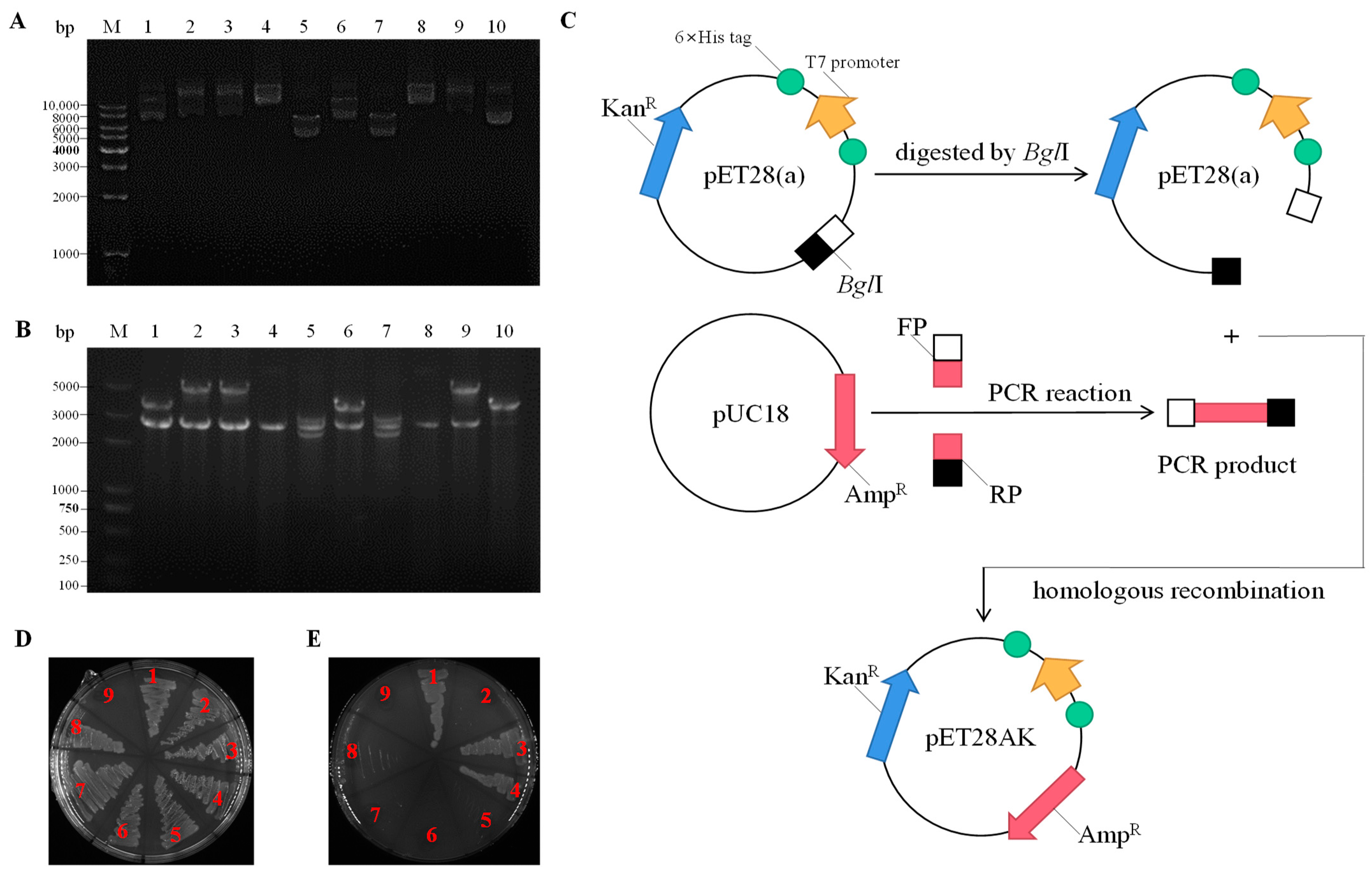
Figure 1. Screening of ORFs that exert Na+/H+ antiport function. (A) The agarose gel electrophoresis of positive clone plasmids that could restore the growth of E. coli KNabc on LBK medium containing 0.2 M NaCl; (B) The agarose gel electrophoresis after double enzyme digestion of positive clone plasmids by EcoRI and HindIII (the corresponding lanes in A&B are Marker and pUC-1028-1 to pUC1028-10 from left to right); (C) Construction schematic of the expression vector pET28AK in this study; (D,E) E. coli KNabc transformants were grown on the LBK medium and LBK medium containing 0.2 M NaCl, (1) KNabc/pUC-1028-1-ORF3, (2) KNabc/pET-1028-1-ORF4, (3) KNabc/pUC-1028-7-ORF2, (4) KNabc/pUC-1028-9-ORF2, (5) KNabc/pET1028-9-ORF3, (6) KNabc/pET1028-9-ORF4, (7) KNabc/pUC18, (8) KNabc/pET28AK, (9) blank.
Ijms 24 10786 g001
Figure 2. Hydrophobicity analysis and transmembrane segment (TMS) prediction of NhaC1 and NhaC2. (A,B) Hydrophobicity analysis of NhaC1 and NhaC2; (C,D) 11 and 12 predicted TMSs of NhaC1 and NhaC2.
Ijms 24 10786 g002
Figure 3. Phylogenetic trees of NhaC1 (A) and NhaC2 (B) with their closest homologs and an identified protein with Na+/H+ antiport activity based on the neighbour-joining algorithm. To construct phylogenetic trees, the 24 and 33 closest homologs with 72.84–96.47% and 63.62–98.28% identities, respectively, were selected from 100 sequences. An identical protein from the NhaC family was selected, as shown in blue in Figure 3. Bootstrap values > 50% (based on 1000 replications) are shown at branch points. Both NhaC1 and NhaC2 and their closest homologs clustered with the bootstrap values of 100%; both are shown in bold red in Figure 3.
Ijms 24 10786 g003
Figure 4. (A) Alignment between NhaC1 and homologs of NhaC family proteins. The 11 homologs with percent identities from 80.43% to 96.47% were selected from Halopiger, Haloterrigena, Natronorubrum, Halostagnicola, Natronococcus, and Natronorubrum. (B) Alignment between NhaC2 and 14 homologs of NhaC family proteins. A total of 14 homologs from Natronococcus, Halobiforma, Natronorubrum, and Halostagnicola with the percent identity ranging from 85.11% to 93.28% were selected. Shading homology corresponds to 100% (black), ≥75% (grey), ≥50% (light grey), and <50% (white) amino acid identity.
Ijms 24 10786 g004
Figure 5. Salt tolerance and alkaline pH resistance of nhaC1 and nhaC2. To test the salt tolerance of KNabc/pUC-nhaC1 and KNabc/pUC-nhaC2, 1% overnight cultures (OD600 was adjusted to 0.8 before inoculation) were inoculated to LBK medium containing 0–0.8 M NaCl (A) or 0–50 mM LiCl (B) and 50 mg∙mL−1 of ampicillin, respectively, at pH 7.0, and cultured at 37 °C for 12 h, with KNabc/pUC18 as the negative control, and the OD600 nm was measured; (C) 1% overnight cultures (OD600 was adjusted to 0.8 before inoculation) were inoculated to LBK medium containing 50 mM NaCl and 50 mg∙mL−1 of ampicillin (pH 7.0–10.0) and cultured at 37 °C for 12 h; KNabc/pUC18 was used as negative control, and the OD600 was measured. Data in the figure represent the average of three independent trials.
Ijms 24 10786 g005
Figure 6. Detection of molecular weights and cell localizations of NhaC1 and NhaC2. (A,B) SDS-PAGE of NhaC1 fused with 6×His tag and NhaC2 fused with 6×His tag, purified by Ni-NTA affinity chromatography, and the staining method was Coomassie brilliant blue staining; (C,D) Cell localizations of NhaC1 and NhaC2 detected by Western blots. Total protein, cytoplasmic protein, and membrane protein of NhaC1 fused with 6×His tag and NhaC2 fused with 6×His tag are shown in Lanes 1, 3, and 5, respectively; Total protein, cytoplasmic protein, and membrane protein from KNabc/pET28AK are shown as a negative control in Lanes 2, 4, and 6, respectively.
Ijms 24 10786 g006
Figure 7. Detection of Na+(K+, Li+)/H+ antiport activity. (A) The measurements for Na+(K+, Li+)/H+ antiport activities were performed in everted membrane vesicles prepared by E. coli KNabc containing pET28AK (as negative controls), pET28AK-nhaC1, and pET28AK-nhaC2 at pH 7.0 with monovalent cations (final concentration 5 mM), respectively; (B,C) Detection of pH-dependent activity profile of NhaC1 and NhaC2. Data in the figure represent the average of three independent trials.
Ijms 24 10786 g007
Figure 8. Calculation of K0.5 values of Na+, K+, and Li+ by NhaC1 and NhaC2. With the final concentration of monovalent cations as the abscissa and the fluorescence dequenching rate as the ordinate, origin2017 was used for nonlinear regression analysis to fit the curve and calculate the final concentration on monovalent cations added, which could reach half of the maximum fluorescence dequenching. (AC) The curves and K0.5 values of Na+, K+, and Li+ NhaC1, respectively; (DF) The curves and K0.5 values of Na+, K+, and Li+ NhaC2 respectively. Data in the figure represents the average of three independent trials.
Ijms 24 10786 g008
Table 1. Characteristics of the hypothetical proteins corresponding to the identified ORFs.
Characteristics1028-1-ORF31028-9-ORF2
Gene IDBB347_RS08225BB347_RS08285
Gene abbreviated namenhaC1nhaC2
Protein abbreviated nameNhaC1NhaC2
AccessionWP_076580434.1WP_076580456.1
Amino acid sequence length(aa)566524
Number of hydrophobic amino acids (aa)302301
Predicted molecular weight (Da)60,72254,786
Number of TMSs1112
DefinitionNa+/H+ antiporter NhaC family proteinNa+/H+ antiporter NhaC family protein
Table 2. Strains and plasmids employed in this study.
Strain or PlasmidDescriptionSource or Reference
Strains
N.daqingense JX313TOriginal strain, an extremely halophilic archaeaIsolated and identified by our lab [27]
E. coli DH5αHost strain for cloningVazyme Biotech Co., Ltd.
E. coli KNabcNa+/H+ antiporter-deficient strain,
nhaA::KmR, nhaB::EmR, chaA::CmR
Donated by Prof. Juquan Jiang [31]
Plasmids
pUC18Cloning vector, AmpRComate Biosciences Co., Ltd. (Changchun, China)
pET-28aProkaryotic expression vector, KanR Comate Biosciences Co., Ltd.
pET-28AKProkaryotic expression vector, KanR and AmpRThis study
pUC1028-1pUC18 carrying 3.5 kb DNA fragment with Na+/H+ antiport activityThis study
pUC1028-9pUC18 carrying 4.9 kb DNA fragment with Na+/H+ antiport activityThis study
pET28AK-nhaC1Heterologous expression vector of nhaC1This study
pET28AK-nhaC2Heterologous expression vector of nhaC2This study
Table 3. Protein purification buffer formula.
ComponentSoluble Film BufferBinding BufferWashing BufferElution Buffer
Choline chloride140 mM140 mM140 mM140 mM
Tris25 mM25 mM25 mM25 mM
Glycerol10%10%10%10%
N-Dodecyl-β-D-maltoside0.02%0.02%0.02%0.02%
Imidazole-10 M25/45/50/55/60/65/85 mM300 mM
Table 4. Primers of this study.
PrimersDescriptionSequence(from 5′ to 3′)Source or Reference
22FArchaea 16S rDNAATTCCGGTTGATCCTGCX.W.Xu, et al. [46]
1540RAGGAGGTGATCCAGCCGCAG
M13-47FSequencing primers of pUC18CGCCAGGGTTTTCCCAGTCACGACThis study
M13RCACACAGGAAACAGCTATGACThis study
T7Sequencing primers of pET-28a and pET28AKTAATACGACTCACTATAGGGThis study
T7tGCTAGTTATTGCTCAGCGGThis study
Amp-FTo insertAmpR into pET-28aCTGCHCGTTGGTGCGGATATCCGCGGAACCCCTATTTGTTThis study
Amp-RGTATCCCACTACCGAGATATCTTACCAATGCTTAATCAGTGAGGCThis study
1-3FPTo insert DNA sequence of pUC1028-1-ORF3 into pUC18TATGACCATGATTACGAATTCATGTCTGACTTTGGAGCGCTTTThis study
1-3RPCAGGTCGACTCTAGAGGATCCTTACTCCTCAGGGTCCGTCCCThis study
9-2FPTo insert DNA sequence of pUC1028-9-ORF2 into pUC18TATGACCATGATTACGAATTCATGAGTGAAGCCAACGATAATTCAThis study
9-2RPCAGGTCGACTCTAGAGGATCCTCATAGTCGTGCCACCTCCTCGThis study
NhaC1-EFTo insert nhaC1 into pET28AKCAGCAAATGGGTCGCGGATCCATGTCTGACTTTGGAGCGCTTTThis study
NhaC1-ERTTGTCGACGGAGCTCGAATTCTTACTCCTCAGGGTCCGTCCCThis study
NhaC1-EFTo insert nhaC2 into pET28AKCAGCAAATGGGTCGCGGATCCATGAGTGAAGCCAACGATAATTCAThis study
NhaC1-ERTTGTCGACGGAGCTCGAATTCTCACACCCCCCAGAAGAACGThis study
Table 5. Accessions of homologs of NhaC1 and NhaC2 to NhaC family proteins used for analysis.
NhaC1 HomologsAccessionNhaC2 HomologsAccession
Nd_NhaC1(this study)WP_076580434.1Nd_NhaC2(this study)WP_076580456.1
Ns_NhaCWP_090507012.1Ns_NhaCWP_090507022.1
NsA_NhaCWP_278304797.1Nt_NhaCWP_076607379.1
Nti_NhaCWP_006090653.1Np_spaTYL39413.1
Nte_NhaCWP_090303449.1NsuJ_NhaCELY48302.1
No_NhaCWP_015320610.1Nh_NhaC1WP_170972344.1
Na_NhaCWP_005555905.1Nh_NhaC2WP_162989723.1
Hk_NhaCWP_092904960.1Nsu_NhaCWP_049890100.1
Nh_NhaCWP_137288726.1Np_NhaCWP_187432893.1
Ht_NhaCWP_012943592.1Hi_NhaCWP_049954249.1
Ha_NhaCWP_120244443.1Na_NhaCWP_152938558.1
Hx_NhaCWP_013881651.1Nt_NhaCWP_006090630.1
NsA_NhaCWP_278304817.1
HiA_NhaCEMA28167.1
NsC_NhaCWP_252487510.1
Disclaimer/Publisher’s Note: The statements, opinions and data contained in all publications are solely those of the individual author(s) and contributor(s) and not of MDPI and/or the editor(s). MDPI and/or the editor(s) disclaim responsibility for any injury to people or property resulting from any ideas, methods, instructions or products referred to in the content.