Abstract
Mast cells have existed for millions of years in species that never suffer from allergic reactions. Hence, in addition to allergies, mast cells can play a critical role in homeostasis and inflammation via secretion of numerous vasoactive, pro-inflammatory and neuro-sensitizing mediators. Secretion may utilize different modes that involve the cytoskeleton, but our understanding of the molecular mechanisms regulating secretion is still not well understood. The Ezrin/Radixin/Moesin (ERM) family of proteins is involved in linking cell surface-initiated signaling to the actin cytoskeleton. However, how ERMs may regulate secretion from mast cells is still poorly understood. ERMs contain two functional domains connected through a long α-helix region, the N-terminal FERM (band 4.1 protein-ERM) domain and the C-terminal ERM association domain (C-ERMAD). The FERM domain and the C-ERMAD can bind to each other in a head-to-tail manner, leading to a closed/inactive conformation. Typically, phosphorylation on the C-terminus Thr has been associated with the activation of ERMs, including secretion from macrophages and platelets. It has previously been shown that the ability of the so-called mast cell “stabilizer” disodium cromoglycate (cromolyn) to inhibit secretion from rat mast cells closely paralleled the phosphorylation of a 78 kDa protein, which was subsequently shown to be moesin, a member of ERMs. Interestingly, the phosphorylation of moesin during the inhibition of mast cell secretion was on the N-terminal Ser56/74 and Thr66 residues. This phosphorylation pattern could lock moesin in its inactive state and render it inaccessible to binding to the Soluble NSF attachment protein receptors (SNAREs) and synaptosomal-associated proteins (SNAPs) critical for exocytosis. Using confocal microscopic imaging, we showed moesin was found to colocalize with actin and cluster around secretory granules during inhibition of secretion. In conclusion, the phosphorylation pattern and localization of moesin may be important in the regulation of mast cell secretion and could be targeted for the development of effective inhibitors of secretion of allergic and inflammatory mediators from mast cells.
Keywords:
ERMs; flavonoids; luteolin; mast cells; mediators; moesin; phosphorylation; secretion; SNAREs; SNAPs; tryptase 1. Introduction
Mast cells are specialized bone marrow-derived cells that play an important role in health [1] and in allergies [2,3,4,5,6,7,8,9,10,11,12] but also in innate and in adaptive immune processes [13,14,15,16], antigen presentation [16,17], regulation of T-cell responses [18,19,20], autoimmunity [21] and inflammation [10,22,23,24,25] in response to allergic and immunologic stress [4,26,27] but also non-allergic stress and toxic stimuli [10,28]. Mast cells are increased in number and are more reactive in mastocytosis [26] and mast cell activation syndrome (MCAS) [26,29,30], but they can also participate in other disorders [4,10,31,32,33], including neurotrauma, neuroinflammatory and neurodegenerative diseases [34,35,36].
2. Mast Cell Mediators and Mechanisms of Secretion
Mast cells are located in all tissues at the interface with the external environment [37] such as eyes, nose, lungs, skin and gastrointestinal tract. However, perivascular mast cells also sense the blood vessel lumen by extending filopodia through endothelial gaps and binding circulating immunoglobulin E (IgE) [38]. Mast cells are well known for their involvement in allergic and anaphylactic reactions via activation of the high-affinity surface receptor for IgE (FcεRI). Multivalent allergen binding leads to aggregation of FcεRI, leading to an influx of calcium ions, thus initiating a cascade of downstream events that involve phosphorylation of phosphatidyl inositol (IP3) and various Tyr kinases [39,40,41,42]. In addition to allergens, mast cells are also stimulated by a variety of triggers that include drugs, foods, pathogens and “danger signals” [26], as well as certain neuropeptides, especially substance P (SP) [43], via activation of their high-affinity receptors. Mast cells are also stimulated/activated by several cytokines, chemokines, hormones, such as corticotropin-releasing hormone (CRH), toxins and extreme external environmental changes [23,36,44,45].
Upon stimulation, mast cells secrete multiple biologically active mediators [46], some of which are preformed and stored in as many as 1000 secretory granules per cell, such as β-hexosaminidase (β-hex), heparin, histamine, tumor necrosis factor (TNF) and the serine proteases chymase and tryptase through rapid (1–5 min) degranulation by exocytosis [47]. Histamine and tryptase are the main mediators commonly associated with mast cells [48]. Tryptase is found in all mast cells, but unlike mucosal mast cells (MMCs), which contain only tryptase, connective tissue mast cells (CTMCs) contain both chymase and tryptase. Even though these proteases are considered to be stored in the same secretory granules, there is evidence that this may not necessarily be true. For instance, serum tryptase was not elevated in many patients with MCAS [28] or in cutaneous mastocytosis [49]. In one paper, it was shown that IgE-mediated degranulation of primary murine MMCs and CTMCs released phenotypically different extracellular vesicle (EV) populations depending on the stimulus [50]. In particular, unstimulated mast cells constitutively released CD9+ EVs, while degranulation was accompanied by the release of CD63+ EVs that contained different proteases [50].
Mast cells also release newly synthesized phospholipid products such as prostaglandin D2 (PGD2) and leukotrienes (LTs) [51,52,53], as well as numerous de novo synthesized protein mediators 6–24 h after stimulation such as interleukins [54], including interleukin-1beta (IL-1β) [55], IL-6 [45,56], IL-31 [57], IL-33 [55] and TNF [43].
Mast cells can secrete their numerous mediators [25,47,58] utilizing different signaling [11,59,60,61,62] and secretory [60,63,64] pathways sometimes referred to as the “secretome” [65]. The secretory pathways include degranulation by exocytosis, compound exocytosis, piecemeal degranulation, transgranulation, directed degranulation, vesicular (differential) release of mediators, extracellular nanovesicles (exosomes), nanotubules [66] and antibody-dependent “immunologic synapses for dedicated secretion” [67,68] (Table 1). The term “secretion” is used in this review to include both degranulation by exocytosis, which is the main means of secretion of granule-stored mediators [69], as well as differential release via which chemokines and cytokines are released without degranulation [59]. For instance, it was first reported that serotonin [45,52,56], and later, vascular endothelial growth factor (VEGF) [70] and IL-6 [45,56], could be secreted from mast cells without degranulation and without the release of histamine or tryptase [59]. It has also been reported that mast cells can release the content of individual secretory granules [71] or individual mediators without degranulation [52]. This process was distinct from “piecemeal degranulation” [72], granule-associated vesicle transport [63] or the release of extracellular vesicles [67,73,74,75,76,77,78].

Table 1.
Different modes of secretion of mediators from mast cells.
Moreover, mast cell mediators could have autocrine actions affecting the expression of receptors or the overall reactivity of mast cells. For instance, mast cells can release the “alarmin” IL-33 themselves [55]. IL-33 then could stimulate mast cells via the activation of its own specific surface receptor ST2 and significantly increase the ability of substance P (SP) to stimulate secretion of VEGF [79,80], IL-31 [57], TNF [43] and IL-1β [55]. Mast cell-derived IL-1β or histamine could further stimulate the release of IL-1β from macrophages [81]. IL-1β could, in turn, stimulate mast cells to release IL-6, which was shown to stimulate mast cell proliferation [82]. The presence of the D816V-KIT mutation in mast cells was associated with constitutive release of IL-6 [83]. Serum levels of IL-6 were reported to be elevated in mastocytosis [84,85,86] and correlated with disease severity. Mast cells could also undergo directional degranulation and secretion of TNF and possibly other pro-inflammatory mediators into the bloodstream [87]. It is also important to note that mast cells exhibit different phenotypes including expression of different receptors depending on the tissue microenvironment [88]. Moreover, different receptors may interact and increase mast cell reactivity [89], as shown for FcεRI and MRGPRX2, which were reported to have an additive effect in stimulating degranulation of human skin mast cells [90].
IL-33 increased the expression of the SP receptor neurokinin-1 (NK-1), while SP increased expression of the IL-33 receptor ST2 [55]. SP also induced the expression of the receptor CRHR-1 for the key stress hormone CRH in human mast cells [91]. Instead, SP downregulated the expression of FcεRI in human mast cells [92]. CRH stimulated mast cells to release VEGF without degranulation, an action that was augmented by the peptide neurotensin (NT) [93]; during this process, CRH stimulated the expression of the NT receptor NT3, while NT stimulated the expression of CRHR-1 [94]. These findings could help explain why many atopic patients worsen dramatically after a major stressful episode [95,96].
Mast cell-derived mediators could also induce epigenetic effects as shown for tryptase, which could catalyze histone clipping [97] and could regulate modification of histones in mast cell leukemia cells [98]. The expression of Ten-eleven translocation-2 (TET2), an epigenetic regulator, was induced in response to the activation of mast cells [99,100]. Hence, mast cells are very dynamic cells that respond not only to external but also to innate stimuli. Such findings have prompted the re-evaluation of the secretory processes and their regulation in mast cells [101].
3. Regulation of Mediator Secretion from Mast Cells
Our understanding of the regulation of mediator release via the different modes of secretion and its regulation is still poorly understood. Even though the stimulus–response coupling pathway has been well delineated for activation of the high-affinity surface receptor for IgE (FcεRI) [42,102,103], and, more recently, of the low-affinity receptor for cationic peptides, Mas-Related G Protein-Coupled Receptor-X2 (MRGPRX2) [104,105,106,107,108], there is still a lack of understanding of the molecular events regulating secretion, whether by degranulation, selective release of mediators or any other mode of secretion (Table 1). The mode and extent of mast cell responsiveness ultimately depend on the interplay between stimulatory and inhibitory signaling pathways, such as CD300 [109,110] and Singlets [111], especially Siglec-7 [112], and the β subunit of FcεRI (FcεRIβ) [113].
SNAREs and SNAPs
One possible mechanism of how mast cell secretion may be regulated could involve the Soluble NSF attachment protein receptors (SNAREs) and synaptosomal-associated proteins (SNAPs) discovered by Dr. J.E. Rothman, who was awarded the 2013 Nobel Prize in Physiology and Medicine for delineating the principles for secretory membrane fusion [114]. The existence of distinct secretory vesicle calcium-sensitive proteins responsible for “snapping” with corresponding proteins on the plasma membrane during secretion by exocytosis from mast cells had actually been proposed much earlier by one of the authors (TCT) in his doctoral thesis examination at Yale University in 1974, with the examiner being Dr. G. Palade, who had just received the 1974 Nobel Prize in Physiology and Medicine for his discovery that secreted proteins are carried from the endoplasmic reticulum (ER) to the cell surface in specialized compartments or transport vesicles.
SNAREs [115,116,117] and synaptosomal-associated protein of 23 kDa (SNAP-23) [118,119,120,121,122,123] have been shown to be involved in mast cell secretion. In fact, there may be different mechanisms regulating exocytosis in mast cells [124], and mast cell distinct secretory granule subsets may be regulated by different SNARE isoforms [125] and different vesicle-associated membrane proteins (VAMPs), especially VAMP2- and VAMP8 [126,127].
Mast cells express Munc18-2, which interacts with SNARE syntaxin 2 or 3, as well as Munc18-3, which interacts with syntaxin 4. Munc18-2 was localized to secretory granules, whereas Munc18-3 was found on the plasma membrane. Increased expression of Munc18-2 inhibited IgE-triggered exocytosis, while increased expression of Munc18-3 had no effect. Upon stimulation, Munc18-2 redistributed on granules that were aligned along microtubules, but was excluded from F-actin ruffles, suggesting a role for Munc18-2 and the microtubule network in the regulation of secretion by degranulation in mast cells [128]. In addition, a number of so-called ‘adapters’ have been reported to regulate secretion from mast cells by binding multiple signaling proteins and localizing them to specific cellular compartments [40].
It Is of note that the degranulation of different mast cell vesicle subsets was differentially and selectively regulated by various polyphenols via interfering with two SNARE complexes, Syn (syntaxin) 4/SNAP-23/VAMP2 and Syn4/SNAP23/VAMP8 [129]. Similarly, polyphenols were shown to interfere with “zippering” of SNARES in the neuron [130]. The structure of the phenolic flavonol quercetin is somewhat similar to cromolyn [131] but is a more potent inhibitor of mast cells than cromolyn [132]. Quercetin inhibited rat mast cell degranulation [133,134], possibly via the inhibition of protein kinase C (PKC) [135,136], but it also induced the phosphorylation of moesin [136]. Quercetin also inhibited the release of pro-inflammatory cytokines [135], including IL-6 [134], from cultured human mast cells. The quercetin-related flavone luteolin and the luteolin analogue tetramethoxyluteolin were even more potent inhibitors of both the degranulation [137] as well as of release of TNF [43] and IL-1β [55] from human mast cells. The ability of flavonoids to inhibit mast cell secretion via phosphorylation of moesin led to conjectures about the design of more potent inhibitors [131].
In spite of the advances briefly outlined above, there is still no effective inhibitor of mediator secretion from mast cells. Antihistamines interfere with histamine binding to its receptors after it has been secreted. There has been considerable progress in developing drugs that block tyrosine kinases involved in mast cell proliferation [138].
4. Ezrin, Radixin and Moesin (ERM) Family of Proteins
Ezrin, radixin and moesin (ERMs) are fairly homologous proteins (73% amino acid identity) that link the actin cytoskeleton to the cytoplasmic tail of transmembrane proteins in the plasma membrane, thus regulating the formation of F-actin-based structures [139,140,141,142,143,144]. ERMs localize to cell surface protrusions such as microvilli, filopodia and cell–cell junctions. ERMs are critical for signal transduction from the cell surface into the cell. Given the high degree of homology and their co-expression to various degrees in many cell types, overlapping or even compensatory functions have been proposed.
Ezrin was named after Ezra Cornell University where it was first isolated from microvilli in chicken intestinal epithelial cells, while radixin (from the Latin meaning root) was isolated from the adherens junctions of rat liver hepatocytes. Moesin (membrane-organizing extension spike protein) was isolated from smooth muscle cells of the bovine uterus. ERMs contain two functional domains connected through a long α-helix region (Figure 1A): the N-terminal FERM (band 4.1 protein-ERM) domain, which is critical for the function of the ERMs, and the C-terminal ERM association domain (C-ERMAD). The FERM domain is composed of three subdomains (F1, a ubiquitin-like domain; F2, with four α-helices; and F3, a pleckstrin homology domain). The FERM domain and the C-ERMAD can bind each other in a head-to-tail manner, leading to a closed/inactive conformation (Figure 1B).
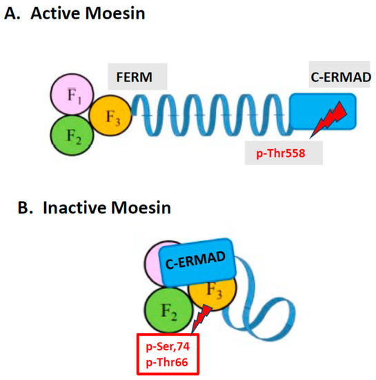
Figure 1.
Diagrammatic representation of the active and inactive forms of moesin. Phosphorylation of moesin at Thr558 opens up actin-binding sites. In contrast, phosphorylation of moesin at Ser56/Thr66 changes the conformational structure of moesin so that Thr558 is no longer accessible to bind to actin, thus preventing secretion.
The release of the C-ERMAD from the FERM domain is necessary for the activation of ERMs, unmasking their F-actin- and PM-binding sites. Activation of ERMs occurs first by phosphatidylinositol 4,5-bisphosphate (PIP2) binding to the N-terminus and changing the 3D structure exposing a C-terminal Threonine (Thr567 in ezrin, Thr564 in radixin and Thr558 in moesin) for phosphorylation [140,145] by the Rho family of GTPases (RhoA/Rac/Cdc42). This step transitions ERMs from a closed (inactive, Figure 1B) to an open (active, Figure 1A) conformation [146] that exposes the C-terminal F-actin-binding domain that cross-links plasma membrane proteins with actin filaments (Figure 2) [140,143,144,145,146].
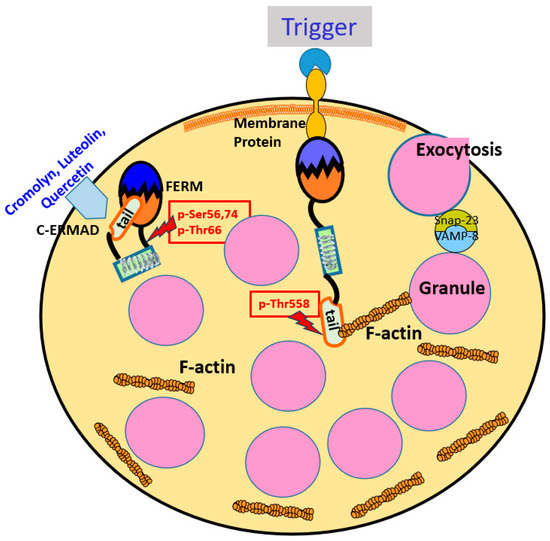
Figure 2.
Moesin in Mast Cell Secretion. Diagrammatic representation of how differential phosphorylation of moesin could regulate secretion from mast cells. Phosphorylation of moesin at Thr558 in response to triggers opens up binding sites permitting granules to travel to the cell surface and secrete granule-stored mediators via degranulation. In contrast, phosphorylation of moesin at Ser56/Thr66 by cromolyn or flavonoids changes the conformational structure of moesin so that Thr558 is no longer accessible to bind to actin, thus preventing secretion.
Moesin in Mast Cells
The expression of particular ERM members varies among different cells. Moesin is mainly expressed in endothelial cells, with ezrin in intestinal epithelial cells and radixin in hepatocytes. However, moesin is the most abundant ERM in leukocytes and mast cells, whereas ezrin is less expressed, and radixin is nearly absent [142].
Mast cells, like any other secretory cell, require the actin cytoskeleton [147] that is necessary for signal transduction and movement of secretory granules or vesicles destined for secretion to the cell surface. For instance, the aggregation of IgE bound to FcεRI by a multivalent antigen stimulates mast cell secretion and rapidly depolymerizes actin filaments, with the actin-severing protein cofilin being dephosphorylated several minutes after stimulation [148]. In contrast, the disaggregation of IgE terminates degranulation mediated by dephosphorylation of Syk associated with a decrease in intracellular Ca2+ concentration and rapid recovery of actin polymerization. Upon FcεRI stimulation, Dok-1 (downstream of tyrosine kinase 1) undergoes Tyr phosphorylation, which negatively regulates Ras/Erk signaling and subsequent secretion by inhibiting calcium influx and calcium-dependent disassembly of actin filaments [149]. It was previously shown that Rho GTPases regulate exocytosis and possibly secretory granule transport. One paper used live-cell imaging to analyze cytoskeleton assembly and secretory granule transport in real-time of mast cells or rat basophil cells (RBL-1) during antigen stimulation. This paper showed that granule transport to the cell periphery was coordinated by de novo microtubule formation and not F-actin since kinesore, which activates the microtubule motor kinesin-1 inhibited microtubule-granule association and significantly reduced degranulation [150]. However, how F-actin or microtubules communicate with secretory granules (or vesicles) and the plasma membrane is still not well understood. Knockdown of the unconventional long-tailed myosin (MYO1F), which localizes with cortical F-actin by short hairpin RNA, reduced human mast cell degranulation stimulated by both IgE and MRGPRX2, and was accompanied by reduced reassembly of the cortical actin ring and fewer secretory granules localized close to the cell surface [151]. Interestingly, MYO1F knockdown also resulted in fewer fissioned mitochondria and deficient mitochondria translocation to sites of degranulation by exocytosis [151]. Mitochondria fission was also reported to accompany secretion by degranulation, but not during secretion of de novo synthesized mediators from human mast cells stimulated by SP [18] and also in skin biopsies from patients with atopic dermatitis [152]. It was further shown that stimulation of mast cells resulted in extracellular secretion of mitochondrial DNA (mtDNA) that acted as an “innate pathogen” and triggered an autoinflammatory response. Increased levels of mtDNA have been reported in patients with COVID-19 [153,154,155,156], psoriasis [157], as well as in EVs from patients with myalgic encephalomyelitis/chronic fatigue syndrome (ME/CFS) [158] and from children with autism spectrum disorder (ASD), and in both cases, mtDNA activated cultured human microglia to secrete IL-1β [159].
The ability of the so-called “mast cell stabilizer” disodium cromoglycate (cromolyn) to inhibit secretion from rat mast cells in response to the cationic Compound 48/80 (C48/80) was shown to closely parallel the phosphorylation of a 78 kDa protein [135,160,161] on the N-terminal Ser56, Ser74 and Thr66 residues (Figure 1B) [162]. We found that this protein was subsequently cloned from mast cells and was shown to be moesin [163], but we named it Mast Cell Degranulation Inhibitory Agent (MACEDONIA) [164]. It is important to note that phosphorylation of at least the N-terminal Ser56/74 and Thr66 residues during inhibition is different to the well-known phosphorylation of C-ERMAD Thr558 associated with moesin activation. In support of the involvement of additional phosphorylation sites than Thr558, there is evidence that, at least in ezrin, Thr235 is phosphorylated by cyclin-dependent kinase 5 (CDK5) and cooperates with Thr576 for its full activation [165].
Using confocal microscopy and ultra cryo-immuno-electron microscopy to preserve the antigenicity of ERMs, it was shown that mast cells contain almost exclusively moesin (with a small amount of ezrin), which was critically localized primarily at the plasma membrane and filopodia, with less around secretory granules; it was further shown that cromolyn induced the clustering of moesin around secretory granules [163]. It was therefore hypothesized that conformational changes in moesin due to phosphorylation/dephosphorylation events could possibly regulate mast cell secretion via positional rearrangements with respect to the membrane/cytoskeleton [163]. It was further hypothesized that moesin could, in fact, serve a dual function depending on its phosphorylation pattern, which occurs after a trigger or an inhibitor interacts with the cell surface [131]. In other words, moesin phosphorylation at C-terminal Thr558 would switch moesin to its active form (Figure 1A) and permit mast secretory granules to move to the surface, fuse with the plasma membrane and undergo exocytosis (Figure 2). In contrast, phosphorylation of N-terminal Ser/Thr sites would switch moesin to its inactive state (Figure 1B) resulting in either (a) the prevention of phosphorylation of Thr558 and moesin activation, (b) the interaction with secretory granules preventing them from moving to the cell surface or (c) affecting the structure of the cell cortex and block secretion indirectly (Figure 2). However, it remains unknown how the phosphorylation of moesin at different sites affects secretion from mast cells in response to different triggers, and how phosphorylation at the N-terminal sites mechanistically leads to the inhibition of mast cell secretion. Moreover, it is not presently known if phosphorylation of moesin may affect modes of secretion other than degranulation by exocytosis. One paper identified a number of Ser/Thr-phosphorylated proteins in activated mast cells, including moesin, but these were involved in different processes such as metabolism and cell structure [166]. Even though ezrin has been mostly discussed for its involvement in cancer [167], it is not known if ezrin could compensate for moesin should the latter be absent or “incapacitated” in mast cells. In fact, ezrin, has been implicated in asthma [168]. The phosphorylation of ezrin at Thr567 was associated with trophoblast motility [169].
Interestingly, moesin knock-out mice were shown to have lymphopenia [170], but mast cell numbers were apparently intact; however, the authors did not investigate mast cell secretion [170]. One X-linked moesin-associated immunodeficiency (X-MAID) has been identified and is characterized by a primary immunodeficiency associated with severe lymphopenia leading to recurrent infections. X-MAID is caused by a single-point mutation leading to a R171W amino acid change in moesin (moesinR171W) [171]. In fact, a mouse model with global expression of moesinR171W exhibited lymphopenia, but it was still characterized by systemic inflammation [171].
The phosphorylation of moesin has also been studied in other secretory systems. Moesin was shown to be phosphorylated at Thr558 within seconds of thrombin-induced activation of platelets [172,173]. Instead, the tyrosine phosphorylation of moesin was reported during the activation of platelets with arachidonic acid [174]. These phosphorylation patterns are reversed by protein phosphatase 2C, which inactivates the F-actin-binding site of activated platelets [175]. Phosphorylation at Thr558 was also reported in activated RAW264.7 macrophages [176]. ERM proteins have been shown to be involved in T-cell polarization and immune synapse formation [177]. It is interesting that anti-moesin autoantibodies were isolated from patients with aplastic anemia [178] and autoimmune vasculitis [179]. However, the significance of these autoantibodies is not apparent, nor is their potential presence in patients with allergies and inflammatory disorders.
5. Mast Cells and Moesin in Neuroinflammation
Mast cells communicate with microglia [180,181] and can activate them [181,182,183,184] via the release of mediators such as histamine [185] and tryptase [186], leading to neuroinflammation [180,182] (Figure 3). The activation of mast cells and microglia in the brain [187] could affect neurodevelopment [188], resulting in neuronal apoptosis [189], and lead to cognitive dysfunction [189]. In fact, the activation of mast cells and microglia has been linked to the pathogenesis of autism spectrum disorder (ASD) [190,191,192,193,194], neurodegenerative diseases [35,195] and traumatic brain injury (TBI) [24,196]. It is, therefore, of interest that moesin has been reported to be involved in the activation of microglia [197]. Moreover, the moesin pseudogene 1 antisense (MSNP1AS) gene was shown to decrease the number and length of neurites, reduce neural viability and promote apoptosis via the inhibition of moesin protein expression, while moesin improved social interactions and reduced repetitive behaviors in BTBR mice [198].
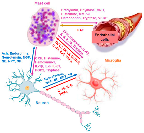
Figure 3.
Diagrammatic representation of the key role of mast cells in neuroinflammation. Mediators released from mast cells can stimulate endothelial cells, microglia and neurons directly to promote inflammation; in turn, molecules secreted from the other cells can stimulate mast cells, thus further promoting neuroinflammation. ERMs could regulate secretion of mediators from mast cells, but also from the other cell types involved. Ach = acetylcholine; CRH = corticotropin-releasing hormone; MMP9 = metalloproteinase-9; NGF = nerve growth factor; NE = norepinephrine; NPY = neuropeptide Y; NT = neurotensin; PAF = platelet activating factor; PGD2 = prostaglandin D2; SP = substance P; VEGF = vascular endothelial growth factor.
Moreover, one paper reported that ezrin, radixin and moesin had distinct roles of in maintaining the plasma membrane integrity and functions of the blood–brain barrier (BBB) transporters [199], which is important because mast cells can regulate the permeability of the BBB [200], the disruption of which has been implicated in ASD [201], in Alzheimer’s disease [33] and in neuro-COVID-19 [202]. ERMs could regulate the secretion of mediators from mast cells but also from the other cell types involved in neuroinflammation.
In this context, it is relevant that flavonoids could have anti-inflammatory [34,203,204,205,206,207,208,209] and neuroprotective effects [210], as well as reduce cognitive dysfunction [211,212,213,214,215], especially brain fog [216,217,218]. In particular, luteolin inhibited both microglia [219,220,221] and mast cells [222,223]. One formulation containing liposomal luteolin in olive pomace (fruit) oil (NeuroProtek®) resulted in significant improvement in children with ASD [224], with a concomitant decrease in serum inflammatory markers [225]. Other papers reported the beneficial actions of luteolin in Long-COVID-19-associated brain fog [216,226] and neurotrauma [207].
6. Conclusions
The studies reviewed indicate that the pattern of phosphorylation and localization of moesin may be important in the regulation of exocytotic secretion of at least secretory granule-associated mediators such as histamine, TNF and tryptase.
It will be important to investigate the expression of total and phosphorylated moesin in human mast cells of different degrees of reactivity/types, such as the leukemic human mast cell line-1 (HMC-1), the Laboratory of allergic diseases-2 (LAD2) and LADR mast cells [227], as well as primary human umbilical cord blood-derived cultured mast cells (hCBMCs), mast cells developed from pluripotent stem cells [228,229,230], but also mast cells from cutaneous mastocytosis or urticaria lesions. Other future studies should investigate whether the knockdown of moesin using small interfering ribonucleic acid (siRNA) would affect the extent of secretion or interfere with the ability of cromolyn or flavonoids to inhibit mast cell secretion. Additionally, studies should also investigate which specific sites are phosphorylated in response to triggers or inhibitors of either the degranulation or differential release of select mediators using trypsin-digested moesin peptides analyzed via mass spectrometry and validated with site-specific phospho-antibodies and point mutant analysis.
It will also be important to investigate the possible presence of some innate molecule(s) or identify novel molecules that could keep moesin in its inactive state, for the development of new effective anti-allergic and anti-inflammatory drugs.
Funding
This research received no external funding.
Institutional Review Board Statement
Not applicable.
Informed Consent Statement
Not applicable.
Conflicts of Interest
The author declare no conflict of interest.
References
- Krystel-Whittemore, M.; Dileepan, K.N.; Wood, J.G. Mast Cell: A Multi-Functional Master Cell. Front. Immunol. 2015, 6, 620. [Google Scholar] [CrossRef] [PubMed]
- Parwaresch, M.R.; Horny, H.P.; Lennert, K. Tissue mast cells in health and disease. Pathol. Res. Pract. 1985, 179, 439–461. [Google Scholar] [CrossRef] [PubMed]
- Csaba, G. Mast cell, the peculiar member of the immune system: A homeostatic aspect. Acta Microbiol. Immunol. Hung. 2015, 62, 207–231. [Google Scholar] [CrossRef] [PubMed]
- Siebenhaar, F.; Redegeld, F.A.; Bischoff, S.C.; Gibbs, B.F.; Maurer, M. Mast Cells as Drivers of Disease and Therapeutic Targets. Trends Immunol. 2018, 39, 151–162. [Google Scholar] [CrossRef]
- Phillips, R.E.; Looareesuwan, S.; White, N.J.; Silamut, K.; Kietinun, S.; Warrell, D.A. Quinine pharmacokinetics and toxicity in pregnant and lactating women with falciparum malaria. Br. J. Clin. Pharmacol. 1986, 21, 677–683. [Google Scholar] [CrossRef]
- Falduto, G.H.; Pfeiffer, A.; Luker, A.; Metcalfe, D.D.; Olivera, A. Emerging mechanisms contributing to mast cell-mediated pathophysiology with therapeutic implications. Pharmacol. Ther. 2021, 220, 107718. [Google Scholar] [CrossRef]
- Dahlin, J.S.; Maurer, M.; Metcalfe, D.D.; Pejler, G.; Sagi-Eisenberg, R.; Nilsson, G. The ingenious mast cell: Contemporary insights into mast cell behavior and function. Allergy 2022, 77, 83–99. [Google Scholar] [CrossRef]
- Kolkhir, P.; Elieh-Ali-Komi, D.; Metz, M.; Siebenhaar, F.; Maurer, M. Understanding human mast cells: Lesson from therapies for allergic and non-allergic diseases. Nat. Rev. Immunol. 2022, 22, 294–308. [Google Scholar] [CrossRef]
- Levi-Schaffer, F.; Gibbs, B.F.; Hallgren, J.; Pucillo, C.; Redegeld, F.; Siebenhaar, F.; Vitte, J.; Mezouar, S.; Michel, M.; Puzzovio, P.G.; et al. Selected recent advances in understanding the role of human mast cells in health and disease. J. Allergy Clin. Immunol. 2022, 149, 1833–1844. [Google Scholar] [CrossRef]
- Olivera, A.; Beaven, M.A.; Metcalfe, D.D. Mast cells signal their importance in health and disease. J. Allergy Clin. Immunol. 2018, 142, 381–393. [Google Scholar] [CrossRef]
- Sibilano, R.; Frossi, B.; Pucillo, C.E. Mast cell activation: A complex interplay of positive and negative signaling pathways. Eur. J. Immunol. 2014, 44, 2558–2566. [Google Scholar] [CrossRef] [PubMed]
- Gallenga, C.E.; Pandolfi, F.; Caraffa, A.; Kritas, S.K.; Ronconi, G.; Toniato, E.; Martinotti, S.; Conti, P. Interleukin-1 family cytokines and mast cells: Activation and inhibition. J. Biol. Regul. Homeost. Agents 2019, 33, 1–6. [Google Scholar] [PubMed]
- Galli, S.J.; Tsai, M.; Piliponsky, A.M. The development of allergic inflammation. Nature 2008, 454, 445–454. [Google Scholar] [CrossRef] [PubMed]
- Toniato, E.; Frydas, I.; Robuffo, I.; Ronconi, G.; Caraffa, A.; Kritas, S.K.; Conti, P. Activation and inhibition of adaptive immune response mediated by mast cells. J. Biol. Regul. Homeost. Agents 2017, 31, 543–548. [Google Scholar] [PubMed]
- Avila, M.; Gonzalez-Espinosa, C. Signaling through Toll-like receptor 4 and mast cell-dependent innate immunity responses. IUBMB Life 2011, 63, 873–880. [Google Scholar] [CrossRef]
- Forsythe, P. Microbes taming mast cells: Implications for allergic inflammation and beyond. Eur. J. Pharmacol. 2016, 778, 169–175. [Google Scholar] [CrossRef]
- Carroll-Portillo, A.; Cannon, J.L.; te Riet, J.; Holmes, A.; Kawakami, Y.; Kawakami, T.; Cambi, A.; Lidke, D.S. Mast cells and dendritic cells form synapses that facilitate antigen transfer for T cell activation. J. Cell Biol. 2015, 210, 851–864. [Google Scholar] [CrossRef]
- Zhang, B.; Weng, Z.; Sismanopoulos, N.; Asadi, S.; Therianou, A.; Alysandratos, K.D.; Angelidou, A.; Shirihai, O.; Theoharides, T.C. Mitochondria distinguish granule-stored from de novo synthesized tumor necrosis factor secretion in human mast cells. Int. Arch. Allergy Immunol. 2012, 159, 23–32. [Google Scholar] [CrossRef]
- Ishii, T.; Wang, J.; Zhang, W.; Mascarenhas, J.; Hoffman, R.; Dai, Y.; Wisch, N.; Xu, M. Pivotal role of mast cells in pruritogenesis in patients with myeloproliferative disorders. Blood 2009, 113, 5942–5950. [Google Scholar] [CrossRef]
- Mekori, Y.A.; Hershko, A.Y.; Frossi, B.; Mion, F.; Pucillo, C.E. Integrating innate and adaptive immune cells: Mast cells as crossroads between regulatory and effector B and T cells. Eur. J. Pharmacol. 2016, 778, 84–89. [Google Scholar] [CrossRef]
- Christy, A.L.; Brown, M.A. The multitasking mast cell: Positive and negative roles in the progression of autoimmunity. J. Immunol. 2007, 179, 2673–2679. [Google Scholar] [CrossRef] [PubMed]
- Hakim-Rad, K.; Metz, M.; Maurer, M. Mast cells: Makers and breakers of allergic inflammation. Curr. Opin. Allergy Clin. Immunol. 2009, 9, 427–430. [Google Scholar] [CrossRef]
- Theoharides, T.C.; Alysandratos, K.D.; Angelidou, A.; Delivanis, D.A.; Sismanopoulos, N.; Zhang, B.; Asadi, S.; Vasiadi, M.; Weng, Z.; Miniati, A.; et al. Mast cells and inflammation. Biochim. Biophys. Acta 2012, 1822, 21–33. [Google Scholar] [CrossRef]
- Kempuraj, D.; Ahmed, M.E.; Selvakumar, G.P.; Thangavel, R.; Dhaliwal, A.S.; Dubova, I.; Mentor, S.; Premkumar, K.; Saeed, D.; Zahoor, H.; et al. Brain Injury-Mediated Neuroinflammatory Response and Alzheimer’s Disease. Neuroscientist 2020, 26, 134–155. [Google Scholar] [CrossRef] [PubMed]
- Mukai, K.; Tsai, M.; Saito, H.; Galli, S.J. Mast cells as sources of cytokines, chemokines, and growth factors. Immunol. Rev. 2018, 282, 121–150. [Google Scholar] [CrossRef]
- Theoharides, T.C.; Valent, P.; Akin, C. Mast Cells, Mastocytosis, and Related Disorders. N. Engl. J. Med. 2015, 373, 163–172. [Google Scholar] [CrossRef]
- Theoharides, T.C. Atopic conditions in search of pathogenesis and therapy. Clin. Ther. 2013, 35, 544–547. [Google Scholar] [CrossRef] [PubMed]
- Theoharides, T.C.; Leeman, S.E. Effect of IL-33 on de novo synthesized mediators from human mast cells. J. Allergy Clin. Immunol. 2019, 143, 451. [Google Scholar] [CrossRef]
- Akin, C. Mast cell activation disorders. J. Allergy Clin. Immunol. Pract. 2014, 2, 252–257.e1. [Google Scholar] [CrossRef]
- Theoharides, T.C.; Tsilioni, I.; Ren, H. Recent advances in our understanding of mast cell activation—Or should it be mast cell mediator disorders? Expert Rev. Clin. Immunol. 2019, 15, 639–656. [Google Scholar] [CrossRef]
- Galli, S.J.; Gaudenzio, N.; Tsai, M. Mast Cells in Inflammation and Disease: Recent Progress and Ongoing Concerns. Annu. Rev. Immunol. 2020, 38, 49–77. [Google Scholar] [CrossRef] [PubMed]
- Kempuraj, D.; Selvakumar, G.P.; Ahmed, M.E.; Raikwar, S.P.; Thangavel, R.; Khan, A.; Zaheer, S.A.; Iyer, S.S.; Burton, C.; James, D.; et al. COVID-19, Mast Cells, Cytokine Storm, Psychological Stress, and Neuroinflammation. Neuroscientist 2020, 26, 402–414. [Google Scholar] [CrossRef] [PubMed]
- Kempuraj, D.; Mentor, S.; Thangavel, R.; Ahmed, M.E.; Selvakumar, G.P.; Raikwar, S.P.; Dubova, I.; Zaheer, S.; Iyer, S.S.; Zaheer, A. Mast Cells in Stress, Pain, Blood-Brain Barrier, Neuroinflammation and Alzheimer’s Disease. Front. Cell. Neurosci. 2019, 13, 54. [Google Scholar] [CrossRef] [PubMed]
- Theoharides, T.C.; Conti, P.; Economu, M. Brain inflammation, neuropsychiatric disorders, and immunoendocrine effects of luteolin. J. Clin. Psychopharmacol. 2014, 34, 187–189. [Google Scholar] [CrossRef]
- Kempuraj, D.; Selvakumar, G.P.; Thangavel, R.; Ahmed, M.E.; Zaheer, S.; Raikwar, S.P.; Iyer, S.S.; Bhagavan, S.M.; Beladakere-Ramaswamy, S.; Zaheer, A. Mast Cell Activation in Brain Injury, Stress, and Post-traumatic Stress Disorder and Alzheimer’s Disease Pathogenesis. Front. Neurosci. 2017, 11, 703. [Google Scholar] [CrossRef]
- Kempuraj, D.; Thangavel, R.; Selvakumar, G.P.; Zaheer, S.; Ahmed, M.E.; Raikwar, S.P.; Zahoor, H.; Saeed, D.; Natteru, P.A.; Iyer, S.; et al. Brain and Peripheral Atypical Inflammatory Mediators Potentiate Neuroinflammation and Neurodegeneration. Front. Cell. Neurosci. 2017, 11, 216. [Google Scholar] [CrossRef]
- Galli, S.J.; Grimbaldeston, M.; Tsai, M. Immunomodulatory mast cells: Negative, as well as positive, regulators of immunity. Nat. Rev. Immunol. 2008, 8, 478–486. [Google Scholar] [CrossRef] [PubMed]
- Cheng, L.E.; Hartmann, K.; Roers, A.; Krummel, M.F.; Locksley, R.M. Perivascular mast cells dynamically probe cutaneous blood vessels to capture immunoglobulin E. Immunity 2013, 38, 166–175. [Google Scholar] [CrossRef]
- Metzger, H.; Eglite, S.; Haleem-Smith, H.; Reischl, I.; Torigoe, C. Quantitative aspects of signal transduction by the receptor with high affinity for IgE. Mol. Immunol. 2002, 38, 1207–1211. [Google Scholar] [CrossRef] [PubMed]
- Alvarez-Errico, D.; Lessmann, E.; Rivera, J. Adapters in the organization of mast cell signaling. Immunol. Rev. 2009, 232, 195–217. [Google Scholar] [CrossRef] [PubMed]
- Ando, T.; Kitaura, J. Tuning IgE: IgE-Associating Molecules and Their Effects on IgE-Dependent Mast Cell Reactions. Cells 2021, 10, 1697. [Google Scholar] [CrossRef] [PubMed]
- Nagata, Y.; Suzuki, R. FcεRI: A Master Regulator of Mast Cell Functions. Cells 2022, 11, 622. [Google Scholar] [CrossRef]
- Taracanova, A.; Alevizos, M.; Karagkouni, A.; Weng, Z.; Norwitz, E.; Conti, P.; Leeman, S.E.; Theoharides, T.C. SP and IL-33 together markedly enhance TNF synthesis and secretion from human mast cells mediated by the interaction of their receptors. Proc. Natl. Acad. Sci. USA 2017, 114, E4002–E4009. [Google Scholar] [CrossRef]
- Theoharides, T.C.; Konstantinidou, A.D. Corticotropin-releasing hormone and the blood-brain-barrier. Front. Biosci. 2007, 12, 1615–1628. [Google Scholar] [CrossRef] [PubMed]
- Kandere-Grzybowska, K.; Letourneau, R.; Kempuraj, D.; Donelan, J.; Poplawski, S.; Boucher, W.; Athanassiou, A.; Theoharides, T.C. IL-1 induces vesicular secretion of IL-6 without degranulation from human mast cells. J. Immunol. 2003, 171, 4830–4836. [Google Scholar] [CrossRef] [PubMed]
- Schwartz, L.B. Mediators of human mast cells and human mast cell subsets. Ann. Allergy 1987, 58, 226–235. [Google Scholar] [PubMed]
- Wernersson, S.; Pejler, G. Mast cell secretory granules: Armed for battle. Nat. Rev. Immunol. 2014, 14, 478–494. [Google Scholar] [CrossRef]
- Uvnas, B. Histamine storage and release. Fed. Proc. 1974, 33, 2172–2176. [Google Scholar] [CrossRef]
- Awan, S.F.; Schwartz, L.B.; Maric, I.; Metcalfe, D.D.; Carter, M.C. Acute increases in total serum tryptase unassociated with hemodynamic instability in diffuse cutaneous mastocytosis. Ann. Allergy Asthma Immunol. 2022, 129, 249–252. [Google Scholar] [CrossRef]
- Groot Kormelink, T.; Arkesteijn, G.J.; van de Lest, C.H.; Geerts, W.J.; Goerdayal, S.S.; Altelaar, M.A.; Redegeld, F.A.; Nolte-’t Hoen, E.N.; Wauben, M.H. Mast Cell Degranulation Is Accompanied by the Release of a Selective Subset of Extracellular Vesicles That Contain Mast Cell-Specific Proteases. J. Immunol. 2016, 197, 3382–3392. [Google Scholar] [CrossRef]
- Picard, M.; Giavina-Bianchi, P.; Mezzano, V.; Castells, M. Expanding spectrum of mast cell activation disorders: Monoclonal and idiopathic mast cell activation syndromes. Clin. Ther. 2013, 35, 548–562. [Google Scholar] [CrossRef] [PubMed]
- Theoharides, T.C.; Bondy, P.K.; Tsakalos, N.D.; Askenase, P.W. Differential release of serotonin and histamine from mast cells. Nature 1982, 297, 229–231. [Google Scholar] [CrossRef] [PubMed]
- Theoharides, T.C.; Cochrane, D.E. Critical role of mast cells in inflammatory diseases and the effect of acute stress. J. Neuroimmunol. 2004, 146, 1–12. [Google Scholar] [CrossRef] [PubMed]
- Solimando, A.G.; Desantis, V.; Ribatti, D. Mast Cells and Interleukins. Int. J. Mol. Sci. 2022, 23, 14004. [Google Scholar] [CrossRef] [PubMed]
- Taracanova, A.; Tsilioni, I.; Conti, P.; Norwitz, E.R.; Leeman, S.E.; Theoharides, T.C. Substance P and IL-33 administered together stimulate a marked secretion of IL-1beta from human mast cells, inhibited by methoxyluteolin. Proc. Natl. Acad. Sci. USA 2018, 115, E9381–E9390. [Google Scholar] [CrossRef]
- Gagari, E.; Tsai, M.; Lantz, C.S.; Fox, L.G.; Galli, S.J. Differential release of mast cell interleukin-6 via c-kit. Blood 1997, 89, 2654–2663. [Google Scholar] [CrossRef] [PubMed]
- Petra, A.I.; Tsilioni, I.; Taracanova, A.; Katsarou-Katsari, A.; Theoharides, T.C. Interleukin 33 and interleukin 4 regulate interleukin 31 gene expression and secretion from human laboratory of allergic diseases 2 mast cells stimulated by substance P and/or immunoglobulin E. Allergy Asthma Proc. 2018, 39, 153–160. [Google Scholar] [CrossRef] [PubMed]
- Theoharides, T.C.; Kalogeromitros, D. The critical role of mast cells in allergy and inflammation. Ann. N. Y. Acad. Sci. 2006, 1088, 78–99. [Google Scholar] [CrossRef]
- Theoharides, T.C.; Kempuraj, D.; Tagen, M.; Conti, P.; Kalogeromitros, D. Differential release of mast cell mediators and the pathogenesis of inflammation. Immunol. Rev. 2007, 217, 65–78. [Google Scholar] [CrossRef]
- Xu, H.; Bin, N.R.; Sugita, S. Diverse exocytic pathways for mast cell mediators. Biochem. Soc. Trans. 2018, 46, 235–247. [Google Scholar] [CrossRef]
- Gilfillan, A.M.; Tkaczyk, C. Integrated signalling pathways for mast-cell activation. Nat. Rev. Immunol. 2006, 6, 218–230. [Google Scholar] [CrossRef] [PubMed]
- Gaudenzio, N.; Sibilano, R.; Marichal, T.; Starkl, P.; Reber, L.L.; Cenac, N.; McNeil, B.D.; Dong, X.; Hernandez, J.D.; Sagi-Eisenberg, R.; et al. Different activation signals induce distinct mast cell degranulation strategies. J. Clin. Invest. 2016, 126, 3981–3998. [Google Scholar] [CrossRef] [PubMed]
- Crivellato, E.; Nico, B.; Gallo, V.P.; Ribatti, D. Cell secretion mediated by granule-associated vesicle transport: A glimpse at evolution. Anat. Rec. 2010, 293, 1115–1124. [Google Scholar] [CrossRef] [PubMed]
- Moon, T.C.; Befus, A.D.; Kulka, M. Mast cell mediators: Their differential release and the secretory pathways involved. Front. Immunol. 2014, 5, 569. [Google Scholar] [CrossRef] [PubMed]
- Vukman, K.V.; Forsonits, A.; Oszvald, A.; Toth, E.A.; Buzas, E.I. Mast cell secretome: Soluble and vesicular components. Semin. Cell Dev. Biol. 2017, 67, 65–73. [Google Scholar] [CrossRef]
- Weng, Z.; Zhang, B.; Tsilioni, I.; Theoharides, T.C. Nanotube Formation: A Rapid Form of “Alarm Signaling”? Clin. Ther. 2016, 38, 1066–1072. [Google Scholar] [CrossRef]
- Carroll-Portillo, A.; Surviladze, Z.; Cambi, A.; Lidke, D.S.; Wilson, B.S. Mast cell synapses and exosomes: Membrane contacts for information exchange. Front. Immunol. 2012, 3, 46. [Google Scholar] [CrossRef]
- Joulia, R.; Gaudenzio, N.; Rodrigues, M.; Lopez, J.; Blanchard, N.; Valitutti, S.; Espinosa, E. Mast cells form antibody-dependent degranulatory synapse for dedicated secretion and defence. Nat. Commun. 2015, 6, 6174. [Google Scholar] [CrossRef]
- Cochrane, D.E.; Douglas, W.W. Calcium-induced extrusion of secretory granules (exocytosis) in mast cells exposed to 48/80 or the ionophores A-23187 and X-537A. Proc. Natl. Acad. Sci. USA 1974, 71, 408–412. [Google Scholar] [CrossRef]
- Asadi, S.; Theoharides, T.C. Corticotropin-releasing hormone and extracellular mitochondria augment IgE-stimulated human mast-cell vascular endothelial growth factor release, which is inhibited by luteolin. J. Neuroinflammation 2012, 9, 85. [Google Scholar] [CrossRef]
- Theoharides, T.C.; Douglas, W.W. Secretion in mast cells induced by calcium entrapped within phospholipid vesicles. Science 1978, 201, 1143–1145. [Google Scholar] [CrossRef] [PubMed]
- Dvorak, A.M. Piecemeal degranulation of basophils and mast cells is effected by vesicular transport of stored secretory granule contents. Chem. Immunol. Allergy 2005, 85, 135–184. [Google Scholar] [PubMed]
- Skokos, D.; Le Panse, S.; Villa, I.; Rousselle, J.C.; Peronet, R.; David, B.; Namane, A.; Mecheri, S. Mast cell-dependent B and T lymphocyte activation is mediated by the secretion of immunologically active exosomes. J. Immunol. 2001, 166, 868–876. [Google Scholar] [CrossRef]
- Skokos, D.; Goubran-Botros, H.; Roa, M.; Mecheri, S. Immunoregulatory properties of mast cell-derived exosomes. Mol. Immunol. 2002, 38, 1359–1362. [Google Scholar]
- Shefler, I.; Salamon, P.; Hershko, A.Y.; Mekori, Y.A. Mast cells as sources and targets of membrane vesicles. Curr. Pharm. Des. 2011, 17, 3797–3804. [Google Scholar] [CrossRef]
- Lecce, M.; Molfetta, R.; Milito, N.D.; Santoni, A.; Paolini, R. FcεRI Signaling in the Modulation of Allergic Response: Role of Mast Cell-Derived Exosomes. Int. J. Mol. Sci. 2020, 21, 5464. [Google Scholar] [CrossRef]
- Shefler, I.; Salamon, P.; Mekori, Y.A. Extracellular Vesicles as Emerging Players in Intercellular Communication: Relevance in Mast Cell-Mediated Pathophysiology. Int. J. Mol. Sci. 2021, 22, 9176. [Google Scholar] [CrossRef]
- Phukan, P.; Barman, B.; Chengappa, N.K.; Lynser, D.; Paul, S.; Nune, A.; Sarma, K. Diffusion tensor imaging analysis of rheumatoid arthritis patients with neuropsychiatric features to determine the alteration of white matter integrity due to vascular events. Clin. Rheumatol. 2022, 41, 3169–3177. [Google Scholar] [CrossRef]
- Theoharides, T.C.; Zhang, B.; Kempuraj, D.; Tagen, M.; Vasiadi, M.; Angelidou, A.; Alysandratos, K.D.; Kalogeromitros, D.; Asadi, S.; Stavrianeas, N.; et al. IL-33 augments substance P-induced VEGF secretion from human mast cells and is increased in psoriatic skin. Proc. Natl. Acad. Sci. USA 2010, 107, 4448–4453. [Google Scholar] [CrossRef]
- Cristinziano, L.; Poto, R.; Criscuolo, G.; Ferrara, A.L.; Galdiero, M.R.; Modestino, L.; Loffredo, S.; de Paulis, A.; Marone, G.; Spadaro, G.; et al. IL-33 and Superantigenic Activation of Human Lung Mast Cells Induce the Release of Angiogenic and Lymphangiogenic Factors. Cells 2021, 10, 145. [Google Scholar] [CrossRef]
- Conti, P.; Caraffa, A.; Tete, G.; Gallenga, C.E.; Ross, R.; Kritas, S.K.; Frydas, I.; Younes, A.; Di Emidio, P.; Ronconi, G. Mast cells activated by SARS-CoV-2 release histamine which increases IL-1 levels causing cytokine storm and inflammatory reaction in COVID-19. J. Biol. Regul. Homeost. Agents 2020, 34, 1629–1632. [Google Scholar] [PubMed]
- Kaur, D.; Gomez, E.; Doe, C.; Berair, R.; Woodman, L.; Saunders, R.; Hollins, F.; Rose, F.R.; Amrani, Y.; May, R.; et al. IL-33 drives airway hyper-responsiveness through IL-13-mediated mast cell: Airway smooth muscle crosstalk. Allergy 2015, 70, 556–567. [Google Scholar] [CrossRef]
- Tobio, A.; Bandara, G.; Morris, D.A.; Kim, D.K.; O’Connell, M.P.; Komarow, H.D.; Carter, M.C.; Smrz, D.; Metcalfe, D.D.; Olivera, A. Oncogenic D816V-KIT signaling in mast cells causes persistent IL-6 production. Haematologica 2020, 105, 124–135. [Google Scholar] [CrossRef]
- Theoharides, T.C.; Boucher, W.; Spear, K. Serum interleukin-6 reflects disease severity and osteoporosis in mastocytosis patients. Int. Arch. Allergy Immunol. 2002, 128, 344–350. [Google Scholar] [CrossRef] [PubMed]
- Brockow, K.; Akin, C.; Huber, M.; Metcalfe, D.D. IL-6 levels predict disease variant and extent of organ involvement in patients with mastocytosis. Clin. Immunol. 2005, 115, 216–223. [Google Scholar] [CrossRef]
- Mayado, A.; Teodosio, C.; Garcia-Montero, A.C.; Matito, A.; Rodriguez-Caballero, A.; Morgado, J.M.; Muniz, C.; Jara-Acevedo, M.; Alvarez-Twose, I.; Sanchez-Munoz, L.; et al. Increased IL6 plasma levels in indolent systemic mastocytosis patients are associated with high risk of disease progression. Leukemia 2016, 30, 124–130. [Google Scholar] [CrossRef] [PubMed]
- Dudeck, J.; Kotrba, J.; Immler, R.; Hoffmann, A.; Voss, M.; Alexaki, V.I.; Morton, L.; Jahn, S.R.; Katsoulis-Dimitriou, K.; Winzer, S.; et al. Directional mast cell degranulation of tumor necrosis factor into blood vessels primes neutrophil extravasation. Immunity 2021, 54, 468–483.e5. [Google Scholar] [CrossRef]
- Lyons, D.O.; Pullen, N.A. Beyond IgE: Alternative Mast Cell Activation Across Different Disease States. Int. J. Mol. Sci. 2020, 21, 1498. [Google Scholar] [CrossRef]
- Franke, K.; Wang, Z.; Zuberbier, T.; Babina, M. Cytokines Stimulated by IL-33 in Human Skin Mast Cells: Involvement of NF-κB and p38 at Distinct Levels and Potent Co-Operation with FcεRI and MRGPRX2. Int. J. Mol. Sci. 2021, 22, 3580. [Google Scholar] [CrossRef]
- Babina, M.; Wang, Z.; Li, Z.; Franke, K.; Guhl, S.; Artuc, M.; Zuberbier, T. FcεRI- and MRGPRX2-evoked acute degranulation responses are fully additive in human skin mast cells. Allergy 2022, 77, 1906–1909. [Google Scholar] [CrossRef]
- Asadi, S.; Alysandratos, K.D.; Angelidou, A.; Miniati, A.; Sismanopoulos, N.; Vasiadi, M.; Zhang, B.; Kalogeromitros, D.; Theoharides, T.C. Substance P (SP) induces expression of functional corticotropin-releasing hormone receptor-1 (CRHR-1) in human mast cells. J. Invest. Dermatol. 2012, 132, 324–329. [Google Scholar] [CrossRef] [PubMed]
- McCary, C.; Tancowny, B.P.; Catalli, A.; Grammer, L.C.; Harris, K.E.; Schleimer, R.P.; Kulka, M. Substance P downregulates expression of the high affinity IgE receptor (FcεRI) by human mast cells. J. Neuroimmunol. 2010, 220, 17–24. [Google Scholar] [CrossRef] [PubMed]
- Donelan, J.; Boucher, W.; Papadopoulou, N.; Lytinas, M.; Papaliodis, D.; Dobner, P.; Theoharides, T.C. Corticotropin-releasing hormone induces skin vascular permeability through a neurotensin-dependent process. Proc. Natl. Acad. Sci. USA 2006, 103, 7759–7764. [Google Scholar] [CrossRef] [PubMed]
- Alysandratos, K.D.; Asadi, S.; Angelidou, A.; Zhang, B.; Sismanopoulos, N.; Yang, H.; Critchfield, A.; Theoharides, T.C. Neurotensin and CRH interactions augment human mast cell activation. PLoS ONE 2012, 7, e48934. [Google Scholar] [CrossRef] [PubMed]
- Theoharides, T.C. Effect of Stress on Neuroimmune Processes. Clin. Ther. 2020, 42, 1007–1014. [Google Scholar] [CrossRef]
- Theoharides, T.C. The impact of psychological stress on mast cells. Ann. Allergy Asthma Immunol. 2020, 125, 388–392. [Google Scholar] [CrossRef] [PubMed]
- Melo, F.R.; Wallerman, O.; Paivandy, A.; Calounova, G.; Gustafson, A.M.; Sabari, B.R.; Zabucchi, G.; Allis, C.D.; Pejler, G. Tryptase-catalyzed core histone truncation: A novel epigenetic regulatory mechanism in mast cells. J. Allergy Clin. Immunol. 2017, 140, 474–485. [Google Scholar] [CrossRef]
- Alanazi, S.; Rabelo Melo, F.; Pejler, G. Tryptase Regulates the Epigenetic Modification of Core Histones in Mast Cell Leukemia Cells. Front. Immunol. 2021, 12, 804408. [Google Scholar] [CrossRef]
- Monticelli, S.; Leoni, C. Epigenetic and transcriptional control of mast cell responses. F1000Research 2017, 6, 2064. [Google Scholar] [CrossRef]
- Rigo, R.; Chelbi, R.; Agopian, J.; Letard, S.; Griffon, A.; Ghamlouch, H.; Vernerey, J.; Ladopoulos, V.; Voisset, E.; De Sepulveda, P.; et al. TET2 regulates immune tolerance in chronically activated mast cells. JCI Insight 2022, 7, e154191. [Google Scholar] [CrossRef]
- Theoharides, T.C.; Perlman, A.I.; Twahir, A.; Kempuraj, D. Mast cell activation: Beyond histamine and tryptase. Expert Rev. Clin. Immunol. 2023, 19, 639–654. [Google Scholar] [CrossRef] [PubMed]
- Blank, U.; Huang, H.; Kawakami, T. The high affinity IgE receptor: A signaling update. Curr. Opin. Immunol. 2021, 72, 51–58. [Google Scholar] [CrossRef] [PubMed]
- Li, Y.; Leung, P.S.C.; Gershwin, M.E.; Song, J. New Mechanistic Advances in FcεRI-Mast Cell-Mediated Allergic Signaling. Clin. Rev. Allergy Immunol. 2022, 63, 431–446. [Google Scholar] [CrossRef] [PubMed]
- Babina, M.; Wang, Z.; Artuc, M.; Guhl, S.; Zuberbier, T. MRGPRX2 is negatively targeted by SCF and IL-4 to diminish pseudo-allergic stimulation of skin mast cells in culture. Exp. Dermatol. 2018, 27, 1298–1303. [Google Scholar] [CrossRef]
- Wang, Z.; Babina, M. MRGPRX2 signals its importance in cutaneous mast cell biology: Does MRGPRX2 connect mast cells and atopic dermatitis? Exp. Dermatol. 2020, 29, 1104–1111. [Google Scholar] [CrossRef]
- Ogasawara, H.; Noguchi, M. Therapeutic Potential of MRGPRX2 Inhibitors on Mast Cells. Cells 2021, 10, 2906. [Google Scholar] [CrossRef]
- Wang, Z.; Li, Z.; Bal, G.; Franke, K.; Zuberbier, T.; Babina, M. beta-arrestin-1 and beta-arrestin-2 Restrain MRGPRX2-Triggered Degranulation and ERK1/2 Activation in Human Skin Mast Cells. Front. Allergy 2022, 3, 930233. [Google Scholar] [CrossRef]
- Wang, Z.; Franke, K.; Bal, G.; Li, Z.; Zuberbier, T.; Babina, M. MRGPRX2-Mediated Degranulation of Human Skin Mast Cells Requires the Operation of G(αi), G(αq), Ca++ Channels, ERK1/2 and PI3K-Interconnection between Early and Late Signaling. Cells 2022, 11, 953. [Google Scholar] [CrossRef]
- Bulfone-Paus, S.; Nilsson, G.; Draber, P.; Blank, U.; Levi-Schaffer, F. Positive and Negative Signals in Mast Cell Activation. Trends Immunol. 2017, 38, 657–667. [Google Scholar] [CrossRef]
- Vitalle, J.; Terren, I.; Orrantia, A.; Bilbao, A.; Gamboa, P.M.; Borrego, F.; Zenarruzabeitia, O. The Expression and Function of CD300 Molecules in the Main Players of Allergic Responses: Mast Cells, Basophils and Eosinophils. Int. J. Mol. Sci. 2020, 21, 3173. [Google Scholar] [CrossRef]
- Bochner, B.S.; O’Sullivan, J.A.; Chang, A.T.; Youngblood, B.A. Siglecs in allergy and asthma. Mol. Asp. Med. 2023, 90, 101104. [Google Scholar] [CrossRef] [PubMed]
- Mizrahi, S.; Gibbs, B.F.; Karra, L.; Ben-Zimra, M.; Levi-Schaffer, F. Siglec-7 is an inhibitory receptor on human mast cells and basophils. J. Allergy Clin. Immunol. 2014, 134, 230–233. [Google Scholar] [CrossRef] [PubMed]
- Arthur, G.K.; Cruse, G. Regulation of Trafficking and Signaling of the High Affinity IgE Receptor by FcεRIβ and the Potential Impact of FcεRIβ Splicing in Allergic Inflammation. Int. J. Mol. Sci. 2022, 23, 788. [Google Scholar] [CrossRef] [PubMed]
- Sudhof, T.C.; Rothman, J.E. Membrane fusion: Grappling with SNARE and SM proteins. Science 2009, 323, 474–477. [Google Scholar] [CrossRef] [PubMed]
- Blank, U.; Cyprien, B.; Martin-Verdeaux, S.; Paumet, F.; Pombo, I.; Rivera, J.; Roa, M.; Varin-Blank, N. SNAREs and associated regulators in the control of exocytosis in the RBL-2H3 mast cell line. Mol. Immunol. 2002, 38, 1341–1345. [Google Scholar] [CrossRef]
- Lorentz, A.; Baumann, A.; Vitte, J.; Blank, U. The SNARE Machinery in Mast Cell Secretion. Front. Immunol. 2012, 3, 143. [Google Scholar] [CrossRef]
- Woska, J.R., Jr.; Gillespie, M.E. SNARE complex-mediated degranulation in mast cells. J. Cell. Mol. Med. 2012, 16, 649–656. [Google Scholar] [CrossRef]
- Suzuki, K.; Verma, I.M. Phosphorylation of SNAP-23 by IκB kinase 2 regulates mast cell degranulation. Cell 2008, 134, 485–495. [Google Scholar] [CrossRef]
- Janowicz, Z.A.; Melber, K.; Merckelbach, A.; Jacobs, E.; Harford, N.; Comberbach, M.; Hollenberg, C.P. Simultaneous expression of the S and L surface antigens of hepatitis B, and formation of mixed particles in the methylotrophic yeast, Hansenula polymorpha. Yeast 1991, 7, 431–443. [Google Scholar] [CrossRef]
- Frank, S.P.; Thon, K.P.; Bischoff, S.C.; Lorentz, A. SNAP-23 and syntaxin-3 are required for chemokine release by mature human mast cells. Mol. Immunol. 2011, 49, 353–358. [Google Scholar] [CrossRef]
- Hepp, R.; Puri, N.; Hohenstein, A.C.; Crawford, G.L.; Whiteheart, S.W.; Roche, P.A. Phosphorylation of SNAP-23 regulates exocytosis from mast cells. J. Biol. Chem. 2005, 280, 6610–6620. [Google Scholar] [CrossRef]
- Naskar, P.; Puri, N. Phosphorylation of SNAP-23 regulates its dynamic membrane association during mast cell exocytosis. Biol. Open 2017, 6, 1257–1269. [Google Scholar] [CrossRef] [PubMed]
- Yang, Y.; Kong, B.; Jung, Y.; Park, J.B.; Oh, J.M.; Hwang, J.; Cho, J.Y.; Kweon, D.H. Soluble N-Ethylmaleimide-Sensitive Factor Attachment Protein Receptor-Derived Peptides for Regulation of Mast Cell Degranulation. Front. Immunol. 2018, 9, 725. [Google Scholar] [CrossRef]
- Gilliam, F.R., 3rd; Rivas, P.A.; Wendt, D.J.; Starmer, C.F.; Grant, A.O. Extracellular pH modulates block of both sodium and calcium channels by nicardipine. Am. J. Physiol. 1990, 259, H1178–H1184. [Google Scholar] [CrossRef] [PubMed]
- Puri, N.; Roche, P.A. Mast cells possess distinct secretory granule subsets whose exocytosis is regulated by different SNARE isoforms. Proc. Natl. Acad. Sci. USA 2008, 105, 2580–2585. [Google Scholar] [CrossRef]
- Paumet, F.; Le Mao, J.; Martin, S.; Galli, T.; David, B.; Blank, U.; Roa, M. Soluble NSF attachment protein receptors (SNAREs) in RBL-2H3 mast cells: Functional role of syntaxin 4 in exocytosis and identification of a vesicle-associated membrane protein 8-containing secretory compartment. J. Immunol. 2000, 164, 5850–5857. [Google Scholar] [CrossRef]
- Sander, L.E.; Frank, S.P.; Bolat, S.; Blank, U.; Galli, T.; Bigalke, H.; Bischoff, S.C.; Lorentz, A. Vesicle associated membrane protein (VAMP)-7 and VAMP-8, but not VAMP-2 or VAMP-3, are required for activation-induced degranulation of mature human mast cells. Eur. J. Immunol. 2008, 38, 855–863. [Google Scholar] [CrossRef]
- Martin-Verdeaux, S.; Pombo, I.; Iannascoli, B.; Roa, M.; Varin-Blank, N.; Rivera, J.; Blank, U. Evidence of a role for Munc18-2 and microtubules in mast cell granule exocytosis. J. Cell Sci. 2003, 116, 325–334. [Google Scholar] [CrossRef]
- Yang, Y.; Oh, J.M.; Heo, P.; Shin, J.Y.; Kong, B.; Shin, J.; Lee, J.C.; Oh, J.S.; Park, K.W.; Lee, C.H.; et al. Polyphenols differentially inhibit degranulation of distinct subsets of vesicles in mast cells by specific interaction with granule-type-dependent SNARE complexes. Biochem. J. 2013, 450, 537–546. [Google Scholar] [CrossRef] [PubMed]
- Yang, Y.; Kim, S.H.; Heo, P.; Kong, B.; Shin, J.; Jung, Y.H.; Yoon, K.; Chung, W.J.; Shin, Y.K.; Kweon, D.H. SNARE zippering is hindered by polyphenols in the neuron. Biochem. Biophys. Res. Commun. 2014, 450, 831–836. [Google Scholar] [CrossRef][Green Version]
- Theoharides, T.C.; Alexandrakis, M.; Kempuraj, D.; Lytinas, M. Anti-inflammatory actions of flavonoids and structural requirements for new design. Int. J. Immunopathol. Pharmacol. 2001, 14, 119–127. [Google Scholar] [PubMed]
- Weng, Z.; Zhang, B.; Asadi, S.; Sismanopoulos, N.; Butcher, A.; Fu, X.; Katsarou-Katsari, A.; Antoniou, C.; Theoharides, T.C. Quercetin is more effective than cromolyn in blocking human mast cell cytokine release and inhibits contact dermatitis and photosensitivity in humans. PLoS ONE 2012, 7, e33805. [Google Scholar] [CrossRef]
- Fox, C.C.; Wolf, E.J.; Kagey-Sobotka, A.; Lichtenstein, L.M. Comparison of human lung and intestinal mast cells. J. Allergy Clin. Immunol. 1988, 81, 89–94. [Google Scholar] [CrossRef] [PubMed]
- Kandere-Grzybowska, K.; Kempuraj, D.; Cao, J.; Cetrulo, C.L.; Theoharides, T.C. Regulation of IL-1-induced selective IL-6 release from human mast cells and inhibition by quercetin. Br. J. Pharmacol. 2006, 148, 208–215. [Google Scholar] [CrossRef] [PubMed]
- Kempuraj, D.; Madhappan, B.; Christodoulou, S.; Boucher, W.; Cao, J.; Papadopoulou, N.; Cetrulo, C.L.; Theoharides, T.C. Flavonols inhibit proinflammatory mediator release, intracellular calcium ion levels and protein kinase C theta phosphorylation in human mast cells. Br. J. Pharmacol. 2005, 145, 934–944. [Google Scholar] [CrossRef] [PubMed]
- Sieghart, W.; Theoharides, T.C.; Douglas, W.W.; Greengard, P. Phosphorylation of a single mast cell protein in response to drugs that inhibit secretion. Biochem. Pharmacol. 1981, 30, 2737–2738. [Google Scholar] [CrossRef]
- Patel, A.B.; Theoharides, T.C. Methoxyluteolin Inhibits Neuropeptide-stimulated Proinflammatory Mediator Release via mTOR Activation from Human Mast Cells. J. Pharmacol. Exp. Ther. 2017, 361, 462–471. [Google Scholar] [CrossRef]
- Gamperl, S.; Stefanzl, G.; Peter, B.; Smiljkovic, D.; Bauer, K.; Willmann, M.; Valent, P.; Hadzijusufovic, E. Effects of ibrutinib on proliferation and histamine release in canine neoplastic mast cells. Vet. Comp. Oncol. 2019, 17, 553–561. [Google Scholar] [CrossRef]
- Ponuwei, G.A. A glimpse of the ERM proteins. J. Biomed. Sci. 2016, 23, 35. [Google Scholar] [CrossRef]
- Tsukita, S.; Yonemura, S. Cortical actin organization: Lessons from ERM (ezrin/radixin/moesin) proteins. J. Biol. Chem. 1999, 274, 34507–34510. [Google Scholar] [CrossRef]
- Neisch, A.L.; Fehon, R.G. Ezrin, Radixin and Moesin: Key regulators of membrane-cortex interactions and signaling. Curr. Opin. Cell Biol. 2011, 23, 377–382. [Google Scholar] [CrossRef]
- Garcia-Ortiz, A.; Serrador, J.M. ERM Proteins at the Crossroad of Leukocyte Polarization, Migration and Intercellular Adhesion. Int. J. Mol. Sci. 2020, 21, 1502. [Google Scholar] [CrossRef]
- Iontcheva, I.; Amar, S.; Zawawi, K.H.; Kantarci, A.; Van Dyke, T.E. Role for moesin in lipopolysaccharide-stimulated signal transduction. Infect. Immun. 2004, 72, 2312–2320. [Google Scholar] [CrossRef][Green Version]
- Lopez, J.P.; Turner, J.R.; Philipson, L.H. Glucose-induced ERM protein activation and translocation regulates insulin secretion. Am. J. Physiol. Endocrinol. Metab. 2010, 299, E772–E785. [Google Scholar] [CrossRef][Green Version]
- Ben-Aissa, K.; Patino-Lopez, G.; Belkina, N.V.; Maniti, O.; Rosales, T.; Hao, J.J.; Kruhlak, M.J.; Knutson, J.R.; Picart, C.; Shaw, S. Activation of moesin, a protein that links actin cytoskeleton to the plasma membrane, occurs by phosphatidylinositol 4,5-bisphosphate (PIP2) binding sequentially to two sites and releasing an autoinhibitory linker. J. Biol. Chem. 2012, 287, 16311–16323. [Google Scholar] [CrossRef]
- Matsui, T.; Maeda, M.; Doi, Y.; Yonemura, S.; Amano, M.; Kaibuchi, K.; Tsukita, S.; Tsukita, S. Rho-kinase phosphorylates COOH-terminal threonines of ezrin/radixin/moesin (ERM) proteins and regulates their head-to-tail association. J. Cell Biol. 1998, 140, 647–657. [Google Scholar] [CrossRef]
- Lazki-Hagenbach, P.; Klein, O.; Sagi-Eisenberg, R. The actin cytoskeleton and mast cell function. Curr. Opin. Immunol. 2021, 72, 27–33. [Google Scholar] [CrossRef]
- Suzuki, R.; Inoh, Y.; Yokawa, S.; Furuno, T.; Hirashima, N. Receptor dynamics regulates actin polymerization state through phosphorylation of cofilin in mast cells. Biochem. Biophys. Res. Commun. 2021, 534, 714–719. [Google Scholar] [CrossRef]
- Du, H.; Sun, N.; Han, S.; Song, R.; Che, H. Dok-1 regulates mast cell degranulation negatively through inhibiting calcium-dependent F-actin disassembly. Clin. Immunol. 2022, 238, 109008. [Google Scholar] [CrossRef]
- Ibanga, J.; Zhang, E.L.; Eitzen, G.; Guo, Y. Mast cell granule motility and exocytosis is driven by dynamic microtubule formation and kinesin-1 motor function. PLoS ONE 2022, 17, e0265122. [Google Scholar] [CrossRef]
- Navines-Ferrer, A.; Ainsua-Enrich, E.; Serrano-Candelas, E.; Proano-Perez, E.; Munoz-Cano, R.; Gastaminza, G.; Olivera, A.; Martin, M. MYO1F Regulates IgE and MRGPRX2-Dependent Mast Cell Exocytosis. J. Immunol. 2021, 206, 2277–2289. [Google Scholar] [CrossRef] [PubMed]
- Zhang, B.; Alysandratos, K.D.; Angelidou, A.; Asadi, S.; Sismanopoulos, N.; Delivanis, D.A.; Weng, Z.; Miniati, A.; Vasiadi, M.; Katsarou-Katsari, A.; et al. Human mast cell degranulation and preformed TNF secretion require mitochondrial translocation to exocytosis sites: Relevance to atopic dermatitis. J. Allergy Clin. Immunol. 2011, 127, 1522–1531.e8. [Google Scholar] [CrossRef]
- Storci, G.; Bonifazi, F.; Garagnani, P.; Olivieri, F.; Bonafe, M. The role of extracellular DNA in COVID-19, Clues from inflamm-aging. Ageing Res. Rev. 2021, 66, 101234. [Google Scholar] [CrossRef]
- Andargie, T.E.; Tsuji, N.; Seifuddin, F.; Jang, M.K.; Yuen, P.S.; Kong, H.; Tunc, I.; Singh, K.; Charya, A.; Wilkins, K.; et al. Cell-free DNA maps COVID-19 tissue injury and risk of death and can cause tissue injury. JCI Insight 2021, 6, e147610. [Google Scholar] [CrossRef] [PubMed]
- Costa, T.J.; Potje, S.R.; Fraga-Silva, T.F.C.; da Silva-Neto, J.A.; Barros, P.R.; Rodrigues, D.; Machado, M.R.; Martins, R.B.; Santos-Eichler, R.A.; Benatti, M.N.; et al. Mitochondrial DNA and TLR9 activation contribute to SARS-CoV-2-induced endothelial cell damage. Vascul. Pharmacol. 2022, 142, 106946. [Google Scholar] [CrossRef] [PubMed]
- Edinger, F.; Edinger, S.; Koch, C.; Markmann, M.; Hecker, M.; Sander, M.; Schneck, E. Peak Plasma Levels of mtDNA Serve as a Predictive Biomarker for COVID-19 in-Hospital Mortality. J. Clin. Med. 2022, 11, 7161. [Google Scholar] [CrossRef] [PubMed]
- Therianou, A.; Vasiadi, M.; Delivanis, D.A.; Petrakopoulou, T.; Katsarou-Katsari, A.; Antoniou, C.; Stratigos, A.; Tsilioni, I.; Katsambas, A.; Rigopoulos, D.; et al. Mitochondrial dysfunction in affected skin and increased mitochondrial DNA in serum from patients with psoriasis. Exp. Dermatol. 2019, 28, 72–75. [Google Scholar] [CrossRef] [PubMed]
- Tsilioni, I.; Natelson, B.; Theoharides, T.C. Exosome-Associated Mitochondrial DNA from Patients with ME/CFS Stimulates Human Cultured Microglia to Release IL-1beta. Eur. J. Neurosci. 2022, 56, 5784–5794. [Google Scholar] [CrossRef]
- Zhang, B.; Angelidou, A.; Alysandratos, K.D.; Vasiadi, M.; Francis, K.; Asadi, S.; Theoharides, A.; Sideri, K.; Lykouras, L.; Kalogeromitros, D.; et al. Mitochondrial DNA and anti-mitochondrial antibodies in serum of autistic children. J. Neuroinflammation 2010, 7, 80. [Google Scholar] [CrossRef]
- Sieghart, W.; Theoharides, T.C.; Alper, S.L.; Douglas, W.W.; Greengard, P. Calcium-dependent protein phosphorylation during secretion by exocytosis in the mast cell. Nature 1978, 275, 329–331. [Google Scholar] [CrossRef]
- Theoharides, T.C.; Sieghart, W.; Greengard, P.; Douglas, W.W. Antiallergic drug cromolyn may inhibit histamine secretion by regulating phosphorylation of a mast cell protein. Science 1980, 207, 80–82. [Google Scholar] [CrossRef]
- Wang, L.; Correia, I.; Basu, S.; Theoharides, T.C. Ca2+ and phorbol ester effect on the mast cell phosphoprotein induced by cromolyn. Eur. J. Pharmacol. 1999, 371, 241–249. [Google Scholar] [CrossRef]
- Theoharides, T.C.; Wang, L.; Pang, X.; Letourneau, R.; Culm, K.E.; Basu, S.; Wang, Y.; Correia, I. Cloning and cellular localization of the rat mast cell 78-kDa protein phosphorylated in response to the mast cell “stabilizer” cromolyn. J. Pharmacol. Exp. Ther. 2000, 294, 810–821. [Google Scholar] [PubMed]
- Theoharides, T.C. The mast cell: A neuroimmunoendocrine master player. Int. J. Tissue React. 1996, 18, 1–21. [Google Scholar]
- Yang, H.S.; Hinds, P.W. Phosphorylation of ezrin by cyclin-dependent kinase 5 induces the release of Rho GDP dissociation inhibitor to inhibit Rac1 activity in senescent cells. Cancer Res. 2006, 66, 2708–2715. [Google Scholar] [CrossRef] [PubMed][Green Version]
- Olson, F.J.; Ludowyke, R.I.; Karlsson, N.G. Discovery and identification of serine and threonine phosphorylated proteins in activated mast cells: Implications for regulation of protein synthesis in the rat basophilic leukemia mast cell line RBL-2H3. J. Proteome Res. 2009, 8, 3068–3077. [Google Scholar] [CrossRef]
- Kawaguchi, K.; Asano, S. Pathophysiological Roles of Actin-Binding Scaffold Protein, Ezrin. Int. J. Mol. Sci. 2022, 23, 3246. [Google Scholar] [CrossRef]
- Zhao, S.; Luo, J.; Hu, J.; Wang, H.; Zhao, N.; Cao, M.; Zhang, C.; Hu, R.; Liu, L. Role of Ezrin in Asthma-Related Airway Inflammation and Remodeling. Mediat. Inflamm. 2022, 2022, 6255012. [Google Scholar] [CrossRef]
- Tabrizi, M.E.A.; Gupta, J.K.; Gross, S.R. Ezrin and Its Phosphorylated Thr567 Form Are Key Regulators of Human Extravillous Trophoblast Motility and Invasion. Cells 2023, 12, 711. [Google Scholar] [CrossRef]
- Doi, Y.; Itoh, M.; Yonemura, S.; Ishihara, S.; Takano, H.; Noda, T.; Tsukita, S. Normal development of mice and unimpaired cell adhesion/cell motility/actin-based cytoskeleton without compensatory up-regulation of ezrin or radixin in moesin gene knockout. J. Biol. Chem. 1999, 274, 2315–2321. [Google Scholar] [CrossRef] [PubMed]
- Avery, L.; Robertson, T.F.; Wu, C.F.; Roy, N.H.; Chauvin, S.D.; Perkey, E.; Vanderbeck, A.; Maillard, I.; Burkhardt, J.K. A Murine Model of X-Linked Moesin-Associated Immunodeficiency (X-MAID) Reveals Defects in T Cell Homeostasis and Migration. Front. Immunol. 2021, 12, 726406. [Google Scholar] [CrossRef] [PubMed]
- Nakamura, F.; Amieva, M.R.; Furthmayr, H. Phosphorylation of threonine 558 in the carboxyl-terminal actin-binding domain of moesin by thrombin activation of human platelets. J. Biol. Chem. 1995, 270, 31377–31385. [Google Scholar] [CrossRef] [PubMed]
- Shcherbina, A.; Kenney, D.M.; Bretscher, A.; Remold, O.D.E. Dynamic association of moesin with the membrane skeleton of thrombin-activated platelets. Blood 1999, 93, 2128–2129. [Google Scholar] [PubMed]
- Meyer, T.; Uher, T.; Schwartz, P.; Buchwald, A.B. Tyrosine Phosphorylation of Moesin in Arachidonic Acid-Stimulated Human Platelets. J. Thromb. Thrombolysis 1998, 6, 117–124. [Google Scholar] [CrossRef] [PubMed]
- Hishiya, A.; Ohnishi, M.; Tamura, S.; Nakamura, F. Protein phosphatase 2C inactivates F-actin binding of human platelet moesin. J. Biol. Chem. 1999, 274, 26705–26712. [Google Scholar] [CrossRef][Green Version]
- Nakamura, F.; Amieva, M.R.; Hirota, C.; Mizuno, Y.; Furthmayr, H. Phosphorylation of 558T of moesin detected by site-specific antibodies in RAW264.7 macrophages. Biochem. Biophys. Res. Commun. 1996, 226, 650–656. [Google Scholar] [CrossRef]
- Charrin, S.; Alcover, A. Role of ERM (ezrin-radixin-moesin) proteins in T lymphocyte polarization, immune synapse formation and in T cell receptor-mediated signaling. Front. Biosci. 2006, 11, 1987–1997. [Google Scholar] [CrossRef] [PubMed]
- Takamatsu, H.; Espinoza, J.L.; Lu, X.; Qi, Z.; Okawa, K.; Nakao, S. Anti-moesin antibodies in the serum of patients with aplastic anemia stimulate peripheral blood mononuclear cells to secrete TNF-alpha and IFN-gamma. J. Immunol. 2009, 182, 703–710. [Google Scholar] [CrossRef]
- Suzuki, K.; Nagao, T.; Itabashi, M.; Hamano, Y.; Sugamata, R.; Yamazaki, Y.; Yumura, W.; Tsukita, S.; Wang, P.C.; Nakayama, T.; et al. A novel autoantibody against moesin in the serum of patients with MPO-ANCA-associated vasculitis. Nephrol. Dial. Transpl. 2014, 29, 1168–1177. [Google Scholar] [CrossRef][Green Version]
- Sandhu, J.K.; Kulka, M. Decoding Mast Cell-Microglia Communication in Neurodegenerative Diseases. Int. J. Mol. Sci. 2021, 22, 1093. [Google Scholar] [CrossRef]
- Hendriksen, E.; van Bergeijk, D.; Oosting, R.S.; Redegeld, F.A. Mast cells in neuroinflammation and brain disorders. Neurosci. Biobehav. Rev. 2017, 79, 119–133. [Google Scholar] [CrossRef]
- Skaper, S.D.; Facci, L.; Giusti, P. Neuroinflammation, microglia and mast cells in the pathophysiology of neurocognitive disorders: A review. CNS Neurol. Disord. Drug Targets 2014, 13, 1654–1666. [Google Scholar] [CrossRef]
- Skaper, S.D.; Facci, L.; Zusso, M.; Giusti, P. Neuroinflammation, Mast Cells, and Glia: Dangerous Liaisons. Neuroscientist 2017, 23, 478–498. [Google Scholar] [CrossRef]
- Zhang, X.; Wang, Y.; Dong, H.; Xu, Y.; Zhang, S. Induction of Microglial Activation by Mediators Released from Mast Cells. Cell. Physiol. Biochem. 2016, 38, 1520–1531. [Google Scholar] [CrossRef]
- Zhang, W.; Zhang, X.; Zhang, Y.; Qu, C.; Zhou, X.; Zhang, S. Histamine Induces Microglia Activation and the Release of Proinflammatory Mediators in Rat Brain Via H1R or H4R. J. Neuroimmune Pharmacol. 2020, 15, 280–291. [Google Scholar] [CrossRef]
- Zhang, S.; Zeng, X.; Yang, H.; Hu, G.; He, S. Mast cell tryptase induces microglia activation via protease-activated receptor 2 signaling. Cell. Physiol. Biochem. 2012, 29, 931–940. [Google Scholar] [CrossRef]
- Wang, Y.; Sha, H.; Zhou, L.; Chen, Y.; Zhou, Q.; Dong, H.; Qian, Y. The Mast Cell Is an Early Activator of Lipopolysaccharide-Induced Neuroinflammation and Blood-Brain Barrier Dysfunction in the Hippocampus. Mediat. Inflamm. 2020, 2020, 8098439. [Google Scholar] [CrossRef]
- Theoharides, T.C.; Kavalioti, M.; Martinotti, R. Factors adversely influencing neurodevelopment. J. Biol. Regul. Homeost. Agents 2019, 33, 1663–1667. [Google Scholar]
- Zhang, X.; Dong, H.; Li, N.; Zhang, S.; Sun, J.; Zhang, S.; Qian, Y. Activated brain mast cells contribute to postoperative cognitive dysfunction by evoking microglia activation and neuronal apoptosis 1. J. Neuroinflammation 2016, 13, 127. [Google Scholar] [CrossRef]
- Theoharides, T.C.; Kavalioti, M.; Tsilioni, I. Mast Cells, Stress, Fear and Autism Spectrum Disorder. Int. J. Mol. Sci. 2019, 20, 3611. [Google Scholar] [CrossRef]
- Theoharides, T.C. Ways to Address Perinatal Mast Cell Activation and Focal Brain Inflammation, including Response to SARS-CoV-2, in Autism Spectrum Disorder. J. Pers. Med. 2021, 11, 860. [Google Scholar] [CrossRef]
- Hu, C.; Li, H.; Li, J.; Luo, X.; Hao, Y. Microglia: Synaptic modulator in autism spectrum disorder. Front. Psychiatry 2022, 13, 958661. [Google Scholar] [CrossRef]
- Lampiasi, N.; Bonaventura, R.; Deidda, I.; Zito, F.; Russo, R. Inflammation and the Potential Implication of Macrophage-Microglia Polarization in Human ASD: An Overview. Int. J. Mol. Sci. 2023, 24, 2703. [Google Scholar] [CrossRef]
- Liao, X.; Chen, M.; Li, Y. The glial perspective of autism spectrum disorder convergent evidence from postmortem brain and PET studies. Front. Neuroendocrinol. 2023, 70, 101064. [Google Scholar] [CrossRef]
- Kempuraj, D.; Thangavel, R.; Selvakumar, G.P.; Ahmed, M.E.; Zaheer, S.; Raikwar, S.P.; Zahoor, H.; Saeed, D.; Dubova, I.; Giler, G.; et al. Mast Cell Proteases Activate Astrocytes and Glia-Neurons and Release Interleukin-33 by Activating p38 and ERK1/2 MAPKs and NF-κB. Mol. Neurobiol. 2019, 56, 1681–1693. [Google Scholar] [CrossRef]
- Kempuraj, D.A.M.; Selvakumar, G.P.; Thangavel, R.; Raikwar, S.P.; Zaheer, S.A.; Iyer, S.S.; Burton, C.; James, D.; Zaheer, A. Mast cell activation, neuroinflammation, and tight junction protein derangement in acute traumatic brain injury. Mediat. Inflamm. 2020, 2020, 4243953. [Google Scholar] [CrossRef]
- Okazaki, T.; Saito, D.; Inden, M.; Kawaguchi, K.; Wakimoto, S.; Nakahari, T.; Asano, S. Moesin is involved in microglial activation accompanying morphological changes and reorganization of the actin cytoskeleton. J. Physiol. Sci. 2020, 70, 52. [Google Scholar] [CrossRef]
- Luo, T.; Ou, J.N.; Cao, L.F.; Peng, X.Q.; Li, Y.M.; Tian, Y.Q. The Autism-Related lncRNA MSNP1AS Regulates Moesin Protein to Influence the RhoA, Rac1, and PI3K/Akt Pathways and Regulate the Structure and Survival of Neurons. Autism Res. 2020, 13, 2073–2082. [Google Scholar] [CrossRef]
- Hoshi, Y.; Uchida, Y.; Kuroda, T.; Tachikawa, M.; Couraud, P.O.; Suzuki, T.; Terasaki, T. Distinct roles of ezrin, radixin and moesin in maintaining the plasma membrane localizations and functions of human blood-brain barrier transporters. J. Cereb. Blood Flow Metab. 2020, 40, 1533–1545. [Google Scholar] [CrossRef]
- Theoharides, T.C. Mast cells: The immune gate to the brain. Life Sci. 1990, 46, 607–617. [Google Scholar] [CrossRef]
- Theoharides, T.C.; Zhang, B. Neuro-inflammation, blood-brain barrier, seizures and autism. J. Neuroinflammation 2011, 8, 168. [Google Scholar] [CrossRef] [PubMed]
- Theoharides, T.C.; Kempuraj, D. Role of SARS-CoV-2 Spike-Protein-Induced Activation of Microglia and Mast Cells in the Pathogenesis of Neuro-COVID. Cells 2023, 12, 688. [Google Scholar] [CrossRef]
- Middleton, E., Jr.; Kandaswami, C.; Theoharides, T.C. The effects of plant flavonoids on mammalian cells: Implications for inflammation, heart disease, and cancer. Pharmacol. Rev. 2000, 52, 673–751. [Google Scholar]
- Leyva-Lopez, N.; Gutierrez-Grijalva, E.P.; Ambriz-Perez, D.L.; Heredia, J.B. Flavonoids as Cytokine Modulators: A Possible Therapy for Inflammation-Related Diseases 1. Int. J. Mol. Sci. 2016, 17, 921. [Google Scholar] [CrossRef] [PubMed]
- Jager, A.K.; Saaby, L. Flavonoids and the CNS. Molecules 2011, 16, 1471–1485. [Google Scholar] [CrossRef]
- Calfio, C.; Gonzalez, A.; Singh, S.K.; Rojo, L.E.; Maccioni, R.B. The Emerging Role of Nutraceuticals and Phytochemicals in the Prevention and Treatment of Alzheimer’s Disease. J. Alzheimers Dis. 2020, 77, 33–51. [Google Scholar] [CrossRef]
- Kempuraj, D.; Thangavel, R.; Kempuraj, D.D.; Ahmed, M.E.; Selvakumar, G.P.; Raikwar, S.P.; Zaheer, S.A.; Iyer, S.S.; Govindarajan, R.; Chandrasekaran, P.N.; et al. Neuroprotective effects of flavone luteolin in neuroinflammation and neurotrauma. Biofactors 2021, 47, 190–197. [Google Scholar] [CrossRef]
- Calis, Z.; Mogulkoc, R.; Baltaci, A.K. The Roles of Flavonols/Flavonoids in Neurodegeneration and Neuroinflammation. Mini Rev. Med. Chem. 2020, 20, 1475–1488. [Google Scholar] [CrossRef]
- Theoharides, T.C. Could SARS-CoV-2 Spike Protein Be Responsible for Long-COVID Syndrome? Mol. Neurobiol. 2022, 13, 1850–1861. [Google Scholar] [CrossRef]
- Ashaari, Z.; Hadjzadeh, M.A.; Hassanzadeh, G.; Alizamir, T.; Yousefi, B.; Keshavarzi, Z.; Mokhtari, T. The Flavone Luteolin Improves Central Nervous System Disorders by Different Mechanisms: A Review. J. Mol. Neurosci. 2018, 65, 491–506. [Google Scholar] [CrossRef]
- Rezai-Zadeh, K.; Douglas, S.R.; Bai, Y.; Tian, J.; Hou, H.; Mori, T.; Zeng, J.; Obregon, D.; Town, T.; Tan, J. Flavonoid-mediated presenilin-1 phosphorylation reduces Alzheimer’s disease beta-amyloid production. J. Cell. Mol. Med. 2009, 13, 574–588. [Google Scholar] [CrossRef] [PubMed]
- Yao, Z.H.; Yao, X.L.; Zhang, Y.; Zhang, S.F.; Hu, J.C. Luteolin Could Improve Cognitive Dysfunction by Inhibiting Neuroinflammation. Neurochem. Res. 2018, 43, 806–820. [Google Scholar] [CrossRef] [PubMed]
- Gratton, G.; Weaver, S.R.; Burley, C.V.; Low, K.A.; Maclin, E.L.; Johns, P.W.; Pham, Q.S.; Lucas, S.J.E.; Fabiani, M.; Rendeiro, C. Dietary flavanols improve cerebral cortical oxygenation and cognition in healthy adults. Sci. Rep. 2020, 10, 19409. [Google Scholar] [CrossRef] [PubMed]
- Devi, S.A.; Chamoli, A. Polyphenols as an Effective Therapeutic Intervention Against Cognitive Decline During Normal and Pathological Brain Aging. Adv. Exp. Med. Biol. 2020, 1260, 159–174. [Google Scholar]
- Theoharides, T.C.; Stewart, J.M.; Hatziagelaki, E.; Kolaitis, G. Brain “fog,” inflammation and obesity: Key aspects of 2 neuropsychiatric disorders improved by luteolin. Front. Neurosci. 2015, 9, 225. [Google Scholar] [CrossRef]
- Theoharides, T.C.; Cholevas, C.; Polyzoidis, K.; Politis, A. Long-COVID syndrome-associated brain fog and chemofog: Luteolin to the rescue. Biofactors 2021, 47, 232–241. [Google Scholar] [CrossRef]
- Stefano, G.B.; Buttiker, P.; Weissenberger, S.; Martin, A.; Ptacek, R.; Kream, R.M. Editorial: The Pathogenesis of Long-Term Neuropsychiatric COVID-19 and the Role of Microglia, Mitochondria, and Persistent Neuroinflammation: A Hypothesis. Med. Sci. Monit. 2021, 27, e933015. [Google Scholar] [CrossRef]
- Hugon, J.; Msika, E.F.; Queneau, M.; Farid, K.; Paquet, C. Long COVID: Cognitive complaints (brain fog) and dysfunction of the cingulate cortex. J. Neurol. 2022, 269, 44–46. [Google Scholar] [CrossRef]
- Rezai-Zadeh, K.; Ehrhart, J.; Bai, Y.; Sanberg, P.R.; Bickford, P.; Tan, J.; Shytle, R.D. Apigenin and luteolin modulate microglial activation via inhibition of STAT1-induced CD40 expression. J. Neuroinflammation 2008, 5, 41. [Google Scholar] [CrossRef]
- Jang, S.; Kelley, K.W.; Johnson, R.W. Luteolin reduces IL-6 production in microglia by inhibiting JNK phosphorylation and activation of AP-1. Proc. Natl. Acad. Sci. USA 2008, 105, 7534–7539. [Google Scholar] [CrossRef]
- Patel, A.B.; Tsilioni, I.; Leeman, S.E.; Theoharides, T.C. Neurotensin stimulates sortilin and mTOR in human microglia inhibitable by methoxyluteolin, a potential therapeutic target for autism. Proc. Natl. Acad. Sci. USA 2016, 113, E7049–E7058. [Google Scholar] [CrossRef]
- Weng, Z.; Patel, A.B.; Panagiotidou, S.; Theoharides, T.C. The novel flavone tetramethoxyluteolin is a potent inhibitor of human mast cells. J. Allergy Clin. Immunol. 2015, 135, 1044–1052.e5. [Google Scholar] [CrossRef] [PubMed]
- Seelinger, G.; Merfort, I.; Schempp, C.M. Anti-oxidant, anti-inflammatory and anti-allergic activities of luteolin. Planta Med. 2008, 74, 1667–1677. [Google Scholar] [PubMed]
- Taliou, A.; Zintzaras, E.; Lykouras, L.; Francis, K. An open-label pilot study of a formulation containing the anti-inflammatory flavonoid luteolin and its effects on behavior in children with autism spectrum disorders. Clin. Ther. 2013, 35, 592–602. [Google Scholar] [CrossRef] [PubMed]
- Tsilioni, I.; Taliou, A.; Francis, K.; Theoharides, T.C. Children with autism spectrum disorders, who improved with a luteolin-containing dietary formulation, show reduced serum levels of TNF and IL-6. Transl. Psychiatry 2015, 5, e647. [Google Scholar] [CrossRef] [PubMed]
- Theoharides, T.C.; Guerra, L.; Patel, K. Successful Treatment of a Patient With Severe COVID-19 Using an Integrated Approach Addressing Mast Cells and Their Mediators. Int. J. Infect. Dis. 2022, 118, 164–166. [Google Scholar] [CrossRef]
- Kirshenbaum, A.S.; Yin, Y.; Sundstrom, J.B.; Bandara, G.; Metcalfe, D.D. Description and Characterization of a Novel Human Mast Cell Line for Scientific Study. Int. J. Mol. Sci. 2019, 20, 5520. [Google Scholar] [CrossRef]
- Luo, Y.; Fernandez Vallone, V.; He, J.; Frischbutter, S.; Kolkhir, P.; Monino-Romero, S.; Stachelscheid, H.; Streu-Haddad, V.; Maurer, M.; Siebenhaar, F.; et al. A novel approach for studying mast cell-driven disorders: Mast cells derived from induced pluripotent stem cells. J. Allergy Clin. Immunol. 2022, 149, 1060–1068.e4. [Google Scholar] [CrossRef]
- L’Homme, L.; Dombrowicz, D. In vitro models of human mast cells: How to get more and better with induced pluripotent stem cells? J. Allergy Clin. Immunol. 2022, 149, 904–906. [Google Scholar] [CrossRef]
- De Toledo, M.A.S.; Fu, X.; Maie, T.; Buhl, E.M.; Gotz, K.; Schmitz, S.; Kaiser, A.; Boor, P.; Braunschweig, T.; Chatain, N.; et al. KIT D816V Mast Cells Derived from Induced Pluripotent Stem Cells Recapitulate Systemic Mastocytosis Transcriptional Profile. Int. J. Mol. Sci. 2023, 24, 5275. [Google Scholar] [CrossRef]
Disclaimer/Publisher’s Note: The statements, opinions and data contained in all publications are solely those of the individual author(s) and contributor(s) and not of MDPI and/or the editor(s). MDPI and/or the editor(s) disclaim responsibility for any injury to people or property resulting from any ideas, methods, instructions or products referred to in the content. |
© 2023 by the authors. Licensee MDPI, Basel, Switzerland. This article is an open access article distributed under the terms and conditions of the Creative Commons Attribution (CC BY) license (https://creativecommons.org/licenses/by/4.0/).