Article

Proteomic and Mutant Analysis of Hydrogenase Maturation Protein Gene hypE in Symbiotic Nitrogen Fixation of Mesorhizobium huakuii

Hubei Provincial Engineering and Technology Research Center for Resources and Utilization of Microbiology, College of Life Sciences, South-Central Minzu University, Wuhan 430074, China
*
Author to whom correspondence should be addressed.
These authors have contributed equally to this work.
Int. J. Mol. Sci. 2023, 24(16), 12534; https://doi.org/10.3390/ijms241612534
Submission received: 28 June 2023 / Revised: 1 August 2023 / Accepted: 4 August 2023 / Published: 8 August 2023
(This article belongs to the Special Issue Molecular Plant-Microbe Interactions 2.0)

Abstract

:
Hydrogenases catalyze the simple yet important redox reaction between protons and electrons and H2, thus mediating symbiotic interactions. The contribution of hydrogenase to this symbiosis and anti-oxidative damage was investigated using the M. huakuii hypE (encoding hydrogenase maturation protein) mutant. The hypE mutant grew a little faster than its parental 7653R and displayed decreased antioxidative capacity under H2O2-induced oxidative damage. Real-time quantitative PCR showed that hypE gene expression is significantly up-regulated in all the detected stages of nodule development. Although the hypE mutant can form nodules, the symbiotic ability was severely impaired, which led to an abnormal nodulation phenotype coupled to a 47% reduction in nitrogen fixation capacity. This phenotype was linked to the formation of smaller abnormal nodules containing disintegrating and prematurely senescent bacteroids. Proteomics analysis allowed a total of ninety differentially expressed proteins (fold change > 1.5 or <0.67, p < 0.05) to be identified. Of these proteins, 21 are related to stress response and virulence, 21 are involved in transporter activity, and 18 are involved in energy and nitrogen metabolism. Overall, the HypE protein is essential for symbiotic nitrogen fixation, playing independent roles in supplying energy and electrons, in bacterial detoxification, and in the control of bacteroid differentiation and senescence.

1. Introduction

Molecular hydrogen is an environmentally clean fuel that generates no toxic by-products, and the reversible (bi-directional) hydrogenase (H2ases) hold great promise for hydrogen uptake and hydrogen production [1]. Hydrogenases catalyze the simple yet important redox reaction between protons and electrons and H2 (2H+ + 2e ↔ H2) [2]. Hydrogenases are found throughout prokaryotes, archaea and lower eukaryotes such as cyanobacteria, sulfate-reducing bacteria, and anaerobic fungi [3]. According to the type of catalytically active metal center, hydrogenases are classified into three classes, the [NiFe]-hydrogenases, the [FeFe]-hydrogenases, and the iron sulfur cluster-free [Fe]-hydrogenases [4]. [FeFe]-hydrogenases mainly produce molecular hydrogen, and [Fe]-hydrogenases catalyze a specific reaction utilizing H2 [5], whereas [NiFe] hydrogenases present in the periplasmic space of the bacteria are hydrogen-uptake enzymes that play a crucial role in energy-conservation processes [6].
Biological nitrogen fixation is vital to nutrient cycling in the biosphere and involves the reduction of atmospheric N2 to ammonia by the bacterial enzyme nitrogenase [7]. In the nitrogen fixation process, nitrogenases catalyze the reduction of N2 with the following limiting stoichiometry: N2 + 8H+ + 8e = H2 + 2NH3 [8]; therefore, a large amount of H2 is produced as an obligate by-product of nitrogen fixation. This hydrogen production is a major factor limiting the efficiency of symbiotic nitrogen fixation [9]. Some rhizobia induce a hydrogen uptake (Hup) system with a [NiFe] hydrogenase along with nitrogenases to utilize H2 to reduce energy losses [10]. The biosynthesis of [NiFe] hydrogenase is a complex process requiring the function of an 18 gene cluster (hupSLCDEFGHIJK-hypABFCDEX) [11]. Among the hypE gene cluster, the hypE gene is confirmed to be involved in the maturation process of hydrogenase, and may also be involved in the transport of Ni [12].
One of the most interesting and least well-understood of these accessory proteins for the hydrogenase production system is the hydrogenase maturation protein HypE. The pleiotropically acting protein HypE serves an essential function in the biosynthesis of the CN ligands of the active-site iron by catalyzing ATP-dependent dehydration of the carbamoyl group to produce a nitrile group [13]. An E. coli mutant hypE variant established by amino acid replacements in the nucleoside triphosphate binding region showed no intrinsic ATPase activity [14]. HypE has been involved in the synthesis of the CN ligand, and, additionally, cyano groups from thiocyanate have been transferred to the HypC-HypD complex to modify Fe atoms [15,16]. The HypCDE ternary complex formation results in the opening movements of HypE, which causes the HypE C-terminal tail to adopt the outward conformation, which favors cyanide transfer [15]. The E. coli hypE mutant has no hydrogenase activity and exhibits impaired in neutral red-mediated iron reduction activity [17].
The hydrogenase systems in Bradyrhizobium japonicum and R. leguminosarum show highly conserved sequence and gene organization, and are adequately characterized. In both cases, a membrane-bound heterodimeric [NiFe] hydrogenase is in charge of hydrogen uptake [9]. The R. leguminosarum hyp gene cluster is necessary for the production of a functional uptake [NiFe] hydrogenase system and is controlled by the nitrogen fixation regulatory protein NifA [18,19]. It has been shown that the R. leguminosarum hypA gene is specifically expressed in bacteroids and required for hydrogenase activity and processing [20], the HypB protein from B. japonicum is required for the nickel-dependent transcriptional regulation of hydrogenase [21], and HypC and HypD are involved in the synthesis and transfer of the Fe(CN)2CO cofactor precursor [22]. However, the function and mechanism of rhizobial HypE in the symbiotic fixation of nitrogen is not well-established. Here, we investigated the hydrogenase gene hypE in another rhizobial genus, Mesorhizobium, and the roles of M. huakuii hypE in free-living bacteria and during N2-fixing symbiosis with Astragalus sinicus by analyzing the phenotypes of the hypE mutant strain. This study was also designed to characterize the proteome profiling of the nodules of A. sinicus in an attempt to uncover the molecular mechanisms regulating nodule formation and development. To our knowledge, this work represents the first proteome analysis of the hypE gene in symbiotic root nodules reported to date.

2. Results

2.1. Bioinformation Analysis of the M. huakuii hypE Gene

M. huakuii MCHK_8345, encoding the hydrogenase maturation protein HypE, is expressed at high levels during symbiosis. The hypE gene is predicted to encode a polypeptide of 355 amino acids, with an expected molecular mass of 36.96 kDa and a theoretical pI value of 5.04. HypE catalyzes the synthesis of the CN ligands of the active-site iron of [NiFe] hydrogenases using carbamoylphosphate as a substrate [14]. During the nitrogen-fixation process, rhizobia can induce [NiFe] hydrogenases to recycle the hydrogen evolved by nitrogenase [23].

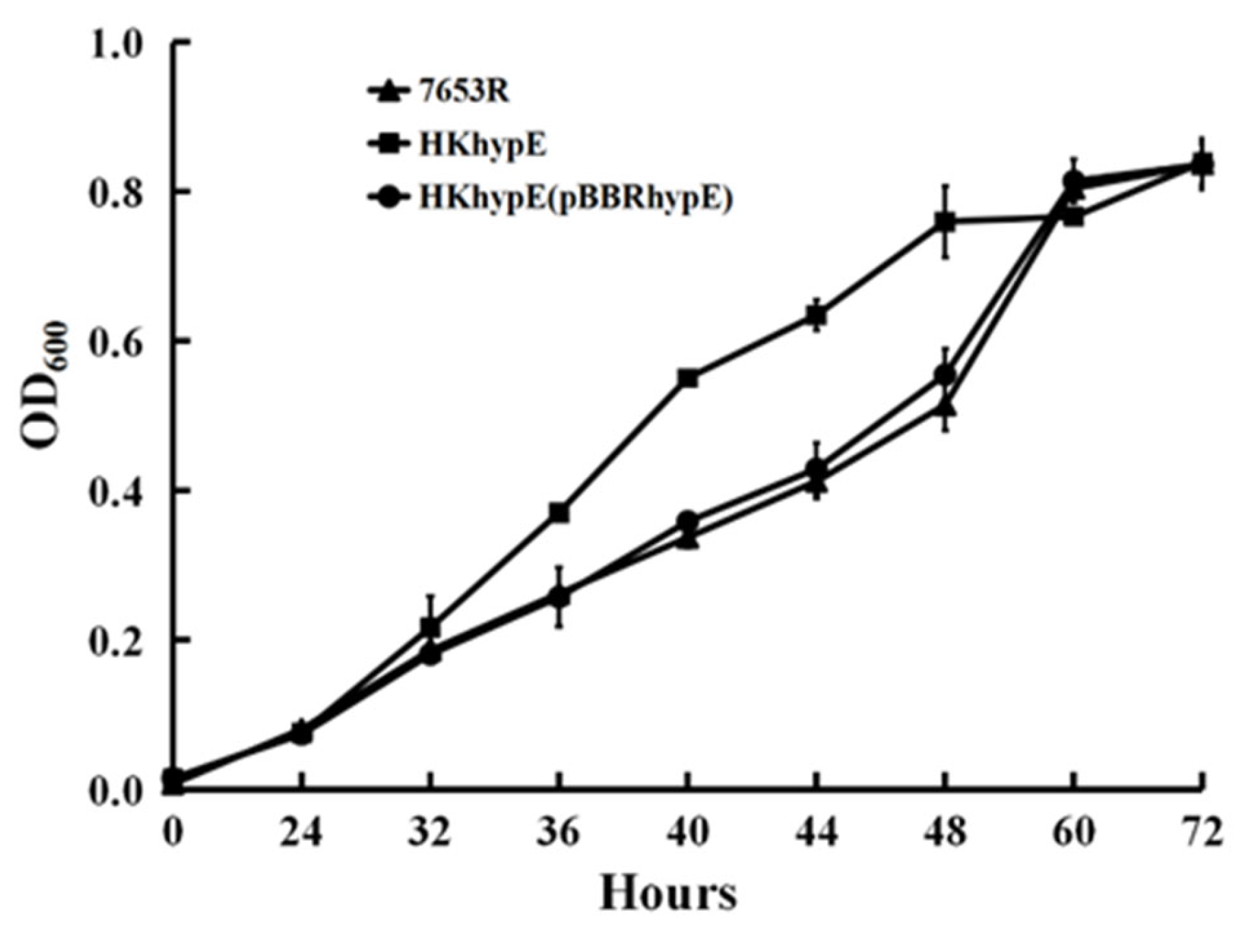
2.2. Growth and Antioxidative Activity in M. huakii hypE Mutant in Free Living Condition

To experimentally confirm the potential function of the hydrogenase maturation protein, a hypE gene mutant HKhypE was made by means of mutagenesis. The growth of the HKhypE strain was compared with that of wild-type 7653R. In liquid AMS minimal medium with glucose as a carbon source and NH4Cl as a nitrogen source, the mutant HKhypE grew slightly faster, and entered a logarithmic growth phase earlier than the parent strain 7653R (Figure 1), while both mutant HKhypE and wild-type strain 7653R achieved the same maximum density. When hypE on plasmid (pBBR1MCS-5) was introduced into mutant HKhypE, the resulting strain HKhypE(pBBRhypE) showed the same growth rate as the wild-type 7653R strain (Figure 1).
In order to study the sensitivity to oxidative stress, growth of mutant HKhypE and wild-type strain 7653R was evaluated by disk diffusion method (Table 1). When H2O2 was given at concentrations of 25, 100, 250, and 1000 mmol/L, the mutant HKhypE showed a clear sensitivity to the H2O2 treatment, and compared with the wild-type strain 7653R, its growth was significantly inhibited, indicating that HypE has critical roles in protecting cells from hydrogen peroxide stress. At concentrations of 25, 100, and 250, the complemented strain HKhypE(pBBRhypE) showed a lower sensitivity compared with the mutant HKhypE (Table 1).

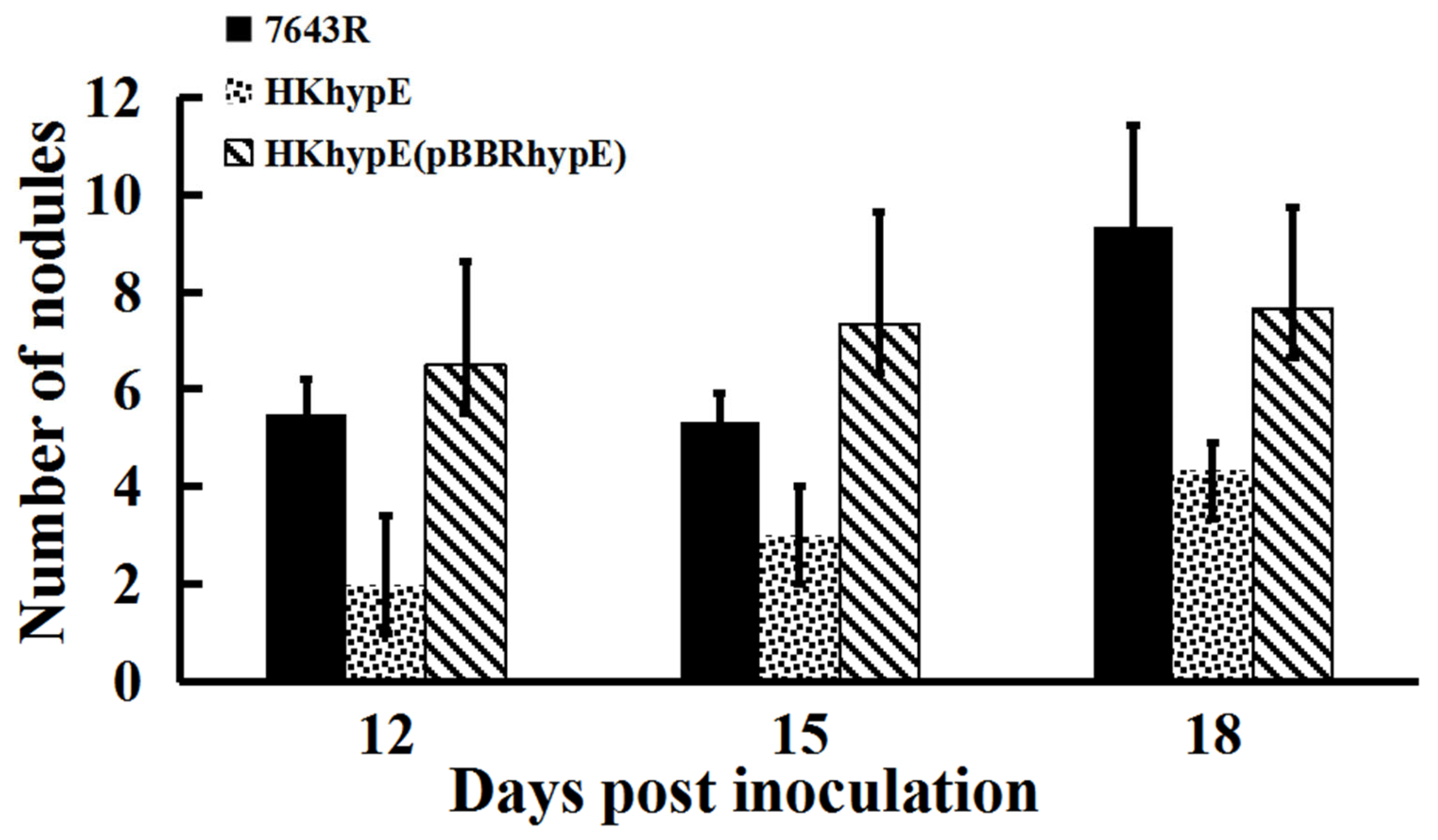
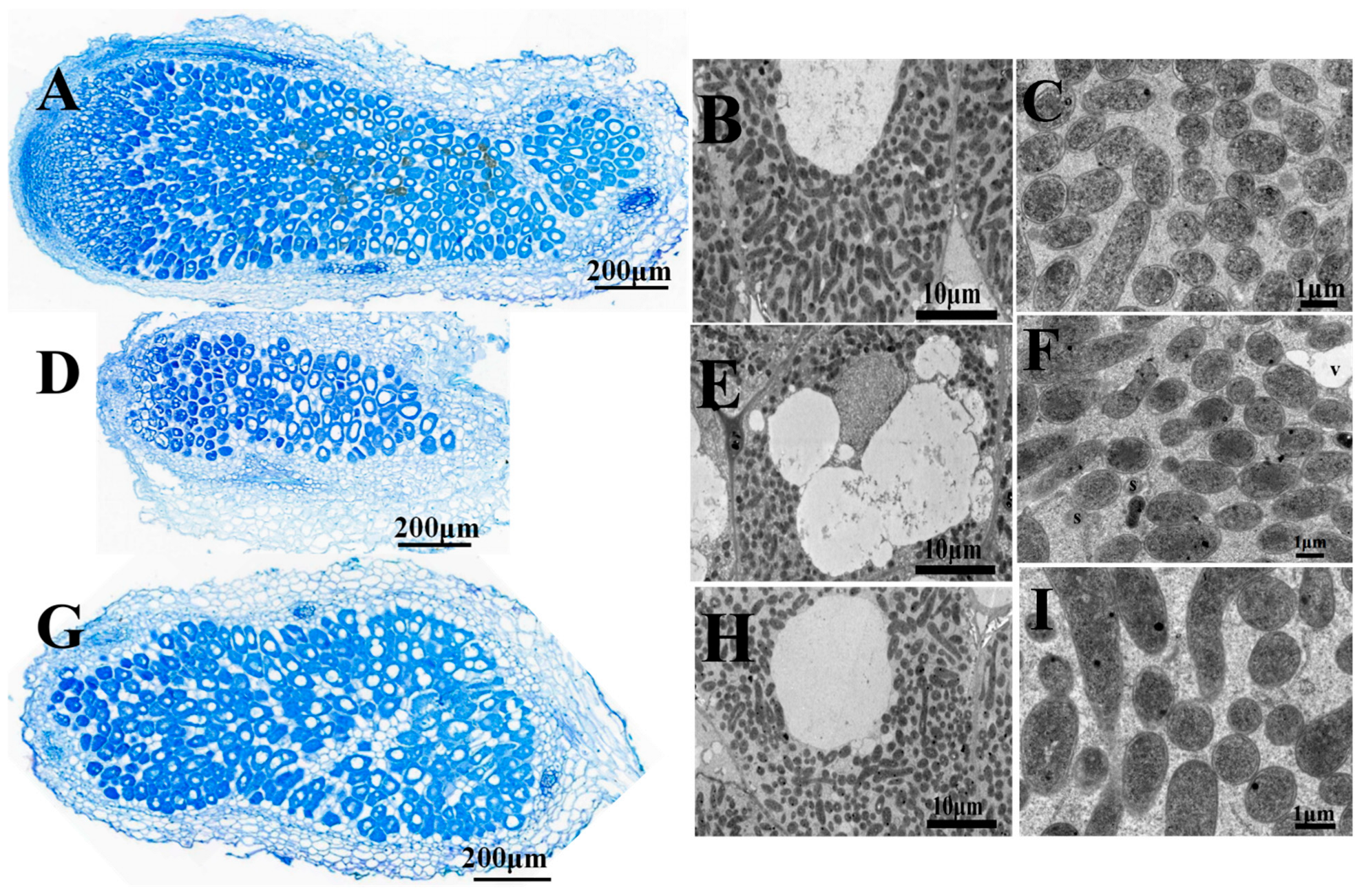
2.3. Symbiotic Properties of hypE Mutant Strain

To determine the function of HypE during symbiotic interactions with an A. sinicus host, the symbiotic performance of the hypE mutant strain was evaluated. During the early stage of nodule formation (at 12, 15, and 18 days post inoculation), nodule number was significantly reduced via the inoculation of mutant HKhypE compared with that of the wild-type strain (Figure 2). However, when root nodules were harvested 28 days after inoculation, no statistically significant difference was observed in the number of nodules between plants inoculated with mutant HKhypE and plants inoculated with wild-type 7653R (Table 2). The wild-type strain showed a normal spherical shape of the nodules (2–4 mm), while the hypE mutant elicited small-size nodules (0.5–1.5 mm), and the nodule weight per plant inoculated with HKhypE was lower than that inoculated with the wild-type strain. A. sinicus plants inoculated with the hypE mutant were shorter and thinner, with more yellow leaves, and the fresh weight of the HKhypE-inoculated plant was 64.39% compared to that of the 7653R-inoculated plant (Table 2). The control plants without rhizobial inoculation had no nodules on their roots and showed clear symptoms of nitrogen deficiency. A notable feature of our study was that plants inoculated with mutant HKhypE showed a significant decrease of more than 47% in acetylene reduction activity compared to that inoculated with wild-type 7653R. Plants inoculated with strain HKhypE(pBBRhypE), in which the mutation in hypE is complemented by a full-length hypE gene cloned on a plasmid, have wild-type properties (Table 2 and Figure 2).
Four-week-old nodules have been examined by light and scanning electron microscopy (SEM). Microscopic analysis of the nodules obtained with mutant HKhypE showed that they were small and filled by rhizobia-infected cells, but contained an abnormally thick cortex (Figure 3D). SEM analysis demonstrated nodule cells infected by hypE mutant contained several more cavities as compared with nodule cells infected by wild-type 7653R (Figure 3B,E). Moreover, the mutant-infected nodule cells showed signs of early senescence with disintegrating bacteria and vacuolation of infected cells (Figure 3F). The results suggested that the hypE-mutant-infected nodules were functionally defective.

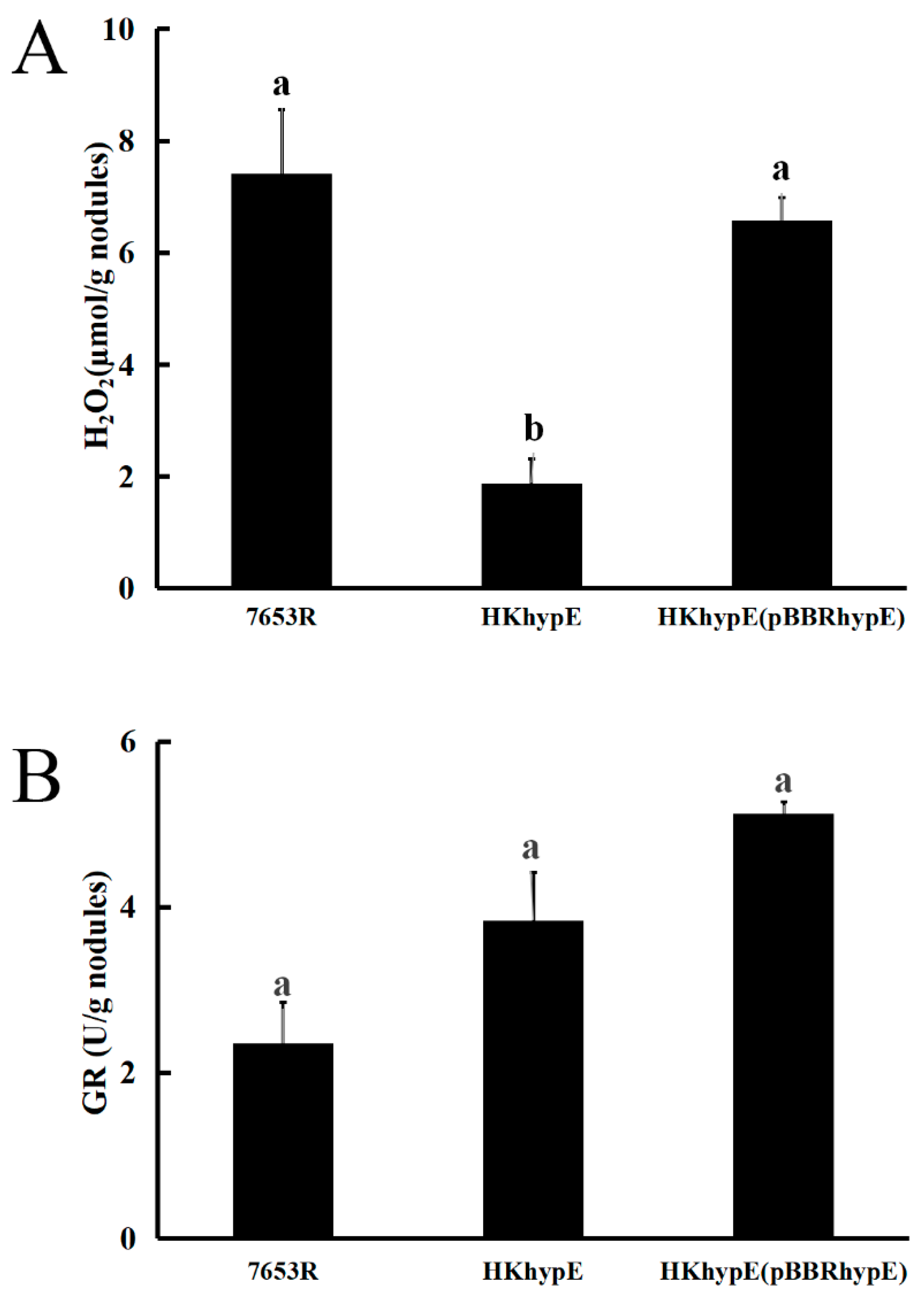
2.4. Effect of hypE Deletion on H2O2 Concentration and Glutathione Reductase Activity in Nodules

To investigate the possible reasons for the changes in symbiotic phenotype resulting from hydrogenase HypE absence, the H2O2 content and glutathione reductase activity in the nodules at 28 days post-inoculation were analyzed. The data showed that the glutathione reductase activity had no significant effect in the hypE mutant, while the quantification results of H2O2 indicated a remarkable decrease in nodules induced by HKhypE compared with plants infected by the wild-type strain (Figure 4). This reduction could be rescued by constitutionally expressing hypE from a plasmid in the mutant (Figure 4). These results indicate that HypE is not associated with glutathione reductase activity and that hydrogenase maturation protein deficiency abolishes the protective effect of H2 against H2O2-induced oxidative damage.

2.5. Rhizosphere Colonization and Competition by M. huakuii Strains

The colonization ability of M. huakuii strains for growth and competition in the rhizosphere of A. sinicus was evaluated after inoculating a low microbial population (103 or 104 bacteria per seedling) into the short-term colonization of the plant rhizosphere and counting the total number of bacteria after one week [24]. When the mutant HKhypE and the wild-type 7653R were inoculated alone into the A. sinicus rhizosphere, the mutant HKhypE was at a significant advantage (36.37% ration of 7653R to HKhypE) compared to the wild-type 7653R (Figure 5). However, when both strains were inoculated together in equal proportion, mutant HKhypE was at a significant disadvantage (53.62% of bacteria recovered) compared to the wild-type 7653R (Student’s t-test; p ≤ 0.01). Even when strain HKhypE was inoculated at a 10-fold excess over 7653R, it accounted for only 273% of bacteria recovered (Figure 5). The results showed that HypE was essential for competition in the host plant rhizosphere by M. huakuii.

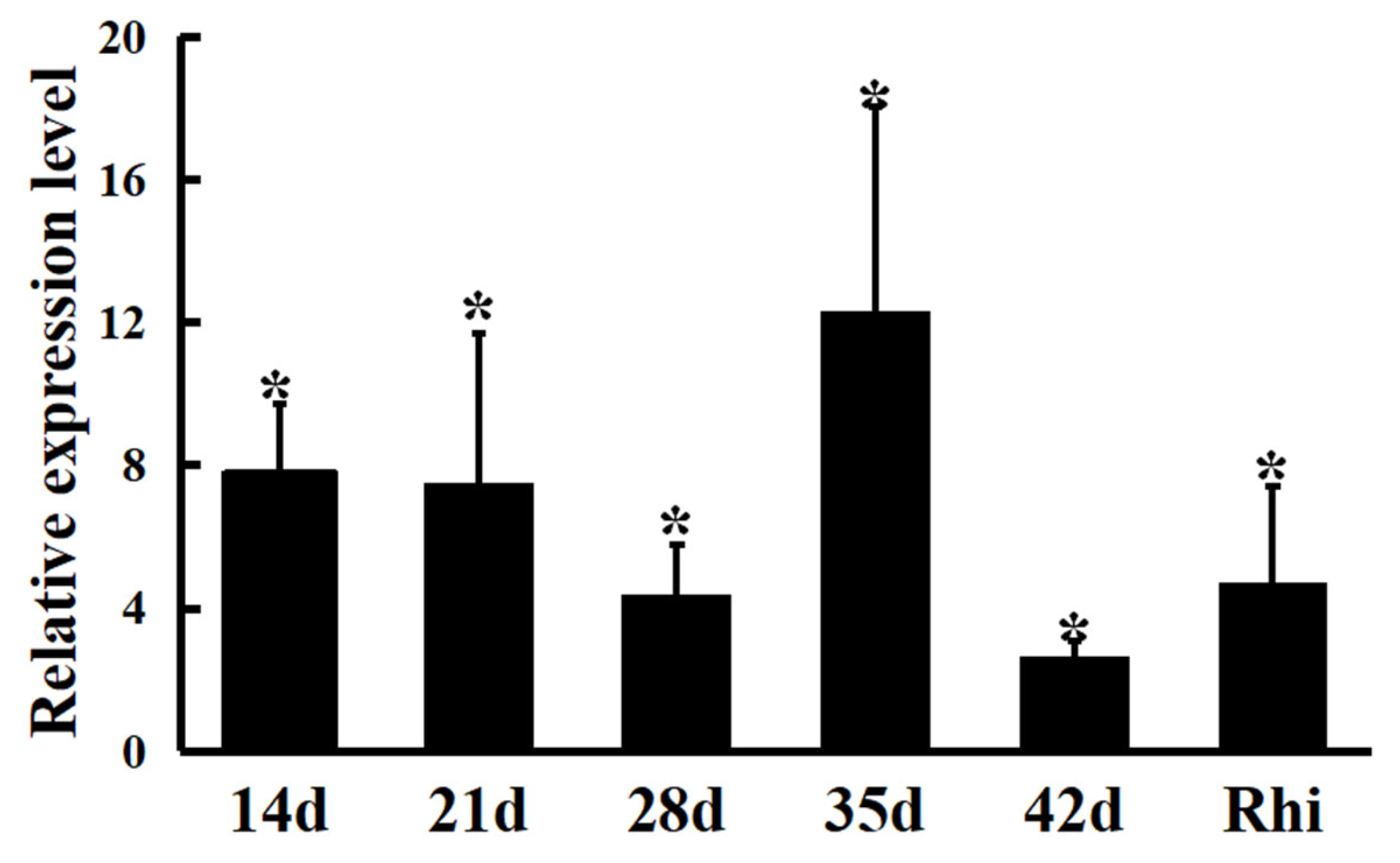
2.6. mRNA Expression Levels of HypE Gene in Nodules Induced by M. huakuii 7653R

The expression levels of the hypE gene in wild-type nodules among different growth stages of host plants (14, 21, 28, 35, and 42 days after inoculation) were determined via quantitative real-time fluorescence polymerase chain reaction (qRT-PCR). In all the treatments, the mRNA levels of hypE gene were significantly up-regulated by 3.5–12.1 fold as compared to wild-type strains in AMS medium, and the hypE gene had the highest expression level (more than 12-fold) in nodules at the nodule maturation stage (35 d) (Figure 6). In the 7-day plant rhizosphere, hypE mRNA levels were also increased (4.2 times the control levels, p < 0.01) (Figure 6). Therefore, hypE gene expression was induced during A. sinicus-M. huakuii symbiosis and was indispensable for nodule/bacteroid development and maturation.

2.7. Proteomic Analysis of Differential Protein Expression in Nodule Bacteroids

A quantitative proteomic approach was performed to examine the influence of HypE deficiency on root nodule symbiosis. The high-throughput analytical method was developed using ultra-performance liquid chromatography coupled with tandem mass spectrometry (UPLC-MS/MS) for protein quantification in the HKhypE mutant and 7653R bacteroids. Proteomics analysis allowed the identification of peptides derived from a total of 2852 protein groups in the hypE gene mutant and wild-type bacteroids, with molecular weights ranging from 6.3 to 317.0 kDa. A total of ninety differentially expressed proteins (fold change > 1.5 or <0.67, p < 0.05) were identified (Table 3). Seven proteins were up-regulated and eighty-three proteins were down-regulated in hypE mutant bacteroids. Eighty-two (91.11%) differential protein-encoding genes were located on the chromosome, and eight (8.89%) were localized in symbiotic megaplasmid pMHb. However, no differential genes were localized on the megaplasmid pMHa.
To categorize these differences into modules of biological relevance, the 90 differential proteins were assigned to six functional categories, which were mainly involved in stress response and virulence (n = 21, 23.33%), energy and nitrogen metabolism (n = 18, 20.20%), transporter activity (n = 15, 16.67%), carbohydrate metabolism (n = 9, 10.00%), nucleotide metabolism (n = 8, 8.89%), and unknown function proteins (n = 19, 21.11%). In particular, all the differential proteins linked to stress response and virulence are down-regulated in the mutant bacteroids. The number of affected oxidoreductase, hydroperoxide reductase, oxygenase, dehydrogenase, thioredoxin, glutathione S-transferase, and antibiotic biosynthesis monooxygenase also suggests that HypE functions in an antioxidant capacity in the root nodules and that the loss of these proteins could result in antioxidant defect. Further analysis of the differentially expressed genes identified a subset involved in electron transport and nitrogen metabolism. Two genes, MCHK_2003 and MCHK_5582, encoding for nitrogen utilization are up-regulated, while all the proteins relative to electron transport and nitrogen fixation ammonia assimilation are down-regulated in the mutant bacteroids. The nitrogenase enzyme is composed of the Fe and MoFe proteins [25]. Three key nitrogen-fixation genes, nifX, nifD, and nifK, are required for nitrogenase component proteins, and nifE is required for the synthesis of the iron–molybdenum cofactor (FeMoco) of nitrogenase. Iron is required for the synthesis of iron-containing proteins in bacteroids for nitrogenase and cytochromes of the electron transport chain [26]. Proteins MCHK_8220 and MCHK_4952 associated with iron metabolism were found to show decreased expression in mutant nodules. The number of differentially expressed nitrogenase genes and nitrogen-fixation-associated genes indicated that hydrogenase maturation protein HypE affects the expression of a wide range of genes involved in the legume–Rhizobium symbiosis interaction.
Furthermore, 15 of the differentially expressed proteins identified are transport proteins, of which 5 are ABC-type nitrate/nitrite transporters. In addition, MgtE is the lone up-regulated transport protein and has been suggested to be essential for N2 fixation [27]. Finally, all the differential expression proteins in the process of nucleotide metabolism were down-regulated in the mutant bacteroids. qRT-PCR was further performed to confirm the validity of the proteome changes. The expression of four genes in four different functional categories were significantly lower in 28-day-old nodules infected by mutant HKhypE compared to wild-type 7653R (Table 3). These results are largely consistent with the changes seen in the proteomic assay results.

3. Discussion

In the process of symbiosis with rhizobium, nitrogen fixation is dependent on a source of ATP and the generation of a reductant at low enough redox potential to transfer electrons to nitrogenase [28]. The nitrogenase complex catalyzes the following reactions: N2 + 8e + 16ATP + 16H2O → 2NH3 + H2 + 16ADP + 16Pi + 8H+. ATP is required for biological nitrogen fixation processes [29], and large amounts of H2 are produced as an obligate by-product of nitrogen fixation in the nodules of legume plants during the nitrogen fixation process. Therefore, this hydrogen production is an important factor limiting the efficiency of symbiotic N2-fixation. An important issue is that a [NiFe] hydrogenase HypE, along with nitrogenase, can utilize H2 to reduce energy losses, and hypE expression is elevated in nitrogen-fixing bacteroids of the root nodules, but the function of HypE in bacteroid nitrogen-fixing systems is poorly understood. Here, we examine the hydrogenase HypE, which is essential for symbiotic nitrogen fixation. Our data demonstrated that HypE is required for root nodule formation and cellular detoxification with regard to its nitrogen fixation capacity and electron transfer.
In this study, mutant HKhypE was constructed using homologous recombination technology, and bioinformatics analysis showed that the HypE protein was involved in the biosynthesis of Ni-Fe hydrogenase and was considered to bind to ATP. The mutation of M. huakuii hypE had less influence on the growth of free-living bacteria. Three hydrogenase minus (Hup-) mutants of Azotobacter chroococcum also gave similar yields to the parent under N2-fixing conditions; however, in carbon-limited mixed cultures, the parent strain outgrew the mutant at high D values, and a Rhodobacter sphaeroides Hup~-/Phb~- mutant strain did not grow well and degraded only 19% of acetic acid, implying that hydrogenase has little effect on the steady-state growth but otherwise can be crucially important to the maintenance of a sustainable rate of growth under stress [30,31]. The mutation of M. huakuii hypE led to decreased antioxidative capacity under hydrogen peroxide H2O2 stress. The direct link between hydrogenase and H2O2 detoxification has been less reported, while in Oligotropha carboxidovorans, the reduction of protons to H2 by CO dehydrogenase is interpreted as a detoxification reaction for electrons to prevent cell damage [32]. It has been reported that the [FeFe] hydrogenase enzymes are excellent catalysts for H2 evolution but rapidly become inactivated in the presence of O2 [33].
HypE plays a prominent role in nodulation and nitrogen fixation, as A. sinicus plants inoculated with mutant HKhypE exhibited large decreases in the nodule number and nitrogen-fixing activity of rhizobial inoculated plants. The hypE-derived mutants formed smaller nodules filled with disintegrating bacteria and vacuolation of infected cells with signs of early senescence. It has been reported that during nitrogen fixation, nitrogenases catalyze the reduction of N2 into NH3 by using protons and electrons with the evolution of H2 [34], [NiFe] hydrogenases, are hydrogen-uptake enzymes that are probably acting to regulate the flow of electrons through electron transport and play a crucial role in energy-conservation processes [35]. Glutathione reductase activity in mutant-inoculated nodules was not different from that wild-type nodules, but the absence of HypE was associated with a 73.7% decrease in nodule H2O2 content. H2O2 appears to play an important signaling role in the establishment and the functioning of the interaction between rhizobia and host plants [36]. It has been reported that A. chroococcurn hydrogenase can benefit bacteria under N2-fixing but not NH4+-utilizing conditions, suggesting that hydrogenase assists the organism either by providing extra energy or by protecting nitrogenase against the inhibition by O2, rather than by protecting nitrogenase against the inhibition of N2 reduction by H2 [31].
Proteomic experiments were performed to provide a foundation for evaluating the effect of hydrogenase on symbiotic nitrogen fixation. Among the 90 differentially expressed proteins, the “stress response and toxicity”-related proteins in bacteroids induced by mutant HKhypE were down-regulated, and most of the proteins related to “electron transport and nitrogen metabolism”, “transport activity”, “carbohydrate metabolism”, and “nucleotide metabolism” were significantly down-regulated. Firstly, hydrogenase was required to supply energy and electrons for the nitrogen fixation reaction. phaR gene was significantly down-regulated, and its expressed protein product regulated the production of polyhydroxyalkanoate (PHA). Studies have shown that Xanthomonas oryzae pv. Oryzae knocks out the phaR gene, showing a decrease in growth rate and a significant decrease in the yield of extracellular polysaccharide [37]. Acidic extracellular polysaccharide is essential for the establishment of nitrogen-fixation symbiosis in leguminous plants. The lack of acidic extracellular polysaccharide will lead to the inability of the Rhizobium to effectively recognize specific hosts [38]. The expression products of the dctA gene are mainly closely related to the transport of C4-dicarboxylate, which is a prerequisite for achieving symbiotic nitrogen fixation [39]. The significant down-regulation of the dctA gene is bound to affect the nitrogen-fixation network. Active nitrogen fixation requires a continuous supply of energy and electrons. PHA can be used as a carbon source and energy to provide energy for the nitrogen-fixation network. The significant down-regulation of the phaR gene was consistent with the results of electron microscope section experiment in which the bacteroids of HKhypE-mutant-infected nodules were significantly smaller than those of the control group. In addition, C4-dicarboxylate plays an isomorphic role in supporting the tricarboxylic acid cycle. Based on the above, it is speculated that the phaP and dctA genes may affect the efficiency of the nitrogen-fixation network through energy efficiency.
Secondly, the hydrogenase HypE is essential for the development of nodule bacteroids. ExbB forms pentamers as a scaffold to form a Ton system with ExbD and TonB, which is used to transport nutrients such as iron and vitamin B12 [40]. In differential proteomics analysis, the significant down-regulation of the exbB gene will undoubtedly hinder the iron transport. In addition, it is interesting to find that the sbmA gene is also significantly down-regulated. The SbmA protein is extremely homologous with BacA protein. The bacA gene has been proven to be closely related to the early development of bacteroids. A lack of the bacA gene will lead to premature senescence of bacteroids in the root nodules [41]. In addition, the hfq gene was also found to be significantly down-regulated. Fhq has been reported to be involved in nodule development, the intracellular activity of bacteroids, and nitrogen fixation [42].
Thirdly, HypE plays a prominent role in rhizobial bacteroid detoxification. Thioredoxins act as antioxidants and function as redox regulators in the bacteroids, and the significant down-regulation of the trxA gene will cause serious damage to biological macromolecules. The Phaseolus vulgaris (common bean) Trxh gene family had the highest expression in the nodule primordium (NP), and their expression patterns in the NP were positively correlated with the symbiotic N2-fixing efficiency of the Rhizobium strain, concomitantly with increased amounts of H2O2 [43]. Another interesting observation is that the recR gene is significantly down-regulated in proteomic analysis. The recR gene is involved in regulating the large amount of DNA synthesis and is an indispensable component [44,45]. Among the “transport activity”-related genes, the mgtE gene is the only up-regulated transporter, which is predicted to be an R. leguminosarum channel and is essential for growth and N2-fixation when both Mg2⁺ is limited and the pH is low [27].
The hypE gene expression is significantly up-regulated during the whole nodulation process, and its highest expression level occurred at 35 days after inoculation. Moreover, the M. huakuii hypE mutant was unable to compete efficiently in the rhizosphere with its parent, which shows that rhizobial HypE is essential for the adaptation of the plant host microenvironment. Taken together, M. huakuii [NiFe] hydrogenase plays an important role in root nodule symbiosis by providing energy and electrons, ROS and pH-dependent detoxification, and control of bacteroid differentiation and senescence.

4. Materials and Methods

4.1. Strains, Plasmids, Primers, and Culture Conditions

All the bacterial strains, plasmids, and primers used in this work and their relevant characteristics are listed in Table 4. M. huakuii strains were grown at 28 °C in either tryptone yeast extract (TY) [46] or acid minimal salts (AMS) medium [47] supplemented with D-glucose (10 mM) as a carbon source and NH4Cl (10 mM) as a nitrogen source. Antibiotics were used at the following concentrations (μg/mL): streptomycin (Str), 250; kanamycin (Km), 20; gentamicin (Gm), 20; neomycin(Neo), 80 or 250 (for generating the hypE mutant); spectinomycin (Spe), 100. To monitor culture growth, strains were grown at 28 °C in AMS liquid medium with shaking (200 rpm), and optical density at 600 nm (OD600) was measured in three independent cultures.

4.2. Construction and Complementation of a hypE Mutant of M. huakuii 7653R

A 657bp hypE (MCHK_8345) fragment was PCR-amplified using primers hypEfor and hypErev. The fragment was cloned into the BamH I and Hind III sites of pK19mob, resulting in plasmid pKhypE. Plasmid pKhypE was conjugated into strain 7653R via triparental mating using the helper plasmid pRK2013, as previously described [47]. Insertions into hypE gene of strain 7653R were confirmed via PCR using hypEmap and a pK19mob-specific primer (either pK19A or pK19B).
To construct a plasmid for complementation of the hypE mutant, a 1.82 kb DNA fragment of the complete hypE gene was PCR-amplified from the genomic DNA of M. huakuiii 7653R using primers chypEfor and chypErev. The PCR product was cloned into Xba I and BamH I sites of pBBR1MCS-5, and the resultant plasmid was named pBBRhypE. Plasmid pBBRhypE was conjugated into the HKhypE recipient strain via triparental mating using pRK2013 as a helper plasmid. Using selection for gentamicin resistance, complemented strain HKhypE(pBBRhypE) was isolated as previously described [52].

4.3. Cellular Sensitivity to H2O2

The H2O2 resistance assay was performed using the disk diffusion method as previously described by M. huakuiii 7653R, and HKhypE and HKhypE(pBBRhypE) were grown aerobically in AMS Glc/NH4+ [53]. An amount of 100 µL of each bacterial suspension (approximately OD600 = 0.4) was spotted onto solid TY medium. Sterile paper disks of 6 mm in diameter were laid on the inoculated plates. A total of 10 µL of 25, 100, 250, 1000 mM H2O2 was pipetted onto the surface of separate disks. The plates were incubated at 28 °C until circular clear zones could be observed. The diameter of zone of inhibition (mm) observed was measured for the mutant and compared with that for wild-type 7653R to provide an estimate of its relative susceptibility to oxidants. The experiment was repeated three times, and the data were analyzed using two-way ANOVA (p < 0.05).

4.4. Plant Experiments and Microscope Study of Nodules

Seeds of Astragalus sinicus were surface-sterilized for 5 min in 75% ethanol, soaked 20 min in 2% sodium hypochlorite, and then rinsed 10 times with sterile water. The plants were grown in 500 mL pots containing sterile vermiculite and watered with nitrogen-free Fahraeus solution. Inoculation with M. huakuii strains was conducted on 7-day-old seeds. Plants were incubated in a controlled-environment chamber with a cycle of 8 h at 20 °C in the dark and a 16 h photoperiod at 22 °C in the light. Number of root nodules in the early growing stage was counted at 12, 15, and 18 days post-inoculation. At 28 days post-inoculation, nodule number per plant, nodule fresh weight per plant, fresh weight per plant, and above-ground fresh weight per plant were measured. Acetylene reduction activity was determined by gas chromatographic measurement, as previously described [54]. Briefly, The plants were extracted and placed in a 50 mL milled bottle, and 1.28 mL of acetylene was injected into the bottle, which was then incubated in a growth chamber at 28 °C for 1 h. Then, 1 mL of gas from the bottle was aspirated and injected into a SP-2100A gas chromatograph (Beijing Beifen-Ruili Analytical Instrument (Group) Co., Ltd., Beijing, China), and the ethylene peak area was separated and detected using an OV-101 capillary column (1.5 m × 0.32 mm × 0.5 μm). The experiment consisted of two independent experiments, each of which had at least five repeats, and statistical differences were analyzed using two-way ANOVA (p  <  0.05).
Root nodules at 28 days post-inoculation were obtained and examined by the use of both light and electron microscopes. Nodules were washed and fixed in 2.5% glutaraldehyde, postfixed in 1.5% osmium tetroxide, and frozen under liquid nitrogen. Semi-thin sections (1–3 µm) of nodules were cut, stained with toluidine-blue solution, and evaluated using a light microscope (SZX16, Olympus Corporation; Tokyo, Japan). Ultra-thin sections were stained with uranyl acetate and lead citrate and examined using a Hitachi H-7100 transmission electron microscope.

4.5. Measurement of H2O2 Concentration and Glutathione Reductase Activity in the Nodules

The roots of 7-day-old seedlings were inoculated with M. huakuii strains. Nodules at 28 days post-inoculation were collected, ground into fine powders in liquid nitrogen, and then suspended in precooled extraction buffer (10 mM Tris-HCl, pH 7.0). H2O2 concentration and glutathione reductase activity were measured using corresponding kits (catalogue numbers BC3590-50 for H2O2 Content Assay and BC1160-50 for glutathione reductase activity, Solarbio life sciences) following the manufacturers’ instructions. The experiment was repeated three times, and the data were analyzed using two-way ANOVA (p  <  0.05).

4.6. Rhizosphere Colonization

Rhizosphere colonization was performed as described previously [53]. A. sinicus seedlings were germinated and grown in 20 mL centrifuge tubes filled with sterile vermiculite, as described above for acetylene reduction. The 7 day-old plants were inoculated with M. huakuii 7653R and HKhypE in the cfu ratios 1000:0, 0:1000, 1000:1000, and 1000:10,000. After 7 days (14 days post-inoculation), shoots were cut off, and 10 mL of sterile phosphate-buffered saline (PBS) buffer (pH 7.4) was added to the roots and vortexed for 15 min [55]. After vortexing, the samples were serially diluted and plated on TY medium containing either streptomycin or streptomycin and neomycin, giving the total number of viable rhizosphere- and root-associated bacteria. The plates were incubated at 28 °C for 72 h before the colonies were counted. Each treatment consisted of ten replications, and each test consisted of a single plant. Statistical differences were evaluated using one-way ANOVA (p  <  0.05).

4.7. RNA Isolation and Quantitative Reverse Transcription-PCR (qRT-PCR)

qRT-PCR was used to determine differences in the expression of genes. M. huakuii samples were collected in triplicates from free-living M. huakuii 7653R cultured in AMS medium, rhizosphere strains at 7 days post-inoculation, and plant nodules, which were harvested from A. sinicus inoculated with M. huakuii after 14, 21, 28, 35, and 42 days. The nodules of plants were ground into a fine powder with liquid nitrogen. Total RNA was isolated using the Trizol Reagent (Invitrogen). First-strand cDNA synthesis and double-strand cDNA amplification were performed using the PrimeScript RT reagent kit with gDNA Eraser (Takara, Dalian, China) according to the manufacturer’s instructions. qRT-PCR was performed using SYBR Premix ExTaq kit (Takara, Dalian, China) following the manufacturer’s instructions on the BIO-RAD CFX96 Real-Time PCR Detection System. The sequences of primers for qRT-PCR were listed in Table 4. The gyrB gene of M. huakuii was used as a reference. For each experiment, three independent biological replicates were performed, and the relative expression levels of the mRNAs of the target genes were normalized using the 2−ΔΔCT method.

4.8. Protein Extraction, Digestion, and Peptide Labeling

The 28-day nodules induced by wild-type 7653R or mutant HKhypE were harvested and ground into fine powder in liquid nitrogen. The tissue powder was transferred into a 5 mL ice-cold centrifuge tube. Four volumes of lysis buffer (1% protease inhibitor cocktail in 8 M urea) were then added to the cell powder, and the lysates were sonicated three times on ice using a high-intensity ultrasonic processor. The insoluble fraction was removed via centrifugation in a cooled centrifuge (at 12,000 rpm for 10 min at 4 °C). Then, the clarified supernatant was collected, and the total protein content was quantified using a BCA protein assay kit (Pierce, Rockland, IL, USA). Equal amounts of proteins were reduced with 10 mM dithiothreitol (DTT) and alkylated with 11 mM iodoacetamide (IAA) for 15 min at room temperature in the dark. The protein sample was diluted with 100 mM tetraethyl ammonium bromide (TEAB) so that the final concentration of urea was less than 2 M. For digestion of protein, trypsin was added at a protein-to-enzyme ratio of 100:1 (100 μg total protein was added to 1 μg trypsin), and the digestion was performed overnight at 37 °C. After digestion, the peptides were desalted using a Strata X C18 SPE column and vacuum-dried. Then, the dried peptide was reconstituted in 0.5 M TEAB and processed by following the manufacturer’s protocol of the TMT kit (ThermoFisher Scientific, Bremen, GA, USA). Briefly, one unit of tandem mass tag (TMT) reagent, together with 100 μg of sample peptides, was dissolved and reconstituted in acetonitrile. After incubation at room temperature for 2 h, the peptide mixtures were desalted and dried via vacuum centrifugation.

4.9. Fractionation of Tryptic Peptides and LC-MS/MS Analysis

The labeled peptide was mixed and fractionated via high-performance liquid chromatography (Thermo Scientific EASY-nLC 1000) using an Agilent 300 Extend C18 column (5 μm × 4.6 mm × 250 mm). Concisely, the peptides were first separated using a gradient of 2–60% acetonitrile in ammonium bicarbonate (10 mM, pH 10). The extracts were fractionated into 80 fractions in 80 min, and these fractions were then combined into 18 fractions and dried via vacuum centrifugation.
The tryptic peptides were resuspended in buffer solvent A (30% acetonitrile, 70% water, 0.1% formic acid) and separated with a reversed-phase analytical column (75 μm × 15 cm). For whole-cell proteome analysis, the gradient was run at a constant flow rate of 400 nL min−1 for 45 min, starting from 6% to 22% solvent B (0.1% formic acid in 98% acetonitrile) over 26 min, followed by 23% to 35% solvent B in 8 min, and increased to 80% solvent B in 3 min, then maintained at 80% solvent B for another 3 min.
The eluted peptides were further analyzed using a Q Exactive™ plus tandem mass spectrometer (Thermo Fisher Scientific, Massachusetts, USA) coupled with ultra-performance liquid chromatography (UPLC) (Thermo Scientific EASY-nLC 1000, Massachusetts, USA). UPLC was performed using homemade analytical column with integrated spray tip (100 μm i.d. × 25 cm) packed with 1.9 μm/120 Å ReproSil-PurC18 resins (Dr. Maisch GmbH, Ammerbuch, Germany). The intact peptides were detected in the Orbitrap at a high resolution of 70,000. Peptides were selected for MS/MS using a normalized collision energy (NCE) setting of 28, and ion fragments were detected in the Orbitrap at a low resolution of 17,500. A data-dependent procedure that alternated between one MS scan followed by 20 MS/MS scans with 10 s dynamic exclusion, and the electrospray voltage applied was 2.0 kV. In order to prevent overfilling of the ion trap, automatic gain control (AGC) was set to accumulate 5 × 10⁴ ions for generation of MS/MS spectra. For the full-scan mode, the mass range for the MS scans was 350 to 1800 m/z, and the MS analysis alternated between MS and data-dependent MS2 scans using dynamic exclusion. For each sample, three independent biological replicates were performed.

4.10. Data Analysis

The resulting MS/MS data were processed using the MaxQuant search engine (version 1.5.2.8). Tandem mass spectra were searched against the M. huakuii 7653R genome database. Trypsin/P was specified as a cleavage enzyme allowing up to 4 missing cleavages. The mass error was set to 20 ppm for the precursor ions, and the mass tolerance was set to 0.05 Da for the MS/MS fragment ion matches. Carbamidomethylation of cysteine was selected as fixed modification, while methionine oxidation was set as variable modification. TMT 6-plex was selected in Mascot for the protein quantification method. A false discovery rate (FDR) of 1% was imposed for protein and peptide identifications. The minimum peptide length was set to 7, and the minimal peptide score for modified peptides was set to 40. The site localization probability was set to > 0.75. Statistical significance for protein expression was analyzed using Student’s t tests (p < 0.05). Only proteins identified in at least two of three biological replicates with a fold change higher than 1.5 or lower than 0.67 were considered to indicate statistically significant difference in protein abundance between mutant and wild-type strains. The mass spectrometry proteomics data have been uploaded to the ProteomeXchange Consortium via the PRIDE partner repository with the dataset identifier PXD026564.

5. Conclusions

We discovered that the mutant in the hydrogenase maturation protein HypE can affect symbiotic nitrogen fixation between rhizobia and host via the formation of smaller and defective nodules. The hypE mutant displayed decreased antioxidative capacity and competition ability in the host plant rhizosphere. The proteomic results show that HypE is mainly involved in stress response and virulence, energy and nitrogen metabolism, and transporter activity. As a result, we believe that M. huakuii [NiFe] hydrogenase HypE plays an important role in root nodule symbiosis by providing energy and electrons, ROS, and control of bacteroid differentiation and senescence.

Author Contributions

Conceptualization, G.C.; methodology, S.L., M.S. and F.Y.; formal analysis, G.C., S.L., M.S., Q.Z. and A.H.; investigation, G.C., S.L. and X.C.; writing—original draft preparation, G.C., S.L. and M.S.; writing—review and editing, G.C., S.L. and X.C.; visualization, S.L. and M.S.; supervision, G.C.; funding acquisition, G.C. All authors have read and agreed to the published version of the manuscript.

Funding

This study was supported by the National Natural Science Foundation of China (grant no. 31772399).

Institutional Review Board Statement

Not applicable.

Informed Consent Statement

Not applicable.

Data Availability Statement

The datasets presented in this study can be found in online repositories. The names of the repository/repositories and accession number(s) can be found below: https://www.ebi.ac.uk/pride/archive/projects/PXD026564 (accessed on 8 August 2021).

Acknowledgments

The authors thank PTM-Biolabs Co., Ltd. (Hangzhou, China) for mass spectrometry analysis.

Conflicts of Interest

The authors declare no conflict of interest.

References

  1. Maeda, T.; Vardar, G.; Self, W.T.; Wood, T.K. Inhibition of hydrogen uptake in Escherichia coli by expressing the hydrogenase from the cyanobacterium Synechocystis sp. PCC 6803. BMC. Biotechnol. 2007, 7, 25. [Google Scholar] [CrossRef] [PubMed] [Green Version]
  2. Sickerman, N.S.; Hu, Y. Hydrogenases. Methods Mol. Biol. 2019, 1876, 65–88. [Google Scholar]
  3. Calusinska, M.; Happe, T.; Joris, B.; Wilmotte, A. The surprising diversity of clostridial hydrogenases: A comparative genomic perspective. Microbiology 2010, 156, 1575–1588. [Google Scholar] [CrossRef] [Green Version]
  4. Forzi, L.; Sawers, R.G. Maturation of [NiFe]-hydrogenases in Escherichia coli. Biometals 2007, 20, 565–578. [Google Scholar] [CrossRef]
  5. Lubitz, W.; Ogata, H.; Rüdiger, O.; Reijerse, E. Hydrogenases. Chem. Rev. 2014, 114, 4081–4148. [Google Scholar] [CrossRef] [PubMed]
  6. Shomura, Y.; Hagiya, K.; Yoont, K.S.; Nishihara, H.; Higuchi, Y. Crystallization and preliminary X-ray diffraction analysis of membrane-bound respiratory [NiFe] hydrogenase from Hydrogenovibrio marinus. Acta Crystallogr. Sect. F Struct. Biol. Cryst. Commun. 2011, 67, 827–829. [Google Scholar] [CrossRef] [PubMed] [Green Version]
  7. Terpolilli, J.J.; Hood, G.A.; Poole, P.S. What determines the efficiency of N(2)-fixing Rhizobium-legume symbioses? Adv. Microb. Physiol. 2012, 60, 325–389. [Google Scholar]
  8. Jensen, B.B.; Cox, R.P. Direct measurements of steady-state kinetics of cyanobacterial n(2) uptake by membrane-leak mass spectrometry and comparisons between nitrogen fixation and acetylene reduction. Appl. Environ. Microbiol. 1983, 45, 1331–1337. [Google Scholar] [CrossRef]
  9. Brito, B.; Palacios, J.M.; Imperial, J.; Ruiz-Argüeso, T. Engineering the Rhizobium leguminosarum bv. viciae hydrogenase system for expression in free-living microaerobic cells and increased symbiotic hydrogenase activity. Appl. Environ. Microbiol. 2002, 68, 2461–2467. [Google Scholar] [CrossRef] [Green Version]
  10. Annan, H.; Golding, A.L.; Zhao, Y.; Dong, Z. Choice of hydrogen uptake (Hup) status in legume-rhizobia symbioses. Ecol. Evol. 2012, 2, 2285–2290. [Google Scholar] [CrossRef]
  11. Brito, B.; Prieto, R.I.; Cabrera, E.; Mandrand-Berthelot, M.A.; Palacios, J.M. Rhizobium leguminosarum hupe encodes a nickel transporter required for hydrogenase activity. J. Bacteriol. 2009, 192, 925–935. [Google Scholar] [CrossRef] [PubMed] [Green Version]
  12. Benoit, S.; Mehta, N.; Wang, G.; Gatlin, M.; Maier, R.J. Requirement of hydD, hydE, hypC and hypE genes for hydrogenase activity in Helicobacter pylori. Microb. Pathog. 2004, 36, 153–157. [Google Scholar] [CrossRef] [PubMed]
  13. Shomura, Y.; Komori, H.; Miyabe, N.; Tomiyama, M.; Shibata, N.; Higuchi, Y. Crystal structures of hydrogenase maturation protein HypE in the Apo and ATP-bound forms. J. Mol. Biol. 2007, 372, 1045–1054. [Google Scholar] [CrossRef] [PubMed]
  14. Blokesch, M.; Paschos, A.; Bauer, A.; Reissmann, S.; Drapal, N.; Böck, A. Analysis of the transcarbamoylation-dehydration reaction catalyzed by the hydrogenase maturation proteins HypF and HypE. Eur. J. Biochem. 2004, 271, 3428–3436. [Google Scholar] [CrossRef]
  15. Miki, K.; Atomi, H.; Watanabe, S. Structural insight into [NiFe] hydrogenase maturation by transient complexes between Hyp proteins. Acc. Chem. Res. 2020, 53, 875–886. [Google Scholar] [CrossRef] [PubMed]
  16. Blokesch, M.; Albracht, S.P.; Matzanke, B.F.; Drapal, N.M.; Jacobi, A.; Böck, A. The complex between hydrogenase-maturation proteins HypC and HypD is an intermediate in the supply of cyanide to the active site iron of [NiFe]-hydrogenases. J. Mol. Biol. 2004, 344, 155–167. [Google Scholar] [CrossRef]
  17. McKinlay, J.B.; Zeikus, J.G. Extracellular iron reduction is mediated in part by neutral red and hydrogenase in Escherichia coli. Appl. Environ. Microbiol. 2004, 70, 3467–3474. [Google Scholar] [CrossRef] [Green Version]
  18. Rey, L.; Imperial, J.; Palacios, J.M.; Ruiz-Argüeso, T. Purification of Rhizobium leguminosarum HypB, a nickel-binding protein required for hydrogenase synthesis. J. Bacteriol. 1994, 176, 6066–6073. [Google Scholar] [CrossRef] [Green Version]
  19. Brito, B.; Martínez, M.; Fernández, D.; Rey, L.; Cabrera, E.; Palacios, J.M.; Imperial, J.; Ruiz-Argüeso, T. Hydrogenase genes from Rhizobium leguminosarum bv. viciae are controlled by the nitrogen fixation regulatory protein nifA. Proc. Natl. Acad. Sci. USA 1997, 94, 6019–6024. [Google Scholar] [CrossRef]
  20. Hernando, Y.; Palacios, J.; Imperial, J.; Ruiz-Argüeso, T. Rhizobium leguminosarum bv. viciae hypA gene is specifically expressed in pea (Pisum sativum) bacteroids and required for hydrogenase activity and processing. FEMS Microbiol. Lett. 1998, 169, 295–302. [Google Scholar] [CrossRef] [Green Version]
  21. Olson, J.W.; Fu, C.; Maier, R.J. The HypB protein from Bradyrhizobium japonicum can store nickel and is required for the nickel-dependent transcriptional regulation of hydrogenase. Mol. Microbiol. 1997, 24, 119–128. [Google Scholar] [CrossRef]
  22. Albareda, M.; Pacios, L.F.; Manyani, H.; Rey, L.; Brito, B.; Imperial, J.; Ruiz-Argüeso, T.; Palacios, J.M. Maturation of Rhizobium leguminosarum hydrogenase in the presence of oxygen requires the interaction of the chaperone HypC and the scaffolding protein HupK. J. Biol. Chem. 2014, 289, 21217–21229. [Google Scholar] [CrossRef] [Green Version]
  23. Albareda, M.; Manyani, H.; Imperial, J.; Brito, B.; Ruiz-Argüeso, T.; Böck, A.; Palacios, J.M. Dual role of HupF in the biosynthesis of [NiFe] hydrogenase in Rhizobium leguminosarum. BMC Microbiol. 2012, 12, 256. [Google Scholar] [CrossRef] [PubMed] [Green Version]
  24. Cheng, G.; Karunakaran, R.; East, A.K.; Munozazcarate, O.; Poole, P.S. Glutathione affects the transport activity of Rhizobium leguminosarum 3841 and is essential for efficient nodulation. FEMS Microbiol. Lett. 2017, 364, fnx045. [Google Scholar] [CrossRef] [PubMed] [Green Version]
  25. Lahiri, S.; Cole, B.; Pulakat, L.; Gavini, N. The NifX Protein is Involved in the Final Stages of FeMo-cofactor Transport to the MoFe Protein. Am. J. Biochem. Biotechnol. 2007, 3, 92–102. [Google Scholar] [CrossRef]
  26. Brear, E.M.; Day, D.A.; Smith, P.M. Iron: An essential micronutrient for the legume-rhizobium symbiosis. Front. Plant. Sci. 2013, 4, 359. [Google Scholar] [CrossRef] [Green Version]
  27. Hood, G.; Karunakaran, R.; Downie, J.A.; Poole, P. MgtE from Rhizobium leguminosarum is a Mg2+ channel essential for growth at low pH and N2 fixation on specific plants. Mol. Plant Microbe Interact. 2015, 28, 1281–1287. [Google Scholar] [CrossRef] [Green Version]
  28. Lindblad, A.; Nordlund, S. Electron transport to nitrogenase in Rhodospirillum rubrum: Role of energization of the chromatophore membrane. Photosynth. Res. 1997, 53, 23–28. [Google Scholar] [CrossRef]
  29. Eady, R.R.; Postgate, J.R. Nitrogenase. Nature 1974, 249, 805–810. [Google Scholar] [CrossRef]
  30. Kim, M.S.; Baek, J.S.; Lee, J.K. Comparison of H2 accumulation by Rhodobacter sphaeroides KD131 and its uptake hydrogenase and PHB synthase deficient mutant. Int. J. Hydrogen Energy 2006, 31, 121–127. [Google Scholar] [CrossRef]
  31. Aguilar, O.M.; Yates, M.G.; Postgate, J.R. The beneficial effect of Hydrogenase in Azotobacter chroococcum under Nitrogen-fixing, carbon-limiting conditions in continuous and batch cultures. Microbiology 1985, 131, 3141–3145. [Google Scholar] [CrossRef] [Green Version]
  32. Beatrix, S.; Ortwin, M. Characterization of hydrogenase activities associated with the molybdenum CO dehydrogenase from oligotropha carboxidovorans. FEMS Microbiol. Lett. 1996, 136, 157–162. [Google Scholar]
  33. Wang, V.C.; Esmieu, C.; Redman, H.J.; Berggren, G.; Hammarström, L. The reactivity of molecular oxygen and reactive oxygen species with [FeFe] hydrogenase biomimetics: Reversibility and the role of the second coordination sphere. Dalton Trans. 2020, 49, 858–865. [Google Scholar] [CrossRef] [PubMed] [Green Version]
  34. Seiji, O.; Bunsho, K.; Hidetaka, N.; Yoshihito, W.; Shunichi, F. Why do nitrogenases waste electrons by evolving dihydrogen? Appl. Organomet. Chem. 2004, 18, 589–594. [Google Scholar]
  35. Appel, J.; Phunpruch, S.; Steinmüller, K.; Schulz, R. The bidirectional hydrogenase of Synechocystis sp. PCC 6803 works as an electron valve during photosynthesis. Arch. Microbiol. 2000, 173, 333–338. [Google Scholar] [CrossRef]
  36. Puppo, A.; Pauly, N.; Boscari, A.; Mandon, K.; Brouquisse, R. Hydrogen peroxide and nitric oxide: Key regulators of the Legume-Rhizobium and mycorrhizal symbioses. Antioxid. Redox. Sign. 2013, 18, 2202–2219. [Google Scholar] [CrossRef]
  37. Long, J.Y.; Song, K.L.; He, X.; Zhang, B.; Cui, X.F.; Song, C.F. Mutagenesis of PhaR, a regulator gene of polyhydroxyalkanoate biosynthesis of Xanthomonas oryzae pv. oryzae-caused pleiotropic phenotype changes. Front. Microbiol. 2018, 9, 3046. [Google Scholar] [CrossRef]
  38. Gray, J.X.; Rolfe, B.G. Exopolysaccharide production in Rhizobium and its role in invasion. Mol. Microbiol. 1990, 4, 1425–1431. [Google Scholar] [CrossRef]
  39. Van Slooten, J.C.; Bhuvanasvari, T.V.; Bardin, S.; Stanley, J. Two C4-dicarboxylate transport systems in Rhizobium sp. NGR234: Rhizobial dicarboxylate transport is essential for nitrogen fixation in tropical legume symbioses. Mol. Plant Microbe Interact. 1992, 5, 179–186. [Google Scholar] [CrossRef]
  40. Maki-Yonekura, S.; Matsuoka, R.; Yamashita, Y.; Shimizu, H.; Tanaka, M.; Iwabuki, F.; Yonekura, K. Hexameric and pentameric complexes of the ExbBD energizer in the Ton system. eLife 2018, 7, e35419. [Google Scholar] [CrossRef]
  41. Glazebrook, J.; Ichige, A.; Walker, G.C. A Rhizobium meliloti homolog of the Escherichia coli peptide-antibiotic transport protein SbmA is essential for bacteroid development. Genes Dev. 1993, 7, 1485–1497. [Google Scholar] [CrossRef] [PubMed] [Green Version]
  42. Becker, A.; Overlöper, A.; Schlüter, J.P.; Reinkensmeier, J.; Robledo, M.; Giegerich, R.; Narberhaus, F.; Evguenieva-Hackenberg, E. Riboregulation in plant-associated α-proteobacteria. RNA Biol. 2014, 11, 550–562. [Google Scholar] [CrossRef] [Green Version]
  43. Boubakri, H.; Chihaoui, S.A.; Najjar, E.; Gargouri, M.; Barhoumi, F.; Jebara, M. Genome-wide analysis and expression profiling of H-type Trx family in Phaseolus vulgaris revealed distinctive isoforms associated with symbiotic N2-fixing performance and abiotic stress response. J. Plant Physiol. 2021, 260, 153410. [Google Scholar] [CrossRef]
  44. Bentchikou, E.; Servant, P.; Coste, G.; Sommer, S. A major role of the RecFOR pathway in DNA double-strand-break repair through ESDSA in Deinococcus radiodurans. PLoS Genet. 2010, 6, e1000774. [Google Scholar] [CrossRef] [Green Version]
  45. Satoh, K.; Kikuchi, M.; Ishaque, A.M.; Ohba, H.; Yamada, M.; Tejima, K.; Onodera, T.; Narumi, I. The role of Deinococcus radiodurans RecFOR proteins in homologous recombination. DNA Repair 2012, 11, 410–418. [Google Scholar] [CrossRef]
  46. Beringer, J.E.; Hopwood, D.A. Chromosomal recombination and mapping in Rhizobium leguminosarum. Nature 1976, 264, 291–293. [Google Scholar] [CrossRef] [PubMed]
  47. Poole, P.S.; Blyth, A.; Reid, C.; Walters, K. myo-Inositol catabolism and catabolite regulation in Rhizobium leguminosarum bv. viciae. Microbiology 1994, 140, 2787–2795. [Google Scholar] [CrossRef] [Green Version]
  48. Cheng, G.J.; Li, Y.G.; Zhou, J.C. Cloning and identification of opa22, a new gene involved in nodule formation by Mesorhizobium huakuii. FEMS Microbiol. Lett. 2006, 257, 152–157. [Google Scholar] [CrossRef] [Green Version]
  49. Schäfer, A.; Tauch, A.; Jäger, W.; Kalinowski, J.; Thierbach, G.; Pühler, A. Small mobilizable multi-purpose cloning vectors derived from the Escherichia coli plasmids pK18 and pK19: Selection of defined deletions in the chromosome of Corynebacterium glutamicum. Gene 1994, 145, 69–73. [Google Scholar] [CrossRef]
  50. Figurski, D.H.; Helinski, D.R. Replication of an origin-containing derivative of plasmid RK2 dependent on a plasmid function provided in trans. Proc. Natl. Acad. Sci. USA 1979, 76, 1648–1652. [Google Scholar] [CrossRef]
  51. Kovach, M.E.; Elzer, P.H.; Hill, D.S.; Robertson, G.T.; Farris, M.A.; Roop, R.M., 2nd; Peterson, K.M. Four new derivatives of the broad-host-range cloning vector pBBR1MCS, carrying different antibiotic-resistance cassettes. Gene 1995, 166, 175–176. [Google Scholar] [CrossRef] [PubMed]
  52. Karunakaran, R.; Haag, A.F.; East, A.K.; Ramachandran, V.K.; Prell, J.; James, E.K.; Scocchi, M.; Ferguson, G.P.; Poole, P.S. BacA is essential for bacteroid development in nodules of galegoid, but not phaseoloid, legumes. J. Bacteriol. 2010, 192, 2920–2928. [Google Scholar] [CrossRef] [Green Version]
  53. Zou, Q.; Zhou, Y.; Cheng, G.; Peng, Y.; Luo, S.; Wu, H.; Yan, C.; Li, X.; He, D. Antioxidant ability of glutaredoxins and their role in symbiotic nitrogen fifixation in Rhizobium leguminosarum bv. viciae 3841. Appl. Environ. Microbiol. 2021, 87, e01956-20. [Google Scholar] [CrossRef] [PubMed]
  54. Allaway, B.; Lodwig, E.; Crompton, L.A.; Wood, M.; Parsons, R.; Wheeler, T.R.; Poole, P.S. Identification of alanine dehydrogenase and its role in mixed secretion of ammonium and alanine by pea bacteroids. Mol. Microbiol. 2000, 36, 508–515. [Google Scholar] [CrossRef] [PubMed] [Green Version]
  55. Karunakaran, R.; Ebert, K.; Harvey, S.; Leonard, M.E.; Ramachandran, V.; Poole, P.S. Thiamine is synthesized by a salvage pathway in Rhizobium leguminosarum bv. viciae strain 3841. J. Bacteriol. 2006, 188, 6661–6668. [Google Scholar] [CrossRef] [PubMed] [Green Version]
Figure 1. Growth of 7653R, hypE mutant HKhypE and complemented strain in AMS medium. Data are from three biological samples plus and minus the standard deviation (±SD).
Ijms 24 12534 g001
Figure 2. Numbers of nodules per plant, assessed12, 15, and 18 days post inoculation. The data represent means _x0005_ standard deviations (n = 32). The experiments were repeated four times, and a representative experiment is shown. Significant differences (p < 0.05) were identified by one-way ANOVA, followed by Tukey’s post hoc test, and are noted as different letters (p < 0.05).
Ijms 24 12534 g002
Figure 3. Structure of 4-week-old Astragalus sinicus nodules and bacteroids. Nodules were induced by M. huakuii 7653R (AC), HkhyE (DF), HKhypE(pBBRhydA) (GI). Scale bars = 200 μm (A,D,G), 10 μm (B,E,H), 10 μm (C,F,I). S, Senescing bacteroid; V, vacuole.
Ijms 24 12534 g003
Figure 4. H2O2 concentration and glutathione reductase activity in M. huakuii nodules. (A), Levels of H2O2 in 28-day-old nodules; (B), glutathione reductase activity in 28-day-old nodules. Data are the average of three independent biological samples. ab Different superscript letters indicate significant difference according to ANOVA test (p < 0.05).
Ijms 24 12534 g004
Figure 5. Rhizosphere colonization and competition of the wild-type and the hypE mutant HKhypE. The mutant HKhypE and the wild-type for growth in the rhizosphere was measured by inoculating 103 bacteria alone or in mixed strains. Inoculation ratios are given on the x axis, with 1 corresponding to 1000 CFU. Seven days after inoculation, the bacterial numbers were measured. Bacterial numbers recovered from 10 plants (mean ± SEM) are shown.
Ijms 24 12534 g005
Figure 6. Expression patterns of hypE gene in symbiotic nodules and rhizosphere. Gene expression levels were examined via real-time RT-PCR. Nodules were collected on different days after inoculation with 7653R. Relative expression of genes involved in pea rhizosphere or nodule bacteroids at different growth stages compared with 7653R cells growth in AMS Glc/NH4+. Rhi, rhizosphere strains at 7 days post-inoculation. Data are the average of three independent biological samples (each with three technical replicates). The gyrB gene was used for calibration, and * indicates significant difference according to ANOVA test (p < 0.05).
Ijms 24 12534 g006
Table 1. The inhibition zone diameters of M. huakuii stains in different concentrations of H2O2.
StrainDiameter (cm)
251002501000
7653R0.96 ± 0.09 a2.12 ± 0.17 a3.04 ± 0.21 a4.22 ± 0.17 a
HKhypE2.13 ± 0.25 b2.75 ± 0.12 b3.88 ± 0.18 b4.73 ± 0.11 b
HKhypE(pBBRhypE)1.40 ± 0.26 a2.60 ± 0.28 ab3.47 ± 0.51 ab5.30 ± 0.61 b
Data are averages (±SEM) from 3 independent experiments. a,b Different superscript letters in the same row indicate significant difference (two-way ANOVA, p < 0.05).
Table 2. Symbiotic phenotype of 7653R and HKhypE α.
Strain
M. huakuii
The Aboveground Fresh Weight per Plant (mg) βNumber of Total Nodules per Plant βAcetylene Reduction Activity (nmol of Ethylene/Plant/h) βNodule Fresh Weight per Plant (mg of Plant) βFresh Weight (mg of Plant) β
7653R96.41 ± 23.97 a17.5 ± 3.5 a64.87 ± 10.9 a12.0 3± 1.18 a129.05 ± 16.55 a
HKhypE77.56 ± 20.53 a11.7 ± 3.8 a34.20 ± 0.72 b10.10 ± 1.56 a83.10 ± 20.68 ab
HKhypE(pBBRhypE)98.70 ± 18.52 a13.7 ± 2.1 a52.70 ± 7.50 a17.53 ± 9.28 a124.03 ± 21.77 a
Control γ 00046.08 ± 13.60 b
α All data are averages (± SEM) from at least ten independent plants. Acetylene reduction activity of nodules induced by hypE mutant strain HKhypE was compared to that of nodules induced by the wild-type strain 7653R. β a,b Values in each column followed by the same letter are not significantly different (p ≤ 0.05). γ Control: without inoculation.
Table 3. Differential expression proteins in 4-week nodule hypE mutant bacteroids relative to wild-type bacteroids α.
Gene IDGene NameProtein DescriptionMW [kDa]Ratiop ValueqRT-PCR
Stress response and virulence
MCHK_6427 Formate dehydrogenase17.090.670.000082
MCHK_0466 Isopenicillin N synthase family oxygenase37.220.670.000192
MCHK_4591 Oxidoreductase27.820.670.000901
MCHK_4254 Accessory factor10.760.670.000011
MCHK_10150 Glutathione S-transferase24.740.660.001701
MCHK_6266sfnGDimethyl sulfone monooxygenase40.680.660.000086
MCHK_1994 Thioredoxin domain-containing protein28.470.660.000105
MCHK_4344 Antibiotic biosynthesis monooxygenase10.830.660.018067
MCHK_8201 * Cold-shock protein7.460.650.007124
MCHK_6140trxAThioredoxin11.450.660.000015
MCHK_4761 Copper chaperone PCu(A)C18.790.630.000415
MCHK_1937 Competence/damage-inducible protein26.350.630.000727
MCHK_4145 Cold-shock protein7.360.620.000013
MCHK_6264ssuDAlkanesulfonate monooxygenase42.280.610.000139
MCHK_3694 Universal stress protein15.680.610.001310
MCHK_1730bamEOuter membrane protein assembly factor BamE18.380.600.000001
MCHK_3913 Response regulator22.200.580.012234
MCHK_6272 Sulfur acquisition oxidoreductase44.720.580.000003
MCHK_4242 Cold-shock protein7.360.560.000046
MCHK_5101 Alkyl hydroperoxide reductase24.020.550.000007
MCHK_3970 Blue-light-activated histidine kinase38.900.310.000327
Electron transport and nitrogen metabolism β
MCHK_2003 Peptide chain release factor 241.931.590.000011
MCHK_5582asdAspartate-semialdehyde dehydrogenase37.681.560.000005
MCHK_8172 *nifENitrogenase iron-molybdenum cofactor biosynthesis protein54.250.760.000009
MCHK_8175 *nifDNitrogenase protein alpha chain55.520.750.000001
MCHK_11255 *nifXNitrogen fixation protein NifX18.300.740.0002580.19
MCHK_8174 *nifKNitrogenase molybdenum-iron protein beta chain57.540.730.000005
MCHK_1461 Glycine-zipper protein10.770.670.007390
MCHK_4867argJArginine biosynthesis bifunctional protein ArgJ43.530.670.000191
MCHK_1729hppAK(+)-insensitive pyrophosphate-energized proton pump72.530.670.000133
MCHK_2808 TonB-dependent hemoglobin/transferrin/lactoferrin receptor78.120.630.000009
MCHK_4872 Parvulin-like PPIase32.620.630.000006
MCHK_8220 * Ferredoxin-like protein11.220.630.000046
MCHK_4952 Iron–sulfur metabolism protein11.030.610.000100
MCHK_4282 Cytochrome b48.570.600.000122
MCHK_5860 Lipoprotein30.600.600.000001
MCHK_2579 NAD(P)H nitroreductase21.170.590.004028
MCHK_5859 ATP-binding cassette protein39.110.570.001336
MCHK_12735 * Nif11-like leader peptide family natural product12.540.500.000007
Transporter activity
MCHK_2240mgtEMagnesium transporter49.341.590.004613
MCHK_6148 PTS fructose transporter14.150.660.000040
MCHK_5406exbBBiopolymer transport protein26.060.650.000174
MCHK_0751dctAC4-dicarboxylate transport protein46.130.650.0045470.30
MCHK_5366 MFS transporter48.530.640.002941
MCHK_0068 Sugar ABC transporter permease44.820.640.000491
MCHK_0065 Transporter substrate-binding protein27.370.640.000205
MCHK_5842 Extracellular solute-binding protein45.460.600.000002
MCHK_1547 ABC transporter substrate-binding protein34.130.590.000174
MCHK_1725 ABC transporter substrate-binding protein31.800.560.000000
MCHK_4896 Extracellular solute-binding protein35.200.560.000000
MCHK_0900sbmAPeptide antibiotic transporter47.590.550.000059
MCHK_0625 Transporter substrate-binding domain-containing protein34.910.520.000281
MCHK_5677tauATaurine ABC transporter substrate-binding protein35.370.510.000039
MCHK_3276 Sulfate ABC transporter substrate-binding protein36.020.510.000002
Carbohydrate metabolism
MCHK_4339 Anthranilate synthase81.052.320.000014
MCHK_3715 N-acetyltransferase17.311.580.018854
MCHK_6064 Phospho-2-dehydro-3-deoxyheptonate aldolase38.821.530.000117
MCHK_5496leuC2-methyl-cis-aconitate hydratase50.891.500.000007
MCHK_1778 Enoyl-CoA hydratase29.060.670.000008
MCHK_4672 Tripartite tricarboxylate transporter substrate binding protein33.480.660.000291
MCHK_4592 Dehydratase17.620.650.017197
MCHK_5108 Fructose-bisphosphate aldolase36.320.630.000001
MCHK_5188phaRPolyhydroxyalkanoate synthesis repressor PhaR23.010.540.0000420.45
Nucleotide metabolism
MCHK_5898 Ribosome biogenesis GTP-binding protein23.760.670.002586
MCHK_3722 Ester cyclase14.750.670.014111
MCHK_5344 Pyridoxal phosphate homeostasis protein23.620.660.001619
MCHK_4655cddCytidine deaminase13.980.660.000198
MCHK_6508recRRecombination protein RecR21.460.650.000427
MCHK_1792 DNA-binding protein HU9.180.630.000009
MCHK_2180hfqRNA chaperone Hfq\11.570.630.0000420.40
MCHK_3965 Transcriptional regulator23.350.270.000081
Unknown function proteins
MCHK_2160 Uncharacterized protein13.090.670.001235
MCHK_3617 Uncharacterized protein7.020.670.003551
MCHK_1009 Uncharacterized protein14.090.670.000005
MCHK_3463 Uncharacterized protein11.220.660.000455
MCHK_5458 Uncharacterized protein10.600.640.000965
MCHK_3109 Uncharacterized protein11.200.640.000074
MCHK_6262 Uncharacterized protein14.250.640.000203
MCHK_1978 Uncharacterized protein15.740.640.000033
MCHK_4545 Uncharacterized protein17.290.640.000246
MCHK_6162 Uncharacterized protein7.900.640.000537
MCHK_0805 Uncharacterized protein37.470.630.000066
MCHK_1574 Uncharacterized protein24.450.630.000404
MCHK_5383 Uncharacterized protein23.590.620.000388
MCHK_6128 Uncharacterized protein18.160.610.000374
MCHK_5147 Uncharacterized protein6.780.610.000204
MCHK_1260 Uncharacterized protein7.600.600.000010
MCHK_5627 Uncharacterized protein18.690.590.001819
MCHK_5063 Uncharacterized protein11.400.570.000147
MCHK_12690 * Uncharacterized protein9.320.470.000240
α Protein expression was analyzed statistically using Student’s t tests (p < 0.05). All proteins except four NifEDFK with a fold change > 1.5 or <0.67 were considered significantly differentially expressed. β Four proteins NifEDFK with a fold change < 0.77 were considered down-regulated. * Gene is located in symbiotic megaplasmid pMHb. The bold font in the table represents the functional classification of proteins.
Table 4. Strains, plasmids, and primers used in this experiment.
StrainsDescriptionReference, Source, Sequence
M. huakuii 7653RWild type, Nod+ on Astragalus sinicus[48]
M. huakuii HKhypE7653R hypE:pk19mob, Strr NeorThis study
M. huakuii HKhypE(pBBRhypE)HKhypE carrying pBBRhypE; Strr Neor GmrThis study
DH5αF lacZDM15 recA1 hsdR17 supE44 D(lacZYA argF)This study
Plasmids
pK19mobpUC19 derivative lacZ mob Kmr[49]
pRK2013Helper plasmid for mobilizing plasmids Kmr[50]
pKhypEhypEfor/hypErev PCR product in pK19mob, KmrThis study
pBBR1MCS-5lacPOZmob, broad host range, Gmr[51]
pBBRhypE
Primer *
hypEforSense primer for hypE mutationTTTAAGCTTATCGAGGAAGGCATGAAGG
hypErevAntisense primer for hypE mutationTTTTCTAGACTGCATGGTCACGCGCCCCG
hypEmapMapping PCR primer for hypE mutationGCCAAGCCGCTCTATCTGTC
pK19A pK19mob mapping primerATCAGATCTTGATCCCCTGC
pK19B pK19mob mapping primerGCACGAGGGAGCTTCCAGGG
chypEforSense PCR primer for complementation of hypE mutantTTTGGATCCGGTGATCATGGTCATGCGAA
chypErevAntisense PCR primer for complementation of hypE mutantTTTTCTAGACAGTATGGCGGCGTCAAGAA
M13-FSense primer for LacZCGCCAGGGTTTTCCCAGTCACGAC
M13-RAntisense primer for LacZCACACAGGAAACAGCTATGAC
QhypE_FSense primer for qRT-PCR of hypETGAAAGACCTGATCGACGAC
QhypE_RAntisense primer for qRT-PCR of hypECAAGCCGGTCGCCATGTTTT
QgyrB_FSense primer for qRT-PCR of gyrBTTCGACCAGAATTCCTACAA
QgyrB_RAntisense primer for qRT-PCR of gyrBGCTCATTTCGAAGATCTGGC
MCHK_11255FSense primer for qRT-PCR of MCHK_11255GCCTCTCACTCGTCACTGAC
MCHK_11255RAntisense primer for qRT-PCR of MCHK_11255GCCGAAATGGGCATTGAGGT
MCHK_0751FSense primer for qRT-PCR of MCHK_0751AAGATGATCATCGCCCCGGT
MCHK_0751RAntisense primer for qRT-PCR of MCHK_0751CCAGCGTCGAGAAGGTGAGG
MCHK_5188FSense primer for qRT-PCR of MCHK_5188GGGGACGAGCACCTATGTGA
MCHK_5188RAntisense primer for qRT-PCR of MCHK_5188AAAATGATCTGAGTCAGCAC
MCHK_2180FSense primer for qRT-PCR of MCHK_2180GATGATGTTTTCCCAGGTCA
MCHK_2180RAntisense primer for qRT-PCR of MCHK_2180TGCCCATCAACGTGCATGTG
* Restriction sites in primer sequences are underlined.
Disclaimer/Publisher’s Note: The statements, opinions and data contained in all publications are solely those of the individual author(s) and contributor(s) and not of MDPI and/or the editor(s). MDPI and/or the editor(s) disclaim responsibility for any injury to people or property resulting from any ideas, methods, instructions or products referred to in the content.

