Article

Anti-Inflammatory Activities of 8-Benzylaminoxanthines Showing High Adenosine A2A and Dual A1/A2A Receptor Affinity

by
Michał Załuski
1,†,
Dorota Łażewska
1,*,†,
Piotr Jaśko
2,
Ewelina Honkisz-Orzechowska
1,
Kamil J. Kuder
1,
Andreas Brockmann
2,
Gniewomir Latacz
1,
Małgorzata Zygmunt
3,
Maria Kaleta
1,
Beril Anita Greser
1,
Agnieszka Olejarz-Maciej
1,
Magdalena Jastrzębska-Więsek
4,
Christin Vielmuth
2,
Christa E. Müller
2 and
Katarzyna Kieć-Kononowicz
1
1
Department of Technology and Biotechnology of Drugs, Faculty of Pharmacy, Jagiellonian University Medical College in Kraków, Medyczna 9, 30-688 Krakow, Poland
2
Department of Pharmaceutical & Medicinal Chemistry, Pharma Center Bonn & Pharmaceutical Institute, University of Bonn, An der Immenburg 4, 53121 Bonn, Germany
3
Department of Pharmacodynamics, Jagiellonian University Medical College in Kraków, Medyczna 9, 30-688 Kraków, Poland
4
Department of Clinical Pharmacy, Jagiellonian University Medical College in Kraków, Medyczna 9, 30-688 Kraków, Poland
*
Author to whom correspondence should be addressed.
These authors contributed equally to this work.
Int. J. Mol. Sci. 2023, 24(18), 13707; https://doi.org/10.3390/ijms241813707
Submission received: 27 July 2023 / Revised: 26 August 2023 / Accepted: 31 August 2023 / Published: 5 September 2023

Abstract

:
Chronic inflammation plays an important role in the development of neurodegenerative diseases, such as Parkinson’s disease (PD). In the present study, we synthesized 25 novel xanthine derivatives with variable substituents at the N1-, N3- and C8-position as adenosine receptor antagonists with potential anti-inflammatory activity. The compounds were investigated in radioligand binding studies at all four human adenosine receptor subtypes, A1, A2A, A2B and A3. Compounds showing nanomolar A2A and dual A1/A2A affinities were obtained. Three compounds, 19, 22 and 24, were selected for further studies. Docking and molecular dynamics simulation studies indicated binding poses and interactions within the orthosteric site of adenosine A1 and A2A receptors. In vitro studies confirmed the high metabolic stability of the compounds, and the absence of toxicity at concentrations of up to 12.5 µM in various cell lines (SH-SY5Y, HepG2 and BV2). Compounds 19 and 22 showed anti-inflammatory activity in vitro. In vivo studies in mice investigating carrageenan- and formalin-induced inflammation identified compound 24 as the most potent anti-inflammatory derivative. Future studies are warranted to further optimize the compounds and to explore their therapeutic potential in neurodegenerative diseases.

1. Introduction

Parkinson’s disease is a progressive neurodegenerative disorder characterized by motor and non-motor symptoms. It is prevalent in older people, but in certain cases, younger patients can be affected [1]. The pathogenesis of Parkinson’s disease is related to the degeneration of dopaminergic neurons in the cortical part of the black matter, as well as the accumulation of Lewy bodies, i.e., abnormal aggregates containing proteins such as α-synuclein, which disrupts normal cell function [1,2]. Signs of PD are usually observed when approximately 60–70% of dopaminergic neurones have been destroyed [1]. However, the appearance of the first pathological changes can precede the onset of visible symptoms by up to 20 years. Increasing evidence suggests that the development of PD begins in the gut, and from there spreads to the brain via the vagus nerve [2]. In recent years, much attention has also been paid to the role of inflammatory factors, particularly the ongoing inflammatory processes in the nervous system (neuroinflammation) playing an important role in the pathogenesis of PD. Neuroinflammation is a process that occurs at the molecular and cellular levels, and results from interactions between different factors. In PD, it especially concerns cell types in the brain that are sensitive to α-synuclein aggregates, i.e., neurones, astrocytes, microglia or endothelial cells. This leads to an impairment of their function and increases the secretion of pro-inflammatory cytokines (such as IL6, IL1, TNF and IFN) and chemokines (e.g., CCL2 and CXCL1). In addition, peripheral immune cells (such as CD4+ T lymphocytes) enter the brain. All this causes an increase in the pro-inflammatory environment, i.e., activation of microglia, production of toxic factors or pro-inflammatory cytokines and chemokines, and together with oxidative stress, this results in neuroinflammation and consequently neurodegeneration [3]. In fact, some studies have shown a genetic similarity between patients with PD and other autoimmune and inflammatory diseases [4]. Moreover, with age, the normal function of the immune system declines, the number of naive T and B lymphocytes decreases and the accumulation of memory cells (T and B) increases, leading to an increased sensitivity to infection and the formation of autoantibodies [4].
The main treatment strategy for PD is based on increasing dopaminergic transmission. Increases in dopamine levels are primarily achieved using its precursor levodopa together with drugs that inhibit its metabolism as aromatic amino acid decarboxylase inhibitors (in the periphery: carbidopa, benserazide), catechol-O-methyltransferase inhibitors (tolcapone, entacapone, opicapone) or monoamine oxidase B inhibitors (selegiline, rasagiline, safinamide). However, long-term use of levodopa leads to a decrease in its efficacy and duration of action. Therefore, drugs with other targets and mechanisms of action, such as amantadine (NMDA antagonist) or istradefylline (adenosine A2A receptor antagonist), are applied in combination therapies [5].
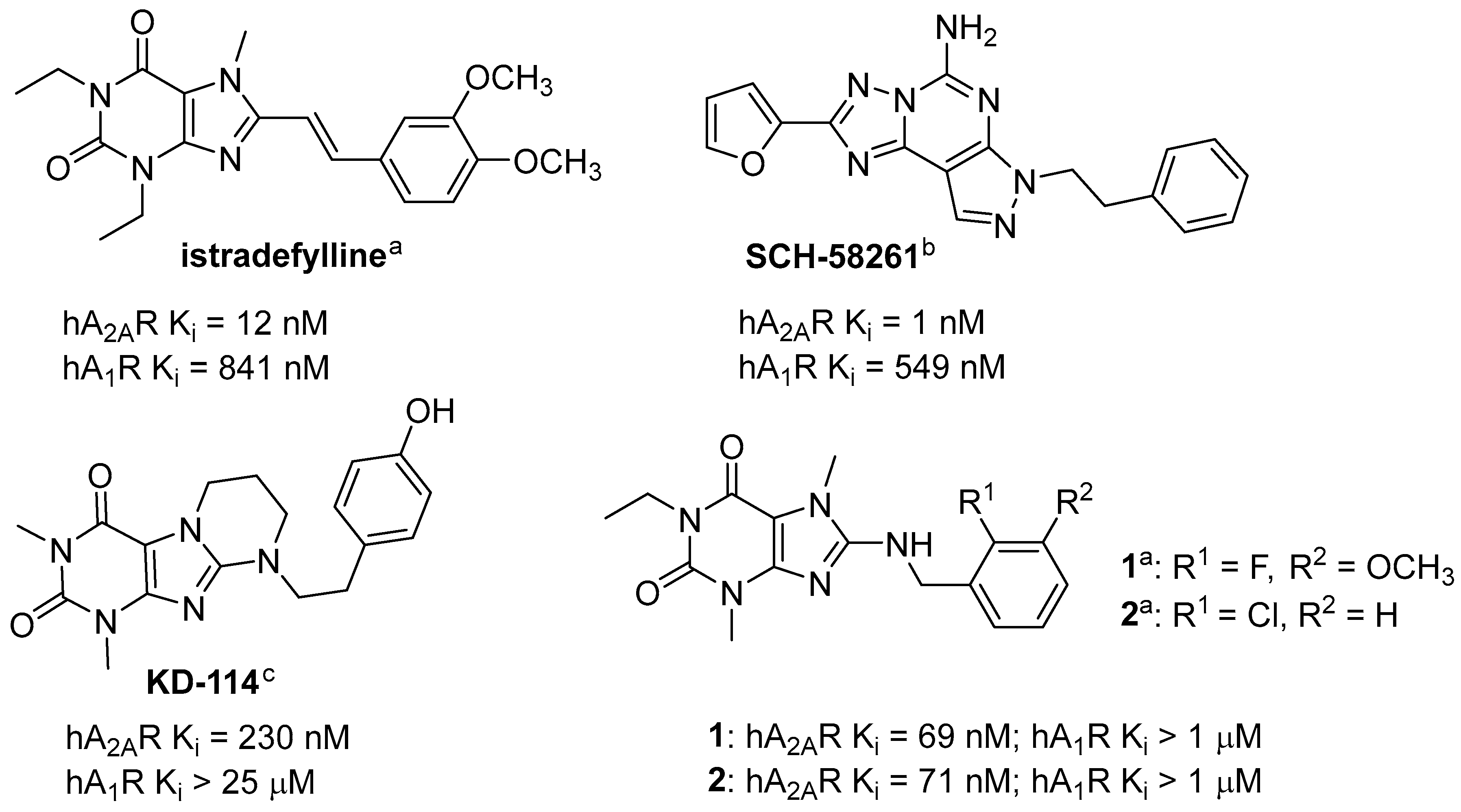
Istradefylline (Figure 1) is an adenosine A2A receptor (A2AR) antagonist with a xanthine core structure, which has been approved for therapeutic use in Japan (2013) and in the USA (2019) [6]. The A2AR belongs to the adenosine receptor family, which includes three further receptor subtypes: A1, A2B and A3. At all of them, the endogenous nucleoside adenosine acts as the cognate agonist, while xanthine derivatives act as antagonists [7]. Under physiological conditions, adenosine levels are low, but increase as a result of hypoxia, tissue damage and inflammation [8]. In the body, adenosine can have a strong effect on inflammation.
Animal studies applying an A2AR antagonist (SCH-58261 [9]; Figure 1) have shown its beneficial effects in a model of inflammation induced by lipopolysaccharide (LPS) administration [10]. This compound inhibited the increase in p38 and JNK phosphorylation and caspase 3 activation. In addition, it stopped activated microglial recruitment and the increase in the level of interleukin 1ß (IL-1ß) [10]. A similar beneficial effect of this compound was also observed in both in vivo and in vitro studies in two models of perinatal brain injury: the low-protein diet (LPD) model (chronic) and ibotenate model (acute) [11]. In both of these models, an increase in A2AR expression in microglia cells as well as in pro-inflammatory factors (IL-1ß, IL-6, inducible nitric oxide synthase and tumor necrosis factor alpha) was observed. The application of SCH-58261 caused a decrease in the above-mentioned cytokines, whereas treatment with the A2AR agonist (CGS-21680) showed the opposite effect.
Our previous studies with A2AR antagonists (xanthine derivatives) showed the anti-inflammatory activity of tested compounds, KD-114 and compound 1 (Figure 1), in carrageenan-induced edema in mice and rats, respectively [12,13]. KD-114, tested at a dose of 100 mg/kg intraperitoneally (ip), significantly (>90%) reduced edema volume in mice at the tested time points (1st, 2nd and 3rd hour). An especially strong effect was observed at the first hour with a reduction from 64% (control) to 2% (with the tested compound) [12]. For compound 1 tested at a dose of 20 mg/kg ip, the strongest inhibition of edema in rats was observed at the 2nd and 3rd hour (44% and 53%, respectively) [13]. Both compounds display good affinity for the A2AR (Ki = 230 nM, KD-114; Ki = 69 nM, 1) and selectivity towards other adenosine receptors (Ki > 1 μM).
Figure 1. Structures of adenosine A2A receptor antagonists; h = human; a information from [13]; b information from [9]; c information from [12].
Ijms 24 13707 g001
The aim of this work was to synthesize novel substituted xanthine derivatives, mainly with a 3,7-dimethylxanthine core, and to evaluate their affinity for adenosine receptors. Further pharmacological studies, both in vitro and in vivo, were then planned for selected derivatives. Compound 2 (Figure 1) from our previous publication was chosen as a lead structure for the design of new compounds [13]. This compound was characterized by a high affinity for A2AR (Ki = 71 nM) and the lack of significant interactions with other adenosine receptors (Ki > 1000 nM), similar to compound 1. However, compound 2 was chosen for economic reasons as an alternative. The planned structural modifications are shown in Figure 2.
Two series of compounds were designed: Series I (blue—a variable substituent in the N1 position) and series II (brown—a cyclopropyl substituent in the N3 position). In series I, three groups of compounds were planned: series IA—a divergent substituent in the N1 position; IB—a propyl substituent; IC—a propargyl substituent. In each of these series (I and II), the substituents in the benzylamine ring (green color) were changed as shown in Figure 2.

2. Results and Discussion

2.1. Synthesis of Target Compounds

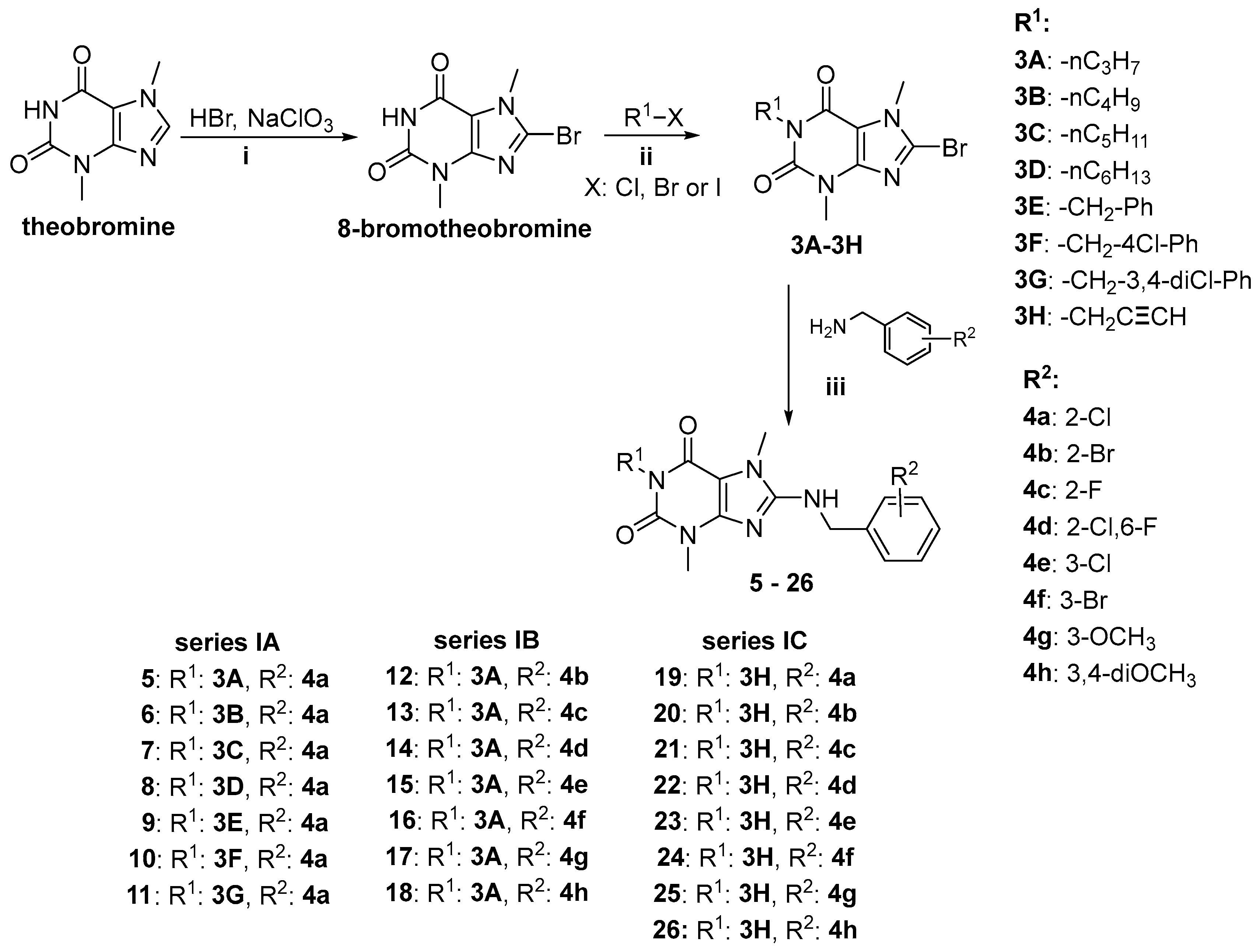
The synthetic routes are shown in Scheme 1 and Scheme 2. Compounds were obtained according to the methods previously described [13]. For most compounds (Scheme 1), the starting substrate for the synthesis was theobromine, which was first oxidatively brominated at position C8, followed by N-alkylation at position 1 and finally followed by a nucleophilic substitution with benzylamine derivatives. Reactions with benzylamines were carried out either in a microwave oven or by traditional heating. All compounds of series I (526) were obtained in this way. The yields of the reactions carried out using the microwave oven were higher, ranging from 39–72%, whereas under traditional conditions, only 4–28% were obtained (except for product 9: 50%).
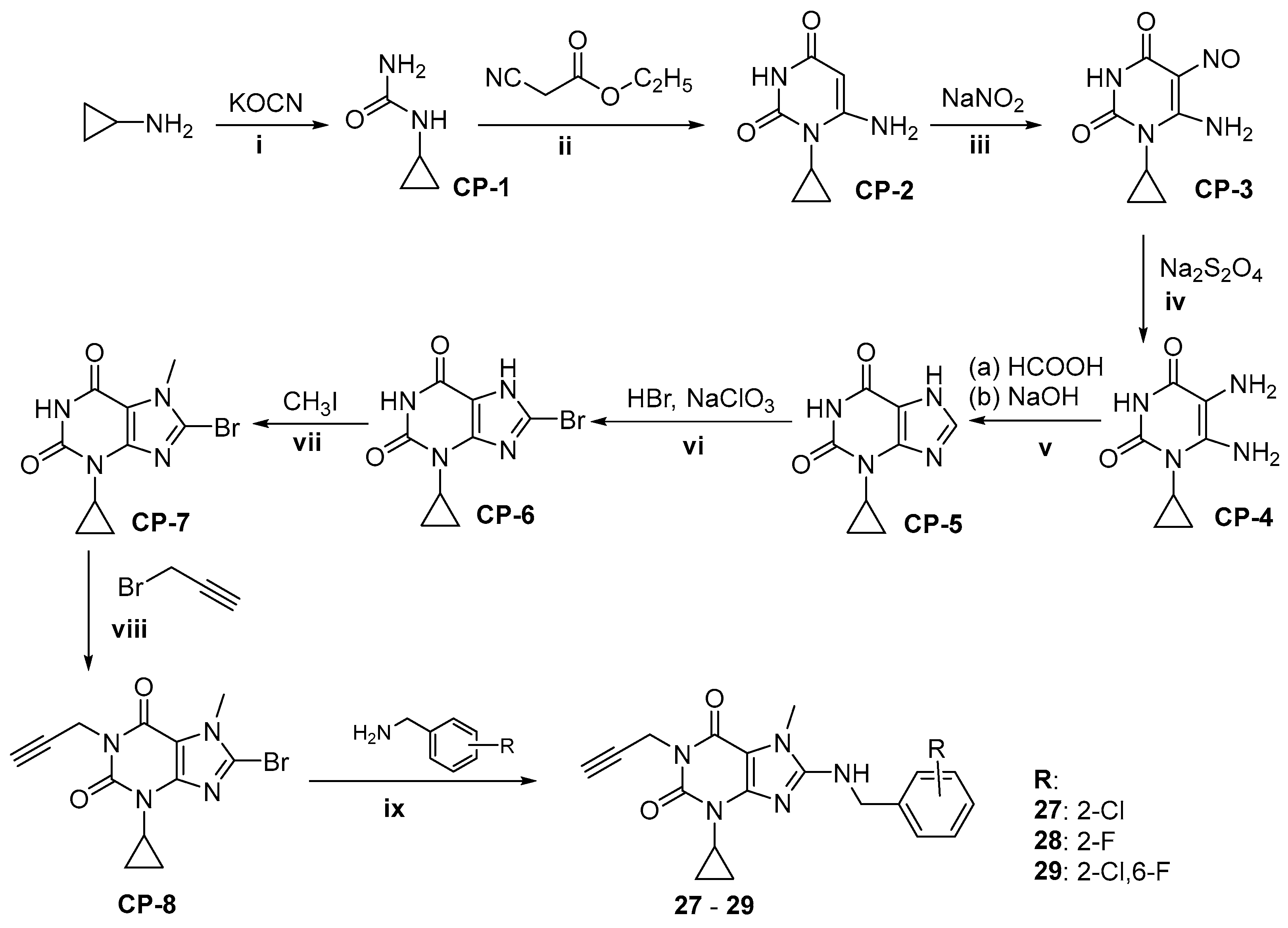
For compounds 2729, the starting material was cyclopropylamine, which, through a series of reactions shown in Scheme 2, provided 1-cyclopropylxanthine (CP-5). Then, compound CP-5 was sequentially brominated with hydrobromide in the presence of sodium chlorate at position C8 (CP-6), N-methylated with methyl iodide at position N7 (CP-7), N-alkylated with propargyl bromide at position N1 (CP-8) and finally condensed with benzylamines. The final reaction was carried out using a microwave oven with good yields (44–48%).
The structures of the synthesized compounds were confirmed by spectral analysis including 1H NMR, 13C NMR and LC-MS (spectra are provided in Supplementary Materials). Melting points were determined for all new compounds, and the purity of the compounds was determined to be at least 95% using ultra-performance liquid chromatography—mass spectrometry (UPLC-MS) (except 27: 93.19%).

2.2. Pharmacological Activity In Vitro

2.2.1. Structure-Activity Relationships at Adenosine Receptors

Affinities of the two series of 8-benzylaminoxanthine derivatives at human adenosine receptor subtypes were determined in radioligand binding studies (see Table 1). All modifications introduced (i.e., type of substituent at the N1, N3 and C8 positions) affected affinities. All compounds (except five: 7, 8, 10, 11 and 13) showed affinities for adenosine, the A2AR in the nanomolar range (60 nM < Ki < 800 nM), whereas only ten compounds (16, 19, 2125, 2729) displayed affinities for the A1R in the nanomolar range (70 nM < Ki < 470 nM). None of the compounds had significant affinities for the A3R (Ki > 1 µM), while only two compounds showed moderate affinities for the A2BR (5: Ki = 832 nM; 14: Ki = 687 nM).
Although all of the introduced modifications affected binding affinity, the strongest effect was observed for substituents in the N1-position, and this was observed at both A1R and A2AR. This influence is particularly evident in compounds with a 2-chlorobenzylamine substituent in the C8-position (compounds 511, 19). An elongation of the alkyl chain from ethyl (lead 2; Ki = 71 nM) to propyl (5; Ki = 302, nM), butyl (6; Ki = 318 nM), pentyl (7; Ki > 1000 nM) or hexyl (8; Ki > 1000 nM) led to a decrease in affinity for A2AR. A benzyl substituent at the N1-position (9; Ki = 323 nM) also caused a similar decrease in affinity for the A2AR as the propyl (5) or butyl (6) substituent. In contrast, the introduction of chlorine atom(s) into the benzyl ring (10 and 11: Ki > 1000 nM) resulted in a lack of affinity. In contrast, the introduced propargyl substituent at N1 (19; Ki = 96 nM) induced a small decrease in affinity for A2AR compared to compound 2, but contributed to good affinity for A1R (19: Ki = 407 nM). This beneficial effect of the propargyl group was observed throughout the series IC, with all compounds showing good affinities for A2AR (60 nM < Ki < 300 nM) and good affinities for A1R (70 < Ki < 470 nM) with the exception of compounds 20 and 26 (A1R Ki > 1000 nM). In this series, the type and position of the substituent in the benzylamine ring determined the affinity for these receptors. In this group, there are usually compounds with higher affinities for A2AR than A1R, but there is also compound 24 with a similar effect on both receptors (A2AR: Ki = 77 nM; A1R: Ki = 72 nM). This compound (24) and compound 22 (A2AR: Ki = 62 nM; A1R: Ki = 130 nM) showed the highest affinities for A2AR among all synthesized compounds.
The introduction of a cyclopropyl moiety at the N3-position (compounds 2729) caused a change in the affinity profile: an increased affinity for A1R and a slightly decreased affinity for A2AR (compare 19 vs. 27; 21 vs. 28 and 22 vs. 29). In this group is compound 29 with a higher affinity for A1R than A2AR (A1R: Ki = 78 nM, A2AR: Ki = 204 nM).

2.2.2. Human Monoamine Oxidase B (MAO B) Inhibition

The inhibitory activity of selected compounds for human MAO B (hMAO B) was determined by a spectrofluorometric method [13]. For these studies, compounds with propyl (5, 1218) and propargyl (1926) moieties were tested at a concentration of 1 µM. None of them showed hMAO B inhibition of >50% at this concentration, therefore, no IC50 values were determined (Table 2). Generally, the percent of hMAO B inhibition was lower than 20% (except for 20, 32%).

2.3. Molecular Docking to Adenosine A1 and A2A Receptors and Molecular Dynamics (MD) Simulations

For docking studies, we selected receptor structures 5N2S and 5N2R [14], solved with the xanthine derivative PSB-36 [15], which shares common structural elements with the series of herein studied ligands: substitution with an alkyl at the N1 and N3 positions, and a spatially large substituent at the C8 position. For both of the biological targets tested, the ligands showed a number of interactions overlapping with those previously described in other resolved structures. Furthermore, in both cases, the so-called ionic-lock between R3.50 and E6.30 residues was maintained, which is typical for the inactive state of the receptors.

2.3.1. Docking to the 5N2S (Adenosine A1 Receptor) and MD Simulations

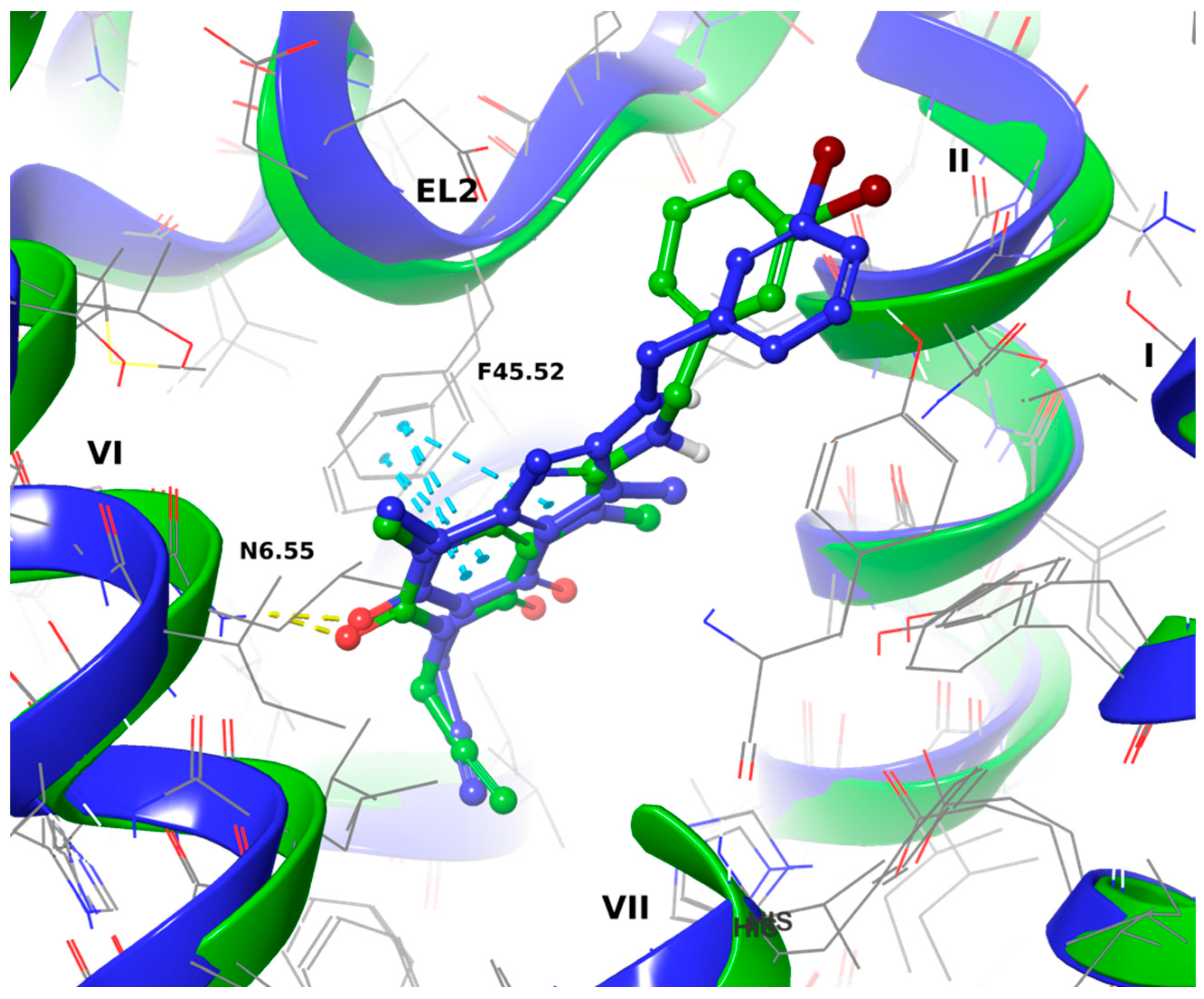
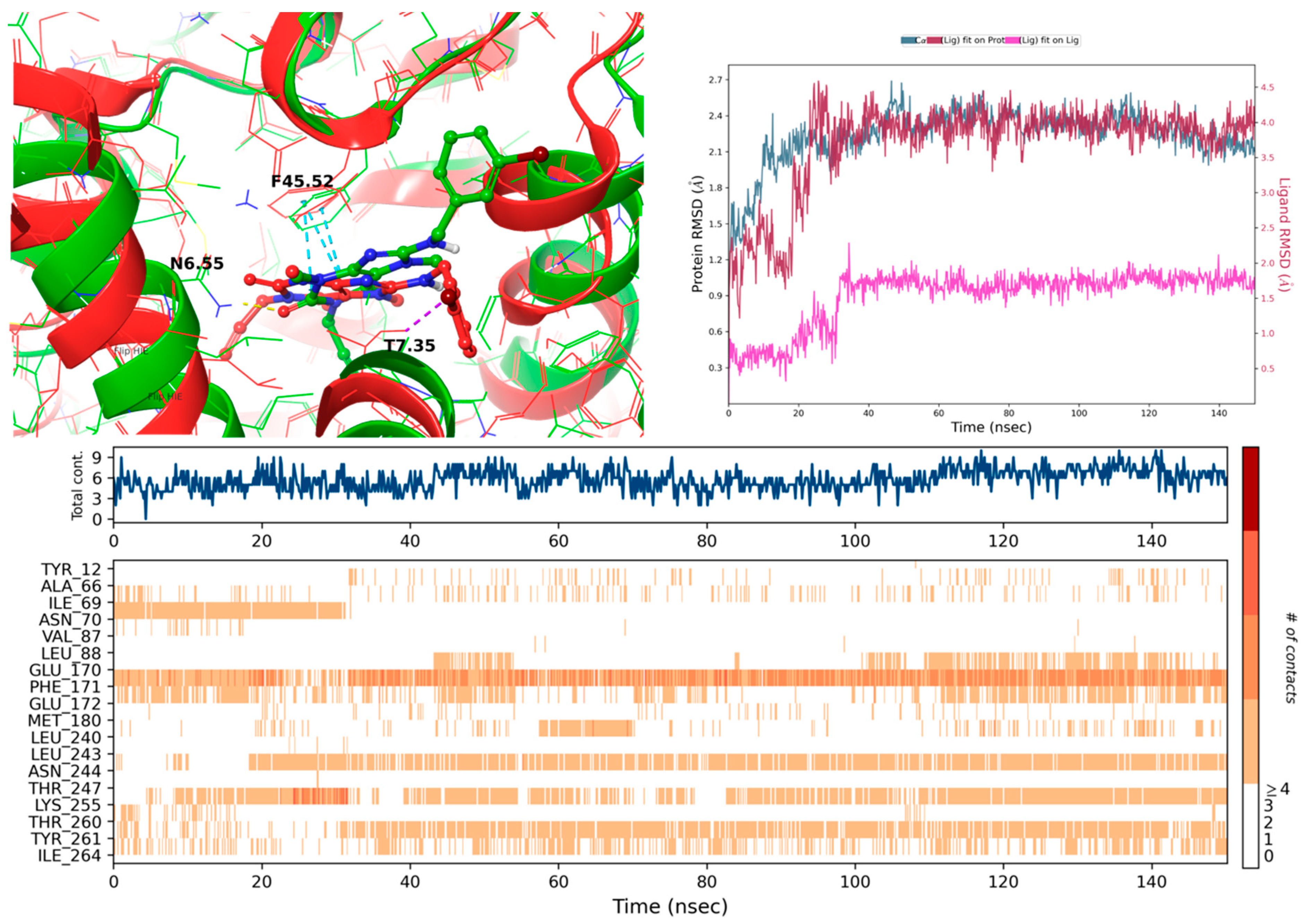
Three complexes were chosen for the estimation of stability, with MM-GBSA values [kcal/mol] of 19 (MZ1483): −79.01, 22 (MZ1490): −77.77 and 24 (MZ1495): −75.26. All three complexes showed similar, yet nonclassical, binding poses. The propargyl fragments are likely enclosed in a TM6 cage formed by L6.51, H6.52 on the sides and W6.48. Due to N7- and N3-methyl substitution, the ligands are expected to be placed in a rather parallel orientation with the membrane, and a key hydrogen bond may be formed between N6.55 and the C2 carbonyl oxygen. The xanthine ring is likely stabilized by π-π interactions with F45.52, and the chlorine atom of 19 may form a halogen bond with N2.65. During a 150-ns simulation, 19 remained stable from the ~20th ns of the trajectory, yet displayed additional interactions with the aminoalkyl group and H7.43 most of the time during the simulation. However, it did not display additional contacts, such as those seen with 24, where K265 interacted through water with N9, and the distal substituted benzene ring was stabilized by Y7.36, which could explain its higher affinity to A1AR when compared to other tested ligands, despite the slight translation in the binding pocket at the beginning of the simulation. Compound 22, on the other hand, lost the interaction with key N6.55 at ~20 ns of the recorded trajectory and was mainly stabilized by hydrophobic interactions with F171(45.52) and Y7.36 or I7.39. The comparison of the putative binding modes for 24 in both proteins is depicted in Figure 3.

2.3.2. Docking to 5N2R (Adenosine A2A Receptor) and MD Simulations

Selected complexes displayed MM-GBSA values [kcal/mol] of −63.22 (19), −71.26 (22) and −64.76 (24). Values correlate with their biological affinity values. The orientation of the ligands in the 5N2R structure was similar to that of 5N2S. A key hydrogen bond likely exists between N6.55 and the carbonyl oxygen C2, and the xanthine core may be stabilized through π-π interactions with F45.52. An additional interaction of the alkyl linker amine group forming H-bond with E45.53 was also found. However, due to structural differences between the two receptor subtypes, the propargyl moiety appeared to interact with V3.32, L6.51 and W6.48. However, during the simulations, the ligands changed their positions in the binding pocket at the beginning of the recorded trajectories. In the case of 19 in the 20th ns of the simulation, the ligand moved toward N6.55 and retained the formed H-bond with N9 through most of the remaining simulation time and occupied the very position until the end of the simulation. A similar shift of the ligand in the binding pocket was observed for the 22/5N2R complex in ca. the 15th ns of the trajectory, so that the C2 carbonyl oxygen interacted through most of the simulation with H6.52 and the N9 formed a water-bridged H bond with N6.55. The least stable interactions through the recorded trajectory were found for 24, mostly stabilized through π-π stacking with F45.52 and an H bond between the alkyl NH group and S2.65, lost when the substituted benzene ring targeted EL3, in the second half of the simulation performed. A detailed RMSD and RMSE, contact timeline and histogram for all recorded trajectories (including apo proteins) can be found in Supplementary Data (Figures S1–S3). Figure 4 shows the exemplary behavior of the 24/5N2S complex in the recorded trajectory.

2.4. Selected ADMET Properties—Evaluation In Vitro

Based on our results of the binding affinities (Table 1) three compounds, namely the most promising dual A1R/A2AR ligands, were selected for further studies: 19 (four times more potent as A2AR ligand), 22 (twice more potent as A2AR ligand) and 24 (with an equal potency for both receptors).

2.4.1. Toxicity Evaluation

Drug safety is a critical step in the process of drug development. It is important to ensure that these drugs do not cause adverse effects on important organ systems such as the central nervous system (CNS) or the liver. In our studies, neurotoxicity and hepatotoxicity were evaluated in SH-SY5Y and HepG2 cells, respectively. The compounds were tested at 11 concentrations from 0.098 μM to 100 μM (see Figure 5). Cell viability was assessed after incubation for 24 h with the SH-SY5Y line and for 48 h with the HepG2 line.
Dose-response analysis showed that compounds 19 (MZ-1483), 22 (MZ-1490) and 24 (MZ-1495) displayed a high safety profile in these cell lines (Figure 5). An examination of cultures under an inverted microscope during incubation with the tested compounds revealed that, at the highest concentrations (25 μM, 50 μM, 100 μM), the compounds precipitated and formed crystals in the culture medium.

2.4.2. Metabolic Stability

Metabolic stability is related to the ability of a compound to undergo biotransformation. As most therapeutic compounds undergo biotransformation in liver tissue, liver microsomes of various species, e.g., human liver microsomes (HLMs), are commonly used for preliminary in vitro assessment of metabolic stability [16].
In our study, the compounds were incubated for 120 min with HLMs, followed by liquid chromatography-mass spectrometry (LC-MS) to identify the formed metabolites. The tested compounds were found to be metabolically stable compared to the unstable reference drug verapamil. The reaction mixtures after incubation showed a similar % remaining for all substrates, which was more than 90% (Table 3, Figures S4–S6 in Supplementary Materials). The same calculated value for the unstable drug verapamil was only 31%, after incubation with HLMs under the same conditions (Table 3; Figure S11). Furthermore, 19 formed two metabolites, while 22 and 24 only formed one metabolite. Moreover, according to the MS data (m/z~249) and retention times (t~6.26 s) in UPLC chromatograms, all compounds were most likely metabolized in the same way (Table 3, Figures S4–S9 in Supplementary Materials). The most probable metabolic pathways for all compounds were proposed with the support of MetaSite 6.0.1 software (Figure S4 in Supplementary Materials), such as fragmentation and hydroxylation. Results are shown in Figures S4, S6, S8 and S10 in Supplementary Materials.

2.4.3. Potential for Drug-Drug Interactions

To predict potential drug-drug interactions (DDI) effects on three of the most relevant cytochrome P450 (CYP), isoforms CYP3A4, CYP2D6 and CYP2C9 were investigated at a concentration of 10 µM. The results were compared with the effects of 1 µM of the selective inhibitors ketoconazole (CYP3A4), quinidine (CYP2D6) and sulfaphenazole (CYP2C9). Compound 22 (MZ1490) showed the smallest effect on CYP3A4 activity (around 75% of the control activity), while 19 (MZ1483) and 24 (MZ1495) were found to be strong CYP3A4 inhibitors, decreasing its activity to ~20% at 10 µM (Figure 6A). Regarding CYP2D6, a slight, statistically significant inhibition was only observed for 24 (Figure 6B). All compounds similarly inhibited the activity of CYP2C9 to around 65–70% of control activity (Figure 6C).

2.4.4. Blood Brain Barrier Permeability

The ability of compounds, especially those intended to act in the CNS, to penetrate the blood-brain barrier (BBB) is an important property to consider in the initial stages of drug candidate screening. The PAMPA assay is a commonly used test to assess the passive permeation of the BBB [17]. In our study, caffeine was used as a highly permeable compound. The tested compounds (19, 22 and 24) were dissolved in phosphate-buffered saline (PBS) (100 μM); due to precipitation, the solutions were sonicated for 20 min before adding them to the donor plate. After 5 h of incubation at room temperature, the results were analyzed by LC-MS, and the permeability coefficients (Pe) were calculated. The results (Table 4) showed that only one compound, 19, could penetrate the BBB as indicated by a calculated Pe parameter of greater than 1.5 × 10⁻⁶ cm/s. This value is the manufacturer’s stated breakpoint for permeable compounds. For compounds 22 and 24, the values were much lower than 1.5 × 10⁻⁶ cm/s (0.37 and 0.89 × 10⁻⁶ cm/s, respectively). This is puzzling, as these compounds do not structurally differ that much from each other, nor are there any significant differences in the calculated lipophilicity parameter logP (2.19, 2.35, 2.46, respectively, calculated in ChemDraw). It is possible that the value of this parameter may have been influenced by their poor solubility in PBS and, although sonication improved it, it may not have been the same in every case, although the differences were not visually apparent.

2.5. Anti-Inflammatory Activity In Vitro

2.5.1. Preliminary Screening of Anti-Inflammatory Activity in Griess Assay

There are several types of immune cells that synthesize nitric oxide (NO), which is a signaling molecule in response to inflammation [18]. NO plays an important role in the pathogenesis of inflammation. To assess the ability of compounds to inhibit NO production, the Griess assay was applied. This is a colorimetric assay to measure the levels of nitrite (NO2), which is one of the primary and stable metabolites of NO. The anti-inflammatory activity of compounds was examined in BV-2 cells treated with LPS as previously described [19]. Briefly, BV-2 cells were first pre-treated with increasing concentrations of tested compounds for 1 h, followed by LPS (1 μg/mL) stimulation for 24 h. Together with the Griess assay, the 3-(4,5-dimethylthiazol-2-yl)-5-(3-carboxymethoxyphenyl)-2-(4-sulfophenyl)-2H-tetrazolium (MTS) assay was performed to ensure that the decrease in NO level was not an effect of toxicity. No significant toxic effect of the compounds tested on BV2 cell viability was observed during the experiment. The results showed that only 19 (MZ-1483) and 22 (MZ-1490) showed moderate anti-inflammatory activity at the lowest concentrations (19: 100 nM and 22: 100 and 200 nM) (Figure 7). While in the presence of LPS, the amount of NO produced increased by 140% as compared to the control cells; in the presence of the tested compounds, NO levels were only 60% to 90% higher than in the control cells.

2.5.2. Phagocytic Activity

Phagocytosis is a cellular process that allows a cell to properly function by removing harmful agents from it. Microglia, as resident macrophages of the CNS, naturally participate in phagocytosis during neural development, maintaining homeostasis, and diseased states [20]. It has not been clearly established which phagocytic phenotype (less or more active) is beneficial or deleterious for neurodegenerative disease progression. In PD, when α-synuclein aggregates are formed, both autophagy and microglia phagocytosis are impaired, affecting the degradation and clearance of α-synuclein. This dysfunction of autophagy and phagocytosis leads to predominantly M1-type (pro-inflammatory) microglia and the release of pro-inflammatory cytokines (IL-1ß, IL-6 and IL-12). This, in turn, contributes to the transformation of M2-type microglial cells into M1-type cells; i.e., an intensification of the entire inflammatory process [21].
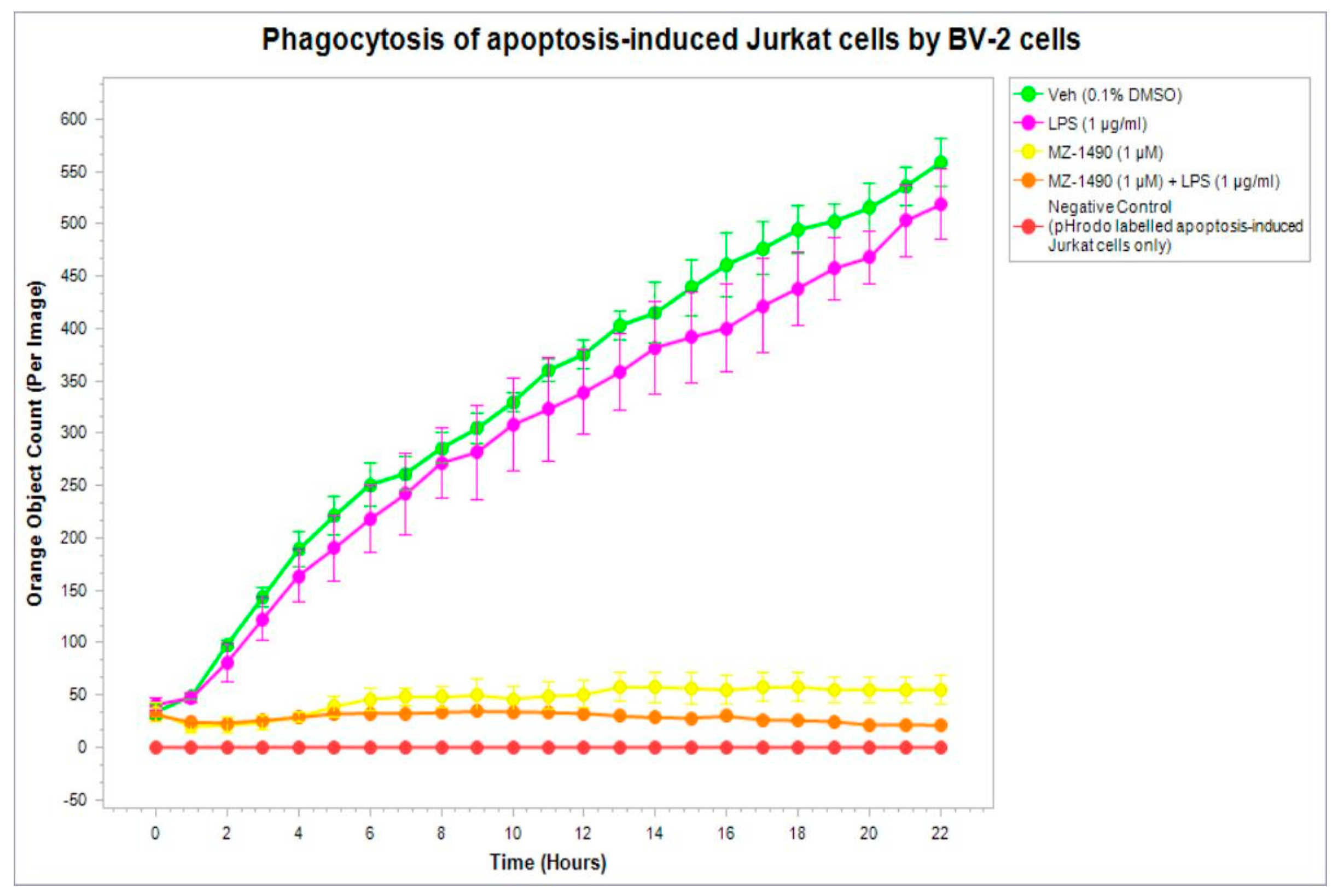
For the evaluation of phagocytic activity, compound 22 (MZ1490) was chosen as the most promising one on the basis of the results from the Griess assay. This compound was tested at a concentration of 1 µM, and phagocytosis was monitored in real time using an IncuCyte® pHrodo® Red Cell Labeling Kit (catalog no. 4766, Sartorius, Göttingen, Germany). IncuCyte analysis showed that BV-2 cells without or after treatment with LPS have a high phagocytic potential, which is in line with previous findings [22]. It is interesting to note that the phagocytic activity exerted by untreated BV-2 cells is also related to the serum concentration. A healthy CNS maintains the BBB, and microglia are often highly ramified surveillance microglia without enhanced phagocytosis [23]. However, in pathological states of the CNS, such as neurodegenerative diseases, the BBB is compromised, resulting in vascular leaks. This is a valuable finding in understanding the behavior of microglia. In our experimental conditions, BV-2 cells were cultured in the presence of 10% fetal bovine serum. Thus, it mimics a pathological state in which BV-2 cells have the morphology of fully activated microglia typically found in the injured brain [24]. An interesting finding is that 22 protects microglia from LPS-induced inflammation and, therefore, also from phagocytosis (Figure 8). Similar results were obtained, where BV-2 cells were treated with 22 alone.

2.6. Antinociceptive and Antiinflammatory Activity In Vivo

The positive results of the anti-inflammatory in vitro studies encouraged us to carry out preliminary in vivo studies to confirm the activity of tested compounds observed in vitro. For this purpose, a model with inflammation induction was used by the administration of carrageenan. This model has been used for years to evaluate the anti-inflammatory effects of compounds [25,26]. Furthermore, analgesic and anti-inflammatory activities were carried out in a formalin test [27].

2.6.1. Anti-Inflammatory (Antiedematous) Effect in the Carrageenan-Induced Edema Model

The carrageenan test is used to evaluate the anti-inflammatory effect, as the injection of this compound into the hind paw of an animal induces long-lasting edema. The aim of the present study was to evaluate the anti-inflammatory (antiedematous) activity of the compounds tested: 19, 22 and 24. The compounds were administered at a dose of 20 mg/kg of body weight (bw). Ketoprofen, used as a reference compound and administered ip at a dose of 20 mg kg bw, inhibited edema formation by 60%, 63.3% and 45.1% in three consecutive hours of the experiment, respectively (Table 5). All tested compounds decreased the volume of edema induced by subcutaneous carrageenan injection into the hind paw of rats (Table 5). The strongest anti-inflammatory effect in this assay was produced by 24. A statistically significant effect was observed in the first, second and third hour of the experiment, inhibiting the development of edema by 51.2%, 43.8% and 48.1%, respectively. Compound 22 inhibited the formation of edema by 37.1%, 18.7% and 30.8% in three consecutive hours of the experiment, respectively, but the effect was not statistically significant. Pretreatment with compound 19 reduced paw edema by 28.2%, 35.7% and 17.3%, in the first, second and third hour of the experiment, respectively, compared to the control group, but the effect was not statistically significant.

2.6.2. Anti-Nociceptive Activity in the Formalin Test

In the formalin test, which is a model of chronic pain induced by the administration of a 5% formalin solution, the evaluated compounds showed antinociceptive activity. Injection of formalin into the dorsal surface of the hind paw of a mouse produces a biphasic nocifensive behavioral response; i.e., licking, biting, flinching or lifting of the injected paw. The acute (neurogenic) nociceptive phase lasts for the first 5 min and is followed by a period of little activity for the next 10 min. The first phase of the test is directly associated with nociceptor stimulation and the development of neurogenic inflammation. The second (late) phase occurs between 15 and 30 min after formalin injection. The second phase depends on peripheral inflammation and the central sensitization of pain. Since this phase reflects the activation of inflammatory processes, the compounds active in this phase of the experiment also have an anti-inflammatory effect. The tested compounds 19, 22 and 24, were investigated in three doses: 10, 20 and 40 mg/kg bw. ASA (acetylsalicylic acid) was used as a reference compound and administered in doses of 50, 100 and 200 mg/kg. All compounds only reduced the licking/biting time of the right hind paw of mice in the II phase of the test (15–30 min) (Table 6) and showed antinociceptive activity. This effect was observed for each of these compounds, only at a dose of 40 mg/kg. The strongest effect in this assay was produced by 24. The calculated ED50 value for this compound is 9.1 mg/kg. In addition, this anti-nociceptive and anti-inflammatory effect was approximately 13.8 times greater than that observed for the reference compound (ASA). Compounds 19 and 22 significantly reduced the duration of the licking response in the late (inflammatory) phase by 47.2% (p < 0.05) and 48.0% (p < 0.05), respectively. ASA also attenuated statistically significant pain responses in the late phases of the formalin test. The calculated value of ED50 is 126.3 mg/kg.

3. Conclusions

Twenty-five novel xanthine derivatives with varied substituents in the N1-, N3- and C8-positions were designed and synthesized. Compounds were divided into two series (I and II). The division depended on the position of the introduced substituents: series I—N1 and C8; series II—N1, N3 and C8. In vitro binding studies at human adenosine receptors showed that these compounds had:
interaction with A3R at submicromolar concentrations,
no interaction with A2BR at submicromolar concentrations (except for compounds 5 and 14),
affinities for A1R in the nanomolar range in the series IC and II (except 20 and 26), and
variable affinities for A2AR (most compounds had Ki values in the range from 62 nM to 773 nM)
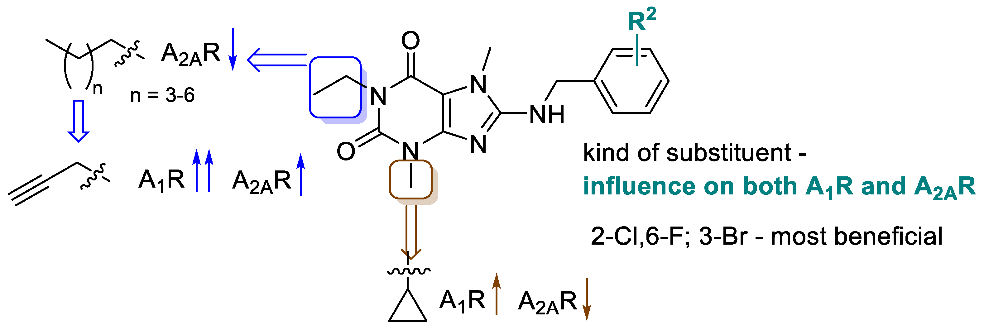
Analysis of SARs indicated chemical features in the presented scaffold that are important for the enhancement of affinity and selectivity for the subtypes of adenosine receptors. A summary is shown in Figure 9.
The propargyl substituent in the N1-position led to affinities for A1R in the nanomolar range. Additionally, the exchange of the methyl for a cyclopropyl in the N3-position increased affinities for A1R, but slightly decreased those for the A2AR. The most beneficial substituents in the benzylamine group were 2-chloro-6-fluro and 3-bromo.
Among all synthesized compounds, only two compounds, 22 and 24 (IC series), had affinities for A2AR comparable to that of the lead structure 2 (22: Ki = 62 nM; 24: Ki = 77 nM vs. 2: Ki = 71 nM). Compound 24 (the 3-bromobenzylamine derivative) also showed a simultaneously strong affinity for the A1R and is, therefore, a dual ligand with balanced affinities for both receptors (A1R Ki = 72 nM; A2AR Ki = 77 nM). In series II, compound 29 was obtained, which had a two-fold higher affinity for the A1R than for the A2AR (A1R Ki = 78 nM; A2AR Ki = 204 nM). Three compounds (19, 22 and 24), being the most promising dual ligands, were selected for further studies.
Docking to the A1R and A2AR showed differences in positioning and interactions with amino acid residues in the binding pocket, suggesting the reasons for differences in affinities of the compounds for these receptors. Toxicity studies confirmed low hepato- and neurotoxicity up to a concentration of 12.5 µM. Higher concentrations caused compound precipitation in the culture medium (PBS). The compounds displayed high metabolic stability. The main metabolite formed for all of them was a product with a molecular weight of 249, which suggests that this product is a result of fragmentation (after dissociation of the benzyl substituent from the amino group at the C8 position of the xanthine moiety) with simultaneous hydroxylation in the propargyl chain.
Anti-inflammatory studies in the Griess assay showed that compounds 19 and 22 had a moderate positive effect on nitric oxide production in BV2 cells, but only in the lowest tested concentrations (100 and/or 200 nM). Therefore, the most promising compound 22 was chosen for further in vitro evaluation and its influence on phagocytosis was tested in BV-2 cells using the IncuCyte imaging platform. Results showed that compound 22 (tested at a concentration of 1 µM) protected from phagocytosis.
Furthermore, in vivo studies showed that the most promising of the three tested compounds (19, 22 and 24) was compound 24, which, at the tested doses (20 mg/kg bw in the carrageenan test and 40 mg/kg in the formalin test), displayed anti-inflammatory activity.
Undoubtedly, our work has certain limitations, which may result from the conditions and procedure of the conducted studies, and may have influenced the results obtained. First of all, we can see that the problem is the limited solubility of our compounds in various solvents, especially in higher concentrations, which are indicated for the determination of a special parameter; e.g., Pe in the PAMPA assay. In this assay, it is necessary to use high concentrations (from 100 to 200 µM [28]) in order to increase the sensitivity of the assay and mimic the composition of specific target barriers [29]. We did not accomplish better solubility of our compounds by increasing the concentrations of DMSO up to 5%, which is the highest concentration compatible with pre-coated filter plate. Moreover, xanthine derivatives are molecules, which are difficult to convert into salts because they do not have a sufficiently basic center (it is known that the formation of salts usually increases the solubility of compounds [30]). Furthermore, this work is our initial work that evaluates the anti-inflammatory activity of compounds from this group in the Griess assay and using the IncuCyte Live-Cell Analysis System. It has led to interesting results that encourage further studies to verify the anti-inflammatory activity of dual A2A/A1 receptor ligands.
Thus, dual ligands with promising anti-inflammatory activity, found in the group of novel tri-substituted xanthines, could be useful tools for the future development of adenosine receptor antagonists as drugs for neuroinflammatory diseases.

4. Material and Methods

4.1. Chemistry

4.1.1. General Information

All commercially available reagents and solvents were purchased and used without further purification. Melting points (mp.) were determined on a MEL-TEMP II (LD Inc., Long Beach, CA, USA) melting point apparatus and were uncorrected. 1H-NMR spectra were recorded on a Varian Mercury 300 MHz or JEOL FT-NMR 500 MHz apparatus in CDCl3 or in DMSO-d6 using tetramethylsilane as an internal standard. 13C NMR data were recorded on 75 MHz on a Varian-Mercury-VX 300 MHz PFG or JEOL FT-NMR 500 MHz spectrometer. The J values are reported in Hertz (Hz), and the splitting patterns are designated as follows: br s (broad singlet), d (doublet), dd (doublet of doublets), dt (doublet of triplets), m (multiplet), s (singlet), t (triplet), tt (triplet of triplets) and quin (quintet). Data are reported in order: Chemical shift, multiplicity, coupling constant, number of protons and proton’s position (cyclopropyl, phe-phenyl, xan-xanthine). The purity of the tested compounds was determined (%) on a Waters TQD mass spectrometer coupled with a Waters ACQUITY UPLC system. Retention times (tR) are given in minutes. The reactions were monitored by thin layer chromatography (TLC) using aluminum sheets coated with silica gel 60F254 (Merck) using as a developing system dichloromethane/metanol 9:1. Spots were detected under UV light.

4.1.2. Synthesis of Compounds 526

Synthesis of 8-bromotheobromine
To theobromine (40 mmol, 7.21 g) in 28 mL of glacial acetic acid, 48% HBr was added and the mixture was heated in a water bath (bath temperature 80 °C) for 30 min. Then, a solution of sodium chlorate (V) in water was dropped over a period of 1 h. Next, the temperature of the bath was increased to boiling and heating was continued with stirring for 1.5–2 h. After that time, the reaction mixture was left at room temperature (for several hours). The precipitate was filtered off and washed several times with cold water. The reaction was repeated several times. Yield: 40–50%.
General procedure of synthesis of 1-substituted 8-bromotheobromines (3A3H)
A mixture of 8-bromotheobromine (20 mmol, 5.18 g), halide (22 mmol) and K2CO3 (60 mmol, 8.30 g) in 40 mL of DMF was heated at 110 °C from 4 h to 18 h. Next, water was added and the resulting precipitate was filtered and washed with water and/or 1% NaOH. The product was directly used for the next step of the synthesis or purified by boiling in ethanol.
1-Propyl-8-bromotheobromine (3A)
Synthesis from propyl iodide. Yield: 74%, C10H13BrN4O2 (MW 301.14).
1-Butyl-8-bromotheobromine (3B)
Synthesis from n-butyl iodide. Yield: 15%, C11H15ClN4O2 (MW 315.17). Synthesis of this compound from n-butyl bromide was described by Patrushev et al. [31] (yield: 81%; mp: 133–136 °C)
1-Pentyl-8-bromotheobromine (3C)
Synthesis from pentyl bromide. Yield: 73%, C12H17BrN4O2 (MW 329.20).
1-Heksyl-8-bromotheobromine (3D)
Synthesis from hexyl bromide. Yield: 27%, C13H19BrN4O2 (MW 343.23).
1-Benzyl-8-bromotheobromine (3E)
Synthesis from benzyl chloride. Yield: 42%, C14H13BrN4O2 (MW 349.19).
1-(4-Chlorobenzyl-8-bromotheobromine (3F)
Synthesis from 4-chlorobenzyl chloride. Yield: 67%, C14H12BrClN4O2 (MW 383.63).
1-(3,4-Dichlorobenzyl-8-bromotheobromine (3G)
Synthesis from 3,4-dichlorobenzyl chloride. Yield: 41%, C14H11BrCl2N4O2 (MW 418.07).
1-(Prop-2-yn-1-yl)-8-bromotheobromine (3H)
Synthesis from 3-bromoprop-1-yne. Yield: 41%, C10H9BrN4O2 (MW 297.11)
General procedure for the synthesis of 8-substitued benzylaminoxanthines: 5, 1226
A mixture of 0.55 mmoles of 8-bromo-1-propyl-3,7-dimethyl-3,7-dihydro-1H-purine-2,6-dione (3A), 1.1 mmoles of appropriate benzylamine, 1.6 mmoles of TEA and 1.00 mL of propanol was heated in closed vessels in a microwave oven (CEM Discover SC, 300 Watt, Power Max Off, 150 °C, 10 bar) for 1 h. The solvent was removed and the residue was treated with DCM. The products were purified by crystallization from ethanol or flash column chromatography over silica gel with CH2Cl2:MeOH (100:0 to 80:20).
8-((2-Chlorobenzyl)amino)-3,7-dimethyl-1-propyl-3,7-dihydro-1H-purine-2,6-dione (5)
Synthesis from 3A (0.55 mmol; 0.157 g) and 2-chlorobenzylamine (1.1 mmol, 0.156 g). Yield: 48% (96 mg), mp: 190–191 °C, C17H20ClN5O2 (MW 361.83). 1H NMR (300 MHz, DMSO-d6) δ: 0.78–0.86 (m, 3H, N1CH2CH2CH3) 1.42–1.57 (m, 2H, N1CH2CH2) 3.26 (s, 3H, N3CH3) 3.62 (s, 3H, N7CH3) 3.70–3.79 (m, 2H, N1CH2) 4.60 (d, J = 5.86 Hz, 2H, NHCH2) 7.24–7.34 (m, 2H, phe-4,5-H) 7.40–7.47 (m, 2H, phe-3,6-H) 7.60 (t, J = 5.86 Hz, 1H, NHCH2). 13C NMR (DMSO-d6) δ: 11.7 (N1CH2CH2CH3), 21.4 (N1CH2CH2), 29.7 (N7CH3), 30.3 (N3CH3), 42.0 (N1CH3), 43.9 (NHCH2), 102.6 (Xan-C5), 127.6 (phe-C5), 129.1 (phe-C4), 129.4 (phe-C6), 129.6 (phe-C3), 132.5 (phe-C2), 136.9 (phe-C1), 148.6 (xan-C4), 151.1 (xan-C2), 153.3 (xan-C6), 154.2 (xan-C8). UPLC/MS purity > 99%; tR = 6.42, (ESI) m/z [M]+ 362.2.
8-((2-Bromobenzyl)amino)-3,7-dimethyl-1-propyl-3,7-dihydro-1H-purine-2,6-dione (12)
Synthesis from 3A (0.55 mmol; 0.157 g) and 2-bromobenzylamine (1.1 mmol, 0.204 g). Yield: 98 mg (44%), mp: 206–207 °C, C17H20BrN5O2 (MW 406.28). 1H NMR (300 MHz, DMSO-d6) δ: 0.78–0.85 (m, 3H, N1CH2CH2CH3) 1.43–1.56 (m, 2H, N1CH2CH2) 3.26 (s, 3H, N3CH3) 3.63 (s, 3H, N7CH3) 3.71–3.78 (m, 2H, N1CH2) 4.56 (d, J = 5.86 Hz, 2H, NHCH2) 7.16–7.24 (m, 1H, phe-4-H) 7.31–7.45 (m, 2H, phe-5,6-H) 7.57–7.66 (m, 2H, phe-3-H + NHCH2). 13C NMR (DMSO-d6) δ: 11.7 (N1CH2CH2CH3), 21.5 (N1CH2CH2), 29.8 (N7CH3), 30.4 (N3CH3), 42.0 (N1CH3), 46.5 (NHCH2), 102.7 (xan-C5), 122.9 (phe-C2), 128.3 (phe-C5), 129.4 (phe-C4), 129.5 (phe-C6), 132.9 (phe-C3), 138.5 (phe-C1), 148.7 (xan-C4), 151.2 (xan-C2), 153.4 (xan-C6), 154.3 (xan-C8). UPLC-MS purity 97.5%, tR = 6.57, (ESI) m/z [M]+ 406.13.
8-((2-Fluorobenzyl)amino)-3,7-dimethyl-1-propyl-3,7-dihydro-1H-purine-2,6-dione (13)
Synthesis from 3A (0.55 mmol; 0.157 g) and 2-flurobenzylamine (1.1 mmol, 0.138 g). Yield: 74 mg (39%), mp: 224–226 °C, C17H20FN5O2 (MW 345.38). 1H NMR (300 MHz, DMSO-d6) δ: 0.78–0.85 (m, 3H, N1CH2CH2CH3), 1.42–1.56 (m, 2H, N1CH2CH2), 3.28 (s, 3H, N3CH3), 3.58 (s, 3H, N7CH3), 3.70–3.77 (m, 2H, N1CH2), 4.56 (d, J = 5.86 Hz, 2H, NHCH2), 7.11–7.20 (m, 2H, phe-5,6-H), 7.24–7.34 (m, 1H, phe-3-H), 7.44 (td, J = 7.77, 1.47 Hz, 1H, phe-4-H), 7.57 (t, J = 5.86 Hz, 1H, NHCH2). 13C NMR (DMSO-d6) δ: 11.6 (N1CH2CH2CH3), 21.4 (N1CH2CH2), 29.6 (N7CH3), 30.3 (N3CH3), 41.9 (N1CH2), 102.6 (xan-C5), 115.5 (d, 2JC,F = 21.8 Hz, phe-C3), 124.7 (d, 4JC,F = 3.5 Hz, phe-C5), 126.6 (d, 2JC,F = 15.0 Hz, phe-C4), 129.4 (d, 3JC,F = 8.1 Hz, phe-C6), 130.1 (d, 3JC,F = 3.4 Hz, phe-C1), 148.6 (xan-C4), 151.1 (xan-C2), 153.3 (xan-C6), 154.2 (xan-C8), 160.6 (d, 1JC,F = 244.2 Hz, phe-C2). UPLC/MS purity > 99%; tR = 5.98, (ESI) m/z [M]+ 346.18.
8-((2-Chloro-6-fluorobenzyl)amino)-3,7-dimethyl-1-propyl-3,7-dihydro-1H-purine-2,6-dione (14)
Synthesis from 3A (0.55 mmol; 0.157 g) and 2-chloro-6-flurobenzylamine (1.1 mmol, 0.176 g). Yield: 110 mg (53%), mp: 241–243 °C, C17H19ClFN5O2 (MW 379.82). 1H NMR (300 MHz, CDCl3-d) δ: 0.92 (t, J = 7.33 Hz, 3H, N1CH2CH2CH3), 1.56–1.72 (m, 2H, N1CH2CH2), 3.52 (s, 3H, N3CH3), 3.65 (s, 3H, N7CH3), 3.82–3.97 (m, 2H, N1CH2), 4.83 (s, 3H, NHCH2 + NHCH2) 6.94–7.06 (m, 1H, phe-4-H) 7.15–7.24 (m, 2 H, phe-3,5-H); 13C NMR (CDCl3-d) δ: 11,4 (N1CH2CH2CH3), 21.4 (N1CH2CH2), 29.7 (N7CH3/N3CH3), 38.8 (d, 3JC,F = 4.6 Hz, NHCH2), 42.5 (N1CH3), 103.4 (xan-C5), 114.4 (d, 2JC,F = 24.0 Hz, phe-C5), 123.7 (d, 2JC,F = 17.3 Hz, phe-C1), 125.3 (d, 4JC,F = 4.2 Hz, phe-C3), 130.0 (d, 3JC,F = 10.3 Hz, phe-C4), 135.5 (phe-C2), 148.1 (xan-C4), 151.5 (xan-C2), 152.5 (xan-C6), 154.2 (xan-C8), 161.7 (d, 1JC,F = 251.0 Hz, phe-C6). UPLC-MS purity > 99%; tR = 6.39; (ESI) m/z [M]+ 380.21.
8-((3-Chlorobenzyl)amino)-3,7-dimethyl-1-propyl-3,7-dihydro-1H-purine-2,6-dione (15)
Synthesis from 3A (0.55 mmol; 0.157 g) and 3-chlorobenzylamine (1.1 mmol, 0.156 g).
Yield: 111 (56%), mp: 185–187 °C, C17H20ClN5O2 (MW 361.83). 1H NMR (500 MHz, DMSO-d6) δ: 0.79 (t, J = 7.45 Hz, 3H, N1CH2CH2CH3), 1.47 (dq, J = 14.61, 7.35 Hz, 2H, N1CH2CH2), 3.26 (s, 3H, N3CH3), 3.56 (s, 3H, N7CH3), 3.72 (t, J = 7.45 Hz, 2H, N1CH2), 4.50 (d, J = 5.73 Hz, 2H, NHCH2), 7.23–7.34 (m, 3H, phe-2,5,6-H), 7.38–7.43 (m, 1H, phe-4-H), 7.54–7.63 (m, 1H, NHCH2); 13C NMR (DMSO-d6) δ: 11.7 (N1CH2CH2CH3), 21.5 (N1CH2CH2), 29.7 (N7CH3), 30.4 (N3CH3), 42.0 (N1CH2), 45.7 (NHCH2), 102.7 (xan-C5), 126.6 (phe-C4), 127.4 (phe-C2), 127.7 (phe-C6), 130.7 (phe-C5), 133.5 (phe-C1), 142.8 (phe-C3), 148.7 (xan-C4), 151.2 (xan-C2), 153.4 (xan-C6), 154.4 (xan-C8). UPLC-MS purity 96.2%; tR = 6.48; (ESI) m/z [M]+ 362.2.
8-((3-Bromobenzyl)amino)-3,7-dimethyl-1-propyl-3,7-dihydro-1H-purine-2,6-dione (16)
Synthesis from 3A (0.55 mmol; 0.157 g) and 3-bromobenzylamine (1.1 mmol, 0.204 g). Yield: 92 mg (41%), mp: 220–222 °C, C17H20BrN5O2 (MW 406.28). 1H NMR (300 MHz, DMSO-d6) δ: 0.81 (t, J = 7.33 Hz, 3H, N1CH2CH2CH3), 1.43–1.57 (m, 2H, N1CH2CH2), 3.29 (s, 3H, N3CH3), 3.58 (s, 3H, N7CH3), 3.70–3.78 (m, 2H, N1CH2), 4.51 (d, J = 5.86 Hz, 2H, NHCH2), 7.23–7.46 (m, 3H, phe-2,5,6-H), 7.56 (s, 1H, phe-4-H), 7.62 (t, J = 6.15 Hz, 1H, NHCH2); 13C NMR (DMSO-d6) δ: 11.7 (N1CH2CH2CH3), 21.4 (N1CH2CH2), 29.7 (N7CH3), 30.3 (N3CH3), 41.9 (NHCH2), 45.6 (N1CH2), 102.6 (xan-C5), 122.0 (phe-C3), 126.9 (phe-C6), 130.2 (phe-C5), 130.6 (phe-C4), 130.9 (phe-C2), 142.9 (phe-C1), 148.6 (xan-C4), 151.1 (xan-C2), 153.3 (xan-C6), 154.2 (xan-C8). UPLC-MS purity 96.8%; tR = 6.62; (ESI) m/z [M + 2]+ 408.12.
8-((3-methoxybenzyl)amino)-3,7-dimethyl-1-propyl-3,7-dihydro-1H-purine-2,6-dione (17)
Synthesis from 3A (0.55 mmol; 0.157 g) and 3-methoxybenzylamine (1.1 mmol, 0.151 g). Yield: 116 mg (59%), mp: 173–175 °C, C18H23N5O3 (MW 357.41). 1H NMR (500 MHz, DMSO-d6) δ: 0.80 (t, J = 7.16 Hz, 3H, N1CH2CH2CH3), 1.44–1.52 (m, 2H, N1CH2CH2), 3.28 (s, 3H, N3CH3), 3.56 (s, 3H, N7CH3), 3.68–3.74 (m, 5H, N1CH2 + OCH3), 4.47 (d, J = 5.16 Hz, 2H, NHCH2), 6.77 (d, J = 6.30 Hz, 1H, phe-4-H), 6.88–6.93 (m, 2H, phe-2,6-H), 7.20 (t, J = 7.73 Hz, 1H, phe-5-H), 7.53 (br. s., 1H, NHCH2); 13C NMR (DMSO-d6) δ: 11.7 (N1CH2CH2CH3), 21.5 (N1CH2CH2), 29.7 (N7CH3), 30.3 (N3CH3), 42.0 (N1 CH2CH2CH3), 46.2 (NHCH2), 55.5 (OCH3), 102.6 (xan-C5), 112.8 (phe-C2), 113.6 (phe-C4), 120.1 (phe-C6), 129.9 (phe-C5), 141.8 (phe-C1), 148.8 (xan-C4), 151.3 (xan-C2), 153.3 (xan-C6), 154.6 (xan-C8), 159.8 (phe-C3). UPLC-MS purity 98.5%; tR = 5.79; (ESI) m/z [M + H]+ 358.27.
8-((3,4-dimethoxybenzyl)amino)-3,7-dimethyl-1-propyl-3,7-dihydro-1H-purine-2,6-dione (18)
Synthesis from 3A (0.55 mmol; 0.157 g) and 3,4-dimethoxybenzylamine (1.1 mmol, 0.184 g). Yield: 140 mg (66%), mp: 200–201 °C, C19H25N5O4 (MW 387.44). 1H NMR (300 MHz, DMSO-d6) δ: 0.81 (t, J = 7.33 Hz, 3H, N1CH2CH2CH3), 1.43–1.55 (m, 2H, N1CH2CH2), 3.32 (s, 3H, N3CH3), 3.57 (s, 3H, N7CH3), 3.68–3.75 (m, 8H, N1CH2 +2 x OCH3), 4.43 (d, J = 5.86 Hz, 2H, NHCH2), 6.87 (s, 2H, phe-5,6-H), 7.01 (s, 1H, phe-1-H), 7.49 (t, J = 5.86 Hz, 1H, NHCH2); 13C NMR (DMSO-d6) δ: 11.7 (N1CH2CH2CH3), 21.4 (N1CH2CH2), 29.6 (N7CH3), 30.2 (N3CH3), 41.9 (N1CH3), 46.0 (NHCH2), 55.8 (OCH3), 56.0 (OCH3), 102.4 (xan-C5), 112.0 (phe-C2), 112.1 (phe-C5), 120.1 (phe-C6), 132.4 (phe-C1), 148.3 (phe-C4), 148.7 (xan-C4), 149.0 (phe-C3), 151.2 (xan-C2), 153.2 (xan-C6), 154.5 (xan-C8). UPLC-MS purity 98.7%; tR = 5.23; (ESI) m/z [M + H]+ 388.25.
8-((2-Chlorobenzyl)amino)-3,7-dimethyl-1-(prop-2-yn-1-yl)-3,7-dihydro-1H-purine-2,6-dione (19)
Synthesis from 3H (0.55 mmol; 0.065 g) and 2-chlorobenzylamine (1.1 mmol, 0.156 g). Yield: 110 mg (56%), mp: 223–225 °C, C17H16ClN5O2 (MW 357.80). 1H NMR (300 MHz, DMSO-d6) δ: 3.02 (t, J = 2.34 Hz, 1H, C≡CH), 3.28 (s, 3H, N3CH3), 3.62 (s, 3H, N7CH3), 4.52 (d, J = 2.34 Hz, 2H, N1CH2), 4.61 (d, J = 5.27 Hz, 2H, NHCH2), 7.24–7.34 (m, 2H, phe-5,6-H), 7.41–7.46 (m, 2H, phe-3,4-H), 7.68 (t, J = 5.86 Hz, 1H, NHCH2); 13C NMR (DMSO-d6) δ: 29.8 (N7CH3), 29.9 (N3CH3), 30.4 (N1CH2), 43.9, (NHCH2), 72.9 (C≡CH), 80.5 (C≡CH), 102.5 (xan-C5), 127.6 (phe-C5), 129.2 (phe-C4), 129.4 (phe-C6), 129.6 (phe-C3), 132.5 (phe-C2), 136.8 (phe-C1), 149.0 (xan-C4), 150.6 (xan-C2), 152.2 (xan-C6), 154.5 (xan-C8). UPLC-MS purity 96.7%; tR = 5.97; (ESI) m/z [M]+ 358.21.
8-((2-Bromobenzyl)amino)-3,7-dimethyl-1-(prop-2-yn-1-yl)-3,7-dihydro-1H-purine-2,6-dione (20)
Synthesis from 3H (0.55 mmol; 0.065 g) and 2-bromobenzylamine (1.1 mmol, 0.204 g). Yield: 137 mg (62%), mp: 210–212 °C, C17H16BrN5O2 (MW 402.25). 1H NMR (300 MHz, DMSO-d6) δ: 3.02 (t, J = 2.34 Hz, 1H, C≡CH), 3.29 (s, 3H, N3CH3), 3.63 (s, 3H, N7CH3), 4.52 (d, J = 2.34 Hz, 2H, N1CH2), 4.57 (d, J = 5.86 Hz, 2H, NHCH2), 7.17–7.24 (m, 1H, phe-4-H), 7.31–7.45 (m, 2H, phe-5,6-H), 7.60 (dd, J = 7.62, 1.17 Hz, 1H, phe-3-H), 7.70 (t, J = 5.86 Hz, 1H, NHCH2); 13C NMR (DMSO-d6) δ: 29.8 (N7CH3), 29.9 (N3CH3), 30.4 (N1CH2), 46.4 (NHCH2), 72.9 (C≡CH), 80.5 (C≡CH), 102.5 (xan-C5), 122.8 (phe-C2), 128.2 (phe-C5), 129.4 (phe-C4), 129.5 (phe-C6), 132.8 (phe-C3), 138.3 (phe-C1), 149.0 (xan-C4), 150.1 (xan-C2), 152.2 (xan-C6), 154.4 (xan-C8). UPLC-MS purity 95.1%; tR = 6.10; (ESI) m/z [M]+ 402.14.
8-((2-Fluorobenzyl)amino)-3,7-dimethyl-1-(prop-2-yn-1-yl)-3,7-dihydro-1H-purine-2,6-dione (21)
Synthesis from 3H (0.55 mmol; 0.065 g) and 2-flurobenzylamine (1.1 mmol, 0.138 g). Yield: 90 mg (48%), mp: 224–226 °C, C17H16FN5O2 (MW 341.35). 1H NMR (300 MHz, DMSO-d6) δ: 3.02 (t, J = 2.34 Hz, 1H, C≡CH), 3.31 (s, 3H, N3CH3), 3.59 (s, 3H, N7CH3), 4.52 (d, J = 2.34 Hz, 2H, N1CH2), 4.57 (d, J = 5.86 Hz, 2H, NHCH2), 7.12–7.21 (m, 2H, phe-5,6-H), 7.26–7.35 (m, 1H, phe-3-H), 7.44 (td, J = 7.77, 1.47 Hz, 1H, phe-4-H), 7.65 (t, J = 5.57 Hz, 1H, NHCH2); 13C NMR (DMSO-d6) δ: 29.8 (N7CH3), 29.9 (N3CH3), 30.4 (N1CH2), 72.9 (C≡CH), 80.5 (C≡CH), 102.4 (xan-C5), 115.9 (d, 2JC,F = 20.7 Hz, phe-C3), 124.8 (d, 4JC,F = 3.5 Hz, phe-C5), 126.5 (d, 2JC,F = 15.0 Hz, phe-C4), 129.5 (d, 3JC,F = 8.0 Hz, phe-C6), 130.2 (d, 3JC,F = 3.5 Hz, phe-C1), 149.0 (xan-C4), 150.6 (xan-C2), 152.2 (xan-C6), 154.5 (xan-C8), 160.6 (d, 1JC,F = 244.2 Hz, phe-C2). UPLC-MS purity 98.7%; tR = 5.53; (ESI) m/z [M + H]+ 342.19.
8-((2-Chloro-6-fluorobenzyl)amino)-3,7-dimethyl-1-(prop-2-yn-1-yl)-3,7-dihydro-1H-purine-2,6-dione (22)
Synthesis from 3H (0.55 mmol; 0.157 g) and 2-chloro-6-flurobenzylamine (1.1 mmol, 0.176 g). Yield: 114 mg (55%), mp: 276–278 °C, C17H15ClFN5O2 (MW 375.79). 1H NMR (300 MHz, DMSO-d6) δ: 2.99–3.02 (m, 1H, C≡CH), 3.35 (s, 3H, N3CH3), 3.53 (s, 3H, N7CH3), 4.51 (d, J = 2.34 Hz, 2H, N1CH2), 4.62 (dd, J = 5.27, 1.17 Hz, 2H, NHCH2), 7.18–7.26 (m, 1H, phe-4-H), 7.30–7.41 (m, 2H, phe-3,5-H), 7.41–7.46 (m, 1H, NHCH2); 13C NMR (DMSO-d6) δ: 29.7 (N7CH3) 29.9 (N3CH3), 30.5 (N1CH2), 38.5 (d, 3JC,F = 4.6 Hz, NHCH2), 72.8 (C≡CH), 80.6 (C≡CH), 102.4 (xan-C5), 114.9 (d, 2JC,F = 23.0 Hz, phe-C5), 124.1 (d, 2JC,F = 17.3 Hz, phe-C1), 125.9 (d, 4JC,F = 4.6 Hz, phe-C3), 130.8 (d, 3JC,F = 9.3 Hz, phe-C4), 135.0 (d, 3JC,F = 5.8 Hz, phe-C2), 149.0 (xan-C4), 150.6 (xan-C2), 152.2 (xan-C6), 154.1 (xan-C8), 162.0 (d, 1JC,F = 248.8 Hz, phe-C6). UPLC-MS purity 96.0%; tR = 5.92; (ESI) m/z [M]+ 376.15.
8-((3-Chlorobenzyl)amino)-3,7-dimethyl-1-(prop-2-yn-1-yl)-3,7-dihydro-1H-purine-2,6-dione (23)
Synthesis from 3H (0.55 mmol; 0.065 g) and 3-chlorobenzylamine (1.1 mmol, 0.156 g). Yield: 116 mg (59%), mp: 234–236 °C, C17H16ClN5O2 (MW 357.80). 1H NMR (300 MHz, DMSO-d6) δ: 3.01 (t, J = 2.34 Hz, 1H, C≡CH), 3.31 (s, 3H, N3CH3), 3.59 (s, 3H, N7CH3), 4.51–4.55 (m, 4H, N1CH2 + NHCH2), 7.27–7.36 (m, 3H, phe-4,5,6-H), 7.42 (s, 1H, phe-2-H), 7.68 (t, J = 6.15 Hz, 1H, NHCH2); 13C NMR (DMSO-d6) δ: 29.8 (N7CH3), 29.9 (N3CH3), 30.3 (N1CH2), 45.6 (NHCH2), 72.9 (C≡CH), 80.5 (C≡CH), 102.4 (xan-C5), 126.5 (phe-C6), 127.4 (phe-C5), 127.7 (phe-C4), 130.6 (phe-C2), 133.4 (phe-C1), 142.6 (phe-C3), 149.0 (xan-C4), 150.6 (xan-C2), 152.2 (Cxan-6), 154.5 (xan-C8). UPLC-MS purity 97.2%; tR = 6.03; (ESI) m/z [M]+ 358.21.
8-((3-Bromobenzyl)amino)-3,7-dimethyl-1-(prop-2-yn-1-yl)-3,7-dihydro-1H-purine-2,6-dione (24)
Synthesis from 3H (0.55 mmol; 0.065 g) and 3-bromobenzylamine (1.1 mmol, 0.204 g). Yield: 91 mg (41%), mp: 248–250 °C, C17H16BrN5O2 (MW 402.25). 1H NMR (300 MHz, DMSO-d6) δ: 3.01–3.04 (m, 1H, C≡CH), 3.31 (s, 3H, N3CH3), 3.59 (s, 3H, N7CH3), 4.50–4.54 (m, 4H, N1CH2 +NHCH2), 7.24–7.46 (m, 3H, phe-2,5,6-H), 7.56 (d, J = 1.76 Hz, 1H, phe-4-H), 7.69 (t, J = 6.15 Hz, 1H, NHCH2); 13C NMR (DMSO-d6) δ: 29.8 (N7CH3), 29.9 (N3CH3), 30.4 (N1CH2), 45.6 (NHCH2), 72.9 (C≡CH), 80.5 (C≡CH), 110.0 (xan-C5), 122.0 (phe-C3), 126.9 (phe-C6), 130.3 (phe-C5), 130.6 (phe-C4), 131.0 (phe-C2), 142.8 (phe-C1), 149.0 (xan-C4), 150.6 (xan-C2), 152.2 (xan-C6), 154.5 (xan-C8). UPLC-MS purity 97.8%; tR = 6.16; (ESI) m/z [M]+ 402.07.
8-((3-Methoxybenzyl)amino)-3,7-dimethyl-1-(prop-2-yn-1-yl)-3,7-dihydro-1H-purine-2,6-dione (25)
Synthesis from 3H (0.55 mmol; 0.065 g) and 3-methoxybenzylamine (1.1 mmol, 0.151 g).Yield: 128 mg (66%), mp: 217–219 °C, C18H19N5O3 (MW 353.38). 1H NMR (300 MHz, DMSO-d6) δ: 3.01 (t, J = 2.64 Hz, 1H, C≡CH), 3.32 (s, 3H, N3CH3), 3.58 (s, 3H, N7CH3), 3.72 (s, 3H, OCH3), 4.48–4.53 (m, 4H, N1CH2 +NHCH2), 6.77–6.83 (m, 1H, ph-4-H), 6.89–6.94 (m, 2H, phe-2,6-H), 7.18–7.26 (m, 1H, phe-5-H), 7.63 (t, J = 5.86 Hz, 1H, NHCH2); 13C NMR (DMSO-d6) δ: 29.8 (N7CH3), 29.9 (N3CH3), 30.3 (N1CH2), 46.1 (NHCH2), 55.4 (OCH3), 72.9 (C≡CH), 80.6 (C≡CH), 102.3 (xan-C5), 112.7 (phe-C2), 113.5 (phe-C4), 120.0 (phe-C6), 129.8 (phe-C5), 141.5 (phe-C1), 149.1 (xan-C4), 150.6 (xan-C2), 152.1 (xan-C6), 154.7 (xan-C8), 159.7 (phe-C3). UPLC-MS purity > 99%; tR = 5.35; (ESI) m/z [M]+ 354.22.
8-((3,4-dimethoxybenzyl)amino)-3,7-dimethyl-1-(prop-2-yn-1-yl)-3,7-dihydro-1H-purine-2,6-dione (26)
Synthesis from 3H (0.55 mmol; 0.157 g) and 3,4-dimethoxybenzylamine (1.1 mmol, 0.184 g). Yield: 152 mg (72%), mp: 247–249 °C, C19H21N5O4 (MW 383.41). 1H NMR (300 MHz, DMSO-d6) δ: 3.02 (t, J = 2.34 Hz, 1H, C≡CH), 3.33 (s, 3H, N3CH3), 3.57 (s, 3H, N7CH3), 3.70 (s, 3H, OCH3), 3.72 (s, 3H, OCH3), 4.44 (d, J = 5.86 Hz, 2H, NHCH2), 4.51 (d, J = 2.34 Hz, 2H, N1CH2), 6.87 (s, 2H, phe-5,6-H), 7.01 (s, 1H, phe-2-H), 7.56 (t, J = 5.86 Hz, 1H, NHCH2); 13C NMR (DMSO-d6) δ: 29.8 (N7CH3), 29.9 (N3CH3), 30.3 (N1CH2), 46.1 (NHCH2), 55.8 (OCH3), 56.0 (OCH3),72.9 (C≡CH), 80.6 (C≡CH), 102.3 (xan-C5), 112.1 (phe-C2), 116.5 (phe-C5), 120.1 (phe-C6), 132.3 (phe-C1), 148.3 (phe-C4), 149.0 (phe-C3), 149.1 (xan-C4), 150.6 (xan-C2), 152.1 (xan-C6), 154.7 (xan-C8). UPLC/MS purity 96.0%; tR = 4.81; (ESI) m/z [M + H]+ 384.20.
General procedure for the synthesis of 8-substitued benzylaminoxanthines: 611
The corresponding N1-substututed derivative of 8-bromothobromine (3B3E) (1 eq) and 2-chlorobenzylamnie (2 eq) in methoxyethanol (3–10 mL) were refluxed for 15–17 h. Next, water was added and a solid was filtered off. A product was purified by column chromatography (CC; eluent:CH2Cl2:CH3OH from 98:12) or crystallized from ethanol.
1-Butyl-8-((2-chlorobenzyl)amino)-3,7-dimethyl-3,7-dihydro-1H-purine-2,6-dione (6)
Synthesis from 3B (0.8 mmol; 0.25 g) and 2-chlorobenzylamine (1.6 mmol, 0.23 g). Purified by CC. Yield: 60 mg (20%) mp: 181–183 °C, C18H22ClN5O2 (MW 375.86). 1H NMR (400 MHz, DMSO-d6) δ: 0.89 (t, J = 7.24 Hz, 3H, N1(CH2)3CH3), 1.22–1.32 (m, 2H, N1(CH2)2CH2), 1.44–1.53 (m, 2H, N1CH2CH2), 3.29 (s, 3H, N3CH3), 3.65 (s, 3H, N7CH3), 3.78–3.84 (m, 2H, N1CH2), 4.62 (d, J = 5.87 Hz, 2H, NHCH2), 7.27–7.36 (m, 2H, phe-5,6-H), 7.44–7.48 (m, 2H, phe-3,4-H), 7.62 (t, J = 5.87 Hz, 1H, NHCH2); 13C NMR (DMSO-d6) δ: 14.2 (N1CH2CH2CH2CH3), 20.1 (N1CH2CH2CH2), 29.7 (N7CH3), 30.3 (N3CH3), 30.3 (N1CH2CH2), 43.9 (NHCH2/N1CH2), 102.7 (xan-C5), 127.6 (phe-C5), 129.2 (phe-C4), 129.4 (phe-C6), 129.6 (phe-C3), 132.5 (phe-C2), 136.9 (phe-C1), 148.6 (xan-C4), 151.1 (xan-C2), 153.3 (xan-C6), 154.2 (xan-C8). UPLC-MS purity > 99%; tR = 6.99; m/z [M + H]+ 376.15.
8-((2-Chlorobenzyl)amino)-3,7-dimethyl-1-pentyl-3,7-dihydro-1H-purine-2,6-dione (7)
Synthesis from 3C (0.5 mmol; 0.16 g) and 2-chlorobenzylamine (1.0 mmol, 0.14 g). Purified by CC. Yield: 40 mg (21%) mp: 177–180 °C, C19H24ClN5O2 (MW 389.88). 1H NMR (500 MHz, DMSO-d6) δ: 0.81 (t, J = 7.09 Hz, 3H, N1(CH2)4CH3), 1.12–1.29 (m, 4H, N1CH2(CH2)2CH2CH3), 1.45 (quin, J = 7.09 Hz, 2H, N1(CH2)3CH2CH3), 3.24 (s, 3H, N3CH3), 3.60 (s, 3H, N7CH3), 3.75 (t, J = 7.09 Hz, 2H, N1CH2), 4.58 (d, J = 5.73 Hz, 2H, NHCH2), 7.21–7.33 (m, 2H, phe-3,6-H), 7.41 (dd, J = 7.30, 1.58 Hz, 2H, phe-4,5-H), 7.59 (t, J = 5.80 Hz, 1H, NHCH2). 13C NMR (DMSO-d6) δ: 14.3 (N1(CH2)4CH3), 22.3 (N1(CH2)3CH2), 27.8 (N1(CH2)2CH2), 29.0 (N1CH2CH2), 29.7 (N7CH3), 30.3 (N3CH3), 31.6 (N1CH2), 43.9 (NHCH2), 102.7 (xan-C5), 127.6 (phe-C5), 129.2 (phe-C6 + phe-C4), 129.6 (phe-C3), 132.5 (phe-C2), 136.9 (phe-C1), 148.6 (xan-C4), 151.1 (xan-C2), 153.3 (xan-C6), 154.2 (xan-C8). UPLC-MS+: Purity: > 99%, tR = 7.55, (ESI) m/z [M + H]+ 390.18.
8-((2-Chlorobenzyl)amino)-1-hexyl-3,7-dimethyl-3,7-dihydro-1H-purine-2,6-dione (8)
Synthesis from 3D (0.5 mmol; 0.16 g) and 2-chlorobenzylamine (1.0 mmol, 0.343 g). Yield: 100 mg (25%), mp: 191–194 °C, C20H26ClN5O2 (MW 403.91). 1H NMR (400 MHz, DMSO-d6) δ: 0.83–0.88 (m, 3H, N1(CH2)5CH3), 1.26 (s, 6H, N1(CH2)4CH2 + N1(CH2)3CH2 + N1(CH2)2CH2), 1.45–1.53 (m, 2H, N1CH2CH2), 3.29 (s, 3H, N3CH3), 3.65 (s, 3H, N7CH3), 3.77–3.82 (m, 2H, N1CH2), 4.62 (d, J = 5.87 Hz, 2H, NHCH2), 7.28–7.36 (m, 2H, phe-5,6-H), 7.44–7.48 (m, 2H, phe-3,4-H), 7.62 (t, J = 5.87 Hz, 1H, NHCH2); 13C NMR (DMSO-d6) δ: 14.4 (N1(CH2)5CH3), 22.4 (N1(CH2)4CH2), 26.5 (N1CH2CH2CH2), 28.1 (N1CH2CH2), 29.7 (N7CH3), 30.3 (N3CH3), 31.5 (N1CH2CH2CH2), 43.9 (NHCH2 + N1CH2), 102.7 (xan-C5), 127.6 (phe-C5), 129.2 (phe-C4), 129.4 (phe-C6), 129.6 (phe-C3), 132.5 (phe-C2), 136.9 (phe-C1), 148.6 (xan-C4), 151.1 (xan-C2), 153.3 (xan-C6), 154.2 (xan-C8). UPLC-MS purity 99%; tR = 8.10; (ESI) m/z [M]+ 404.20.
1-Benzyl-8-((2-chlorobenzyl)amino)-3,7-dimethyl-1-propyl-3,7-dihydro-1H-purine-2,6-dione (9)
Synthesis from 3E (3 mmol; 1.05 g) and 2-chlorobenzylamine (6 mmol, 0.85 g). Yield: 620 mg (50%), mp: 205–208 °C, C21H20ClN5O2 (MW 409.87). 1H NMR (400 MHz, DMSO-d6) δ: 3.31 (s, 3H, N3CH3), 3.66 (s, 3H, N7CH3), 4.64 (d, J = 5.48 Hz, 2H, NHCH2), 5.01 (s, 2H, N1CH2), 7.19–7.37 (m, 7H, phe’-2,3,4,5,6-H + phe-5,6-H), 7.44–7.49 (m, 2H, phe-3,4-H), 7.68 (t, J = 5.87 Hz, 1H, NHCH2); 13C NMR (DMSO-d6) δ: 29.8 (N7CH3), 30.4 (N3CH3), 43.6 (N1CH2), 43.9 (NHCH2), 102.6 (xan-C5), 127.3 (phe’-C4′), 127.6 (phe-C5), 127.9 (phe’-C2′,C6′), 128.6 (phe’-C3′,C5′), 129.2 (phe-C4), 129.4 (phe-C6), 129.6 (phe-C3), 132.6 (phe-C2), 136.9 (phe-C1), 138.6 (phe’-C1′), 148.9 (xan-C4), 151.3 (xan-C2), 153.2 (xan-C6), 154.4 (xan-C8). UPLC-MS purity 98.7%, tR = 7.12, (ESI) m/z [M]+ 410.25.
1-(4-Chlorobenzyl)-8-((2-chlorobenzyl)amino)-3,7-dimethyl-3,7-dihydro-1H-purine-2,6-dione (10)
Synthesis from 3F (3 mmol; 1.15 g) and 2-chlorobenzylamine (6 mmol, 0.85 g). Yield: 50 mg (4%), mp: 228–230 °C, C21H19Cl2N5O2 (MW 444.32). 1H NMR (400 MHz, DMSO-d6) δ: 3.31 (s, 3H, N3CH3), 3.65 (s, 3H, N7CH3), 4.64 (d, J = 5.87 Hz, 2H, NHCH2), 4.99 (s, 2H, N1CH2), 7.28–7.36 (m, 6H, phe-5,6-H + phe’-2,3-5,6-H), 7.44–7.48 (m, 2H, phe-3,4-H), 7.69 (t, J = 5.87 Hz, 1H, NHCH2); 13C NMR (DMSO-d6) δ: 29.8 (N7CH3), 30.4 (N3CH3), 43.0 (N1CH2), 43.9 (NHCH2), 102.7 (xan-C5), 127.6 (phe-C5), 128.6 (phe’-C3′,C5′), 129.2 (phe-C4), 129.4 (phe-C6), 129.6 (phe-C3), 129.9 (phe’-C2′,C6′), 131.9 (phe’-C1′), 132.6 (phe-C2), 136.9 (phe-C1), 137.6 (phe’-C4′), 149.0 (xan-C4), 151.3 (xan-C2), 153.1 (xan-C6), 154.5 (xan-C8). UPLC-MS purity 98.7%; tR = 7.77, (ESI) m/z [M]+ 444.21.
8-((2-Chlorobenzyl)amino)-1-(3,4-dichlorobenzyl)-3,7-dimethyl-3,7-dihydro-1H-purine-2,6-dione (11)
Synthesis from 3G (3 mmol; 1.25 g) and 2-chlorobenzylamine (6 mmol, 0.85 g). Yield: 15% (220 mg), mp: 198–203 °C, C21H18Cl3N5O2 (MW 477.05). 1H NMR (400 MHz, DMSO-d6) δ: 3.30 (s, 3H, N3CH3), 3.65 (s, 3H, N7CH3), 4.64 (d, J= 5.87 Hz, 2H, NHCH2), 4.98 (s, 2H, N1CH2), 7.24–7.36 (m, 3H, Phe-5,6-H + phe’-6′-H), 7.44–7.49 (m, 2H, phe-3,4-H), 7.51–7.56 (m, 2H, phe’-2′,5′-H), 7.70 (t, J = 5.87 Hz, 1H, NHCH2); 13C NMR (DMSO-d6) δ: 29.9 (N7CH3), 30.4 (N3CH3), 42.8 (N1CH2), 43.9 (NHCH2), 102.7 (xan-C5), 127.6 (phe-C5), 128.4 (phe’-C6′), 129.2 (phe-C4), 129.4 (phe-C6), 129.6 (phe-C3), 130.0 (phe’-C1′), 130.1 (phe’-C3′), 130.9 (phe’-C5′), 131.2 (phe-C4′),132.6 (phe-C2), 136.9 (phe-C1, phe), 139.8 (phe’-C1), 149.1 (xan-C4), 151.3 (xan-C2), 153.0 (xan-C6), 154.5 (xan-C8). UPLC-MS purity > 99%, tR = 8.35, (ESI) m/z [M]+ 478.11.

4.1.3. Synthesis of 8-substitued 3-cyclopropyl-7-methyl-1-propargylxanthines 2729

Synthesis of Substrates

Synthesis of cyclopropylurea (CP-1)
To a mixture of cyclopropylamine (0.13 mol) in 26 mL of 5 N HCl was added KOCN (0.13 mol) and then heated for 4 h at 70 °C. Next, the reaction mixture was evaporated to dryness and ethanol was added. The precipitated KCl was filtered off and the filtrate was concentrated on an evaporator and petroleum ether was added. The solid was then filtered and washed with petroleum ether, and recrystallized with acetone. Yield 41% (5.2 g), mp: 115–117 °C, C4H8N2O (MW 100.12). UPLC-MS: Purity 92.9%, tR = 1.26, (ESI) m/z [M]+ 106.96.
Synthesis of 6-amino-1-cyclopropyl uracil (CP-2)
A mixture of cyclopropylurea (CP-1) (0.045 mol), cyanoacetic acid ethyl ester (0.045 mol) and 67 mL of sodium ethanolate was heated for 4 h at 100 °C. Then, the solvent was evaporated and water was added to the residue. The resulting solution was adjusted to pH–7 with acetic acid. The precipitate was filtered and washed with acetone. Yield: 27% (2.1 g), mp: 246–248 °C, C7H10N3O2 (MW 168.17). UPLC-MS: Purity > 99%, tR = 1.46, (ESI) m/z [M]+ 169.07.
Synthesis of 6-amino-1-cyclopropyl-5-nitrosouracil (CP-3)
A mixture of 6-amino-1-cyclopropylouracil (CP-3) (0.02 mol) in 45 mL of 50% acetic acid was heated until completely dissolved at 60 °C. Then, 1.2 g NaNO2 was gradually added until brown vapors formed and the violet precipitate was formed. The solid was filtered off, washed with water, dried and directly used for the next step. Yield: 48% (1.9 g), C7H8N4O3 (MW 196.16).
Synthesis of 1-cyclopropyl-5,6-diaminouracil (CP-4)
A mixture of 6-amino-1-cyclopropyl-5-nitrosouracil (CP-3) (0.01 mol) in 60 mL of 12.5% NH3 was heated at 70 °C until a clear solution was obtained. At this temperature, about 4.2 g of sodium dithionite was gradually added over a period of 10 min until the color changed from red to yellow. The resulting solution was concentrated until the product began to crystallize, then cooled to 4 °C. The product was filtered off, washed with water and directly used in the next reaction. Yield: 80% (1.4 g), C7H10N4O2 (MW 182.18).
Synthesis of 3-cyclopropyl-3,7-dihydro-1H-purine-2,6-dione (CP-5)
A mixture of 1-cyclopropyl-5,6-diaminouracil (CP-4) (0.01 mol) and 15 mL of a 95–97% solution of formic acid was refluxed for 1 h. Then, the excess formic acid was distilled off and the formed precipitate was directly cyclized by adding 15 mL of a 10% sodium hydroxide solution to alkalize the reaction medium. The mixture was then refluxed for 2 h, cooled and acidified with 10% HCl to pH = 5. The precipitated xanthine was filtered off and dried. Yield: 65% (1.24 g), C8H8N4O2 (MW 192.18).
Synthesis of 8-bromo-3-cyclopropyl-3,7-dihydro-1H-purine-2,6-dione (CP-6)
A mixture of 3-cyclopropylxanthine (CP-5) (8 mmol), 99.5% acetic acid (8.8 mL) and 40% HBr (1.22 mL) was heated in a water bath at 58 °C until a clear solution was obtained. Next, NaClO3 (0.4 g dissolved in 2.50 mL water) was slowly dropped. The resulting mixture was heated for 2 h and the precipitate was filtered, washed with water and dried. Yield: 62% (1.35 g), C8H7BrN4O2 (MW 271.07).
Synthesis of 8-bromo-3-cyclopropyl-7-methyl-3,7-dihydro-1H-purine-2,6-dione (CP-7)
A mixture of 8-bromo-3-cyclopropylxanthine (CP-6) (4 mmol), iodomethane (4.8 mmol) and DIPEA (8 mmol) in 8 mL of DMF was heated at 40 °C for 4 h. Then, water was added and the resulting precipitate was filtered, washed with water, dried and directly used for the next step of the synthesis. C9H9BrN4O2 (MW 285.10).
Synthesis of 8-bromo-3-cyclopropyl-7-methyl-1-(prop-2-yn-1-yl)-3,7-dihydro-1H-purine-2,6-dione (CP-8)
A mixture of 8-bromo-3-cyclopropylo-7-methyl-3,7-dihydro-1H-purine-2,6-dione (CP-7) (4 mmol), propargyl bromide (8 mmol) and K2CO3 (8 mmol) in 4 mL of DMF was heated at 75 °C for 4 h. Then, water was added and the resulting precipitate was filtered, washed with water, dried and directly used for the next step of the synthesis. C12H11BrN4O2 (MW 323.15).

General Procedure of Synthesis of Compounds 2729

A mixture of 8-bromo-3-cyclopropyl-7-methyl-1-(prop-2-yn-1-yl)-3,7-dihydro-1H-purine-2,6-dione (CP-8) (0.55 mmol, 0.178 g), an appropriate benzylamine (1.1 mmol), TEA (1.6 mmol) and 1.00 mL of n-propanol was heated in closed vessels in a microwave oven (CEM Discover SC, 300 Watt, Power Max Off, 150 °C, 10 bar) for 1 h. Then, the solvent was removed and the residue was treated with CH2Cl2. The products were purified by crystallization from ethanol or flash column chromatography over silica gel with CH2Cl2:MeOH (100:0 to 80:20).
8-((2-Chlorobenzyl)amino)-3-cyclopropyl-7-methyl-1-(prop-2-yn-1-yl)-3,7-dihydro-1H-purine-2,6-dione (27)
Synthesis from 2-chlorobenzylamine (1.1 mmol, 0.156 g). Yield: 46% (97 mg), mp: 172–173 °C, C19H18ClN5O2 (MW 383.84). 1H NMR (300 MHz, DMSO-d6) δ: 0.78–0.87 (m, 2H, cyclopropyl-2,3-H), 0.87–0.96 (m, 2H, cyclopropyl-2,3-H), 2.86 (tt, J = 7.03, 3.52 Hz, 1H, cyclopropyl-1-H), 3.00 (s, 1H, C≡CH), 3.60 (s, 3H, N7CH3), 4.49 (d, J = 2.34 Hz, 2H, N1CH2), 4.60 (d, J = 5.86 Hz, 2H, NHCH2), 7.24–7.33 (m, 2H, phe-5,6-H), 7.40–7.50 (m, 2H, phe-3,4-H), 7.64 (t, J = 5.57 Hz, 1H, NHCH2); 13C NMR (DMSO-d6) δ: 8.0 (cyclopropyl-C2,C3), 26.5 (N3CH2), 29.9 (N7CH3), 30.2 (N1CH2), 44.0 (NHCH2), 72.8 (C≡CH), 80.6 (C≡CH), 102.8 (xan-C5), 127.5 (phe-C5), 129.1 (phe-C4), 129.5 (phe-C6), 129.9 (phe-C3), 132.7 (phe-C2), 137.0 (phe-C1), 149.5 (xan-C4), 151.1 (xan-C2), 152.4 (xan-C6), 154.0 (xan-C8). UPLC-MS purity 93.19%, tR = 6.25, m/z [M]+ 383.93.
3-Cyclopropyl-8-((2-fluorobenzyl)amino)-7-methyl-1-(prop-2-yn-1-yl)-3,7-dihydro-1H-purine-2,6-dione (28)
Synthesis from 2-fluorobenzylamine (1.1 mmol, 0.138 g). Yield: 48% (97 mg), mp: 190–192 °C, C19H18FN5O2 (MW 367.38). 1H NMR (300 MHz, DMSO-d6) δ: 0.80–0.99 (m, 4H, cyclopropyl-2,3-H), 2.88 (tt, J = 7.11, 3.74 Hz, 1H, cyclopropyl-1-H,), 2.99 (s, 1H, C≡CH), 3.57 (s, 3H, N7CH3), 4.49 (d, J = 2.34 Hz, 2H, N1CH2), 4.55 (d, J = 5.28 Hz, 2H, NHCH2), 7.11–7.20 (m, 2H, phe-5,6-H), 7.25–7.34 (m, 1H, phe-3-H), 7.48 (t, J = 7.62 Hz, 1H, phe-4-H), 7.62 (t, J = 5.57 Hz, 1H, NHCH2); 13C NMR (DMSO-d6) δ: 8.0 (cyclopropyl-C2,C3), 26.5 (N3CH2), 29.9 (N7CH3), 30.2 (N1CH2), 72.8 (C≡CH), 80.6 (C≡CH), 102.8 (xan-C5), 115.5 (d, 2JC,F = 20.7 Hz, phe-C3), 124.3 (d, 4JC,F = 3.4 Hz, phe-C5), 126.7 (d, 2JC,F = 15.0 Hz, phe-C4), 129.4 (d, 2JC,F = 8.1 Hz, phe-C1), 130.6 (d, 3JC,F = 3.4 Hz, phe-C6), 149.5 (xan-C4), 151.1 (xan-C2), 152.4 (xan-C6), 154.1 (xan-C8), 160.7 (d, 1JC,F = 244.1 Hz, phe-C2). UPLC-MS purity 97.5%, tR = 5.77, m/z [M]+ 368.04.
8-((2-Chloro-6-fluorobenzyl)amino)-3-cyclopropyl-7-methyl-1-(prop-2-yn-1-yl)-3,7-dihydro-1H-purine-2,6-dione (29)
Synthesis from 2-chloro-6-fluorobenzylamine (1.1 mmol, 0.176 g). Yield: 44% (97 mg), mp: 186–187 °C, C19H17ClFN5O2 (MW 401.83). 1H NMR (300 MHz, DMSO-d6) δ: 0.83–1.02 (m, 4 H, C2H2, C3H2, cyclopropyl), 2.84–2.93 (m, 1 H, C1H, cyclopropyl), 3.00 (t, J = 2.34 Hz, 1H, C≡CH), 3.52 (s, 3H, N7CH3), 4.49 (d, J = 2.34 Hz, 2H, N1CH2), 4.63 (d, J = 4.10 Hz, 2H, NHCH2), 7.17–7.26 (m, 1H, phe-4-H), 7.28–7.44 (m, 3H, phe-3,5-H + NHCH2); 13C NMR (DMSO-d6) δ: 8.0 (cyclopropyl-C2,C3,), 26.5 (N3CH2), 29.9 (N7CH3), 30.3 (N1CH2), 38.4 (d, 3JC,F = 4.6 Hz, NHCH2), 72.8 (C≡CH), 80.6 (C≡CH), 102.7 (xan-C5), 114.9 (d, 2JC,F = 23.1 Hz, phe-C5), 124.4 (d, 2JC,F = 17.3 Hz, phe-C1), 125.9 (d, 4JC,F = 3.4 Hz, phe-C3), 130.6 (d, 3JC,F = 9.2 Hz, phe-C4), 135.4 (d, 3JC,F = 5.7 Hz, phe-C2), 149.5 (xan-C4), 152.2 (xan-C2), 152.5 (xan-C6), 153.7 (xan-C8), 162.0 (d, 1JC,F = 248.8 Hz, phe-C6). UPLC-MS purity 96.5%, tR = 6.20, m/z [M]+ 402.21.

4.2. Radioligand Binding Assays at Human Adenosine Receptors

Affinities for adenosine receptors were evaluated in radioligand binding assays as previously described [13,32]. Human adenosine receptors were stably expressed in CHO cells. The following radioligands were used: [3H]2-chloro-N6-cyclopentyladenosine ([3H]CCPA) for A1R; [3H]3-(3-hydroxypropyl)-7-methyl-8-(m-methoxystyryl)-1-propargylxanthine ([3H]MSX-2) for A2AR; [3H]8-(4-(4-(4-chlorophenyl)piperazine-1-sulfonyl)phenyl)-1-propylxanthine ([3H]PSB-603) for A2BR; [3H]phenyl-8-ethyl-4-methyl-(8R)-4,5,7,8-tetrahydro-1H-imidazo [2,1-i]purine-5-one ([3H]PSB-11) for A3R. Initially, compounds were tested at a concentration of 1 µM. In cases where the percent inhibition was > 50, full concentration-inhibition curves were determined to calculate Ki values. At least three independent experiments were performed. Data were analyzed using GraphPad PRISM version 5.0 or higher (Graph Pad, San Diego, CA, USA). Ki were calculated using the Cheng–Prusoff equation.

4.3. Human MAO B Inhibitory Activity

The compounds were tested twice in duplicates at a concentration of 1 μM by fluorometric method as previously described [13]. Paratyramine (200 μM) was used as a substrate for the enzyme. Safinamide (1 μM; reversible) and rasagiline (1 μM; irreversible) were used as reference inhibitors.

4.4. Molecular Modeling Studies to Adenosine A1 and A2A Receptors

For docking studies, Schrodinger 2022-4 [33] was used. Bioactive conformations were generated using ConfGen [34] (water environment, target number of conformers—20). For all the compounds, the five lowest energy conformers were selected for docking studies. Docking to a rigid form of the receptor (bound ligand-centered grid) was performed using a standard docking protocol [35]. To validate the methods used, the ligand present in the protein structures 5N2S and 5N2R (adenosine A1R and A2AR, respectively) [14] was redocked with high confidence. The binding free energy of the docked pose was calculated using Prime MM-GBSA [36]. Proteins were prepared with Protein Preparation Workflow using default settings. OPLS4 forcefield was used for calculations.
Dynamics simulations (for 150 ns, T = 300 K) were run in Desmond [37]. The protein orientation in the membrane was obtained from the OPM database [38]. The simulation lasted for 150 ns and the POPC (300 K) membrane / TIP3P solvent model was applied [39]. A total of 1000 frames were produced for the run. The obtained trajectories were then visually analyzed, as well as using the Simulation Interaction Analysis tool of Desmond/Maestro. All of the figures are derived from the Schrödinger package and were prepared using freely available graphics software.

4.5. ADMET Properties

4.5.1. Toxicity Evaluation

To evaluate neurotoxicity and hepatotoxicity, the human neuroblastoma cell line SH-SY5Y (ATCC® no. CRL-2266™) and hepatoma cell line HepG2 (ATCC® no. HB-8065™) were used, respectively. The cells (SH-SY5Y: 5 × 10³ cells/100μL/well and HepG2: 7 × 10³ cells/100 μL/well) were seeded in transparent 96-well plates (Nunc) in DMEM supplemented with 10% FBS and cultured overnight. The next day, the medium was changed to medium containing dimethylsulfoxide (DMSO < 0.1%, vehicle control) or an increasing concentration of compounds MZ-1483 (19), MZ-1490 (22) and MZ-1495 (24) (9.8 × 10⁻⁶–100 × 10−6 M). To perform dose-response analysis, two-fold serial dilutions (11 points) were prepared. Treatment with compounds was performed for 48 h. After the incubation time, the cell viability was examined using an MTS-based [3-(4,5-dimethylthiazol-2-yl)-5-(3-carboxymethoxyphenyl)-2-(4-sulfophenyl)-2 H tetrazolium] CellTiter96® AQueous One Solution Cell Proliferation Assay (Promega, Madison, WI, USA) following the manufacturer’s protocol. Briefly, 20 μL of MTS solution was pipetted into each well containing 100 μL of culture or culture medium (negative control) and incubated at 37 °C for 1 h. After incubation time, formazan product turnover absorbance was measured at 490 nm using the microplate reader (Tecan Spark®, Tecan Group Ltd., Maennedorf, Switzerland). A reference wavelength of 630 nm was used.

4.5.2. Metabolic Stability in Human Liver Microsomes

Human liver microsomes (HLMs) were purchased from Sigma-Aldrich (St. Louis, MO, USA; catalog nr M9066-1VL). The studies with microsomes were supported by MetaSite 6.0.1 software provided by Molecular Discovery Ltd. (Hertfordshire, UK), which allowed for the determination of the most probable sites of metabolism. The compounds were incubated with HLMs for 120 min. After the addition of methanol and centrifugation, the supernatants were analyzed with the Waters ACQUITY™ TQD system (Waters, Milford, CT, USA). All reference drugs used (caffeine, ketoconazole, quinidine and sulfaphenazole) were purchased from Sigma-Aldrich (St. Louis, MO, USA).

4.5.3. Drug—Drug Interactions

The influence of the tested compounds on recombinant human cytochromes was determined with the use of CYP3A4, CYP2D6 and CYP2C9 P450-Glo™ kits and protocols provided by Promega (Madison, WI, USA). Compounds were tested at a concentration of 10 μM. Reference inhibitors: Ketoconazole (CYP3A4), quinidine (CYP2D6) and sulfaphenazole (CYP2C9) were tested at 1 μM. The luminescent signals were measured using a microplate reader EnSpire PerkinElmer (Waltham, MA, USA).

4.5.4. Blood Brain Barrier Permeability

For permeability evaluation, a PAMPA Gentest™ pre-coated platelet system, provided by Corning (Tewksbury, MA, USA), was used. The precise procedure was previously described [40]. The permeability coefficient Pe was calculated based on the given equations. Concentrations of the tested compounds in the apical and basolateral wells were determined by LC/MS analysis using an internal standard. The results were compared with a high-permeability reference compound caffeine (CFN).

4.6. Anti-Inflammatory Activity In Vitro

4.6.1. Preliminary Screening for Anti-Inflammatory Activity (Griess Assay)

Griess reagent (Sigma-Aldrich, Saint Louis, MO, USA, G4410) was freshly prepared by dissolving 1 g in ultrapure water and mixing by inversion for about 4 min under protection from light. After 24 h of incubation, 100 μL of culture medium was transferred to a transparent 96-well plate and mixed with the same volume of Griess reagent. The plate was incubated for 15 min at room temperature and the absorbance was read at 540 nm using the EnSpire microplate reader (PerkinElmer, Waltham, MA, USA). Data were analyzed by direct comparison based on the average absorbance of LPS treatment, compound treatment or co-treatment with LPS, performed by normalizing treated cells (compound alone or co-treatment with LPS) to LPS-treated cells set at 100%.

4.6.2. Phagocytic Activity

Phagocytosis was monitored in real time using the IncuCyte® pHrodo® Red Cell Labeling Kit. The principle of this test is based on the ability of the pHrodo to emit fluorescence in an acidic environment, which is precisely the environment inside the phagosome. If effector cells engulf labeled target cells and these enter the acidic phagosome, a substantial increase in fluorescence is observed. In this way, we can quantify phagocytosis. In our experiment, Jurkat cells were used as target cells. In order to induce apoptosis in these cells, staurosporine was applied at a final concentration of 5 μM for 24 h. Following this, apoptotic Jurkat cells were labeled with IncuCyte pHrodo at 250 ng/mL for 1 h. According to the manufacturer’s instructions, the following steps were taken. BV-2 cells were used as effector cells in this study. One day prior to the experiment, BV-2 cells were seeded in 96-well plates at a density of 1 × 10⁴ cells/well/100 μL. The next day, BV-2 cells were treated for 1 h according to the scheme to create four experimental groups: (1) 0.1% DMSO—Vehicle; (2) LPS 1 μg/mL; (3) MZ-1490 (22) at 1 μM; (4) MZ-1490 (22) at 1 μM + LPS 1 μg/mL. After this time, the labeled Jurkat apoptotic cells were added to the BV-2 cells at a density of 100,000 cells/well and the cells were monitored in an IncuCyte Live-Cell Analysis System. Repeat scanning to record phase and fluorescence images was carried out every 1 h, for up to 22 h. Objective 10× and 800 ms acquisition was applied.

4.7. Antinociceptive and Anti-Inflammatory Activity In Vivo

4.7.1. Animals

The in vivo experiments were carried out on male albino Swiss mice weighing 18–26 g and male Wistar rats weighing 150–180 g. The animals were housed in constant temperature facilities exposed to a 12:12 light-dark cycle and maintained on a standard pellet diet and tap water given ad libitum. The control and experimental groups consisted of 6–8 animals each. The investigated compounds were intraperitoneally (ip) administered in the form of a suspension in 0.5% methylcellulose. Control animals received the equivalent volume of solvent. All procedures were conducted according to the guidelines of ICLAS (International Council on Laboratory Animals Science) and were approved by the Local Ethics Committee of the Jagiellonian University in Kraków.

4.7.2. Statistical Analysis

The data are expressed as the mean ± SEM (standard error of the mean). Differences between vehicle control and treatment groups were tested using two-way ANOVA followed by Bonferroni multiple comparison test. The difference in means was statistically significant if p < 0.05.

4.7.3. The Formalin Test

The mice were pretreated with the test compound or the vehicle and were allowed to acclimate in Plexiglas observation chambers (20 × 30 × 15 cm) for 30 min before the test. Then, 20 μL of a 5% formalin solution was intraplantarly injected into the right hind paw using a 26-gauge needle. Immediately after formalin injection, the animals were individually placed into glass beakers and were observed during the next 30 min. Time (in seconds) spent on licking or biting the injected paw in selected intervals, 0–5, 15–20, 20–25 and 25–30 min, was measured in each experimental group and was an indicator of nociceptive behavior [41]. The ED50 values and their confidence limits were estimated by the method of Litchfield and Wilcoxon [42].

4.7.4. Carrageenan-Induced Edema Model

Rats were divided into four groups, one of them being the control. In order to produce inflammation, 0.1 mL of 1% carrageenan solution in water was injected into the hind paw subplantar tissue of rats, according to the modified method of C. A. Winter [25] and P. Lence [43]. The development of paw edema was measured with a plethysmometr (Plethysmometr 7140, Ugo Basile). Prior to the administration of test substances, paw diameters were measured by dividers and recorded. The investigated compounds were administered at a dose of 20 mg/kg, ip (as a suspension in methylcellulose), prior to carrageenan injection. Methylcellulose was administered by the same route to the control group (methylcelullose had no effect on edema, data not shown). After these administrations, paw diameters were measured at 1, 2 and 3 h. The edema % and edema inhibition % were calculated according to the formulas given below.
Edema% = (N′ × 100)/N
Edema inhibition% = (N – N′ × 100)/N
N: Paw diameters measured 1, 2 and 3 h after injection of carrageenan to the control group—paw diameters at the beginning.
N′: Paw diameters measured 1, 2 and 3 h after injection of carrageenan to the test groups—paw diameters at the beginning.

Supplementary Materials

The following supporting information can be downloaded at: https://www.mdpi.com/article/10.3390/ijms241813707/s1.

Author Contributions

Conceptualization, D.Ł. and K.K.-K.; synthesis of compounds, M.Z. (Michał Załuski) and M.K.; molecular modelling studies, K.J.K.; in vitro adenosine receptors evaluation, A.B., C.V., P.J. and C.E.M.; MAO B evaluation, A.O.-M.; ADMET studies, G.L. and E.H.-O.; in vitro anti-inflammatory activities, E.H.-O. and B.A.G.; in vivo studies, M.Z. (Małgorzata Zygmunt) and M.J.-W.; writing—original draft preparation, D.Ł., E.H.-O., G.L., K.J.K., M.Z. (Michał Załuski) and M.Z. (Małgorzata Zygmunt); writing—review and editing, C.E.M., D.Ł. and K.K.-K.; supervision, C.E.M. and K.K.-K.; project administration, K.K.-K.; funding acquisition, K.K.-K. All authors have read and agreed to the published version of the manuscript.

Funding

This research was funded in part by Jagiellonian University Medical College in Kraków grant no N42/DBS/000333 (K.K.-K.).

Institutional Review Board Statement

Animal’s studies were conducted in accordance with the guidelines of the International Council on Laboratory Animals Science (ICLAS) and approved by the Local Ethics Committee on Animal Experimentation of the Jagiellonian University in Kraków (agreement no. 219/2022).

Informed Consent Statement

Not applicable.

Data Availability Statement

Not applicable.

Acknowledgments

E.H.-O. sincerely appreciate the Sartorius company for a free in-lab IncuCyte S3 demo to perform the experiments.

Conflicts of Interest

The authors declare no conflict of interest.

References

  1. Bloem, B.R.; Okun, M.S.; Klein, C. Parkinson’s Disease. Lancet 2021, 397, 2284–2303. [Google Scholar] [CrossRef] [PubMed]
  2. Liddle, R.A. Parkinson’s disease from the gut. Brain Res. 2018, 1693 Pt B, 201–206. [Google Scholar] [CrossRef]
  3. Pajares, M.; Rojo, A.I.; Manda, G.; Boscá, L.; Cuadrado, A. Inflammation in Parkinson’s Disease: Mechanisms and Therapeutic Implications. Cells 2020, 9, 1687. [Google Scholar] [CrossRef] [PubMed]
  4. Tansey, M.G.; Wallings, R.L.; Houser, M.C.; Herrick, M.K.; Keating, C.E.; Joers, V. Inflammation and immune dysfunction in Parkinson disease. Nat. Rev. Immunol. 2022, 22, 657–673. [Google Scholar] [CrossRef]
  5. Armstrong, M.J.; Okun, M.S. Diagnosis and Treatment of Parkinson Disease: A Review. JAMA 2020, 323, 548–560. [Google Scholar] [CrossRef] [PubMed]
  6. Jenner, P.; Mori, A.; Aradi, S.D.; Hauser, R.A. Istradefylline—A first generation adenosine A2A antagonist for the treatment of Parkinson’s disease. Expert. Rev. Neurother. 2021, 21, 317–333. [Google Scholar] [CrossRef]
  7. Müller, C.E.; Jacobson, K.A. Xanthines as adenosine receptor antagonists. Handb. Exp. Pharmacol. 2011, 200, 151–199. [Google Scholar] [CrossRef]
  8. Pasquini, S.; Contri, C.; Borea, P.A.; Vincenzi, F.; Varani, K. Adenosine and Inflammation: Here, There and Everywhere. Int. J. Mol. Sci. 2021, 22, 7685. [Google Scholar] [CrossRef]
  9. Ongini, E. SCH 58261: A selective A2A adenosine receptor antagonist. Drug Develop. Res. 1997, 42, 63–70. [Google Scholar] [CrossRef]
  10. Rebola, N.; Simões, A.P.; Canas, P.M.; Tomé, A.R.; Andrade, G.M.; Barry, C.E.; Agostinho, P.M.; Lynch, M.A.; Cunha, R.A. Adenosine A2A receptors control neuroinflammation and consequent hippocampal neuronal dysfunction. J. Neurochem. 2011, 117, 100–111. [Google Scholar] [CrossRef]
  11. Colella, M.; Zinni, M.; Pansiot, J.; Cassanello, M.; Mairesse, J.; Ramenghi, L.; Baud, O. Modulation of Microglial Activation by Adenosine A2a Receptor in Animal Models of Perinatal Brain Injury. Front. Neurol. 2018, 9, 605. [Google Scholar] [CrossRef] [PubMed]
  12. Zygmunt, M.; Gołembiowska, K.; Drabczyńska, A.; Kieć-Kononowicz, K.; Sapa, J. Anti-inflammatory, antioxidant, and antiparkinsonian effects of adenosine A2A receptor antagonists. Pharmacol. Biochem. Behav. 2015, 32, 71–78. [Google Scholar] [CrossRef]
  13. Załuski, M.; Schabikowski, J.; Jaśko, P.; Bryła, A.; Olejarz-Maciej, A.; Kaleta, M.; Głuch-Lutwin, M.; Brockmann, A.; Hinz, S.; Zygmunt, M.; et al. 8-Benzylaminoxanthine scaffold variations for selective ligands acting on adenosine A2A receptors. Design, synthesis and biological evaluation. Bioorg. Chem. 2020, 101, 104033. [Google Scholar] [CrossRef]
  14. Cheng, R.K.Y.; Segala, E.; Robertson, N.; Deflorian, F.; Doré, A.S.; Errey, J.C.; Fiez-Vandal, C.; Marshall, F.H.; Cooke, R.M. Structures of Human A 1 and A 2A Adenosine Receptors with Xanthines Reveal Determinants of Selectivity. Structure 2017, 25, 1275–1285.E4. [Google Scholar] [CrossRef] [PubMed]
  15. Weyler, S.; Fülle, F.; Diekmann, M.; Schumacher, B.; Hinz, S.; Klotz, K.N.; Müller, C.E. Improving potency, selectivity, and water solubility of adenosine A1 receptor antagonists: Xanthines modified at position 3 and related pyrimido[1,2,3-cd]purinediones. ChemMedChem 2006, 1, 891–902. [Google Scholar] [CrossRef]
  16. Słoczyńska, K.; Gunia-Krzyżak, A.; Koczurkiewicz, P.; Wójcik-Pszczoła, K.; Żelaszczyk, D.; Popiół, J.; Pękala, E. Metabolic stability and its role in the discovery of new chemical entities. Acta Pharm. 2019, 69, 345–361. [Google Scholar] [CrossRef] [PubMed]
  17. Neumaier, F.; Zlatopolskiy, B.D.; Neumaier, B. Drug Penetration into the Central Nervous System: Pharmacokinetic Concepts and In Vitro Model Systems. Pharmaceutics 2021, 13, 1542. [Google Scholar] [CrossRef]
  18. Sharma, J.N.; Al-Omran, A.; Parvathy, S.S. Role of nitric oxide in inflammatory diseases. Inflammopharmacology 2007, 15, 252–259. [Google Scholar] [CrossRef]
  19. Honkisz-Orzechowska, E.; Popiołek-Barczyk, K.; Linart, Z.; Filipek-Gorzała, J.; Rudnicka, A.; Siwek, A.; Werner, T.; Stark, H.; Chwastek, J.; Starowicz, K.; et al. Anti-inflammatory effects of new human histamine H3 receptor ligands with flavonoid structure on BV-2 neuroinflammation. Inflamm. Res. 2023, 72, 181–194. [Google Scholar] [CrossRef]
  20. Galloway, D.A.; Phillips, A.E.M.; Owen, D.R.J.; Moore, C.S. Phagocytosis in the Brain: Homeostasis and Disease. Front. Immunol. 2019, 10, 790. [Google Scholar] [CrossRef]
  21. Lv, Q.K.; Tao, K.X.; Wang, X.B.; Yao, X.Y.; Pang, M.Z.; Liu, J.Y.; Wang, F.; Liu, C.F. Role of α-synuclein in microglia: Autophagy and phagocytosis balance neuroinflammation in Parkinson’s disease. Inflamm. Res. 2023, 72, 443–462. [Google Scholar] [CrossRef] [PubMed]
  22. Kinetic Analysis of Microglial Function and Morphology. News-Medical. Available online: https://www.news-medical.net/whitepaper/20200714/Kinetic-Analysis-of-Microglial-Function-and-Morphology.aspx (accessed on 14 March 2023).
  23. Ronaldson, P.T.; Davis, T.P. Regulation of blood-brain barrier integrity by microglia in health and disease: A therapeutic opportunity. J. Cereb. Blood Flow. Metab. 2020, 40 (Suppl. 1), S6–S24. [Google Scholar] [CrossRef] [PubMed]
  24. Jolivel, V.; Brun, S.; Binamé, F.; Benyounes, J.; Taleb, O.; Bagnard, D.; De Sèze, J.; Patte-Mensah, C.; Mensah-Nyagan, A.G. Microglial Cell Morphology and Phagocytic Activity Are Critically Regulated by the Neurosteroid Allopregnanolone: A Possible Role in Neuroprotection. Cells 2021, 10, 698. [Google Scholar] [CrossRef] [PubMed]
  25. Winter, C.A.; Risely, E.A.; Nuss, G.W. Carrageenan induced edema in hind paw of the rat as an assay for anti-inflammatory. Proc. Soc. Exp. Biol. Med. 1962, 111, 544–547. [Google Scholar] [CrossRef] [PubMed]
  26. Morris, C.J. Carrageenan-Induced Paw Edema in the Rat and Mouse. In Inflammation Protocols. Methods in Molecular Biology; Winyard, P.G., Willoughby, D.A., Eds.; Humana Press Inc.: Totowa, NJ, USA, 2003; Volume 225, pp. 115–121. [Google Scholar] [CrossRef]
  27. López-Cano, M.; Fernández-Dueñas, V.; Llebaria, A.; Ciruela, F. Formalin Murine Model of Pain. Bio-Protoc. 2017, 7, e2628. [Google Scholar] [CrossRef] [PubMed]
  28. Chen, X.; Murawski, A.; Patel, K.; Crespi, C.L.; Balimane, P.V. A novel design of artificial membrane for improving the PAMPA model. Pharm. Res. 2008, 25, 1511–1520. [Google Scholar] [CrossRef] [PubMed]
  29. Kansy, M.; Fischer, H.; Kratzat, K.; Senner, F.; Wagner, B.; Parrilla, I. High-Throughput Artificial Membrane Permeability Studies in Early Lead Discovery and Development. In Pharmacokinetic Optimization in Drug Research; Testa, B., Van de Waterbeend, H., Folkers, G., Guy, R., Eds.; Verlag Helvetica Chimica Acta/Wiley/VCH: Zurich, Switzerland, 2001; pp. 447–464. [Google Scholar]
  30. Gupta, D.; Bhatia, D.; Dave, V.; Sutariya, V.; Varghese Gupta, S. Salts of Therapeutic Agents: Chemical, Physicochemical, and Biological Considerations. Molecules 2018, 23, 1719. [Google Scholar] [CrossRef]
  31. Patrushev, S.S.; Rybalova, T.V.; Ivanov, I.D.; Vavilin, V.A.; Shults, E.E. Synthesis of a new class of bisheterocycles via the Heck reaction of eudesmane type methylene lactones with 8-bromoxanthines. Tetrahedron 2017, 73, 2717–2726. [Google Scholar] [CrossRef]
  32. Drabczyńska, A.; Karcz, T.; Szymańska, E.; Köse, M.; Müller, C.E.; Paskaleva, M.; Karolak-Wojciechowska, J.; Handzlik, J.; Yuzlenko, O.; Kieć-Kononowicz, K. Synthesis, biological activity and molecular modelling studies of tricyclic alkylimidazo-, pyrimido- and diazepinopurinediones. Purinergic Signal. 2013, 9, 395–414. [Google Scholar] [CrossRef]
  33. Schrödinger, LLC. Schrödinger Release 2022-4: Schrödinger Suite 2022-4; Schrödinger, LLC: New York, NY, USA, 2017. [Google Scholar]
  34. Watts, K.S.; Dalal, P.; Murphy, R.B.; Sherman, W.; Friesner, R.A.; Shelley, J.C. ConfGen: A Conformational Search Method for Efficient Generation of Bioactive Conformers. J. Chem. Inf. Model. 2010, 50, 534–546. [Google Scholar] [CrossRef]
  35. Friesner, R.A.; Banks, J.L.; Murphy, R.B.; Halgren, T.A.; Klicic, J.J.; Mainz, D.T.; Repasky, M.P.; Knoll, E.H.; Shelley, M.; Perry, J.K.; et al. Glide: A new approach for rapid, accurate docking and scoring. 1. Method and assessment of docking accuracy. J. Med. Chem. 2004, 47, 1739–1749. [Google Scholar] [CrossRef] [PubMed]
  36. Jacobson, M.P.; Pincus, D.L.; Rapp, C.S.; Day, T.J.F.; Honig, B.; Shaw, D.E.; Friesner, R. A Hierarchical Approach to All-Atom Protein Loop Prediction. Proteins Struct. Funct. Genet. 2004, 55, 351–367. [Google Scholar] [CrossRef]
  37. Bowers, K.J.; Chow, E.; Xu, H.; Dror, R.O.; Eastwood, M.P.; Gregersen, B.A.; Klepeis, J.L.; Kolossvary, I.; Moraes, M.A.; Sacerdoti, F.D.; et al. Scalable Algorithms for Molecular Dynamics Simulations on Commodity Clusters. In Proceedings of the 2006 ACM/IEEE Conference on Supercomputing, Tampa, FL, USA, 11–17 November 2006; Association for Computing Machinery: New York, NY, USA, 2006; p. 84. [Google Scholar] [CrossRef]
  38. Lomize, M.A.; Lomize, A.L.; Pogozheva, I.D.; Mosberg, H. OPM: Orientations of proteins in membranes database. Bioinformatics 2006, 22, 623–625. [Google Scholar] [CrossRef] [PubMed]
  39. Jorgensen, W.L.; Chandrasekhar, J.; Madura, J.D.; Impey, R.W.; Klein, M. Comparison of simple potential functions for simulating liquid water. J. Chem. Phys. 1983, 79, 926–935. [Google Scholar] [CrossRef]
  40. Lubelska, A.; Latacz, G.; Jastrzębska-Więsek, M.; Kotańska, M.; Kurczab, R.; Partyka, A.; Marć, M.A.; Wilczyńska, D.; Doroz-Płonka, A.; Łażewska, D.; et al. Are the Hydantoin-1,3,5-triazine 5-HT6R Ligands a Hope to a Find New Procognitive and Anti-Obesity Drug? Considerations Based on Primary In Vivo Assays and ADME-Tox Profile In Vitro. Molecules 2019, 24, 4472. [Google Scholar] [CrossRef]
  41. Kolesnikov, Y.; Cristea, M.; Oksman, G.; Torosjan, A.; Wilson, R. Evaluation of teh tail formalin test in mice as a new model to assess local analgesic effects. Brain Res. 2004, 1029, 217–223. [Google Scholar] [CrossRef] [PubMed]
  42. Litchfield, J.T.; Wilcoxon, F. A simplifield method of evaluating dose-effect experiments. J. Pharmacol. Exp. Ther. 1948, 96, 99–113. [Google Scholar]
  43. Lence, P. A new device for plethysmoscopic measuring of small objects. Arch. Int. Pharmacodyn. Ther. 1962, 136, 237–240. [Google Scholar]
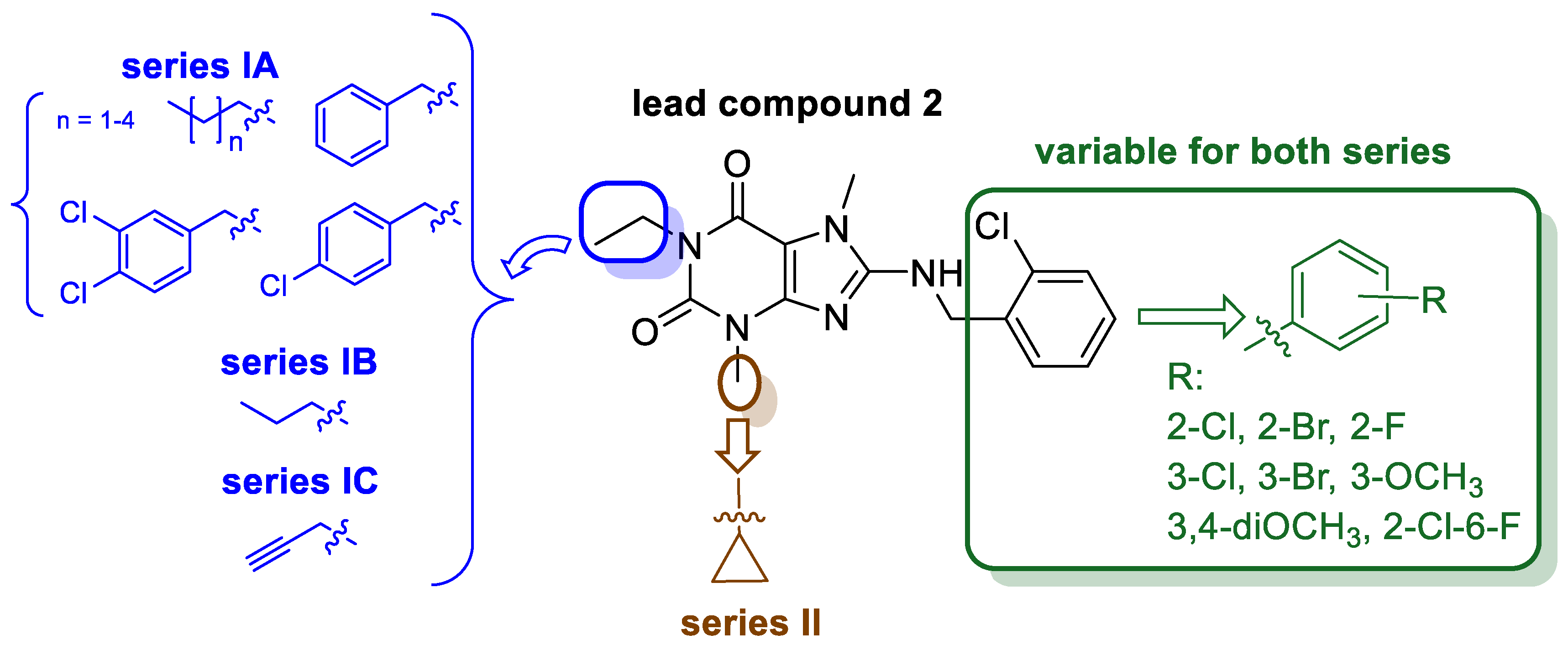
Figure 2. Lead compound 2 and structural modifications.
Ijms 24 13707 g002
Scheme 1. Synthesis of compounds 526. Reagents and conditions: (i) NaClO3, CH3COOH, H2O, 80 °C, 2 h; (ii) K2CO3, DMF, 75 °C, 4 h; (iii) triethylamine (TEA), n−C3H7OH, 150 °C, microwave irradiation (300 W; 10 bar), 1 h or CH3OCH2OH, reflux 15–17 h.
Ijms 24 13707 sch001
Scheme 2. Synthetic route of compounds 2729. Reagents and conditions: (i) 5 N HCl, 70 °C, 4 h; (ii) CH3CH2ONa, 100 °C, 4 h; (iii) 50% CH3COOH, 60 °C; (iv) 12.5% NH3, 70 °C; (v) (a) reflux 1 h, (b) reflux 2 h; (vi) CH3COOH, first 58 °C, then: reflux 2 h; (vii) DIPEA, DMF, 40 °C, 4 h; (viii) K2CO3, DMF, 75 °C, 4 h; (ix) TEA, n−C3H7OH, 150 °C, microwave irradiation (300 W; 10 bar), 1 h.
Ijms 24 13707 sch002
Figure 3. Putative binding mode of 24 in 5N2S (green ribbons) and 5N2R (blue ribbons) complexes. Yellow dashed lines depict H-bonds, blue π-π interaction, Roman numbers denote TM helices. TM7 was partially removed from both structures to increase figure visibility.
Ijms 24 13707 g003
Figure 4. Top left panel—alignment of 1st (green) and 1000th (red) frame of 24-5N2S complex trajectory. TM7 and EL3 were partially removed for better image visibility; top right panel RMSD evolution of a protein (left Y-axis) and ligand (right Y-axis; pink—Fit on ligand, maroon—Fit on protein). Bottom panel: Protein-ligand contacts timeline; the darker the color, the more interactions recorded.
Ijms 24 13707 g004
Figure 5. Toxicity evaluation of compounds 19 (MZ-1483), 22 (MZ-1490) and 24 (MZ-1495) in HepG2 cells (A) and SH-SY5Y cells (B). The graphs were generated using GraphPad Prism software 8.0. Each point represents the mean ± SEM of three independent experiments, each of which consisted of three replicates per treatment group. Statistical significance was evaluated by one-way ANOVA with post hoc Dunnett test at significance level α = 0.05 and p-values are detailed in the Figure.
Ijms 24 13707 g005
Figure 6. The effect of compounds 19 (MZ-1483), 22 (MZ-1490) and 24 (MZ-1495) on the activity of recombinant human cytochromes. (A): The influence on CYP3A4 activity. Statistical significance was evaluated by one-way ANOVA, followed by Bonferroni’s comparison test (**** p < 0.001). KE—Ketoconazole. (B): The influence on CYP2D6 activity. Statistical significance was evaluated by one-way ANOVA, followed by Bonferroni’s comparison test (* p < 0.05, **** p < 0.001). QD—Quinidine. (C): The influence on CYP2C9 activity. Statistical significance was evaluated by one-way ANOVA, followed by Bonferroni’s comparison test (**** p < 0.001). SE—Sulfaphenazole.
Ijms 24 13707 g006
Figure 7. The effect of compounds 19 (MZ-1483), 22 (MZ-1490) and 24 (MZ-1495) on nitric oxide (NO) production in BV-2 cells assessed by the Griess assay (column graph) and cell viability assessed by MTS assay (line graph). Data are expressed as mean ± SEM of three independent experiments, each of which consisted of three replicates per treatment group and is expressed as a percentage of NO production versus vehicle cells (untreated cells) set at 100%. Statistical analysis by one-way ANOVA showed significant differences between the groups (α = 0.05) and was followed by Dunnett’s multiple comparison test. Data indicated with * p = 0.033; ** p = 0.002 reflect statistically significant differences between the control and experimental groups. Data indicated with # p = 0.033; ### p < 0.001 reflect statistically significant differences between LPS = treated cells and experimental groups.
Ijms 24 13707 g007
Figure 8. Real-time efferocytosis activity of BV−2 cells. The graph shows the kinetic curve of phagocytosis of pH−rodo labeled apoptotic Jurkat cells.
Ijms 24 13707 g008
Figure 9. SAR analysis of synthesized xanthine derivatives 529. (Arrow up: increase of affinity; arrow down: decrease of affinity).
Ijms 24 13707 g009
Table 1. Affinities of xanthine derivatives 529 for human adenosine receptor subtypes a.
Compd.StructureA1R vs.
[³H]CCPA b
A2AR vs.
[³H]MSX-2 c
A2BR vs.
[³H]PSB-603 d
A3R vs.
[H³]PSB-11 e
Ki ± SEM (nM) (or % Inhibition ± SEM at 1 µM)
Series IA—N1-Different Substituents
Compound 2
(lead)
Ijms 24 13707 i001>1000 f
(22 ± 1)
71 ± 6 f>1000 f
(44 ± 10)
>1000 f
(14 ± 1)
5Ijms 24 13707 i002>1000
(26 ± 8)
302 ± 34832 ± 133>1000
(4 ± 4)
6Ijms 24 13707 i003>1000
(21 ± 6)
318 ± 141>1000
(32 ± 0)
>1000
(11 ± 2)
7Ijms 24 13707 i004>1000
(35 ± 2)
>1000
(38 ± 4)
>1000
(41 ± 8)
>1000
(13 ± 3)
8Ijms 24 13707 i005>1000
(17 ± 6)
>1000
(9 ± 8)
>1000
(13 ± 5)
>1000
(31 ± 3)
9Ijms 24 13707 i006>1000
(23 ± 5)
323 ± 150>1000
(9 ± 1)
>1000
(24 ± 7)
10Ijms 24 13707 i007>1000
(12 ± 1)
>1000>1000
(14 ± 11)
>1000
(31 ± 2)
11Ijms 24 13707 i008>1000
(10 ± 2)
>1000
(23 ± 7)
>1000
(4 ± 7)
>1000
(40 ± 9)
Series IB—N1-propyl derivatives
12Ijms 24 13707 i009>1000
(30 ± 1)
361 ± 82>1000
(33 ± 10)
>1000
(8 ± 4)
13Ijms 24 13707 i010>1000
(32 ± 4)
>1000
(47 ± 2)
>1000
(21 ± 11)
>1000
(6 ± 8)
14Ijms 24 13707 i011>1000
(29 ± 10)
124± 26687 ± 147>1000
(18 ± 4)
15Ijms 24 13707 i012>1000
(40 ± 3)
588 ± 74>1000
(34 ± 8)
>1000
(0 ± 1)
16Ijms 24 13707 i013340 ± 73254 ± 59>1000
(30 ± 1)
>1000
(11 ± 11)
17Ijms 24 13707 i014>1000
(17 ± 2)
605 ± 120>1000
(20 ± 1)
>1000
(6 ± 3)
18Ijms 24 13707 i015>1000
(7 ± 12)
773 ± 127>1000
(17 ± 9)
>1000
(−5 ± 10)
Series IC—N1-propargyl derivatives
19
(MZ-1483)
Ijms 24 13707 i016407 ± 7596 ± 31>1000
(14 ± 6)
>1000
(9 ± 1)
20Ijms 24 13707 i017>1000
(44 ± 5)
137 ± 35>1000
(25 ± 10)
>1000
(13 ± 9)
21Ijms 24 13707 i018353 ± 42287 ± 90>1000
(41 ± 3)
>1000
(21 ± 1)
22
(MZ-1490)
Ijms 24 13707 i019130 ± 3462 ± 5>1000
(41 ± 6)
>1000
(21 ± 6)
23Ijms 24 13707 i020334 ± 122126± 15>1000
(26 ± 2)
>1000
(14 ± 4)
24
(MZ-1495)
Ijms 24 13707 i02172 ± 877 ± 24>1000
(25 ± 8)
>1000
(7 ± 8)
25Ijms 24 13707 i022460 ± 99225 ± 40>1000
(35 ± 9)
>1000
(0 ± 2)
26Ijms 24 13707 i023>1000
(−2 ± 7)
108 ± 22>1000
(28 ± 6)
>1000
(2 ± 0)
Series II—N1-propargyl-N3-cyclopropyl derivatives
27Ijms 24 13707 i024247 ± 43311 ± 167>1000
(9 ± 1)
>1000
(16 ± 2)
28Ijms 24 13707 i025166 ± 65386 ± 61>1000
(14 ± 4)
>1000
(−2 ± 6)
29Ijms 24 13707 i02678 ± 9204 ± 38>1000
(16 ± 3)
>1000
(16 ± 1)
a Human recombinant receptors expressed in CHO cells. Ki values were determined as means from three independent experiments ± standard error of the mean (SEM); b [³H]2-Chloro-N6-cyclopentyladenosine; c [³H] (3-(3-Hydroxypropyl)-7-methyl-8-(m-methoxystyryl)-1-propargylxanthine); d [³H]8-(4-(4-(4-hClorophenyl)piperazine-1-sulfonyl)phenyl)-1-propylxanthine; e [3H]8-Ethyl-4-methyl-2-phenyl-(8R)-4,5,7,8-tetrahydro-1H-imidazo [2,1-i]-purin-5-one; f data from [13].
Table 2. Inhibitory activity of compounds 5, 1226 at human MAO B evaluated in a fluorescence assay.
Compound% of hMAO B Inhibition at 1 µM aCompound% of hMAO B Inhibition at 1 µM a
58196
1272032
1315218
149227
1510238
16112410
1752511
182263
a Mean value of two independent experiments.
Table 3. The most probable metabolic pathways for compounds 19, 22 and 24, and the reference drug verapamil determined in vitro using human liver microsomes.
Drug% Remaining in the Reaction Mixture aMolecular Mass (m/z)Molecular Mass of
the Metabolite (m/z)/
Retention Time (min)
Metabolic Pathway
19>95358.16249.07/
6.27 (M1)
fragmentation and hydroxylation
234.16/
3.59 (M2)
fragmentation,
demethylation and hydroxylation
22>90376.12249.53/
6.26 (M1)
fragmentation, and hydroxylation
24>95402.13249.13/
6.26 (M1)
fragmentation, and hydroxylation
Verapamil b31455.31441.35/
4.95 (M1)
demethylation
291.32/
4.00 (M2)
fragmentation
165.09/
3.38 (M3)
fragmentation
441.29/
4.62 (M4)
demethylation
427.33/4.54
(M5)
double-demethylation
277.26/
3.92 (M6)
fragmentation
a after 120 min of incubation with HLMs; b reference drug (low metabolic stability).
Table 4. Permeability coefficient of compounds (19, 22 and 24) and caffeine.
Compounda,b Pe
(10−6 cm/s) ± SD
PermeableDrug Retention (%)
Caffeine6.57 ± 1.57yes29.46
193.13 ± 0.82yes51.78
220.37 ± 0.01no68.62
240.89 ± 0.71no93.30
a PAMPA plate’s manufacturer breakpoint for permeable compounds: Pe ≥ 1.5. b Tested in triplicate.
Table 5. Anti-inflammatory (antiedematous) effect of the compounds in the carrageenan-induced paw edema test in rats.
TreatmentDose (mg/kg)Change in Edema Volume [mL]
1 h2 h 3 h
Control 0.78 ± 0.031.12 ± 0.131.33 ± 0.13
19200.56 ± 0.130.72 ± 0.091.10 ± 0.21
22200.49 ± 0.080.91 ± 0.050.92 ± 0.06
24200.38 ± 0.02 a0.63 ± 0.14 a0.69 ± 0.01 b
ketoprofen200.32 ± 0.03 a0.41 ± 0.04 a0.43 ± 0.03 b
Data are presented as the means ± SEM of six to eight animals per group, a p < 0.05, b p < 0.01.
Table 6. The influence of the investigated compounds on the duration of pain reaction in the formalin test in mice a.
TreatmentDose (mg/kg)Licking of the Hind Paw (sec)
Early Phase
(0–5 min)
Inhibition
(%)
Late Phase
(15–30 min)
Inhibition
(%)
Mean ± SEM Mean ± SEM
Control 41.6 ± 15.2-119.4 ± 32.8-
194042.6 ± 2.8063.0 ± 6.6 b47.2
2048.2 ± 10.0076.8 ± 26.435.7
1041.8 ± 5.523.188.4 ± 5.626.0
224039.2 ± 1.35.862.0 ± 5.2 b48.0
2041.0 ± 8.6078.2 ± 20.30
1041.6 ± 5.4092.2 ± 6.522.8
244030.8 ± 6.726.012.2 ± 3.3 d89.7
2031.4 ± 5.024.520.4 ± 10.6 d82.9
1043.2 ± 6.6061.2 ± 6.4 b48.7
Control 63.8 ± 2.0-120.8 ± 3.4-
ASA20062.0 ± 8.69.240.6 ± 14.0 c66.3
10058.5 ± 4.514.360.3 ± 9.5 c50.0
5059.8 ± 6.312.4108.7 ± 13.810.0
a Data are presented as the means ± SEM of 6–8 mice per group. b p < 0.05, c p < 0.01, d p < 0.001 vs. control.
Disclaimer/Publisher’s Note: The statements, opinions and data contained in all publications are solely those of the individual author(s) and contributor(s) and not of MDPI and/or the editor(s). MDPI and/or the editor(s) disclaim responsibility for any injury to people or property resulting from any ideas, methods, instructions or products referred to in the content.