Article

Defining the Basal and Immunomodulatory Mediator-Induced Phosphoprotein Signature in Pediatric B Cell Acute Lymphoblastic Leukemia (B-ALL) Diagnostic Samples

by
Aaruni Khanolkar
1,2,*,
Guorong Liu
1 and
Bridget M. Simpson Schneider
1
1
Department of Pathology, Ann and Robert H. Lurie Children’s Hospital of Chicago, Chicago, IL 60611, USA
2
Department of Pathology, Northwestern University, Chicago, IL 60611, USA
*
Author to whom correspondence should be addressed.
Int. J. Mol. Sci. 2023, 24(18), 13937; https://doi.org/10.3390/ijms241813937
Submission received: 29 June 2023 / Revised: 29 August 2023 / Accepted: 31 August 2023 / Published: 11 September 2023

Abstract

:
It is theorized that dysregulated immune responses to infectious insults contribute to the development of pediatric B-ALL. In this context, our understanding of the immunomodulatory-mediator-induced signaling responses of leukemic blasts in pediatric B-ALL diagnostic samples is rather limited. Hence, in this study, we defined the signaling landscape of leukemic blasts, as well as normal mature B cells and T cells residing in diagnostic samples from 63 pediatric B-ALL patients. These samples were interrogated with a range of immunomodulatory-mediators within 24 h of collection, and phosflow analyses of downstream proximal signaling nodes were performed. Our data reveal evidence of basal hyperphosphorylation across a broad swath of these signaling nodes in leukemic blasts in contrast to normal mature B cells and T cells in the same sample. We also detected similarities in the phosphoprotein signature between blasts and mature B cells in response to IFNγ and IL-2 treatment, but significant divergence in the phosphoprotein signature was observed between blasts and mature B cells in response to IL-4, IL-7, IL-10, IL-21 and CD40 ligand treatment. Our results demonstrate the existence of both symmetry and asymmetry in the phosphoprotein signature between leukemic and non-leukemic cells in pediatric B-ALL diagnostic samples.

1. Introduction

Our awareness of the association between inflammation and cancer stretches back over one hundred and fifty years when Rudolf Virchow made his astute observation documenting the presence of leukocytes in cancerous tissues [1,2,3,4,5,6,7]. However, only 15–20% of malignancies are associated with pre-existing localized inflammation, and inflammation impacts critical steps in the neoplastic process, such as tumor growth, metastatic spread and the outcome of therapeutic interventions [2,3,4,8,9,10,11,12,13,14]. Multiple lines of evidence point to the fact that the tumor micro-environment (TME) tends to be replete in inflammatory mediators, such as cytokines and chemokines [2]. In addition, consistent with this evidence, clinical data also reveal that both non-specific as well as targeted inhibition of inflammatory signals can attenuate the risk for developing certain types of cancers [15,16,17]. Even malignancies that lack microbial drivers experience inflammatory stress induced by oncogenic transformation, metabolic alterations, cell death and hypoxia [4,8,18]. Indeed, genetic lesions that disrupt the tumor suppressor function can trigger DNA damage-induced inflammatory responses, and conversely, the activation of oncogenes can augment the expression of cytokines and chemokines that can drive tumor progression and/or simultaneously inhibit anti-tumor immune responses [8,19,20,21,22,23,24].
B cell acute lymphoblastic leukemia (B-ALL) is the most frequent cancer in children [25]. In fact, pre-leukemic clones harboring the ETV6-RUNX1 gene fusion are observed in almost 5% of healthy newborns, but frank leukemic transformation occurs in only a small fraction (~0.2%) of children that carry this genetic aberration [26,27,28]. Prevailing wisdom suggests that dysregulated immune responses to childhood infectious insults contribute to the accumulation of additional genetic hits that ultimately drive the development of clinically apparent B-ALL [26]. Thus, B-ALL represents an aberrant outgrowth of a primitive B cell clone that is endowed with a selective growth advantage aided and abetted by a varied set of genomic alterations involving coding DNA sequences as well as non-coding genetic elements [25]. This underlying genetic heterogeneity underpins the spectrum of clinical phenotypes and therapy-related outcomes observed in pediatric B-ALL patients. Although the conventional five-year survival rate in pediatric B-ALL patients exceeds 85%, disease relapse is associated with a high mortality rate [25,29]. Collectively, available data suggest that a combination of at least two factors can influence the development of pediatric B-ALL. These factors include, first, an underlying genetic predisposition characterized by the aforementioned variably penetrant, somatic or germline genetic variants; and second, infectious insults that elicit atypical responses, including a disordered cytokine profile, from an untrained immune system [26].
Beginning with Kinlen’s “population-mixing” theory outlined in 1988, in the ensuing years, at least five other theoretical models have been proposed, aimed at enhancing our understanding of the elements that might promote the onset of pediatric B-ALL [26,30]. These alternate models include Greaves’ delayed immune exposure theory; the infective lymphoid recovery hypothesis; Smith’s theory, which entails the vertical transmission of infections from the mother to the fetus; Schmiegelow’s adrenal hypothesis model; and an emerging model based on the concept of “trained immunity” [31,32,33,34,35]. A common thread that runs through these theoretical models is the putative link between childhood infections and perturbations in immunomodulatory cytokines elaborated by the host’s immune system that is still “learning the ropes”. Previous investigations have revealed perturbations in cytokine profiles of neonates that later go on to develop B-ALL in comparison to children that remain healthy [26,36,37,38]. It has also been demonstrated that allelic polymorphisms that affect the production of key effector cytokines, such as IFNγ, can influence the timing of the onset of B-ALL in children, and a subset of pediatric B-ALL patients that relapse and display IL-15 receptor α (IL15Rα) gene expression levels in blasts that are above the median level experience better subsequent event-free and overall survival [39,40]. Altogether, these lines of scientific evidence further support the notion that either direct cross-talk between precursor B cells and T cells, or perhaps bystander responses to soluble mediators such as cytokines, elicited during childhood immune responses could potentially support the malignant outgrowth of precursor B cells that harbor deleterious genetic variants.
Cell signaling undergirds almost every aspect of human biology, and signaling associated with immunomodulatory mediators, such as cytokines and co-stimulatory molecules, plays a pivotal role in the lineage commitment, maturation, differentiation and functional polarization of many immune cell types [41,42,43,44,45,46,47]. Very little is known about immunomodulatory-mediator-induced signaling responses in B-ALL blasts and/or co-existing non-malignant lymphocytes in pediatric B-ALL samples, and our knowledge of the signaling potential and properties of the leukemic blasts existent in diagnostic samples of pediatric B-ALL patients is incomplete [40]. Therefore, gaining a deeper understanding of the signaling properties of malignant immune cells and their normal counterparts in diagnostic samples of pediatric B-ALL subjects might offer clues that could potentially be leveraged in the future as actionable biomarkers that allow for more precise disease classification as well as to identify novel therapeutic targets. Because the behavior of leukemic B-ALL blasts is so clearly aberrant compared to non-leukemic B cells, we hypothesized that the immunomodulatory-mediator-driven phosphoprotein signature in the blasts completely diverges from that of the non-leukemic B cells residing in the diagnostic B-ALL samples. To test this hypothesis, we assembled a data set defining the immunomodulatory-mediator-induced signaling landscape of leukemic blasts, as well as normal mature B cells and T cells residing in diagnostic samples from 63 pediatric B-ALL patients. We utilized phosflow to evaluate the phosphorylation status of proximal signaling nodes following the treatment of diagnostic B-ALL samples with immunomodulatory mediators that are often generated and/or upregulated in the setting of pathogen-induced immune responses. Specifically, recombinant human (rh) IFNγ, IL-2, IL-4, IL-7, IL-10, IL-21 and trimerized CD40 ligand (CD40L) were used as signaling inputs to define this phosphoprotein signature. These signaling studies were complemented by a detailed evaluation of the surface expression patterns for the following cytokine receptors: IFNγR1, IFNγR2, CD25 (IL-2Rα), CD122 (IL-2Rβ), CD124 (IL-4Rα), CD127 (IL-7Rα), CD132 (common γ chain), IL10Rα, IL-10Rβ, CD360 (IL-21R) and CD40 in a subset of the patients. To the best of our knowledge, this study represents one of the few (if not the only) report(s) of a comprehensive assessment of basal and immunomodulatory-mediator-induced phosphoprotein signatures in diagnostic samples of pediatric B-ALL subjects. Furthermore, the identification of significantly elevated basal phosphorylation levels of STAT(s)-1, 5, 6 and p65-NFκB in the diagnostic samples of patients that experienced disease-related mortality is a novel observation, as it underscores the potential prognostic value of assessing phosphorylation signals at the time of diagnosis.

2. Results

2.1. Basal Hyperphosphorylation in Leukemic Blasts

Basal hyperphosphorylation refers to the existence of an elevated background phosphorylation state of a signaling node, detected directly ex vivo in the absence of any exogenous treatment with stimulants such as immunomodulatory mediators. The detection of basal hyperphosphorylation indirectly suggests the in vivo presence of low-level stimuli that could potentially contribute to persistent low-grade inflammation [48]. As the neoplastic process is intimately associated with inflammation, the assessment of basal phosphorylation signals can provide critical early clues relating to underlying low-grade persistent inflammatory states [1,2,3,4,5,6,7,9,10,11,12,13,14]. We examined and compared the basal phosphorylation states of leukemic blasts and non-leukemic mature B cells and T cells residing in diagnostic B-ALL samples obtained from sixty-three patients with a confirmed diagnosis of pediatric B-ALL leukemia (Figure 1; for the gating strategy, please refer to Supplementary Figure S1a,b). The signaling nodes evaluated for this assessment included the following Signal Transducer and Activator of Transcription (STAT) molecules: STAT1, STAT3, STAT5 and STAT6, and we also assessed the phosphorylation of p65-NFκB (Figure 1). The basal phosphorylation state of the leukemic blasts was significantly elevated compared to the neighboring mature B cells in the same sample for STAT1, STAT3, STAT5, STAT6 and p65-NFκB (Figure 1). Four to eight samples obtained from non-leukemic donors were similarly evaluated to assess basal hyperphosphorylation among hematogones and mature B and T cells. We were not able to detect similar evidence of basal hyperphosphorylation in hematogones compared to mature B cells and CD3+ T cells that were present in non-leukemic bone marrow samples (for the gating strategy, please refer to Supplementary Figure S1c). We also assessed the basal phosphorylation status of the leukemic blasts against the hematogones, which revealed that leukemic blasts had significantly elevated basal phosphorylation for STAT(s)3, 5, 6 and p65-NFκB but not STAT1 (Supplementary Figure S2). Overall, these data suggest that an altered response to local environmental cues is hardwired into the signaling machinery of leukemic cells.

2.2. Phosphoprotein Signature of Pediatric B-ALL Blasts Treated with Immunomodulatory Mediators

Immunomodulatory mediators imprint the trajectory of development and maturation, as well as the functional attributes of lymphocyte types [41,42,43,44,45,46,47]. Therefore, we undertook a comprehensive survey of the immunomodulatory-mediator-induced signaling properties of leukemic blasts and mature B and T cells present in pediatric B-ALL diagnostic samples. As part of this effort, we evaluated the phosphorylation status of proximal signaling nodes that engage with the cognate receptors for the following cytokines: IL-7, IL-2, IL-4, IL-21, IFNγ and IL-10, as well as the phosphorylation of p65-NFκB following the engagement of the CD40 receptor (Figure 2a–g) [49,50,51,52,53,54,55]. The majority of responses were clearly divergent between the leukemic blasts and mature B cells. For instance, the IL-7-induced pSTAT5 signal was absent in mature B cells but clearly detectable in leukemic blasts, whereas the IL-4, IL-21, IL-10 and CD40L-induced signaling responses were attenuated in leukemic blasts but robust in mature B cells (Figure 2a,c,d,f,g). Interestingly, the signaling responses induced by IL-2 and IFNγ displayed good symmetry between leukemic blasts and mature B cells (Figure 2b,e). Specifically, treatment with IFNγ evoked a robust pSTAT1 signal in both leukemic blasts and mature B cells, and IL-2-induced STAT5 phosphorylation was undetectable in both subsets. In a few experiments, we also examined the signaling properties of hematogones present in non-leukemic bone marrow specimens, and this allowed us to separately compare the phosphoprotein profile of hematogones with that of the leukemic blasts. This set of assessments also revealed evidence of signaling symmetry as well as asymmetry between these two populations. IFNγ-induced STAT1, IL-2-induced STAT5 and IL-10-induced STAT3 phosphorylation was comparable between the leukemic blasts and hematogones, and the IL-4, IL-7, IL-21 and CD40L-induced phosphorylation of STAT(s)6, 5, 3 and p65-NFκB displayed significantly elevated signals in leukemic blasts (Supplementary Figure S3). For both leukemic and non-leukemic samples, T cells were assessed in parallel and served as crucial internal controls for the phosflow assays (Figure 2). Overall, these data indicate both the preservation as well as the loss of select signaling properties associated with leukemic blasts that diverge from the normal maturation pathway and fail to differentiate into mature B cells.

2.3. Cytokine Receptor Expression Profile

In an attempt to better comprehend the observed symmetry and asymmetry in the signaling profiles of leukemic blasts and mature B cells in diagnostic leukemic samples as well as hematogones in non-leukemic specimens, we undertook an evaluation of the cognate surface receptors for the immunomodulatory mediators in a separate series of experiments. Specifically, we examined the expression of IL-2Rα (CD25), IL-2Rβ (CD122), IL-4Rα (CD124), IL-7Rα (CD127), IL-10Rα (CD210), IL-10Rβ, IL-21R (CD360), CD40, IFNγR1 (CD119), IFNγR2 and the common γ chain (CD132) on the surface of the relevant cellular subsets.

2.3.1. IL-7Rα (CD127) and the Common γ Chain (CD132)

As has been described in the literature, a greater frequency of leukemic blasts expressed IL-7Rα on their surfaces compared to mature B cells, and a similar trend was observed when we compared the median fluorescence intensity (MFI) of surface IL-7Rα (Figure 3a,b) [56,57,58,59,60]. This observation was consistent with our initial finding that IL-7-induced STAT5 phosphorylation was significantly elevated in leukemic blasts compared to mature B cells (Figure 2a). Hematogones also expressed higher levels of IL-7Rα compared to their mature counterparts in non-leukemic specimens, but this level was significantly lower compared to leukemic blasts (Figure 3c). The common γ chain (CD132) is the signaling subunit shared by IL-2, IL-4, IL-7, IL-9, IL-15 and IL-21 [49,50]. In order to further resolve the disparate signaling profile observed in leukemic blasts and mature B cells associated with IL-7 treatment, we also examined the surface expression of CD132. A greater frequency of leukemic blasts expressed CD132 compared to mature B cells in diagnostic B-ALL samples, and additionally, the density of CD132 surface expression was also slightly greater in the leukemic blasts (Figure 3d,e). Interestingly, we did not detect any significant difference between leukemic blasts and hematogones in non-leukemic bone marrow in terms of the frequency of these cells expressing CD132, as well as the MFI of CD132 expression (Figure 3f).

2.3.2. IL-2Rα (CD25) and IL-2Rβ (CD122)

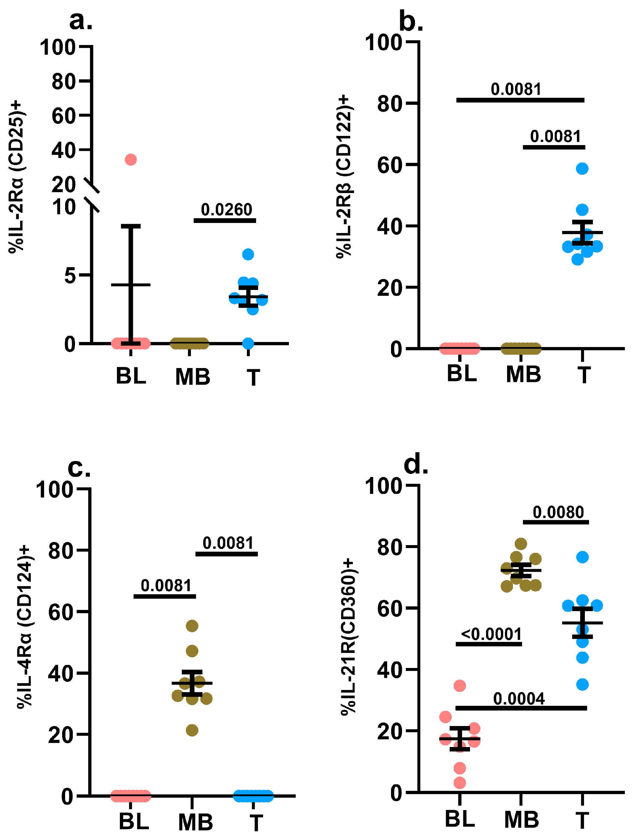
In addition to its engagement downstream of IL-7R, STAT5 is also engaged downstream of IL-2R, and stimulation with IL-2 revealed a symmetrical lack of STAT5 phosphorylation in leukemic blasts and mature B cells (Figure 2b). This symmetry was preserved in the pattern of the expression of IL-2Rα (CD25) and IL-2Rβ (CD122) between the leukemic blasts and mature B cells, as well as between hematogones and mature B cells in non-leukemic specimens (Figure 4a,b and Supplementary Figure S4a,b).

2.3.3. IL-4Rα (CD124)

As mentioned above, CD132 is also paired with the IL-4 receptor [49,50]. With respect to IL-4-driven STAT6 phosphorylation, we detected a much stronger signal in the mature B cells in the leukemic samples compared to the blasts, and accordingly, we only detected IL-4Rα surface expression on mature B cells (Figure 2c and Figure 4c). This surface receptor pattern was mirrored in the hematogones in non-leukemic specimens (Supplementary Figure S4c).

2.3.4. IL-21R (CD360)

The fourth immunomodulatory mediator in our study that also utilizes CD132 is IL-21 [49,50]. In the context of B cells, IL-21 is predominantly involved in regulating the germinal center reaction and associated humoral immune responses, as well as plasma cell differentiation [52,61,62,63]. Under the right conditions, IL-21 has also been reported to induce apoptosis of non-Hodgkin’s B lymphoma cells, and in certain instances, it can also equip B cells to express cytotoxic effector molecules such as granzyme B [64,65,66]. Our phosflow data revealed robust IL-21 signaling in mature B cells and T cells but not in leukemic blasts (Figure 2d). Consistent with this observation, only a small frequency of leukemic blasts expressed surface IL-21R compared to mature B cells (as well as T cells) (Figure 4d). A similar trend was observed in non-leukemic bone marrow specimens, where hematogones displayed the lowest proportion of cells displaying surface IL-21R expression (Supplementary Figure S4d).

2.3.5. IFNγR1 (CD119) and IFNγR2

Notably, symmetrical yet diametrically opposite to the IL-2-induced signaling response, IFNγ-induced STAT1 phosphorylation was observed to be equally robust between leukemic blasts and mature B cells in leukemic samples (Figure 2e). When we assessed the surface expression of IFNγ receptors, there were no significant differences in the frequencies of these two cell subsets expressing IFNγR1, although a higher frequency of mature B cells expressed IFNγR2 (Figure 5a,c). The MFI for both IFNγ receptors were slightly lower in the blasts compared to the mature B cell subset (Figure 5b,d). The proportion of leukemic blasts expressing IFNγR1 was slightly higher compared to hematogones in non-leukemic specimens, but there was no such difference noted for IFNγR2 (Supplementary Figure S5a,b). Taken together, these findings suggest that the reduced levels in the surface expression patterns of IFNγR on the leukemic blasts do not significantly hinder the formation of the IFNγ-IFNγR1-IFNγR2 ternary complex that is essential and required for the signaling cascade that engages STAT1 further downstream in the IFNγ signaling axis [67,68].

2.3.6. IL-10Rα (CD210) and IL-10Rβ

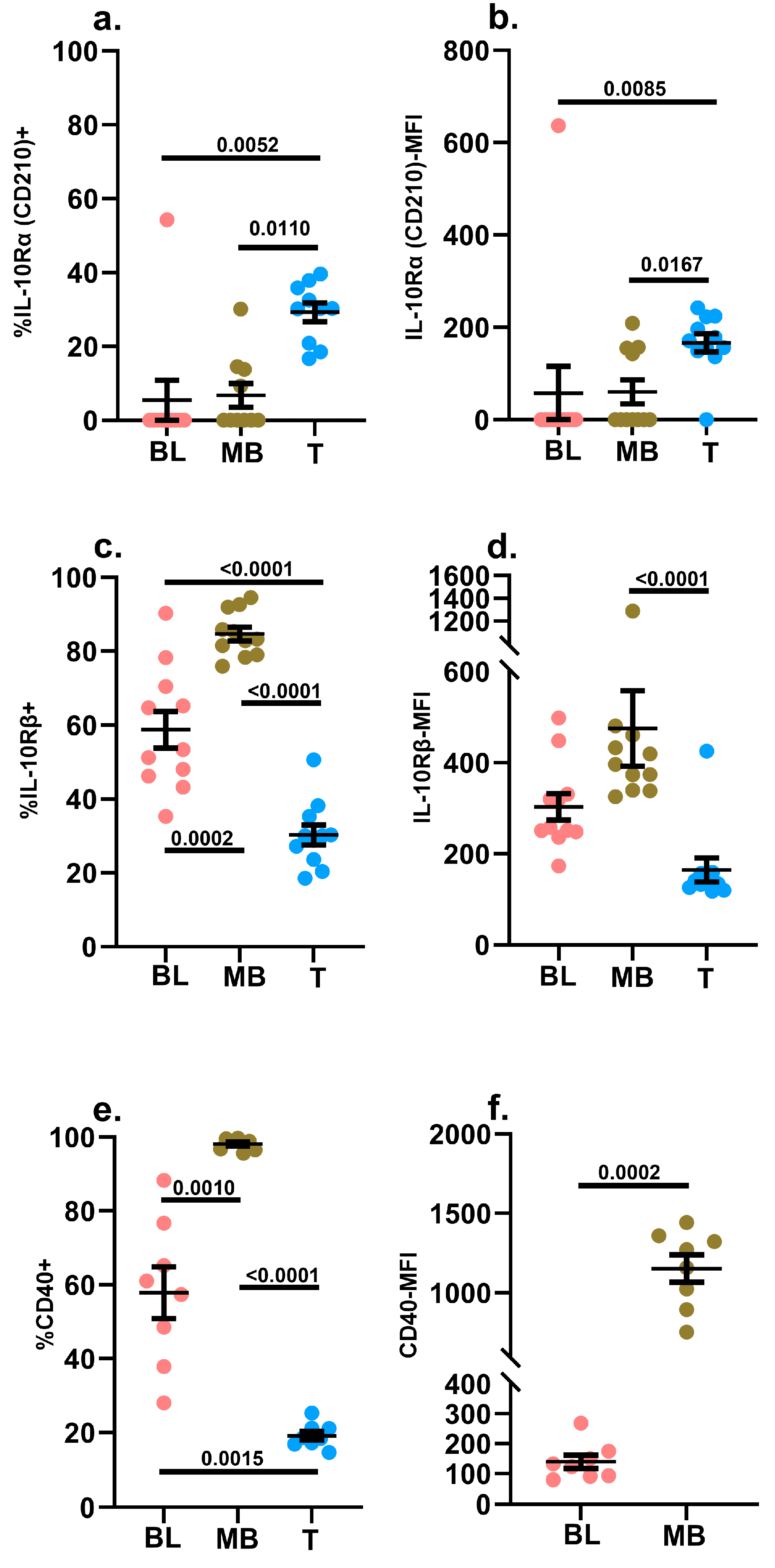
STAT3 phosphorylation induced by IL-10 treatment was significantly depressed in leukemic blasts compared to mature B cells (Figure 2f). Neither the blasts nor the mature B cells expressed much IL-10Rα on their surfaces. However, greater than 80% of mature B cells in leukemic samples expressed IL-10Rβ, and ~60% of leukemic blasts displayed IL-10Rβ on their surface. However, there was no significant difference in the MFI of IL-10Rβ expression between the blasts and mature B cells in the leukemic samples (Figure 6a–d). Given this, the markedly attenuated IL-10-induced pSTAT3 signal in the blasts was intriguing. Overall, this might suggest that, in the absence of IL-10Rα expression, >80% of B lineage cells need to express IL-10Rβ to transduce IL-10 signals effectively. Alternatively, the precise molecular requirements for IL-10 signaling could also differ between blasts and mature B cells. Hematogones in non-leukemic specimens closely mirrored the leukemic blasts in terms of their IL-10 receptor expression profile as well as their IL-10 signaling potential (fold change) (Supplementary Figure S6a–d).

2.3.7. CD40

CD40 is a member of the tumor necrosis factor (TNF)-receptor superfamily [69]. It is expressed on several hematopoietically-derived antigen presenting cells, including mature B cells, as well as non-hematopoietic cells [69]. In our study, consistent with published data, all mature B cells expressed CD40 on their surfaces at high levels (Figure 6e,f). It has also been previously reported that B-ALL blasts express CD40, and we did observe CD40 expression on leukemic blasts in our diagnostic samples. However, both the frequency of the blasts expressing CD40 as well as the level of surface expression was significantly lower compared to mature B cells (Figure 6e,f) [70,71,72,73,74,75,76,77,78]. This finding could potentially explain the reduced phosphorylation of p65-NFκB in the leukemic blasts following treatment with trimerized CD40L (Figure 2g) [55]. Similar trends were observed between mature B cells and hematogones in non-leukemic specimens (Supplementary Figure S6e,f).

2.4. Phosphoprotein Signature and Overall Survival Analyses

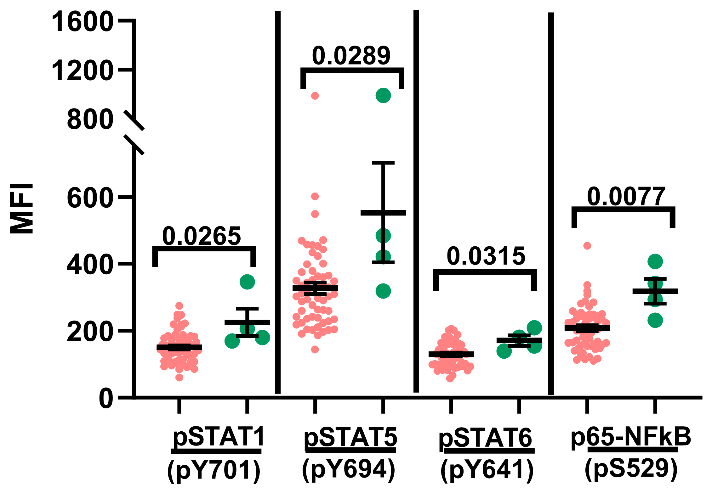
We also examined the phosphoprotein signature in the context of overall survival (OS) (Figure 7) [79]. The OS group included study subjects that had been classified as Standard Risk (SR), High Risk (HR) and Very High Risk (VHR) per established criteria at the time of diagnosis [25,29,80], as well as the study subjects that experienced disease relapse [25,29,80]. The study subjects that succumbed to the disease were excluded from the OS group. Interestingly, basal phosphorylation levels of STAT1, STAT5, STAT6 and p65-NFκB were significantly higher in the diagnostic samples obtained from the study subjects that experienced mortality (Figure 7). No significant differences were observed when event-free survival (EFS) was evaluated [79]. The amplified basal phosphorylation signal in the study subjects that experienced mortality might suggest the likelihood of a markedly dysregulated inflammatory milieu that may or may not be directly related to their underlying disease. Furthermore, these data also highlight the potential prognostic value of measuring the basal phosphorylation signal in diagnostic samples in terms of predicting the likelihood of survival from the disease.

3. Discussion

In this study, we utilized phosflow to examine the phosphoprotein signature of leukemic blasts in diagnostic samples of 63 pediatric B-ALL patients that span the spectrum of established diagnostic risk classifications (SR; HR; VHR) [25,29,80]. Accumulating evidence implicates a disordered immune response to early-life microbial insults that colludes with pre-existing (in utero) genetic lesions as well as those acquired postnatally before the disease manifests as frank B-ALL [26,30,31,32,33,34,35]. Hence, we investigated the signaling responses at proximal cytosolic signaling hubs following treatment with a defined suite of immunomodulatory mediators that are commonly generated by the host immune system in response to pathogens. These phosphoprotein signals were measured in leukemic blasts and benchmarked against the same signal detected in neighboring mature (un-transformed) B cells and T cells present in the diagnostic samples. In an effort to better understand the perceived differences in the phosphoprotein signals among the blasts, mature B cells and T cells, we also conducted an assessment of cytokine receptor and CD40 surface expression patterns on these cellular subsets in a smaller group of patient samples.
We detected evidence of basal hyperphosphorylation in leukemic blasts compared to mature B cells in diagnostic B-ALL samples across all five signaling nodes, STAT(s)-1,3,5,6 and p65-NFκB, assessed in this study (Figure 1). This finding is consistent with the notion that inflammation is writ large in the tumor microenvironment (TME) [2,8,14,81]. We speculate that this persistent hyperphosphorylation is quite likely the result of a combination of ongoing low-grade cytokine-induced signaling in the TME, coupled with altered calibrations in the sensitivity thresholds for cytokine responsiveness in the leukemic blasts. The fact that neighboring untransformed mature B cells in the diagnostic samples and hematogones in non-leukemic samples do not display evidence of basal hyperphosphorylation suggests that aberrations in the signaling machinery are specifically localized to the blast population in the sample (Figure 1 and Supplementary Figure S2). In future studies, we will endeavor to define the cytokine landscape existent in the B-ALL diagnostic samples in order to obtain a better handle on the specific cytokines that are likely contributing to this basal hyperphosphorylation. Given that signaling pathways for different cytokines can intersect and converge at several signaling hubs, dissecting this network and parsing out these signaling pathways will entail in-depth computational analyses that are beyond the scope of this current study. Another limitation of our study is the bulk analyses of the B-ALL blasts and mature B cells. Follow-up studies will include assessments at the single-cell level to enhance the resolution of the analyses.
In general, the amplitude of the phosphorylation signal for the leukemic blasts assessed in response to the immunomodulatory mediators used in the study was not particularly robust, with the exception of IFNγ-induced signaling (Figure 2e). The reduced signaling potential in response to IL-2, IL-4 and IL-21 may be attributable to the reduced surface expression levels of the cognate receptor subunits [IL-2Rα (CD25), IL-2Rβ (CD122), IL-4Rα (CD124) and IL-21R (CD360)] because the frequency and density of the expression of the common γ chain (CD132), the signaling subunit shared by these three cytokines, was higher in the leukemic blasts versus the mature B cells (Figure 2b–d, Figure 3d,e and Figure 4a–d). Furthermore, a significantly lower proportion of leukemic blasts expressed IL-10Rβ on their surfaces compared to mature B cells in the diagnostic samples, and this might be one contributing factor to the dampened IL-10 signaling potential of the blasts (Figure 2f and Figure 6c,d). Similarly, the reduced proportion of blasts expressing surface CD40 coupled with the markedly reduced level of CD40 expression (CD40 MFI) could explain the attenuated phosphorylation of p65-NFkB in the blasts treated with trimerized CD40L compared to mature B cells in the diagnostic samples (Figure 2g and Figure 6e,f).
There is a plethora of published evidence highlighting the role of IFNγ in immunosurveillance, but there also exists an expanding body of work attributing a pro-tumorigenic and immune-editing role for IFNγ [53,67,82,83,84,85,86,87,88,89]. For instance, to minimize inflammation-induced collateral tissue damage, IFNγ signaling in normal cells can upregulate immune checkpoint molecules such as PD-L1, PD-L2 and CTLA-4 [67,89,90,91,92,93,94]. Signaling mediated by PD-L1, PD-L2 and CTLA-4 functions as a molecular brake that dampens T cell responses and can also orchestrate the functional exhaustion of T cells [95,96]. Cancer cells have learned to co-opt this protective mechanism as a means of counteracting immunosurveillance, as they can upregulate PD-L1 in response to IFNγ exposure [67,89,90,91,92,93,94,97,98]. Along similar lines, IFNγ signaling can also boost the expression of non-classical MHC class Ia genes, such as HLA-G, -E and -F, which are known to endow cancer cells with ability to evade CTL and NK cell responses [67,85]. Furthermore, IFNγ signaling is also involved in the trafficking of myeloid-derived suppressor cells into the TME [67,88]. Data from IFNγ transgenic mouse models have also underscored the role of elevated IFNγ levels in arresting B cell maturation at the pro-B cell stage [99]. Hence, in light of these observations, it is tempting to speculate that, with reference to our data demonstrating the preservation of IFNγ signaling in B-ALL blasts, this signaling pathway in leukemic B-ALL blasts might serve to facilitate immune escape and promote the survival of these cells (Figure 2e).
STAT5 signaling exerts considerable influence over B cell ontogeny, as evidenced by the fact that its abrogation arrests B cell maturation at the progenitor stage [100,101]. Appropriate checks and balances for STAT5 signaling are essential for preserving the integrity of the B cell developmental pathway because constitutive STAT5 activation is associated with the development of several myeloid and lymphoid malignancies [100,102]. It has also been reported that constitutively active STAT5b can collude with aberrant pre-B cell receptor (pre-BCR) signaling entities to prompt the development of B-ALL [100,103,104,105]. Interestingly, even in the absence of IL-7Rα mutations, the over-expression of wild-type IL-7Rα can itself be oncogenic, given that IL-7 signaling promotes survival (via the JAK3-STAT5 pathway) as well as proliferation (via the PI3Kinase-mTOR pathway) [58,105,106]. In our study, treatment with exogenous IL-7 significantly augmented STAT5 phosphorylation in leukemic blasts compared to both mature B cells in leukemic samples and hematogones in non-leukemic bone marrow specimens (Figure 2a and Supplementary Figure S3e). It is possible that IL-7Rα variants could be contributing to the enhanced IL-7Rα-mediated STAT5 phosphorylation in the leukemic blasts; hence, molecular analyses of the IL7R gene will be the focus of a follow-up study [56,105]. Consistent with the observation of enhanced IL-7 Rα-mediated STAT5 phosphorylation in the leukemic blasts, a significantly greater proportion of leukemic blasts expressed IL-7Rα compared to mature B cells in the diagnostic samples, as well as hematogones in non-leukemic bone marrow samples (Figure 3a,c). In addition, in the leukemic samples, the density of surface IL-7Rα expression on the blasts was significantly higher compared to the mature B cells (Figure 3b). Overall, these data and the aforementioned basal hyperphosphorylation of STAT5 align well with reports in the literature that implicate an oncogenic role for IL-7Rα over-expression and dysregulated STAT5 activation in B-ALL.
We also examined STAT3 activation in the context of IL-10 and IL-21 signaling. The leukemic blasts displayed significantly elevated levels of STAT3 basal phosphorylation compared with mature B cells (in diagnostic B-ALL samples) and hematogones (in non-leukemic bone marrow samples) (Figure 1b; Supplementary Figure S2b). However, STAT3 phosphorylation in response to IL-21 and IL-10 treatment was significantly diminished in the leukemic blasts compared to the mature B cells (Figure 2d,f). There is some evidence in the literature linking IL-10 deficiency with dysbiosis, which can in turn promote B cell DNA damage and corrupt B cell development, thus predisposing the host to pediatric B-ALL [36,107], and IL-21 has been shown to promote apoptotic activity in B cells [108]. Both the frequency of cells expressing IL-21R and the density of its expression were significantly lower for the leukemic blasts compared to the mature B cells (Figure 4d; Supplementary Figure S7). Given these findings, it is tempting to speculate that primitive B cells that transform into leukemic blasts in B-ALL could exploit reduced levels of IL-21R expression to their advantage to avoid IL-21-induced programmed cell death, which might in turn contribute to the proliferative advantage that leukemic cells display. Given the putative link between infectious insults and pediatric B-ALL development, it is worth noting that many pathogens, pathogen products and stressors can engage STAT3 [109,110,111,112,113,114,115,116,117]. Furthermore, persistently activated STAT3 can promote a pro-oncogenic inflammatory environment and subvert anti-tumor immunity [109,118,119,120,121,122,123,124,125,126,127,128]. STAT3 can also cooperate with NFκB to enhance the stemness of cancer cells, and it can also trap the RELA (p. 65) subunit of NFκB in the nucleus, thereby regulating the expression of oncogenic and inflammatory genes that include inflammatory mediators such as IL-6, which can facilitate the establishment of a “feed forward” loop of STAT3 activation [109,118,119,121,122,123,124,129,130,131,132,133,134,135,136,137,138].
It is believed that a dysregulated immune response to pathogens heightens the risk for the development of childhood B-ALL [26,30,31,32,33,34,35]. In this report, we studied the signaling responses to key immunomodulatory mediators to define the phosphoprotein signature in diagnostic samples obtained from pediatric B-ALL patients. The responses measured in leukemic blasts were directly compared against mature B cells and T cells residing in the same samples. Our data reveal evidence of generalized basal hyperphosphorylation restricted to the blast population, and exposure to immunomodulatory mediators displayed evidence of both symmetry and asymmetry in the phosphoprotein signature between the blasts and mature B cells. This indicates the existence of persistent inflammatory stimuli within the TME, and the similarities and disparities in the immunomodulatory-induced signaling patterns likely reflect differences in cognate receptor expression patterns between blasts and mature B cells in the diagnostic pediatric B-ALL samples. Additionally, the phosphoprotein signature detected in the B-ALL blasts in this study is also quite distinct from that detected in lymphocytes that we previously evaluated in healthy control subjects, as well as in perinatally infected pediatric subjects with Human Immunodeficiency virus (HIV) infection and early anti-retroviral therapy (ART) initiation but displaying disparate patterns of viral control and attendant underlying inflammation [48]. Finally, another notable aspect of our study is the evidence of significantly elevated basal phosphorylation in the leukemic blasts present in the diagnostic samples of the study subjects that did not survive the disease compared with those that did (Figure 7). In future studies, we will further explore this interesting observation as a potential prognostic indicator of the disease.

4. Materials and Methods

4.1. Study Subjects

We are a Clinical Laboratory Improvement Amendments (CLIA)-certified and College of American Pathologists (CAP)-accredited diagnostic immunology and flow cytometry laboratory. Sixty-three pediatric subjects were recruited into this study after a confirmed diagnosis of B-acute lymphoblastic leukemia (B-ALL) rendered by a board-certified hematopathologist following a flow cytometric evaluation of peripheral blood (n = 53) or bone marrow (n = 10) samples in our laboratory (Supplementary Table S1). Diagnostic samples from each study subject were also independently evaluated for chromosomal aberrations (including but not limited to hyper-diploidy, chromosomal translocations, fusions, rearrangements and loss) by a board-certified cytogeneticist. For assays requiring assessments of hematogones, left-over samples from de-identified leukemia-negative bone marrow specimens were analyzed. This study was carried out in accordance with the recommendations of the Human Research Protection Program Guidelines of our Institutional Review Board (IRB). This protocol was approved by our IRB (No. 2014-15677; Study Title: To Define a Cellular Signature That Predicts Disease Relapse in Precursor-B cell Acute Lymphocytic Leukemia in Pediatric Patients; and No. 2020-3756: Validation of new screening tests to aid in the diagnosis of hematologic malignancies (leukemias and lymphomas)). All B-ALL patients ≥ 18 years of age gave written informed consent in accordance with the Declaration of Helsinki. B-ALL patients between the ages of 12 and 17 years provided written assent, and in addition, we obtained written informed consent from their legal guardians. Written informed consent was obtained from the legal guardians of all B-ALL patients under the age of 12 years.

4.2. Phosflow

We performed phosflow staining using established protocols [48,50,139,140]. Please refer to the gating strategy in Supplementary Figure S1 [141,142,143,144,145]. Briefly, aliquots from peripheral blood or bone marrow diagnostic samples were separately treated for 15–20 min at 37 °C with either phosphate-buffered saline (PBS) or the following stimulants: recombinant human (rh) IFNγ, rhIL-4, rhIL-10 (BD Biosciences, San Jose, CA, USA), rhIL-2 (PeproTech, Rocky Hill, NJ, USA), rhIL-7 (Thermo Fisher Scientific, San Diego, CA, USA), rhIL-21 (Biolegend, San Diego, CA, USA) and trimerized rhCD40L (Enzo Life Sciences, Inc. Farmingdale, NY, USA), within 24 h following sample collection. The cells were then fixed, permeabilized and stained with monoclonal antibodies (MAb) targeting the following molecules: anti-human cytoplasmic CD3 (cyCD3; clone UCHT1) and CD10 (clone HI10a), CD34 (clone 8G12), anti-human phospho-STAT1 (pY701; clone 4a), phospho-STAT3 (pY705; clone 4/P-STAT3), phospho-STAT5 [clone 47/Stat5(pY694)], phospho-STAT6 (pY641; clone 18/P-Stat6), anti-NFκB p65 (pS529; clone K10-895.12.50) (all from BD Biosciences, San Jose, CA, USA), anti-human cyCD79a (clone HM57;Agilent [Dako]; Santa Clara, CA, USA) and anti-human CD45 (clone HI30; ThermoFisher Scientific, Waltham, MA, USA). Our preliminary analyses revealed a preponderant lack of statistically significant differences in the phosphorylation signal measured for the blasts and mature B cells between the peripheral blood and bone marrow samples). Hence, the phosphorylation signal values for the peripheral blood and bone marrow sample donors were not further segregated based on sample type for this study. A total of 50,000–100,000 events/tube were acquired on FACS Canto-II instruments (Becton-Dickinson, Franklin Lakes, NJ, USA), and the data were analyzed using Cytobank software (version ≤ 10.3) (Cytobank (now part of Beckman Coulter) Santa Clara, CA, USA). Each study subject was analyzed once. The phosflow data for the leukemic blasts and mature B cells and T cells, evaluated for the specimens obtained from each study subject and based on the clinical subtypes used for the stratification of BCP-ALL patients, are depicted in Supplementary Table S2.

4.3. Cell Surface Receptor Expression

Flow cytometry was utilized to examine the cell surface expression of IL-2Rα (CD25), IL-4Rα (CD124), IL-7Rα (CD127), IL-10Rα (CD210), IL-10Rβ, IL-21R (CD360), IFNγR1 (CD119), IFNγR2, common γ chain (CD132) and CD40 on T cells, non-leukemic B cells and leukemic blasts in peripheral blood or bone marrow samples within 24 h of collection (please refer to the gating strategy in Supplementary Figure S1) [141]. Hematogones were evaluated in de-identified leukemia-negative bone marrow samples within 24 h of collection (please refer to the gating strategy in Supplementary Figure S1) [142,143,144,145]. The following Ab reagents were utilized for this assessment: (a) From Biolegend (San Diego, CA, USA): anti-human IL-7Rα (clone A019D5), anti-human IL-10Rα (clone 3F9), anti-human IFNγR1 (clone GIR-94), anti-human IL-2Rα (clone BC96), anti-human IL-4Rα (clone G077F6), anti-human IL-21R (clone 2G1-K12), anti-human CD40 (clone 5C3), anti-human common γ chain (clone TUGh4), mouse IgG1,κ (clone MOPC-21), rat IgG2a,κ (clone RTK2758), mouse IgG2b,κ (clone MPC-11), mouse IgG2a,κ (clone MOPC-173) and rat IgG2b,κ (clone RTK4530). (b) From BD Biosciences (San Jose, CA, USA): anti-human IL-10Rβ (clone 90220) and mIgG1,κ (clone X40). (c) From R&D Systems (Minneapolis, MN, USA): anti-human IFNγR2 (polyclonal goat IgG) and polyclonal goat IgG isotype control. The isotype control Abs were used at matching doses to the Abs targeting the indicated cell surface receptors, and both the isotype control and cell-surface-receptor-directed Abs were cross-adsorbed with normal mouse, rat and goat serum (2% v/v), as appropriate, prior to use in the assays. T cells, non-leukemic B cells, leukemic blasts and hematogones were identified via Abs targeting CD45, CD3, CD19 and CD10 (or CD34 for samples lacking CD10 expression). A total of 250,000 events/tube were acquired on FACS Canto-II instruments (Becton-Dickinson, Franklin Lakes, NJ, USA), and the data were analyzed using FlowJoTM v10.7 (or higher) software (BD Life Sciences). Each study subject was analyzed once.

4.4. Statistical Analyses

In the experiments where we compared data from more than two groups, the D’Agostino–Pearson omnibus normality test (α  =  0.05) for normal distributions was performed followed by the Kruskal–Wallis and Dunn’s multiple comparisons tests (α  =  0.05) to identify any potential statistically significant differences in the patterns measured between the groups (Prism version ≥ 8.0, GraphPad, San Diego, CA, USA). In some of the experiments where we compared more than two groups, if the numeric values being compared represented matched data, the Friedman and Dunn’s multiple comparisons tests (α  =  0.05) were utilized. When we compared data from two groups, the test for normality was run as described above, following which we employed either the unpaired two-tailed Student’s t test (with Welch’s correction) or the two-tailed Mann–Whitney U test, as appropriate (Prism version ≥ 8.0, GraphPad, San Diego, CA, USA). For all statistical analyses, measurements were taken from distinct samples. The figure legends indicate the number of times each sample was evaluated.

Supplementary Materials

The following supporting information can be downloaded at https://www.mdpi.com/article/10.3390/ijms241813937/s1.

Author Contributions

A.K. designed the study, performed the experiments and wrote and reviewed the manuscript. G.L. and B.M.S.S. performed the diagnostic testing on the patient samples and reviewed the manuscript. All authors have read and agreed to the published version of the manuscript.

Funding

This study was partially supported by funds provided by the Becton-Dickinson (BD) Biosciences Immunology Reagents Grant Program (1001177229).

Institutional Review Board Statement

This study was carried out in accordance with the guidelines of the Declaration of Helsinki as well as recommendations of the Human Research Protection Program Guidelines of our Institutional Review Board (IRB). This protocol was approved by our IRB: (a) Protocol Number 2014-15677 [Study Title: To Define a Cellular Signature That Predicts Disease Relapse in Precursor-B cell Acute Lymphocytic Leukemia in Pediatric Patients; Date of Approval: 21 July 2014] and (b) Protocol Number No. 2020-3756 [Study Title: Validation of new screening tests to aid in the diagnosis of hematologic malignancies (leukemias and lymphomas) (Date of Approval: 20 May 2020) (Re-submission of legacy version: No. 2014-15585; Date of Approval: 28 October 2013)].

Informed Consent Statement

All B-ALL patients ≥ 18 years of age gave written informed consent in accordance with the Declaration of Helsinki. B-ALL patients between the ages of 12 and 17 years provided written assent, and in addition, we obtained written informed consent from their legal guardians. Written informed consent was obtained from the legal guardians of all B-ALL patients under the age of 12 years.

Data Availability Statement

The data presented in this study are available upon reasonable request from the corresponding author.

Acknowledgments

We are very grateful to the various faculty and staff in the Division of Hematology, Oncology, Neuro-Oncology and Stem Cell Transplantation at the Ann and Robert H. Lurie Children’s Hospital of Chicago for their assistance with consenting the study subjects.

Conflicts of Interest

The authors declare no conflict of interest.

References

  1. Balkwill, F.; Mantovani, A. Inflammation and cancer: Back to Virchow? Lancet 2001, 357, 539–545. [Google Scholar] [CrossRef] [PubMed]
  2. Shalapour, S.; Karin, M. Immunity, inflammation, and cancer: An eternal fight between good and evil. J. Clin. Investig. 2015, 125, 3347–3355. [Google Scholar] [CrossRef] [PubMed]
  3. Grivennikov, S.I.; Greten, F.R.; Karin, M. Immunity, inflammation, and cancer. Cell 2010, 140, 883–899. [Google Scholar] [CrossRef] [PubMed]
  4. Mantovani, A.; Allavena, P.; Sica, A.; Balkwill, F. Cancer-related inflammation. Nature 2008, 454, 436–444. [Google Scholar] [CrossRef]
  5. Trinchieri, G. Cancer and inflammation: An old intuition with rapidly evolving new concepts. Annu. Rev. Immunol. 2012, 30, 677–706. [Google Scholar] [CrossRef]
  6. Karin, M. Nuclear factor-kappaB in cancer development and progression. Nature 2006, 441, 431–436. [Google Scholar] [CrossRef]
  7. Hanahan, D.; Weinberg, R.A. Hallmarks of cancer: The next generation. Cell 2011, 144, 646–674. [Google Scholar] [CrossRef]
  8. Greten, F.R.; Grivennikov, S.I. Inflammation and Cancer: Triggers, Mechanisms, and Consequences. Immunity 2019, 51, 27–41. [Google Scholar] [CrossRef]
  9. Coussens, L.M.; Werb, Z. Inflammation and cancer. Nature 2002, 420, 860–867. [Google Scholar] [CrossRef]
  10. Chavez, J.S.; Rabe, J.L.; Loeffler, D.; Higa, K.C.; Hernandez, G.; Mills, T.S.; Ahmed, N.; Gessner, R.L.; Ke, Z.; Idler, B.M.; et al. PU.1 enforces quiescence and limits hematopoietic stem cell expansion during inflammatory stress. J. Exp. Med. 2021, 218, e20201169. [Google Scholar] [CrossRef]
  11. Higa, K.C.; Goodspeed, A.; Chavez, J.S.; De Dominici, M.; Danis, E.; Zaberezhnyy, V.; Rabe, J.L.; Tenen, D.G.; Pietras, E.M.; DeGregori, J. Chronic interleukin-1 exposure triggers selection for Cebpa-knockout multipotent hematopoietic progenitors. J. Exp. Med. 2021, 218, e20200560. [Google Scholar] [CrossRef] [PubMed]
  12. Zhao, H.; Wu, L.; Yan, G.; Chen, Y.; Zhou, M.; Wu, Y.; Li, Y. Inflammation and tumor progression: Signaling pathways and targeted intervention. Signal Transduct. Target. Ther. 2021, 6, 263. [Google Scholar] [CrossRef] [PubMed]
  13. Recher, C. Clinical Implications of Inflammation in Acute Myeloid Leukemia. Front. Oncol. 2021, 11, 623952. [Google Scholar] [CrossRef] [PubMed]
  14. Vilchis-Ordonez, A.; Contreras-Quiroz, A.; Vadillo, E.; Dorantes-Acosta, E.; Reyes-Lopez, A.; Quintela-Nunez del Prado, H.M.; Venegas-Vazquez, J.; Mayani, H.; Ortiz-Navarrete, V.; Lopez-Martinez, B.; et al. Bone Marrow Cells in Acute Lymphoblastic Leukemia Create a Proinflammatory Microenvironment Influencing Normal Hematopoietic Differentiation Fates. Biomed. Res. Int. 2015, 2015, 386165. [Google Scholar] [CrossRef]
  15. Rothwell, P.M.; Fowkes, F.G.; Belch, J.F.; Ogawa, H.; Warlow, C.P.; Meade, T.W. Effect of daily aspirin on long-term risk of death due to cancer: Analysis of individual patient data from randomised trials. Lancet 2011, 377, 31–41. [Google Scholar] [CrossRef]
  16. Rothwell, P.M.; Wilson, M.; Price, J.F.; Belch, J.F.; Meade, T.W.; Mehta, Z. Effect of daily aspirin on risk of cancer metastasis: A study of incident cancers during randomised controlled trials. Lancet 2012, 379, 1591–1601. [Google Scholar] [CrossRef] [PubMed]
  17. Ridker, P.M.; MacFadyen, J.G.; Thuren, T.; Everett, B.M.; Libby, P.; Glynn, R.J.; Group, C.T. Effect of interleukin-1beta inhibition with canakinumab on incident lung cancer in patients with atherosclerosis: Exploratory results from a randomised, double-blind, placebo-controlled trial. Lancet 2017, 390, 1833–1842. [Google Scholar] [CrossRef]
  18. Mantovani, A.; Ponzetta, A.; Inforzato, A.; Jaillon, S. Innate immunity, inflammation and tumour progression: Double-edged swords. J. Intern. Med. 2019, 285, 524–532. [Google Scholar] [CrossRef]
  19. Andriani, G.A.; Almeida, V.P.; Faggioli, F.; Mauro, M.; Tsai, W.L.; Santambrogio, L.; Maslov, A.; Gadina, M.; Campisi, J.; Vijg, J.; et al. Whole Chromosome Instability induces senescence and promotes SASP. Sci. Rep. 2016, 6, 35218. [Google Scholar] [CrossRef]
  20. Komarova, E.A.; Krivokrysenko, V.; Wang, K.; Neznanov, N.; Chernov, M.V.; Komarov, P.G.; Brennan, M.L.; Golovkina, T.V.; Rokhlin, O.W.; Kuprash, D.V.; et al. p53 is a suppressor of inflammatory response in mice. FASEB J. 2005, 19, 1030–1032. [Google Scholar] [CrossRef]
  21. Schwitalla, S.; Ziegler, P.K.; Horst, D.; Becker, V.; Kerle, I.; Begus-Nahrmann, Y.; Lechel, A.; Rudolph, K.L.; Langer, R.; Slotta-Huspenina, J.; et al. Loss of p53 in enterocytes generates an inflammatory microenvironment enabling invasion and lymph node metastasis of carcinogen-induced colorectal tumors. Cancer Cell 2013, 23, 93–106. [Google Scholar] [CrossRef]
  22. Liao, W.; Overman, M.J.; Boutin, A.T.; Shang, X.; Zhao, D.; Dey, P.; Li, J.; Wang, G.; Lan, Z.; Li, J.; et al. KRAS-IRF2 Axis Drives Immune Suppression and Immune Therapy Resistance in Colorectal Cancer. Cancer Cell 2019, 35, 559–572.e7. [Google Scholar] [CrossRef] [PubMed]
  23. Davalos, A.R.; Coppe, J.P.; Campisi, J.; Desprez, P.Y. Senescent cells as a source of inflammatory factors for tumor progression. Cancer Metastasis Rev. 2010, 29, 273–283. [Google Scholar] [CrossRef] [PubMed]
  24. Kortlever, R.M.; Sodir, N.M.; Wilson, C.H.; Burkhart, D.L.; Pellegrinet, L.; Brown Swigart, L.; Littlewood, T.D.; Evan, G.I. Myc Cooperates with Ras by Programming Inflammation and Immune Suppression. Cell 2017, 171, 1301–1315.e14. [Google Scholar] [CrossRef] [PubMed]
  25. Hunger, S.P.; Mullighan, C.G. Redefining ALL classification: Toward detecting high-risk ALL and implementing precision medicine. Blood 2015, 125, 3977–3987. [Google Scholar] [CrossRef]
  26. Hauer, J.; Fischer, U.; Borkhardt, A. Toward prevention of childhood ALL by early-life immune training. Blood 2021, 138, 1412–1428. [Google Scholar] [CrossRef]
  27. Wiemels, J.L.; Cazzaniga, G.; Daniotti, M.; Eden, O.B.; Addison, G.M.; Masera, G.; Saha, V.; Biondi, A.; Greaves, M.F. Prenatal origin of acute lymphoblastic leukaemia in children. Lancet 1999, 354, 1499–1503. [Google Scholar] [CrossRef]
  28. Greaves, M.F.; Wiemels, J. Origins of chromosome translocations in childhood leukaemia. Nat. Rev. Cancer 2003, 3, 639–649. [Google Scholar] [CrossRef] [PubMed]
  29. Vrooman, L.M.; Blonquist, T.M.; Harris, M.H.; Stevenson, K.E.; Place, A.E.; Hunt, S.K.; O’Brien, J.E.; Asselin, B.L.; Athale, U.H.; Clavell, L.A.; et al. Refining risk classification in childhood B acute lymphoblastic leukemia: Results of DFCI ALL Consortium Protocol 05-001. Blood Adv. 2018, 2, 1449–1458. [Google Scholar] [CrossRef]
  30. Kinlen, L. Evidence for an infective cause of childhood leukaemia: Comparison of a Scottish new town with nuclear reprocessing sites in Britain. Lancet 1988, 2, 1323–1327. [Google Scholar] [CrossRef]
  31. Greaves, M.F. Speculations on the cause of childhood acute lymphoblastic leukemia. Leukemia 1988, 2, 120–125. [Google Scholar] [PubMed]
  32. Richardson, R.B. Promotional etiology for common childhood acute lymphoblastic leukemia: The infective lymphoid recovery hypothesis. Leuk. Res. 2011, 35, 1425–1431. [Google Scholar] [CrossRef] [PubMed]
  33. Smith, M.A.; Simon, R.; Strickler, H.D.; McQuillan, G.; Ries, L.A.; Linet, M.S. Evidence that childhood acute lymphoblastic leukemia is associated with an infectious agent linked to hygiene conditions. Cancer Causes Control 1998, 9, 285–298. [Google Scholar] [CrossRef] [PubMed]
  34. He, J.R.; Ramakrishnan, R.; Hirst, J.E.; Bonaventure, A.; Francis, S.S.; Paltiel, O.; Haberg, S.E.; Lemeshow, S.; Olsen, S.; Tikellis, G.; et al. Maternal Infection in Pregnancy and Childhood Leukemia: A Systematic Review and Meta-analysis. J. Pediatr. 2020, 217, 98–109.e8. [Google Scholar] [CrossRef] [PubMed]
  35. Schmiegelow, K.; Vestergaard, T.; Nielsen, S.M.; Hjalgrim, H. Etiology of common childhood acute lymphoblastic leukemia: The adrenal hypothesis. Leukemia 2008, 22, 2137–2141. [Google Scholar] [CrossRef]
  36. Chang, J.S.; Zhou, M.; Buffler, P.A.; Chokkalingam, A.P.; Metayer, C.; Wiemels, J.L. Profound deficit of IL10 at birth in children who develop childhood acute lymphoblastic leukemia. Cancer Epidemiol. Biomarkers Prev. 2011, 20, 1736–1740. [Google Scholar] [CrossRef]
  37. Ouyang, W.; O’Garra, A. IL-10 Family Cytokines IL-10 and IL-22: From Basic Science to Clinical Translation. Immunity 2019, 50, 871–891. [Google Scholar] [CrossRef]
  38. Soegaard, S.H.; Rostgaard, K.; Skogstrand, K.; Wiemels, J.L.; Schmiegelow, K.; Hjalgrim, H. Neonatal Inflammatory Markers Are Associated with Childhood B-cell Precursor Acute Lymphoblastic Leukemia. Cancer Res. 2018, 78, 5458–5463. [Google Scholar] [CrossRef]
  39. Cloppenborg, T.; Stanulla, M.; Zimmermann, M.; Schrappe, M.; Welte, K.; Klein, C. Immunosurveillance of childhood ALL: Polymorphic interferon-gamma alleles are associated with age at diagnosis and clinical risk groups. Leukemia 2005, 19, 44–48. [Google Scholar] [CrossRef]
  40. Wu, S.; Gessner, R.; von Stackelberg, A.; Kirchner, R.; Henze, G.; Seeger, K. Cytokine/cytokine receptor gene expression in childhood acute lymphoblastic leukemia: Correlation of expression and clinical outcome at first disease recurrence. Cancer 2005, 103, 1054–1063. [Google Scholar] [CrossRef]
  41. Belardelli, F.; Ferrantini, M. Cytokines as a link between innate and adaptive antitumor immunity. Trends Immunol. 2002, 23, 201–208. [Google Scholar] [CrossRef]
  42. Takatsu, K. Cytokines involved in B-cell differentiation and their sites of action. Proc. Soc. Exp. Biol. Med. 1997, 215, 121–133. [Google Scholar] [CrossRef]
  43. Acosta-Rodriguez, E.V.; Merino, M.C.; Montes, C.L.; Motran, C.C.; Gruppi, A. Cytokines and chemokines shaping the B-cell compartment. Cytokine Growth Factor. Rev. 2007, 18, 73–83. [Google Scholar] [CrossRef]
  44. Cyster, J.G.; Allen, C.D.C. B Cell Responses: Cell Interaction Dynamics and Decisions. Cell 2019, 177, 524–540. [Google Scholar] [CrossRef]
  45. Dong, C. Cytokine Regulation and Function in T Cells. Annu. Rev. Immunol. 2021, 39, 51–76. [Google Scholar] [CrossRef]
  46. Zhu, J.; Paul, W.E. Peripheral CD4+ T-cell differentiation regulated by networks of cytokines and transcription factors. Immunol. Rev. 2010, 238, 247–262. [Google Scholar] [CrossRef]
  47. Elgueta, R.; Benson, M.J.; de Vries, V.C.; Wasiuk, A.; Guo, Y.; Noelle, R.J. Molecular mechanism and function of CD40/CD40L engagement in the immune system. Immunol. Rev. 2009, 229, 152–172. [Google Scholar] [CrossRef]
  48. Khanolkar, A.; Muller, W.J.; Simpson, B.M.; Cerullo, J.; Williams, R.; Sowers, S.B.; Matthews, K.; Mercader, S.; Hickman, C.J.; D’Aquila, R.T.; et al. Preservation of lymphocyte functional fitness in perinatally-infected and treated HIV+ pediatric patients displaying sub-optimal viral control. Commun. Med. 2022, 2, 1–15. [Google Scholar] [CrossRef]
  49. Roifman, C.M. 35—Primary T-Cell Immunodeficiencies. In Clinical Immunology, 5th ed.; Rich, R.R., Fleisher, T.A., Shearer, W.T., Schroeder, H.W., Frew, A.J., Weyand, C.M., Eds.; Elsevier: London, UK, 2019; pp. 489–508.e1. [Google Scholar]
  50. Khanolkar, A.; Wilks, J.D.; Jennings, L.J.; Davies, J.L.; Zollett, J.A.; Lott, L.L.; Fullmer, E.R.; Bensen, N.E.; Carlson-Leuer, K.M.; Tse, W.T.; et al. Signaling impairments in maternal T cells engrafted in an infant with a novel IL-2 receptor gamma mutation. J. Allergy Clin. Immunol. 2015, 135, 1093–1096.e8. [Google Scholar] [CrossRef]
  51. Khanolkar, A.; Kirschmann, D.A.; Caparelli, E.A.; Wilks, J.D.; Cerullo, J.M.; Bergerson, J.R.E.; Jennings, L.J.; Fuleihan, R.L. CD4 T cell-restricted IL-2 signaling defect in a patient with a novel IFNGR1 deficiency. J. Allergy Clin. Immunol. 2018, 141, 435–439.e7. [Google Scholar] [CrossRef]
  52. Spolski, R.; Leonard, W.J. Interleukin-21: A double-edged sword with therapeutic potential. Nat. Rev. Drug Discov. 2014, 13, 379–395. [Google Scholar] [CrossRef]
  53. Schroder, K.; Hertzog, P.J.; Ravasi, T.; Hume, D.A. Interferon-gamma: An overview of signals, mechanisms and functions. J. Leukoc. Biol. 2004, 75, 163–189. [Google Scholar] [CrossRef]
  54. Saraiva, M.; Vieira, P.; O’Garra, A. Biology and therapeutic potential of interleukin-10. J. Exp. Med. 2020, 217, e20190418. [Google Scholar] [CrossRef]
  55. Chen, D.; Ireland, S.J.; Remington, G.; Alvarez, E.; Racke, M.K.; Greenberg, B.; Frohman, E.M.; Monson, N.L. CD40-Mediated NF-kappaB Activation in B Cells Is Increased in Multiple Sclerosis and Modulated by Therapeutics. J. Immunol. 2016, 197, 4257–4265. [Google Scholar] [CrossRef]
  56. Shochat, C.; Tal, N.; Bandapalli, O.R.; Palmi, C.; Ganmore, I.; te Kronnie, G.; Cario, G.; Cazzaniga, G.; Kulozik, A.E.; Stanulla, M.; et al. Gain-of-function mutations in interleukin-7 receptor-alpha (IL7R) in childhood acute lymphoblastic leukemias. J. Exp. Med. 2011, 208, 901–908. [Google Scholar] [CrossRef]
  57. Hertzberg, L.; Vendramini, E.; Ganmore, I.; Cazzaniga, G.; Schmitz, M.; Chalker, J.; Shiloh, R.; Iacobucci, I.; Shochat, C.; Zeligson, S.; et al. Down syndrome acute lymphoblastic leukemia, a highly heterogeneous disease in which aberrant expression of CRLF2 is associated with mutated JAK2: A report from the International BFM Study Group. Blood 2010, 115, 1006–1017. [Google Scholar] [CrossRef]
  58. Barata, J.T.; Durum, S.K.; Seddon, B. Flip the coin: IL-7 and IL-7R in health and disease. Nat. Immunol. 2019, 20, 1584–1593. [Google Scholar] [CrossRef]
  59. Dias, S.; Silva, H., Jr.; Cumano, A.; Vieira, P. Interleukin-7 is necessary to maintain the B cell potential in common lymphoid progenitors. J. Exp. Med. 2005, 201, 971–979. [Google Scholar] [CrossRef]
  60. Clark, M.R.; Mandal, M.; Ochiai, K.; Singh, H. Orchestrating B cell lymphopoiesis through interplay of IL-7 receptor and pre-B cell receptor signalling. Nat. Rev. Immunol. 2014, 14, 69–80. [Google Scholar] [CrossRef]
  61. Spolski, R.; Leonard, W.J. The Yin and Yang of interleukin-21 in allergy, autoimmunity and cancer. Curr. Opin. Immunol. 2008, 20, 295–301. [Google Scholar] [CrossRef]
  62. Spolski, R.; Leonard, W.J. IL-21 is an immune activator that also mediates suppression via IL-10. Crit. Rev. Immunol. 2010, 30, 559–570. [Google Scholar] [CrossRef]
  63. Deenick, E.K.; Avery, D.T.; Chan, A.; Berglund, L.J.; Ives, M.L.; Moens, L.; Stoddard, J.L.; Bustamante, J.; Boisson-Dupuis, S.; Tsumura, M.; et al. Naive and memory human B cells have distinct requirements for STAT3 activation to differentiate into antibody-secreting plasma cells. J. Exp. Med. 2013, 210, 2739–2753. [Google Scholar] [CrossRef]
  64. Lindner, S.; Dahlke, K.; Sontheimer, K.; Hagn, M.; Kaltenmeier, C.; Barth, T.F.; Beyer, T.; Reister, F.; Fabricius, D.; Lotfi, R.; et al. Interleukin 21-induced granzyme B-expressing B cells infiltrate tumors and regulate T cells. Cancer Res. 2013, 73, 2468–2479. [Google Scholar] [CrossRef]
  65. Hagn, M.; Schwesinger, E.; Ebel, V.; Sontheimer, K.; Maier, J.; Beyer, T.; Syrovets, T.; Laumonnier, Y.; Fabricius, D.; Simmet, T.; et al. Human B cells secrete granzyme B when recognizing viral antigens in the context of the acute phase cytokine IL-21. J. Immunol. 2009, 183, 1838–1845. [Google Scholar] [CrossRef]
  66. Bhatt, S.; Sarosiek, K.A.; Lossos, I.S. Interleukin 21—Its potential role in the therapy of B-cell lymphomas. Leuk. Lymphoma 2017, 58, 17–29. [Google Scholar] [CrossRef]
  67. Zaidi, M.R. The Interferon-Gamma Paradox in Cancer. J. Interferon Cytokine Res. 2019, 39, 30–38. [Google Scholar] [CrossRef]
  68. Haan, C.; Kreis, S.; Margue, C.; Behrmann, I. Jaks and cytokine receptors—An intimate relationship. Biochem. Pharmacol. 2006, 72, 1538–1546. [Google Scholar] [CrossRef]
  69. Vonderheide, R.H. CD40 Agonist Antibodies in Cancer Immunotherapy. Annu. Rev. Med. 2020, 71, 47–58. [Google Scholar] [CrossRef]
  70. Troeger, A.; Glouchkova, L.; Ackermann, B.; Escherich, G.; Meisel, R.; Hanenberg, H.; den Boer, M.L.; Pieters, R.; Janka-Schaub, G.E.; Goebel, U.; et al. High expression of CD40 on B-cell precursor acute lymphoblastic leukemia blasts is an independent risk factor associated with improved survival and enhanced capacity to up-regulate the death receptor CD95. Blood 2008, 112, 1028–1034. [Google Scholar] [CrossRef]
  71. Law, C.L.; Wormann, B.; LeBien, T.W. Analysis of expression and function of CD40 on normal and leukemic human B cell precursors. Leukemia 1990, 4, 732–738. [Google Scholar]
  72. Uckun, F.M.; Gajl-Peczalska, K.; Myers, D.E.; Jaszcz, W.; Haissig, S.; Ledbetter, J.A. Temporal association of CD40 antigen expression with discrete stages of human B-cell ontogeny and the efficacy of anti-CD40 immunotoxins against clonogenic B-lineage acute lymphoblastic leukemia as well as B-lineage non-Hodgkin’s lymphoma cells. Blood 1990, 76, 2449–2456. [Google Scholar] [CrossRef] [PubMed]
  73. Saeland, S.; Duvert, V.; Caux, C.; Pandrau, D.; Favre, C.; Valle, A.; Durand, I.; Charbord, P.; de Vries, J.; Banchereau, J. Distribution of surface-membrane molecules on bone marrow and cord blood CD34+ hematopoietic cells. Exp. Hematol. 1992, 20, 24–33. [Google Scholar] [PubMed]
  74. Ranheim, E.A.; Kipps, T.J. Activated T cells induce expression of B7/BB1 on normal or leukemic B cells through a CD40-dependent signal. J. Exp. Med. 1993, 177, 925–935. [Google Scholar] [CrossRef] [PubMed]
  75. Yellin, M.J.; Sinning, J.; Covey, L.R.; Sherman, W.; Lee, J.J.; Glickman-Nir, E.; Sippel, K.C.; Rogers, J.; Cleary, A.M.; Parker, M.; et al. T lymphocyte T cell-B cell-activating molecule/CD40-L molecules induce normal B cells or chronic lymphocytic leukemia B cells to express CD80 (B7/BB-1) and enhance their costimulatory activity. J. Immunol. 1994, 153, 666–674. [Google Scholar] [CrossRef]
  76. Van den Hove, L.E.; Van Gool, S.W.; Vandenberghe, P.; Bakkus, M.; Thielemans, K.; Boogaerts, M.A.; Ceuppens, J.L. CD40 triggering of chronic lymphocytic leukemia B cells results in efficient alloantigen presentation and cytotoxic T lymphocyte induction by up-regulation of CD80 and CD86 costimulatory molecules. Leukemia 1997, 11, 572–580. [Google Scholar] [CrossRef]
  77. Buhmann, R.; Nolte, A.; Westhaus, D.; Emmerich, B.; Hallek, M. CD40-activated B-cell chronic lymphocytic leukemia cells for tumor immunotherapy: Stimulation of allogeneic versus autologous T cells generates different types of effector cells. Blood 1999, 93, 1992–2002. [Google Scholar] [CrossRef]
  78. Trentin, L.; Perin, A.; Siviero, M.; Piazza, F.; Facco, M.; Gurrieri, C.; Galvan, S.; Adami, F.; Agostini, C.; Pizzolo, G.; et al. B7 costimulatory molecules from malignant cells in patients with b-cell chronic lymphoproliferative disorders trigger t-cell proliferation. Cancer 2000, 89, 1259–1268. [Google Scholar] [CrossRef] [PubMed]
  79. Yin, J.; LaPlant, B.; Uy, G.L.; Marcucci, G.; Blum, W.; Larson, R.A.; Stone, R.M.; Mandrekar, S.J. Evaluation of event-free survival as a robust end point in untreated acute myeloid leukemia (Alliance A151614). Blood Adv. 2019, 3, 1714–1721. [Google Scholar] [CrossRef]
  80. Schultz, K.R.; Pullen, D.J.; Sather, H.N.; Shuster, J.J.; Devidas, M.; Borowitz, M.J.; Carroll, A.J.; Heerema, N.A.; Rubnitz, J.E.; Loh, M.L.; et al. Risk- and response-based classification of childhood B-precursor acute lymphoblastic leukemia: A combined analysis of prognostic markers from the Pediatric Oncology Group (POG) and Children’s Cancer Group (CCG). Blood 2007, 109, 926–935. [Google Scholar] [CrossRef]
  81. Burgler, S.; Nadal, D. Pediatric precursor B acute lymphoblastic leukemia: Are T helper cells the missing link in the infectious etiology theory? Mol. Cell Pediatr. 2017, 4, 6. [Google Scholar] [CrossRef]
  82. Zaidi, M.R.; Merlino, G. The two faces of interferon-gamma in cancer. Clin. Cancer Res. 2011, 17, 6118–6124. [Google Scholar] [CrossRef] [PubMed]
  83. Schreiber, R.D.; Old, L.J.; Smyth, M.J. Cancer immunoediting: Integrating immunity’s roles in cancer suppression and promotion. Science 2011, 331, 1565–1570. [Google Scholar] [CrossRef] [PubMed]
  84. Algarra, I.; Garcia-Lora, A.; Cabrera, T.; Ruiz-Cabello, F.; Garrido, F. The selection of tumor variants with altered expression of classical and nonclassical MHC class I molecules: Implications for tumor immune escape. Cancer Immunol. Immunother. 2004, 53, 904–910. [Google Scholar] [CrossRef]
  85. Gobin, S.J.; van den Elsen, P.J. Transcriptional regulation of the MHC class Ib genes HLA-E, HLA-F, and HLA-G. Hum. Immunol. 2000, 61, 1102–1107. [Google Scholar] [CrossRef] [PubMed]
  86. Wischhusen, J.; Waschbisch, A.; Wiendl, H. Immune-refractory cancers and their little helpers—An extended role for immunetolerogenic MHC molecules HLA-G and HLA-E? Semin. Cancer Biol. 2007, 17, 459–468. [Google Scholar] [CrossRef] [PubMed]
  87. Schurch, C.; Riether, C.; Amrein, M.A.; Ochsenbein, A.F. Cytotoxic T cells induce proliferation of chronic myeloid leukemia stem cells by secreting interferon-gamma. J. Exp. Med. 2013, 210, 605–621. [Google Scholar] [CrossRef]
  88. Ostrand-Rosenberg, S.; Sinha, P. Myeloid-derived suppressor cells: Linking inflammation and cancer. J. Immunol. 2009, 182, 4499–4506. [Google Scholar] [CrossRef]
  89. Mo, X.; Zhang, H.; Preston, S.; Martin, K.; Zhou, B.; Vadalia, N.; Gamero, A.M.; Soboloff, J.; Tempera, I.; Zaidi, M.R. Interferon-gamma Signaling in Melanocytes and Melanoma Cells Regulates Expression of CTLA-4. Cancer Res. 2018, 78, 436–450. [Google Scholar] [CrossRef]
  90. Abiko, K.; Matsumura, N.; Hamanishi, J.; Horikawa, N.; Murakami, R.; Yamaguchi, K.; Yoshioka, Y.; Baba, T.; Konishi, I.; Mandai, M. IFN-gamma from lymphocytes induces PD-L1 expression and promotes progression of ovarian cancer. Br. J. Cancer 2015, 112, 1501–1509. [Google Scholar] [CrossRef]
  91. Bellucci, R.; Martin, A.; Bommarito, D.; Wang, K.; Hansen, S.H.; Freeman, G.J.; Ritz, J. Interferon-gamma-induced activation of JAK1 and JAK2 suppresses tumor cell susceptibility to NK cells through upregulation of PD-L1 expression. Oncoimmunology 2015, 4, e1008824. [Google Scholar] [CrossRef]
  92. Garcia-Diaz, A.; Shin, D.S.; Moreno, B.H.; Saco, J.; Escuin-Ordinas, H.; Rodriguez, G.A.; Zaretsky, J.M.; Sun, L.; Hugo, W.; Wang, X.; et al. Interferon Receptor Signaling Pathways Regulating PD-L1 and PD-L2 Expression. Cell Rep. 2017, 19, 1189–1201. [Google Scholar] [CrossRef]
  93. Sharma, P.; Hu-Lieskovan, S.; Wargo, J.A.; Ribas, A. Primary, Adaptive, and Acquired Resistance to Cancer Immunotherapy. Cell 2017, 168, 707–723. [Google Scholar] [CrossRef]
  94. Benci, J.L.; Xu, B.; Qiu, Y.; Wu, T.J.; Dada, H.; Twyman-Saint Victor, C.; Cucolo, L.; Lee, D.S.M.; Pauken, K.E.; Huang, A.C.; et al. Tumor Interferon Signaling Regulates a Multigenic Resistance Program to Immune Checkpoint Blockade. Cell 2016, 167, 1540–1554.e12. [Google Scholar] [CrossRef] [PubMed]
  95. Blank, C.U.; Haining, W.N.; Held, W.; Hogan, P.G.; Kallies, A.; Lugli, E.; Lynn, R.C.; Philip, M.; Rao, A.; Restifo, N.P.; et al. Defining ‘T cell exhaustion’. Nat. Rev. Immunol. 2019, 19, 665–674. [Google Scholar] [CrossRef] [PubMed]
  96. McLane, L.M.; Abdel-Hakeem, M.S.; Wherry, E.J. CD8 T Cell Exhaustion During Chronic Viral Infection and Cancer. Annu. Rev. Immunol. 2019, 37, 457–495. [Google Scholar] [CrossRef]
  97. Chihara, N.; Madi, A.; Kondo, T.; Zhang, H.; Acharya, N.; Singer, M.; Nyman, J.; Marjanovic, N.D.; Kowalczyk, M.S.; Wang, C.; et al. Induction and transcriptional regulation of the co-inhibitory gene module in T cells. Nature 2018, 558, 454–459. [Google Scholar] [CrossRef]
  98. Landsberg, J.; Kohlmeyer, J.; Renn, M.; Bald, T.; Rogava, M.; Cron, M.; Fatho, M.; Lennerz, V.; Wolfel, T.; Holzel, M.; et al. Melanomas resist T-cell therapy through inflammation-induced reversible dedifferentiation. Nature 2012, 490, 412–416. [Google Scholar] [CrossRef] [PubMed]
  99. Young, H.A.; Klinman, D.M.; Reynolds, D.A.; Grzegorzewski, K.J.; Nii, A.; Ward, J.M.; Winkler-Pickett, R.T.; Ortaldo, J.R.; Kenny, J.J.; Komschlies, K.L. Bone marrow and thymus expression of interferon-gamma results in severe B-cell lineage reduction, T-cell lineage alterations, and hematopoietic progenitor deficiencies. Blood 1997, 89, 583–595. [Google Scholar] [CrossRef]
  100. Wang, Z.; Bunting, K.D. STAT5 activation in B-cell acute lymphoblastic leukemia: Damned if you do, damned if you don’t. Cancer Cell Microenviron. 2016, 3, e1186. [Google Scholar] [CrossRef]
  101. Dai, X.; Chen, Y.; Di, L.; Podd, A.; Li, G.; Bunting, K.D.; Hennighausen, L.; Wen, R.; Wang, D. Stat5 is essential for early B cell development but not for B cell maturation and function. J. Immunol. 2007, 179, 1068–1079. [Google Scholar] [CrossRef]
  102. Benekli, M.; Baer, M.R.; Baumann, H.; Wetzler, M. Signal transducer and activator of transcription proteins in leukemias. Blood 2003, 101, 2940–2954. [Google Scholar] [CrossRef]
  103. Katerndahl, C.D.S.; Heltemes-Harris, L.M.; Willette, M.J.L.; Henzler, C.M.; Frietze, S.; Yang, R.; Schjerven, H.; Silverstein, K.A.T.; Ramsey, L.B.; Hubbard, G.; et al. Antagonism of B cell enhancer networks by STAT5 drives leukemia and poor patient survival. Nat. Immunol. 2017, 18, 694–704. [Google Scholar] [CrossRef] [PubMed]
  104. Heltemes-Harris, L.M.; Willette, M.J.; Ramsey, L.B.; Qiu, Y.H.; Neeley, E.S.; Zhang, N.; Thomas, D.A.; Koeuth, T.; Baechler, E.C.; Kornblau, S.M.; et al. Ebf1 or Pax5 haploinsufficiency synergizes with STAT5 activation to initiate acute lymphoblastic leukemia. J. Exp. Med. 2011, 208, 1135–1149. [Google Scholar] [CrossRef]
  105. Almeida, A.R.M.; Neto, J.L.; Cachucho, A.; Euzebio, M.; Meng, X.; Kim, R.; Fernandes, M.B.; Raposo, B.; Oliveira, M.L.; Ribeiro, D.; et al. Interleukin-7 receptor alpha mutational activation can initiate precursor B-cell acute lymphoblastic leukemia. Nat. Commun. 2021, 12, 7268. [Google Scholar] [CrossRef] [PubMed]
  106. Silva, A.; Almeida, A.R.M.; Cachucho, A.; Neto, J.L.; Demeyer, S.; de Matos, M.; Hogan, T.; Li, Y.; Meijerink, J.; Cools, J.; et al. Overexpression of wild-type IL-7Ralpha promotes T-cell acute lymphoblastic leukemia/lymphoma. Blood 2021, 138, 1040–1052. [Google Scholar] [CrossRef] [PubMed]
  107. Fitch, B.A.; Zhou, M.; Situ, J.; Surianarayanan, S.; Reeves, M.Q.; Hermiston, M.L.; Wiemels, J.L.; Kogan, S.C. Decreased IL-10 accelerates B-cell leukemia/lymphoma in a mouse model of pediatric lymphoid leukemia. Blood Adv. 2022, 6, 854–865. [Google Scholar] [CrossRef] [PubMed]
  108. Tsuruoka, N.; Arima, M.; Arguni, E.; Saito, T.; Kitayama, D.; Sakamoto, A.; Hatano, M.; Tokuhisa, T. Bcl6 is required for the IL-4-mediated rescue of the B cells from apoptosis induced by IL-21. Immunol. Lett. 2007, 110, 145–151. [Google Scholar] [CrossRef]
  109. Yu, H.; Pardoll, D.; Jove, R. STATs in cancer inflammation and immunity: A leading role for STAT3. Nat. Rev. Cancer 2009, 9, 798–809. [Google Scholar] [CrossRef]
  110. Samavati, L.; Rastogi, R.; Du, W.; Huttemann, M.; Fite, A.; Franchi, L. STAT3 tyrosine phosphorylation is critical for interleukin 1 beta and interleukin-6 production in response to lipopolysaccharide and live bacteria. Mol. Immunol. 2009, 46, 1867–1877. [Google Scholar] [CrossRef]
  111. Bronte-Tinkew, D.M.; Terebiznik, M.; Franco, A.; Ang, M.; Ahn, D.; Mimuro, H.; Sasakawa, C.; Ropeleski, M.J.; Peek, R.M., Jr.; Jones, N.L. Helicobacter pylori cytotoxin-associated gene A activates the signal transducer and activator of transcription 3 pathway in vitro and in vivo. Cancer Res. 2009, 69, 632–639. [Google Scholar] [CrossRef]
  112. Choudhari, S.R.; Khan, M.A.; Harris, G.; Picker, D.; Jacob, G.S.; Block, T.; Shailubhai, K. Deactivation of Akt and STAT3 signaling promotes apoptosis, inhibits proliferation, and enhances the sensitivity of hepatocellular carcinoma cells to an anticancer agent, Atiprimod. Mol. Cancer Ther. 2007, 6, 112–121. [Google Scholar] [CrossRef] [PubMed]
  113. Sun, S.; Steinberg, B.M. PTEN is a negative regulator of STAT3 activation in human papillomavirus-infected cells. J. Gen. Virol. 2002, 83 Pt 7, 1651–1658. [Google Scholar] [CrossRef] [PubMed]
  114. Muromoto, R.; Ikeda, O.; Okabe, K.; Togi, S.; Kamitani, S.; Fujimuro, M.; Harada, S.; Oritani, K.; Matsuda, T. Epstein-Barr virus-derived EBNA2 regulates STAT3 activation. Biochem. Biophys. Res. Commun. 2009, 378, 439–443. [Google Scholar] [CrossRef] [PubMed]
  115. Kortylewski, M.; Kujawski, M.; Herrmann, A.; Yang, C.; Wang, L.; Liu, Y.; Salcedo, R.; Yu, H. Toll-like receptor 9 activation of signal transducer and activator of transcription 3 constrains its agonist-based immunotherapy. Cancer Res. 2009, 69, 2497–2505. [Google Scholar] [CrossRef]
  116. Chiron, D.; Bekeredjian-Ding, I.; Pellat-Deceunynck, C.; Bataille, R.; Jego, G. Toll-like receptors: Lessons to learn from normal and malignant human B cells. Blood 2008, 112, 2205–2213. [Google Scholar] [CrossRef]
  117. Migone, T.S.; Lin, J.X.; Cereseto, A.; Mulloy, J.C.; O’Shea, J.J.; Franchini, G.; Leonard, W.J. Constitutively activated Jak-STAT pathway in T cells transformed with HTLV-I. Science 1995, 269, 79–81. [Google Scholar] [CrossRef]
  118. Kortylewski, M.; Kujawski, M.; Wang, T.; Wei, S.; Zhang, S.; Pilon-Thomas, S.; Niu, G.; Kay, H.; Mule, J.; Kerr, W.G.; et al. Inhibiting Stat3 signaling in the hematopoietic system elicits multicomponent antitumor immunity. Nat. Med. 2005, 11, 1314–1321. [Google Scholar] [CrossRef]
  119. Lee, H.; Herrmann, A.; Deng, J.H.; Kujawski, M.; Niu, G.; Li, Z.; Forman, S.; Jove, R.; Pardoll, D.M.; Yu, H. Persistently activated Stat3 maintains constitutive NF-kappaB activity in tumors. Cancer Cell 2009, 15, 283–293. [Google Scholar] [CrossRef]
  120. Kujawski, M.; Kortylewski, M.; Lee, H.; Herrmann, A.; Kay, H.; Yu, H. Stat3 mediates myeloid cell-dependent tumor angiogenesis in mice. J. Clin. Investig. 2008, 118, 3367–3377. [Google Scholar] [CrossRef]
  121. Kortylewski, M.; Xin, H.; Kujawski, M.; Lee, H.; Liu, Y.; Harris, T.; Drake, C.; Pardoll, D.; Yu, H. Regulation of the IL-23 and IL-12 balance by Stat3 signaling in the tumor microenvironment. Cancer Cell 2009, 15, 114–123. [Google Scholar] [CrossRef]
  122. Wang, L.; Yi, T.; Kortylewski, M.; Pardoll, D.M.; Zeng, D.; Yu, H. IL-17 can promote tumor growth through an IL-6-Stat3 signaling pathway. J. Exp. Med. 2009, 206, 1457–1464. [Google Scholar] [CrossRef] [PubMed]
  123. Yu, H.; Jove, R. The STATs of cancer--new molecular targets come of age. Nat. Rev. Cancer 2004, 4, 97–105. [Google Scholar] [CrossRef] [PubMed]
  124. Yu, H.; Kortylewski, M.; Pardoll, D. Crosstalk between cancer and immune cells: Role of STAT3 in the tumour microenvironment. Nat. Rev. Immunol. 2007, 7, 41–51. [Google Scholar] [CrossRef]
  125. Cheng, F.; Wang, H.W.; Cuenca, A.; Huang, M.; Ghansah, T.; Brayer, J.; Kerr, W.G.; Takeda, K.; Akira, S.; Schoenberger, S.P.; et al. A critical role for Stat3 signaling in immune tolerance. Immunity 2003, 19, 425–436. [Google Scholar] [CrossRef] [PubMed]
  126. Cheng, P.; Corzo, C.A.; Luetteke, N.; Yu, B.; Nagaraj, S.; Bui, M.M.; Ortiz, M.; Nacken, W.; Sorg, C.; Vogl, T.; et al. Inhibition of dendritic cell differentiation and accumulation of myeloid-derived suppressor cells in cancer is regulated by S100A9 protein. J. Exp. Med. 2008, 205, 2235–2249. [Google Scholar] [CrossRef]
  127. Ho, H.H.; Ivashkiv, L.B. Role of STAT3 in type I interferon responses. Negative regulation of STAT1-dependent inflammatory gene activation. J. Biol. Chem. 2006, 281, 14111–14118. [Google Scholar] [CrossRef]
  128. Wang, T.; Niu, G.; Kortylewski, M.; Burdelya, L.; Shain, K.; Zhang, S.; Bhattacharya, R.; Gabrilovich, D.; Heller, R.; Coppola, D.; et al. Regulation of the innate and adaptive immune responses by Stat-3 signaling in tumor cells. Nat. Med. 2004, 10, 48–54. [Google Scholar] [CrossRef]
  129. Catlett-Falcone, R.; Landowski, T.H.; Oshiro, M.M.; Turkson, J.; Levitzki, A.; Savino, R.; Ciliberto, G.; Moscinski, L.; Fernandez-Luna, J.L.; Nunez, G.; et al. Constitutive activation of Stat3 signaling confers resistance to apoptosis in human U266 myeloma cells. Immunity 1999, 10, 105–115. [Google Scholar] [CrossRef]
  130. Grivennikov, S.; Karin, E.; Terzic, J.; Mucida, D.; Yu, G.Y.; Vallabhapurapu, S.; Scheller, J.; Rose-John, S.; Cheroutre, H.; Eckmann, L.; et al. IL-6 and Stat3 are required for survival of intestinal epithelial cells and development of colitis-associated cancer. Cancer Cell 2009, 15, 103–113. [Google Scholar] [CrossRef]
  131. Shain, K.H.; Yarde, D.N.; Meads, M.B.; Huang, M.; Jove, R.; Hazlehurst, L.A.; Dalton, W.S. Beta1 integrin adhesion enhances IL-6-mediated STAT3 signaling in myeloma cells: Implications for microenvironment influence on tumor survival and proliferation. Cancer Res. 2009, 69, 1009–1015. [Google Scholar] [CrossRef]
  132. Karin, M.; Greten, F.R. NF-kappaB: Linking inflammation and immunity to cancer development and progression. Nat. Rev. Immunol. 2005, 5, 749–759. [Google Scholar] [CrossRef]
  133. Pikarsky, E.; Porat, R.M.; Stein, I.; Abramovitch, R.; Amit, S.; Kasem, S.; Gutkovich-Pyest, E.; Urieli-Shoval, S.; Galun, E.; Ben-Neriah, Y. NF-kappaB functions as a tumour promoter in inflammation-associated cancer. Nature 2004, 431, 461–466. [Google Scholar] [CrossRef]
  134. Baud, V.; Karin, M. Is NF-kappaB a good target for cancer therapy? Hopes and pitfalls. Nat. Rev. Drug Discov. 2009, 8, 33–40. [Google Scholar] [CrossRef] [PubMed]
  135. Bromberg, J.F.; Wrzeszczynska, M.H.; Devgan, G.; Zhao, Y.; Pestell, R.G.; Albanese, C.; Darnell, J.E., Jr. Stat3 as an oncogene. Cell 1999, 98, 295–303. [Google Scholar] [CrossRef] [PubMed]
  136. Karin, M.; Cao, Y.; Greten, F.R.; Li, Z.W. NF-kappaB in cancer: From innocent bystander to major culprit. Nat. Rev. Cancer 2002, 2, 301–310. [Google Scholar] [CrossRef]
  137. Ghosh, S.; May, M.J.; Kopp, E.B. NF-kappa B and Rel proteins: Evolutionarily conserved mediators of immune responses. Annu. Rev. Immunol. 1998, 16, 225–260. [Google Scholar] [CrossRef] [PubMed]
  138. Chen, L.F.; Greene, W.C. Shaping the nuclear action of NF-kappaB. Nat. Rev. Mol. Cell Biol. 2004, 5, 392–401. [Google Scholar] [CrossRef] [PubMed]
  139. Irish, J.M.; Myklebust, J.H.; Alizadeh, A.A.; Houot, R.; Sharman, J.P.; Czerwinski, D.K.; Nolan, G.P.; Levy, R. B-cell signaling networks reveal a negative prognostic human lymphoma cell subset that emerges during tumor progression. Proc. Natl. Acad. Sci. USA 2010, 107, 12747–12754. [Google Scholar] [CrossRef]
  140. Wu, S.; Jin, L.; Vence, L.; Radvanyi, L.G. Development and application of ‘phosphoflow’ as a tool for immunomonitoring. Expert. Rev. Vaccines 2010, 9, 631–643. [Google Scholar] [CrossRef]
  141. McKenna, R.W.; Asplund, S.L.; Kroft, S.H. Immunophenotypic analysis of hematogones (B-lymphocyte precursors) and neoplastic lymphoblasts by 4-color flow cytometry. Leuk. Lymphoma 2004, 45, 277–285. [Google Scholar] [CrossRef]
  142. McKenna, R.W.; Washington, L.T.; Aquino, D.B.; Picker, L.J.; Kroft, S.H. Immunophenotypic analysis of hematogones (B-lymphocyte precursors) in 662 consecutive bone marrow specimens by 4-color flow cytometry. Blood 2001, 98, 2498–2507. [Google Scholar] [CrossRef] [PubMed]
  143. Sevilla, D.W.; Colovai, A.I.; Emmons, F.N.; Bhagat, G.; Alobeid, B. Hematogones: A review and update. Leuk. Lymphoma 2010, 51, 10–19. [Google Scholar] [CrossRef]
  144. Lucio, P.; Parreira, A.; van den Beemd, M.W.; van Lochem, E.G.; van Wering, E.R.; Baars, E.; Porwit-MacDonald, A.; Bjorklund, E.; Gaipa, G.; Biondi, A.; et al. Flow cytometric analysis of normal B cell differentiation: A frame of reference for the detection of minimal residual disease in precursor-B-ALL. Leukemia 1999, 13, 419–427. [Google Scholar] [CrossRef] [PubMed]
  145. Sedek, L.; Bulsa, J.; Sonsala, A.; Twardoch, M.; Wieczorek, M.; Malinowska, I.; Derwich, K.; Niedzwiecki, M.; Sobol-Milejska, G.; Kowalczyk, J.R.; et al. The immunophenotypes of blast cells in B-cell precursor acute lymphoblastic leukemia: How different are they from their normal counterparts? Cytometry B Clin. Cytom. 2014, 86, 329–339. [Google Scholar] [CrossRef] [PubMed]
Figure 1. Basal hyperphosphorylation of proximal signaling nodes. The basal phosphorylation signal for the signaling nodes listed along the y-axes was measured following PBS treatment via phosflow (ae). The phosphorylation signal data are depicted as MFI for the blasts (BL; salmon-colored filled circles), normal mature B cells (MB; green-colored filled circles) and T cells (T; blue-colored filled circles) identified in the diagnostic samples of confirmed cases of pediatric B-ALL (n = 63). Statistically significant differences were identified based on the statistical tests applied, as listed in the Materials and Methods section. Individual data points are shown in addition to the mean ± standard error of mean (SEM). A p-value < 0.05 was considered significant. Each study subject was evaluated once.
Ijms 24 13937 g001
Figure 2. Immunomodulatory mediator-induced phosphorylation of proximal signaling nodes in pediatric B-ALL diagnostic samples. Representative data depicting the phosphorylation signal for the signaling nodes listed along the x-axes were measured via phosflow following treatment with the indicated immunomodulatory mediators (rhIL-7; rhIL-2; rhIL-4; rhIL-21; rhIFNγ; rhIL-10, trimerized CD40L) (ag). The phosphorylation signal data are depicted in the form of stacked histogram overlaid plots. The numerical values in each stacked histogram plot denote the mean ± standard deviation (SD) of the fold change in the MFI of the phosphorylation signal of the signaling node following treatment with the signaling input compared to PBS treatment, which is normalized to one. Statistically significant differences were identified based on the statistical tests applied as listed in the Materials and Methods section. A p-value < 0.05 was considered statistically significant (ns: not significant). Each study subject was evaluated once. n = 63 for IFNγ, IL-2, IL-4, IL-10 and CD40L; n = 59 for IL-7; n = 43 for IL-21. Bl: Blasts; B: Mature B cells; T: Mature T cells.
Ijms 24 13937 g002
Figure 3. Assessment of IL-7Rα (CD127) and common γ chain (CD132) surface expression. The background-subtracted frequencies (%) of blasts (BL), mature B cells (MB) and mature T cells (T) in diagnostic samples of pediatric B-ALL patients (n = 11) expressing CD127 (a) and its signaling subunit (CD132) (d) on their surfaces were assessed via flow cytometry. The background-subtracted MFI of CD127 (b) and CD132 (e) surface expression was also assessed. The background signal was determined by staining an aliquot of each specimen with dose-matched isotype control antibodies. Individual data points are shown in addition to the mean ± SEM. The background-subtracted frequency (%) of CD127 (c) and CD132 (f) expressing cells was also compared between the blasts present in leukemic samples, and hematogones present in non-leukemic bone marrow specimens (n = 4–8). A p-value < 0.05 was considered significant. Each study subject was evaluated once.
Ijms 24 13937 g003
Figure 4. Evaluation of IL-2R, IL-4R and IL-21R surface expression. The background-subtracted frequencies (%) of blasts (BL), mature B cells (MB) and mature T cells (T) in diagnostic samples of pediatric B-ALL patients (n = 8) expressing IL-2Rα (CD25) (a), IL-2Rβ (CD122) (b), IL-4Rα (CD124) (c) and IL-21R (CD360) (d) on their surfaces were assessed via flow cytometry. The background signal was determined by staining an aliquot of each specimen with dose-matched isotype control antibodies. Individual data points are shown in addition to the mean ± SEM. A p-value < 0.05 was considered significant. Each study subject was evaluated once.
Ijms 24 13937 g004
Figure 5. Determination of IFNγR1 (CD119) and IFNγR2 surface expression. The background-subtracted frequencies (%) of blasts (BL), mature B cells (MB) and mature T cells (T) in diagnostic samples of pediatric B-ALL patients (n = 8) expressing IFNγR1 (CD119) (a) and IFNγR2 (c) on their surfaces were assessed via flow cytometry. The background-subtracted MFI of IFNγR1 (b) and IFNγR2 (d) surface expression was also assessed. The background signal was determined by staining an aliquot of each specimen with dose-matched isotype control antibodies. Individual data points are shown in addition to the mean ± SEM. A p-value < 0.05 was considered significant. Each study subject was evaluated once.
Ijms 24 13937 g005
Figure 6. Measurement of IL-10Rα (CD210), IL-10Rβ and CD40 surface expression. The background-subtracted frequencies (%) of blasts (BL), mature B cells (MB) and mature T cells (T) in diagnostic samples of pediatric B-ALL patients (n = 8–11) expressing IL-10Rα (CD210) (a), IL-10Rβ (c) and CD40 (e) on their surfaces were assessed via flow cytometry. The background-subtracted MFI of IL-10Rα (CD210) (b), IL-10Rβ (d) and CD40 (f) surface expression was also assessed. The background signal was determined by staining an aliquot of each specimen with dose-matched isotype control antibodies. Individual data points are shown in addition to the mean ± SEM. A p-value < 0.05 was considered significant. Each study subject was evaluated once.
Ijms 24 13937 g006
Figure 7. Pattern of basal hyperphosphorylation of select proximal signaling nodes in diagnostic samples based on overall survival (OS) analyses. The basal phosphorylation signal for the signaling nodes listed along the x-axes was measured following PBS treatment via phosflow. The phosphorylation signal data are depicted in the form of median fluorescence intensity (MFI) for the leukemic blasts in patients that either survived (pink circles; n = 58) or died (green circles; n = 4). A p-value < 0.05 was considered significant. Each study subject was evaluated once.
Ijms 24 13937 g007
Disclaimer/Publisher’s Note: The statements, opinions and data contained in all publications are solely those of the individual author(s) and contributor(s) and not of MDPI and/or the editor(s). MDPI and/or the editor(s) disclaim responsibility for any injury to people or property resulting from any ideas, methods, instructions or products referred to in the content.