Review

Druggable Biomarkers Altered in Clear Cell Renal Cell Carcinoma: Strategy for the Development of Mechanism-Based Combination Therapy

1
Department of Internal Medicine, Carver College of Medicine, University of Iowa, Iowa City, IA 52242, USA
2
Department of Cancer Biology, Roswell Park Comprehensive Cancer Center, Buffalo, NY 14203, USA
3
Department of Dermatology, College of Medicine Medical Scientist Training Program, University of Iowa Carver, Iowa City, IA 52242, USA
4
Department of Dermatology, University of Iowa Hospitals and Clinics, Iowa City, IA 52242, USA
5
Roswell Park Cancer Institute, University of Pittsburgh, Pittsburgh, PA 15260, USA
*
Author to whom correspondence should be addressed.
Int. J. Mol. Sci. 2023, 24(2), 902; https://doi.org/10.3390/ijms24020902
Submission received: 20 October 2022 / Revised: 15 December 2022 / Accepted: 15 December 2022 / Published: 4 January 2023

Abstract

:
Targeted therapeutics made significant advances in the treatment of patients with advanced clear cell renal cell carcinoma (ccRCC). Resistance and serious adverse events associated with standard therapy of patients with advanced ccRCC highlight the need to identify alternative ‘druggable’ targets to those currently under clinical development. Although the Von Hippel-Lindau (VHL) and Polybromo1 (PBRM1) tumor-suppressor genes are the two most frequently mutated genes and represent the hallmark of the ccRCC phenotype, stable expression of hypoxia-inducible factor-1α/2α (HIFs), microRNAs-210 and -155 (miRS), transforming growth factor-beta (TGF-ß), nuclear factor erythroid 2-related factor 2 (Nrf2), and thymidine phosphorylase (TP) are targets overexpressed in the majority of ccRCC tumors. Collectively, these altered biomarkers are highly interactive and are considered master regulators of processes implicated in increased tumor angiogenesis, metastasis, drug resistance, and immune evasion. In recognition of the therapeutic potential of the indicated biomarkers, considerable efforts are underway to develop therapeutically effective and selective inhibitors of individual targets. It was demonstrated that HIFS, miRS, Nrf2, and TGF-ß are targeted by a defined dose and schedule of a specific type of selenium-containing molecules, seleno-L-methionine (SLM) and methylselenocystein (MSC). Collectively, the demonstrated pleiotropic effects of selenium were associated with the normalization of tumor vasculature, and enhanced drug delivery and distribution to tumor tissue, resulting in enhanced efficacy of multiple chemotherapeutic drugs and biologically targeted molecules. Higher selenium doses than those used in clinical prevention trials inhibit multiple targets altered in ccRCC tumors, which could offer the potential for the development of a new and novel therapeutic modality for cancer patients with similar selenium target expression. Better understanding of the underlying mechanisms of selenium modulation of specific targets altered in ccRCC could potentially have a significant impact on the development of a more efficacious and selective mechanism-based combination for the treatment of patients with cancer.

1. Introduction

Over the last decade, there has been a significantly expanded knowledge base of the molecular, immunological, and biological profile of tumor cells and their adjacent microenvironment, as well as of the mechanisms of altered expression of specific targets implicated in increased tumor angiogenesis, metastasis, and drug resistance. The knowledge gained has provided the rationale for the development of new drugs and therapies for cancer patients. Specifically, the development of targeted therapeutics, including immune checkpoint, multi-targeted tyrosine kinase, and mTOR inhibitors, revolutionized the treatment of patients with clear cell renal cell carcinoma (ccRCC) [1,2]. Durable and sustained responses were documented in approximately 30% of treated patients. However, the documented innate and/or acquired resistance to these drugs in the majority of ccRCC tumors highlights the need to identify and therapeutically evaluate alternative druggable targets to those under clinical evaluation. Identification of new druggable targets that are altered in the majority of ccRCC tumors could provide the basis for the development of mechanism-based treatment combinations that may further enhance the response to standard targeted therapeutics. ccRCC tumors are molecularly and immunologically heterogeneous tumors that express several dysregulated targets (Figure 1, Table 1). These include the following:
(1)
Biomarkers associated with unstable and leaky tumor vasculature [3,4,5].
(2)
Overexpression of the mutant Von Hippel-Lindau gene [6,7] and the Polybromo-1 (PBRM-1) tumor-suppressor gene [8,9,10].
(3)
The high incidence and stable expression of the hypoxia-inducible factor-1α and -2α (HIFS) proteins [11,12,13,14,15,16] and the upregulated oncogenic miRS-210/-155 [17,18,19,20,21,22,23].
(4)
The altered expression of mitochondrial lipid-metabolizing enzymes, carnitine palmitoyl transferase-1A and Perilipin-2 [24,25,26], enzymes regulated by the cooperative interaction of HIFs and oncogenic miRs.
Table 1. Reported expression levels of biomarkers in ccRCC tumors with and without sarcomatoid differentiation.
ReferenceMarkersSarc.ccRCCNno-Sarc ccRCC
[27]PD-L1/TIL90%62%
[27]PD-L154%17%
[27]PD1/PD-L150%3%
[11]HIF183%67%
[11]HIF1/250% not expressed32%
[28]miRs-210/-155over expressedover expressed
[29]Nrf2not reported78%
Randomized phase III clinical trials of immune checkpoint inhibitor-based combination therapy have recently demonstrated superiority of the combination therapy over monotherapy in naïve patients with metastatic ccRCC (Figure 2, Table 2). Moreover, ccRCC with sarcomatoid features are more sensitive to immune checkpoint inhibitors than to VEGF(R) inhibitors, and express higher levels of the programmed death ligand-I (PD-L1) protein. Resistance and grade 3 and 4 toxicities, however, continue to represent major clinical challenges, and highlight the need to identify additional druggable targets. Although ccRCC tumors are molecularly and immunologically heterogeneous, the focus of this minireview is to identify additional biomarkers altered in ccRCC other than VEGF and PD-L1 that can be effectively downregulated by nontoxic doses of selenium. This minireview is not intended to provide a comprehensive review of ‘druggable ’targets altered in ccRCC. The focus, rather, is on specific targets, HIFS, miRS, and Nrf2, that are overexpressed in ccRCC tumors where selenium was determined an effective modulator. With the knowledge that ccRCC tumors are considered unresponsive to anticancer cytotoxic drugs that are active in other malignancies, along with the need to identify cytotoxic drugs that can potentially further enhance the antitumor activity of biologically targeted molecules, the overexpressed thymidine phosphorylase in ccRCC is being introduced as a target for the activation of 5-Fluorouracil pro-drug. The hope is that this minireview will stimulate preclinical and clinical research to confirm the therapeutic potential of targeting specific types of biomarkers altered in ccRCC by a defined dose and schedule of specific types of selenium molecules.

2. Clear Cell Renal Cell Carcinoma

Clear cell renal cell carcinoma (ccRCC) represents over 75% of RCC, is highly immunogenic, and is molecularly, immunologically, and histologically heterogeneous. Although mutation or inactivation of the Von Hippel-Lindau (VHL) tumor-suppressor gene is considered the hallmark of ccRCC, the second most mutated gene after VHL is PBRM1 [6,7,8,9,10]. Histologically, 5–15% of ccRCC express sarcomatoid features, which hold a unique molecular and immunological profile. While non-sarcomatoid ccRCC is generally considered chemotherapy-resistant, efficacy to specific types of chemotherapeutic agents alone and in combination with targeted molecules demonstrated some degree of efficacy with dose-limiting toxicity. Four phase III trials have been completed that evaluated the combination of VEGF/VEGFR-targeted therapy and immune checkpoint inhibitors (biologic molecules) vs. targeted therapy alone, namely with sunitinib, and results have been collected in untreated sarcomatoid and non-sarcomatoid ccRCC [1] (Table 2). For the 1753 patients without sarcomatoid differentiation treated with the combination of biologic-targeted molecules, the reported intention-to-treat (ITT) responses were as follows: overall response rate (ORR), complete response rate (CR), median progression-free survival (mPFS), and median overall survival (OS) were 47.4%, 6.3%, 12.1 months, and 34.8 months, respectively. For the 1756 patients without sarcomatoid differentiation treated with sunitinib alone, the reported ORR, CR, mPFS, and mOS were 30.9%, 1.8%, 9.1 months, and 30.8 months, respectively. For the 226 patients with sarcomatoid differentiation treated with the combination of biologic molecules, the ORR, CR, mPFS, and mOS were 52.8%, 11.1%, 7.9 months, and 24.8 months, respectively. For the 241 patients with sarcomatoid differentiation treated with sunitinib alone, the reported ORR, CR, mPFS, and mOS were 21.5%, 1.4%, 5.7 months, and 14.3 months, respectively. Collectively, the data in Figure 2 and Table 2 indicate that both with and without sarcomatoid differentiation exhibited higher response rates with the combination of biologic-targeted molecules than with sunitinib alone (Figure 2). For patients with ccRCC treated with axitinib alone in second-line therapy, the reported ORR, mPFS, and mOS were 18%, 8 months, and 18 months, respectively [30,31].
Regarding the expression patterns seen in ccRCC, research has shown that, while PD-L1 is expressed in 69/98 (70.4%) total ccRCC, PD-L1 is only expressed in 11.2% of cells with the wild Von Hippel-Lindau (VHL) tumor-suppressor gene [32]. Next-generation sequencing techniques have advanced our understanding of the biologic and molecular profile of ccRCC with identified loss of chromosome 3p and mutations of VHL and PBMR1. Sarcomatoid-ccRCC is associated with TP53-, PTEN-, and RELN-mutated genes, as well as TGF-β, while ccRCC is associated with VHL- and PBRM1-mutated genes [33,34,35]. ccRCC tumors with wild expression of VHL are characterized by a high-frequency presence of sarcomatoid features, chromosomal instability, and aggressiveness. It is well-documented that the tumor microenvironment and associated vasculature are unstable, leaky, and likely contributors to the instability of tumor vasculature, decreased delivery of drugs to tumor cells, and resistance of tumor cells to standard therapies [3,36]. The crosstalk between tumor vasculature and tumor tissues not only contributes to tumor growth and proliferation but also limits the delivery of drug concentrations sufficient for inhibition of tumor growth and metastasis. Thus, modulation of biomarkers that promote normalized tumor vasculature may offer the opportunity for the development of new, novel, and selective therapies for cancer patients.

3. Druggable Targets Altered in ccRCC

3.1. MicroRNAs-210/-155 (miRs) and Hypoxia-Inducible Factor-1α and -2α (HIFS)

Although many solid tumors express multiple tumor-suppressor and tumor-promoter microRNAs (miRS), the focus is on miRS-210 and miR-155, which are ubiquitously overexpressed in many solid tumors [37,38,39,40] and on the demonstration that these miRs are selenium targets in ccRCC [18]. miRs-210/-155 have been reported to regulate the expression of VHL [40], PBRM1 [9,10], HIFs, PD-L1, multidrug-resistant proteins [41,42], nuclear factor erythroid 2-related factor 2 (Nrf2) forkhead box p3 (FoxP3) transcription factors [43], and key mitochondrial lipid-metabolizing enzymes [44,45]. miR-210 and -155 are induced by hypoxia and considered hypoxia biomarkers, and these miRs are significantly overexpressed in normoxic ccRCC cell lines expressing HIFs and mutated VHL [17,46,47,48,49,50,51,52]. miRs also function as epigenetic modulators and as modifiers of DNA methylation. The oncogenic miRs regulate gene expression at the transcriptional levels by binding to the 3′ untranslated region (UTR) of the protein-coding target mRNA [50], thus resulting in translational repression and degradation. The 3′UTR of PD-L1 contains two binding sites for the oncogenic miR-155 to bind, resulting in the inhibition of translation of PD-L1 mRNA. Further, the promoter region of oncogenic miRs contains a functional hypoxia-responsive element to which HIFs and their regulated genes bind [50]. Functionally, HIF-1α is reported to regulate glycolysis, which is predominately involved in early stages of ccRCC development, while HIF-2α regulates genes associated with lipoprotein metabolism and is predominately involved in late stages [13]. Se-methylselenocysteine (MSC) and seleno-L-methionine (SLM) (selenium) were identified as highly effective inhibitors of constitutively expressed and HIFs [52,53,54]. Inhibition of HIFs by selenium is prolyl hydroxylase 2 (PHD2)- and proteasome degradation-dependent, independent of VHL status. The effects were associated with significant enhancement of the antitumor activity of anticancer therapeutics [55,56,57].
Histologically, ccRCC tumors are characterized by large and lipid-rich cytoplasmic deposits, and high vasculature density. HIFs and miRs are known to regulate the metabolic enzymes associated with the accumulation of these lipid droplets. Published data have demonstrated that the oncogenic miRs interact with carnitine palmitoyl transferase 1A and Perilipin 2 [24,25] key enzymes that regulate the cytoplasmic accumulation of lipid droplets with succinate dehydrogenase (SDH) and glycerol-3-phosphate dehydrogenase 1-like, which target prolyl hydroxylase (Figure 1). In tumors with mutated SDH or fumarate hydratase, the accumulated levels of succinate and fumarate inhibit prolyl hydroxylase, resulting in decreased PHD-dependent hydroxylation of HIFs, along with stable expression of HIFs [58,59,60,61]. Downregulation of GPD1L by miRS and HIFS in ccRCC tumors has been attributed to the inhibition of PHD-dependent hydroxylation. The overexpressed miR-210 was reported to inhibit GPD-1L, resulting in stable expression of HIFs. Although considerable efforts are underway to develop inhibitors of miRs and HIFs, in vivo toxicity, stability, and limited efficacy continue to represent considerable challenges [62,63,64,65,66]. Thus, sustained inhibition of miRs and HIFs by therapeutic doses and a set schedule of SLM could lead to the activation of GPD-L1 and CPT1A and hyper-hydroxylation of PHD, resulting in the degradation of HIFs and their regulated genes, including VEGF, GLUT1, and PD-L1. The role of miRs-210/-155 and HIFs as critical therapeutic targets is well-established. The emphasis herein, however, is on the discovery that inhibition of these targets by SLM and MSC is dose- and schedule-dependent. Inhibition of these targets is necessary but not sufficient for achieving durable responses.

3.2. Transcription Factor Nrf2

Nuclear factor erythroid 2-related factor 2 (Nrf2) is a transcription factor that regulates the expression of an antioxidant response pathway, allowing cells to regulate reactive oxygen species and oxidative damage. Its activation promotes the antioxidant defense of normal cells. In normal tissues, Nrf2 protects tissues against oxidative damage [67,68,69,70,71,72,73,74,75,76]. In many cancers, however, the upregulated Nrf2 regulates growth and contributes to drug resistance [69,70]. Under oxidative stress, Nrf2 dissociates from KEAP1 and translocate into the nucleus, binding to the antioxidant response element and leading to the protection of cells from the oxidative stress induced by chemotherapy and radiation therapy. Nrf2 is regulated by multiple factors, including hypoxia, HIFs, miRs, cytokines, and TGF-ß, and regulates multiple target genes which promote tumor growth, metastasis, and drug resistance (Figure 3). Cellular expression of Nrf2 promotes the EMT [71]. The Nrf2 protein is positive in 119 out of 151 ccRCC tumors, compared to 87 out of 151 in normal tissue [69] (Table 3). It was demonstrated that selenium deficiency promotes the upregulation of Nrf2 [72,73,74,75]. We reported that a defined dose and schedule of MSC can differentially modulate the expression of Nrf2 in normal tissue vs. lung A549 and HT29 colon carcinoma cell lines and xenografts [72]. Treatment with MSC resulted in increased expression levels of Nrf2 in normal mouse tissues and decreased levels in tumor tissues. A schematic representation of the deferential effects of selenium in normal vs. tumor tissues was proposed, as outlined in Figure 4 [72]. Data generated in preclinical models demonstrated that treatment with a specific type, dose, and schedule of a selenium-containing molecule has dual effects on the expression levels of Nrf2 in tumor vs. normal tissues, downregulating its expression in tumors and upregulating it in normal tissues (Figure 4). These differential effects were associated with the selective sensitization of tumor tissues to subsequent treatment with chemotherapy. The documented protection of normal tissues from drug-induced toxicity by selenium may also be due, in part, to the activation of Nrf2, thus resulting in diminished levels of ROS and activation of PHDs, resulting in enhanced HIF hydroxylation and degradation. In brief, Nrf2 is a pleiotropic transcription factor that regulates multiple targets associated with increased tumor angiogenesis, tumor growth, mitochondrial metabolism, and drug response. For these reasons, it is potentially a critical druggable target. Hyperactivated Nrf2 plays dual roles, protecting normal tissues against oxidative damage and functioning in tumor tissue as an oncogenic protein. In tumor tissues, the Kelch-like ECH-associated protein (Keap1), an inhibitor of Nrf2, is inactivated, resulting in the stable expression of Nrf2, translocation to the nucleus, and activation of target genes. Based on preclinical data demonstrating that treatment with a defined dose and schedule of selenium resulted in the activation of Nrf2 in normal tissues and its downregulation in lung tumor tissue, it is expected that modulation of the dual function of Nrf2 by selenium will result in selective tumor tissue sensitization to subsequent treatment with immune checkpoint inhibitor-based immunotherapy alone and in combination with chemotherapy.

3.3. Transforming Growth Factor-Beta (TGF-β)

TGF-β, a multifunctional extracellular cytokine, acts as a tumor suppressor in normal tissues and as an immune-suppressor oncogene in advanced tumor tissues. It may be regulated differentially by the specific types of miRNAs, HIFs, and cytokines expressed in tumors and their associated microenvironments. TGF-β induces stabilization of HIFs under hypoxic and normoxic conditions by regulating biomarkers that regulate prolyhydroxylase-2 activity, namely glycogen synthase kinase 3 beta, which is overexpressed in 93% of ccRCC [77], glycerol-3-phosphate dehydrogenase-1 like, which is significantly expressed at lower levels in ccRCC than normal renal tissue [78], fumarate hydratase [79], succinate dehydrogenase [80], and pVHL [81]. TGF activity was attenuated by the reintroduction of VHL in ccRCC [81,82,83,84]. The altered expression of these biomarkers likely contributes to the instability of the tumor microenvironment that could result in decreased drug delivery to tumor cells. It was reported that TGF-β negatively regulates pericyte recruitment during blood vessel stabilization [85]. In addition, TGF-β regulates the expression of targets implicated in increased tumor angiogenesis and drug resistance, including programmed death-l and its ligand (PD-L/PD-L1), vascular endothelial growth factor (VEGF), and an immune response regulator, NKG2D [86]. NKG2D was also reported to be modulated by selenium [87]. TGF-β inhibits immune responses via the activation of FOXP3 and the regeneration of Treg. TGF-β is associated with increased Treg cells, IFN-g, decreased NK cytotoxicity, immune-suppressive tumor microenvironment, and increased tumor angiogenesis, due, in part, to the stable expression of PD-L1 and VEGF, as well as the induction of the EMT. Additionally, recent results demonstrated that, in patients with advanced COVID-19, TGF-β is overexpressed. Several studies have shown an association with blood selenium deficiency [88,89]. Patients with tumors overexpressing TGF-β in 76–100% of their cells demonstrated a median survival of 20 months, compared to 60 months for patients with only 0–25% of their cells overexpressing TGF-β. Evaluation of 32 renal cell carcinomas reported higher expression of FOXP3 expression levels in ccRCC compared with adjacent normal renal tissue [90]. Thus, TGF-β is overexpressed in the majority of advanced ccRCC tumors, is involved in the regulation of biomarkers implicated in drug resistance, increased tumor angiogenesis, and mitochondrial lipid metabolism, and is shown to be a target of selenium.

3.4. P-Glycoprotein (Pgp)

The multidrug-resistance glycoprotein (P-gp) is a member of the ABC transporter family and is an energy-dependent proton pump that facilitates the efflux of DNA-reactive drugs and protein kinase inhibitors from cancer cells, limiting the accumulation of therapeutically effective concentrations of drugs at target sites and rendering tumor cells resistant to structurally and mechanistically unrelated anticancer drugs. Pgp is expressed in ccRCC, and its expression is associated with hypoxia, HIFs, Nrf2, and specific types of microRNAs reported to modulate the expression of MDR1 mRNA and P-glycoprotein [41,91,92,93,94,95,96]. miRNAs interact directly with the 3′ untranslated region (UTR) of the MDR1 mRNA and suppress P-glycoprotein expression. Although the functional role of P-gp and other multidrug-resistant markers in resistance to cytotoxic drugs and radiation therapy has been documented, the potential role of P-gp in resistance to the anti-angiogenic TKIs and immune checkpoint inhibitors has not yet been fully elucidated. Unlike other human solid tumors, ccRCC is generally considered chemo/radiotherapy resistant. Recent data indicate that the biologically targeted molecules serve either as a substrate or as an inhibitor of P-gp, depending on the dose and treatment duration [41,92,93,94,95,96]. A study by Jedesko et al. demonstrates potentiation of the antitumor activity of pazopanib by topotecan, an inhibitor of HIFs [97]. To date, however, agents developed to inhibit the functional role of Pgp are toxic and exhibit limited clinical therapeutic value. Due to the demonstration that HIFs, Nrf2, and miRs that regulate Pgp are selenium targets, it is reasonable to expect that selenium may directly or indirectly regulate the expression of Pgp, and that its effective and selective inhibition may result in the mitigation of resistance to biologically targeted molecules and chemotherapeutic drugs.

4. Modulators of the Proposed Druggable Targets

The current trend in the treatment of patients with advanced ccRCC is the continued development of clinical combinations of biologics that target the upregulated pro-angiogenic pathways with immune checkpoint inhibitors and mTOR inhibitors. The fact that most of these patients are nonresponsive, and that those who do respond eventually relapse, suggests the need to identify additional ‘druggable’ targets for those under current clinical development. miRs-210/-155, HIF-1α, HIF-2α, Nrf2, and TGF-β are highly expressed in the majority of advanced ccRCC tumors and cooperatively regulate the expression of biomarkers critical for the pathogenesis of ccRCC and potentially critical therapeutic targets. Recent clinical data demonstrated the potential benefit derived from the inhibition of HIF-2α [98,99,100]. Data generated suggests the possibility that while inhibition of a single target, HIF2α, may be necessary, it is not sufficient for achieving the necessary durable responses. Although significant efforts are underway to develop miRs and TGF-β inhibitors, toxicity and limited efficacy continue to represent major clinical challenges [101].
Two alternative approaches are presented as potential inhibitors of the indicated ‘druggable targets’, including: (1) the use of defined types and doses of multitarget selenium-containing molecules, and (2) topotecan as a transcriptional inhibitor of HIFs and a topoisomerase 1 inhibitor. In addition, the overexpressed thymidine phosphorylase, a proangiogenic enzyme, offers the scientific rationale for its use as a substrate for activation of 5-Flourouracil pro-drugs to cytotoxic metabolites.

4.1. Selenium-Containing Molecules

Although several selenium-containing molecules are under preclinical and clinical evaluation, the focus of this mini-review is on Se-methyl selenocysteine (MSC) and seleno-L-methionine (SLM). Both agents are “pro-drugs” and are activated by β-lyase to the presumed active moiety, methylselenol [87]. These selenium compounds act as antioxidants, and therapeutic doses modulate multiple targets associated with increased tumor angiogenesis and drug resistance [102,103,104]. Using several human tumor xenograft models, it was demonstrated that miRS -210/-155, HIF-1/2α, Nrf2, and regulated targets, PD-L1 and VEGF, are selenium targets, and their downregulation was associated with enhanced antitumor activity in multiple mouse models [53,54,55,56,57,105,106,107]. The proposed clinical use of specific types of selenium is not as a cytotoxic drug but rather as a selective modulator of the therapeutic efficacy and toxicity of anticancer therapeutics through targeting HIFS, miRs, and Nrf2, that regulate targets implicated in ‘drug’ resistance and increased tumor angiogenesis.
Selenium was evaluated in clinical prevention trials [108,109] and as a protector from induced chemotherapy and radiation toxicity [52,56,110,111,112,113]. Unlike the use of 200 mcg of SLM in the prevention trials, molecularly effective doses of SLM/MSC in sequential combination with anticancer drugs was synergistic in combination with anticancer drugs in several preclinical models. A recent laboratory-based clinical phase 1 trial (www.clinicaltrials.gov, NCT02535533, accessed on 1 December 2022), of up to a 4000 mcg total dose administered twice daily for fourteen days, then daily with axitinib, a tyrosine kinase inhibitor that targets VEGFRs, demonstrated significant efficacy in previously treated patients with advanced ccRCC [114]. The reported increased prostate cancer and diabetes in patients treated with selenium [107] was not seen in the 35 patients continuously treated with the high SLM dose for more than one year.

4.2. Topotecan

Topotecan is a topoisomerase 1 inhibitor and potent transcriptional inhibitor of HIFs that regulates multiple targets, including VEGF and PD-L1 [115,116,117,118]. The doses and schedules of topotecan clinically utilized have exhibited limited antitumor activity in ccRCC patients and are associated with significant dose-limiting toxicities. To reduce its toxicity and enhance its antitumor activity, metronomic use of topotecan has been evaluated in preclinical models, with promising outcomes [118]. We demonstrated that protracted administration of topotecan in combination with selenium was necessary for pronounced inhibition of HIFS in ccRCC xenografts expressing a high intensity of HIFs and miRS-155 and -210. The protracted oral administration of this molecularly effective dose and schedule of topotecan in combination with MSC was not toxic and was synergistic in combination with biologic-targeted therapeutics [55]. The protracted administration of topotecan could exert dual effects that inhibit topoisomerase 1, leading to the induction of DNA double-strand breaks, a cytotoxic effect, while also inhibiting HIFS synthesis, leading to inhibition of glycolytic enzymes required for anaerobic metabolism and of transcriptionally regulated proteins VEGF and PD-L1, an anti-angiogenic effect. Given the fact that over 80% of tumors from patients with advanced ccRCC express both HIF-1α and -2α [11,12], and with the knowledge that stable expression of HIFs is regulated by the rate of synthesis and degradation, it is expected that the combination of topotecan’s dose-sufficient inhibition of HIFS synthesis with SLM that targets HIFs degradation will result in a more selective and efficacious treatment modality. Furthermore, since HIFS regulate, in part, the stable expression of PD-L1 and VEGF, inhibition of HIFS should sensitize tumor cells to treatment with anti-angiogenic and immune checkpoint inhibitors. The administration of a molecularly effective dose of topotecan, rather than the maximum tolerated dose, should result in less toxicity to host tissue with the potential for greater efficacy.

5. Thymidine Phosphorylase, an Activator of 5-Flourouracil Pro-Drugs

Thymidine phosphorylase (TP), also known as platelet-derived endothelial growth factor, is overexpressed in tumors of patients with ccRCC and other cancers [119,120] (Table 4). A study by Huang et al. demonstrated that in 127 ccRCC tumors, 54% expressed high levels of TP and 46% expressed low levels. Furthermore, the reported 5-year survival was 88.1% for high TP and 68.1% for low TP expression [118]. Therapeutically, the overexpression of TP in tumor tissues exerts dual effects: the promotion of tumor angiogenesis and metastasis, and an enzyme for the activation of several clinically approved 5-Flourouracil pro-drugs, such as capecitabine and S-1 [121,122]. Thus, utilization of TP for greater and selective activation of 5-FU pro-drugs could result in decreased tumor angiogenesis and a higher tumor level accumulation of 5-FU cytotoxic metabolites. Since pretreatment with therapeutically and molecularly effective doses of selenium resulted in stabilization of tumor vasculature [57,105], it is expected that higher drug concentrations will be delivered to tumor tissues, resulting in greater accumulation of 5-FU cytotoxic metabolites. Using ccRCC xenografts expressing TP, treatment with protracted low doses of capecitabine or S-1 were more active and selective than the schedule employed clinically, including the once-monthly or daily for 5 days every 21 days. With the demonstrated molecular and biological properties of selenium, the sequential combination of a defined dose of SLM in combination with capecitabine and targeted molecules should be clinically evaluated.

6. Discussion

The communication between tumor cells and the surrounding microenvironment contributes to ccRCC’s biological and molecular heterogeneity and resistance to standard therapies. The instability of tumor vasculature, a component of the TME, likely plays a critical role in limiting the delivery and distribution of treatment to tumor cells [3,39,123,124]. While the major focus of research has been on tumor cells, significant efforts are underway to characterize and understand the TME as a potential ‘druggable’ therapeutic target. While significant advances have been achieved, in the treatment of ccRCC patients, resistance and grade 3/4 dose-limiting toxicities [1] highlight the need to identify new ‘druggable’ targets and prioritize cancer immunotherapy. The proangiogenic and drug-resistant biomarkers, HIF-1α and -2α, miRs-210/-155, Nrf2, and TGF-β, are overexpressed in the majority of primary and metastatic ccRCC tumors. The FDA-approved belzutifan, an inhibitor of HIF-2α, is under clinical development, with promising efficacy in patients with advanced ccRCC [98,99,100]. ccRCC tumors, however, express both HIF-1α and -2α, which regulate independent and overlapping target genes [125,126,127]. Similarly, responses of patients with advanced ccRCC treated with immune checkpoint inhibitors were observed independent of PD-L1 expression. Thus, inhibition of a single target altered in a heterogeneous tumor and associated microenvironment is not sufficient for optimal treatment outcomes. Although the focus of this review is on specific druggable biomarkers altered in ccRCC tumors by a defined dose of specific types of selenium, advanced ccRCC tumors express multiple other molecular, immunologic, biologic, and metabolic biomarkers (Figure 1). ccRCC tumors are also characterized by cytoplasmic lipid accumulation [128,129] altered expression of a number of metabolic biomarkers [130,131,132,133,134,135], and nutritional status [136]. The unstable tumor microenvironment composed of multiple cell types plays a critical role in the regulation of tumor angiogenesis, metastasis, and the response to a variety of anticancer treatments [137,138]. The documented metabolic and molecular alteration associated with ccRCC tumors offers the potential for the development of drug inhibitors.
Using several xenograft models, it was demonstrated that specific types of selenium-containing molecules, Seleno-L-methionine (SLM) and Se-methyl-selenocysteine (MSC), are effective inhibitors of biomarkers overexpressed in the majority of ccRCC tumors, including HIF-1/2α, miRS-210/-155, Nrf2, and TGF-β. Downregulation of these altered biomarkers is associated with dual and complimentary effects, including the following: (1) normalizing tumor vasculature, resulting in enhanced drug delivery to tumor cells, and (2) increasing the time window for the administration of anticancer therapeutics. Therapeutic synergy was documented only when anticancer therapeutics were administered at a time during which optimal downregulation of these biomarkers and stabilization of tumor vasculature was achieved via 7–14 days of treatment with SLM or MSC. While ccRCC tumors are considered resistant to chemotherapy, the recognition that TP is overexpressed in ccRCC tumors provided the rationale for the use of capecitabine, a 5-FU pro-drug activated by TP, in combination with selenium and biologically targeted molecules. Utilization of TP as a therapeutic target could achieve dual effects, decreasing tumor angiogenesis and generating higher 5-FU cytotoxic metabolites, an approach that may make drug-resistant tumors sensitive.
Since SLM is FDA-approved clinically, we are introducing SLM, not as a cytotoxic molecule but as a pleiotropic modulator of multiple biomarkers altered in tumor tissues and their associated microenvironment, implicated in increased tumor angiogenesis, drug resistance, and immune evasion. SLM as multi-target modulators could avoid the use of individual target inhibitors and minimize associated toxicities. Preclinical results in several xenograft models demonstrated that durable responses were only achieved when the molecularly effective dose and schedule of SLM/MSC were administered sequentially in combination with chemotherapy and biologically targeted molecules. The therapeutic value of selenium in modulation of the efficacy of axitinib in previously treated patients with advanced ccRCC was demonstrated by the completed phase clinical trial [114]. Durable responses were documented in patients treated with an SLM dose that yielded blood selenium concentrations that were determined to be molecularly effective and therapeutically synergistic with anticancer drugs in xenografts. Delineation of the underlying mechanisms of action of therapeutic doses of SLM at the level of tumor cells and the associated tumor microenvironment could provide the scientific rationale for the development of more efficacious and selective mechanism-based combination therapies.
Recent research investigating the potential role of the nutritional status of patients infected with COVID-19 indicated that these patients are selenium-deficient, and that the selenium level is a factor associated with the severity of infection and morbidity [139,140]. In addition, with the knowledge that selenium targets immune response biomarkers, along with the data that we generated confirming that TGF-β is a selenium target by selenium concentrations that can be achieved clinically without host toxicity, as well as reports that TGF-β is overexpressed in patients with COVID-19 [88,141,142], sufficient evidence is available to evaluate the potential role of a defined selenium type and dose in the treatment of patients with COVID-19. Selenium supplementation alone and in combination with other treatments under evolution may reduce the onset of infection and may also reduce the severity of infection and morbidity.

7. Take Home Message

The intent of this minireview was not to provide a comprehensive review of all targets altered in ccRCC but to illustrate that specific targets overexpressed in ccRCC, such as HIFS, miRs-210/-155, TGF-β, and Nrf2, which are targets that can be downregulated by nontoxic, clinically achievable selenium doses. In addition, this minireview highlights that therapeutic synergy with a variety of anticancer drugs is associated with the downregulation of the intended targets that are implicated in multidrug resistance, an unstable tumor microenvironment, and increased tumor angiogenesis. The preclinical and clinical molecular effects and therapeutic benefits generated should be confirmed by other laboratories and should stimulate research to better understand the underlying mechanisms of action of molecularly effective and therapeutically efficacious doses of SLM or MSC. The SELECT prevention trial in prostate cancer that generated a negative impression as to the therapeutic value of selenium should not impede future research. Evidence for the overexpression of multidrug resistance molecules as selenium targets, topotecan as an inhibitor of HIFS synthesis and a cytotoxic drug, together with the overexpression of thymidine phosphorylase, a proangiogenic and an enzyme essential for the activation of 5-Flourouracil pro-drugs, should provide the scientific rationale for the development of a combination of molecularly targeted molecules in combination with targeted chemotherapy.

Author Contributions

First draft by Y.M.R.; conceptualization, Y.M.R., R.R. and T.M.R.; investigation, Y.M.R., R.R. and T.M.R.; final editing, R.R., T.M.R. and Y.M.R.; writing—final review and editing, Y.M.R., R.R. and T.M.R. All authors have read and agreed to the published version of the manuscript.

Funding

This research received no external funding.

Institutional Review Board Statement

Not applicable.

Informed Consent Statement

Not applicable.

Data Availability Statement

Not applicable.

Conflicts of Interest

The authors declare no conflict of interest.

References

  1. Buonerba, C.; Dolce, P.; Iaccarino, S.; Scafuri, L.; Verde, A.; Costabile, F.; Pagliuca, M.; Morra, R.; Riccio, V.; Ribera, D.; et al. Outcomes Associated with First-Line anti-PD-1/PD-L1 agents vs. Sunitinib in Patients with Sarcomatoid Renal Cell Carcinoma: A Systematic Review and Meta-Analysis. Cancers 2020, 12, 408. [Google Scholar] [CrossRef] [PubMed] [Green Version]
  2. Powles, T. Treatment Choices for Front-line Metastatic Clear Cell Renal Cancer. Eur. Urol. 2020, 77, 454–456. [Google Scholar] [CrossRef] [PubMed]
  3. Jain, R.K. Normalizing tumor vasculature with anti-angiogenic therapy: A new paradigm for combination therapy. Nat. Med. 2001, 7, 987–989. [Google Scholar] [CrossRef]
  4. Goel, S.; Duda, D.G.; Xu, L.; Boucher, Y.; Fukumura, D.; Jain, R.K. Normalization of the Vasculature for Treatment of Cancer and Other Diseases. Physiol. Rev. 2011, 91, 1071–1121. [Google Scholar] [CrossRef]
  5. Martin, J.D.; Fukumura, D.; Duda, D.G.; Boucher, Y.; Jain, R.K. Reengineering the Tumor Microenvironment to Alleviate Hypoxia and Overcome Cancer Heterogeneity. Cold Spring Harb. Perspect. Med. 2016, 6, a027094. [Google Scholar] [CrossRef] [Green Version]
  6. Gossage, L.; Eisen, T.; Maher, E.R. VHL, the story of a tumour suppressor gene. Nat. Rev. Cancer 2014, 15, 55–64. [Google Scholar] [CrossRef] [PubMed]
  7. Kaelin, W.G. Treatment of kidney cancer. Cancer 2009, 115, 2262–2272. [Google Scholar] [CrossRef]
  8. Gao, W.; Li, W.; Xiao, T.; Liu, X.S.; Kaelin, W.G., Jr. Inactivation of the PBRM1 tumor suppressor gene amplifies the HIF-response in VHL−/−clear cell renal carcinoma. Proc. Natl. Acad. Sci. USA 2017, 114, 1027–1032. [Google Scholar] [CrossRef] [PubMed] [Green Version]
  9. Hogner, A.; Kraus, H.; Jandrig, B.; Kasim, M.; Jandrig, B.; Kasim, M.; Fuller, T.F.; Schostak, M.; Erbersdobler, A.; Patzak, A.; et al. BRM1 and VHL Expression Correlate in Human Clear Cell Renal Cell Carcinoma with Differential Association with Patient’s Overall Survival. In Urologic Oncology; Springer: Cham, Switzerland, 2018; Volume 36. [Google Scholar]
  10. Brugarolas, J. PBRM1 and BAP1 as Novel Targets for Renal Cell Carcinoma. Cancer J. 2013, 19, 324–332. [Google Scholar] [CrossRef] [Green Version]
  11. Ku, J.H.; Park, Y.H.; Myung, J.K.; Moon, J.K.; Kwak, C.; Kim, H.H. Expression of hypoxia inducible factor-1α and 2α in conventional renal cell carcinoma with or without sarcomatoid differentiation. Urol. Oncol. 2009, 29, 731–737. [Google Scholar] [CrossRef]
  12. Tóth, K.; Chintala, S.; Rustum, Y.M. Constitutive Expression of HIF-α Plays a Major Role in Generation of Clear-cell Phenotype in Human Primary and Metastatic Renal Carcinoma. Appl. Immunohistochem. Mol. Morphol. 2014, 22, 642–647. [Google Scholar] [CrossRef] [PubMed] [Green Version]
  13. Hoefflin, R.; Harlander, S.; Schafer, S.; Metzger, P.; Kuo, F.; Schönenberger, D.; Adlesic, M.; Peighambari, A.; Seidel, P.; Chen, C.Y.; et al. HIF-1α and HIF-2α differently regulate tumour development and inflammation of clear cell renal carcinoma in mice. Nat. Commun. 2020, 11, 4111. [Google Scholar] [CrossRef] [PubMed]
  14. Hu, C.-J.; Wang, L.-Y.; Chodosh, L.A.; Keith, B.; Simon, C. Differential Roles of Hypoxia- Inducible Factor 1α (HIF-1α) and HIF-2α in Hypoxic Gene Regulation. Mol. Cell. Biol. 2003, 23, 9361–9374. [Google Scholar] [CrossRef] [PubMed] [Green Version]
  15. Semenza, G.L. Targeting HIF-1 for cancer therapy. Nat. Rev. Cancer 2003, 3, 721–732. [Google Scholar] [CrossRef]
  16. Raval, R.R.; Lau, K.W.; Tran, M.G.B.; Sowter, H.M.; Mandriota, S.J.; Li, J.-L.; Pugh, C.W.; Maxwell, P.H.; Harris, A.L.; Ratcliffe, P.J. Contrasting Properties of Hypoxia-Inducible Factor 1 (HIF-1) and HIF-2 in von Hippel-Lindau-Associated Renal Cell Carcinoma. Mol. Cell. Biol. 2005, 25, 5675–5686. [Google Scholar] [CrossRef] [Green Version]
  17. Li, M.; Wang, Y.; Song, Y.; Bu, R.; Yin, B.; Fei, X.; Guo, Q.; Wu, B. MicroRNAs in renal cell carcinoma: A systematic review of clinical implications (Review). Oncol. Rep. 2015, 33, 1571–1578. [Google Scholar] [CrossRef] [Green Version]
  18. Rustum, Y.M.; Chintala, S.; Durrani, F.A.; Bhattacharya, A. Non-Coding Micro RNAs and Hypoxia-Inducible Factors Are Selenium Targets for Development of a Mechanism-Based Combination Strategy in Clear-Cell Renal Cell Carcinoma—Bench-to-Bedside Therapy. Int. J. Mol. Sci. 2018, 19, 3378. [Google Scholar] [CrossRef] [Green Version]
  19. Mytsyk, Y.; Dosenko, V.; Skrzypczyk, M.A.; Borys, Y.; Diychuk, Y.; Kucher, A.; Kowalskyy, V.; Pasichnyk, S.; Mytsyk, O.; Manyuk, L. Potential clinical application of micrRNAs as biomarker for renal cell carcinoma. Cent. Eur. J. Urol. 2018, 71, 295–303. [Google Scholar]
  20. Shiomi, E.; Sugai, T.; Ishida, K.; Osakabe, M.; Tsuyukubo, T.; Kato, Y.; Takata, R.; Obara, W. Analysis of Expression Patterns of MicroRNAs That Are Closely Associated With Renal Carcinogenesis. Front. Oncol. 2019, 9. [Google Scholar] [CrossRef]
  21. Redova, M.; Svoboda, M.; Slaby, O. MicroRNAs and their target gene networks in renal cell carcinoma. Biochem. Biophys. Res. Commun. 2011, 405, 153–156. [Google Scholar] [CrossRef]
  22. Zhang, J.; Ye, Y.; Chang, D.W.; Lin, S.-H.; Huang, M.; Lin, S.H.; Huang, M.; Tannir, N.M.; Matin, S.; Karam, J.A.; et al. Global and Targeted miRNA Expression Profiling in Clear Cell Renal Cell Carcinoma Tissues Potentially Links miR-155-5p and miR-210- 3p to both Tumorigenesis and Recurrence. Am. J. Pathol. 2018, 188, 2487–2496. [Google Scholar] [CrossRef] [PubMed]
  23. Du, W.; Zhang, L.; Brett-Morris, A.; Agujila, A.; Kerner, J.; Hoppel, C.L.; Puchowicz, M.; Serra, D.; Herrero, L.; Rini, B.R.; et al. HIF drives lipid deposition and cancer in ccRCC via repression of fatty acid metabolism. Nat. Commun. 2017, 8, 1769. [Google Scholar] [CrossRef] [PubMed] [Green Version]
  24. Wang, K.; Ruan, H.; Song, Z.S.; Cao, Q.; Bao, L.; Liu, D.; Xu, T.; Xiao, H.; Wang, C.; Cheng, G.; et al. PLIN3 is upregulated and correlates with poor prognosis in clear cell renal cell carcinoma. Urol. Oncol. 2018, 36, 343.e9–343.e19. [Google Scholar] [CrossRef] [PubMed]
  25. Cao, Q.; Ruan, H.; Wang, K.; Song, Z.; Bao, L.; Xu, T.; Xiao, H.; Wang, C.; Cheng, G.; Tong, J.; et al. Overexpression of PLIN2 is a prognostic marker and attenuates tumor progression in clear cell renal cell carcinoma. Int. J. Oncol. 2018, 53, 137–147. [Google Scholar] [CrossRef] [Green Version]
  26. Okamoto, E.; Osaki, M.; Kase, S.; Adachi, H.; dachi, H.; Kaibara, N.; Ito, H. Thymidine phosphorylase expression causes both the increase of intertumoral microvessels and decrease of apoptosis in human esophageal carcinomas. Pathol. Int. 2008, 51, 158–164. [Google Scholar] [CrossRef]
  27. Joseph, R.W.; Millis, S.Z.; Carballido, E.M.; Bryant, D.; Gatalica, Z.; Reddy, S.; Bryce, A.H.; Vogelzang, N.J.; Stanton, M.L.; Castle, E.P.; et al. PD-1 and PD-L1 Expression in Renal Cell Carcinoma with Sarcomatoid Differentiation. Cancer Immunol. Res. 2015, 3, 1303–1307. [Google Scholar] [CrossRef] [Green Version]
  28. Ji, H.; Tian, D.; Zhang, B.; Zhang, Y.; Yan, D.; Wu, S. Overexpression of miR-155 in clear-cell renal cell carcinoma and its oncogenic effect through targeting FOXO3a. Exp. Ther. Med. 2017, 13, 2286–2292. [Google Scholar] [CrossRef] [Green Version]
  29. Deng, Y.; Wu, Y.; Zhao, P.; Weng, W.; Ye, M.; Sun, H.; Xu, M.; Wang, C. The Nrf2/HO-1 axis can be a prognostic factor in clear cell renal cell carcinoma. Cancer Manag. Res. 2019, ume 11, 1221–1230. [Google Scholar] [CrossRef] [Green Version]
  30. Facchini, G.; Rossetti, S.; Berretta, M.; Cavaliere, C.; Scagliarini, S.; Vitale, M.G.; Ciccarese, C.; Di Lorenzo, G.; Palesandro, E.; Conteduca, V.; et al. Second line therapy with axitinib after only prior sunitinib in metastatic renal cell cancer: Italian multicenter real world SAX study final results. J. Transl. Med. 2019, 17, 1–11. [Google Scholar] [CrossRef]
  31. Motzer, R.J.; Escudier, B.; Tomczak, P.; Hutson, T.E. Axitinib versus sorafenib as second-line treatment for advanced renal cell carcinoma: Overall survival analysis and updated results from a randomized phase 3 trial. Lancet Oncol. 2013, 14, 552–562. [Google Scholar] [CrossRef]
  32. Kammerer-Jacquet, S.-F.; Crouzet, L.; Brunot, A.; Dagher, J.; Pladys, A.; Edeline, J.; Laguerre, B.; Peyronnet, B.; Mathieu, R.; Verhoest, G.; et al. Independent association of PD-L1 expression with noninactivated VHL clear cell renal cell carcinoma-A finding with therapeutic potential. Int. J. Cancer 2016, 140, 142–148. [Google Scholar] [CrossRef] [PubMed]
  33. Wang, Z.; Kim, T.B.; Peng, B.; Karam, J.; Creighton, C.; Joon, A.; Kawakami, F.; Trevisan, P.; Jonasch, E.; Chow, C.-W.; et al. Sarcomatoid Renal Cell Carcinoma Has a Distinct Molecular Pathogenesis, Driver Mutation Profile, and Transcriptional Landscape. Clin. Cancer Res. 2017, 23, 6686–6696. [Google Scholar] [CrossRef] [PubMed] [Green Version]
  34. Bi, M.; Zhao, S.; Said, J.W.; Merino, M.J.; Adeniran, A.J.; Xie, Z.; Nawaf, C.B.; Choi, J.; Belldegrun, A.S.; Pantuck, A.J.; et al. Genomic characterization of sarcomatoid transformation in clear cell renal cell carcinoma. Proc. Natl. Acad. Sci. USA 2016, 113, 2170–2175. [Google Scholar] [CrossRef] [PubMed] [Green Version]
  35. Blum, K.A.; Gupta, S.; Tichoo, S.K.; Chan, T.A.; Russo, P.; Motzer, R.J.; karam, J.A.; Hakima, A. Sarcomatoid renal cell carci-noma: Biology, natural history and management. Nat. Rev. Urol. 2020, 17, 659–678. [Google Scholar] [CrossRef] [PubMed]
  36. Bishop-Bailey, D. Tumor vascularization: A druggable target. Curr. Opin. Pharmacol. 2009, 9, 96–100. [Google Scholar] [CrossRef]
  37. Lin, Y.-C.; Chen, T.-H.; Huang, Y.-M.; Wei, P.-L.; Lin, J.-C. Involvement of microRNA in Solid Cancer: Role and Regulatory Mechanisms. Biomedicines 2021, 9, 343. [Google Scholar] [CrossRef]
  38. Cheng, T.; Wang, L.; Li, Y.; Huang, C.; Zeng, L.; Yang, J. Differential microRNA expression in renal cell carcinoma. Oncol. Lett. 2013, 6, 769–776. [Google Scholar] [CrossRef] [Green Version]
  39. Heidegger, I.; Pircher, A.; Pichler, R. Targeting the Tumor Microenvironment in Renal Cell Cancer Biology and Therapy. Front. Oncol. 2019, 9, 490. [Google Scholar] [CrossRef] [Green Version]
  40. Schanza, L.-M.; Seles, M.; Stotz, M.; Fosselteder, J.; Hutterer, G.C.; Pichler, M.; Stiegelbauer, V. MicroRNAs Associated with Von Hippel–Lindau Pathway in Renal Cell Carcinoma: A Comprehensive Review. Int. J. Mol. Sci. 2017, 18, 2495. [Google Scholar] [CrossRef] [Green Version]
  41. An, X.; Sarmiento, C.; Tan, T.; Zhu, H. Regulation of multidrug resistance by microRNAs in anti-cancer therapy. Acta Pharm. Sin. B 2017, 7, 38–51. [Google Scholar] [CrossRef]
  42. Ma, J.; Dong, C.; Ji, C. MicroRNA and drug resistance. Cancer Gene Ther. 2010, 17, 523–531. [Google Scholar] [CrossRef] [PubMed]
  43. Fabrizio, F.P.; Sparaneo, A.; Muscarella, L.A. NRF2 Regulation by Noncoding RNAs in Cancers: The Present Knowledge and the Way Forward. Cancers 2020, 12, 3621. [Google Scholar] [CrossRef] [PubMed]
  44. Puissegur, M.-P.; Mazure, N.M.; Bertero, T.; Pradelli, L.; Grosso, S.; Robbe-Sermesant, K.; Maurin, T.; Lebrigand, K.; Cardinaud, B.; Hofman, V.; et al. miR-210 is over expressed in late-stage Lung cancer and mediate mitochondrial alterations associated with modulation of HIE-1 activity. Cell Death Differ. 2011, 18, 465–478. [Google Scholar] [CrossRef] [PubMed] [Green Version]
  45. Wen, C.; Zhao, L.; Sun, H.; Pan, K.; Sun, H.; Zhang, Z.; Zhou, M.; Cao, G.; Wang, M. GPD1L is down regulated by three- mi-croRNA signature in Pancreatic Cancer. Transl. Cancer Res. 2017, 6, 106. [Google Scholar] [CrossRef]
  46. Kelly, T.J.; Souza, A.L.; Clish, C.B.; Puigserver, P. A Hypoxia-Induced Positive Feedback Loop Promotes Hypoxia-Inducible Factor 1α Stability through miR-210 Suppression of Glycerol-3-Phosphate Dehydrogenase 1-Like. Mol. Cell. Biol. 2011, 31, 2696–2706. [Google Scholar] [CrossRef] [Green Version]
  47. Kulshreshtha, R.; Ferracin, M.; Wojcik, S.; Garzon, R.; Alder, H.; Agosto-Perez, F.J.; Davuluri, R.; Liu, C.G.; Croce, C.M.; Negrini, M.; et al. A microRNA signature of hypoxia. Mol. Cell. Biol. 2007, 27, 1859–1867. [Google Scholar] [CrossRef] [PubMed] [Green Version]
  48. Fedorko, M.; Pacik, D.; Wasserbauer, R.; Juracek, J.; Varga, G.; Ghazal, M.; Nussir, M.I. MicroRNAs in the Pathogenesis of Renal Cell Carcinoma and Their Diagnostic and Prognostic Utility as Cancer Biomarkers. Int. J. Biol. Markers 2016, 31, 26–37. [Google Scholar] [CrossRef]
  49. Braga, E.A.; Fridman, M.V.; Loginov, V.I.; Dmitriev, A.A.; Morozov, S.G. Molecular Mechanisms in Clear Cell Renal Cell Carcinoma: Role of miRNAs and Hypermethylated miRNA Genes in Crucial Oncogenic Pathways and Processes. Front. Genet. 2019, 10. [Google Scholar] [CrossRef]
  50. Van Peer, G.; Mets, E.; Claeys, S.; De Punt, I.; Lefever, S.; Ongenaert, M.; Rondou, P.; Speleman, F.; Mestdagh, P.; Vandesompele, J. A high-throughput 3′ UTR reporter screening identifies microRNA interactomes of cancer genes. PLoS ONE 2018, 13, e0194017. [Google Scholar] [CrossRef] [Green Version]
  51. Serocki, M.; Bartoszewska, S.; Janaszak-Jasiecka, A.; Ochocka, R.J.; Collawn, J.F.; Bartoszewski, R. miRNAs regulate the HIF switch during hypoxia: A novel therapeutic target. Angiogenesis 2018, 21, 183–202. [Google Scholar] [CrossRef] [Green Version]
  52. Zakharia, Y.; Bhattacharya, A.; Rustum, Y.M. Selenium targets resistance biomarkers enhancing efficacy while reducing tox-icity of anti-cancer drugs: Preclinical and clinical development. Oncotarget 2018, 9, 10765–10783. [Google Scholar] [CrossRef] [PubMed]
  53. Chintala, S.; Najrana, T.; Toth, K.; Cao, S.; Durrani, F.A.; Pili, R.; Rustum, Y.M. Prolyl hydroxylase 2 dependent and Von-Hippel-Lindau independent degradation of hypoxia-inducible factor 1 and 2 alpha by selenium in clear cell renal cell carcinoma leads to tumor growth inhibition. BMC Cancer 2012, 12, 293–302. [Google Scholar] [CrossRef] [PubMed] [Green Version]
  54. Chintala, S.; Toth, K.; Cao, S.; Durrani, F.A.; Vaughan, M.M.; Jensen, R.L.; Rustum, Y.M. Se-methylselenocysteine sensitizes hypoxic tumor cells by targeting hypoxia inducible factor1α. Cancer Chemother. Pharm. 2010, 66, 899–911. [Google Scholar] [CrossRef] [PubMed] [Green Version]
  55. Duranni, F.; Chintala, S.; Toth, K.; Rustum, Y.M. Mechanism-based drug combination targeting HIF2α and VEGF. Trends Cell Mol. Biol. 2015, 10, 2561–2569. [Google Scholar]
  56. Cao, S.; Durrani, F.A.; Rustum, Y.M. Selective modulation of the therapeutic efficacy of anticancer drugs by selenium con-taining compounds against human tumor xenografts. Clin. Cancer Res. 2004, 10, 256. [Google Scholar] [CrossRef] [Green Version]
  57. Bhattacharya, A.; Seshadri, M.; Oven, S.D.; Tóth, K.; Vaughan, M.M.; Rustum, Y.M. Tumor Vascular Maturation and Improved Drug Delivery Induced by Methylselenocysteine Leads to Therapeutic Synergy with Anticancer Drugs. Clin. Cancer Res. 2008, 14, 3926–3932. [Google Scholar] [CrossRef] [Green Version]
  58. Ooi, A.; Furge, K.A. Fumarate hydratase inactivation in renal tumors: HIF1 alpha, NRF2, and “cryptic targets” of transcription factors. Chin. J. Cancer 2012, 31, 413–420. [Google Scholar] [CrossRef] [Green Version]
  59. Cornejo, K.M.; Lu, M.; Yang, P.; Wu, S.; Cai, C.; Zhong, W.-D.; Olumi, A.; Young, R.H.; Wu, C.-L. Succinate dehydrogenase B: A new prognostic biomarker in clear cell renal cell carcinoma. Hum. Pathol. 2015, 46, 820–826. [Google Scholar] [CrossRef] [Green Version]
  60. Zhao, T.; Mu, X.; You, Q. Succinate: An initiator in tumorigenesis and progression. Oncotarget 2017, 8, 53819–53828. [Google Scholar] [CrossRef] [Green Version]
  61. Bruning, U.; Cerone, L.; Neufeld, Z.; Fitzpatrick, S.F.; Fitzpatrick, S.F.; Cheong, A.; Scholz, C.C.; Simpson, D.A.; Leonard, M.O.; Tambuwala, M.M.; et al. MicroRNA-155 promote resolution of hypoxia-inducible factor 1 alpha activity during prolonged hypoxia. Mol. Cell Biol. 2011, 19, 4087–4096. [Google Scholar] [CrossRef] [Green Version]
  62. Stenvang, J.; Petri, A.; Lindow, M.; Obad, S.; Kauppinen, S. Inhibition of microRNA function by antimiR oligonucleotides. Silence 2012, 3, 1–17. [Google Scholar] [CrossRef] [PubMed] [Green Version]
  63. Nguyen, D.-D.; Chang, S. Development of Novel Therapeutic Agents by Inhibition of Oncogenic MicroRNAs. Int. J. Mol. Sci. 2018, 19, 65. [Google Scholar] [CrossRef] [PubMed]
  64. Fallah, J.; Rini, B.I. HIF Inhibitors: Status of Current Clinical Development. Curr. Oncol. Rep. 2019, 21, 6. [Google Scholar] [CrossRef] [PubMed]
  65. Scheuermann, T.H.; Li, Q.; Ma, H.W.; Key, J.; Zhang, L.; Chen, R.; Garcia, J.A.; Naidoo, J.; Longgood, J.; Frantz, D.E.; et al. Allosteric inhibition of hypoxia inducible factor-2 with small molecules. Nat. Chem. Biol. 2013, 9, 271–276. [Google Scholar] [CrossRef] [PubMed]
  66. Scheuermann, T.H.; Tomchick, D.R.; Machius, M.; Guo, Y.; Bruick, R.K.; Gardner, K.H. Artificial ligand binding within the HIF2α PAS-B domain of the HIF2 transcription factor. Proc. Natl. Acad. Sci. USA 2009, 106, 450–455. [Google Scholar] [CrossRef] [PubMed] [Green Version]
  67. Nezu, M.; Suzuki, N. Roles of Nrf2 in Protecting the Kidney from Oxidative Damage. Int. J. Mol. Sci. 2020, 21, 2951. [Google Scholar] [CrossRef] [Green Version]
  68. Ma, Q. Role of Nrf2 in Oxidative Stress and Toxicity. Annu. Rev. Pharmacol. Toxicol. 2013, 53, 401–426. [Google Scholar] [CrossRef] [Green Version]
  69. Yuki, H.; Kamai, T.; Murakami, S.; Higasi, S.; Narimatsu, T.; Higashi, S.; Narimatsu, T.; Kambara, T.; Betsunoh, H.; Abe, H. Increased Nrf2 expression by renal cell carcinoma is associated with postoperative chronic kidney disease and an unfavorable prognosis. Oncotarget 2018, 9, 28351–28352. [Google Scholar] [CrossRef]
  70. Clerici, S.; Boletta, A. Role of the KEAP1-NRF2 Axis in Renal Cell Carcinoma. Cancers 2020, 12, 3458. [Google Scholar] [CrossRef]
  71. Bocci, F.; Tripathi, S.C.; Mercedes, S.A.V.; George, J.T.; Casabar, J.P.; Wong, P.K.; Hanash, S.M.; Levine, H.; Onuchic, J.N.; Jolly, M.K. NRF2 activates a partial epithelial-mesenchymal transition and is maximally present in a hybrid epithelial/mesenchymal phenotype. Integr. Biol. 2019, 11, 251–263. [Google Scholar] [CrossRef] [Green Version]
  72. Kim, Y.-C.; Baek, S.-H.; Bogner, P.N.; Ip, C.; Rustum, y.; Fakih, M.; Ramnath, N.; Park, Y.-M. Targeting the Nrf2-Prx1 pathway with selenium to enhance the efficacy and selectivity of cancer therapy. J. Cancer Mol. 2007, 3, 37–43. [Google Scholar]
  73. Müller, M.; Banning, A.; Brigelius-Flohé, R.; Kipp, A. Nrf2 target genes are induced under marginal selenium-deficiency. Genes Nutr. 2010, 5, 297–307. [Google Scholar] [CrossRef] [PubMed] [Green Version]
  74. Tindell, R.; Wall, S.B.; Li, Q.; Li, R.; Dunigan, K.; Wood, R.; Tipple, T.E. Selenium supplementation of lung epithelial cells enhances nuclear factor E2-related factor 2 (Nrf2) activation following thioredoxin reductase inhibition. Redox Biol. 2018, 19, 331–338. [Google Scholar] [CrossRef] [PubMed]
  75. He, F.; Ru, X.; Wen, T. NRF2, a Transcription Factor for Stress Response and Beyond. Int. J. Mol. Sci. 2020, 21, 4777. [Google Scholar] [CrossRef] [PubMed]
  76. Reszka, E.; Wieczorek, E.; Jablonska, E.; Janasik, B.; Fendler, W.; Wasowicz, W. Association between plasma selenium level and NRF2 target genes expression in humans. J. Trace Elements Med. Biol. 2015, 30, 102–106. [Google Scholar] [CrossRef]
  77. Bilim, V.; Ougolkov, A.; Yuuki, K.; Naito, S.; Kawazoe, H.; Muto, A.; Oya, M.; Billadeau, D.; Motoyama, T.; Tomita, Y. Glycogen synthase kinase-3: A new therapeutic target in renal cell carcinoma. Br. J. Cancer 2009, 101, 2005–2014. [Google Scholar] [CrossRef] [Green Version]
  78. Ren, L.; Xiaoming, X.; Yong, L.; Yuanfa, F.; Rui, Z.; Weide, Z. Expression and clinical significance of GPD1L in clear cell renal carcinoma. J. New Med. 2020, 51, 127–132. [Google Scholar] [CrossRef]
  79. Sudarshan, S.; Sourbier, C.; Kong, H.S.; Block, K.; Valera Romero, V.A.; Yang, Y.; Galindo, C.; Mollapour, M.; Scroggins, B.; Goode, N.; et al. Fumarate hydratase deficiency in renal cancer induces glycolytic addiction and hypoxia-inducible transcription factor 1alpha stabilization by glucose-dependent generation of reactive oxygen species. Mol. Cell Biol. 2009, 29, 4080–4090. [Google Scholar] [CrossRef] [Green Version]
  80. Fang, Z.; Sun, Q.; Yang, H.; Zheng, J. SDHB Suppresses the Tumorigenesis and Development of ccRCC by Inhibiting Glycolysis. Front. Oncol. 2021, 11. [Google Scholar] [CrossRef]
  81. Zhou, J.; Dabiri, Y.; Gama-Brambila, R.A.; Ghafoory, S.; Altinbay, M.; Mehrabi, A.; Golriz, M.; Blagojevic, B.; Reuter, S.; Han, K.; et al. pVHL-mediated SMAD3 degradation suppresses TGF-β signaling. J. Cell Biol. 2021, 221. [Google Scholar] [CrossRef]
  82. McMahon, S.; Charbonneau, M.; Grandmont, S.; Richard, D.E.; Dubois, C.M. Transforming growth factor beta1 induces hypoxia-inducible factor-1 stabilization through selective inhibition of PHD2 expression. J. Biol. Chem. 2006, 281, 24171–24181. [Google Scholar] [CrossRef] [PubMed] [Green Version]
  83. Mallikarjuna, P. The Role of Transforming Growth Factor-B Signaling and Hypoxia-Inducible Factors in Renal Cell Carcinoma. Ph.D. Thesis, Umea University, Umea, Sweden, 2019. [Google Scholar]
  84. Mallikarjuna, P.; Sitaram, R.T.; Aripaka, K.; Ljungberg, B.; Landström, M. Interactions between TGF-β type I receptor and hypoxia-inducible factor-α mediates a synergistic crosstalk leading to poor prognosis for patients with clear cell renal cell carcinoma. Cell Cycle 2019, 18, 2141–2156. [Google Scholar] [CrossRef] [PubMed] [Green Version]
  85. Zonneville, J.; Safina, A.; Truskinovsky, A.M.; Arteaga, C.L.; Bakin, A.V. TGF-β signaling promotes tumor vasculature by enhancing the pericyte-endothelium association. BMC Cancer 2018, 18, 670. [Google Scholar] [CrossRef] [PubMed]
  86. Lazarova, M.; Steinle, A. Impairment of NKG2D-Mediated Tumor Immunity by TGF-β. Front. Immunol. 2019, 10, 2689. [Google Scholar] [CrossRef] [Green Version]
  87. Hagemann-Jensen, M.; Uhlenbrock, F.; Kehlet, S.; Andresen, L.; Gabel-Jensen, C.; Ellgaard, L.; Gammelgaard, B.; Skov, S. The Selenium Metabolite Methylselenol Regulates the Expression of Ligands That Trigger Immune Activation through the Lymphocyte Receptor NKG2D. J. Biol. Chem. 2014, 289, 31576–31590. [Google Scholar] [CrossRef] [Green Version]
  88. Chen, W. A potential treatment of COVID-19 with TGF-β blockade. Int. J. Biol. Sci. 2020, 16, 1954–1955. [Google Scholar] [CrossRef]
  89. Khatiwada, S.; Subedi, A. A Mechanistic Link Between Selenium and Coronavirus Disease 2019 (COVID-19). Curr. Nutr. Rep. 2021, 10, 125–136. [Google Scholar] [CrossRef]
  90. Sell, K.; Barth, P.J.; Moll, R.; Thomas, M.A.; Zimmer, N.; Oplesch, E.; Gudo, M.; Schrader, M.; Hofmann, R.; Schrader, A.J. Localization of FOXP3-positive cells in renal cell carcinoma. Tumor Biol. 2011, 33, 507–513. [Google Scholar] [CrossRef]
  91. Naito, S.; Sakamoto, N.; Kotoh, S.; Goto, K.; Matsumoto, T.; Kumazawa, J. Expression of P- glycoprotein and multidrug re-sistance in renal cell carcinoma. Eur. Urol. 1993, 24, 156–160. [Google Scholar]
  92. Bak, J.M.; Efferth, T.; Mickisch, G.; Mattern, J.; Volm, M. Detection of Drug Resistance and P-Glycoprotein in Human Renal Cell Carcinomas. Eur. Urol. 1990, 17, 72–75. [Google Scholar] [CrossRef]
  93. Mignogna, C.; Staibano, S.; Altieri, V.; De Rosa, G.; Pannone, G.; Santoro, A.; Zamparese, R.; D’Armiento, M.; Rocchetti, R.; Mezza, E.; et al. Prognostic significance of multidrug-resistance protein (MDR-1) in renal clear cell carcinomas: A five year follow-up analysis. BMC Cancer 2006, 6, 293. [Google Scholar] [CrossRef] [PubMed] [Green Version]
  94. Beretta, G.L.; Cassinelli, G.; Pennati, M.; Zuco, V.; Gatti, L. Overcoming ABC transporter-mediated multidrug resistance: The dual role of tyrosine kinase inhibitors as multitargeting agents. Eur. J. Med. Chem. 2017, 142, 271–289. [Google Scholar] [CrossRef]
  95. Shibayama, Y.; Nakano, K.; Maeda, H.; Taguchi, M.; Ikeda, R.; Sugawara, M.; Iseki, K.; Takeda, Y.; Yamada, K. Multidrug Resistance Protein 2 Implicates Anticancer Drug-Resistance to Sorafenib. Biol. Pharm. Bull. 2011, 34, 433–435. [Google Scholar] [CrossRef] [Green Version]
  96. Bielecka, Z.F.; Czarnecka, A.M.; Solarek, W.; Kornakiewicz, A. Mechanisms of Acquired Resistance to Tyrosine Kinase Inhib-itors in Clear-Cell Renal Cell Carcinoma (ccRCC). Curr. Signal Transduct. Ther. 2013, 8, 219–222. [Google Scholar] [CrossRef] [PubMed]
  97. Jedeszko, C.; Paez-Ribes, M.; Di Desidero, T.; Man, S.; Lee, C.R.; Xu, P.; Bjarnason, G.A.; Bocci, G.; Kerbel, R.S. Postsurgical adjuvant or metastatic renal cell carcinoma therapy models reveal potent antitumor activity of metronomic oral topotecan with pazopanib. Sci. Transl. Med. 2015, 7, 282ra50-282ra50. [Google Scholar] [CrossRef] [PubMed]
  98. Courtney, K.D.; Infante, J.R.; Lam, E.T. Phase I Dose-Escalation Trial of PT2385, a First-in-Class Hypoxia-Inducible Fac-tor-2alpha Antagonist in Patients with Previously Treated Advanced Clear Cell Renal Cell Carcinoma. J. Clin. Oncol. 2018, 36, 867–874. [Google Scholar] [CrossRef]
  99. Raja, R.; Kuziora, M.; Brohawn, P.Z.; Higgs, B.W.; Gupta, A.; Dennis, P.A.; Ranade, K. Early reduction in ctDNA predicts sur-vival in patients with lung and bladder cancer treated with durvalumab. Clin. Cancer Res. 2018, 24, 6212–6222. [Google Scholar] [CrossRef] [Green Version]
  100. Bauer, T.M.; Choueiri, T.K.; Papadopoulos, K.P.; Plimack, E.R.; Merchan, J.R.; McDermott, D.F.; Michaelson, M.D.; Appleman, L.J.; Thamake, S.; Perini, R.F.; et al. The oral HIF-2 α inhibitor MK-6482 in patients with advanced clear cell renal cell carcinoma (RCC): Updated follow-up of a phase I/II study. J. Clin. Oncol. 2021, 39, 273. [Google Scholar] [CrossRef]
  101. Fu, Z.; Wang, L.; Li, S.; Chen, F.; Au-Yeung, K.K.-W.; Shi, C. MicroRNA as an Important Target for Anticancer Drug Development. Front. Pharmacol. 2021, 12. [Google Scholar] [CrossRef]
  102. Ebert, R.; Ulmer, M.; Zeck, S.; Meissner-Weigl, J.; Schneider, D.; Stopper, H.; Schupp, N.; Kassem, M.; Jakob, F. Selenium sup-plementation restores the antioxidative capacity and prevents cell damage in bone marrow stromal cells in vitro. Stem Cells 2006, 24, 1226–1235. [Google Scholar] [CrossRef]
  103. Zoidis, E.; Seremelis, I.; Kontopoulos, N.; Danezis, G.P. Selenium-Dependent Antioxidant Enzymes: Actions and Properties of Selenoproteins. Antioxidants 2018, 7, 66. [Google Scholar] [CrossRef] [PubMed] [Green Version]
  104. Burk, R.F. Selenium, an Antioxidant Nutrient. Nutr. Clin. Care 2002, 5, 75–79. [Google Scholar] [CrossRef] [PubMed]
  105. Bhattacharya, A. Methylselenocysteine—A promising antiangiogenic agent for overcoming drug delivery barriers in solid malignancies for therapeutic synergy with anticancer drugs. Expert Opin. Drug Deliv. 2011, 8, 749–763. [Google Scholar] [CrossRef] [PubMed] [Green Version]
  106. Azrak, R.G.; Cao, S.; Durrani, F.A.; Toth, K.; Bhattacharya, A.; Rustum, Y.M. Augmented therapeutic efficacy of irinotecan is associated with enhanced drug accumulation. Cancer Lett. 2011, 311, 219–229. [Google Scholar] [CrossRef]
  107. Rustum, Y.M.; Tóth, K.; Seshadri, M.; Sen, A.; Durrani, F.A.; Stott, E.; Morrison, C.D.; Cao, S.; Bhattacharya, A. Architectural Heterogeneity in Tumors Caused by Differentiation Alters Intratumoral Drug Distribution and Affects Therapeutic Synergy of Antiangiogenic Organoselenium Compound. J. Oncol. 2010, 2010, 1–13. [Google Scholar] [CrossRef] [PubMed] [Green Version]
  108. Lippman, S.M.; Goodman, P.J.; Klein, E.A.; Parnes, H.L.; Thompson, I.M.; Kristal, A.; Santella, R.M.; Probstfield, J.L.; Moinpour, C.M.; Albanes, D.; et al. Designing the Selenium and Vitamin E Cancer Prevention Trial (SELECT). JNCI: J. Natl. Cancer Inst. 2005, 97, 94–102. [Google Scholar] [CrossRef] [Green Version]
  109. Klein, E.A.; Thompson, I.M., Jr.; Tangen, C.M.; Crowley, J.J.; Lucia, M.S.; Goodman, P.J.; Minasian, L.M.; Ford, L.G.; Parnes, H.L.; Gaziano, J.M.; et al. Vitamin E and the Risk of Prostate Cancer: The selenium and vitamin e cancer prevention trial (SELECT). JAMA 2011, 306, 1549–1556. [Google Scholar] [CrossRef] [Green Version]
  110. Farhood, B.; Mortezaee, K.; Motevaseli, E.; Mirtavoos-Mahyari, H.; Shabeeb, D.; Musa, A.E.; Sanikhani, N.S.; Najafi, M.; Ahmadi, A. Selenium as an adjuvant for modification of radiation response. J. Cell Biochem. 2019, 120, 18559–18571. [Google Scholar] [CrossRef]
  111. Lobb, R.J.; Jacobson, G.M.; Cursons, R.T.; Jameson, M.B. The Interaction of Selenium with Chemotherapy and Radiation on Normal and Malignant Human Mononuclear Blood Cells. Int. J. Mol. Sci. 2018, 19, 3167. [Google Scholar] [CrossRef] [Green Version]
  112. Fritz, H.; Kennedy, D.; Fergusson, D.; Fernandes, R.; Cooley, K.; Seely, A.; Sagar, S.; Wong, R.; Seely, D. Selenium and Lung Cancer: A Systematic Review and Meta Analysis. PLoS ONE 2011, 6, e26259. [Google Scholar] [CrossRef] [Green Version]
  113. Wang, L.; Wang, Z.; Cao, Y.; Lu, W.; Kuang, L.; Hua, D. Strategy for Highly Efficient Radioprotection by a Selenium-Containing Polymeric Drug with Low Toxicity and Long Circulation. ACS Appl. Mater. Interfaces 2020, 12, 44534–44540. [Google Scholar] [CrossRef] [PubMed]
  114. Zakharia, Y.; Sieren, J.; Reis, R.; Garje, R.; Born, J.; Rajput, M.; Humble, R.; Bellizzi, A.; Rustum, Y.M. Potential Role of Sele-no-L-Methionine (SLM) in the Stabilization of Tumor Vasculature and Enhanced Efficacy of Axitinib in Previously Treated Patients with Advanced Clear Cell Renal Cell Carcinoma (ccRCC). In Proceedings of the Kidney Cancer Research Summit, Philadelphia, PA, USA, 7–8 October 2021. [Google Scholar]
  115. Puppo, M.; Battaglia, F.; Ottaviano, C.; Delfino, S.; Ribatti, D.; Varesio, L.; Bosco, M.C. Topotecan inhibits vascular endothelial growth factor production and angiogenic activity induced by hypoxia in human neuroblastoma by targeting hypoxia-inducible factor-1α and -2α. Mol. Cancer Ther. 2008, 7, 1974–1984. [Google Scholar] [CrossRef] [PubMed] [Green Version]
  116. Rapisarda, A.; Zalek, J.; Hollingshead, M.; Braunschweig, T.; Uranchimeg, B.; Bonomi, C.A.; Borgel, S.D.; Carter, J.P.; Hewitt, S.M.; Shoemaker, R.H.; et al. Schedule-dependent inhibition of hypoxia- inducible factor-1alpha protein accumulation, an-giogenesis, and tumor growth by topotecan in U251-HRE glioblastoma xenografts. Cancer Res. 2004, 64, 6845–6882. [Google Scholar] [CrossRef] [Green Version]
  117. Bernstock, J.D.; Ye, D.; Gessle, F.A.; Luca Peruzzotti-Jametti, L.; Gilbert, M.R.; Pommier, Y.; Pluchino, S.; Nikano, I.; Hallenbeck, J. Topotecan Decreases the Expression of Programmed Death-Ligand 1 in Glioblastoma Cell Lines; Implications for Immunotherapy. Matters 2017, 3. [Google Scholar] [CrossRef]
  118. Hashimoto, K.; Man, S.; Xu, P.; Cruz-Munoz, W.; Tang, T.; Kumar, R.; Kerbel, R.S. Potent Preclinical Impact of Metronomic Low-Dose Oral Topotecan Combined with the Antiangiogenic Drug Pazopanib for the Treatment of Ovarian Cancer. Mol. Cancer Ther. 2010, 9, 996–1006. [Google Scholar] [CrossRef] [PubMed] [Green Version]
  119. Takayama, T.; Mugiya, S.; Sugiyama, T.; Aoki, T.; Furuse, H.; Liu, H.; Hirano, Y.; Kai, F.; Ushiyama, T.; Ozono, S. High Levels of Thymidine Phosphorylase as an Independent Prognostic Factor in Renal Cell Carcinoma. Jpn. J. Clin. Oncol. 2006, 36, 564–569. [Google Scholar] [CrossRef] [Green Version]
  120. Huang, X.; Wang, L.; Chen, Y.; Zheng, X.; Wang, X. Poor Prognosis Associated with High Levels of Thymidine Phosphorylase and Thrombocytosis in Patients with Renal Cell Carcinoma. Urol. Int. 2016, 98, 162–168. [Google Scholar] [CrossRef]
  121. Oevermann, K.; Buer, J.; Hoffmann, R.; Franzke, A.; Schrader, A.; Patzelt, T.; Kirchner, H.; Atzpodien, J. Capecitabine in the treatment of metastatic renal cell carcinoma. Br. J. Cancer 2000, 83, 583–587. [Google Scholar] [CrossRef] [Green Version]
  122. Elamin, Y.Y.; Rafee, S.; Osman, N.; O’Byrne, K.J.; Gately, K. Thymidine Phosphorylase in Cancer; Enemy or Friend? Cancer Microenviron. 2016, 9, 33–43. [Google Scholar] [CrossRef] [Green Version]
  123. Viallard, C.; Larrivée, B. Tumor angiogenesis and vascular normalization: Alternative therapeutic targets. Angiogenesis 2017, 20, 409–426. [Google Scholar] [CrossRef]
  124. Farha, M.; Vince, R.; Nallandhighal, S.; Stangl-Kremser, J.; Goldenthal, S.; Triner, D.; Morgan, T.M.; Palapattu, G.S.; Udager, A.M.; Salami, S.S. Characterization of the tumor immune microenvironment in clear cell renal cell carcinoma (ccRCC): Prognostic value and therapeutic implications of an M0-macrophage enriched subtype. J. Clin. Oncol. 2021, 39, 4572-4572. [Google Scholar] [CrossRef]
  125. Arreola, A.; Cowey, C.L.; Coloff, J.L.; Rathmell, J.C.; Rathmell, W.K. HIF1α and HIF2α Exert Distinct Nutrient Preferences in Renal Cells. PLoS ONE 2014, 9, e98705. [Google Scholar] [CrossRef] [Green Version]
  126. Branco-Price, C.; Zhang, N.; Schnelle, M.; Evans, C.; Katschinski, D.M.; Liao, D.; Ellies, L.; Johnson, R.S. Endothelial Cell HIF-1α and HIF-2α Differentially Regulate Metastatic Success. Cancer Cell 2012, 21, 52–65. [Google Scholar] [CrossRef] [PubMed] [Green Version]
  127. Hamidian, A.; Stedingk, K.; Munksgaard, M.; Mohlin, T.S.; Pahlman, S. Differential regulation of HIF-1α and HIF-2α in neuroblastoma: Estrogen-related receptor alpha (ERRα) regulates HIF2Atranscription and to poor outcome. Biochem. Biophys. Res. Commun. 2015, 461, 560–567. [Google Scholar] [CrossRef]
  128. Bombelli, S.; Torsello, B.; De Marco, S.; Lucarelli, G.; Cifola, I.; Grasselli, C.; Strada, G.; Bovo, G.; Perego, R.A.; Bianchi, C. 36-kDa Annexin A3 Isoform Negatively Modulates Lipid Storage in Clear Cell Renal Cell Carcinoma Cells. Am. J. Pathol. 2020, 190, 2317–2326. [Google Scholar] [CrossRef] [PubMed]
  129. Bianchi, C.; Meregalli, C.; Bombelli, S.; Di Stefano, V.; Salerno, F.; Torsello, B.; De Marco, S.; Bovo, G.; Cifola, I.; Mangano, E.; et al. The glucose and lipid metabolism reprogramming is grade-dependent in clear cell renal cell carcinoma primary cultures and is targetable to modulate cell viability and proliferation. Oncotarget 2017, 8, 113502–113515. [Google Scholar] [CrossRef] [Green Version]
  130. Lucarelli, G.; Loizzo, D.; Franzin, R.; Battaglia, S.; Ferro, M.; Cantiello, F.; Castellano, G.; Bettocchi, C.; Ditonno, P.; Battaglia, M. Metabolomic insights into pathophysiological mechanisms and biomarker discovery in clear cell renal cell carcinoma. Expert Rev. Mol. Diagn. 2019, 19, 397–407. [Google Scholar] [CrossRef]
  131. di Meo, N.A.; Lasorsa, F.; Rutigliano, M.; Loizzo, D.; Ferro, M.; Stella, A.; Bizzoca, C.; Vincenti, L.; Pandolfo, S.D.; Autorino, R.; et al. Renal Cell Carcinoma as a Metabolic Disease: An Update on Main Pathways, Potential Biomarkers, and Therapeutic Targets. Int. J. Mol. Sci. 2022, 23, 14360. [Google Scholar] [CrossRef]
  132. De Marco, S.; Torsello, B.; Minutiello, E.; Morabito, I.; Grasselli, C.; Bombelli, S.; Zucchini, N.; Lucarelli, G.; Strada, G.; Perego, R.A.; et al. The cross-talk between Abl2 tyrosine kinase and TGFβ1 signalling modulates the invasion of clear cell Renal Cell Carcinoma cells. FEBS Lett. 2022. [Google Scholar] [CrossRef]
  133. Lucarelli, G.; Rutigliano, M.; Sallustio, F.; Ribatti, D.; Giglio, A.; Signorile, M.L.; Grossi, V.; Sanese, P.; Napoli, A.; Maiorano, E.; et al. Integrated multi-omics characterization reveals a distinctive metabolic signature and the role of NDUFA4L2 in promoting angiogenesis, chemoresistance, and mitochondrial dysfunction in clear cell renal cell carcinoma. Aging 2018, 10, 3957–3985. [Google Scholar] [CrossRef]
  134. Lucarelli, G.; Rutigliano, M.; Loizzo, D.; di Meo, N.A.; Lasorsa, F.; Mastropasqua, M.; Maiorano, E.; Bizzoca, C.; Vincenti, L.; Battaglia, M.; et al. MUC1 Tissue Expression and Its Soluble Form CA15-3 Identify a Clear Cell Renal Cell Carcinoma with Distinct Metabolic Profile and Poor Clinical Outcome. Int. J. Mol. Sci. 2022, 23, 13968. [Google Scholar] [CrossRef] [PubMed]
  135. Lucarelli, G.; Rutigliano, M.; Ferro, M.; Giglio, A.; Intini, A.; Triggiano, F.; Palazzo, S.; Gigante, M.; Castellano, G.; Ranieri, E.; et al. Activation of the kynurenine pathway predicts poor outcome in patients with clear cell renal cell carcinoma. Urol. Oncol. Semin. Orig. Investig. 2017, 35, 461.e15–461.e27. [Google Scholar] [CrossRef] [PubMed]
  136. Shim, S.R.; Kim, S.I.; Kim, S.J.; Cho, D.S. Prognostic nutritional index as a prognostic factor for renal cell carcinoma: A sys-tematic review and meta-analysis. PLoS ONE 2022, 17, e0271821. [Google Scholar] [CrossRef]
  137. Vuong, L.; Kotecha, R.R.; Voss, M.H.; Hakimi, A.A. Tumor Microenvironment Dynamics in Clear-Cell Renal Cell Carcinoma. Cancer Discov. 2019, 9, 1349–1357. [Google Scholar] [CrossRef] [PubMed]
  138. Tamma, R.; Rutigliano, M.; Lucarelli, G.; Annese, T.; Ruggieri, S.; Cascardi, E.; Napoli, A.; Battaglia, M.; Ribatti, D. Microvascular density, macrophages, and mast cells in human clear cell renal carcinoma with and without bevacizumab treatment. Urol. Oncol. Semin. Orig. Investig. 2019, 37, 355.e11–355.e19. [Google Scholar] [CrossRef]
  139. Zhang, J.; Saad, R.; Taylor, E.W.; Rayman, M.P. Selenium and selenoproteins in viral infection with potential relevance to COVID-19. Redox Biol. 2020, 37, 101715. [Google Scholar] [CrossRef]
  140. Moghaddam, A.; Heller, R.A.; Sun, Q.; Seelig, J.; Cherkezov, A.; Seibert, L.; Hackler, J.; Seemann, P.; Diegmann, J.; Pilz, M.; et al. Selenium Deficiency Is Associated with Mortality Risk from COVID-19. Nutrients 2020, 12, 2098. [Google Scholar] [CrossRef]
  141. Zhang, J.; Taylor, E.W.; Bennett, K.; Saad, R.; Rayman, M.P. Association between regional selenium status and reported outcome of COVID-19 cases in China. Am. J. Clin. Nutr. 2020, 111, 1297–1299. [Google Scholar] [CrossRef]
  142. Ferreira-Gomes, M.; Kruglov, A.; Durek, P.; Heinrich, F.; Tizian, C.; Heinz, G.A.; Pascual-Reguant, A.; Du, W.; Mothes, R.; Fan, C.; et al. SARS-CoV-2 in severe COVID-19 induces a TGF-β-dominated chronic immune response that does not target itself. Nat. Commun. 2021, 12, 1–14. [Google Scholar] [CrossRef]
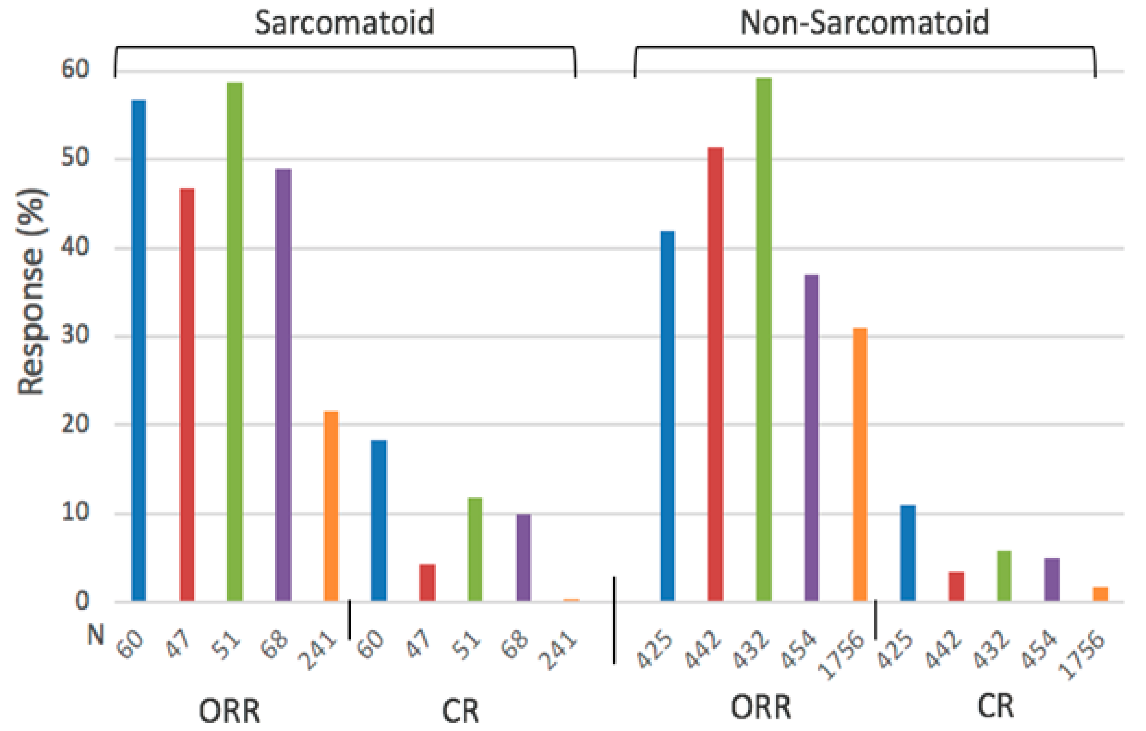
Figure 1. Molecular and immunologic targets expressed in ccRCC tumors and associated microenvironment implicated in tumor angiogenesis, unstable microenvironment, drug resistance, and immune evasion. TGF-ß, transforming growth factor-ß; HIFs, hypoxia-inducible factor1α/2α; onco-miRs, oncogenic microRNAs; TSmiRS, tumor-suppressor microRNAS; P-gp, p-glycoprotein; PHD, prolyl hydroxylase; PD-1/PD-L1, programmed death-1 and its ligand; GPDL1, glycerol-3-phosphate dehydrogenase 1-like; TP, thymidine phosphorylase; CPT1A, carnitine palmitoyl transferase IA; PLIN2, Perilipin 2; mVHL, mutated Von Hippel-Lindau tumor-suppressor gene; ↑ upregulated; ↓ downregulated. *: SLM/MSC targets.
Ijms 24 00902 g001
Figure 2. Objective response rate (ORR, (CR + PR)) and complete response (CR) of previously untreated patients with advanced ccRCC with and without sarcomatoid differentiation treated with a combination of biologically targeted molecules: randomized phase 3 clinical trial [1]. The number indicated on the x-axis represents the number of patients included in each clinical trial. Ipilimumab/Nivolumab (blue), Avelumab/axitinib (red), Pembrolizumab/Axitinib (green), Atezolizumab/Bevacizumab (purple), sunitinib (orange).
Ijms 24 00902 g002
Figure 3. Biomarkers’ expression associated with the overexpressed nuclear factor erythroid 2-related factor 2 (Nrf2). ↑ Activator and inhibitor. Onco-miRs, oncogenic microRNAs; HIFs, hypoxia-inducible factor-1α/2α; KRAS, BRAF, c-myc, Kristen rat sarcoma virus, B-raf proto-oncogene, serine/threonine kinase, and c-myc is a proto-oncogene; TGF-ß, transforming growth factor-ß; Th17, T-helper 17 cells; Foxp3/Treg, transcription factor forkhead box protein/T regulatory cell; PD-L1, programmed death ligand 1; GSK-3ß, glycogen synthase kinase-3-beta; VEGF, vascular endothelial growth factor; Th1/2, T-helper-type 1/2 cells; MDSC, myeloid-derived suppressor cells [75].
Ijms 24 00902 g003
Figure 4. Differential regulation of Nrf2 expression by methylselenocystein (Se) in tumor and normal tissues [72]. Se upregulates Nrf2 in normal mouse tissues and downregulates its expression in tumor tissues.
Ijms 24 00902 g004
Table 2. Summary of objective response rate (ORR), median progression-free survival (mPFS), and median overall survival (mOS) of ccRCC patients with and without sarcomatoid differentiation treated with a combination of biologically targeted molecules vs. sunitinib [1], ( ): range.
OutcomePatients with Sarc. DifferentiationPatients without Sarc. Differentiation
ComboSunitinibComboSunitinib
ORR (%)52.8 (46.8–58.8)21.5 (14–31.5)47.6 (37–59.8)30.9 (25.7–35.7)
mPFS (Months)7.9 (7.0–8.4)5.7 (4.0–8.3)12.1 (8.2–15.1)9.1 (8.3–11.1)
mOS (Months)24.8 (18.3, 31.2)14.3 (13.6, 18.3)34.6 (33.6, 35.6)30.8 (26.6, 34.9)
Table 3. Expression of Nrf2 in ccRCC tumors and normal tissue [69]. Difference in the expression levels between tumor and normal tissue is significant, with p < 0.001.
SitenPositive Nrf2 Expression
ccRCC tumor152119 (78.3%)
Normal tissue15187 (57.6%)
Table 4. Thymidine phosphorylase (TP) is preferentially expressed at a higher activity level in renal cell carcinoma (RCC) tumor cells than non-neoplastic kidney tissue specimens [119], range: ( ). Difference in the activity of TP between RCC and kidney is significant, with p < 0.0001.
TissuenMedian TP Activity (u/mg Protein)
RCC11612.8 (3.2–933.9)
Kidney9011.79 (0–128.0)
Disclaimer/Publisher’s Note: The statements, opinions and data contained in all publications are solely those of the individual author(s) and contributor(s) and not of MDPI and/or the editor(s). MDPI and/or the editor(s) disclaim responsibility for any injury to people or property resulting from any ideas, methods, instructions or products referred to in the content.

