Review

Structural Modifications of siRNA Improve Its Performance In Vivo

by
Ivan V. Chernikov
,
Ulyana A. Ponomareva
and
Elena L. Chernolovskaya
*
Institute of Chemical Biology and Fundamental Medicine, Siberian Branch of the Russian Academy of Sciences, Acad. Lavrentiev Ave., 8, 630090 Novosibirsk, Russia
*
Author to whom correspondence should be addressed.
Int. J. Mol. Sci. 2023, 24(2), 956; https://doi.org/10.3390/ijms24020956
Submission received: 1 December 2022 / Revised: 23 December 2022 / Accepted: 26 December 2022 / Published: 4 January 2023
(This article belongs to the Special Issue Oligonucleotide, Therapy, and Applications 3.0)

Abstract

:
The use of small interfering RNA (siRNA) in the clinic gives a wide range of possibilities for the treatment of previously incurable diseases. However, the main limitation for biomedical applications is their delivery to target cells and organs. Currently, delivery of siRNA to liver cells is a solved problem due to the bioconjugation of siRNA with N-acetylgalactosamine; other organs remain challenging for siRNA delivery to them. Despite the important role of the ligand in the composition of the bioconjugate, the structure and molecular weight of siRNA also play an important role in the delivery of siRNA. The basic principle is that siRNAs with smaller molecular weights are more efficient at entering cells, whereas siRNAs with larger molecular weights have advantages at the organism level. Here we review the relationships between siRNA structure and its biodistribution and activity to find new strategies for improving siRNA performance.

1. Introduction

RNA interference (RNAi) is an evolutionarily conservative biological process of suppression of gene expression, implemented at the post-transcriptional level [1]. Therefore, RNAi is a very convenient method for suppressing gene expression in experimental biology and clinical practice; five drugs based on small interfering RNAs have been approved by the FDA and are commercially available [2,3]. However, a huge number of potential siRNA-based drugs have failed clinical trials because many factors limit the use of siRNA in biomedicine, such as the sensitivity of siRNA to the action of ribonucleases, filtration by the kidneys, uptake by immune cells, off-target effects, low efficiency of penetration through a hydrophobic cell membrane, and release of siRNA from endosomes.
The use of siRNA bioconjugates is the most promising method for siRNA delivery, as it is characterized by low toxicity and high efficiency in vivo. Currently, most drugs from the pipeline of leading pharmaceutical companies (Alnylam Pharmaceuticals, Silence Therapeutic, Arrowhead Pharmaceuticals, Dicerna Pharmaceuticals) are siRNA and N-acetylgalactosamine conjugates and are designed to deliver siRNA to liver cells, so we can assume that siRNA delivery to this organ is a solved problem (reviewed in [4,5]) since the addition of N-acetylgalactosamine to siRNA contributes to its significant accumulation in hepatocytes and provides a long-term effect. Therefore, the search for new molecules to conjugate with siRNA to deliver siRNA to other organs is one of the most urgent tasks (reviewed in [6,7]). However, the accumulation in the target organs and the overall biodistribution of bioconjugates of nucleic acids in the body and cells depend not only on the delivery molecule but also on the molecular weight and structure of siRNA [8]. Nucleic acids with a molecular weight smaller than the classic 21 bp duplex, such as ASO, are better accumulated within cells, while longer >21 nt dsRNAs tend to have benefits at the organism level. In this review, we will analyze available literature data on the relationship of the performance of siRNA bioconjugates with their structure.

2. Mechanism of RNA Interference

The requirements for the structure of siRNA are determined by the mechanism of RNAi and the structure of the proteins involved in it. The mechanism of RNA interference can be divided into two stages: the initiation phase, cleavage of long dsRNA into short fragments (siRNA), and the effector phase, the formation of the activated RISC* complex. At the first stage, the dsRNA that entered the cytoplasm is cut by the Dicer enzyme into short fragments 21–23 bp long with two nucleotides protruding at the 3′-end [9]. After that, Dicer and siRNA dissociate; however, it is assumed that under certain conditions, Dicer can remain associated with siRNA, facilitating its further processing (Figure 1) [10].
At the second stage of RNAi, a dimer consisting of Dicer and TRBP interacts with siRNA and forms an intermediate RLC complex, with TRBP binding to the thermodynamically more stable end of the siRNA duplex, and Dicer to the less stable one [11,12]. At the next stage, the Ago2 protein binds to the siRNA, displacing the protein dimer from the RLC, while the MID and PIWI domains of Ago2 bind the 5’-end of the “guide” strand of the siRNA, where TRBP is located [13], while the PAZ domain is attached to the 3’ end of this strand, where Dicer is located [14], forming the RISC complex. This is followed by local unwinding of the duplex and cutting of the “passenger” strand between the ninth and tenth nucleotides from the 5′-phosphate of the “guiding” strand by the PIWI domain of Ago2, which has an RNase H activity. After the “passenger” strand dissociates from the complex, an activated RISC* complex is formed, which includes Ago2 and the “guiding” siRNA strand.
Thus, for a 21 bp siRNA, the thermodynamic asymmetry of the duplex ends is one of the key factors determining the emplacement of the siRNA in the RLC complex and the choice of the “guide” strand in RISC* [15]. Another strand selection mechanism can be implemented for dsRNAs longer than 21 bp, which are Dicer’s substrate; it is assumed that since Dicer may not dissociate after cleavage, but remain associated with the protruding 3’-end of siRNA, then it will spatially orientate the RLC formation and, accordingly, determine the choice of the “guide” circuit that will be included in RISC* (Figure 1). Finding the optimal structure for Dicer Substrate Small Interfering RNA (DsiRNA) recognition to incorporate the antisense strand into RISC* is a developing field of research [10].
It should be noted that the incorporation of siRNAs into RISC* can be carried out without Dicer’s participation. Here, dsRNA forms a complex with Ago2 by direct loading: during this process, the 5’-phosphate-binding pocket formed by the MID and PIWI domains of Ago selectively binds the 5’-terminal phosphate of the nucleotide located at the less stable end of the siRNA duplex via a Dicer-independent pathway [16] (reviewed in [17,18]).

3. Chemical Modifications

Chemical modifications are essential for the use of siRNA in bioconjugates in vivo since siRNA is sensitive to ribonucleases and, unlike liposomes and nanoparticles, is not protected in the conjugate; this topic is widely discussed in numerous reviews [6,19,20]. Chemical modifications of siRNA are an actively developing field; in short, there are modifications of ribose (LNA, UNA, GNA, CeNA, HNA, 2’O-Me, 2’O-MOE, 2’F and 4′-thioribonucleosides [21,22,23,24,25,26,27]), phosphate (Tert-butyl-S-acyl-2-thioethyl, boranophosphate, 5′(E)-vinylphosphonate, phosphorothioate [PS] [28,29,30]) and base modifications (pseudouridine, 2′ thiouridine [31]). Ribose modifications affecting the 2’OH group are the most effective in blocking endoribonucleases, which make the main contribution to siRNA cleavage, whereas phosphate modifications block exonucleases better. The cleavage mechanism by endonucleases involves the 2’OH group of the ribose, so replacement of all this group with a 2’F fluorine or 2’O-Me group is necessary throughout the duplex. It was shown that the introduction of alternating 2’F and 2’O-Me into both strands of siRNA does not block its interaction with RISC components and increases the duration of gene silencing. The bulkier 2’O-Me modification effectively protects siRNA from the action of nucleases; however, this modification more significantly reduces the activity of RNAi, especially when applied at certain positions of the duplex [32,33]. Therefore, a balance between the number of 2’O-Me and 2’F modifications is necessary to ensure the effectiveness of siRNA action. To achieve this goal, attempts were made to increase the proportion of 2’O-Me modifications in siRNA by reducing the proportion of 2’F in the alternating pattern of modifications [34,35]. It has been shown that a decrease in the proportion of 2’F to 18% increases the efficiency and duration of action of such siRNAs in GalNAc conjugates. The modification pattern [34] was chosen as a statistical consensus on 1890 siRNAs and does not usually block the RNAi process for most siRNAs; further optimization of the 2’F/2’O-Me composition for each siRNA sequence can increase its activity [4,36,37]. Detailed optimization of 2’F/2’O-Me composition was carried out for asymmetric anti-FLT1 siRNA [38], and it was shown that the introduction of 2’O-Me at all nucleotides except two 2’F (6%) does not impair the silencing activity of siRNA in vitro and increases the accumulation in the liver, kidney, and placenta of the lipophilic conjugate of this siRNA compared to siRNA containing 50% 2’F. The duration of the biological effect of such an optimized siRNA was not specified by the authors.
For the optimization of 2’F/2’O-Me modifications, it should be considered that some positions of 2’F are probably more significant—for example, it was shown that the 14th 2’O-Me instead of 2’F in the antisense strand of fully modified siRNA is not tolerated by RNAi machinery [39]. Several 2′Fs in the center of sense strand (9–11 nt) are preferred, probably due to Ago2 cleavage of the passenger strand at this location and more efficient release of the passenger strand from RISC (Table 1) [34].
For increasing the 2’O-Me/2’F relation in the case of long fully modified dsRNAs, which are Dicer’s substrate, it should be taken into account that the slicing activity of Dicer is blocked by 2’O-Me [40], so 2’F modifications should be placed in the sites of RNA cleavage by Dicer.
Further optimization of siRNA chemical modifications such as GNA, stereoselectivity of modifications, and Alnylam’s IKARIA platform decrease therapeutic dose and improve safety profile of siRNA and, thereby, gives us more opportunities for widespread use of siRNA in the clinic.
Table 1. Structural modifications of ss-siRNA.
#Structure of siRNA—Sense Strand 5’-3’ (S) Above, Antisense Strand 3’-5’ (AS); Below, Pattern of Chemical Modifications 1,2In VitroIn VivoRef.
Cells/
Target
Delivery 3Efficiency 4
-Increase
-Decline
Model/
Target
Dose, Mode of Adm 5Efficiency
-Increase
-Decline
1ss-siRNA, n/mHaCaT/F3Lf200087%, mRNA--- [41]
siRNA, n/m90%(3%), mRNA---
2ss-siRNA, (boranophosphate)
Ijms 24 00956 i001
HeLa/EGFPOligofectami-ne,
25 nM
100%, protein--- [28]
siRNA, n/m70%(30%), protein---
3ss-siRNAIjms 24 00956 i002HeLa/
PTEN
Lf2000IC50 2 nM, mRNA--- [42]
siRNA
Ijms 24 00956 i003
IC50 0.2 nM(×10), mRNA ---
Ijms 24 00956 i004
R: Ijms 24 00956 i005
Prim. hepatocytes/ApoC IIIElectroporationIC50 2 mcM, mRNATransge-nic mice, liver/
ApoC III
14 mg/kg, s.c.65%, IC50 ~10 mg/kg, mRNA
4ss-siRNA
Ijms 24 00956 i006
MCF-7/PRRNAiMAX, 50 nM~85%, mRNA--- [43]
HEK
293/SIN3A
5%, mRNA
siRNA
Ijms 24 00956 i007
MCF-7/PR~90%(5%), mRNA ---
HEK
293/
SIN3A
~60%(55%), mRNA
5ss-siRNA
Ijms 24 00956 i008
Hepa 1–6/CTNNB1RNAiMAX IC50 0.07 nMMice, liver/
CTNNB1
0.05 mg/kg i.v. with LNP50%, mRNA [44]
siRNA
Ijms 24 00956 i009
IC50 0.12 nM (×1.7)65%(15%), mRNA
1 Chemical modifications are indicated: 2’F (red), 2’O-Me (blue), 2’O-MOE (dark blue), dNMP (grey), PS (orange), 5’-(E)-VP (green), inverted thymidine (black), boranophosphate modification (purple) and LNA (yellow). n/m—non-modified. 2 “R”—linker with ligand. 3 Lf2000—transfection agent Lipofectamine 2000. 4 A decrease () or increase () in the effectiveness of a particular modification to silencing activity of siRNA. 5 Route of administration: i.v.—intravenously, s.c.—subcutaneously.

4. Single-Stranded siRNAs

A factor limiting the effective delivery of siRNAs to cells in vivo is the low ability of siRNAs to leave endosomes; therefore, to overcome this limitation, it was proposed to use single-stranded ss-siRNAs (single stranded siRNAs, ss-siRNAs). Since these molecules have a lower molecular weight, they bind efficiently to serum proteins and thus better penetrate the target cell. Such ss-siRNAs are inducers of RNAi and make it possible to avoid undesirable side effects from the side of the “passenger” strand in the case of siRNA duplexes. However, despite these advantages, unmodified ss-siRNAs have lower gene silencing activity compared to canonical double-stranded siRNAs (Table 1, #1) and are unstable in serum (cleavage time of about 5 s); therefore, related studies were aimed at creating chemically modified ss-siRNAs [41].
The first attempt to use modified ss-siRNAs was made in the work [28] (Table 1, #2), where it was shown that ss-siRNAs containing the boranophosphate modification are 30% more effective in silencing the target gene compared to unmodified siRNA duplexes; however, the stability of such ss-siRNAs remains low, requiring additional modifications. Further studies led to the creation of a pattern of chemical modifications of ss-siRNA, including 14 PS modifications and alternating 2’F and 2’O-Me nucleotide analogs [32,33], which contributes to an increase in the stability of ss-siRNA in vitro up to 8 h or more. Optimization of ss-siRNA design by introducing chemical modifications contributes not only to stability in biological fluids, but also increases the duration of ss-siRNA action. It was shown that the half-life of ss-siRNA from mouse tissue is about 10 days, and the duration of the inhibitory effect of ss-siRNA reaches 7 days [45]. The improvement of the pharmacokinetic properties of siRNA was achieved by introducing PS modifications that increase the efficiency of binding of oligonucleotides to serum proteins, which prevents the rapid elimination of ss-siRNA from the body by filtration by the kidneys [46].
A comparison of the interfering properties of a fully modified ss-siRNA and its siRNA duplex with an unmodified sense strand in vitro shows that ss-siRNA is an order of magnitude less active than the siRNA duplex when delivered to cells using a transfection agent (Table 1, #3) [42]. Chemically modified ss-siRNAs were also significantly less active than unmodified siRNA duplexes. It was shown that the modified ss-siRNA suppresses the expression of the SIN3A gene by 5%, while the unmodified siRNA duplex—by 60% when delivered to HEK293 cells using a transfection agent (Table 1, # 4) [43]. However, in in vivo experiments in mice, effective suppression of the target gene occurred in the liver by 65% (ApoC gene) after subcutaneous administration of ss-siRNA (Table 1, #3) [42] and in the brain by 80% (Htt gene) after direct intraventricular injection of ss-siRNA [47]. In another work, the biological activity of fully modified ss-siRNA and siRNA was compared in vitro and in vivo (Table 1, #5) [44]. It was shown that ss-siRNAs have a 1.7-fold lower IC50 value compared to siRNAs when delivered to Hepa1–6 cells using a transfection agent. However, after intravenous injection of 0.05 mg/kg ss-siRNA in mice in combination with lipid nanoparticles, a 50% decrease in the level of CTNNB1 gene mRNA in the liver is observed, while the biological activity of the siRNA duplex is 65%. Despite the fact that chemically modified ss-siRNAs are more effective than their unmodified counterparts, the doses required to effectively suppress gene expression remain relatively high.

5. Segmented siRNAs (sisiRNAs)

Another strategy to increase the specificity of siRNA is to introduce a single-strand break in the sense strand of siRNA (small internally segmented interfering RNA, sisiRNA) [21]. Such sense strand inactivation in the sisiRNA structure prevents off-target effects caused by the sense strand if it is incorrectly chosen as a “guide” and loaded into RISC*. However, the low thermal stability of sisiRNA compared to siRNA duplexes of the same sequence is their significant disadvantage; therefore, to solve this problem, it was proposed to introduce LNA modifications into sisiRNA, which would stabilize it [22]. A comparison of the biological activities of LNA-modified sisiRNA and siRNA when delivered to cells using a transfection agent showed that sisiRNA suppresses the expression of the target gene by 12% less effectively than siRNA. However, in vivo experiments on the MiaPaca-2 xenograft tumor model in mice showed that LNA-modified sisiRNA and siRNA have a similar distribution profile in tissues and accumulate predominantly in the kidneys; in addition, they exhibit similar biological activity and suppress the expression of the EGFP gene by 50% (Table 2, #6) [22].
In another study [48], dimeric sisiRNAs linked by a disulfide bond and delivered to cells by cationic polymers were studied. It is assumed that the presence of a nickname in the sense strand facilitates the formation of complexes with cationic polymers due to its structural flexibility. It was shown that after transfection of MDA-MB-435 cells expressing GFP, complexes of dimeric siRNA and sisiRNA suppress the expression of the target gene by 35% and 45%, respectively, while monomeric siRNA did not show biological activity. It is unlikely that this fact can be unequivocally assessed as evidence of the superiority of non-canonical structures; rather, it indicates an unsuccessful choice of a sequence as a non-canonical siRNA, due to which an incorrect assembly of the RISC complex occurs.
Probably, the differences between sisiRNA and siRNA are due to an increase in the specificity of action. However, since the structure of the sisiRNA duplex does not contribute to a significant increase in biological activity in vitro and in vivo, further optimization of the design of the molecule is required in order to consider it for use in the clinic.
Table 2. Structural modifications of sisiRNA.
#Structure of siRNA—Sense Strand 5’-3’ (S); Above, Antisense Strand 3’-5’ (AS); Below, Pattern of Chemical Modifications 1In VitroIn VivoRef.
Cells/
Target
Delivery 2Efficiency 3
-Increase
-Decline
Model/
Target
Dose, Mode of Adm 4Efficiency -Increase
-Decline
6siRNA
Ijms 24 00956 i010
MiaPaca-2/EGFPLf2000 5 nM~62%, mRNAXeno-graft MiaPaca−2 in mice/
EGFP
0.25 mg/kg, s.c. within 1 week, osmotic pump50%, mRNA [22]
sisiRNA
ss nick
Ijms 24 00956 i011
~50% (12%), mRNA50%, mRNA
1 Chemical modifications are indicated: LNA (yellow). 2 Lf2000—transfection agent Lipofectamine 2000. 3 A decrease () or increase () in the effectiveness of a particular modification to silencing activity of siRNA. 4 Route of administration: s.c.—subcutaneously.

6. Circular Small Interfering RNAs (csiRNA)

Circular RNAs (circRNA) are endogenous regulatory single-stranded, covalently closed RNA molecules that have recently attracted the attention of researchers studying their involvement in disease development as markers and potential molecular targets [49,50,51]. Recently, analogues of these natural RNAs have been proposed as synthetic inducers of RNAi—circular small interfering RNAs (csiRNAs). Natural circular RNA molecules are formed enzymatically as a result of splicing; synthetic analogues of siRNAs are usually obtained as a result of chemical crosslinking using various approaches. A number of approaches have used reversible cross-links that could break down under intracellular conditions and restore the original siRNA duplex.
Thus, in [52] the so-called caged circular RNA was proposed, sense, antisense or both strands of which were cyclized with a photosensitive crosslink using a carboxylic acid attached through a photocleavable linker at the 5’ end. They showed that such siRNAs have increased stability in the presence of serum, despite the fact that additional chemical modifications to increase nuclease resistance were not used, and are able to suppress the expression of the target gene in vitro and in vivo. Cyclization of the sense strand did not interfere with the biological activity of the duplex, while duplexes with a cyclized antisense strand were active only after photocleavage. Thus, it was shown that duplexes with a circular sense strand do not require cleavage and can be used without introducing labile bonds into their composition [53].
An alternative approach was proposed in [54], where circular duplexes were used, obtained either by enzymatic cyclization of strands containing a photo-cleavable linker in the middle or by formation of a reducible disulfide bond. It was found that changing the structure of the duplex reduces unwanted immunostimulation compared to a linear duplex of the same sequence and almost does not reduce the interfering activity.
Another interesting variant of circular macromolecular structures composed of two or three siRNA motifs was proposed in [55], where circular molecules were obtained using a template-mediated ligation method. Circular RNAs contained loops in their composition at the sites of their desired cleavage to obtain siRNAs; such cleavage, according to the authors, will occur in the cell under the action of ribonucleases. It was shown that such structures are indeed capable of suppressing the expression of the target gene by the mechanism of RNAi in cell culture.
The most advanced technology for obtaining chemically modified therapeutic siRNA conjugates was applied to circular RNAs in [56]. Fully modified duplexes were constructed in this work, containing a circular sense strand with an attached GalNAc residue and a linear antisense strand with a non-cleavable (E)-vinylphosphonate. Cyclization was carried out using copper-dependent click chemistry. Cyclization has been shown to impact potency of RNAi-mediated silencing in vitro; however, in vivo this effect was almost offset by increased metabolic stability, and the use of (E)-vinylphosphonate almost completely restored the activity. The reduction in off-target effects caused by the action of the sense strand also argues in favor of considering such a structure for the development of potential therapeutic drugs; however, so far there is not enough data to prove the safety of using such structures.

7. Dicer Substrates

The use of Dicer substrates (DsiRNA) as inducers of RNAi is based on the ability of Dicer to remain associated with the protruding 3’-end of the duplex after processing and facilitate the inclusion of a “guide” strand into RISC*, which makes it possible to reduce the effective concentration of siRNA required to suppress the target gene. Thus, in one of the first works devoted to non-canonical RNAi inducers, it was shown that 27–30 bp dsRNAs, which are Dicer’s substrates, have greater interfering activity compared to the 21-bp canonical siRNA. A comparison of the effectiveness of target gene suppression using DsiRNA and siRNA showed that 25–30-bp duplexes are about 100 times more active than 21-bp siRNAs (Table 3, #7) [57].
To study the effect of DsiRNA structure on recognition by Dicer, three types of DsiRNA conjugated with palmitic acid with the lengths of the sense strand 21, 23, and 25 nucleotides and the antisense strand of 23, 25, and 27 nucleotides, respectively (21/23 DsiRNA, 23/25 DsiRNA, and 25/27 DsiRNA) were synthesized [58]. It was shown that the 21/23 DsiRNA conjugate is not processed by Dicer but, nevertheless, suppresses the expression of the target gene with high efficiency (93%) (Table 3, #8). An increase in the length of the duplex by two nucleotides leads to the recognition of the 23/25 DsiRNA conjugate by Dicer; however, after cleavage, in addition to 21 bp siRNA, a mixture of products of different lengths is formed. A further increase in length to 25 nucleotides in the sense strand and 27 nucleotides in the antisense strand provides high specificity for recognition of the 25/27 DsiRNA conjugate by Dicer to form a 21 bp siRNA. Nevertheless, it was shown that, despite the differences in processing, all the conjugates studied in the work have high biological activity and suppress the expression of the target gene by 91–93%, which slightly exceeded the activity of the conjugate of the canonical 21 bp siRNA (87%).
The high biological activity of 25/27 DsiRNAs in vivo has been shown in a number of studies. Thus, the decrease in the level of mRNA of the CCR2 gene by 80% upon delivery of 25/27 DsiRNA to HEK293 cells using a transfection agent and by 71% after two intrathecal injections of 25/27 DsiRNA in rats together with CPP (transductin) (Table 3, #9) was shown in [59]. In [43,44] the interfering properties of 25/27 DsiRNA bioconjugates were studied in vivo and their high biological activity was shown. For example, the effectiveness of the DsiRNA conjugate with the aptamer was 90% after intravenous administration to mice infected with the human immunodeficiency virus (Table 3, #10) [60]. The conjugate of DsiRNA and α -tocopherol has been shown to reduce the level of mRNA of the ApoB gene in the mouse liver by 80% after 2 days (Table 3, #11) [61].
Despite the fact that Dicer has specificity for a certain duplex structure, in particular the presence of a protruding 3’ end of the DsiRNA antisense strand, it can also process DsiRNAs with blunt ends. It was shown that 27 bp long DsiRNA reduces the expression of the reporter target gene by 35% more effectively than canonical siRNA when delivered to cells with the help of a transfection agent (Table 3, #14) [62]. A comparison of the effectiveness of the conjugates of 27 bp DsiRNA and siRNA with palmitic acid in vitro and in vivo showed that both conjugates have high biological activity when delivered to cells with a transfection agent (the efficiency was 75% and 77% for the conjugates of siRNA and DsiRNA, respectively) [63]. In vivo experiments also showed a high efficiency of these conjugates: 62% and 70% gene silencing by siRNA and DsiRNA conjugates, respectively (Table 3, #15).
To assess Dicer’s specificity for DsiRNA with a hairpin, [10] constructed DsiRNAs containing a four-nucleotide looped hairpin (tetra-looped DsiRNA) at the end of the duplex (Table 3, #12). This structure was chosen on the basis of data showing that RNA with a similar structure increased stability and binding efficiency for RNA and proteins. Tetra-looped DsiRNAs were shown to have high biological activity in Dicer-expressing cells, while activity was reduced in Dicer-knockout cells. A number of studies have shown that short hairpin RNAs (shRNAs) provide a long-term effect of RNAi [64,65,66] due to the interaction of the duplex loop with Dicer’s N-terminal helicase domain [67,68,69,70] and its cofactor TRBP, which increases Dicer’s RNA binding affinity and enhances cleavage fidelity [71]. This suggests that the presence of a loop in small RNAs, as in tetra-looped DsiRNAs, may affect not only the Dicer cleavage activity but also the efficiency of target gene silencing. A comparison of the effectiveness of DsiRNA and looped DsiRNA showed that the presence of a hairpin in tetra-looped DsiRNA increases its biological activity from 4% to 18% when delivered to cells using a transfection agent (Table 3, #12).
One of the most promising approaches to date is the use of a fully modified tetra-looped DsiRNA conjugated with GalNac [72], which suppresses the expression of the LDHA gene by 98% in vitro and 50% in vivo (Table 3, #13).
Thus, DsiRNA bioconjugates have high biological activity in vitro and in vivo and can be more effective than canonical siRNA. Since DsiRNA processing significantly improves the biological activity of DsiRNA, it is important to take into account its specificity for non-canonical siRNAs with other structures.
Table 3. Structural modifications of DsiRNA.
#Structure of siRNA—Sense Strand 5’-3’ (S) Above, Antisense Strand 3’-5’ (AS) Below. Pattern of Chemical Modifications 1,2,3In VitroIn VivoRef.
Cells/
Target
Delivery 4Efficiency 5
-Increase
-Decline
Model/
Target
Dose, Mode of Adm 6Efficiency
-Increase
-Decline
7siRNA, n/mHEK 293/EGFPLf2000 20 nM~47%, IC50 ~30 nM, protein --- [57]
DsiRNA (25/27), n/m~97%(50%), IC50 175 pM (×170), protein ---
8Palmitic acid (C16)-siRNA, n/m
Ijms 24 00956 i012
Ijms 24 00956 i013
Hela/RlucLf2000
1 nM
87%, Rluc exp--- [58]
Palmitic acid (C16)-siRNA (21/23), n/m
Ijms 24 00956 i014
Ijms 24 00956 i015
93% (6%), Rluc exp---
Palmitic acid (C16)-DsiRNA (23/25), n/m
Ijms 24 00956 i016
Ijms 24 00956 i017
91% (4%), Rluc exp---
Palmitic acid (C16)-DsiRNA (25/27), n/m
Ijms 24 00956 i018
Ijms 24 00956 i019
92% (5%), Rluc exp---
9DsiRNA (25/27)
Ijms 24 00956 i020
HEK293/
CCR2
RNAiMAX
1 nM
80%, mRNARat, ganglion/CCR25 mcg, 2× intrathecal/Transductin71%, mRNA [59]
10«RNA aptamer (A-1) to gp120»- DsiRNA
Ijms 24 00956 i021
----HIV-1 in miceRag2−/−γc−/−0.38 mg/kg, 8× i.v. 90%, mRNA (PBMC), 105 fold (in plasma) [60]
1α- tocopherol -DsiRNA
Ijms 24 00956 i022
Ijms 24 00956 i023
----Mice, liver/ApoB32 mg/kg, i.v.80%, IC50 2 mg/kg, mRNA [61]
13DsiRNA
Ijms 24 00956 i024
HeLa/
LDHA
RNAiMax
1 nM
~96.9%, mRNA--- [72]
Tetra-looped DsiRNA
Ijms 24 00956 i025
Ijms 24 00956 i026
~98.5% (1.6%), IC50 20 пM, mRNAMice, liver/
LDHA
10 mg/kg, i.v.50%, mRNA
14siRNA, n/mHela/RlucLf2000
50 nM
48%, Rluc exp--- [62]
DsiRNA (27/27, “blunt” ends), n/m83% (35%)---
Cholesterol-DsiRNA (27/27, “blunt” ends), n/m
Ijms 24 00956 i027
Ijms 24 00956 i028
-
600 nM
30%, Rluc exp---
15Palmitic acid (C16)-siRNA, n/m
Ijms 24 00956 i029
Ijms 24 00956 i030
GCIY-
eGFP/
EGFP
RNAiMAX
100 nM
75%, mRNAXenograft GCIY-
eGFP in nude mice/
EGFP
3 nmol, i.t./Invivofectamine 2.062% [63]
Palmitic acid (C16)-DsiRNA (27/27, “blunt” ends), n/m
Ijms 24 00956 i031
Ijms 24 00956 i032
77%(2%), mRNA70%(8%)
1 Chemical modifications are indicated: 2’F (red), 2’O-Me (blue), 2’O-MOE (dark blue), dNMP (grey), PS (orange), 5’-(E)-VP (green), inverted thymidine (black), boranophosphate modification (purple) and LNA (yellow). n/m—non-modified. 2 “R”—linker with ligand. 3 Putative Dicer cleavage sites are indicated by black arrows. 4 “-”—delivery without transfection agent, Lf2000—transfection agent Lipofectamine 2000. 5 A decrease () or increase () in the effectiveness of a particular modification to silencing activity of siRNA. 6 Route of administration: i.v.—intravenously, s.c.—subcutaneously, i.t.—intratumoral injection, i.c.v.—intracerebroventricular.

8. Multimeric siRNAs

One of the unsolved problems in the development of siRNA-based drugs for clinical use is the rapid elimination of these molecules from the bloodstream due to filtration by the kidneys. Currently, various approaches are in development to increase the circulation time of siRNA in the bloodstream and their accumulation in target organs; however, these approaches have not yet been introduced into clinical practice and are limited to experimental studies [73,74,75]. It is assumed that the use of siRNA duplexes with a higher molecular weight as inducers of RNAi will improve the pharmacodynamic and pharmacokinetic properties of such drugs. Depending on the design of the duplex, long siRNAs may have a linear or branched structure; in the next section, we will take a closer look at each type of duplex architecture (Table 4 and Table 5).

8.1. Linear Multimeric siRNA Structures

Changing the structure of siRNA by increasing the molecular weight of the duplex makes it possible to increase the biological activity and, accordingly, reduce the dose of siRNA necessary to achieve the desired effect. Various approaches to the design of high molecular weight siRNA are actively discussed in the literature. The fundamental difference between these approaches is the method of connecting RNA: through a nucleotide linker or directly during synthesis, or through a non-nucleotide linker by chemical crosslinking.
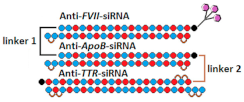
Both approaches were compared in the work [76], where conjugates of GalNac and dimeric siRNA directed to the TTR and FVII genes were synthesized and their interfering properties were studied. These dimeric siRNA conjugates were characterized by an order of magnitude higher IC50 compared to a mixture of conjugates of monomeric siRNA when delivered to cells without a transfection agent (Table 4, #16). However, their biological activity in vivo was comparable to that of a mixture of monomeric siRNA conjugates. Thus, their advantage in the duration of circulation in vivo compensates for the lack of biological activity in vitro. It is assumed that the way monomeric siRNAs are connected can affect the flexibility of the duplex and, thus, the efficiency of interaction with RNAi proteins and suppression of the target gene. A comparison of the effectiveness of dimeric siRNA conjugates with linkers of different nature showed that the most active is the conjugate of dimeric siRNA with a nucleotide-based linker, which suppressed the expression of each of the two target genes by 92% in vivo.
Fully modified, PS-containing asymmetric siRNAs connected through a tetraethylene glycol linker were used in another study [77]. A comparison of the accumulation of Cy3-tagged dimeric and monomeric siRNA in mouse brain showed that dimeric siRNA accumulates in tissue much more efficiently than canonical siRNA, supporting the idea that cooperative interactions between two partially PS-modified siRNAs provide increased tissue accumulation. High accumulation of dimeric siRNA in brain structures correlated with the efficiency of target gene suppression. It has been shown that dimeric siRNA suppresses Htt gene expression by 90%, while PS-free dimeric siRNA is inactive. Based on these data, it can be concluded that the introduction of PS modification into the composition of single-stranded overhangs of asymmetric siRNAs can significantly improve their interfering properties.
The biodistribution and silencing activity of fluorescently labeled dimeric siRNA connected by an oligoethylengrycol linker were studied in Cynomolgus monkeys. A month after a unilateral injection into the brain ventricle, a uniform distribution of dimeric siRNA was observed throughout the primate brain, with the highest accumulation in the cortex (9 μg/g), hippocampus (6 μg/g) and thalamus (6 μg/g) and the lowest accumulation in the caudate nucleus (2 µg/g) and nucleus basalis (1 µg/g) of the striatum [77]. Effective suppression of the HTT protein by 90% in the cortex, 80% in the hippocampus, 50–85% in the caudate nucleus, and 40–70% in the striatal nucleus basalis was achieved using dimeric siRNA (Table 4, #17). Thus, the dimeric siRNAs studied in this work can be considered promising agents for their use in clinical practice for the treatment of diseases of the central nervous system.
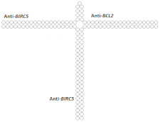
On the other hand, there are a number of studies in which dimeric siRNA was obtained by connecting monomers through a nucleotide linker. For example, in [78], a 66% decrease in the expression of the BIRC5 gene and 83% of the BCL2 gene was achieved when dimeric siRNA was delivered to cells using a transfection agent (Table 4, #20). The rate of BIRC5 gene suppression by monomeric siRNA was 55%, whereas suppression of the BCL2 gene by dimeric siRNA was only slightly less effective.
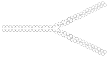
The hybridization of long sense and antisense strands, which contain sequences of monomeric siRNAs directed to different targets, is a linker-free method for obtaining dimeric siRNAs. For example, in [79], dimeric siRNAs were designed to suppress two target genes, LMNA and TIG3; the silencing activity of the dimer for each target did not differ significantly from the activity of individual monomeric siRNAs and reached more than 90% when delivered to HeLa cells using a transfection agent (Table 4, #21).
A further increase in molecular weight for trimeric siRNAs was made in a study [80] comparing the interfering activity of a selectively modified 21 bp siRNA with that of 42 bp siRNA (dimeric siRNA) and 63 bp siRNA (trimeric siRNA), which contain two and three copies of the canonical siRNA sequence, respectively. It was shown that 6 days after transfection of KB-8-5 cells, trimeric siRNA suppresses MDR1 gene expression more effectively than canonical and dimeric siRNA. The IC50 value of trimeric siRNA was two and 4.6 times lower compared to dimeric and canonical siRNA, respectively (Table 4, #22). The study of the mechanism of action of selectively modified dimeric and trimeric siRNAs showed that these duplexes are not subjected to Dicer cleavage. Despite the fact that the potential cleavage sites in the siRNAs studied in this work did not contain modifications, inhibition of Dicer cleavage may be associated with the presence of 2’O-methyl modifications in one or two nucleotides from the putative Dicer cleavage sites, which may prevent processing. An increase in the effectiveness of the action of extended siRNA variants in this case may be due to the fact that such siRNAs can directly be loaded in a complex with Ago2 and, independently of Dicer, participate in RNAi.
Since Dicer’s processing of extended siRNA variants can increase the concentration of functional siRNAs, it makes sense to optimize the pattern of chemical modifications so that the modifications do not interfere with this process. In addition, Dicer treatment may allow the use of extended siRNA variants to effectively suppress the expression of several genes at once. Thus in [81], a 63-bp trimeric siRNA was investigated, containing in one duplex sequences directed to the mRNA of the MDR1, LMP2, and LMP7 genes with a limited number of 2’O-Me modifications at nuclease-sensitive sites. It has been shown that this siRNA is able to suppress the expression of all three target genes with high efficiency, acting through a Dicer-dependent mechanism. In the cell, this trimeric siRNA is cut into duplexes 42 and 21 bp long and suppresses the expression of target genes much more effectively than canonical siRNA with the corresponding monomer sequence.
A study of the properties of cholesterol derivatives of 63 bp trimeric siRNAs consisting of three copies of the canonical siRNA [8] showed that an increase in the length of the siRNA duplex in the cholesterol conjugate increases its biological activity when delivered to KB-8-5 cells using a transfection agent. However, upon delivery in a carrier-free mode, the efficiency of accumulation of conjugates in cells and the efficiency of suppression of the target gene are significantly reduced. An in vitro study of the biological activity of the conjugates showed that the canonical siRNA conjugate suppresses MDR1 gene expression by 50%, while the trimeric siRNA conjugate is inactive (Table 4, #23). In vivo experiments on a mouse model with a subcutaneously grafted KB-8-5 tumor showed that the cholesterol conjugate of monomeric siRNA effectively suppresses the expression of the target gene, while the cholesterol conjugate of trimeric siRNA has no biological activity, despite efficient accumulation in xenograft tumor cells. The lack of correlation between the level of accumulation and biological activity can be explained by the inefficient release of the cholesterol conjugate of trimeric siRNA from endosomes into the cell cytoplasm, where RNAi occurs.
Table 4. Structural modifications of linear multimeric siRNA.
#Structure of siRNA—Sense Strand 5’-3’ (S) Above, Antisense Strand 3’-5’ (AS) Below. Pattern of Chemical Modifications 1,2,3In VitroIn VivoRef.
Cells/
Target
Delivery 4Efficiency 5
-Increase
-Decline
Model/
Target
Dose, mode of Adm 6Cells/
Target
16 siRNA mix
Ijms 24 00956 i033
Ijms 24 00956 i034
Prim. hepatocytes/TTR/FVII-IC50 0.1 nM (TTR)
IC50 ~0.25 nM (FVII)
Mice liver/
TTR/
FVII
3 mg/kg, s.c.~98%/~98% [76]
Dimeric siRNA
Ijms 24 00956 i035
IC50 0.4 nM (×4)/~1 nM(×4)3 mg/kg, s.c.92%(6%)/92% (6%)
Dimeric siRNA
Ijms 24 00956 i036
Ijms 24 00956 i037
IC50 1 nM (×10)/IC50 1 nM (×4)3 mg/kg, s.c. 73%(25%)/~82%(16%)
17siRNA
Ijms 24 00956 i038
HeLa/HTTRNAiMax 0.1 nM70%, IC50 ~25 pM, mRNA --- [77]
Dimeric siRNA
Ijms 24 00956 i039
Ijms 24 00956 i040
80%, (10%)
IC50 ~10 pM (×2.5), mRNA
Mice, brain/
HTT
23.75 mg/kg, i.c.v. bilaterally>90%, protein in the hippocampus
>70%, protein,
in the cortex
M.Cynomolgus brain0.017 mg/kg, i.c.v. >90%, protein, in the cortex
18GalNAc-siRNA
Ijms 24 00956 i041
---Mice, liver/FVII50 mg/kg, s.c. 85%, protein [82]
GalNAc-dimeric siRNA
Ijms 24 00956 i042
Ijms 24 00956 i043
---75%(10%), protein
19GalNAc-siRNA mix---Mice, liver/FVII/ApoB/TTR50 mg/kg, s.c.50%/30%/97%
GalNAc-trimeric siRNA
Ijms 24 00956 i044
Ijms 24 00956 i045
Ijms 24 00956 i046
---62% (12%)/30%/97%
20siRNA
Ijms 24 00956 i047
T24/BIRC5/BCL2Lf2000
10 nM
55%/85%,mRNA--- [78]
Dimeric siRNA
Ijms 24 00956 i048
66% (11%)/83% (2%), mRNA---
21siRNA mix
Hela/LMNA/
TIG3
Lf2000
10 nM
90%/98%, mRNA--- [79]
Dimeric siRNA (34 bp), n/m
Ijms 24 00956 i049
98% (8%)/96% (2%), mRNA---
22siRNA
Ijms 24 00956 i050
KB-8-5/MDR1OligofectamineIC50 23 nM--- [80]
Dimeric siRNA
Ijms 24 00956 i051
IC50 10 nM (×2.3)---
Trimeric siRNA
Ijms 24 00956 i052
MDR1,
IC50 5 nM
(×4.6)
---
23Cholesterol-siRNA
Ijms 24 00956 i053
Ijms 24 00956 i054
KB-8-5-MDR1-GFP/MDR1Lf2000IC50 29 nMXenograft KB-8-5 in SCID mice/MDR18.5 mg/kg i.v.60%, protein [8]
-
5 mcM
48%, protein
Cholesterol—trimeric siRNA
Ijms 24 00956 i055
Ijms 24 00956 i056
Lf2000IC50 16 nM (×1.8)0% (60%), protein
-
5 mcM
2%(46%), protein
1 Chemical modifications are indicated: 2’F (red), 2’O-Me (blue), PS (orange), 5’-(E)-VP (green), inverted thymidine (black). n/m—non-modified. 2 “R”—linker with ligand. 3 Putative Dicer cleavage sites are indicated by black arrows. 4 “-”—delivery without transfection agent, Lf2000—transfection agent Lipofectamine 2000. 5 A decrease () or increase () in the effectiveness of a particular modification to silencing activity of siRNA. 6 Route of administration: i.v.—intravenously, s.c.—subcutaneously, i.t.—intratumoral injection, i.c.v.—intracerebroventricular.
The results of these studies show that an increase in the molecular weight of the duplex and construction of trimeric siRNAs make it possible to achieve a high level of accumulation in the target organ; however, for biological activity in the suppression of the target gene, long siRNAs must leave endosomes. The efficiency of the escape of long siRNAs from endosomes can be affected by the design of such extended siRNA variants. For example, trimeric siRNAs were constructed in [82] by connecting monomers through a cleavable linker. In vivo experiments have shown that the conjugate of GalNac and trimeric siRNA containing sequences directed to the FVII, ApoB, and TTR genes actively suppresses the expression of these target genes by 62%, 30%, and 97%, respectively. The biological activity of the mixture of monomeric siRNA conjugates was similar in suppression of the ApoB and TTR genes and 12% lower in suppression of the FVII gene (Table 4, #19). It can be assumed that the use of a cleavable linker both increases the flexibility of the long siRNA structure and leads to the release of monomeric siRNAs after linker cleavage and, thus, promotes better exit from endosomes into the cell cytoplasm.
Thus, changing the structure of siRNA by increasing the molecular weight and introducing chemical modifications provides highly specific and long-term inhibition of target gene expression without causing a toxic effect. However, a balance between high molecular weight and efficiency of escape from endosomes is required to achieve a high silencing effect.

8.2. Branched Multimeric siRNA Structures

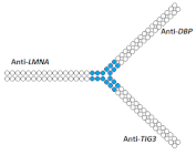
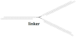
Another multimeric siRNA variant with increased molecular weight is branched siRNA, which contains several copies of siRNAs directed to the same or different target genes. One of the proposed branched siRNAs deployed a structure consisting of three siRNAs interconnected by a linker based on deoxyribonucleotides [83]. This branched siRNA was shown to be 4.7 times more efficient than canonical siRNA when delivered to Hela cells with a transfection agent (Table 5, #24). It is assumed that such branched siRNAs can be processed by Dicer, which leads to an increase in their efficiency.
Table 5. Structural modifications of branched multimeric siRNA.
#Structure of siRNA—Sense Strand 5’-3’ (S) Above, Antisense Strand 3’-5’ (AS) Below, Pattern of Chemical Modifications 1,2,3In VitroIn VivoRef.
Cells/
Target
Delivery 4Efficiency 5
-Increase
-Decline
Model/
Target
Dose, Mode of Adm 6Cells/
Target
24siRNA, n/mHela/
BIRC5
Lf2000IC50 3.89 nM, mRNA--- [83]
Branched siRNA (36 n.), n/m
Ijms 24 00956 i057
IC50 0.83 nM (×4.7), mRNA---
25siRNA mixHeLa/LMNA/
DBP/TIG3
Lf200060%/47%/82%, Rluc exp--- [84]
Branched siRNA (38 n.)
Ijms 24 00956 i058
68% (8%)/62% (15%)/85% (3%)---
26siRNA mixHela/BIRC5/CTNNB/METLf200076%/77%/90%, Rluc exp--- [85]
Branched siRNA (38 n.), n/m
Ijms 24 00956 i059
85% (9%)/45% (32%)/77% (13%)%, Rluc exp---
27siRNA, non-modifiedHepa 1–6/ApoBPEI-Gal
100 nM
50%, mRNAMice, liver/ApoB6 mg/kg, i.v. with PEI-Gal33%, mRNA [86]
Branched siRNA (32 n.), n/m
Ijms 24 00956 i060
ApoB 70%
(20%), mRNA
63% (30%), mRNA
28siRNA, n/mHela/RlucRNAiMax78%, Rluc exp--- [87]
Branched siRNA (21 n.), n/m
Ijms 24 00956 i061
Ijms 24 00956 i062
81% (3%), Rluc exp---
29siRNA mixHela/BIRC5/CTNNB/
STAT3/
MET
PEI
25 nM
39%/30%/40%/30%, mRNA, 1 d.--- [88]
Branched siRNA (38 n.)
Ijms 24 00956 i063
61% (22%)/40% (10%)/70% (30%)/62% (32%), mRNA---
30siRNA, n/mSMMC-7721/
BIRC5/BCL2
Lf200080%/70%, protein, 2 d.--- [89]
Dimeric siRNA
Ijms 24 00956 i064
48% (32%)/45% (25%),
protein
---
Dimeric siRNA
Ijms 24 00956 i065
46% (34%)/57% (13%), protein---
Trimeric siRNA
Ijms 24 00956 i066
68% (12%)/48% (22%), protein---
1 Chemical modifications are indicated: 2’O-Me (blue), n/m—non-modified. 2 “R”—linker with ligand. 3 Putative Dicer cleavage sites are indicated by black arrows. 4 “-”—delivery without transfection agent, Lf2000—transfection agent Lipofectamine 2000. 5 A decrease () or increase () in the effectiveness of a particular modification to silencing activity of siRNA. 6 Route of administration: i.v.—intravenously.
On the other hand, siRNA copies can be linked directly via a phosphodiester bond. It was shown in the following studies [84,85,86] that siRNA with such a branched structure exhibits high efficiency compared to the canonical siRNA in in vitro experiments (Table 5, #25–27). The mechanism of action of such branched siRNAs revealed that Dicer-mediated processing is not required for gene silencing and that RNAi is triggered by a 38 nucleotide long guide strand [84]. In order to evaluate the therapeutic potential of branched siRNAs in vivo, mice were intravenously injected with 6 mg/kg of branched siRNA complexed with a conjugate of polyethyleneimine and galactose (PEI-Gal) [86]. It was shown that 2 days after the administration of the complexes, the mRNA level of the ApoB gene decreased by 63% and 33% respectively, in the case of branched and canonical siRNA (Table 5, #27).
Another option for the design of branched siRNAs is the connection of monomeric siRNAs through a hexaethylene linker [87]. These molecules are stable in serum and have no off-target effects. The study of biological activity showed that the effectiveness of such branched siRNAs in vitro was 81% and was comparable to the efficiency of canonical siRNAs (Table 5, #28). Taken together, the results of this study suggest that such branched siRNAs are promising for in vivo use.
During the study of the interfering properties of branched siRNA, consisting of the monomers directed to four different genes [88], it was shown that the efficiency of such branched siRNA in suppressing the target genes BIRC5, CTNNB, STAT3, and MET is 20–30% higher than canonical siRNA mixtures (Table 5, #29). Thus, it is possible to further increase the molecular weight and to design siRNA with a more complex structure.
To increase the stability of long siRNAs in [89], it was proposed to construct siRNA with a secondary structure of a “loop” or “clover leaf”. It has been shown that such siRNAs are able to suppress the expression of several target genes at once, acting along the Ago2-dependent pathway without causing undesirable immunostimulation. However, the efficiency of siRNAs with such secondary structures was lower than the efficiency of a mixture of canonical siRNAs when delivered to cells with a transfection agent (Table 5, #30). The data obtained in this work suggest that siRNAs with various structural modifications can induce RNAi. However, the question of whether such siRNA constructs solve the problem of rapid removal of siRNA from the body remains open.
Thus, an increase in the molecular weight of siRNA partially solves the problem of their rapid excretion from the body by filtration by the kidneys and contributes to a high accumulation of siRNA in the target organ, but does not always guarantee a high level of activity in suppressing target gene expression since it significantly hinders the release of siRNA from endosomes into the cytoplasm. Therefore, changing the structure and molecular weight of siRNA in order to find a balance between the effective accumulation of siRNA in the cytoplasm of the target cell and its slow excretion from the body remains an urgent task.

9. Conclusions

The described approaches for increasing the molecular weight of siRNA make it possible to increase the circulation time of siRNA in the blood and, consequently, biodistribution. However, for the effective realization of multimeric siRNA, it must be combined with systems that improve the release of siRNA from endosomes. Since no non-toxic in vivo agent has yet been found to escape endosomes, the most promising approach is the use of multimeric siRNAs with cleavable bonds between themselves. Such dsRNAs will be cleaved to monomeric siRNAs, which do not have the same poor endosomal escape as multimeric siRNA. Another important issue for increasing multimeric siRNA efficiency is optimization of the 2′F/2′O-Me composition in fully modified multimeric siRNA. It should be noted that for fully modified dsRNAs, processing to monomeric siRNAs can be blocked in the presence of 2’O-Me at Dicer’s cleavage sites, and Dicer-independent loading might not be very efficient in this case; so, it is necessary to use deoxyribonucleotide or 2’F modifications at Dicer’s cleavage sites. Unexpectedly, fully modified DsiRNA from Dicerna Pharma does not require Dicer’s cleavage activity because it contains a non-cleavable 2’O-Me modified nucleotide at the site of Dicer’s cleavage and still has high efficiency silencing activity. Processing of such DsiRNA occurs in a Dicer-independent manner, and Dicer can solely direct the loading of siRNA into RISC [71]. Another benefit from the DsiRNA structure is the additional protection from nucleases provided by the tetraloop hairpin, which is the most important factor for the long-term silencing activity of siRNA. Thus, combining structural siRNA modification with rational chemical modification design will improve biodistribution and reduce therapeutic dose, allowing the development of new treatments for previously incurable diseases.

10. Future Prospects

Future prospects for the extrahepatic use of siRNA are successful using Alnylam’s advanced chemical modification patterns IKARIA and their lipophilic monomeric siRNAs, which may be in clinical practice in a few years for treating diseases of the lungs, brain, and muscles or other organs [74,75,90,91]. The most effective structural modification for extrahepatic use is a dimeric siRNA from Atalanta Therapeutics [77], which after a single injection silences the target gene in the brain for several months; the company is developing this platform, so in the future we can expect the release of numerous drugs with improved chemical patterns of modifications and aimed at various diseases of the brain. Thus, the use of siRNA in the clinic is progressing toward reducing the cost of treatment due to new chemical modifications and increasing the number of new drugs and, accordingly, the impact that this technology has on the standard of living of humankind.

Author Contributions

Conceptualization, I.V.C., writing—original draft preparation, U.A.P. and I.V.C.; writing—review and editing, E.L.C.; supervision, I.V.C. and E.L.C. All authors have read and agreed to the published version of the manuscript.

Funding

This research was funded by the Russian Science Foundation (grant #19-14-00251).

Institutional Review Board Statement

Not applicable.

Informed Consent Statement

Not applicable.

Data Availability Statement

Publicly available datasets were analyzed in this study.

Conflicts of Interest

The authors declare no conflict of interest.

References

  1. Fire, A.; Xu, S.; Montgomery, M.K.; Kostas, S.A.; Driver, S.E.; Mello, C.C. Potent and Specific Genetic Interference by Double-Stranded RNA in Caenorhabditis Elegans. Nature 1998, 391, 806–811. [Google Scholar] [CrossRef] [PubMed]
  2. Lamb, Y.N. Inclisiran: First Approval. Drugs 2021, 81, 389–395. [Google Scholar] [CrossRef] [PubMed]
  3. Keam, S.J. Vutrisiran: First Approval. Drugs 2022, 82, 1419–1425. [Google Scholar] [CrossRef] [PubMed]
  4. Cui, H.; Zhu, X.; Li, S.; Wang, P.; Fang, J. Liver-Targeted Delivery of Oligonucleotides with N-Acetylgalactosamine Conjugation. ACS Omega 2021, 6, 16259–16265. [Google Scholar] [CrossRef]
  5. Springer, A.D.; Dowdy, S.F. GalNAc-SiRNA Conjugates: Leading the Way for Delivery of RNAi Therapeutics. Nucleic Acid Ther. 2018, 28, 109–118. [Google Scholar] [CrossRef] [Green Version]
  6. Chernikov, I.V.; Vlassov, V.V.; Chernolovskaya, E.L. Current Development of SiRNA Bioconjugates: From Research to the Clinic. Front. Pharmacol. 2019, 10, 444. [Google Scholar] [CrossRef] [Green Version]
  7. Benizri, S.; Gissot, A.; Martin, A.; Vialet, B.; Grinstaff, M.W.; Barthélémy, P. Bioconjugated Oligonucleotides: Recent Developments and Therapeutic Applications. Bioconjug. Chem. 2019, 30, 366–383. [Google Scholar] [CrossRef]
  8. Chernikov, I.V.; Gladkikh, D.V.; Karelina, U.A.; Meschaninova, M.I.; Ven’yaminova, A.G.; Vlassov, V.V.; Chernolovskaya, E.L. Trimeric Small Interfering RNAs and Their Cholesterol-Containing Conjugates Exhibit Improved Accumulation in Tumors, but Dramatically Reduced Silencing Activity. Molecules 2020, 25, 1877. [Google Scholar] [CrossRef]
  9. Elbashir, S.M.; Harborth, J.; Lendeckel, W.; Yalcin, A.; Weber, K.; Tuschl, T. Duplexes of 21-Nucleotide RNAs Mediate RNA Interference in Cultured Mammalian Cells. Nature 2001, 411, 494–498. [Google Scholar] [CrossRef]
  10. Song, M.; Rossi, J.J. The Effect of Dicer Knockout on RNA Interference Using Various Dicer Substrate Interfering RNA Structures. bioRxiv Mol. Biol. 2020. [Google Scholar] [CrossRef]
  11. Tomari, Y.; Matranga, C.; Haley, B.; Martinez, N.; Zamore, P.D. A Protein Sensor for SiRNA Asymmetry. Science 2004, 306, 1377–1380. [Google Scholar] [CrossRef] [PubMed]
  12. Kini, H.K.; Walton, S.P. Effect of SiRNA Terminal Mismatches on TRBP and Dicer Binding and Silencing Efficacy. FEBS J. 2009, 276, 6576–6585. [Google Scholar] [CrossRef] [PubMed] [Green Version]
  13. Frank, F.; Sonenberg, N.; Nagar, B. Structural Basis for 5′-Nucleotide Base-Specific Recognition of Guide RNA by Human AGO2. Nature 2010, 465, 818–822. [Google Scholar] [CrossRef] [PubMed]
  14. Yan, K.S.; Yan, S.; Farooq, A.; Han, A.; Zeng, L.; Zhou, M.M. Structure and Conserved RNA Binding of the PAZ Domain. Nature 2003, 426, 468–474. [Google Scholar] [CrossRef]
  15. Schwarz, D.S.; Hutvágner, G.; Du, T.; Xu, Z.; Aronin, N.; Zamore, P.D. Asymmetry in the Assembly of the RNAi Enzyme Complex. Cell 2003, 115, 199–208. [Google Scholar] [CrossRef] [Green Version]
  16. Suzuki, H.I.; Katsura, A.; Yasuda, T.; Ueno, T.; Mano, H.; Sugimoto, K.; Miyazono, K. Small-RNA Asymmetry Is Directly Driven by Mammalian Argonautes. Nat. Struct. Mol. Biol. 2015, 22, 512–521. [Google Scholar] [CrossRef]
  17. Herrera-Carrillo, E.; Berkhout, B. Dicer-Independent Processing of Small RNA Duplexes: Mechanistic Insights and Applications. Nucleic Acids Res. 2017, 45, 10369–10379. [Google Scholar] [CrossRef] [Green Version]
  18. Liu, Y.P.; Karg, M.; Harwig, A.; Herrera-Carrillo, E.; Jongejan, A.; Van Kampen, A.; Berkhout, B. Mechanistic Insights on the Dicer-Independent AGO2-Mediated Processing of AgoshRNAs. RNA Biol. 2015, 12, 92–100. [Google Scholar] [CrossRef] [Green Version]
  19. Deleavey, G.F.; Damha, M.J. Designing Chemically Modified Oligonucleotides for Targeted Gene Silencing. Chem. Biol. 2012, 19, 937–954. [Google Scholar] [CrossRef] [Green Version]
  20. Khvorova, A.; Watts, J.K. The Chemical Evolution of Oligonucleotide Therapies of Clinical Utility. Nat. Biotechnol. 2017, 35, 238–248. [Google Scholar] [CrossRef]
  21. Bramsen, J.B.; Laursen, M.B.; Damgaard, C.K.; Lena, S.W.; Ravindra Babu, B.; Wengel, J.; Kjems, J. Improved Silencing Properties Using Small Internally Segmented Interfering RNAs. Nucleic Acids Res. 2007, 35, 5886–5897. [Google Scholar] [CrossRef]
  22. Mook, O.R.; Vreijling, J.; Wengel, S.; Wengel, J.; Zhou, C.; Chattopadhyaya, J.; Baas, F.; Fluiter, K. In Vivo Efficacy and Off-Target Effects of Locked Nucleic Acid (LNA) and Unlocked Nucleic Acid (UNA) Modified SiRNA and Small Internally Segmented Interfering RNA (SisiRNA) in Mice Bearing Human Tumor Xenografts. Artif. DNA PNA XNA 2010, 1, 36–44. [Google Scholar] [CrossRef] [Green Version]
  23. Dande, P.; Prakash, T.P.; Sioufi, N.; Gaus, H.; Jarres, R.; Berdeja, A.; Swayze, E.E.; Griffey, R.H.; Bhat, B. Improving RNA Interference in Mammalian Cells by 4′-Thio-Modified Small Interfering RNA (SiRNA): Effect on SiRNA Activity and Nuclease Stability When Used in Combination with 2′-O-Alkyl Modifications. J. Med. Chem. 2006, 49, 1624–1634. [Google Scholar] [CrossRef]
  24. Laursen, M.B.; Pakula, M.M.; Gao, S.; Fluiter, K.; Mook, O.R.; Baas, F.; Langklær, N.; Wengel, S.L.; Wengel, J.; Kjems, J.; et al. Utilization of Unlocked Nucleic Acid (UNA) to Enhance SiRNA Performance in Vitro and in Vivo. Mol. Biosyst. 2010, 6, 862–870. [Google Scholar] [CrossRef] [Green Version]
  25. Song, X.; Wang, X.; Ma, Y.; Liang, Z.; Yang, Z.; Cao, H. Site-Specific Modification Using the 2′-Methoxyethyl Group Improves the Specificity and Activity of SiRNAs. Mol. Ther.-Nucleic Acids 2017, 9, 242–250. [Google Scholar] [CrossRef] [Green Version]
  26. Nauwelaerts, K.; Fisher, M.; Froeyen, M.; Lescrinier, E.; Van Aerschot, A.; Xu, D.; DeLong, R.; Kang, H.; Juliano, R.L.; Herdewijn, P. Structural Characterization and Biological Evaluation of Small Interfering RNAs Containing Cyclohexenyl Nucleosides. J. Am. Chem. Soc. 2007, 129, 9340–9348. [Google Scholar] [CrossRef]
  27. Takahashi, M.; Nagai, C.; Hatakeyama, H.; Minakawa, N.; Harashima, H.; Matsuda, A. Intracellular Stability of 2′-OMe-4′-Thioribonucleoside Modified SiRNA Leads to Long-Term RNAi Effect. Nucleic Acids Res. 2012, 40, 5787–5793. [Google Scholar] [CrossRef]
  28. Hall, A.H.S.; Wan, J.; Spesock, A.; Sergueeva, Z.; Shaw, B.R.; Alexander, K.A. High Potency Silencing by Single-Stranded Boranophosphate SiRNA. Nucleic Acids Res. 2006, 34, 2773–2781. [Google Scholar] [CrossRef] [PubMed] [Green Version]
  29. Berg, D.; Hagopian, J.C.; Springer, A.D.; Eguchi, A.; Kacsinta, A.D. Neutralizing Phosphotriester Backbone Modifications. Nat Biotechnol. 2015, 32, 1256–1261. [Google Scholar]
  30. Wu, S.Y.; Yang, X.; Gharpure, K.M.; Hatakeyama, H.; Egli, M.; McGuire, M.H.; Nagaraja, A.S.; Miyake, T.M.; Rupaimoole, R.; Pecot, C.V.; et al. 2′-OMe-Phosphorodithioate-Modified SiRNAs Show Increased Loading into the RISC Complex and Enhanced Anti-Tumour Activity. Nat. Commun. 2014, 5, 3459. [Google Scholar] [CrossRef] [Green Version]
  31. Sipa, K.; Sochacka, E.; Kazmierczak-Baranska, J.; Maszewska, M.; Janicka, M.; Nowak, G.; Nawrot, B. Effect of Base Modifications on Structure, Thermodynamic Stability, and Gene Silencing Activity of Short Interfering RNA. RNA 2007, 13, 1301–1316. [Google Scholar] [CrossRef]
  32. Cummins, L.L.; Owens, S.R.; Risen, L.M.; Lesnik, E.A.; Freier, S.M.; Mc Gee, D.; Cook, C.J.; Cook, P.D. Characterization of Fully 2’-Modified Oligoribonucleotide Hetero-and Homoduplex Hybridization Andnuclease Sensitivity. Nucleic Acids Res. 1995, 23, 2019–2024. [Google Scholar] [CrossRef] [Green Version]
  33. Takahashi, M.; Minakawa, N.; Matsuda, A. Synthesis and Characterization of 2′-Modified-4′-ThioRNA: A Comprehensive Comparison of Nuclease Stability. Nucleic Acids Res. 2009, 37, 1353–1362. [Google Scholar] [CrossRef] [Green Version]
  34. Foster, D.J.; Brown, C.R.; Shaikh, S.; Trapp, C.; Schlegel, M.K.; Qian, K.; Sehgal, A.; Rajeev, K.G.; Jadhav, V.; Manoharan, M.; et al. Advanced SiRNA Designs Further Improve In Vivo Performance of GalNAc-SiRNA Conjugates. Mol. Ther. 2018, 26, 708–717. [Google Scholar] [CrossRef] [Green Version]
  35. Khvorova, A. Oligonucleotide Therapeutics—A New Class of Cholesterol-Lowering Drugs. N. Engl. J. Med. 2017, 376, 4–7. [Google Scholar] [CrossRef] [Green Version]
  36. Berk, C.; Civenni, G.; Wang, Y.; Steuer, C.; Catapano, C.V.; Hall, J. Pharmacodynamic and Pharmacokinetic Properties of Full Phosphorothioate Small Interfering RNAs for Gene Silencing In Vivo. Nucleic Acid Ther. 2020, 31, 237–244. [Google Scholar] [CrossRef] [Green Version]
  37. Fitzgerald, K.; White, S.; Borodovsky, A.; Bettencourt, B.R.; Strahs, A.; Clausen, V.; Wijngaard, P.; Horton, J.D.; Taubel, J.; Brooks, A.; et al. A Highly Durable RNAi Therapeutic Inhibitor of PCSK9. N. Engl. J. Med. 2017, 376, 41–51. [Google Scholar] [CrossRef] [Green Version]
  38. Davis, S.M.; Hariharan, V.N.; Lo, A.; Turanov, A.A.; Echeverria, D.; Sousa, J.; McHugh, N.; Biscans, A.; Alterman, J.F.; Karumanchi, S.A.; et al. Chemical Optimization of SiRNA for Safe and Efficient Silencing of Placental SFLT1. Mol. Ther.-Nucleic Acids 2022, 29, 135–149. [Google Scholar] [CrossRef]
  39. Shmushkovich, T.; Monopoli, K.R.; Homsy, D.; Leyfer, D.; Betancur-Boissel, M.; Khvorova, A.; Wolfson, A.D. Functional Features Defining the Efficacy of Cholesterol-Conjugated, Self-Deliverable, Chemically Modified SiRNAs. Nucleic Acids Res. 2018, 46, 10905–10916. [Google Scholar] [CrossRef] [Green Version]
  40. Lamontagne, B.; Hannoush, R.N.; Damha, M.J.; Elela, S.A. Molecular Requirements for Duplex Recognition and Cleavage by Eukaryotic RNase III: Discovery of an RNA-Dependent DNA Cleavage Activity of Yeast Rnt1p. J. Mol. Biol. 2004, 338, 401–418. [Google Scholar] [CrossRef]
  41. Holen, T. Similar Behaviour of Single-Strand and Double-Strand SiRNAs Suggests They Act through a Common RNAi Pathway. Nucleic Acids Res. 2003, 31, 2401–2407. [Google Scholar] [CrossRef]
  42. Prakash, T.P.; Lima, W.F.; Murray, H.M.; Li, W.; Kinberger, G.A.; Chappell, A.E.; Gaus, H.; Seth, P.P.; Bhat, B.; Crooke, S.T.; et al. Identification of Metabolically Stable 5′-Phosphate Analogs That Support Single-Stranded SiRNA Activity. Nucleic Acids Res. 2015, 43, 2993–3011. [Google Scholar] [CrossRef] [Green Version]
  43. Pendergraff, H.M.; Debacker, A.J.; Watts, J.K. Single-Stranded Silencing RNAs: Hit Rate and Chemical Modification. Nucleic Acid Ther. 2016, 26, 216–222. [Google Scholar] [CrossRef]
  44. Chang, W.; Pei, Y.; Guidry, E.N.; Zewge, D.; Parish, C.A.; Sherer, E.C.; Dimuzio, J.; Zhang, H.; South, V.J.; Strapps, W.R.; et al. Bioorganic & Medicinal Chemistry Letters Systematic Chemical Modifications of Single Stranded SiRNAs Significantly Improved CTNNB1 MRNA Silencing. Bioorg. Med. Chem. Lett. 2016, 26, 4513–4517. [Google Scholar] [CrossRef]
  45. Lima, W.F.; Prakash, T.P.; Murray, H.M.; Kinberger, G.A.; Li, W.; Chappell, A.E.; Li, C.S.; Murray, S.F.; Gaus, H.; Seth, P.P.; et al. Single-Stranded SiRNAs Activate RNAi in Animals. Cell 2012, 150, 883–894. [Google Scholar] [CrossRef] [Green Version]
  46. Crooke, S.T. Antisense Drug Technology: Principles, Strategies, and Applications, 2nd ed.; CRC Press: Boca Raton, FL, USA, 2007; ISBN 9781439832509. [Google Scholar]
  47. Yu, D.; Pendergraff, H.; Liu, J.; Kordasiewicz, H.B.; Cleveland, D.W.; Swayze, E.E.; Lima, W.F.; Crooke, S.T.; Prakash, T.P.; Corey, D.R. Single-Stranded RNAs Use RNAi to Potently and Allele-Selectively Inhibit Mutant Huntingtin Expression. Cell 2012, 150, 895–908. [Google Scholar] [CrossRef] [Green Version]
  48. Hong, C.A.; Nam, Y.S. Reducible Dimeric Conjugates of Small Internally Segment Interfering RNA for Efficient Gene Silencing. Macromol. Biosci. 2016, 16, 1442–1449. [Google Scholar] [CrossRef]
  49. Tang, X.; Ren, H.; Guo, M.; Qian, J.; Yang, Y.; Gu, C. Review on Circular RNAs and New Insights into Their Roles in Cancer. Comput. Struct. Biotechnol. J. 2021, 19, 910–928. [Google Scholar] [CrossRef]
  50. Zhao, X.; Zhong, Y.; Wang, X.; Shen, J.; An, W. Advances in Circular RNA and Its Applications. Int. J. Med. Sci. 2022, 19, 975–985. [Google Scholar] [CrossRef]
  51. Yu, J.; Xie, D.; Huang, N.; Zhou, Q. Circular RNAs as Novel Diagnostic Biomarkers and Therapeutic Targets in Kidney Disease. Front. Med. 2021, 8, 8–10. [Google Scholar] [CrossRef]
  52. Zhang, L.; Liang, D.; Wang, Y.; Li, D.; Zhang, J.; Wu, L.; Feng, M.; Yi, F.; Xu, L.; Lei, L.; et al. Caged Circular SiRNAs for Photomodulation of Gene Expression in Cells and Mice. Chem. Sci. 2017, 9, 44–51. [Google Scholar] [CrossRef] [PubMed]
  53. Zhang, L.; Liang, D.; Chen, C.; Wang, Y.; Amu, G.; Yang, J.; Yu, L.; Dmochowski, I.J.; Tang, X. Circular SiRNAs for Reducing Off-Target Effects and Enhancing Long-Term Gene Silencing in Cells and Mice. Mol. Ther.-Nucleic Acids 2018, 10, 237–244. [Google Scholar] [CrossRef] [PubMed] [Green Version]
  54. Kimura, Y.; Shu, Z.; Ito, M.; Abe, N.; Nakamoto, K.; Tomoike, F.; Shuto, S.; Ito, Y.; Abe, H. Intracellular Build-up RNAi with Single-Strand Circular RNAs as SiRNA Precursors. Chem. Commun. 2020, 56, 466–469. [Google Scholar] [CrossRef] [PubMed]
  55. Abe, N.; Abe, H.; Ohshiro, T.; Nakashima, Y.; Maeda, M.; Ito, Y. Synthesis and Characterization of Small Circular Double-Stranded RNAs. Chem. Commun. 2011, 47, 2125–2127. [Google Scholar] [CrossRef] [PubMed]
  56. Jahns, H.; Degaonkar, R.; Podbevsek, P.; Gupta, S.; Bisbe, A.; Aluri, K.; Szeto, J.; Kumar, P.; LeBlanc, S.; Racie, T.; et al. Small Circular Interfering RNAs (SciRNAs) as a Potent Therapeutic Platform for Gene-Silencing. Nucleic Acids Res. 2021, 49, 10250–10264. [Google Scholar] [CrossRef] [PubMed]
  57. Kim, D.-H.; Behlke, M.A.; Rose, S.D.; Chang, M.-S.; Choi, S.; Rossi, J.J. Synthetic DsRNA Dicer Substrates Enhance RNAi Potency and Efficacy. Nat. Biotechnol. 2005, 23, 222–226. [Google Scholar] [CrossRef] [Green Version]
  58. Kubo, T.; Yanagihara, K.; Sato, Y.; Nishimura, Y.; Kondo, S.; Seyama, T. Gene-Silencing Potency of Symmetric and Asymmetric Lipid-Conjugated Sirnas and Its Correlation with Dicer Recognition. Bioconjug. Chem. 2013, 24, 2045–2057. [Google Scholar] [CrossRef]
  59. Bégin-Lavallée, V.; Midavaine, É.; Dansereau, M.-A.; Tétreault, P.; Longpré, J.-M.; Jacobi, A.M.; Rose, S.D.; Behlke, M.A.; Beaudet, N.; Sarret, P. Functional Inhibition of Chemokine Receptor CCR2 by Dicer-Substrate-SiRNA Prevents Pain Development. Mol. Pain 2016, 12, 174480691665396. [Google Scholar] [CrossRef] [Green Version]
  60. Neff, C.P.; Zhou, J.; Remling, L.; Kuruvilla, J.; Zhang, J.; Li, H.; Smith, D.D.; Swiderski, P.; Rossi, J.J.; Akkina, R. An Aptamer-SiRNA Chimera Suppresses HIV-1 Viral Loads and Protects from Helper CD4+ T Cell Decline in Humanized Mice. Sci. Transl. Med. 2011, 3, 66ra6. [Google Scholar] [CrossRef] [Green Version]
  61. Alterman, J.F.; Hall, L.M.; Coles, A.H.; Hassler, M.R.; Didiot, M.-C.; Chase, K.; Abraham, J.; Sottosanti, E.; Johnson, E.; Sapp, E.; et al. Hydrophobically Modified SiRNAs Silence Huntingtin MRNA in Primary Neurons and Mouse Brain. Mol. Ther.-Nucleic Acids 2015, 4, e266. [Google Scholar] [CrossRef]
  62. Kubo, T.; Zhelev, Z.; Ohba, H.; Bakalova, R. Modified 27-Nt DsRNAs with Dramatically Enhanced Stability in Serum and Long-Term RNAi Activity. Oligonucleotides 2007, 17, 445–464. [Google Scholar] [CrossRef] [PubMed]
  63. Kubo, T.; Yanagihara, K.; Seyama, T. In Vivo RNAi Efficacy of Palmitic Acid-Conjugated Dicer-Substrate SiRNA in a Subcutaneous Tumor Mouse Model. Chem. Biol. Drug Des. 2016, 87, 811–823. [Google Scholar] [CrossRef] [PubMed]
  64. Rossi, J.J. Expression Strategies for Short Hairpin RNA Interference Triggers. Hum. Gene Ther. 2008, 19, 313–317. [Google Scholar] [CrossRef] [PubMed]
  65. McCaffrey, A.P.; Meuse, L.; Pham, T.-T.T.; Conklin, D.S.; Hannon, G.J.; Kay, M.A. RNA Interference in Adult Mice. Nature 2002, 418, 38–39. [Google Scholar] [CrossRef] [PubMed]
  66. Brummelkamp, T.R. A System for Stable Expression of Short Interfering RNAs in Mammalian Cells. Science 2002, 296, 550–553. [Google Scholar] [CrossRef] [Green Version]
  67. Liu, Z.; Wang, J.; Cheng, H.; Ke, X.; Sun, L.; Zhang, Q.C.; Wang, H.W. Cryo-EM Structure of Human Dicer and Its Complexes with a Pre-MiRNA Substrate. Cell 2018, 173, 1191–1203. [Google Scholar] [CrossRef] [Green Version]
  68. Pokornowska, M.; Milewski, M.C.; Ciechanowska, K.; Szczepańska, A.; Wojnicka, M.; Radogostowicz, Z.; Figlerowicz, M.; Kurzynska-Kokorniak, A. The RNA–RNA Base Pairing Potential of Human Dicer and Ago2 Proteins. Cell. Mol. Life Sci. 2020, 77, 3231–3244. [Google Scholar] [CrossRef] [Green Version]
  69. Hansen, S.R.; Aderounmu, A.M.; Donelick, H.M.; Bass, B.L. Dicer’s Helicase Domain: A Meeting Place for Regulatory Proteins. Cold Spring Harb. Symp. Quant. Biol. 2019, 84, 185–193. [Google Scholar] [CrossRef]
  70. Park, J.-E.; Heo, I.; Tian, Y.; Simanshu, D.K.; Chang, H.; Jee, D.; Patel, D.J.; Kim, V.N. Dicer Recognizes the 5′ End of RNA for Efficient and Accurate Processing. Nature 2011, 475, 201–205. [Google Scholar] [CrossRef] [Green Version]
  71. Chakravarthy, S.; Sternberg, S.H.; Kellenberger, C.A.; Doudna, J.A. Substrate-Specific Kinetics of Dicer-Catalyzed RNA Processing. J. Mol. Biol. 2010, 404, 392–402. [Google Scholar] [CrossRef] [Green Version]
  72. Brown, B.; Dudek, H.; Lai, C. Therapeutic Inhibition of Lactate Dehydrogenase and Agents Therefor. United States Patent U.S. 10,351,854, 16 July 2019. [Google Scholar]
  73. Adams, D.; Gonzalez-Duarte, A.; O’Riordan, W.D.; Yang, C.-C.; Ueda, M.; Kristen, A.V.; Tournev, I.; Schmidt, H.H.; Coelho, T.; Berk, J.L.; et al. Patisiran, an RNAi Therapeutic, for Hereditary Transthyretin Amyloidosis. N. Engl. J. Med. 2018, 379, 11–21. [Google Scholar] [CrossRef] [PubMed]
  74. Tian, G.; Pan, R.; Zhang, B.; Qu, M.; Lian, B.; Jiang, H.; Gao, Z.; Wu, J. Liver-Targeted Combination Therapy Basing on Glycyrrhizic Acid-Modified DSPE-PEG-PEI Nanoparticles for Co-Delivery of Doxorubicin and Bcl-2 SiRNA. Front. Pharmacol. 2019, 10, 4. [Google Scholar] [CrossRef] [PubMed] [Green Version]
  75. Osborn, M.F.; Coles, A.H.; Biscans, A.; Haraszti, R.A.; Roux, L.; Davis, S.; Ly, S.; Echeverria, D.; Hassler, M.R.; Godinho, B.M.D.C.; et al. Hydrophobicity Drives the Systemic Distribution of Lipid-Conjugated SiRNAs via Lipid Transport Pathways. Nucleic Acids Res. 2019, 47, 1070–1081. [Google Scholar] [CrossRef] [PubMed] [Green Version]
  76. Theile, A.C.S.; Zlatev, I.; Castoreno, A.; Bisbe, A.; Williams, S.; Nguyen, T.; Schlegel, M.K.; Waldron, S.; Qin, J.; Fishman, S.; et al. Bis-RNAiTM Conjugates for Simultaneous Silencing of Two Different Gene Transcripts. In Proceedings of the 14th Annual Meeting of the Oligonucleotide Therapeutics Society, Seattle, WA, USA, 30 September–3 October 2018; p. 75. [Google Scholar]
  77. Alterman, J.F.; Godinho, B.M.D.C.; Hassler, M.R.; Ferguson, C.M.; Echeverria, D.; Sapp, E.; Haraszti, R.A.; Coles, A.H.; Conroy, F.; Miller, R.; et al. A Divalent SiRNA Chemical Scaffold for Potent and Sustained Modulation of Gene Expression throughout the Central Nervous System. Nat. Biotechnol. 2019, 37, 884–894. [Google Scholar] [CrossRef] [Green Version]
  78. Peng, W.; Chen, J.; Qin, Y.; Yang, Z.; Zhu, Y.Y. Long Double-Stranded Multiplex SiRNAs for Dual Genes Silencing. Nucleic Acid Ther. 2013, 23, 281–288. [Google Scholar] [CrossRef] [Green Version]
  79. Chang, C.I.; Kang, H.S.; Ban, C.; Kim, S.; Lee, D.K. Dual-Target Gene Silencing by Using Long, Synthetic SiRNA Duplexes without Triggering Antiviral Responses. Mol. Cells 2009, 27, 689–695. [Google Scholar] [CrossRef]
  80. Gvozdeva, O.V.; Dovydenko, I.S.; Venyaminova, A.G.; Zenkova, M.A.; Vlassov, V.V.; Chernolovskaya, E.L. 42- and 63-Bp Anti-MDR1-SiRNAs Bearing 2′-OMe Modifications in Nuclease-Sensitive Sites Induce Specific and Potent Gene Silencing. FEBS Lett. 2014, 588, 1037–1043. [Google Scholar] [CrossRef] [Green Version]
  81. Gvozdeva, O.V.; Gladkih, D.V.; Chernikov, I.V.; Meschaninova, M.I.; Venyaminova, A.G.; Zenkova, M.A.; Vlassov, V.V.; Chernolovskaya, E.L. Nuclease-Resistant 63-Bp Trimeric SiRNAs Simultaneously Silence Three Different Genes in Tumor Cells. FEBS Lett. 2018, 592, 122–129. [Google Scholar] [CrossRef] [Green Version]
  82. Brown, J.M.; Dahlman, J.E.; Neuman, K.K.; Prata, C.A.H.; Krampert, M.C.; Hadwiger, P.M.; Vornlocher, H.-P. Ligand Conjugated Multimeric SiRNAs Enable Enhanced Uptake and Multiplexed Gene Silencing. Nucleic Acid Ther. 2019, 29, 231–244. [Google Scholar] [CrossRef] [Green Version]
  83. Chang, C.I.; Lee, T.Y.; Kim, S.; Sun, X.; Hong, S.W.; Yoo, J.W.; Dua, P.; Kang, H.S.; Kim, S.; Li, C.J.; et al. Enhanced Intracellular Delivery and Multi-Target Gene Silencing Triggered by Tripodal RNA Structures. J. Gene Med. 2012, 14, 138–146. [Google Scholar] [CrossRef]
  84. Chang, C.I.; Lee, T.Y.; Yoo, J.W.; Shin, D.; Kim, M.; Kim, S.; Lee, D. Branched, Tripartite-Interfering RNAs Silence Multiple Target Genes with Long Guide Strands. Nucleic Acid Ther. 2012, 22, 30–39. [Google Scholar] [CrossRef] [PubMed]
  85. Kim, Y.; Kang, Y.G.; Choe, J.Y.; Lee, D.; Shin, C.; Hong, S.W.; Lee, D.K. RNA Interference-Mediated Gene Silencing by Branched Tripodal RNAs Does Not Require Dicer Processing. Nucleic Acid Ther. 2018, 28, 44–49. [Google Scholar] [CrossRef] [PubMed]
  86. Sajeesh, S.; Lee, T.Y.; Kim, J.K.; Son, D.S.; Hong, S.W.; Kim, S.; Yun, W.S.; Kim, S.; Chang, C.; Li, C.; et al. Efficient Intracellular Delivery and Multiple-Target Gene Silencing Triggered by Tripodal RNA Based Nanoparticles: A Promising Approach in Liver-Specific RNAi Delivery. J. Control. Release 2014, 196, 28–36. [Google Scholar] [CrossRef] [PubMed]
  87. Chandela, A.; Ueno, Y. Design, Synthesis and Evaluation of Novel, Branched Trident Small Interfering RNA Nanostructures for Sequence-Specific RNAi Activity. RSC Adv. 2019, 9, 34166–34171. [Google Scholar] [CrossRef] [Green Version]
  88. Lee, T.Y.; Chang, C.I.; Lee, D.; Hong, S.W.; Shin, C.; Li, C.J.; Kim, S.; Haussecker, D.; Lee, D.K. RNA Interference-Mediated Simultaneous Silencing of Four Genes Using Cross-Shaped RNA. Mol. Cells 2013, 35, 320–326. [Google Scholar] [CrossRef] [Green Version]
  89. Li, T.; Zhu, Y.Y.; Ji, Y.; Zhou, S. Interfering RNA with Multi-Targets for Efficient Gene Suppression in HCC Cells. Int. J. Mol. Med. 2018, 41, 3604–3610. [Google Scholar] [CrossRef] [Green Version]
  90. Brown, K.M.; Nair, J.K.; Janas, M.M.; Anglero-Rodriguez, Y.I.; Dang, L.T.H.; Peng, H.; Theile, C.S.; Castellanos-Rizaldos, E.; Brown, C.; Foster, D.; et al. Expanding RNAi Therapeutics to Extrahepatic Tissues with Lipophilic Conjugates. Nat. Biotechnol. 2022, 40, 1500–1508. [Google Scholar] [CrossRef]
  91. Engelbeen, S.; Pasteuning-Vuhman, S.; Boertje-van der Meulen, J.; Parmar, R.; Charisse, K.; Sepp-Lorenzino, L.; Manoharan, M.; Aartsma-Rus, A.; van Putten, M. Efficient Downregulation of Alk4 in Skeletal Muscle After Systemic Treatment with Conjugated SiRNAs in a Mouse Model for Duchenne Muscular Dystrophy. Nucleic Acid Ther. 2022. [Google Scholar] [CrossRef]
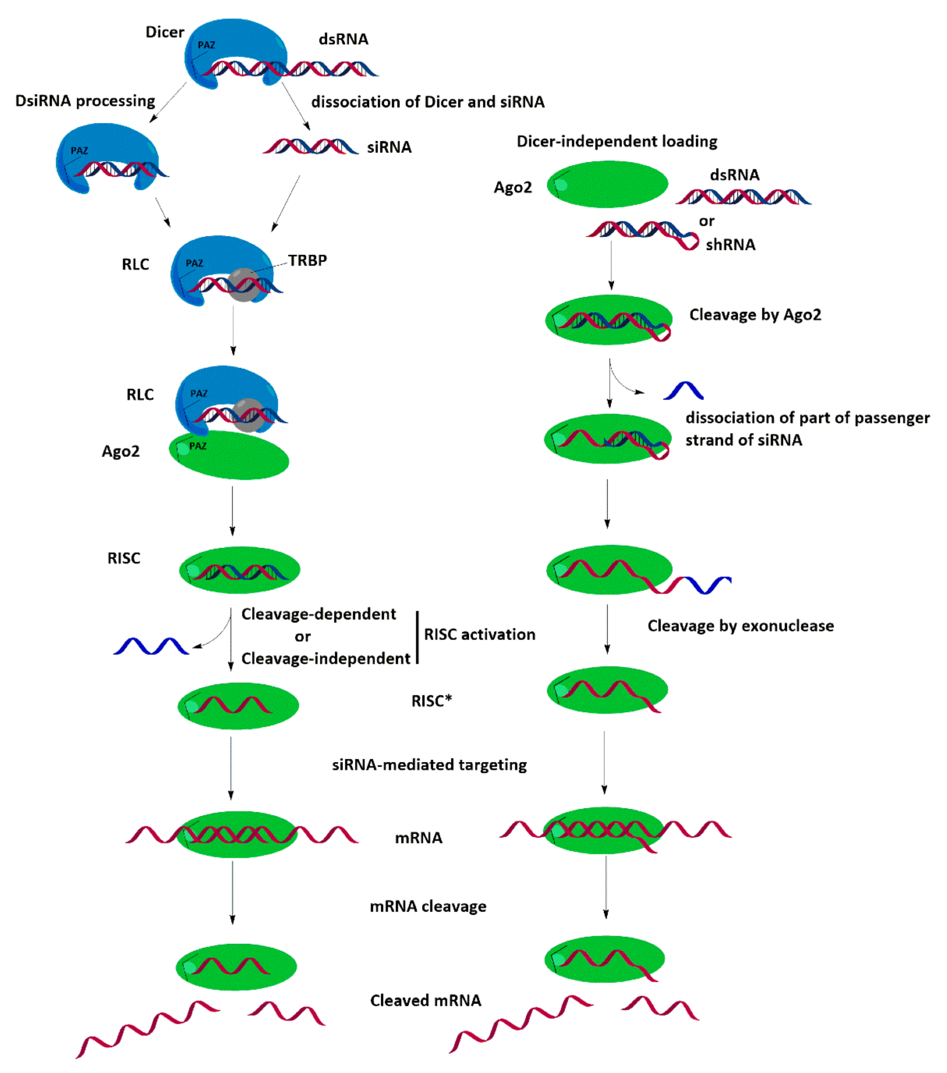
Figure 1. Dicer-dependent and Dicer-independent mechanisms of RNA interference.
Ijms 24 00956 g001
Disclaimer/Publisher’s Note: The statements, opinions and data contained in all publications are solely those of the individual author(s) and contributor(s) and not of MDPI and/or the editor(s). MDPI and/or the editor(s) disclaim responsibility for any injury to people or property resulting from any ideas, methods, instructions or products referred to in the content.