Review

The Crosstalk between Mesenchymal Stromal/Stem Cells and Hepatocytes in Homeostasis and under Stress

by
Irina V. Kholodenko
1,*,
Roman V. Kholodenko
2 and
Konstantin N. Yarygin
1,*
1
Laboratory of Cell Biology, Orekhovich Institute of Biomedical Chemistry, 119121 Moscow, Russia
2
Laboratory of Molecular Immunology, Shemyakin-Ovchinnikov Institute of Bioorganic Chemistry, Russian Academy of Sciences, 117997 Moscow, Russia
*
Authors to whom correspondence should be addressed.
Int. J. Mol. Sci. 2023, 24(20), 15212; https://doi.org/10.3390/ijms242015212
Submission received: 23 September 2023 / Revised: 7 October 2023 / Accepted: 10 October 2023 / Published: 16 October 2023
(This article belongs to the Special Issue Stem Cells in Health and Disease)

Abstract

:
Liver diseases, characterized by high morbidity and mortality, represent a substantial medical problem globally. The current therapeutic approaches are mainly aimed at reducing symptoms and slowing down the progression of the diseases. Organ transplantation remains the only effective treatment method in cases of severe liver pathology. In this regard, the development of new effective approaches aimed at stimulating liver regeneration, both by activation of the organ’s own resources or by different therapeutic agents that trigger regeneration, does not cease to be relevant. To date, many systematic reviews and meta-analyses have been published confirming the effectiveness of mesenchymal stromal cell (MSC) transplantation in the treatment of liver diseases of various severities and etiologies. However, despite the successful use of MSCs in clinical practice and the promising therapeutic results in animal models of liver diseases, the mechanisms of their protective and regenerative action remain poorly understood. Specifically, data about the molecular agents produced by these cells and mediating their therapeutic action are fragmentary and often contradictory. Since MSCs or MSC-like cells are found in all tissues and organs, it is likely that many key intercellular interactions within the tissue niches are dependent on MSCs. In this context, it is essential to understand the mechanisms underlying communication between MSCs and differentiated parenchymal cells of each particular tissue. This is important both from the perspective of basic science and for the development of therapeutic approaches involving the modulation of the activity of resident MSCs. With regard to the liver, the research is concentrated on the intercommunication between MSCs and hepatocytes under normal conditions and during the development of the pathological process. The goals of this review were to identify the key factors mediating the crosstalk between MSCs and hepatocytes and determine the possible mechanisms of interaction of the two cell types under normal and stressful conditions. The analysis of the hepatocyte–MSC interaction showed that MSCs carry out chaperone-like functions, including the synthesis of the supportive extracellular matrix proteins; prevention of apoptosis, pyroptosis, and ferroptosis; support of regeneration; elimination of lipotoxicity and ER stress; promotion of antioxidant effects; and donation of mitochondria. The underlying mechanisms suggest very close interdependence, including even direct cytoplasm and organelle exchange.

1. Introduction

Liver diseases are a global problem due to high morbidity and mortality. Europe has the highest number of cases of diagnosed liver disease in the world [1]; in the EU alone, nearly 30 million people live with chronic liver diseases [2]. Liver cirrhosis kills about 170,000 people every year, accounting for 1.8% of all deaths reported in Europe [2]. Liver diseases are now the second leading cause of disability in Europe after ischemic heart disease [3]. According to the Global Burden of Disease (GBD) Study 2017, the number of deaths from cirrhosis and other chronic liver diseases was 1.32 million in 2017 compared with 889,000 in 1990 [4]. Presumably, the COVID-19 pandemic had a pronounced negative impact on mortality from chronic liver diseases, especially exacerbating the situation in countries where the alcoholic etiology was predominant [5].
The current therapeutic approaches to the treatment of liver diseases are mainly focused on reducing the symptoms and slowing down the progression of the disease, while heterologous organ transplantation remains the only way to treat end-stage liver disease. In this regard, issues related to the development of new effective approaches aimed at stimulating liver regeneration, both by switching on the internal resources of the organism and by enhancing regeneration with drugs and various medicinal procedures, are of high relevance. In the field of regenerative medicine in general and in regenerative hepatology in particular, stem and progenitor cells are considered powerful therapeutic tools [6]. It has been shown that systemic infusion of MSCs effectively induces immunological tolerance [7] and immunosuppression [8] in liver transplant recipients. To date, several systematic reviews and meta-analyses have been published confirming the efficacy of mesenchymal stem cell (MSC) transplantation in the treatment of liver diseases of various severities and etiologies [9,10,11]. However, despite the successful use of MSCs in clinical practice and the promising therapeutic results that have been demonstrated in animal models of liver diseases, the mechanisms of their protective and regenerative action remain poorly understood. The data about the mediators of intercellular communication produced by these cells and responsible for their therapeutic action are fragmented and often contradictory.
MSCs are a specific cell type present in all tissues and organs of the body and participate in the formation of the organs’ stroma and tissue-specific niches. The unique features of these cells include immunomodulatory properties and extensive regenerative potential. As shown by several research groups, the liver contains a pool of resident MSCs, which, in terms of their main characteristics (expression patterns of cell surface markers, adhesion, and morphology), correspond to those of MSCs isolated from other tissue sources [12,13,14]. Liver MSCs have several unique characteristics compared with those obtained from other tissue sources. In particular, liver MSCs express a spectrum of liver-specific genes [15]. Lee et al. [16] showed that liver MSCs are more readily differentiated into the cells of hepatic lineage compared with umbilical cord-derived MSCs, while Yigitbilek et al. [17] demonstrated their more pronounced immunomodulatory properties and pro-angiogenic potential compared with adipose tissue-derived MSCs. As shown by Raicevic et al. [18], inflammatory conditions significantly influenced the immunophenotype of liver MSCs; the expression of the costimulatory molecule CD40 was initiated, the expression of Toll-like receptors (TLRs) 1, 2, and 3 increased, and the expression of the adhesion molecules CD58 and CD106 as well as the expression of the immunoregulatory molecule CD274 (PD-L1 or B7-H1) were substantially enhanced [18]. Girousse et al.’s review article [19] proposed and substantiated a hypothesis claiming that endogenous MSCs can serve as the main cellular components of an interorgan communication network ensuring coordinated functioning of the tissues, organs, and their parts at the cellular level and cooperative responses to stressful stimuli. Originally, the known interorgan communication messages included peptides, proteins, and metabolites, and later, the list was supplemented with extracellular vesicles. Since MSCs or MSC-like cells are present in all tissues and organs, it is likely that most of the key cell-to-cell interaction-dependent processes, including crosstalk between cells, the epithelial–mesenchymal transition, immunomodulation, differentiation, cellular maintenance, and cell death, are dependent on the MSCs present within the tissue niche. In this context, it is important to understand the mechanisms underlying the communication between MSCs and differentiated somatic cells in each particular tissue. This is important both from the perspective of basic science and for the development of therapeutic approaches involving the modulation of the activity of resident MSCs. With regard to the liver, the research should be concentrated on the intercommunication between MSCs and hepatocytes. Elsewhere, we discussed resident liver MSCs different from hepatic stellate cells and reviewed existing data concerning their possible triple origin from the resident liver MSCs formed during histogenesis and organogenesis, hepatic pericytes, and MSCs mobilized from the bone marrow [15]. The latter option is evident in situations of liver injury, such as liver fibrosis [20,21], liver cirrhosis [22,23], partial hepatectomy [24,25], and hepatocellular carcinoma [26], and after living donor liver transplantation [27]. The goal of this review was to identify the main pathways and key factors mediating the crosstalk between MSCs and hepatocytes to determine the possible mechanisms of the direct and indirect interaction of the two cell types under normal or close to normal conditions and under stress. Mainly, we focused on model experiments in vitro since in such artificial systems, the key regulators of intercellular communication are the most reliably detected, while in vivo, it is difficult to determine the true regulators due to the presence of many cell types and other biologically active components in the microenvironment.

2. Crosstalk between MSCs and Hepatocytes in Close-to-Homeostasis Conditions

2.1. Chaperon-Like Functions of MSCs in Mixed 3D Cultures

In liver organogenesis, mesenchymal cells play an important role in the development of the liver bud and the subsequent morphogenesis of the organ. The formation of the liver from the cell condensate begins with the loss of contact between hepatoblasts due to the suppression of E-cadherin expression, which provides contact between neighboring cells [28,29]. This allows hepatoblasts to migrate into the surrounding mesenchyme. Simultaneously, matrix metalloproteinases (MMPs) degrade extracellular matrix proteins, such as collagen, fibronectin, and basement membrane laminin, thereby facilitating hepatoblast migration. This extracellular matrix remodeling is mainly mediated by MMP2 expressed by mesenchymal cells and MMP14 expressed by hepatoblasts [30]. Matsumoto et al. [31] demonstrated that during embryonic development, mesenchymal cells play a critical role in the formation of the liver primordium from the endoderm, controlling hepatic progenitor cells and regulating their fate by creating a specialized hepatocyte embryonic niche.
The fetal liver is a unique tissue in terms of the coexistence of cells of endodermal and mesenchymal origin [32]. Ito et al. [33] showed that in the fetal liver, PDGFRa-positive mesenchymal cells are located in close proximity to albumin-positive hepatoblasts. In vitro, Dlk1midPDGFRa+ mesenchymal cells secreted factors that increased hepatoblast proliferation. At the same time, direct interaction between hepatoblasts and Dlk1midPDGFRa+ mesenchymal cells did not increase proliferation, but it strictly induced the differentiation of hepatoblasts into hepatocytes, as shown by the increased expression of genes, reflecting mature hepatocyte function; these genes included those encoding for albumin, tyrosine aminotransferase (TAT), and carbamoyl-phosphate synthetase (CPS) [33]. In vitro mouse fetal CD49f±Thy1+gp38+CD45 mesenchymal cells cocultured with mouse fetal hepatoblasts induced their maturation, as shown by positive acid–Schiff staining and the upregulation of the expression of mature hepatocyte markers, such as tyrosine aminotransferase, tryptophan-2,3-dioxygenase, and glucose-6-phosphatase [34]. Also, human bone marrow MSC-derived conditioned medium induced the maturation of hepatoblasts derived from induced pluripotent stem cells, as shown by an increase in the production of albumin and urea as well as an increase in the expression and activity of cytochrome P450 (CYP) 3A4 [35].
Many approaches to obtaining liver organoids (sometimes called liver buds) of diverse complexity based on combinations of various cell types have been developed, both to study the mechanisms of liver organogenesis and to obtain functionally active hepatocyte-containing tissue engineering constructs suitable for drug screening with the potential for clinical application [36,37]. Direct cell-to-cell contact, the interaction of cells with extracellular matrix proteins, and the presence of a whole range of soluble factors are critical for the successful formation of liver organoids [38]. In organoids consisting of monocultures of hepatocytes, hepatoblasts, and/or hepatocyte-like cells differentiated from various progenitor and stem cells matrix is a critical component required for the efficient assembly and maintenance of the functional activity. Collagen or matrigel is often used to provide a 3D environment for cells to interact and self-organize. More complex bioengineering approaches, such as the employment of decellularized organ scaffolds or bioprinting technology, can also be applied to generate a native 3D tissue structure. Along with the matrix, soluble factors imitating events occurring in organogenesis can be utilized; among them are a variety of small molecules and biologicals, such as Wnt agonists R-spondin1 and CHIR99021; EGF, FGF7, and FGF10; HGF and the TGF-β inhibitor A83-01; TNFα; etc. [39,40,41]. In liver organoids consisting of hepatocytes, endothelial cells, and mesenchymal cells, the supportive mesenchymal and endothelial cells play a pivotal role in maintaining the viability and functionality of the parenchymal cells. They provide hepatocytes with an appropriate microenvironment in the form of cell contact, the production of extracellular matrix proteins, vasculature, and the secretion of a whole range of soluble factors.
MSCs provide reinforcement of the 3D architecture of liver organoids. It has been established that organoids are not formed in the absence of stromal components, such as MSCs and/or extracellular matrix [37,42]. In particular, it has been shown that 3D spheroids derived from human BM-MSCs and human hepatocytes form an inner core consisting of parenchymal cells that express HNF4α and cytokeratin 18 surrounded by an outer layer of vimentin-expressing stromal cells. Phalloidin staining showed the presence of a peripheral actin belt and F-actin enrichment in the apical region of the cells, indicating hepatocyte polarization. Type I collagen deposits were observed in the intercellular space between mesenchymal cells and the internal parenchymal mass [43].
The formation of liver organoids in the presence of MSCs occurs due to the so-called “contraction mechanism”, which is non-muscular myosin-II (NSM-II)-dependent [36]. Using live cell tracking, Takebe et al. [44] showed that the co-cultivation of iPSC-derived hepatic endodermal cells, HUVECs, and MSCs initiated the assembly of vascularized organ buds after rapid cell convergence. This was followed by spatial rearrangements through self-organization, as evidenced by the formation of an endothelium-like network. During the initial phase of self-convergence, a cohesive multicellular unit was formed consisting of cells that quickly moved to a single center, a process called collective condensation. It is noteworthy that the formation of cell condensates was based not on the active migration of cells but on their contraction. Importantly, cellular condensation did not occur in the absence of MSCs, which indicates that MSCs provide sufficient contractile force for the assembly of the three cell types. During embryonic invagination, a group of cells undergoes contraction. A sharp inward shift of intercellular junctions occurs due to the activity of myosin II (MII), which allows cells to invaginate during embryonic gastrulation [45,46]. MII regulatory light chain diphosphorylation (ppMRLC) has greater actin-activated Mg2+-ATPase activity than MRLC monophosphorylation (pMRLC), which is sufficient to generate contractile force [47]. It was shown that the level of ppMRLC in co-cultures of iPSC-derived hepatic endodermal cells, HUVECs, and MSCs significantly increased after 4 h of plating, and a marked increase in the total amount of MRLC was observed during the formation of cell condensates, which corresponded to the time when the cells began their collective movements. MIIA immunostaining in the liver bud showed that CD90+ MSCs significantly expressed MIIA. These results clearly indicate that MSCs in the liver bud provide the traction force created by the actomyosin complex of the cytoskeleton, which plays an important role in directed cell movements and the formation of cell condensates [44]. Later, the same group of authors developed a system for obtaining liver buds from three types of cells, each of which was derived from human induced pluripotent stem cells (iPSC) by directed differentiation into transitional hepatic endoderm (tHE) cells (co-expressing T-box transcription factor 3 (TBX3) and adrenoceptor alpha 1B (ADRA1B)), septum transversum mesenchyme (STM) cells (co-expressing Wilms tumor 1 homolog (WT1), MIIA, and LHX2), and endothelial progenitors (iPSC-EC) (co-expressing CD144 (VE-cadherin) and CD31), respectively [48]. Global gene array analysis showed that the iPSC-STM signatures were similar to those of human adult hepatic mesenchymal cell signatures [49,50]. In the liver bud, these cells were the driving force of myosin-IIA-dependent self-condensation [48].
Liver organoids generated using decellularized liver tissue as a scaffold made of the native extracellular matrix containing all ECM proteins essential for the maintenance of hepatocytes, such as collagen type I, collagen type IV, fibronectin, and laminin, represent one of the most practical in vitro models for hepatology research [36,51]. It has been shown that MSCs present in liver organoids increase the level of extracellular matrix proteins and enhance the expression of β-integrin by hepatocytes, securing their effective adhesion to the ECM [51]. Also, MSCs, being the cells that form tissue niches for various types of stem cells, are themselves efficient producers of ECM proteins, including laminin, fibronectin, type I collagen, tenascin-C, decorin, etc. [52,53]. Accordingly, extracellular matrix proteins produced by MSCs provide additional supportive substrates for hepatocytes within liver organoids, as demonstrated by Park et al. [42].
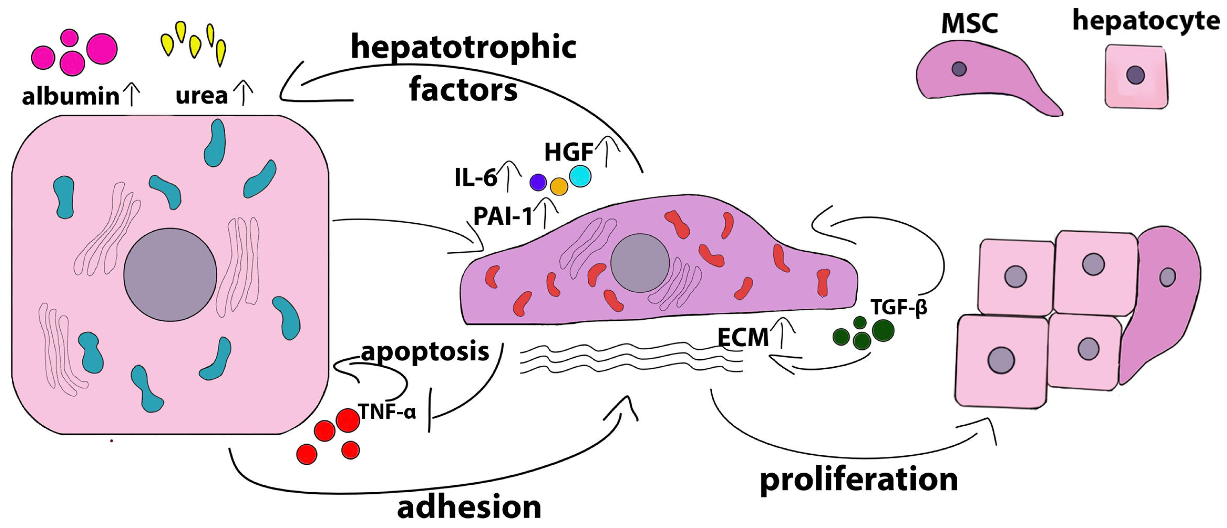
Several studies have shown that liver organoids fail to assemble if MSCs are replaced with an MSC-derived conditioned medium (MSC-CM) [44,54], although the paracrine effects of MSCs may still persist, supporting the differentiation and maturation of hepatocytes [54,55]. For example, paracrine factors, including IL-6, GRO-α, IL-8, MCP1, SDF-1α, LIF, VEGF-A, and PDGF-BB, secreted by human amniotic-derived MSCs (hAMSCs) cultured under 3D conditions had a pronounced effect on hepatogenic differentiation and the functional maturation of EpCAM+ liver progenitor cells in a monoculture organoid, as evidenced by the over-production of albumin and the over-activity of CYP3A4 [55]. The involvement of MSCs in niche formation in liver organoids may not be limited to cell-to-cell contact, paracrine signals, and the production of extracellular matrix proteins, but it may also involve the differentiation of MSCs into endothelial cells with subsequent formation of vascular structures. Kadota et al. [51] demonstrated the co-expression of VEGF and CD31 on BM-MSCs in liver organoids, suggesting that MSCs can be differentiated in endothelial lineage cells, probably through interactions between co-cultured hepatocytes and the microenvironment of the liver post-decellularized ECM. These data demonstrate that under 3D conditions, MSCs play a key role in the creation and maintenance of the tissue architecture of liver organoids, providing niche formation and paracrine signals directed at parenchymal cells. Schematically, the role and functions of MSCs in the liver organoids are shown in Figure 1.
Beneficial effects of MSCs on cultured hepatocytes were observed not only in autologous/syngeneic and allogeneic systems but also in xenogeneic systems. Montanari et al. [56] showed that human bone marrow MSCs co-encapsulated with porcine hepatocytes in alginate- and poly(ethylene glycol) (PEG)-grafted alginate hydrogels significantly prolonged hepatocyte survival and stimulated albumin production without affecting diazepam metabolism [56]. Organoids generated by co-cultivation of primary rat hepatocytes with human umbilical cord-derived MSCs in porcine liver-derived ECM (PLECM gel) survived longer than those made of hepatocytes only, maintained hepatocyte function for more than 20 days, showed a significantly higher production of albumin on day 14, and demonstrated enhanced urea synthesis for more than 3 weeks [36]. On the other hand, in 3D spheroids derived from human BM-MSCs and human hepatocytes, the levels of urea production and albumin secretion did not change compared with those of monocultures of hepatocytes [43]. However, despite the fact that the expression of CYP3A4 increased both in the 3D co-culture of hepatocytes with BM-MSCs and in the monoculture of hepatocytes, the activity of this enzyme persisted for 2 weeks only in the co-culture, while in the monoculture, it decreased after a week. Also, when hepatocytes were co-cultured with BM-MSCs, the expression of the 1A2 and 2C9 isoforms significantly increased compared with monoculture [43], suggesting improved maintenance or activation of the metabolic function of hepatocytes due to the presence of MSCs.

2.2. Crosstalk of Hepatocytes and MSC in 2D Co-Culture

Most papers describing simpler, essentially 2D systems of hepatocyte co-cultivation with MSCs have also demonstrated the supporting role of the latter, manifested by the increased viability of hepatocytes as well as better preservation of their typical morphology and other signs of a differentiated state, such as albumin and urea production and enhanced drug metabolism [57].
Using an allogeneic direct co-culture system in which human primary hepatocytes were seeded on top of a human adipose-derived MSC monolayer, Qin et al. [58] showed that, in addition to providing hepatocyte support, MSCs had pronounced hepatotrophic and anti-apoptotic effects. Minimal hepatotrophic and anti-apoptotic effects were also shown in indirect co-cultures of hepatocytes with MSCs in the transwell plates and could be induced by conditioned media. Co-cultures with MSCs reduced the secretion of TNF-α by hepatocytes, and this inhibitory effect of co-cultures on the hepatocyte autocrine depended on MSC–hepatocyte contact and the intracellular ROS activity in the MSCs. TNF-α is known to have a bidirectional regulatory effect on both hepatocyte proliferation and apoptosis [59]. Co-cultures with MSCs protect hepatocytes from apoptosis induced by high levels of TNF-α [60]. Co-culturing hepatocytes with MSCs also induced an additive or synergistic contact-dependent effect on TGF-β1 production. The autocrine action of TGF-β1 on MSCs, leading to increased production of ECM proteins [61], was required for the trophic effects seen in co-culture conditions. Direct co-culturing significantly increased TGF-β1 secretion compared with monocultures of hepatocytes or MSCs, while in indirect co-cultures, these effects did not take place [58]. Extracellular collagen deposition by MSCs was enhanced by co-cultivation with hepatocytes, and this at least partially contributed to the trophic and anti-apoptotic effects of MSCs. Attachment to extracellular collagen facilitated the entry of hepatocytes into the S phase [62] and mediated hepatocyte aggregation and cell-to-cell contact [63]. Extracellular collagen is mainly localized around MSCs, and the inhibition of the production of type I collagen in MSCs significantly reduces the synthesis of albumin and urea by hepatocytes [64]. Increased deposition of extracellular collagen by mesenchymal cells may predominantly occur due to MSC–hepatocyte contact and is associated with intracellular ROS activity in MSCs. These results suggest that the hepatotrophic effect of co-culturing is mostly mediated by the synergistic effects of ECM and direct cell-to-cell interaction [58].
MSCs isolated from different tissue sources have slightly different effects on hepatocytes in co-cultures. This is probably due to differences in the expression/secretion profiles of hepatotrophic factors. For example, under standard monoculture conditions, human umbilical cord perivascular cells (HUCPVCs) have been shown to express lower levels of hepatotrophic genes, such as cytokine stem cell factor (KITLG), proteoglycan decorin (DCN), notch ligand jagged 1 (JAG1), and HGF, compared with human BM-MSCs. At the same time, HUCPVCs and BM-MSCs equally supported the production and secretion of albumin as well as the expression of the hepatospecific protein tryptophan 2,3-dioxygenase, although the expression of tyrosine aminotransferase was upregulated only in the co-cultures of hepatocytes with HUCPVCs. Unlike BM-MSCs, HUCPVCs effectively supported ureagenesis. BM-MSCs, in turn, were more effective at increasing CYP activity [65]. Figure 2 presents a scheme of the mutual influence of hepatocytes and MSCs during co-cultivation under normal conditions.
It is noteworthy that in addition to the effects of MSCs on hepatocytes, the latter also affect mesenchymal cells. In human HUCPVCs and BM-MSCs, the expression profiles of hepatotrophic factors significantly changed when they were co-cultured with rat hepatocytes. HUCPVCs began to express increased levels of laminin (LAMB1), HGF, IL-6, and connexin 43 (GJA1) compared with BM-MSCs, and BM-MSCs expressed increased levels of KITLG and JAG1 [65]. Porcine hepatocytes induced increased expression and secretion of serpin E1 (plasminogen activator inhibitor-1 (PAI-1)) by human MSCs and did not affect the secretion of pro-inflammatory cytokines such as CCL2, CXCL12, and macrophage migration inhibitory factor (MIF) [56]. Notably, several studies have shown that MSCs do not differentiate in the hepatocyte lineage in co-cultures [65,66].
In the majority of publications describing the direct co-cultivation of hepatocytes and MSCs, it has been shown that cell-to-cell contact is necessary for the implementation of maintenance effects, although paracrine interactions between cells due to the production of bioactive molecules may play a significant role. In several works described above, it was shown that an MSC-CM had no or only partial supporting and/or protective effects on hepatocytes [58,67,68]. For example, a conditioned medium derived from human adipose MSCs stimulated the proliferation of mouse hepatocytes and the accumulation of glycogen but did not affect the activity levels of cytochrome P450 or the production of albumin and urea [67]. Similar results were obtained with regard to the action of the conditioned medium obtained from human MSCs on human hepatocytes, namely improved survival of hepatocytes and a slight increase in their functional activity compared with direct co-cultures [68]. The mechanisms of MSC communication with target cells mediated by cell-free MSC derivatives, including extracellular vesicles, conditioned media, apoptotic bodies, and mitochondria, have been extensively characterized in various disease models [69].

3. MSCs Communication with Hepatocytes in Stressful Environment

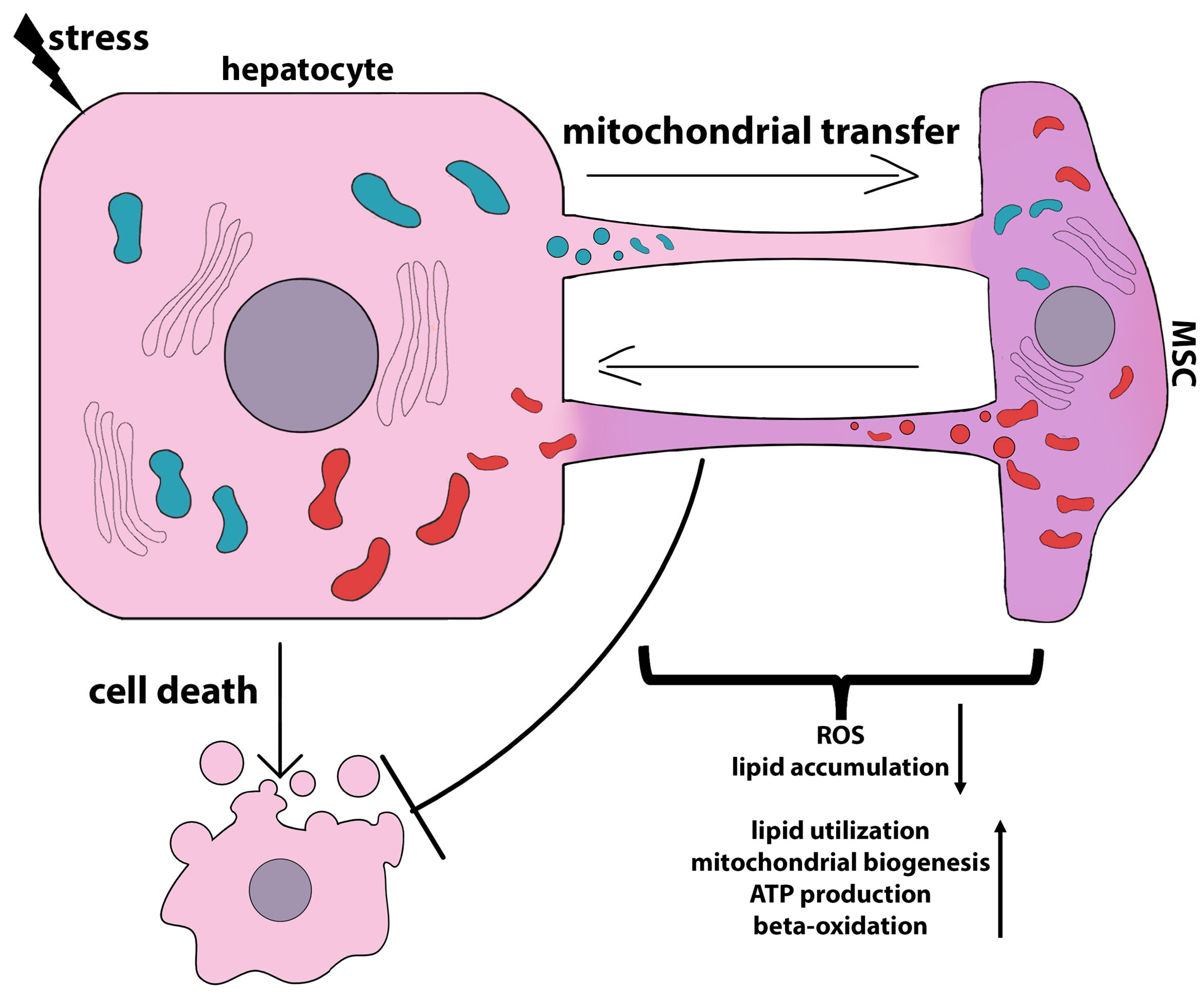
Generally, the communication of MSCs with hepatocytes as well as with other cell types can be carried out not only through direct interaction between cells but also through the secretion of soluble factors [70,71,72], the exchange of extracellular vesicles [73,74], or mitochondrial transfer [75]. For example, mouse BM-MSC-derived exosomes induced the conversion of mature hepatocytes into EpCAMhigh small oval cells. These cells, which are hepatic progenitor cells, also expressed hepatoblast markers, including DLK1, Onecut 2 (OC2), α-fetoprotein, and cytokeratin 19 [76]. On the other hand, the MSC-CM had a supportive effect on hepatocytes, stimulating their metabolism and albumin production [67]. A human amnion-derived MSC (hAMSC)-CM had the potential to increase hepatic stem/progenitor cell differentiation in human liver organoids [55].
The following two sections present the results of the in vitro and in vivo studies demonstrating the protective effects of both MSCs and MSC derivatives on hepatocytes exposed to stress in the context of the crosstalk between the two cell types. The mechanisms involved in hepatocyte protection against apoptosis, ferroptosis, pyroptosis, and lipotoxicity are described in detail.

3.1. Anti-Apoptotic Effects of MSCs

Apoptosis is a ubiquitous form of cell death that regularly occurs in human liver diseases. Under normal physiological conditions and during organ development, apoptosis is carefully regulated spatiotemporally and does not provoke adverse secondary events. Pathological apoptosis, on the contrary, activates secondary signaling cascades that can lead to massive damage to tissues and organs. Such pathological apoptosis-induced cascades stimulate tissue inflammatory responses, injury, and fibrosis [77]. Apoptosis is a trigger for both acute and chronic liver diseases. For example, the number of apoptotic hepatocytes increases significantly in patients with non-alcoholic steatohepatitis and correlates with the severity of the disease and the stage of fibrosis, which indicates the etiopathogenetic role of apoptotic cell death in the progression of this disease [78,79]. Infection with hepatitis B (HBV) or C viruses (HCV) induces liver damage, mainly by activating the host’s immune response to viral proteins expressed by infected hepatocytes. Notably, acute liver injury is mainly characterized by hepatocyte necrosis, while chronic HCV and HBV infections predominantly induce massive apoptosis. In viral hepatitis, cytotoxic T lymphocytes recognize viral antigens on hepatocytes and kill them, leading to liver damage. It has been proven that cytotoxic T lymphocytes kill virus-infected hepatocytes through Fas-dependent apoptosis. Consequently, hepatocyte Fas expression is significantly increased, and the number of FasL-positive infiltrating mononuclear cells in the liver is significantly elevated in patients with hepatitis C [80] or chronic active hepatitis B [81]. Fas/FasL expression levels are closely correlated with inflammatory activity, which initiates the development of the disease and contributes to its progression [82]. Another mechanism by which virus-infected hepatocytes undergo massive apoptosis involves TNF-α [83,84]. Long-term alcohol consumption in large quantities leads to the development of alcoholic liver disease [85]. Alcohol induces the production of high levels of reactive oxygen species (ROS) and a decrease in intracellular antioxidant levels, thereby causing oxidative stress in hepatocytes. Alcohol-induced oxidative stress is the cause of massive hepatocellular apoptosis and the main mechanism leading to liver damage [86]. The mechanisms of hepatocyte apoptosis in various pathological liver conditions have been described in more detail in a number of reviews [77,82,87].

3.1.1. Involvement of Growth Factors and Bioactive Molecules in the Anti-Apoptotic Effects of MSCs

Many authors have noted decreased apoptosis and increased proliferation in hepatocytes after the transplantation of MSCs or cell-free MSC derivatives in various animal models of liver diseases [88,89,90,91]. For example, BM-MSC-derived exosomes entered primary rat hepatocytes within 12–24 h and accumulated in the cytoplasm, preventing hepatocyte apoptosis induced by D-galactosamine and lipopolysaccharide (D-GalN/LPS) by increasing the number of autophagosomes and the expression of the autophagy-related proteins LC3II and Beclin-1. At the same time, the expression levels of the proapoptotic proteins Bax and cleaved caspase 3 significantly decreased and the expression level of the anti-apoptotic protein Bcl-2 increased, indicating the induction of autophagy in hepatocytes due to exosomes preventing apoptosis [92]. Exosomes prepared from hESC-derived HuES9.E1 MSCs stimulated hepatocyte proliferation and reduced the apoptosis of hepatocytes in a mouse carbon tetrachloride liver injury model. In two in vitro models, the H2O2-induced oxidative stress model and the acetaminophen-induced injury model, it was shown that in three lines of mouse and human hepatocytes, exosomes derived from the hESC-derived HuES9.E1 MSCs regulated hepatocyte proliferation, promoting their transition from the G0 phase to the G1 phase of the cell cycle by stimulating the expression of tumor necrosis factor alpha (TNF-α), interleukin 6 (IL-6), inducible nitric oxide synthase (iNOS), cyclooxygenase-2 (COX-2), and macrophage inflammatory protein 2 (MIP-2), which in turn led to the restoration of NF-κB and STAT3 signaling. The anti-apoptotic effect of exosomes was associated mainly with the activation of STAT3 signaling, the inhibition of caspase 3, and increased levels of the anti-apoptotic protein Bcl-xL, but it was not associated with the activation of antioxidative genes, including heme oxygenase-1 (HO-1), glutathione peroxidase 4 (Gpx4), glutathione reductase (GSR), and MnSOD [93]. The level of apoptosis decreased and proliferation increased due to the activation of Notch signaling in direct co-cultures of mouse hepatocytes (H2.35) treated with palmitic acid to induce lipotoxicity and human adipose-derived MSCs [94].
Also, in vitro studies have shown that MSCs suppressed staurosporine-induced apoptosis in hepatocyte co-cultures [58]. Staurosporine is known to activate caspase 3 signaling independently from caspases 8, 9, and 12 [95]. Caspase 9 is an initiator of caspase-mediated apoptosis through the mitochondrial pathway [96]. Co-culture of hepatocytes with MSCs reduced caspase 9 gene expression rather than that of caspases 3, 8, and 12, suggesting that in the co-culture, the anti-apoptotic action of MSCs is mainly confined to the initiation of apoptosis. Bax/Bcl-2 balance regulates hepatocyte apoptosis [97]. BID interacts with Bax and mediates caspases 3 and 8 in apoptotic hepatocytes [98]. Co-cultures with MSCs significantly reduced the Bax/Bcl-2 ratio and BID expression by hepatocytes in a manner dependent on cell contact and MSC ROS activity. The authors suggested that isolated primary human hepatocytes became irresponsive to soluble trophic and anti-apoptotic factors released by MSCs, which was the reason for the weaker anti-apoptotic activity of the MSC-CM [58].
In an in vitro model, rat hepatocytes treated with sera from animals given D-galactosamine to induce acute liver failure underwent massive cell death and began to express high levels of IL-6. When such hepatocytes were co-cultured with human MSCs, their viability was restored, and IL-6 secretion was significantly reduced [66]. In this context, IL-6 expression can be regarded as a response to inflammatory conditions, enhancing hepatocyte damage. On the other hand, in an earlier work by Isoda et al., hepatocytes co-cultured with BM-MSCs maintained higher levels of albumin and urea production compared with hepatocyte monocultures, regardless of intercellular contact; this effect was mainly associated with IL-6 produced by BM-MSCs but not by rat hepatocytes [99]. IL-6 was significantly elevated in direct co-cultures of rat hepatocytes treated with the serum of an acute liver failure patient and human orbital fat-derived stem cells and was the key factor mediating the protective effects of MSCs [100]. Such contradictory results regarding the contribution of IL-6 to the protective effects of MSCs on hepatocytes may be related to the multiplicity of the mechanisms of IL-6 signaling.
IL-6 is known to have a cytoprotective effect in vivo and in vitro [101,102]. IL-6-treated hepatocytes are able to maintain higher levels of albumin and urea secretion and show a better ability to metabolize drugs in the presence of injurious agents [101]. IL-6 has an effect on liver regeneration and can induce hepatocyte proliferation, but in some cases, it can lead to the arrest of cell growth [103,104]. This may be explained by the results of Sun et al., who showed that IL-6 alone induced a resting state in hepatocytes but that the effect was reversed in the presence of non-parenchymal cells. IL-6 stimulates non-parenchymal cells to produce hepatocyte growth factor, which is a potent hepatic mitogen and induces hepatocyte proliferation [104].
The pleiotropic effects of IL-6 on hepatocytes may be associated with the specific regulation of its signaling [105]. IL-6 has an affinity to IL-6R but not to gp130. In turn, IL-6R also does not have a significant affinity to gp130. Only the formed IL-6/IL-6R complex can bind to gp130 [106]. This is a very important sequence of biologically active molecule interactions that plays a significant role in the regulation of IL-6 signaling. While gp130 is expressed by all body cells, IL-6R is expressed by only a few cell types, including hepatocytes, some leukocytes, and some epithelial (biliary epithelial cells) and non-epithelial cells (hepatic stellate cells). Therefore, only cells expressing IL-6R can directly respond to cytokine IL-6 [106]. Other cell types may respond to IL-6 by trans-signaling when the cytokine interacts with the soluble form sIL-6R. In this case, the IL-6/sIL-6R complex interacts with gp130 with high affinity. Since hepatocytes express more gp130 than IL-6R, the presence of IL-6 and sIL-6R results in greater gp130 activation and, therefore, higher IL-6 signal amplitude. Moreover, the IL-6/sIL-6R complex was shown to be internalized less efficiently than IL-6/IL-6R, resulting in longer IL-6 signal duration when mediated by trans-signaling [107]. Accordingly, hepatocytes permanently proliferated in IL-6/sIL-6R double transgenic mice in the absence of any liver insults but did not show any mitogenic response in IL-6 single transgenic mice. Thus, a signal induced via membrane-bound IL-6R on hepatocytes was insufficient to induce a proliferative response [108,109].
As shown in a model of acetaminophen-induced damage, MSCs significantly reduced necrosis through secreted growth factors in indirect co-cultures with the human LO2 cell line [110]. Many authors mistake the LO2 cell line for human hepatocytes, but it is a derivative of the cervical cancer cell line HeLa [111]. Several MSC-expressed factors, such as HGF, epidermal growth factor (EGF), IL-6, vascular endothelial growth factor (VEGF), and insulin-like growth factor-binding protein (IGFBP), may prevent hepatocyte apoptosis, increase hepatocyte survival, and/or stimulate proliferation [112,113]. In the presence of acetaminophen, the expression of some of these cytokines was markedly increased in human umbilical cord MSCs, and the addition of the cytokine-neutralizing antibodies to co-cultures led to a marked decrease in the anti-necrotic effect of the MSCs. It is noteworthy that among the analyzed cytokines, HGF made the greatest contribution to the protective effect of human umbilical cord MSCs on hepatocytes [110].
HGF is a pleiotropic factor that has mitogenic, motogenic, anti-apoptotic, morphogenetic, and immunoregulatory activities [114,115]. Such a wide range of activities warrants the ability of HGF to prevent fibrosis, inflammation, and apoptosis and promote tissue regeneration and angiogenesis [116]. It has been repeatedly shown that the therapeutic efficacy of MSCs in various pathologies partially or completely depends on the secretion of HGF [117,118]. The role of HGF in MSC-mediated therapeutic effects has been reliably proven in many studies using genetically modified MSCs with HGF overexpression [119]. Therapy with HGF-overexpressing MSCs has been shown to be more effective than using cells and cytokines separately or in combination [120,121]. HGF-overexpressing MSCs effectively reduced fibrosis and had a suppressive effect on hepatocyte apoptosis in liver disease models [121,122,123,124,125,126,127]. Also, HGF-overexpressing MSCs showed pronounced antioxidant activity, which is important for the alleviation of the inflammatory response through a reduction in oxidative stress by decreasing lipid peroxidation, probably via the ability to promote the activation of superoxide dismutase (SOD) and the synthesis of glutathione (GSH) [128].
Liu et al. [91] proved the role of MSC-produced prostaglandin E2 (PGE2) in reducing apoptosis and increasing hepatocyte proliferation caused by acute liver failure (ALF). The authors used LPS-treated AML12 cells as an in vitro model of ALF. MSC-CM and PGE2 significantly upregulated Ki67 and YAP expression and activated mTOR signaling in LPS-treated hepatocytes. Yes-associated protein (YAP), a transcriptional coactivator, is a core component of Hippo signaling that plays important roles in liver development, regeneration, and tumorigenesis (reviewed in [129,130,131]). When Hippo signaling is turned on, YAP is phosphorylated at serine S127 and then binds to the scaffolding molecule 14-3-3 and is transported to the proteasome for degradation. However, when Hippo signaling is turned off, YAP is translocated to the nucleus, where it binds the transcription factors of the TEA domain family (TEAD) to regulate genes associated with Hippo signaling [132,133]. In various models of liver injury, it has been shown that YAP activation improved organ regeneration by enhancing hepatocyte proliferation and reducing hepatocyte apoptosis [134,135]. In addition, the protective role of YAP has been shown in the regulation of hepatocyte ferroptosis in a sepsis-induced liver injury model [136]. The mammalian target of the rapamycin (mTOR) pathway regulates cell growth, survival, and metabolism [137]. mTOR plays a critical role in liver regeneration, hepatic lipid metabolism, and liver cancer [138,139]. Recent studies have shown a potential crosstalk between the Hippo and the mTOR pathways under normal physiological and pathological conditions [140]. Thus, the authors showed that MSC-produced PGE2 activates two interconnected signaling pathways involved in liver regeneration: YAP activation is mediated by p-CREB (p-cAMP responsive element binding protein); in turn, YAP activation leads to the suppression of PTEN due to miR-29a-3p and the subsequent activation of mTOR signaling, as evidenced by increased expression of p-S6K, p-AKT, and p-GSK3β [91]. Table 1 summarizes the data on the bioactive molecules and EVs produced by MSCs and protecting hepatocytes from apoptosis.
PGE2 is defined as a lipid-derived cytokine, an inflammatory lipid mediator that is formed from arachidonic acid by the action of cyclooxygenase 2 (COX2) [141]. PGE2 may act as a morphogen, regulating endodermal cell fate and specification in embryonic development, in particular determining the differentiation of endodermal progenitors into liver or pancreatic cells [142]. In the adult liver, PGE2 is produced by hepatocytes [143], Kupffer cells [144], and endothelial cells [145]. The role of PGE2 in the functioning of hepatocytes under normal and pathological conditions was demonstrated quite a long time ago. PGE2 has a significant effect on glycogenesis by stimulating the conversion of glucose to glycogen [146]. However, it also stimulates the breakdown of glycogen (glycogenolysis) in hepatocytes [147]. Specifically, PGE2 can either stimulate glycogen phosphorylase activity (glycogenolytic effect) or inhibit glucagon-stimulated glycogen-phosphorylase activity (antiglycogenolytic effect) in rat hepatocytes, depending on the type of receptor it binds to [148]. Prostaglandins can influence hepatocyte metabolism directly [149] or indirectly through hormones [150,151] or cytokines [152,153].
The effects of PGE2 on the hepatic lipid metabolism described in the literature are contradictory. Some studies have shown that prostaglandins can promote fat accumulation in hepatocytes, thereby facilitating the development of steatosis [154], while others have shown that PGE2 suppresses de novo lipogenesis [155] or does not affect lipogenesis but at the same time attenuates triglyceride incorporation into very-low-density lipoprotein (VLDL) [156]. Henkel et al. [157] have shown that PGE2 increases triglyceride accumulation in hepatocytes, most likely through the inhibition of β-oxidation combined with decreased VLDL production, thus contributing to hepatic steatosis. On the other hand, PGE2 has been shown to be involved in liver regeneration; its production levels in the liver increase biphasically during liver regeneration after partial hepatectomy, causing hepatocyte proliferation [158]. Rudnick et al. [159] showed that prostaglandins are essential for liver regeneration and COX-2, which is involved in the biosynthesis of PGE2, plays a particularly important role in this process. Hepatocyte-specific COX-2 transgenic mice showed reduced hepatocyte necrosis, decreased neutrophil infiltration, diminished oxidative and ER stress, and low levels of pro-inflammatory factors after liver ischemia/reperfusion injury (IRI) compared with wild-type mice [160]. A number of early studies have shown that PGE2 promotes the proliferation of primary rat hepatocytes [161,162] and has a mitogenic effect that is synergistic with EGF action [163,164]. Later, it was shown that hepatocyte proliferation occurs after the interaction of PGE2 with one of its EP3 receptors but not with the EP1 receptor [165]. However, in more recent studies, the stimulatory role of PGE2 in liver regeneration has been questioned. Nishizawa et al. [166] showed that the inhibition of inducible microsomal PGE synthase-1 (mPGES-1), the terminal enzyme of PGE2 generation, resulted in the stimulation of liver repair in animals subjected to hepatic IRI, whereas the activation of this enzyme exacerbated damage and delayed liver repair due to the accumulation of pro-inflammatory macrophages [166]. It has also been shown that COX2 is involved in the development of obesity [167] and fatty liver disease [168]. In the HBV-infected liver, endogenous PGE2 induced M1 polarization of macrophages and suppressed M2 polarization via EP4 receptors, leading to increased inflammation and aggravation of the nonalcoholic steatohepatitis (NASH) phenotype, while the EP4 antagonist, to some extent, improved the disturbance of lipid accumulation in the liver and balanced Kupffer cell polarization [169]. On the contrary, it was shown that in acute liver failure, the therapeutic potential of MSCs depended on the secretion of PGE2. MSC-derived PGE2 inhibited TGF-β-activated kinase 1 (TAK1) signaling and NLRP3 inflammasome activation in hepatic macrophages, resulting in decreased production of inflammatory cytokines. MSC-derived PGE2 via STAT6 and mTOR signaling induced the polarization of M2 macrophages, contributing to the resolution of inflammation and limiting liver damage. It is noteworthy that an EP4 antagonist reduced the M2 polarization of macrophages and their secretion of the anti-inflammatory cytokine IL-10 [170]. In their review, Cheng et al. [171] described in detail the mechanisms of PGE2-mediated tissue regeneration under various pathological conditions and PGE2-based therapeutic strategies. The authors explained the conflicting results regarding the role of PGE2 in IRI by the use of different model animals with varying pathological backgrounds, nonidentical ischemia reperfusion times, and diverse treatment methods, such as local or global I/R and warm or cold reperfusion.

3.1.2. Involvement of miRNAs in the Anti-Apoptotic Effects of MSCs

MiRNAs play an important role in the normal development, function, and regeneration of the liver. MiRNAs are involved in the regulation of hepatocyte proliferation, differentiation, and metabolic activity. A review by Kweon et al. described in detail the spectrum of miRNAs that are produced in the liver and are critical for different liver functions [172].
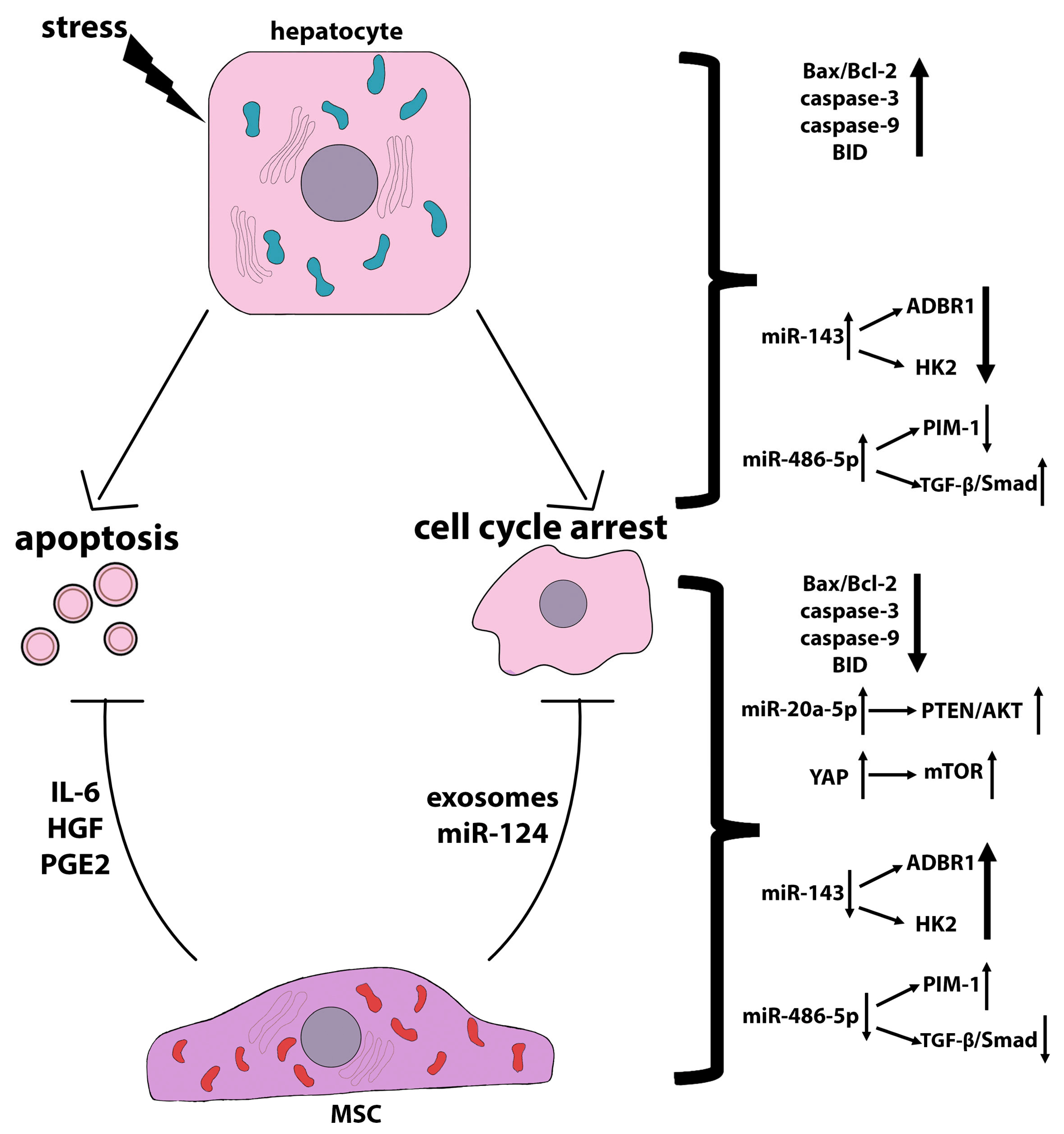
Hydrogen peroxide (H2O2)-induced oxidative stress in LO2 cells resulted in an increased number of apoptotic cells and reduced mitochondrial membrane potential, as well as an increased percentage of the cell population in the G0/G1 phase and a sharp decrease in the S and G2/M phases, showing that H2O2 induces cell cycle arrest in LO2 cells. In addition, H2O2 significantly reduced the Bcl-2/Bax ratio and markedly increased BMF expression. The level of miR143 was significantly elevated in cells treated with H2O2. The targets of miR143 could presumably be ADRB1 (beta-1-adrenergic receptor) and HK2 (hexokinase 2), the expression of which was significantly reduced in H2O2-treated cells. MiR143 inhibited Bcl-2 expression and activated Bax and caspase-9, consequently activating the endogenous mitochondrial pathway that promotes cell apoptosis [173]. A study by Zhang et al. [174] showed that in osteosarcoma cells, Bcl-2 could be the target gene for miR143. In addition, in prostate cancer, miR143 was shown to target HK2 [175] and ADRB1 and play an important role in proliferation and apoptosis during H2O2 injury. It was shown that a decreased level of HK2 in hepatocellular carcinoma cells significantly inhibited glucose flux to pyruvate and lactate. Decreased glycolysis was associated with increased oxidative phosphorylation [176]. MiR143 can directly target the HK2 3’-UTR, thereby suppressing glucose uptake, lactate production, cellular G6P and ATP levels, and liver cell proliferation upon H2O2 injury. It was demonstrated that the hUC-MSC-CM reversed the negative effects of H2O2 on normal hepatocytes. In particular, hUC-MSC-CM reduced apoptosis in H2O2-treated LO2 cells by half, restored the mitochondrial membrane potential, increased the Bcl-2/Bax ratio, and reduced BMF expression within 3 h. Thus, hUC-MSC-CM had a pronounced anti-apoptotic effect under oxidative stress conditions, abolished cell cycle arrest, and promoted the transition of cells to the G1/S phase. In addition, hUC-MSC-CM reduced the miR143 level in cells subjected to oxidative stress and, accordingly, increased the levels of HK2 and ADRB1 [173]. The MSC-CM contains a large number of cytokines, promoting cell growth after H2O2 removal [177,178]. It can also be hypothesized that the hUC-MSC-CM affects H2O2-damaged LO2 cells by modulating glycolysis. In short, the promotion of LO2 cell proliferation and protection from apoptosis may occur through the regulation of the miR143/HK2 axis and the restoration of ADRB1 expression after hUC-MSC-CM treatment [173]. In the same model of oxidative stress, another miR was found, the level of which increased after LO2 cell treatment with H2O2, namely miR-486-5p [179]. The proviral integration site for Moloney murine leukemia virus kinase 1 (PIM1) was verified as the miR-486-5p target; accordingly, PIM1 expression significantly decreased with an increase in the level of miR-486-5p in LO2 cells subjected to oxidative stress; the TGF-β/Smad pathway was also activated in these cells. The HUC-MSC-CM attenuated damage caused by oxidative stress in LO2 cells by inhibiting miR-486-5p, upregulating PIM1, and blocking TGF-β/Smad signaling. Thus, the UCB-MSC-CM inhibits TGF-β/Smad signaling through the miR-486-5p/PIM1 axis [179].
In addition to reducing the expression of miRNAs involved in the progression of pathological states of hepatocytes, MSCs upregulate a number of miRNAs that have anti-apoptotic and pro-proliferative functions. However, the signals transmitted from MSCs to upregulate these miRNAs remain unclear. For example, Zhou et al. [180] showed that hUC-MSCs reduce apoptosis and enhance the proliferation of primary mouse hepatocytes isolated from bile duct ligation (BDL) livers by upregulating miR-148-5p, which plays an important role in hepatocyte differentiation and liver regeneration. It has been shown that miR-148 expression is downregulated in liver damage, and its upregulation leads to a pronounced therapeutic effect [181]. Human BM-MSC-derived extracellular vesicles (EVs) reduced hepatocyte apoptosis and promoted cell proliferation by upregulating miR-20a-5p, which targeted the the PTEN/AKT signaling pathway in mice with LPS/D-GalN-induced ALF [182].
The mechanisms of anti-apoptotic action of MSCs on damaged hepatocytes are shown in Figure 3.
In addition to regulating hepatocyte miRNAs, MSCs also produce various miRNAs that may mediate their regenerative effects on hepatocytes. The delivery of miRNAs from one cell to another is carried out by extracellular vesicles, in particular, by exosomes. Human chorionic plate-derived MSCs (CP-MSCs) were shown to suppress the development of CCl4-induced liver fibrosis in rats due to the increased content of miRNA-125b in exosomes, which inhibited Hedgehog (Hh) signaling in hepatic stellate cells, thereby slowing down fibrogenesis, and stimulated the accumulation of progenitor cells in the liver, inducing regeneration [183]. MiR-181-5p produced by adipose-derived MSCs was shown to exert the anti-fibrotic effect by suppressing the activation of hepatic stellate cells [184]. In a partial rat hepatectomy model, human umbilical cord blood mesenchymal stem cell (hUCB-MSC)-derived exosomal miR-124 promoted liver regeneration by enhancing hepatocyte proliferation through the downregulation of Foxg1 [185].
The detailed mechanisms of the anti-apoptotic effect of MSCs on hepatocytes remain unclear. However, it has been undeniably established that soluble hepatotrophic factors, such as HGF, IL-6, PGE2, etc., as well as extracellular vesicles loaded with protective bioactive molecules, can be considered as MSC apoptosis-reducing signals.

3.2. Regenerative Effects of MSCs

Liver regeneration is a well-studied process. The most common model for deciphering the mechanisms of liver regeneration is partial hepatectomy, which was extensively used to investigate the temporal and spatial processes involved in restoring organ mass, architecture, and function. In most cases, after partial hepatectomy, the liver is restored due to hepatocyte hyperplasia and hypertrophy. However, in real clinical situations, liver surgery does not lead to complete organ regeneration due to pre-existing pathological abnormalities; it entails a number of undesirable events leading to complications, including fibrosis, severe oxidative stress, etc. In this regard, MSCs may become valuable assistants in triggering full-fledged liver regeneration after surgical interventions, as demonstrated in many experimental studies involving the models of partial hepatectomy [70,186,187], liver transplantation [188,189], ischemia/reperfusion injury (IRI) [190,191], bile duct ligation (BDL) [192], etc. In all these studies, MSCs or MSC-derivatives have shown a pro-regenerative potential to reduce apoptosis of hepatocytes, increasing their proliferation and metabolic activity, and suppress inflammation in the liver by reducing infiltrating pro-inflammatory immune cells and inhibiting fibrosis by abolishing the activation of hepatic stellate cells. However, the majority of the MSC-derived factors associated with such a wide range of regeneration-related events remain unknown.
In a number of studies, it has also been demonstrated that MSCs or MSC-derivatives, inducing in liver recovery after surgery, do not always directly affect hepatocytes, or they did not have positive effects on the restoration of liver function at all. In a mouse model of partial hepatectomy, MSCs were shown to exert their therapeutic effect through the secretion of exosomes. However, it was shown that MSC-derived exosomes were not taken up by hepatocytes but were captured by macrophages, causing their polarization into M2 macrophages and thereby inhibiting inflammation [193]. The allogeneic MSC transplantation in a large animal model of repeated biliary obstruction followed by partial hepatectomy promoted the growth of liver tissue without any effect on liver function [194]. After partial hepatectomy of a fibrotic or cirrhotic liver, MSCs injected locally into the liver, but not those injected systemically, had a regenerative effect [195], although most studies have shown the therapeutic effects of MSCs and their derivatives, regardless of the transplantation route [196].
The secretome derived from the adipose tissue-derived MSCs pre-conditioned with the secretome taken from the damaged hepatocyte culture (MSC-iCM) showed higher therapeutic potential compared with the naïve secretome after injection into mice with the thioacetamide-induced hepatic failure and those with partial hepatectomy. Proteomic analysis showed that the MSC-iCM contained a large amount of antioxidant factors, in particular peroxiredoxin-1. After incubation with the MSC-iCM, damaged hepatocytes showed the highest expression of liver regeneration markers (HGF, VEGF, PCNA, and p-ERK) and the lowest expression of apoptotic and inflammation markers (PARP, BIM, BAX, and F4/80) [197]. Peroxiredoxin-1 catalyzes the reduction of H2O2 and alkyl hydroperoxide and thus protects cells from the attack of free radicals. Presumably, peroxiredoxin-1, produced by MSCs, interacts with TLR4 on the surface of hepatocytes, leading to an increase in the expression of the hepatoproliferative marker (p-ERK), thereby stimulating proliferation. Peroxiredoxin-1 levels also increased in damaged hepatocytes treated with the MSC-iCM [197]. The delivery of another antioxidant, glutathione peroxidase 3, by engineered human induced pluripotent stem cell-derived MSCs into the liver of mice with IRI and subsequent partial hepatectomy significantly suppressed hepatocyte senescence and apoptosis [198]. Together, these results demonstrate that MSCs can be a source of antioxidants to reduce oxidative stress in hepatocytes.
An interesting crosstalk mechanism between stressed hepatocytes and MSCs was revealed by Choi et al. using the bile duct ligation model, which is the most common surgical model used to induce cholestasis injury in rodents, causing proliferation of the biliary tract epithelial cells and oval cells and leading to inflammation, fibrosis, and hepatocyte apoptosis [199]. Human chorion plate-derived MSCs (CP-MSCs) have been shown to be effectively engrafted into an injured rat liver and facilitate the resolution of fibrosis, promoting liver regeneration after transplantation in rats undergoing bile duct ligation. In the in vitro experiments, the authors found reciprocal paracrine regulation of PRL-1 between rat hepatocytes treated with lithocholic acid (LCA), a bile acid used to mimic cholestasis, and CP-MSCs in an indirect co-culture system. The phosphatase of the regenerating liver (PRL) family is a group of protein tyrosine phosphatases (PTPs) that consists of three closely related members, PRL-1, PRL-2, and PRL-3, which have 76% to 87% amino acid identity [200]. PRL-1 is not a factor critical for liver development, postnatal growth, and hepatocyte differentiation; however, this tyrosine phosphatase is required to regulate the timing of liver regeneration after partial hepatectomy [201]. PRL-1 is an early growth response gene that is activated shortly after partial hepatectomy and triggers a cascade of regenerative events via the cytokine-independent pathway [202]. PRL-1 promotes G1/S progression by modulating the expression of cell cycle regulators via the activation of the AKT/STAT3 signaling pathway [203]. PRL-2 is expressed at high levels in several cell types, including hippocampal pyramidal neurons, ependymal cells, cone and rod photoreceptor cells, endocardium, vascular and bronchial smooth muscle, and collecting duct epithelium in the kidney. In parenchymal cells of the liver and pancreas, its expression is minute [203]. The role of PRL-2 in vivo [204] and in vitro [205] is modulation of the bioenergetic functions. Knockdown of PRL-2 leads to a significant decrease in intracellular levels of ATP and glutamine uptake. Subsequently, PRLs were also shown to have oncogenic roles. For example, overexpression of PRL-1 has been observed in several types of cancer and is correlated with poor prognosis, including gastric, intrahepatic, and prostate cancer [206,207,208]. PRL-3 is overexpressed in murine melanoma cells, where it plays a key role in cell motility and metastasis [209]. It also promotes endothelial cell migration in vitro [210]. PRL-3 is a mediator of metabolic reprogramming of cancer cells. PRL-3 increases glycolysis in various types of cancer, including myeloma [211,212] and colorectal cancer [213]. Luo et al. [214] showed that PRL-1 overexpression is associated with increased MMP-2 and MMP-9 levels in HEK293 cells through the Src and ERK1/2 pathways. CP-MSCs that expressed PRL-1 promoted liver regeneration through microRNA expression and mitochondrial dynamics in a liver cirrhosis rat model [215]. In the aforementioned work [199], the authors showed that normal hepatocytes and CP-MSCs both express PRL-1. After the treatment of primary rat hepatocytes with lithocholic acid, the expression level of PRL-1 decreased, while co-cultivation with CP-MSCs resulted in the restoration of its expression and subsequent regulation of MSC homing through RhoA-mediated ROCK1 signaling. PRL-1 inhibition by siRNA-PRL-1 reduced CP-MSC migration to damaged primary rat hepatocytes by reducing RhoA and ROCK1 levels as well as by decreasing MMP-2 levels. These results indicate that the expression of the Rho family and MMPs is regulated by PRL-1, which may be a key regulator for chemo-attractive CP-MSC migration (Figure 4). Genetically modified BM-MSCs overexpressing PRL-1 increased anaerobic mitochondrial metabolism in damaged hepatocytes, decreasing cytoplasmic lactate and increasing mitochondrial lactate, which ultimately led to increased ATP synthesis and hepatocyte repair [216]. The scheme shown in Figure 4 demonstrates the PRL-1-dependent mechanism of the MSC protective action.
Thus, hepatocyte proliferation, the process underlying liver regeneration, can be launched by the interaction of MSCs with damaged hepatocytes via the secretion of hepatotrophic factors, exosomes, and miRNAs. Reciprocal paracrine stimulation occurring between damaged hepatocytes and MSCs can lead to increased homing of the latter to the liver and the enhancement of their regenerative abilities.

3.3. Anti-Ferroptosis and Anti-Pyroptosis Effects of MSC

Ferroptosis is a type of “programmed necrosis” that was initially identified in cancer cells as nonapoptotic cell death mainly characterized by iron overload and lipid peroxidation [217]. Later, it was shown that cell death due to ferroptosis is characteristic not only to tumor cells, but it is also involved in the pathogenesis of certain diseases, such as neurodegeneration [218] and acute renal failure [219]. However, the role of ferroptosis in the development and progression of liver diseases remains poorly explored [220]. A number of studies have shown that ferroptosis may be a novel type of cell death associated with liver diseases, such as viral hepatitis, drug-induced liver injury [221], alcoholic liver disease [222], non-alcoholic fatty liver disease (NAFLD) [223], non-alcoholic steatohepatitis (NASH) [224], and hemochromatosis [225]. Tsurusaki et al. [224] identified the type of cell death that occurs at the earliest stage of NASH. NASH is a severe form of NAFLD characterized by the accumulation of lipid droplets in hepatocytes, hepatocyte death, inflammatory cell infiltration, and fibrosis. Earlier studies have shown that apoptosis and necroptosis are major contributors to the pathogenesis of NASH. However, the mechanisms underlying the transition from simple steatosis to steatohepatitis remained unclear until recently. In a mouse choline-deficient, ethionine-supplemented diet model of steatohepatitis, the authors proved that the onset of the disease was associated with massive necrosis of hepatocytes around the portal vein, which preceded apoptosis. They established that the necrotic death of hepatocytes is nothing more than ferroptosis and that ferroptosis is the initial cell death that induces inflammation at the onset of steatohepatitis [224]. From these results, it can be concluded that ferroptosis may be involved not only in the progression of liver diseases but also in their initiation.
As mentioned above, ferroptosis is a specific paradigm of cell death resulting from iron overload, reactive oxygen species generation, and lipid peroxidation, leading to antioxidative system dysfunction and, ultimately, cell membrane damage [226]. Ferroptosis is a form of regulated cell death driven by perturbation of the glutathione (GSH)-dependent lipid hydroperoxide scavenging network [227]. Lipid peroxidation is regulated by the XC− system, which transports glutamate out of the cell in exchange for the transport of cystine, an important precursor for the synthesis of glutathione (GSH), into the cell [228]. The XC− system consists of a light-chain subunit (SLC7A11/xCT) and a heavy-chain subunit (SLC3A2). SLC7A11 is a multi-pass transmembrane protein that operates as the cystine/glutamate antiporter. SLC7A11 transports cystine into the cell, where it is rapidly converted to cysteine, which is the rate-limiting precursor of GSH. Thus, SLC7A11 plays an important role in providing cysteine for glutathione biosynthesis. GSH is important for glutathione peroxidase 4 (GPX4), which protects cells from ferroptosis by converting toxic lipid hydroperoxides to non-toxic fatty alcohol [229].
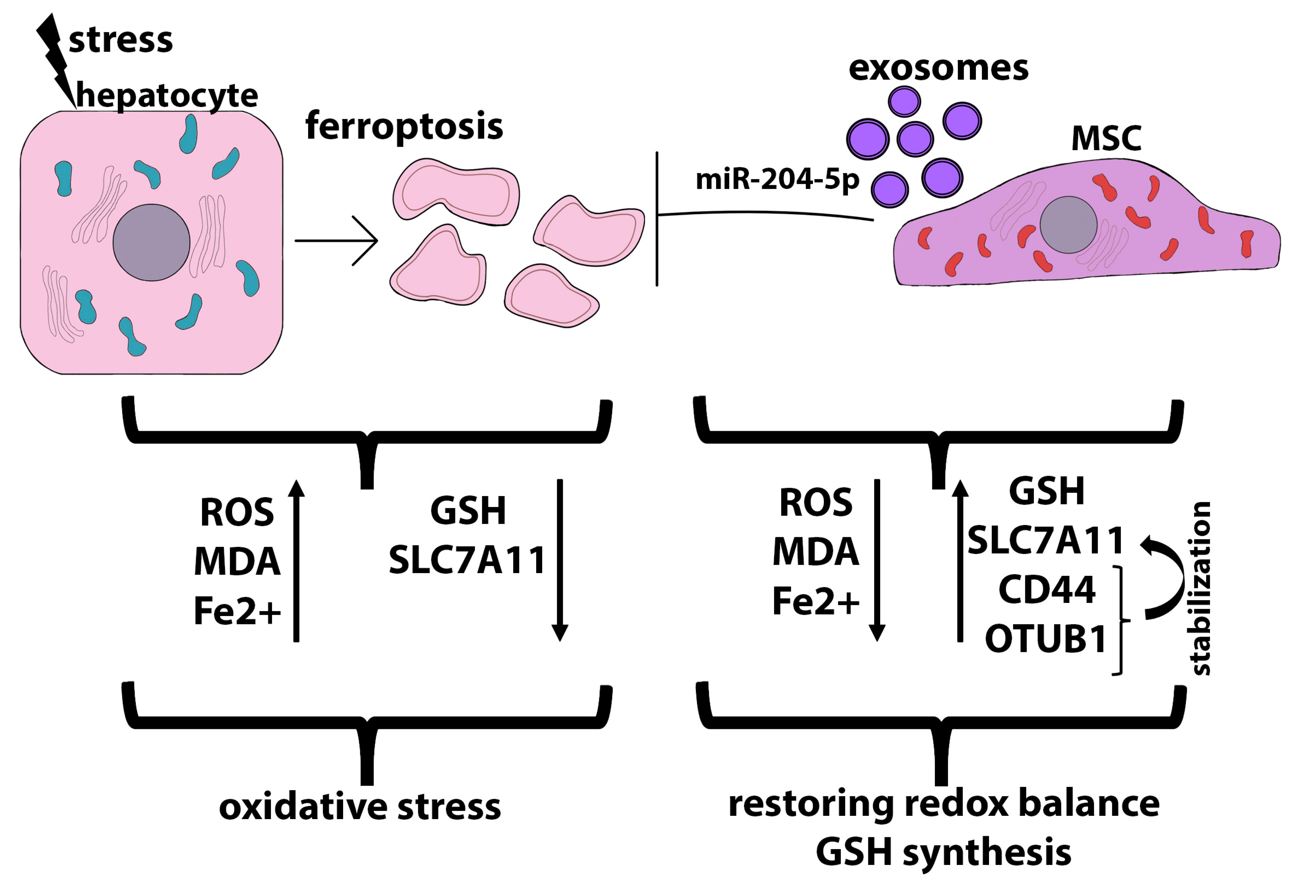
Ferroptosis is a critical driver of CCl4-induced liver injury in mice, a commonly used in vivo model for acute liver injury [230]. MSCs, as well as MSC-derived exosomes, reduced ROS accumulation, downregulated malondialdehyde (MDA) levels, and increased SLC7A11 expression in damaged hepatocytes, thereby exerting a pronounced antioxidant effect, when indirectly co-cultured with primary mouse hepatocytes obtained from the livers of animals with CCl4-induced acute liver injury. At the same time, the MSC-Exo-induced recovery of SLC7A11 was accompanied by the increased expression of CD44 and OTUB1 on hepatocytes. OTUB1 is an ovarian tumor deubiquitinase (OTUB) family member that regulates the SLC7A11 stability through direct protein interaction [231]. CCl4 increased SLC7A11 ubiquitinylation, thereby abolishing its stabilization and leading to its downregulation, while MSC-Exo downregulated SLC7A11 ubiquitinylation, restoring its stabilization, which led to the upregulation of SLC7A11. Additionally, SLC7A11 was stabilized by MSC-Exo-mediated upregulation of CD44 and OTUB1 expression, both of which coprecipitate with SLC7A11 [230]. Several studies have shown that CD44 stabilizes SLC7A11 expression in the plasma membrane, thus promoting cystine uptake and GSH synthesis [228].
In a rat fatty liver transplantation model, exosomes derived from heme oxygenase 1-overexpressed human BM-MSCs were shown to abolish hepatocyte ferroptosis, improve liver function, inhibit Kupffer cell and T cell activation, and prevent long-term biliary fibrosis [232]. Exosome transplantation reduced Fe2+ levels and molecular markers of ferroptosis, such as Ptgs2 mRNA, 4-hydroxynonenal (4-HNE), and malondialdehyde (MDA), which were significantly elevated in animals without exosome treatment. The level of glutathione, an important antioxidant maintaining cellular redox homeostasis, also increased strongly after exosome transplantation. The key negative regulator of ferroptosis in this model was miR-204-5p, which is highly expressed in exosomes derived from heme oxygenase 1-modified human BM-MSCs. The miR-204-5p target is ACSL4, a key regulator of ferroptosis, whose expression was strongly reduced after treatment with exosomes. Simultaneously, the expression of GPX4, a critical protective enzyme that prevents ferroptosis, was upregulated [232]. That is, exosomes obtained from heme oxygenase 1-modified human BM-MSCs, due to miR-204-5p, not only inhibit ferroptosis but also activate the antioxidant system. In the I/R myocardial injury model, it was shown that exosomes derived from hUCB-MSCs and loaded with exosomal miR-23a-3p inhibited ferroptosis by regulating iron metabolism independent of the GPX4 pathway [233]. The role of exosomes in ferroptosis-related pathogenic mechanisms in various diseases is described in detail in this review [234]. Figure 5 shows the scheme of hepatocyte protection from ferroptosis due to MSC and MSC derivatives.
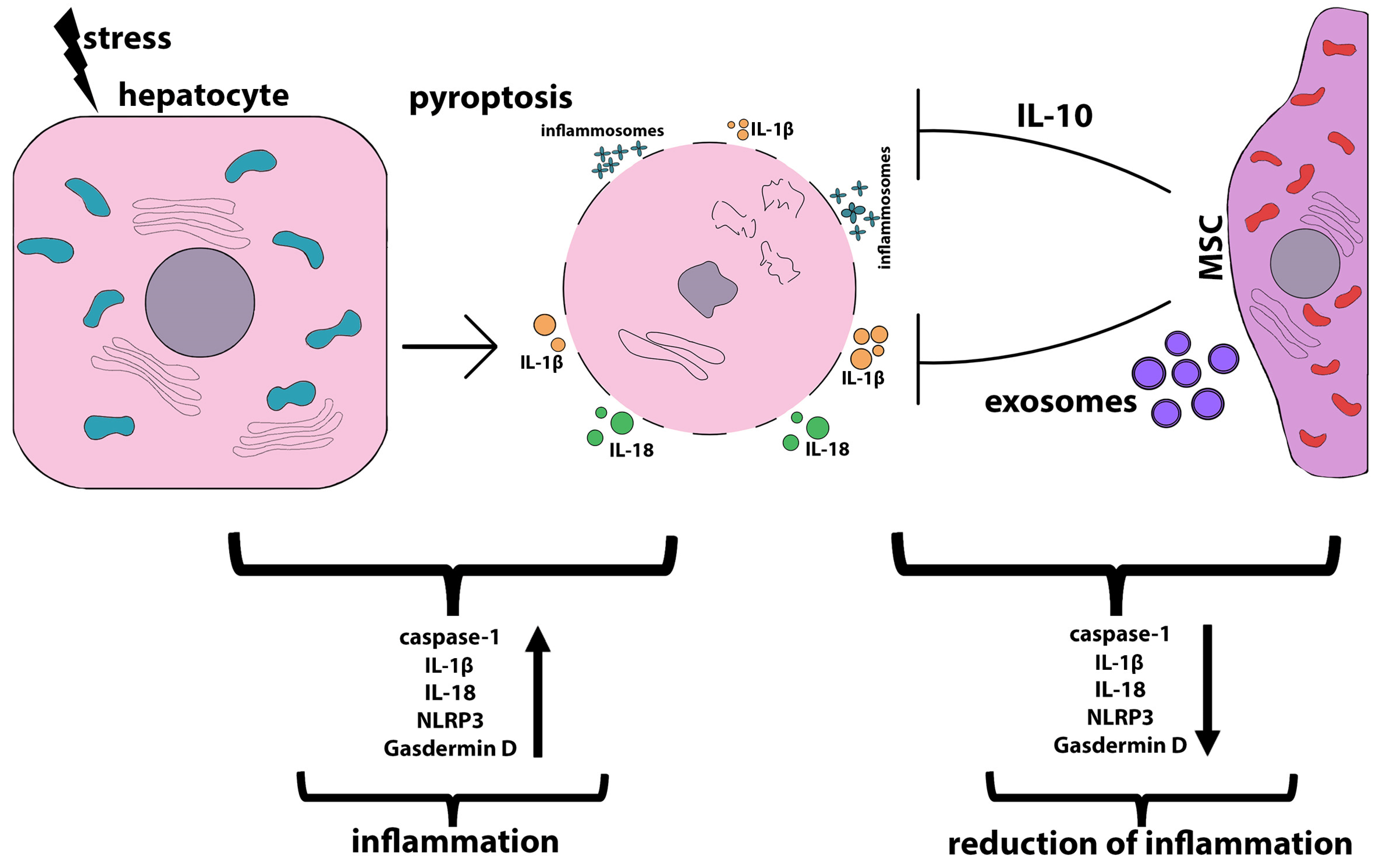
Pyroptosis, a rather recently characterized type of programmed cell death, plays an important role in various liver diseases. Pyroptosis is characterized by caspase-dependent (caspases 1, 4, 5, and 11) pore formation in the cell plasma membrane and the subsequent release of inflammatory mediators (interleukin-18, IL-1β). Inflammasomes are caspase activators in pyroptosis. Inflammasomes are multiprotein complexes that activate caspase 1 in response to stress signals. Cleaved caspase 1 mediates the maturation and secretion of IL-1β and IL-18 and pyroptotic cell death. The hallmarks of pyroptosis are plasma membrane hyperpermeabilization, cell swelling, rapid cell lysis, and the release of cytoplasmic contents and pro-inflammatory cytokines into the microenvironment. Initially, pyroptosis was considered an innate immune mechanism against intracellular bacteria. However, there is increasing evidence that it is also involved in sterile inflammation, such as that in acute or chronic liver diseases. More information about the mechanisms of pyroptosis and its role in liver diseases has been described in several recent review articles [235,236]. Inflammasomes are predominantly expressed by immune cells, although many studies have shown that hepatocytes undergoing pyroptosis due to liver damage release inflammasomes, which are taken up by other cells and thereby mediate inflammatory and pro-fibrogenic stress signals [237].
Recent studies have shown that inflammasome-mediated pyroptosis is a driver of inflammation and plays a critical role in the progression of liver pathologies, such as autoimmune liver diseases [238], liver fibrosis [239], and fatty liver diseases [240,241]. Therefore, pharmacological pyroptosis targeting may become an option for reducing inflammation in liver diseases. It has been found that MSCs or MSC derivatives are able to suppress inflammation by inhibiting pyroptosis [242,243]. In a mouse model of acute liver failure induced by D-galactosamine, the injection of mouse BM-MSCs significantly reduced the expression of the NLRP3 inflammasome protein and the production of pro-inflammatory cytokines associated with the progression of pyroptosis, IL-1β, and IL-18. Similar results were obtained with D-galactosamine-treated primary mouse hepatocytes after indirect co-culturing with mouse BM-MSCs, in which MSCs markedly reduced NLRP3-caspase 1 inflammasomes in hepatocytes. The main driver of the anti-pyroptosis effect of MSCs was shown to be the anti-inflammatory cytokine IL-10, which inhibits the expression of NLRP3, thereby negatively regulating inflammasome-associated pyroptosis and inflammation [244]. In a mouse model of CCl4-induced liver cirrhosis, a triggering role of pyroptosis in the progression of liver injury was also shown, as demonstrated by the high expression of gasdermin D (GSDMD), caspase 1, and NLRP3 and the pro-inflammatory cytokine IL-1β in the animals’ livers. GSDMD, the key pyroptosis executioner, was shown to be the only pyroptosis effector with pore-forming activity [245]. Stem cells isolated from human exfoliated deciduous teeth attenuated liver cirrhosis in animal models by inhibiting pyroptosis. In vitro experiments using the co-cultivation of mouse hepatocytes (mouse hepatic cell line NCTC 1469) treated with CCl4 with human exfoliated deciduous teeth-derived stem cells in the transwell system showed that the pyroptosis-inhibiting ability is associated with decreasing ROS levels and normalization of the membrane mitochondrial potential in hepatocytes induced by soluble factors produced by MSCs [246]. In a mouse model of acute liver failure induced by D-galactosamine (D-GalN) and lipopolysaccharide (LPS), transplantation of human UC-MSCs was shown to inhibit not only apoptosis and inflammation but also pyroptosis by suppressing NLRP3 inflammasome activation, IL-1β maturation, and caspase 1 cleavage [88]. Palmitic acid-treated HepG2 (an in vitro model of fatty liver disease) significantly increased the expression of the characteristic markers of pyroptosis, including NLRP3, GSDMD/-N (activated form of GSDMD), caspase 1, p20 (activated form of caspase 1), IL-1β, and IL-18, while indirect co-cultivation with MSCs significantly reduced their expression and suppressed pyroptosis [247]. Figure 6 shows the scheme of hepatocyte protection from pyroptosis by MSC and MSC derivatives.
Thus, both in liver injuries in vivo and in the course of indirect co-cultivation with hepatocytes subjected to pyroptosis-inducing stress in vitro, MSCs are able to protect hepatocytes from pyroptosis, most likely due to the secreted factors. For example, human UC-MSCs abrogated traumatic brain injury-induced pyroptosis in vivo and lipopolysaccharide/ATP-induced BV2 microglial pyroptosis in vitro due to the secretion of anti-inflammatory factors, such as tumor necrosis factor-stimulated gene 6 (TSG-6) [248]. The inhibition of pyroptosis in dextran sulfate sodium-induced ulcerative colitis in mice by transplantation of hair follicle-derived MSCs is critically associated with exosomes produced by mesenchymal cells since the inhibition of exosome secretion completely abolishes the protective effects of the MSCs in vivo and in vitro. Presumably, MSC-derived exosomes might inhibit pyroptosis through tumor necrosis factor-related apoptosis-inducing ligand (TRAIL) signaling and the IFN-γ pathway [249]. Also, many studies have shown that miRNAs transported by MSC-derived exosomes play a role in the suppression of pyroptosis caused by inflammation or other stimuli [250].

3.4. MSCs Protection from Lypotoxicity and ER Stress

Hepatocytes are the main cells in the body that carry out lipid metabolism. Normal lipid metabolism in hepatocytes includes their synthesis, uptake, efflux, and oxidation. These highly regulated processes maintain homeostatic intracellular lipid levels. Fatty acid transport into hepatocytes requires the action of the fatty acid-binding protein (FABPpm) and fatty acid translocase (FAT)/CD36. Some of the incorporated fatty acids undergo β-oxidation in the mitochondria by carnitine palmitoyltransferase 1a (CPT1A) [251] and mitochondrial trifunctional protein, which catalyzes the last three steps of the mitochondrial beta-oxidation of long-chain fatty acids, hydroxyacyl-CoA dehydrogenase/3-ketoacyl-CoA thiolase/enoyl-CoA hydratase alpha/beta subunit (HadhA/B) [252], followed by ATP generation. Another part of incorporated fatty acids is stored in hepatocytes in the form of fat droplets formed due to the action of endoplasmic reticulum-bound stearoyl-CoA desaturase 1 (SCD1), the key determinant of triglycerides biosynthesis pathway [253]; acetyl-CoA carboxylase 1 and 2 (ACC1 and ACC2), which regulate hepatic fat storage [254]; and fatty acid synthase (FAS), which catalyzes the de novo synthesis of fatty acids producing fat for the storage of energy when nutrients are present in excess [255].
Lipotoxicity associated with abnormal accumulation of lipids in hepatocytes underlies the pathogenesis of liver diseases such as non-alcoholic fatty liver disease (NAFLD) and non-alcoholic steatohepatitis (NASH) [256]. It was also shown that within a short time after liver surgery, the aberrant metabolism and abnormal accumulation of lipids are triggered in hepatocytes, eventually leading to liver injury and liver failure [257].
The abnormal accumulation of lipids in hepatocytes can occur due to stressful conditions, such as overexposure to saturated fatty acids or insulin resistance, which leads to the triggering of several pathological signaling mechanisms, including the accumulation of ROS, ER stress, and chronic inflammation. The endoplasmic reticulum (ER) performs important functions related to the synthesis, folding, and transport of proteins and plays a critical role in lipid synthesis and Ca2+ homeostasis [258]. Prolonged excessive uptake of saturated fatty acids leads to an imbalance between ER protein loading and folding, leading to the hyperactivation of the unfolded protein response (UPR) and the subsequent activation of the cell death pathway [259]. Disruption of ER calcium balance is one of the initial and key events of cell death induced by ER stress [260]. Impaired cellular Ca2+ homeostasis is a hallmark of many ER-related diseases and a key trigger for NAFLD/NASH. The sarcoplasmic/endoplasmic reticulum Ca2+ ATPase (SERCA) transports Ca2+ from the cytosol to the ER lumen, maintaining resting calcium concentrations. Saturated fatty acids inhibit the activity of SERCAs, which leads to the release of calcium from the ER lumen and a high calcium load in the cytosol, ultimately leading to the disruption of ER homeostasis and cellular function [261].
Several studies have shown that MSCs attenuate lipotoxicity in hepatocytes, which is associated with aberrant lipid accumulation and lipid metabolism disorders. However, the mechanisms of the attenuation of lipotoxicity in hepatocytes induced by MSCs remain poorly understood. Using a mouse natural aging model, Ling et al. [262] demonstrated that hUCMSC-exos significantly reduced aging markers and genomic instability in aging livers. Metabolome studies found that hUCMSC-exos reduced lipids associated with lipotoxicity and inflammation, including saturated glycerophospholipids, palmitoyl-glycerols, and eicosanoid derivatives. Using phosphoproteomics approaches, the authors also showed a decrease in the phosphorylation of the metabolic enzyme propionate-CoA ligase. In addition, it was shown that hUCMSC-exos significantly reduced the phosphorylation of proteins involved in nuclear transport and cancer signaling in hepatocytes, including heat shock protein HSP90-beta (Hsp90ab1) and nucleoprotein TPR, and increased the phosphorylation of proteins involved in the intercellular communication, such as calnexin and PDZ domain-containing protein 8 (Pdzd8) [262].
In the HepG2 cell line, palmitic acid (PA) was shown to reduce viability and increase apoptosis and lipotoxicity [247]. PA, a long-chain saturated fatty acid, undergoes β-oxidation in the mitochondria, and PA-induced lipotoxicity can cause mitochondrial dysfunction, which indicates bioenergetics disruption [263]. PA-induced lipotoxicity in HepG2 was manifested by the accumulation of massive fat droplets and an increase in the triglyceride level. The amount of the sterol regulatory element binding protein 1c (SREBP1c) (truncated SREBP1), which is a master transcription factor controlling lipogenesis and lipid uptake, was significantly increased in PA-treated hepatocytes. The indirect co-cultivation of PA-treated HepG2 with MSCs markedly inhibited the accumulation of fat droplets, reduced triglyceride levels, and decreased SREBP1c expression in hepatocytes. Thus, MSCs protected HepG2 from PA-induced lipid accumulation [247]. PA-induced lipotoxicity in hepatocytes resulted in the upregulation of a gene associated with fatty acid transport, CD36l the lipid synthesis genes, including PPARalpha, PPARgamma, FASN, and SREBP1c; as well as CPT1A, a mitochondrial enzyme responsible for the transport of fatty acids into the mitochondria for β-oxidation, while MSCs reduced their expression. Under lipotoxic conditions, abnormal morphological changes in the ER occurred in HepG2, including cistern enlargement and ribosome loss, which were not manifested during co-cultivation with MSCs. Compared with PA-treated monocultures, the co-cultures of HepG2 with MSCs showed reduced expression of typical ER stress marker proteins, including BiP, ATF6, ATF4, p-eIF2a, and CHOP, and genes associated with the UPR pathway, such as GRP78, FKBP11, and GRP94. MSCs restored the calcium balance between the ER and the cytosol, which was disturbed by the treatment of hepatocytes with palmitic acid by increasing the expression and activation of SERCA [247].
Sirtuin 1 (SIRT1) is an NAD+-dependent deacetylase that is a master regulator of the transcriptional network that controls hepatic lipid metabolism. Under energy depletion conditions, SIRT1 deacetylates and alters the expression of key transcriptional regulators involved in hepatic lipogenesis, fatty acid β-oxidation, and cholesterol/bile acid metabolism [264]. Human UC-MSC-CM improved mitochondrial function and reduced inflammation and apoptosis in the PA-induced steatosis in LO2 cells by reducing the levels of the pro-inflammatory cytokines TNFα, IL1β, and IL6 and increasing the protein levels of SIRT1, PGC1α, NRF1, and TFAM, the key molecules in maintaining mitochondrial function. Since after SIRT1 silencing in hepatocytes, the effect of the UC-MSC-CM on the levels of SIRT1, PGC1α, NRF1, and TFAM was significantly reduced and ROS production and the levels of pro-inflammatory cytokines increased, the authors suggested that SIRT1 mediates the protective effects of the UC-MSC-CM [265]. SIRT1 regulates cellular metabolism and mediates the deacylation of target proteins, such as peroxisome proliferator-activated receptor gamma coactivator 1α (PGC1α), which is involved in fatty acid oxidation and mitochondrial function [266]. SIRT1 overexpression effectively reduces obesity and insulin resistance in NAFLD rodents [267]. In patients with obesity or NAFLD, SIRT1 levels are significantly reduced in the plasma and in the liver [268]. Thus, SIRT1 and SERCA can serve as targets in the treatment of fatty liver diseases.
PA-treated mouse hepatocytes accumulated lipid deposits, in particular triglycerides and cholesterol, similar to the situation seen in NAFLD. After treating the hepatocytes with murine adipose-derived MSC (AMSC)-EVs, marked reduction of lipid deposits, normalization of lipid metabolism, and a reduction in liver fibrosis marker proteins (α-SMA, COL1A1, and TGF-β1) was observed. AMSC-EVs efficiently accumulated in hepatocytes over time and not only delivered miR-223-3p to them but also induced the upregulation of this miRNA in the recipient hepatocytes [269]. There were reports showing the involvement of granulocyte-enriched miR-223 [270] in chronic liver injury, such as non-alcoholic steatohepatitis (NASH) and acute hepatitis [271]. An miR-223 analog, miR-223-3p, improves acute and chronic hepatitis through inhibition of NLRP3 inflammasome activation [272]. BM-MSC-derived extracellular vesicles carrying miR-223-3p alleviate autoimmune liver disease by reducing the inflammatory response [273]. miR-223-3p has also been shown to have a mitigating effect against inflammatory diseases by targeting the inhibition of the E2F transcription factor 1 (E2F1) [274]. In turn, E2F1 modulates cholestatic liver fibrosis, highlighting its promise as a fibrogenic marker [275].
In a rat model, 90% hepatectomy induced acute steatosis, which was attenuated after the transplantation of rat BM-MSCs [276]. The expression analysis of the genes associated with different aspects of lipid metabolism, including lipid decomposition (CPT1a, Hadha, and Hadhb), lipid synthesis (Scd1, Acaca, and FASN), and lipid transport (CD36), showed that BM-MSCs mainly influenced the processes of lipid decomposition and metabolism but did not influence lipid synthesis and transport. The expression of lipolysis-related proteins, including peroxisome proliferator-activated receptor alpha (PPARα), CPT1a, and carnitine octanoyltransferase (CROT), increased after MSC transplantation, although there were no significant changes in the expression of lipid synthesis-related proteins, such as ACC and FASN. The expression of the sterol-regulatory element-binding proteins (SREBP1 and SREBP2) was also restored, although the expression of the FASN downstream protein did not change significantly. These results can be explained by the fact that BM-MSCs reduced lipid accumulation and restored lipid metabolism. PPARα, the key transcription factor regulating fatty acid β-oxidation, was upregulated after BM-MSC transplantation, showing that BM-MSC transplantation can improve fatty acid β-oxidation function. BM-MSC transplantation activated the mTOR pathway, and its activation played a critical role in liver recovery after failure caused by partial hepatectomy. The mTOR pathway may also be involved in the effects of MSCs on mitochondria and lipid metabolism. IL-10 was identified as the key BM-MSC-produced factor determining their protective action on hepatocytes subjected to lipotoxicity caused by 90% hepatectomy, as the depletion of this cytokine in BM-MSCs abolished the therapeutic effect [276]. IL-10 is a cytokine that regulates the growth and differentiation of innate immune cells. IL-10 can control cell proliferation, apoptosis, angiogenesis, and inflammation [277,278]. IL-10 is associated with the occurrence of lipid metabolism disorders [279]. IL-10 provides a key link between inflammatory factors and lipid metabolism. IL-10 mainly affects genes associated with lipid catabolism (CPT1a, Hadha, and Hadhb) and proteins associated with lipid β-oxidation (PPARα, CPT1a, and CROT). These results suggest that MSC-derived IL-10 is an important cytokine improving mitochondrial damage in partial hepatectomy liver failure. Also, IL-10 depletion inhibits mTOR pathway activation after MSC transplantation. Thus, MSCs can improve lipid metabolism, mainly by secreting IL-10 and influencing mitochondrial function [276].
MSCs ameliorate lipotoxic kidney injury via a novel microenvironment-dependent paracrine HGF/c-Met signaling mechanism to suppress ER stress and its downstream pro-inflammatory and pro-apoptotic consequences [280]. MSCs were able to attenuate endothelial lipotoxicity by inhibiting ER stress and the endothelial-to-mesenchymal transition by the secretion of stanniocalcin-1 (STC-1) [281].
The above-described data suggest that MSCs can offset lipotoxicity by secreting soluble factors, such as IL-10 or miR-223-3p, which inhibit ER stress and normalize mitochondrial function with the subsequent restoration of lipid metabolism.
There are various ways to increase the therapeutic activity of MSCs, including hypoxic conditions, 3D culturing, pre-treatment of mesenchymal cells with pro-inflammatory cytokines, lipopolysaccharides, and chemical agents [282]. All these methods lead to changes in the MSC secretome, promoting more pronounced immunomodulatory, regenerative, and proangiogenic properties. For example, hypoxic conditions cause the upregulation of prostaglandin E synthase (PTGES) in cultured MSCs, resulting in enhanced PGE2 production, which promotes the switch of macrophages to the anti-inflammatory M2 phenotype. Also, hypoxia upregulates miR120 in MSCs, which reduces hepatocyte apoptosis [283]. In mice, after radical hepatectomy, the hypoxia-induced upregulation of VEGF in transplanted MSCs promotes hepatocyte proliferation and liver regeneration [284]. The conditioned media obtained from the cultures of the adipose-derived stem cells pretreated with LPS contained higher levels of cytokines involved in liver regeneration, including interleukin-6 (IL-6), tumor necrosis factor-alpha (TNF-α), hepatocyte growth factor, and vascular endothelial growth factor. These conditioned media had a more pronounced supportive effect on mouse hepatocytes treated with thioacetamide, increasing their viability compared with the conditioned media obtained from the cultures of MSCs not treated with LPS. In vivo, in partially hepatectomized mice, the LPS-CM significantly enhanced liver regeneration, as manifested by a decrease in serum levels of aspartate transaminase and alanine transaminase [285]. Icariin-treated hUMSCs increased the antioxidant activities in the liver and prevented fibrosis progression in CCl4-induced chronic liver injury in mice [286].

3.5. Mitochondrial Transfer and Antioxidant Effects of MSCs

The main function of the mitochondria is to produce energy in the form of ATP. Furthermore, mitochondria are producers of superoxide anion radical (O2•−), from which reactive oxygen species (ROS) are formed. ROS are essential for regulating cell signaling and maintaining redox homeostasis [287]. Redundant superoxide anion radical (O2•−) and ROS can cause oxidative stress. Mitochondria contain antioxidant systems to quench oxidative stress. For example, manganese superoxide dismutase (MnSOD) converts O2•− to H2O2 and molecular oxygen. Mitochondrial antioxidants, including GSH and thioredoxin, are able to buffer H2O2. The fission and fusion of mitochondria are required for the cell cycle and apoptosis progression as well as the control of their own functional integrity [288]. Together with the ER, mitochondria are involved in the storage and buffering of calcium. An elevated mitochondrial Ca2+ level may trigger cell death, including apoptosis, necrosis, and autophagy [289]. As described above, both the ER and mitochondria regulate lipid metabolism. Mitochondria serve as a site for the β-oxidation of fatty acids, resulting in the generation of acetyl-CoA [290]. In hepatocytes, mitochondria perform unique functions, including the regulation of gluconeogenesis, ammonia detoxification, and anabolic pathways [291]. Therefore, the disruption of the mitochondria functions in hepatocytes leads to general damage to the whole organism’s homeostasis. The most common types of mitochondrial dysfunction in liver diseases include the enhancement of the oxidative processes and/or impairment of the antioxidant mechanisms, leading to oxidative stress [292]. Mitochondrial dysfunction is observed in almost all liver pathologies, including alcoholic liver disease [293], NAFLD [294], viral hepatitis [295], fibrosis, and cirrhosis [296].
In various disease models, it has been shown that MSCs are able to restore the oxidant/antioxidant balance in damaged tissues and cells. Intravenously transplanted human UC-MSCs normalized mitochondrial function in a rat model of D-galactose-induced liver injury. This animal model is characterized by a high level of oxidative stress and mitochondrial dysfunction, which is manifested by a decrease in antioxidant capacity as well as the disturbance of bioenergetic functions, as evidenced by the loss of the mitochondrial membrane potential, increased ROS production, a decreased number of mitochondrial respiratory complexes, and decreased ATP. Human UC-MSCs restored the mitochondrial antioxidant system mainly by increasing the expression of SOD, GPx, and GSH via the activation of the nuclear factor erythroid 2-related factor 2/heme oxygenase-1 (Nrf2/HO-1) pathway [297]. In a mouse model of CCl4-induced liver damage, syngeneic BM-MSCs also had a pronounced antioxidant effect and restored mitochondrial function by increasing the gene expression levels of Hmox-1 (heme oxygenase-1), BI-1 (Bax inhibitor-1), HGF (hepatocyte growth factor), GST (glutathione transferase), and Nrf2 [298]. In the same model of CCl4-induced liver injury, human UC-MSC-Exo exerted antioxidant and anti-apoptotic effects [299]. In the above-described works, the mechanisms of the antioxidant effects of MSCs were not established, and the key molecular factors that were produced by MSCs and were capable of implementing their protective capabilities were not elucidated. That is to say, the question of the communication mechanisms between MSCs and damaged hepatocytes and their dysfunctional mitochondria remains open.
Some studies have suggested that such mechanisms can be mediated by MSC-produced extracellular vesicles. In the liver IRI model, human UC-MSC-derived extracellular vesicles protected against hepatic apoptosis by reducing neutrophil infiltration and alleviating oxidative stress in the liver tissue [300]. In the in vitro experiments, the authors found that human UC-MSC-EVs containing high levels of the MnSOD could transport this antioxidant enzyme into H2O2-damaged cells (LO2 cell lines), thereby increasing its levels in recipient cells and, as a result, restoring the oxidant/antioxidant balance. Importantly, siMnSOD-EVs derived from MSCs with quenched MnSOD expression did not have antioxidant or antiapoptotic effects in vitro or in vivo. Thus, MnSOD is the key protein mediating the protective functions of human UC-MSC-EVs by reducing oxidative stress and the apoptosis of hepatocytes in the IRI model [300]. Another antioxidant protein, glutathione peroxidase 1 (GPx1), has been shown to be delivered by MSC-derived exosomes into damaged hepatocytes, restoring mitochondrial function and reducing oxidative stress [301]. In the mouse model of CCl4-induced liver failure, human UC-MSC-derived exosomes reduced pro-inflammatory cytokines (G-CSF, IL-1α, IL-6, monocyte chemoattractant protein-1 (MCP-1), and TNF-α), and inhibited oxidative stress and apoptosis. In a model of H2O2 and CCl4-induced oxidative stress in LO2 cells, it was shown that human UC-MSC-derived exosomes abolished oxidative stress-induced apoptosis by inducing anti-apoptotic events, ERK1/2 phosphorylation, and Bcl2 expression and inhibiting the pro-apoptotic IKKB/NFkB/casp9/3 pathway. Eventually, it was proven that GPx1 antioxidant enzyme delivery into damaged hepatocytes or liver tissue by human UC-MSC-derived exosomes is the prime process underlying the reduction of oxidative stress and, as a result, leading to the prevention of hepatocyte apoptosis in vitro and in vivo [301].
A reduction in oxidative stress and restoration of mitochondrial function in liver diseases can also be achieved through the direct transfer of MSC-derived mitochondria to damaged hepatocytes. Many studies have demonstrated that MSCs can renew mitochondria in damaged cells through the transfer of their own mitochondria into them [302,303,304]. Transferred mitochondria are able to support the affected cells by improving mitochondrial quality control, the maintenance of macromolecular biosynthesis, and the cytoplasmic pool of NAD+ [304,305]. It was shown that the transfer of mitochondria between MSCs and damaged tissues is crucial for tissue regeneration [306]. Several studies have demonstrated that the mechanism of mitochondrial transfer from MSCs to damaged hepatocytes is a key event in the communication between the two cell types, leading to the restoration of damaged liver tissue. For example, in oxygen–glucose deprivation conditions, the human LO2 cell line underwent apoptosis and accumulated high levels of ROS. Co-cultivation of the LO2 cells with human UC-MSCs in various ratios abolished apoptosis and reduced the ROS level due to the transfer of mitochondria from MSCs into LO2 cells [307].
In NASH livers, lipid metabolism switches from utilization to storage. A hallmark of NASH is the elevation of liver lipid deposition due to excess fatty acid supply from adipose tissue coupled with impaired mitochondrial β-oxidation in the hepatocytes, involving reduced respiratory chain activity and impaired triglyceride secretion via low-density lipoprotein (VLDL) [308]. MSCs support the restoration of tissue homeostasis by attenuating NASH-associated pathomechanisms, such as lipid loading, oxidative stress, acute damage response, and the epithelial-to-mesenchymal transition. Since mitochondrial deterioration may be the cause of lipid accumulation, it is hypothesized that MSCs may improve mitochondrial function. Hsu et al. [75] revealed the lipolytic effect of human BM-MSCs pre-differentiated into a hepatocyte phenotype in the methionine–choline-deficient diet-induced NASH mouse model. The authors suggested that this protective effect may be related to the restoration of mitochondrial function in hepatocytes. The authors created an in vitro model of fatty hepatocytes by culturing primary mouse hepatocytes in a steatosis-inducing medium containing a methionine–choline-deficient medium or palmitic acid (C16:0). Under such conditions, hepatocytes accumulated large numbers of fat droplets. A decrease in hepatocyte lipid content was observed when fatty hepatocytes were directly co-cultured with human MSCs, indicating that MSCs really correct lipid metabolism. The lipolytic effect of MSCs was not manifested when the conditioned medium was added to hepatocytes, which indicates that intercellular contact, rather than soluble factors or exosomes, is necessary for the effect to be realized. The authors found that MSCs communicated directly with hepatocytes in co-cultures through long filopodia-like tubules derived from MSCs and contact with fatty hepatocytes or other MSCs. These tubules were enriched in F-actin, which is one of the essential features of tunneling nanotubes (TNTs). TNTs carry out the exchange of molecular and corpuscular messages between cells, and mitochondria are the usual cargo of TNTs [309,310]. Notably, the cargo exchange between fatty hepatocytes and MSCs is bidirectional. However, the rate of cargo transportation from hepatocytes to MSCs proved to be somewhat lower (627 nm/min) than from MSCs to hepatocytes (1656 nm/min). In addition to mitochondria, peroxisomes are also delivered from MSCs to hepatocytes via TNTs. Human BM-MSCs were found to contain mouse mitochondria after co-culture with mouse fatty hepatocytes. It is not certain that the transfer of mouse mitochondria was also carried out via TNTs. However, the reciprocal exchange of organelles between different types of cells even in xenogeneic systems is a proven fact. An important therapeutic result of the transfer of MSC-derived mitochondria and peroxisomes to fatty hepatocytes was the restoration of lipid metabolism through the two following mechanisms: (1) the activation of the key regulators of lipid utilization, including peroxisome proliferator-activated receptor α (PPARA), by stimulating mitochondrial biogenesis in hepatocytes, as shown by increased co-culture expression of mouse PGC1α (PPARGC1A; PPARγ coactivator 1 α), a regulator of mitochondrial biogenesis; (2) the increase in the lipid-oxidizing capacity in hepatocytes due to the increased numbers of MSC-derived mitochondria in the hepatocytes. Taken together, these results show that human MSCs previously differentiated into the hepatocyte phenotype can deliver mitochondria (and bona fide peroxisomes) to mouse hepatocytes, which in turn can support lipid breakdown both by providing oxidative capacity and by supporting the ability of hepatocytes to utilize lipids, potentially supporting mitochondrial biogenesis. Human MSCs transplanted into mice intrahepatically were shown to donate their mitochondria to host hepatocytes [75]. Using another high-fat-diet-induced NASH model, the same authors later found that mitochondrial donation from human MSCs to hepatocytes represents the primary level of MSC action that secondarily mediates universal mechanisms involved in improving NASH, regardless of disease etiology [311]. Figure 7 illustrates the mitochondrial transfer from MSCs to hepatocytes and explains the recovery mechanisms associated with this process.
In a mouse T2DM-associated NAFLD model, BM-MSC transplantation reversed the abnormally elevated alanine aminotransferase (ALT), aspartate aminotransferase (AST), triglycerides (TGs), and total cholesterol levels; completely reversed steatosis; and restored to normalcy, such hepatocyte mitochondria characteristic morphology, mtDNA copy number, oxygen consumption rate, and ATP production. Also, BM-MSC treatment restored malondialdehyde (MDA), ROS, and mitochondrial membrane potential. The authors showed that in the hepatocytes of the recipient T2DM mice, mitochondria from transplanted MSCs were localized near the nuclei in the cytoplasm of hepatocytes. The mitochondrial transfer from MSCs was confirmed in vitro using HepG2 cultured in the free fatty acid medium. Notably, the negative consequences of T2DM-associated NAFLD, namely steatosis and associated pathophysiological processes, including lipid accumulation, oxidative stress, mitochondrial dysfunction, and decreased ATP levels, were not reversed when mitochondrial transfer from BM-MSCs was blocked [312]. These results suggest that mitochondrial transport, rather than paracrine signaling, plays a central role in the rescue of the steatotic hepatocytes. Another study showed that mitochondrial transfer is the initial mechanism that triggers the processes of restoration of the redox homeostasis of the cell, the normalization of lipid metabolism, and, as a result, the prevention of hepatocyte apoptosis and the subsequent development of inflammation in the damaged liver.
It is noteworthy that at present, the direct targeted delivery of mitochondria to damaged hepatocytes is increasingly being considered as a possible therapeutic approach for the treatment of liver diseases (see more in the review [313]).

4. Conclusions

The data reviewed in this article clearly show that the interaction and mutual influence of hepatocytes, liver parenchymal cells, and MSCs or MSC-like stromal cells present in the liver are crucial for liver tissue integrity and function in health and disease. The interaction involves direct cell-to-cell contact and paracrine mechanisms using soluble factors and extracellular vesicles as messages. Direct contact takes place not only between closely adjacent cells but also via long processes formed by MSCs. A substantial amount of the information concerning the MSC-hepatocyte crosstalk comes from in vitro experiments. The in vivo verification of these data remains partly inconsistent due to the interference of many cell types, including hepatic stellate cells, macrophages, and infiltrating immune cells, as well as other biologically active microenvironmental components, such as extracellular matrix proteins and blood-born factors.
The analysis of the hepatocyte–MSC interaction shows that MSCs carry out chaperone-like functions, including the synthesis of the supportive extracellular matrix proteins, prevention of apoptosis, pyroptosis and ferroptosis, support of regeneration, prevention of lipotoxicity and ER stress, promotion of antioxidant effects, and donation of mitochondria. The underlying mechanisms suggest very close interdependence, including direct cytoplasm and organelle exchange. However, despite the abundance of published papers, the mechanisms of mutual communication between MSCs and hepatocytes are still not fully elucidated.
With regard to parenchyma-stroma crosstalk, the liver is not an exception. MSCs and other stromal cells are present and participate in the arrangement of the parenchymal cell niches in virtually all tissues and organs. It is quite probable that the ways they operate at different locations are similar and that stromal cells in various tissues give a coordinated response to physiological and harmful stimuli. Then, it is time to acknowledge the existence of the fourth system (after the nervous, endocrine, and immune systems) supporting the integrated reactions of the organism. This system can utilize the resident and blood-born MSCs of the bone marrow, adipose tissue, or tissues of another origin for further and deeper integration.

Author Contributions

Conceptualization, I.V.K.; formal analysis, K.N.Y.; investigation, I.V.K.; writing—original draft preparation, I.V.K.; writing—review and editing, R.V.K. and K.N.Y.; project administration, I.V.K. All authors have read and agreed to the published version of the manuscript.

Funding

The research was supported by the Russian Science Foundation grant No. 23-15-00149.

Institutional Review Board Statement

The study did not require ethical approval.

Acknowledgments

The authors thank Ekaterina Popenko for drawing the illustrations.

Conflicts of Interest

The authors declare no conflict of interest.

References

  1. Pimpin, L.; Cortez-Pinto, H.; Negro, F.; Corbould, E.; Lazarus, J.V.; Webber, L.; Sheron, N.; EASL HEPAHEALTH Steering Committee. Burden of liver disease in Europe: Epidemiology and analysis of risk factors to identify prevention policies. J. Hepatol. 2018, 69, 718–735. [Google Scholar] [CrossRef] [PubMed]
  2. Blachier, M.; Leleu, H.; Peck-Radosavljevic, M.; Valla, D.C.; Roudot-Thoraval, F. The burden of liver disease in Europe: A review of available epidemiological data. J. Hepatol. 2013, 58, 593–608. [Google Scholar] [CrossRef] [PubMed]
  3. Karlsen, T.H.; Sheron, N.; Zelber-Sagi, S.; Carrieri, P.; Dusheiko, G.; Bugianesi, E.; Pryke, R.; Hutchinson, S.J.; Sangro, B.; Martin, N.K.; et al. The EASL-Lancet Liver Commission: Protecting the next generation of Europeans against liver disease complications and premature mortality. Lancet 2022, 399, 61–116. [Google Scholar] [CrossRef] [PubMed]
  4. GBD 2017 Cirrhosis Collaborators. The global, regional, and national burden of cirrhosis by cause in 195 countries and territories, 1990-2017: A systematic analysis for the Global Burden of Disease Study 2017. Lancet Gastroenterol. Hepatol. 2020, 5, 245–266. [Google Scholar] [CrossRef]
  5. Fedeli, U.; Barbiellini Amidei, C.; Casotto, V.; Grande, E.; Saia, M.; Zanetto, A.; Russo, F.P. Mortality from chronic liver disease: Recent trends and impact of the COVID-19 pandemic. World J. Gastroenterol. 2023, 29, 4166–4173. [Google Scholar] [CrossRef] [PubMed]
  6. Kholodenko, I.V.; Yarygin, K.N. Cellular Mechanisms of Liver Regeneration and Cell-Based Therapies of Liver Diseases. Biomed. Res. Int. 2017, 2017, 8910821. [Google Scholar] [CrossRef] [PubMed]
  7. Kholodenko, I.V.; Kholodenko, R.V.; Lupatov, A.Y.; Yarygin, K.N. Cell Therapy as a Tool for Induction of Immunological Tolerance after Liver Transplantation. Bull. Exp. Biol. Med. 2018, 165, 554–563. [Google Scholar] [CrossRef] [PubMed]
  8. Zhang, Y.; Zhang, J.; Yi, H.; Zheng, J.; Cai, J.; Chen, W.; Lu, T.; Chen, L.; Du, C.; Liu, J.; et al. A novel MSC-based immune induction strategy for ABO-incompatible liver transplantation: A phase I/II randomized, open-label, controlled trial. Stem Cell Res. Ther. 2021, 12, 244. [Google Scholar] [CrossRef] [PubMed]
  9. Cheng, F.; Huang, Z.; Wei, W.; Li, Z. Mesenchymal Stem Cell Transplantation for Hepatitis B Virus-Related Acute-on-Chronic Liver Failure: A Systematic Review and Meta-Analysis. Curr. Stem Cell Res. Ther. 2023, 18, 834–842. [Google Scholar] [CrossRef] [PubMed]
  10. Shi, M.; Li, Y.Y.; Xu, R.N.; Meng, F.P.; Yu, S.J.; Fu, J.L.; Hu, J.H.; Li, J.X.; Wang, L.F.; Jin, L.; et al. Mesenchymal stem cell therapy in decompensated liver cirrhosis: A long-term follow-up analysis of the randomized controlled clinical trial. Hepatol. Int. 2021, 15, 1431–1441. [Google Scholar] [CrossRef]
  11. Schacher, F.C.; Martins Pezzi da Silva, A.; Silla, L.M.D.R.; Álvares-da-Silva, M.R. Bone Marrow Mesenchymal Stem Cells in Acute-on-Chronic Liver Failure Grades 2 and 3: A Phase I-II Randomized Clinical Trial. Can. J. Gastroenterol. Hepatol. 2021, 2021, 3662776. [Google Scholar] [CrossRef]
  12. Herrera, M.B.; Bruno, S.; Buttiglieri, S.; Tetta, C.; Gatti, S.; Deregibus, M.C.; Bussolati, B.; Camussi, G. Isolation and characterization of a stem cell population from adult human liver. Stem Cells 2006, 24, 2840–2850. [Google Scholar] [CrossRef] [PubMed]
  13. Kholodenko, I.V.; Kholodenko, R.V.; Manukyan, G.V.; Yarygin, K.N. Gepatogennaia differentsirovka stromal’nykh kletok vzrosloĭ i fetal’noĭ pecheni in vitro [The hepatic differentiation of adult and fetal liver stromal cells in vitro]. Biomed. Khim. 2016, 62, 674–682. (In Russian) [Google Scholar] [CrossRef] [PubMed]
  14. Kholodenko, I.V.; Gisina, A.M.; Manukyan, G.V.; Majouga, A.G.; Svirshchevskaya, E.V.; Kholodenko, R.V.; Yarygin, K.N. Resistance of Human Liver Mesenchymal Stem Cells to FAS-Induced Cell Death. Curr. Issues Mol. Biol. 2022, 44, 3428–3443. [Google Scholar] [CrossRef]
  15. Kholodenko, I.V.; Kurbatov, L.K.; Kholodenko, R.V.; Manukyan, G.V.; Yarygin, K.N. Mesenchymal Stem Cells in the Adult Human Liver: Hype or Hope? Cells 2019, 8, 1127. [Google Scholar] [CrossRef]
  16. Lee, J.; Choi, J.; Kang, S.; Kim, J.; Lee, R.; So, S.; Yoon, Y.I.; Kirchner, V.A.; Song, G.W.; Hwang, S.; et al. Hepatogenic Potential and Liver Regeneration Effect of Human Liver-derived Mesenchymal-Like Stem Cells. Cells 2020, 9, 1521. [Google Scholar] [CrossRef]
  17. Yigitbilek, F.; Conley, S.M.; Tang, H.; Saadiq, I.M.; Jordan, K.L.; Lerman, L.O.; Taner, T. Comparable in vitro Function of Human Liver-Derived and Adipose Tissue-Derived Mesenchymal Stromal Cells: Implications for Cell-Based Therapy. Front. Cell Dev. Biol. 2021, 9, 641792. [Google Scholar] [CrossRef] [PubMed]
  18. Raicevic, G.; Najar, M.; Najimi, M.; El Taghdouini, A.; van Grunsven, L.A.; Sokal, E.; Toungouz, M. Influence of inflammation on the immunological profile of adult-derived human liver mesenchymal stromal cells and stellate cells. Cytotherapy 2015, 17, 174–185. [Google Scholar] [CrossRef] [PubMed]
  19. Girousse, A.; Mathieu, M.; Sastourné-Arrey, Q.; Monferran, S.; Casteilla, L.; Sengenès, C. Endogenous Mobilization of Mesenchymal Stromal Cells: A Pathway for Interorgan Communication? Front. Cell Dev. Biol. 2021, 8, 598520. [Google Scholar] [CrossRef]
  20. Liu, Y.; Yang, X.; Jing, Y.; Zhang, S.; Zong, C.; Jiang, J.; Sun, K.; Li, R.; Gao, L.; Zhao, X.; et al. Contribution and Mobilization of Mesenchymal Stem Cells in a mouse model of carbon tetrachloride-induced liver fibrosis. Sci. Rep. 2015, 5, 17762. [Google Scholar] [CrossRef]
  21. Li, C.; Kong, Y.; Wang, H.; Wang, S.; Yu, H.; Liu, X.; Yang, L.; Jiang, X.; Li, L.; Li, L. Homing of bone marrow mesenchymal stem cells mediated by sphingosine 1-phosphate contributes to liver fibrosis. J. Hepatol. 2009, 50, 1174–1183. [Google Scholar] [CrossRef]
  22. Chen, Y.; Xiang, L.X.; Shao, J.Z.; Pan, R.L.; Wang, Y.X.; Dong, X.J.; Zhang, G.R. Recruitment of endogenous bone marrow mesenchymal stem cells towards injured liver. J. Cell. Mol. Med. 2010, 14, 1494–1508. [Google Scholar] [CrossRef] [PubMed]
  23. Skurikhin, E.G.; Zhukova, M.A.; Pan, E.S.; Ermakova, N.N.; Pershina, O.V.; Pakhomova, A.V.; Putrova, O.D.; Sandrikina, L.A.; Krupin, V.A.; Kogai, L.V.; et al. Age-Related Features of the Response of the Liver and Stem Cells during Modeling of Liver Cirrhosis. Bull. Exp. Biol. Med. 2021, 171, 127–133. [Google Scholar] [CrossRef]
  24. Fujii, H.; Hirose, T.; Oe, S.; Yasuchika, K.; Azuma, H.; Fujikawa, T.; Nagao, M.; Yamaoka, Y. Contribution of bone marrow cells to liver regeneration after partial hepatectomy in mice. J. Hepatol. 2002, 36, 653–659. [Google Scholar] [CrossRef] [PubMed]
  25. Zhai, R.; Wang, Y.; Qi, L.; Williams, G.M.; Gao, B.; Song, G.; Burdick, J.F.; Sun, Z. Pharmacological Mobilization of Endogenous Bone Marrow Stem Cells Promotes Liver Regeneration after Extensive Liver Resection in Rats. Sci. Rep. 2018, 8, 3587. [Google Scholar] [CrossRef] [PubMed]
  26. Yin, Z.; Jiang, K.; Li, R.; Dong, C.; Wang, L. Multipotent mesenchymal stromal cells play critical roles in hepatocellular carcinoma initiation, progression and therapy. Mol. Cancer 2018, 17, 178. [Google Scholar] [CrossRef]
  27. Katagiri, H.; Kushida, Y.; Nojima, M.; Kuroda, Y.; Wakao, S.; Ishida, K.; Endo, F.; Kume, K.; Takahara, T.; Nitta, H.; et al. A Distinct Subpopulation of Bone Marrow Mesenchymal Stem Cells, Muse Cells, Directly Commit to the Replacement of Liver Components. Am. J. Transplant. 2016, 16, 468–483. [Google Scholar] [CrossRef]
  28. Gumbiner, B.M. Regulation of cadherin-mediated adhesion in morphogenesis. Nat. Rev. Mol. Cell Biol. 2005, 6, 622–634. [Google Scholar] [CrossRef] [PubMed]
  29. Battle, M.A.; Konopka, G.; Parviz, F.; Gaggl, A.L.; Yang, C.; Sladek, F.M.; Duncan, S.A. Hepatocyte nuclear factor 4alpha orchestrates expression of cell adhesion proteins during the epithelial transformation of the developing liver. Proc. Natl. Acad. Sci. USA 2006, 103, 8419–8424. [Google Scholar] [CrossRef]
  30. Ober, E.A.; Lemaigre, F.P. Development of the liver: Insights into organ and tissue morphogenesis. J. Hepatol. 2018, 68, 1049–1062. [Google Scholar] [CrossRef] [PubMed]
  31. Matsumoto, K.; Yoshitomi, H.; Rossant, J.; Zaret, K.S. Liver organogenesis promoted by endothelial cells prior to vascular function. Science 2001, 294, 559–563. [Google Scholar] [CrossRef] [PubMed]
  32. Giancotti, A.; D’Ambrosio, V.; Corno, S.; Pajno, C.; Carpino, G.; Amato, G.; Vena, F.; Mondo, A.; Spiniello, L.; Monti, M.; et al. Current protocols and clinical efficacy of human fetal liver cell therapy in patients with liver disease: A literature review. Cytotherapy 2022, 24, 376–384. [Google Scholar] [CrossRef] [PubMed]
  33. Ito, K.; Yanagida, A.; Okada, K.; Yamazaki, Y.; Nakauchi, H.; Kamiya, A. Mesenchymal progenitor cells in mouse foetal liver regulate differentiation and proliferation of hepatoblasts. Liver Int. 2014, 34, 1378–1390. [Google Scholar] [CrossRef]
  34. Kamo, N.; Yasuchika, K.; Fujii, H.; Hoppo, T.; Machimoto, T.; Ishii, T.; Fujita, N.; Tsuruo, T.; Yamashita, J.K.; Kubo, H.; et al. Two populations of Thy1-positive mesenchymal cells regulate in vitro maturation of hepatic progenitor cells. Am. J. Physiol. Gastrointest. Liver Physiol. 2007, 292, G526–G534. [Google Scholar] [CrossRef] [PubMed]
  35. Takagi, C.; Yagi, H.; Hieda, M.; Tajima, K.; Hibi, T.; Abe, Y.; Kitago, M.; Shinoda, M.; Itano, O.; Kitagawa, Y. Mesenchymal Stem Cells Contribute to Hepatic Maturation of Human Induced Pluripotent Stem Cells. Eur. Surg. Res. 2017, 58, 27–39. [Google Scholar] [CrossRef]
  36. He, Y.T.; Zhu, X.L.; Li, S.F.; Zhang, B.Q.; Li, Y.; Wu, Q.; Zhang, Y.L.; Zhou, Y.Y.; Li, L.; Qi, Y.N.; et al. Creating rat hepatocyte organoid as an in vitro model for drug testing. World. J. Stem Cells 2020, 12, 1184–1195. [Google Scholar] [CrossRef]
  37. Li, J.; Xing, F.; Chen, F.; He, L.; So, K.F.; Liu, Y.; Xiao, J. Functional 3D Human Liver Bud Assembled from MSC-Derived Multiple Liver Cell Lineages. Cell Transplant. 2019, 28, 510–521, Erratum in Cell Transplant. 2022, 31, 9636897221126859. [Google Scholar] [CrossRef] [PubMed]
  38. Lam, D.T.U.H.; Dan, Y.Y.; Chan, Y.S.; Ng, H.H. Emerging liver organoid platforms and technologies. Cell Regen. 2021, 10, 27. [Google Scholar] [CrossRef] [PubMed]
  39. Prior, N.; Hindley, C.J.; Rost, F.; Meléndez, E.; Lau, W.W.Y.; Göttgens, B.; Rulands, S.; Simons, B.D.; Huch, M. Lgr5+ stem and progenitor cells reside at the apex of a heterogeneous embryonic hepatoblast pool. Development 2019, 146, dev174557. [Google Scholar] [CrossRef] [PubMed]
  40. Hu, H.; Gehart, H.; Artegiani, B.; LÖpez-Iglesias, C.; Dekkers, F.; Basak, O.; van Es, J.; Chuva de Sousa Lopes, S.M.; Begthel, H.; Korving, J.; et al. Long-Term Expansion of Functional Mouse and Human Hepatocytes as 3D Organoids. Cell 2018, 175, 1591–1606.e19. [Google Scholar] [CrossRef]
  41. Peng, W.C.; Logan, C.Y.; Fish, M.; Anbarchian, T.; Aguisanda, F.; Álvarez-Varela, A.; Wu, P.; Jin, Y.; Zhu, J.; Li, B.; et al. Inflammatory Cytokine TNFα Promotes the Long-Term Expansion of Primary Hepatocytes in 3D Culture. Cell 2018, 175, 1607–1619.e15. [Google Scholar] [CrossRef] [PubMed]
  42. Park, J.; Kim, J.; Sullivan, K.M.; Baik, S.; Ko, E.; Kim, M.J.; Kim, Y.J.; Kong, H. Decellularized Matrix Produced by Mesenchymal Stem Cells Modulates Growth and Metabolic Activity of Hepatic Cell Cluster. ACS. Biomater. Sci. Eng. 2018, 4, 456–462. [Google Scholar] [CrossRef]
  43. Rebelo, S.P.; Costa, R.; Silva, M.M.; Marcelino, P.; Brito, C.; Alves, P.M. Three-dimensional co-culture of human hepatocytes and mesenchymal stem cells: Improved functionality in long-term bioreactor cultures. J. Tissue Eng. Regen. Med. 2017, 11, 2034–2045. [Google Scholar] [CrossRef] [PubMed]
  44. Takebe, T.; Enomura, M.; Yoshizawa, E.; Kimura, M.; Koike, H.; Ueno, Y.; Matsuzaki, T.; Yamazaki, T.; Toyohara, T.; Osafune, K.; et al. Vascularized and Complex Organ Buds from Diverse Tissues via Mesenchymal Cell-Driven Condensation. Cell Stem Cell 2015, 16, 556–565. [Google Scholar] [CrossRef] [PubMed]
  45. Bertet, C.; Sulak, L.; Lecuit, T. Myosin-dependent junction remodelling controls planar cell intercalation and axis elongation. Nature 2004, 429, 667–671. [Google Scholar] [CrossRef] [PubMed]
  46. Shindo, A.; Wallingford, J.B. PCP and septins compartmentalize cortical actomyosin to direct collective cell movement. Science 2014, 343, 649–652. [Google Scholar] [CrossRef] [PubMed]
  47. Mizutani, T.; Haga, H.; Koyama, Y.; Takahashi, M.; Kawabata, K. Diphosphorylation of the myosin regulatory light chain enhances the tension acting on stress fibers in fibroblasts. J. Cell. Physiol. 2006, 209, 726–731. [Google Scholar] [CrossRef]
  48. Takebe, T.; Sekine, K.; Kimura, M.; Yoshizawa, E.; Ayano, S.; Koido, M.; Funayama, S.; Nakanishi, N.; Hisai, T.; Kobayashi, T.; et al. Massive and Reproducible Production of Liver Buds Entirely from Human Pluripotent Stem Cells. Cell Rep. 2017, 21, 2661–2670. [Google Scholar] [CrossRef] [PubMed]
  49. Asahina, K.; Tsai, S.Y.; Li, P.; Ishii, M.; Maxson, R.E., Jr.; Sucov, H.M.; Tsukamoto, H. Mesenchymal origin of hepatic stellate cells, submesothelial cells, and perivascular mesenchymal cells during mouse liver development. Hepatology 2009, 49, 998–1011. [Google Scholar] [CrossRef]
  50. Asahina, K.; Zhou, B.; Pu, W.T.; Tsukamoto, H. Septum transversum-derived mesothelium gives rise to hepatic stellate cells and perivascular mesenchymal cells in developing mouse liver. Hepatology 2011, 53, 983–995. [Google Scholar] [CrossRef] [PubMed]
  51. Kadota, Y.; Yagi, H.; Inomata, K.; Matsubara, K.; Hibi, T.; Abe, Y.; Kitago, M.; Shinoda, M.; Obara, H.; Itano, O.; et al. Mesenchymal stem cells support hepatocyte function in engineered liver grafts. Organogenesis 2014, 10, 268–277. [Google Scholar] [CrossRef] [PubMed]
  52. Novoseletskaya, E.; Grigorieva, O.; Nimiritsky, P.; Basalova, N.; Eremichev, R.; Milovskaya, I.; Kulebyakin, K.; Kulebyakina, M.; Rodionov, S.; Omelyanenko, N.; et al. Mesenchymal Stromal Cell-Produced Components of Extracellular Matrix Potentiate Multipotent Stem Cell Response to Differentiation Stimuli. Front. Cell Dev. Biol. 2020, 8, 555378. [Google Scholar] [CrossRef]
  53. Burk, J.; Sassmann, A.; Kasper, C.; Nimptsch, A.; Schubert, S. Extracellular Matrix Synthesis and Remodeling by Mesenchymal Stromal Cells Is Context-Sensitive. Int. J. Mol. Sci. 2022, 23, 1758. [Google Scholar] [CrossRef] [PubMed]
  54. Asai, A.; Aihara, E.; Watson, C.; Mourya, R.; Mizuochi, T.; Shivakumar, P.; Phelan, K.; Mayhew, C.; Helmrath, M.; Takebe, T.; et al. Paracrine signals regulate human liver organoid maturation from induced pluripotent stem cells. Development 2017, 144, 1056–1064. [Google Scholar] [CrossRef] [PubMed]
  55. Lo Nigro, A.; Gallo, A.; Bulati, M.; Vitale, G.; Paini, D.S.; Pampalone, M.; Galvagno, D.; Conaldi, P.G.; Miceli, V. Amnion-Derived Mesenchymal Stromal/Stem Cell Paracrine Signals Potentiate Human Liver Organoid Differentiation: Translational Implications for Liver Regeneration. Front. Med. 2021, 8, 746298. [Google Scholar] [CrossRef] [PubMed]
  56. Montanari, E.; Pimenta, J.; Szabó, L.; Noverraz, F.; Passemard, S.; Meier, R.P.H.; Meyer, J.; Sidibe, J.; Thomas, A.; Schuurman, H.J.; et al. Beneficial Effects of Human Mesenchymal Stromal Cells on Porcine Hepatocyte Viability and Albumin Secretion. J. Immunol. Res. 2018, 2018, 1078547. [Google Scholar] [CrossRef] [PubMed]
  57. Gómez-Aristizábal, A.; Keating, A.; Davies, J.E. Mesenchymal stromal cells as supportive cells for hepatocytes. Mol. Ther. 2009, 17, 1504–1508. [Google Scholar] [CrossRef] [PubMed]
  58. Qin, H.H.; Filippi, C.; Sun, S.; Lehec, S.; Dhawan, A.; Hughes, R.D. Hypoxic preconditioning potentiates the trophic effects of mesenchymal stem cells on co-cultured human primary hepatocytes. Stem Cell Res. Ther. 2015, 6, 237. [Google Scholar] [CrossRef]
  59. Tiegs, G.; Horst, A.K. TNF in the liver: Targeting a central player in inflammation. Semin. Immunopathol. 2022, 44, 445–459. [Google Scholar] [CrossRef] [PubMed]
  60. Shi, X.L.; Gu, J.Y.; Zhang, Y.; Han, B.; Xiao, J.Q.; Yuan, X.W.; Zhang, N.; Ding, Y.T. Protective effects of ACLF sera on metabolic functions and proliferation of hepatocytes co-cultured with bone marrow MSCs in vitro. World J. Gastroenterol. 2011, 17, 2397–2406. [Google Scholar] [CrossRef] [PubMed]
  61. Popova, A.P.; Bozyk, P.D.; Goldsmith, A.M.; Linn, M.J.; Lei, J.; Bentley, J.K.; Hershenson, M.B. Autocrine production of TGF-beta1 promotes myofibroblastic differentiation of neonatal lung mesenchymal stem cells. Am. J. Physiol. Lung. Cell Mol. Physiol. 2010, 298, L735–L743. [Google Scholar] [CrossRef] [PubMed]
  62. Hansen, L.K.; Albrecht, J.H. Regulation of the hepatocyte cell cycle by type I collagen matrix: Role of cyclin D1. J. Cell Sci. 1999, 112 Pt 17, 2971–2981. [Google Scholar] [CrossRef]
  63. Moghe, P.V.; Coger, R.N.; Toner, M.; Yarmush, M.L. Cell–cell interactions are essential for maintenance of hepatocyte function in collagen gel but not on matrigel. Biotechnol. Bioeng. 1997, 56, 706–711. [Google Scholar] [CrossRef]
  64. Gu, J.; Shi, X.; Zhang, Y.; Ding, Y. Heterotypic interactions in the preservation of morphology and functionality of porcine hepatocytes by bone marrow mesenchymal stem cells in vitro. J. Cell. Physiol. 2009, 219, 100–108. [Google Scholar] [CrossRef] [PubMed]
  65. Gómez-Aristizábal, A.; Ng, C.; Ng, J.; Davies, J.E. Effects of two mesenchymal cell populations on hepatocytes and lymphocytes. Liver Transpl. 2012, 18, 1384–1394. [Google Scholar] [CrossRef] [PubMed]
  66. Yagi, H.; Parekkadan, B.; Suganuma, K.; Soto-Gutierrez, A.; Tompkins, R.G.; Tilles, A.W.; Yarmush, M.L. Long-term superior performance of a stem cell/hepatocyte device for the treatment of acute liver failure. Tissue Eng. Part A 2009, 15, 3377–3388. [Google Scholar] [CrossRef] [PubMed]
  67. Azhdari Tafti, Z.; Mahmoodi, M.; Hajizadeh, M.R.; Ezzatizadeh, V.; Baharvand, H.; Vosough, M.; Piryaei, A. Conditioned Media Derived from Human Adipose Tissue Mesenchymal Stromal Cells Improves Primary Hepatocyte Maintenance. Cell J. 2018, 20, 377–387, Erratum in Cell J. 2021, 23, 143–144. [Google Scholar] [CrossRef]
  68. Fitzpatrick, E.; Wu, Y.; Dhadda, P.; Hughes, R.D.; Mitry, R.R.; Qin, H.; Lehec, S.C.; Heaton, N.D.; Dhawan, A. Coculture with mesenchymal stem cells results in improved viability and function of human hepatocytes. Cell Transplant. 2015, 24, 73–83. [Google Scholar] [CrossRef]
  69. Kholodenko, I.V.; Kholodenko, R.V.; Majouga, A.G.; Yarygin, K.N. Apoptotic MSCs and MSC-Derived Apoptotic Bodies as New Therapeutic Tools. Curr. Issues Mol. Biol. 2022, 44, 5153–5172. [Google Scholar] [CrossRef]
  70. Fouraschen, S.M.; Pan, Q.; de Ruiter, P.E.; Farid, W.R.; Kazemier, G.; Kwekkeboom, J.; Ijzermans, J.N.; Metselaar, H.J.; Tilanus, H.W.; de Jonge, J.; et al. Secreted factors of human liver-derived mesenchymal stem cells promote liver regeneration early after partial hepatectomy. Stem Cells Dev. 2012, 21, 2410–2419. [Google Scholar] [CrossRef]
  71. Pinheiro, D.; Dias, I.; Freire, T.; Thole, A.A.; Stumbo, A.C.; Cortez, E.A.C.; de Carvalho, L.; de Carvalho, S.N. Effects of mesenchymal stem cells conditioned medium treatment in mice with cholestatic liver fibrosis. Life Sci. 2021, 281, 119768. [Google Scholar] [CrossRef]
  72. Chen, L.; Zhang, J.; Yang, L.; Zhang, G.; Wang, Y.; Zhang, S. The Effects of Conditioned Medium Derived from Mesenchymal Stem Cells Cocultured with Hepatocytes on Damaged Hepatocytes and Acute Liver Failure in Rats. Stem Cells Int. 2018, 2018, 9156560. [Google Scholar] [CrossRef] [PubMed]
  73. Wu, R.; Fan, X.; Wang, Y.; Shen, M.; Zheng, Y.; Zhao, S.; Yang, L. Mesenchymal Stem Cell-Derived Extracellular Vesicles in Liver Immunity and Therapy. Front. Immunol. 2022, 13, 833878. [Google Scholar] [CrossRef]
  74. Zhang, S.; Hou, Y.; Yang, J.; Xie, D.; Jiang, L.; Hu, H.; Hu, J.; Luo, C.; Zhang, Q. Application of mesenchymal stem cell exosomes and their drug-loading systems in acute liver failure. J. Cell. Mol. Med. 2020, 24, 7082–7093. [Google Scholar] [CrossRef]
  75. Hsu, M.J.; Karkossa, I.; Schäfer, I.; Christ, M.; Kühne, H.; Schubert, K.; Rolle-Kampczyk, U.E.; Kalkhof, S.; Nickel, S.; Seibel, P.; et al. Mitochondrial Transfer by Human Mesenchymal Stromal Cells Ameliorates Hepatocyte Lipid Load in a Mouse Model of NASH. Biomedicines 2020, 8, 350. [Google Scholar] [CrossRef] [PubMed]
  76. Wu, H.H.; Lee, O.K. Exosomes from mesenchymal stem cells induce the conversion of hepatocytes into progenitor oval cells. Stem Cell Res. Ther. 2017, 8, 117. [Google Scholar] [CrossRef] [PubMed]
  77. Najimi, M.; Smets, F.; Sokal, E. Hepatocyte apoptosis. Methods Mol. Biol. 2009, 481, 59–74. [Google Scholar] [CrossRef] [PubMed]
  78. Ribeiro, P.S.; Cortez-Pinto, H.; Sola, S.; Castro, R.E.; Ramalho, R.M.; Baptista, A.; Moura, M.C.; Camilo, M.E.; Rodrigues, C.M. Hepatocyte apoptosis, expression of death receptors, and activation of NF-kappaB in the liver of nonalcoholic and alcoholic steatohepatitis patients. Am. J. Gastroenterol. 2004, 99, 1708–1717. [Google Scholar] [CrossRef]
  79. Feldstein, A.E.; Canbay, A.; Angulo, P.; Taniai, M.; Burgart, L.J.; Lindor, K.D.; Gores, G.J. Hepatocyte apoptosis and fas expression are prominent features of human nonalcoholic steatohepatitis. Gastroenterology 2003, 125, 437–443. [Google Scholar] [CrossRef]
  80. Yoneyama, K.; Goto, T.; Miura, K.; Mikami, K.; Ohshima, S.; Nakane, K.; Lin, J.G.; Sugawara, M.; Nakamura, N.; Shirakawa, K.; et al. The expression of Fas and Fas ligand, and the effects of interferon in chronic liver diseases with hepatitis C virus. Hepatol. Res. 2002, 24, 327–337. [Google Scholar] [CrossRef] [PubMed]
  81. Jing, Z.T.; Liu, W.; Wu, S.X.; He, Y.; Lin, Y.T.; Chen, W.N.; Lin, X.J.; Lin, X. Hepatitis B Virus Surface Antigen Enhances the Sensitivity of Hepatocytes to Fas-Mediated Apoptosis via Suppression of AKT Phosphorylation. J. Immunol. 2018, 201, 2303–2314, Erratum in J. Immunol. 2020, 205, 300–301. [Google Scholar] [CrossRef]
  82. Guicciardi, M.E.; Gores, G.J. Apoptosis: A mechanism of acute and chronic liver injury. Gut 2005, 54, 1024–1033. [Google Scholar] [CrossRef]
  83. Su, F.; Schneider, R.J. Hepatitis B virus HBX protein sensitizes cells to apoptotic killing by tumor necrosis factor alpha. Proc. Natl. Acad. Sci. USA 1997, 94, 8744–8749. [Google Scholar] [CrossRef]
  84. Wang, K. Molecular mechanisms of hepatic apoptosis. Cell Death Dis. 2014, 5, e996. [Google Scholar] [CrossRef] [PubMed]
  85. Woo, G.A.; O’Brien, C. Long-term management of alcoholic liver disease. Clin. Liver Dis. 2012, 16, 763–781. [Google Scholar] [CrossRef] [PubMed]
  86. Tan, H.K.; Yates, E.; Lilly, K.; Dhanda, A.D. Oxidative stress in alcohol-related liver disease. World J. Hepatol. 2020, 12, 332–349. [Google Scholar] [CrossRef]
  87. Lin, S.; Zhang, Y.J. Interference of Apoptosis by Hepatitis B Virus. Viruses 2017, 9, 230. [Google Scholar] [CrossRef] [PubMed]
  88. Liu, M.; He, J.; Zheng, S.; Zhang, K.; Ouyang, Y.; Zhang, Y.; Li, C.; Wu, D. Human umbilical cord mesenchymal stem cells ameliorate acute liver failure by inhibiting apoptosis, inflammation and pyroptosis. Ann. Transl. Med. 2021, 9, 1615. [Google Scholar] [CrossRef]
  89. Zheng, J.; Chen, L.; Lu, T.; Zhang, Y.; Sui, X.; Li, Y.; Huang, X.; He, L.; Cai, J.; Zhou, C.; et al. MSCs ameliorate hepatocellular apoptosis mediated by PINK1-dependent mitophagy in liver ischemia/reperfusion injury through AMPKα activation. Cell Death Dis. 2020, 11, 256. [Google Scholar] [CrossRef]
  90. Quintanilha, L.F.; Takami, T.; Hirose, Y.; Fujisawa, K.; Murata, Y.; Yamamoto, N.; Goldenberg, R.C.; Terai, S.; Sakaida, I. Canine mesenchymal stem cells show antioxidant properties against thioacetamide-induced liver injury in vitro and in vivo. Hepatol. Res. 2014, 44, E206–E217. [Google Scholar] [CrossRef] [PubMed]
  91. Liu, Y.; Ren, H.; Wang, J.; Yang, F.; Li, J.; Zhou, Y.; Yuan, X.; Zhu, W.; Shi, X. Prostaglandin E(2) secreted by mesenchymal stem cells protects against acute liver failure via enhancing hepatocyte proliferation. FASEB J. 2019, 33, 2514–2525. [Google Scholar] [CrossRef]
  92. Zhao, S.; Liu, Y.; Pu, Z. Bone marrow mesenchymal stem cell-derived exosomes attenuate D-GaIN/LPS-induced hepatocyte apoptosis by activating autophagy in vitro. Drug Des. Devel. Ther. 2019, 13, 2887–2897. [Google Scholar] [CrossRef]
  93. Tan, C.Y.; Lai, R.C.; Wong, W.; Dan, Y.Y.; Lim, S.K.; Ho, H.K. Mesenchymal stem cell-derived exosomes promote hepatic regeneration in drug-induced liver injury models. Stem Cell Res. Ther. 2014, 5, 76. [Google Scholar] [CrossRef]
  94. Ishida, K.; Seki, A.; Kawaguchi, K.; Nasti, A.; Yamato, M.; Inui, H.; Komura, T.; Yamashita, T.; Arai, K.; Yamashita, T.; et al. Restorative effect of adipose tissue-derived stem cells on impaired hepatocytes through Notch signaling in non-alcoholic steatohepatitis mice. Stem Cell Res. 2021, 54, 102425. [Google Scholar] [CrossRef]
  95. Feng, G.; Kaplowitz, N. Mechanism of staurosporine-induced apoptosis in murine hepatocytes. Am. J. Physiol. Gastrointest. Liver Physiol. 2002, 282, G825–G834. [Google Scholar] [CrossRef]
  96. Allan, L.A.; Clarke, P.R. Apoptosis and autophagy: Regulation of caspase-9 by phosphorylation. FEBS J. 2009, 276, 6063–6073. [Google Scholar] [CrossRef]
  97. Hikita, H.; Takehara, T.; Kodama, T.; Shimizu, S.; Shigekawa, M.; Hosui, A.; Miyagi, T.; Tatsumi, T.; Ishida, H.; Li, W.; et al. Delayed-onset caspase-dependent massive hepatocyte apoptosis upon Fas activation in Bak/Bax-deficient mice. Hepatology 2011, 54, 240–251. [Google Scholar] [CrossRef] [PubMed]
  98. Li, S.; Zhao, Y.; He, X.; Kim, T.H.; Kuharsky, D.K.; Rabinowich, H.; Chen, J.; Du, C.; Yin, X.M. Relief of extrinsic pathway inhibition by the Bid-dependent mitochondrial release of Smac in Fas-mediated hepatocyte apoptosis. J. Biol. Chem. 2002, 277, 26912–26920. [Google Scholar] [CrossRef] [PubMed]
  99. Isoda, K.; Kojima, M.; Takeda, M.; Higashiyama, S.; Kawase, M.; Yagi, K. Maintenance of hepatocyte functions by coculture with bone marrow stromal cells. J. Biosci. Bioeng. 2004, 97, 343–346. [Google Scholar] [CrossRef] [PubMed]
  100. Chen, X.; Zhang, S.; Liu, T.; Liu, Y.; Wang, Y. Maintenance of rat hepatocytes under inflammation by coculture with human orbital fat-derived stem cells. Cell Mol. Biol. Lett. 2012, 17, 182–195. [Google Scholar] [CrossRef]
  101. De Bartolo, L.; Salerno, S.; Morelli, S.; Giorno, L.; Rende, M.; Memoli, B.; Procino, A.; Andreucci, V.E.; Bader, A.; Drioli, E. Long-term maintenance of human hepatocytes in oxygen-permeable membrane bioreactor. Biomaterials 2006, 27, 4794–4803. [Google Scholar] [CrossRef]
  102. Tiberio, L.; Tiberio, G.A.; Bardella, L.; Cervi, E.; Cerea, K.; Dreano, M.; Garotta, G.; Fra, A.; Montani, N.; Ferrari-Bravo, A.; et al. Mechanisms of interleukin-6 protection against ischemia-reperfusion injury in rat liver. Cytokine 2006, 34, 131–142. [Google Scholar] [CrossRef]
  103. Ren, X.; Hogaboam, C.; Carpenter, A.; Colletti, L. Stem cell factor restores hepatocyte proliferation in IL-6 knockout mice following 70% hepatectomy. J. Clin. Investig. 2003, 112, 1407–1418. [Google Scholar] [CrossRef]
  104. Sun, R.; Jaruga, B.; Kulkarni, S.; Sun, H.; Gao, B. IL-6 modulates hepatocyte proliferation via induction of HGF/p21cip1: Regulation by SOCS3. Biochem. Biophys. Res. Commun. 2005, 338, 1943–1949. [Google Scholar] [CrossRef] [PubMed]
  105. Schmidt-Arras, D.; Rose-John, S. IL-6 pathway in the liver: From physiopathology to therapy. J. Hepatol. 2016, 64, 1403–1415. [Google Scholar] [CrossRef]
  106. Kishimoto, T. Interleukin-6: From basic science to medicine--40 years in immunology. Annu. Rev. Immunol. 2005, 23, 1–21. [Google Scholar] [CrossRef]
  107. Peters, M.; Blinn, G.; Solem, F.; Fischer, M.; Meyer zum Büschenfelde, K.H.; Rose-John, S. In vivo and in vitro activities of the gp130-stimulating designer cytokine Hyper-IL-6. J. Immunol. 1998, 161, 3575–3581. [Google Scholar] [CrossRef]
  108. Peters, M.; Schirmacher, P.; Goldschmitt, J.; Odenthal, M.; Peschel, C.; Fattori, E.; Ciliberto, G.; Dienes, H.P.; Meyer zum Büschenfelde, K.H.; Rose-John, S. Extramedullary expansion of hematopoietic progenitor cells in interleukin (IL)-6-sIL-6R double transgenic mice. J. Exp. Med. 1997, 185, 755–766. [Google Scholar] [CrossRef] [PubMed]
  109. Schirmacher, P.; Peters, M.; Ciliberto, G.; Blessing, M.; Lotz, J.; Meyer zum Büschenfelde, K.H.; Rose-John, S. Hepatocellular hyperplasia, plasmacytoma formation, and extramedullary hematopoiesis in interleukin (IL)-6/soluble IL-6 receptor double-transgenic mice. Am. J. Pathol. 1998, 153, 639–648. [Google Scholar] [CrossRef] [PubMed]
  110. Wang, P.; Cui, Y.; Wang, J.; Liu, D.; Tian, Y.; Liu, K.; Wang, X.; Liu, L.; He, Y.; Pei, Y.; et al. Mesenchymal stem cells protect against acetaminophen hepatotoxicity by secreting regenerative cytokine hepatocyte growth factor. Stem Cell Res. Ther. 2022, 13, 94. [Google Scholar] [CrossRef]
  111. Weiskirchen, R. Letter to the Editor: LO2, a misidentified cell line: Some data should be interpreted with caution. Hepatology 2023, 77, E66. [Google Scholar] [CrossRef]
  112. Lee, C.W.; Chen, Y.F.; Wu, H.H.; Lee, O.K. Historical perspectives and advances in mesenchymal stem cell research for the treatment of liver diseases. Gastroenterology 2018, 154, 46–56. [Google Scholar] [CrossRef]
  113. Van Poll, D.; Parekkadan, B.; Cho, C.H.; Berthiaume, F.; Nahmias, Y.; Tilles, A.W.; Yarmush, M.L. Mesenchymal stem cell-derived molecules directly modulate hepatocellular death and regeneration in vitro and in vivo. Hepatology 2008, 47, 1634–1643. [Google Scholar] [CrossRef]
  114. Matsumoto, K.; Nakamura, T. Hepatocyte growth factor: Renotropic role and potential therapeutics for renal diseases. Kidney Int. 2001, 59, 2023–2038. [Google Scholar] [CrossRef] [PubMed]
  115. Molnarfi, N.; Benkhoucha, M.; Funakoshi, H.; Nakamura, T.; Lalive, P.H. Hepatocyte growth factor: A regulator of inflammation and autoimmunity. Autoimmun. Rev. 2015, 14, 293–303. [Google Scholar] [CrossRef]
  116. Nakamura, T.; Mizuno, S. The discovery of hepatocyte growth factor (HGF) and its significance for cell biology, life sciences and clinical medicine. Proc. Jpn. Acad. Ser. B Phys. Biol. Sci. 2010, 86, 588–610. [Google Scholar] [CrossRef]
  117. Hu, S.; Li, J.; Xu, X.; Liu, A.; He, H.; Xu, J.; Chen, Q.; Liu, S.; Liu, L.; Qiu, H.; et al. The hepatocyte growth factor-expressing character is required for mesenchymal stem cells to protect the lung injured by lipopolysaccharide in vivo. Stem Cell Res. Ther. 2016, 7, 66. [Google Scholar] [CrossRef]
  118. Chen, Q.H.; Wu, F.; Liu, L.; Chen, H.B.; Zheng, R.Q.; Wang, H.L.; Yu, L.N. Mesenchymal stem cells regulate the Th17/Treg cell balance partly through hepatocyte growth factor in vitro. Stem Cell Res. Ther. 2020, 11, 91. [Google Scholar] [CrossRef] [PubMed]
  119. Meng, H.F.; Jin, J.; Wang, H.; Wang, L.S.; Wu, C.T. Recent advances in the therapeutic efficacy of hepatocyte growth factor gene-modified mesenchymal stem cells in multiple disease settings. J. Cell. Mol. Med. 2022, 26, 4745–4755. [Google Scholar] [CrossRef]
  120. Yu, Y.; Yao, A.H.; Chen, N.; Pu, L.Y.; Fan, Y.; Lv, L.; Sun, B.C.; Li, G.Q.; Wang, X.H. Mesenchymal stem cells over-expressing hepatocyte growth factor improve small-for-size liver grafts regeneration. Mol. Ther. 2007, 15, 1382–1389. [Google Scholar] [CrossRef]
  121. Yu, Y.; Lu, L.; Qian, X.; Chen, N.; Yao, A.; Pu, L.; Zhang, F.; Li, X.; Kong, L.; Sun, B.; et al. Antifibrotic effect of hepatocyte growth factor-expressing mesenchymal stem cells in small-for-size liver transplant rats. Stem Cells Dev. 2010, 19, 903–914. [Google Scholar] [CrossRef] [PubMed]
  122. Seo, K.W.; Sohn, S.Y.; Bhang, D.H.; Nam, M.J.; Lee, H.W.; Youn, H.Y. Therapeutic effects of hepatocyte growth factor-overexpressing human umbilical cord blood-derived mesenchymal stem cells on liver fibrosis in rats. Cell Biol. Int. 2014, 38, 106–116. [Google Scholar] [CrossRef]
  123. Moon, S.H.; Lee, C.M.; Park, S.H.; Jin, N.M. Effects of hepatocyte growth factor gene-transfected mesenchymal stem cells on dimethylnitrosamine-induced liver fibrosis in rats. Growth Factors 2019, 37, 105–119. [Google Scholar] [CrossRef]
  124. Zhang, J.; Zhou, S.; Zhou, Y.; Feng, F.; Wang, Q.; Zhu, X.; Ai, H.; Huang, X.; Zhang, X. Hepatocyte growth factor gene-modified adipose-derived mesenchymal stem cells ameliorate radiation induced liver damage in a rat model. PLoS ONE 2014, 9, e114670. [Google Scholar] [CrossRef] [PubMed]
  125. Kim, M.D.; Kim, S.S.; Cha, H.Y.; Jang, S.H.; Chang, D.Y.; Kim, W.; Suh-Kim, H.; Lee, J.H. Therapeutic effect of hepatocyte growth factor-secreting mesenchymal stem cells in a rat model of liver fibrosis. Exp. Mol. Med. 2014, 46, e110. [Google Scholar] [CrossRef] [PubMed]
  126. Ishikawa, H.; Jo, J.; Tabata, Y. Liver anti-fibrosis therapy with mesenchymal stem cells secreting hepatocyte growth factor. J. Biomater. Sci. Polym. Ed. 2012, 23, 2259–2272. [Google Scholar] [CrossRef]
  127. Yin, F.; Wang, W.Y.; Mao, L.C.; Cai, Q.Q.; Jiang, W.H. Effect of human umbilical cord mesenchymal stem cells transfected with HGF on TGF-β1/Smad signaling pathway in carbon tetrachloride-induced liver fibrosis rats. Stem Cells Dev. 2020, 29, 1395–1406. [Google Scholar] [CrossRef] [PubMed]
  128. Tang, Y.; Li, Q.; Meng, F.; Huang, X.; Li, C.; Zhou, X.; Zeng, X.; He, Y.; Liu, J.; Hu, X.; et al. Therapeutic Potential of HGF-Expressing Human Umbilical Cord Mesenchymal Stem Cells in Mice with Acute Liver Failure. Int. J. Hepatol. 2016, 2016, 5452487. [Google Scholar] [CrossRef]
  129. Russell, J.O.; Camargo, F.D. Hippo signalling in the liver: Role in development, regeneration and disease. Nat. Rev. Gastroenterol. Hepatol. 2022, 19, 297–312. [Google Scholar] [CrossRef]
  130. Pibiri, M.; Simbula, G. Role of the Hippo pathway in liver regeneration and repair: Recent advances. Inflamm. Regen. 2022, 42, 59. [Google Scholar] [CrossRef] [PubMed]
  131. Driskill, J.H.; Pan, D. The Hippo Pathway in Liver Homeostasis and Pathophysiology. Annu. Rev. Pathol. 2021, 16, 299–322. [Google Scholar] [CrossRef] [PubMed]
  132. Yimlamai, D.; Fowl, B.H.; Camargo, F.D. Emerging evidence on the role of the Hippo/YAP pathway in liver physiology and cancer. J. Hepatol. 2015, 63, 1491–1501. [Google Scholar] [CrossRef]
  133. Nishina, H. Physiological and pathological roles of the Hippo-YAP/TAZ signaling pathway in liver formation, homeostasis, and tumorigenesis. Cancer Sci. 2022, 113, 1900–1908. [Google Scholar] [CrossRef] [PubMed]
  134. Fan, S.; Gao, Y.; Qu, A.; Jiang, Y.; Li, H.; Xie, G.; Yao, X.; Yang, X.; Zhu, S.; Yagai, T.; et al. YAP-TEAD mediates PPAR α-induced hepatomegaly and liver regeneration in mice. Hepatology 2022, 75, 74–88. [Google Scholar] [CrossRef]
  135. Oh, S.H.; Swiderska-Syn, M.; Jewell, M.L.; Premont, R.T.; Diehl, A.M. Liver regeneration requires Yap1-TGFβ-dependent epithelial-mesenchymal transition in hepatocytes. J. Hepatol. 2018, 69, 359–367. [Google Scholar] [CrossRef] [PubMed]
  136. Wang, J.; Zhu, Q.; Li, R.; Zhang, J.; Ye, X.; Li, X. YAP1 protects against septic liver injury via ferroptosis resistance. Cell Biosci. 2022, 12, 163. [Google Scholar] [CrossRef] [PubMed]
  137. Yang, M.; Lu, Y.; Piao, W.; Jin, H. The Translational Regulation in mTOR Pathway. Biomolecules 2022, 12, 802. [Google Scholar] [CrossRef]
  138. Sun, E.J.; Wankell, M.; Palamuthusingam, P.; McFarlane, C.; Hebbard, L. Targeting the PI3K/Akt/mTOR Pathway in Hepatocellular Carcinoma. Biomedicines 2021, 9, 1639. [Google Scholar] [CrossRef] [PubMed]
  139. Marcondes-de-Castro, I.A.; Reis-Barbosa, P.H.; Marinho, T.S.; Aguila, M.B.; Mandarim-de-Lacerda, C.A. AMPK/mTOR pathway significance in healthy liver and non-alcoholic fatty liver disease and its progression. J. Gastroenterol. Hepatol. 2023, 13, 970292. [Google Scholar] [CrossRef] [PubMed]
  140. Honda, D.; Okumura, M.; Chihara, T. Crosstalk between the mTOR and Hippo pathways. Dev. Growth Differ. 2023, 65, 337–347. [Google Scholar] [CrossRef]
  141. Mahesh, G.; Anil Kumar, K.; Reddanna, P. Overview on the Discovery and Development of Anti-Inflammatory Drugs: Should the Focus Be on Synthesis or Degradation of PGE2? J. Inflamm. Res. 2021, 14, 253–263. [Google Scholar] [CrossRef]
  142. Nissim, S.; Sherwood, R.I.; Wucherpfennig, J.; Saunders, D.; Harris, J.M.; Esain, V.; Carroll, K.J.; Frechette, G.M.; Kim, A.J.; Hwang, K.L.; et al. Prostaglandin E2 regulates liver versus pancreas cell-fate decisions and endodermal outgrowth. Dev. Cell 2014, 28, 423–437. [Google Scholar] [CrossRef]
  143. Masaki, N.; Ohta, Y.; Shirataki, H.; Ogata, I.; Hayashi, S.; Yamada, S.; Hirata, K.; Nagoshi, S.; Mochida, S.; Tomiya, T.; et al. Hepatocyte membrane stabilization by prostaglandins E1 and E2: Favorable effects on rat liver injury. Gastroenterology 1992, 102, 572–576. [Google Scholar] [CrossRef]
  144. Wanner, G.A.; Müller, P.; Ertel, W.; Busch, C.J.; Menger, M.D.; Messmer, K. Differential effect of cyclooxygenase metabolites on proinflammatory cytokine release by Kupffer cells after liver ischemia and reperfusion. Am. J. Surg. 1998, 175, 146–151. [Google Scholar] [CrossRef]
  145. Arai, M.; Peng, X.X.; Currin, R.T.; Thurman, R.G.; Lemasters, J.J. Protection of sinusoidal endothelial cells against storage/reperfusion injury by prostaglandin E2 derived from Kupffer cells. Transplantation 1999, 68, 440–445. [Google Scholar] [CrossRef]
  146. Okumura, T.; Kanemaki, T.; Kitade, H. Stimulation of glucose incorporation into glycogen by E-series prostaglandins in cultured rat hepatocytes. Biochim. Biophys. Acta 1993, 1176, 137–142. [Google Scholar] [CrossRef] [PubMed]
  147. Kanemaki, T.; Kitade, H.; Hiramatsu, Y.; Kamiyama, Y.; Okumura, T. Stimulation of glycogen degradation by prostaglandin E2 in primary cultured rat hepatocytes. Prostaglandins 1993, 45, 459–474. [Google Scholar] [CrossRef] [PubMed]
  148. Püschel, G.P.; Kirchner, C.; Schröder, A.; Jungermann, K. Glycogenolytic and antiglycogenolytic prostaglandin E2 actions in rat hepatocytes are mediated via different signalling pathways. Eur. J. Biochem. 1993, 218, 1083–1089. [Google Scholar] [CrossRef] [PubMed]
  149. Püschel, G.P.; Jungermann, K. Integration of function in the hepatic acinus: Intercellular communication in neural and humoral control of liver metabolism. Prog. Liver Dis. 1994, 12, 19–46. [Google Scholar] [PubMed]
  150. Henkel, J.; Neuschäfer-Rube, F.; Pathe-Neuschäfer-Rube, A.; Püschel, G.P. Aggravation by prostaglandin E2 of interleukin-6-dependent insulin resistance in hepatocytes. Hepatology 2009, 50, 781–790. [Google Scholar] [CrossRef]
  151. Hespeling, U.; Jungermann, K.; Püschel, G.P. Feedback-inhibition of glucagon-stimulated glycogenolysis in hepatocyte/Kupffer cell cocultures by glucagon-elicited prostaglandin production in Kupffer cells. Hepatology 1995, 22, 1577–1583. [Google Scholar]
  152. Fennekohl, A.; Lucas, M.; Puschel, G.P. Induction by interleukin 6 of G(s)-coupled prostaglandin E(2) receptors in rat hepatocytes mediating a prostaglandin E(2)-dependent inhibition of the hepatocyte’s acute phase response. Hepatology 2000, 31, 1128–1134. [Google Scholar] [CrossRef]
  153. Pérez, S.; Aspichueta, P.; Ochoa, B.; Chico, Y. The 2-series prostaglandins suppress VLDL secretion in an inflammatory condition-dependent manner in primary rat hepatocytes. Biochim. Biophys. Acta 2006, 1761, 160–171. [Google Scholar] [CrossRef]
  154. Enomoto, N.; Ikejima, K.; Yamashina, S.; Enomoto, A.; Nishiura, T.; Nishimura, T.; Brenner, D.A.; Schemmer, P.; Bradford, B.U.; Rivera, C.A.; et al. Kupffer cell-derived prostaglandin E(2) is involved in alcohol-induced fat accumulation in rat liver. Am. J. Physiol. Gastrointest. Liver Physiol. 2000, 279, G100–G106. [Google Scholar] [CrossRef]
  155. Mater, M.K.; Thelen, A.P.; Jump, D.B. Arachidonic acid and PGE2 regulation of hepatic lipogenic gene expression. J. Lipid Res. 1999, 40, 1045–1052. [Google Scholar] [CrossRef] [PubMed]
  156. Björnsson, O.G.; Sparks, J.D.; Sparks, C.E.; Gibbons, G.F. Prostaglandins suppress VLDL secretion in primary rat hepatocyte cultures: Relationships to hepatic calcium metabolism. J. Lipid Res. 1992, 33, 1017–1027. [Google Scholar] [CrossRef] [PubMed]
  157. Henkel, J.; Frede, K.; Schanze, N.; Vogel, H.; Schürmann, A.; Spruss, A.; Bergheim, I.; Püschel, G.P. Stimulation of fat accumulation in hepatocytes by PGE₂-dependent repression of hepatic lipolysis, β-oxidation and VLDL-synthesis. Lab. Investig. 2012, 92, 1597–1606. [Google Scholar] [CrossRef]
  158. Tsujii, H.; Okamoto, Y.; Kikuchi, E.; Matsumoto, M.; Nakano, H. Prostaglandin E2 and rat liver regeneration. Gastroenterology 1993, 105, 495–499. [Google Scholar] [CrossRef]
  159. Rudnick, D.A.; Perlmutter, D.H.; Muglia, L.J. Prostaglandins are required for CREB activation and cellular proliferation during liver regeneration. Proc. Natl. Acad. Sci. USA 2001, 98, 8885–8890. [Google Scholar] [CrossRef]
  160. Motiño, O.; Francés, D.E.; Casanova, N.; Fuertes-Agudo, M.; Cucarella, C.; Flores, J.M.; Vallejo-Cremades, M.T.; Olmedilla, L.; Pérez Peña, J.; Bañares, R.; et al. Protective Role of Hepatocyte Cyclooxygenase-2 Expression Against Liver Ischemia-Reperfusion Injury in Mice. Hepatology 2019, 70, 650–665. [Google Scholar] [CrossRef]
  161. Hashimoto, N.; Watanabe, T.; Ikeda, Y.; Yamada, H.; Taniguchi, S.; Mitsui, H.; Kurokawa, K. Prostaglandins induce proliferation of rat hepatocytes through a prostaglandin E2 receptor EP3 subtype. Am. J. Physiol. 1997, 272 Pt 1, G597–G604. [Google Scholar] [CrossRef]
  162. Refsnes, M.; Dajani, O.F.; Sandnes, D.; Thoresen, G.H.; Røttingen, J.A.; Iversen, J.G.; Christoffersen, T. On the mechanisms of the growth-promoting effect of prostaglandins in hepatocytes: The relationship between stimulation of DNA synthesis and signaling mediated by adenylyl cyclase and phosphoinositide-specific phospholipase C. J. Cell. Physiol. 1995, 164, 465–473. [Google Scholar] [CrossRef] [PubMed]
  163. Refsnes, M.; Thoresen, G.H.; Dajani, O.F.; Christoffersen, T. Stimulation of hepatocyte DNA synthesis by prostaglandin E2 and prostaglandin F2 alpha: Additivity with the effect of norepinephrine, and synergism with epidermal growth factor. J. Cell. Physiol. 1994, 159, 35–40. [Google Scholar] [CrossRef]
  164. Dajani, O.F.; Meisdalen, K.; Guren, T.K.; Aasrum, M.; Tveteraas, I.H.; Lilleby, P.; Thoresen, G.H.; Sandnes, D.; Christoffersen, T. Prostaglandin E2 upregulates EGF-stimulated signaling in mitogenic pathways involving Akt and ERK in hepatocytes. J. Cell. Physiol. 2008, 214, 371–380. [Google Scholar] [CrossRef] [PubMed]
  165. Meisdalen, K.; Dajani, O.F.; Christoffersen, T.; Sandnes, D. Prostaglandins enhance epidermal growth factor-induced DNA synthesis in hepatocytes by stimulation of E prostanoid 3 and F prostanoid receptors. J. Pharmacol. Exp. Ther. 2007, 322, 1044–1050. [Google Scholar] [CrossRef]
  166. Nishizawa, N.; Ito, Y.; Eshima, K.; Ohkubo, H.; Kojo, K.; Inoue, T.; Raouf, J.; Jakobsson, P.J.; Uematsu, S.; Akira, S.; et al. Inhibition of microsomal prostaglandin E synthase-1 facilitates liver repair after hepatic injury in mice. J. Hepatol. 2018, 69, 110–120. [Google Scholar] [CrossRef]
  167. Chan, P.C.; Hsiao, F.C.; Chang, H.M.; Wabitsch, M.; Hsieh, P.S. Importance of adipocyte cyclooxygenase-2 and prostaglandin E2-prostaglandin E receptor 3 signaling in the development of obesity-induced adipose tissue inflammation and insulin resistance. FASEB J. 2016, 30, 2282–2297. [Google Scholar] [CrossRef] [PubMed]
  168. Hsieh, P.S.; Jin, J.S.; Chiang, C.F.; Chan, P.C.; Chen, C.H.; Shih, K.C. COX-2-mediated inflammation in fat is crucial for obesity-linked insulin resistance and fatty liver. Obesity 2009, 17, 1150–1157. [Google Scholar] [CrossRef]
  169. Lan, Y.; Qian, B.; Huang, H.Y.; Wang, P.; Li, T.; Yuan, Q.; Zhang, H.Y.; Lin, Y.C.; Lin, Z.N. Hepatocyte-Derived Prostaglandin E2-Modulated Macrophage M1-Type Polarization via mTOR-NPC1 Axis-Regulated Cholesterol Transport from Lysosomes to the Endoplasmic Reticulum in Hepatitis B Virus x Protein-Related Nonalcoholic Steatohepatitis. Int. J. Mol. Sci. 2022, 23, 11660. [Google Scholar] [CrossRef] [PubMed]
  170. Wang, J.; Liu, Y.; Ding, H.; Shi, X.; Ren, H. Mesenchymal stem cell-secreted prostaglandin E2 ameliorates acute liver failure via attenuation of cell death and regulation of macrophage polarization. Stem Cell Res. Ther. 2021, 12, 15. [Google Scholar] [CrossRef] [PubMed]
  171. Cheng, H.; Huang, H.; Guo, Z.; Chang, Y.; Li, Z. Role of prostaglandin E2 in tissue repair and regeneration. Theranostics 2021, 11, 8836–8854. [Google Scholar] [CrossRef] [PubMed]
  172. Kweon, M.; Kim, J.Y.; Jun, J.H.; Kim, G.J. Research Trends in the Efficacy of Stem Cell Therapy for Hepatic Diseases Based on MicroRNA Profiling. Int. J. Mol. Sci. 2020, 22, 239. [Google Scholar] [CrossRef] [PubMed]
  173. Xu, X.; Li, D.; Li, X.; Shi, Q.; Ju, X. Mesenchymal stem cell conditioned medium alleviates oxidative stress injury induced by hydrogen peroxide via regulating miR143 and its target protein in hepatocytes. BMC Immunol. 2017, 18, 51. [Google Scholar] [CrossRef]
  174. Zhang, H.; Cai, X.; Wang, Y.; Tang, H.; Tong, D.; Ji, F. microRNA-143, down-regulated in osteosarcoma, promotes apoptosis and suppresses tumorigenicity by targeting Bcl-2. Oncol. Rep. 2010, 24, 1363–1369. [Google Scholar] [CrossRef]
  175. Zhou, P.; Chen, W.G.; Li, X.W. MicroRNA-143 acts as a tumor suppressor by targeting hexokinase 2 in human prostate cancer. Am. J. Cancer Res. 2015, 5, 2056–2063. [Google Scholar]
  176. DeWaal, D.; Nogueira, V.; Terry, A.R.; Patra, K.C.; Jeon, S.M.; Guzman, G.; Au, J.; Long, C.P.; Antoniewicz, M.R.; Hay, N. Hexokinase-2 depletion inhibits glycolysis and induces oxidative phosphorylation in hepatocellular carcinoma and sensitizes to metformin. Nat. Commun. 2018, 9, 446, Erratum in Nat. Commun. 2018, 9, 2539. [Google Scholar] [CrossRef] [PubMed]
  177. Liang, X.; Ding, Y.; Zhang, Y.; Tse, H.F.; Lian, Q. Paracrine mechanisms of mesenchymal stem cell-based therapy: Current status and perspectives. Cell Transpl. 2014, 23, 1045–1059. [Google Scholar] [CrossRef] [PubMed]
  178. Spees, J.L.; Lee, R.H.; Gregory, C.A. Mechanisms of mesenchymal stem/stromal cell function. Stem Cell Res. Ther. 2016, 7, 125. [Google Scholar] [CrossRef] [PubMed]
  179. Ma, N.; Li, S.; Lin, C.; Cheng, X.; Meng, Z. Mesenchymal stem cell conditioned medium attenuates oxidative stress injury in hepatocytes partly by regulating the miR-486-5p/PIM1 axis and the TGF-β/Smad pathway. Bioengineered 2021, 12, 6434–6447. [Google Scholar] [CrossRef]
  180. Zhou, Q.; Rong, C.; Gu, T.; Li, H.; Wu, L.; Zhuansun, X.; Zhao, X.; Xiao, Z.; Kuang, Y.; Xu, S.; et al. Mesenchymal stem cells improve liver fibrosis and protect hepatocytes by promoting microRNA-148a-5p-mediated inhibition of Notch signaling pathway. Stem Cell Res. Ther. 2022, 13, 354. [Google Scholar] [CrossRef]
  181. Zhu, J.; Luo, Z.; Pan, Y.; Zheng, W.; Li, W.; Zhang, Z.; Xiong, P.; Xu, D.; Du, M.; Wang, B.; et al. H19/miR-148a/USP4 axis facilitates liver fibrosis by enhancing TGF-β signaling in both hepatic stellate cells and hepatocytes. J. Cell. Physiol. 2019, 234, 9698–9710. [Google Scholar] [CrossRef]
  182. Zhang, J.; Gao, J.; Li, X.; Lin, D.; Li, Z.; Wang, J.; Chen, J.; Gao, Z.; Lin, B. Bone marrow mesenchymal stem cell-derived small extracellular vesicles promote liver regeneration via miR-20a-5p/PTEN. Front. Pharmacol. 2023, 14, 1168545. [Google Scholar] [CrossRef] [PubMed]
  183. Hyun, J.; Wang, S.; Kim, J.; Kim, G.J.; Jung, Y. MicroRNA125b-mediated Hedgehog signaling influences liver regeneration by chorionic plate-derived mesenchymal stem cells. Sci. Rep. 2015, 5, 14135. [Google Scholar] [CrossRef] [PubMed]
  184. Qu, Y.; Zhang, Q.; Cai, X.; Li, F.; Ma, Z.; Xu, M.; Lu, L. Exosomes derived from miR-181-5p-modified adipose-derived mesenchymal stem cells prevent liver fibrosis via autophagy activation. J. Cell. Mol. Med. 2017, 21, 2491–2502. [Google Scholar] [CrossRef]
  185. Song, X.J.; Zhang, L.; Li, Q.; Li, Y.; Ding, F.H.; Li, X. hUCB-MSC derived exosomal miR-124 promotes rat liver regeneration after partial hepatectomy via downregulating Foxg1. Life Sci. 2021, 265, 118821. [Google Scholar] [CrossRef] [PubMed]
  186. Papanikolaou, I.G.; Katselis, C.; Apostolou, K.; Feretis, T.; Lymperi, M.; Konstadoulakis, M.M.; Papalois, A.E.; Zografos, G.C. Mesenchymal Stem Cells Transplantation following Partial Hepatectomy: A New Concept to Promote Liver Regeneration-Systematic Review of the Literature Focused on Experimental Studies in Rodent Models. Stem Cells Int. 2017, 2017, 7567958. [Google Scholar] [CrossRef] [PubMed]
  187. Wabitsch, S.; Benzing, C.; Krenzien, F.; Splith, K.; Haber, P.K.; Arnold, A.; Nösser, M.; Kamali, C.; Hermann, F.; Günther, C.; et al. Human Stem Cells Promote Liver Regeneration After Partial Hepatectomy in BALB/C Nude Mice. J. Surg. Res. 2019, 239, 191–200. [Google Scholar] [CrossRef] [PubMed]
  188. You, Y.; Wen, D.G.; Gong, J.P.; Liu, Z.J. Research Status of Mesenchymal Stem Cells in Liver Transplantation. Cell Transpl. 2019, 28, 1490–1506. [Google Scholar] [CrossRef]
  189. Owen, A.; Newsome, P.N. Mesenchymal Stromal Cells, a New Player in Reducing Complications From Liver Transplantation? Front. Immunol. 2020, 11, 1306. [Google Scholar] [CrossRef] [PubMed]
  190. Li, S.; Zheng, X.; Li, H.; Zheng, J.; Chen, X.; Liu, W.; Tai, Y.; Zhang, Y.; Wang, G.; Yang, Y. Mesenchymal Stem Cells Ameliorate Hepatic Ischemia/Reperfusion Injury via Inhibition of Neutrophil Recruitment. J. Immunol. Res. 2018, 2018, 7283703. [Google Scholar] [CrossRef] [PubMed]
  191. Saat, T.C.; van den Engel, S.; Bijman-Lachger, W.; Korevaar, S.S.; Hoogduijn, M.J.; IJzermans, J.N.; de Bruin, R.W. Fate and Effect of Intravenously Infused Mesenchymal Stem Cells in a Mouse Model of Hepatic Ischemia Reperfusion Injury and Resection. Stem Cells Int. 2016, 2016, 5761487. [Google Scholar] [CrossRef] [PubMed]
  192. Meier, R.P.; Mahou, R.; Morel, P.; Meyer, J.; Montanari, E.; Muller, Y.D.; Christofilopoulos, P.; Wandrey, C.; Gonelle-Gispert, C.; Bühler, L.H. Microencapsulated human mesenchymal stem cells decrease liver fibrosis in mice. J. Hepatol. 2015, 62, 634–641. [Google Scholar] [CrossRef]
  193. Xu, J.; Chen, P.; Yu, C.; Shi, Q.; Wei, S.; Li, Y.; Qi, H.; Cao, Q.; Guo, C.; Wu, X.; et al. Hypoxic bone marrow mesenchymal stromal cells-derived exosomal miR-182-5p promotes liver regeneration via FOXO1-mediated macrophage polarization. FASEB J. 2022, 36, e22553. [Google Scholar] [CrossRef] [PubMed]
  194. Vištejnová, L.; Liška, V.; Kumar, A.; Křečková, J.; Vyčítal, O.; Brůha, J.; Beneš, J.; Kolinko, Y.; Blassová, T.; Tonar, Z.; et al. Mesenchymal Stromal Cell Therapy in Novel Porcine Model of Diffuse Liver Damage Induced by Repeated Biliary Obstruction. Int. J. Mol. Sci. 2021, 22, 4304. [Google Scholar] [CrossRef]
  195. Van der Helm, D.; Barnhoorn, M.C.; de Jonge-Muller, E.S.M.; Molendijk, I.; Hawinkels, L.J.A.C.; Coenraad, M.J.; van Hoek, B.; Verspaget, H.W. Local but not systemic administration of mesenchymal stromal cells ameliorates fibrogenesis in regenerating livers. J. Cell. Mol. Med. 2019, 23, 6238–6250. [Google Scholar] [CrossRef]
  196. Ezquer, F.; Bahamonde, J.; Huang, Y.L.; Ezquer, M. Administration of multipotent mesenchymal stromal cells restores liver regeneration and improves liver function in obese mice with hepatic steatosis after partial hepatectomy. Stem Cell Res. Ther. 2017, 8, 20. [Google Scholar] [CrossRef]
  197. Kim, O.H.; Hong, H.E.; Seo, H.; Kwak, B.J.; Choi, H.J.; Kim, K.H.; Ahn, J.; Lee, S.C.; Kim, S.J. Generation of induced secretome from adipose-derived stem cells specialized for disease-specific treatment: An experimental mouse model. World J. Stem Cells 2020, 12, 70–86. [Google Scholar] [CrossRef]
  198. Qi, X.; Ng, K.T.; Lian, Q.; Li, C.X.; Geng, W.; Ling, C.C.; Yeung, W.H.; Ma, Y.Y.; Liu, X.B.; Liu, H.; et al. Glutathione Peroxidase 3 Delivered by hiPSC-MSCs Ameliorated Hepatic IR Injury via Inhibition of Hepatic Senescence. Theranostics 2018, 8, 212–222. [Google Scholar] [CrossRef]
  199. Choi, J.H.; Park, S.; Kim, G.D.; Kim, J.Y.; Jun, J.H.; Bae, S.H.; Baik, S.K.; Hwang, S.G.; Kim, G.J. Increased Phosphatase of Regenerating Liver-1 by Placental Stem Cells Promotes Hepatic Regeneration in a Bile-Duct-Ligated Rat Model. Cells 2021, 10, 2530. [Google Scholar] [CrossRef]
  200. Zeng, Q.; Hong, W.; Tan, Y. Mouse PRL-2 and PRL-3, two potentially prenylated protein tyrosine phosphatases homologous to PRL-1. Biochem. Biophys. Res. Commun. 1998, 244, 421–427. [Google Scholar] [CrossRef]
  201. Jiao, Y.; Ye, D.Z.; Li, Z.; Teta-Bissett, M.; Peng, Y.; Taub, R.; Greenbaum, L.E.; Kaestner, K.H. Protein tyrosine phosphatase of liver regeneration-1 is required for normal timing of cell cycle progression during liver regeneration. Am. J. Physiol. Gastrointest. Liver Physiol. 2015, 308, G85–G91. [Google Scholar] [CrossRef]
  202. Taub, R.; Greenbaum, L.E.; Peng, Y. Transcriptional regulatory signals define cytokine-dependent and -independent pathways in liver regeneration. Semin. Liver Dis. 1999, 19, 117–127. [Google Scholar] [CrossRef]
  203. Gungabeesoon, J.; Tremblay, M.L.; Uetani, N. Localizing PRL-2 expression and determining the effects of dietary Mg2+ on expression levels. Histochem. Cell Biol. 2016, 146, 99–111. [Google Scholar] [CrossRef]
  204. Uetani, N.; Hardy, S.; Gravel, S.P.; Kiessling, S.; Pietrobon, A.; Wong, N.N.; Chénard, V.; Cermakian, N.; St-Pierre, J.; Tremblay, M.L. PRL2 links magnesium flux and sex-dependent circadian metabolic rhythms. JCI Insight 2017, 2, e91722. [Google Scholar] [CrossRef]
  205. Hardy, S.; Kostantin, E.; Wang, S.J.; Hristova, T.; Galicia-Vázquez, G.; Baranov, P.V.; Pelletier, J.; Tremblay, M.L. Magnesium-sensitive upstream ORF controls PRL phosphatase expression to mediate energy metabolism. Proc. Natl. Acad. Sci. USA 2019, 116, 2925–2934. [Google Scholar] [CrossRef]
  206. Dumaual, C.M.; Sandusky, G.E.; Soo, H.W.; Werner, S.R.; Crowell, P.L.; Randall, S. Tissue-specific alterations of PRL-1 and PRL-2 expression in cancer. Am. J. Transl. Res. 2012, 4, 83–101. [Google Scholar]
  207. Liu, L.Z.; He, Y.Z.; Dong, P.P.; Ma, L.J.; Wang, Z.C.; Liu, X.Y.; Duan, M.; Yang, L.X.; Shi, J.Y.; Zhou, J.; et al. Protein tyrosine phosphatase PTP4A1 promotes proliferation and epithelial-mesenchymal transition in intrahepatic cholangiocarcinoma via the PI3K/AKT pathway. Oncotarget 2016, 7, 75210–75220. [Google Scholar] [CrossRef]
  208. Shinmei, S.; Sentani, K.; Hayashi, T.; Sakamoto, N.; Goto, K.; Oo, H.Z.; Naito, Y.; Teishima, J.; Matsubara, A.; Oue, N.; et al. Identification of PRL1 as a novel diagnostic and therapeutic target for castration-resistant prostate cancer by the Escherichia coli ampicillin secretion trap (CAST) method. Urol. Oncol. 2014, 32, 769–778. [Google Scholar] [CrossRef] [PubMed]
  209. Wu, X.; Zeng, H.; Zhang, X.; Zhao, Y.; Sha, H.; Ge, X.; Zhang, M.; Gao, X.; Xu, Q. Phosphatase of regenerating liver-3 promotes motility and metastasis of mouse melanoma cells. Am. J. Pathol. 2004, 164, 2039–2054. [Google Scholar] [CrossRef] [PubMed]
  210. Parker, B.S.; Argani, P.; Cook, B.P.; Liangfeng, H.; Chartrand, S.D.; Zhang, M.; Saha, S.; Bardelli, A.; Jiang, Y.; St Martin, T.B.; et al. Alterations in vascular gene expression in invasive breast carcinoma. Cancer Res. 2004, 64, 7857–7866, Erratum in Cancer Res. 2004, 64, 8794. [Google Scholar] [CrossRef]
  211. Vandsemb, E.N.; Rye, M.B.; Steiro, I.J.; Elsaadi, S.; Rø, T.B.; Slørdahl, T.S.; Sponaas, A.M.; Børset, M.; Abdollahi, P. PRL-3 induces a positive signaling circuit between glycolysis and activation of STAT1/2. FEBS J. 2021, 288, 6700–6715. [Google Scholar] [CrossRef]
  212. Smith, C.N.; Blackburn, J.S. PRL-3 promotes a positive feedback loop between STAT1/2-induced gene expression and glycolysis in multiple myeloma. FEBS J. 2021, 288, 6674–6676. [Google Scholar] [CrossRef]
  213. Xu, H.; Zeng, Y.; Liu, L.; Gao, Q.; Jin, S.; Lan, Q.; Lai, W.; Luo, X.; Wu, H.; Huang, Y.; et al. PRL-3 improves colorectal cancer cell proliferation and invasion through IL-8 mediated glycolysis metabolism. Int. J. Oncol. 2017, 51, 1271–1279. [Google Scholar] [CrossRef]
  214. Luo, Y.; Liang, F.; Zhang, Z.-Y. PRL1 promotes cell migration and invasion by increasing MMP2 and MMP9 expression through Src and ERK1/2 pathways. Biochemistry 2009, 48, 1838–1846. [Google Scholar] [CrossRef] [PubMed]
  215. Kim, J.Y.; Jun, J.H.; Park, S.Y.; Yang, S.W.; Bae, S.H.; Kim, G.J. Dynamic regulation of miRNA expression by functionally enhanced placental mesenchymal stem cells promotes hepatic regeneration in a rat model with bile duct ligation. Int. J. Mol. Sci. 2019, 20, 5299. [Google Scholar] [CrossRef] [PubMed]
  216. Kim, J.Y.; Kim, S.H.; Seok, J.; Bae, S.H.; Hwang, S.G.; Kim, G.J. Increased PRL-1 in BM-derived MSCs triggers anaerobic metabolism via mitochondria in a cholestatic rat model. Mol. Ther. Nucleic Acids 2023, 31, 512–524. [Google Scholar] [CrossRef] [PubMed]
  217. Chen, Z.; Wang, W.; Abdul Razak, S.R.; Han, T.; Ahmad, N.H.; Li, X. Ferroptosis as a potential target for cancer therapy. Cell Death Dis. 2023, 14, 460. [Google Scholar] [CrossRef] [PubMed]
  218. Hambright, W.S.; Fonseca, R.S.; Chen, L.; Na, R.; Ran, Q. Ablation of ferroptosis regulator glutathione peroxidase 4 in forebrain neurons promotes cognitive impairment and neurodegeneration. Redox Biol. 2017, 12, 8–17. [Google Scholar] [CrossRef]
  219. Friedmann Angeli, J.P.; Schneider, M.; Proneth, B.; Tyurina, Y.Y.; Tyurin, V.A.; Hammond, V.J.; Herbach, N.; Aichler, M.; Walch, A.; Eggenhofer, E.; et al. Inactivation of the ferroptosis regulator Gpx4 triggers acute renal failure in mice. Nat. Cell Biol. 2014, 16, 1180–1191. [Google Scholar] [CrossRef] [PubMed]
  220. Macías-Rodríguez, R.U.; Inzaugarat, M.E.; Ruiz-Margáin, A.; Nelson, L.J.; Trautwein, C.; Cubero, F.J. Reclassifying Hepatic Cell Death during Liver Damage: Ferroptosis-A Novel Form of Non-Apoptotic Cell Death? Int. J. Mol. Sci. 2020, 21, 1651. [Google Scholar] [CrossRef]
  221. Yamada, N.; Karasawa, T.; Kimura, H.; Watanabe, S.; Komada, T.; Kamata, R.; Sampilvanjil, A.; Ito, J.; Nakagawa, K.; Kuwata, H.; et al. Ferroptosis driven by radical oxidation of n-6 polyunsaturated fatty acids mediates acetaminophen-induced acute liver failure. Cell Death Dis. 2020, 11, 144. [Google Scholar] [CrossRef] [PubMed]
  222. Liu, J.; He, H.; Wang, J.; Guo, X.; Lin, H.; Chen, H.; Jiang, C.; Chen, L.; Yao, P.; Tang, Y. Oxidative stress-dependent frataxin inhibition mediated alcoholic hepatocytotoxicity through ferroptosis. Toxicology 2020, 445, 152584. [Google Scholar] [CrossRef]
  223. Cheng, Z.; Chu, H.; Zhu, Q.; Yang, L. Ferroptosis in non-alcoholic liver disease: Molecular mechanisms and therapeutic implications. Front. Nutr. 2023, 10, 1090338. [Google Scholar] [CrossRef]
  224. Tsurusaki, S.; Tsuchiya, Y.; Koumura, T.; Nakasone, M.; Sakamoto, T.; Matsuoka, M.; Imai, H.; Yuet-Yin Kok, C.; Okochi, H.; Nakano, H.; et al. Hepatic ferroptosis plays an important role as the trigger for initiating inflammation in nonalcoholic steatohepatitis. Cell Death Dis. 2019, 10, 449. [Google Scholar] [CrossRef]
  225. Wang, H.; An, P.; Xie, E.; Wu, Q.; Fang, X.; Gao, H.; Zhang, Z.; Li, Y.; Wang, X.; Zhang, J.; et al. Characterization of ferroptosis in murine models of hemochromatosis. Hepatology 2017, 66, 449–465. [Google Scholar] [CrossRef]
  226. Jiang, X.; Stockwell, B.R.; Conrad, M. Ferroptosis: Mechanisms, biology and role in disease. Nat. Rev. Mol. Cell Biol. 2021, 22, 266–282. [Google Scholar] [CrossRef] [PubMed]
  227. Dixon, S.J.; Lemberg, K.M.; Lamprecht, M.R.; Skouta, R.; Zaitsev, E.M.; Gleason, C.E.; Patel, D.N.; Bauer, A.J.; Cantley, A.M.; Yang, W.S.; et al. Ferroptosis: An iron-dependent form of nonapoptotic cell death. Cell 2012, 149, 1060–1072. [Google Scholar] [CrossRef]
  228. Ishimoto, T.; Nagano, O.; Yae, T.; Tamada, M.; Motohara, T.; Oshima, H.; Oshima, M.; Ikeda, T.; Asaba, R.; Yagi, H.; et al. CD44 variant regulates redox status in cancer cells by stabilizing the xCT subunit of system xc(-) and thereby promotes tumor growth. Cancer Cell 2011, 19, 387–400. [Google Scholar] [CrossRef] [PubMed]
  229. Yang, W.S.; SriRamaratnam, R.; Welsch, M.E.; Shimada, K.; Skouta, R.; Viswanathan, V.S.; Cheah, J.H.; Clemons, P.A.; Shamji, A.F.; Clish, C.B.; et al. Regulation of ferroptotic cancer cell death by GPX4. Cell 2014, 156, 317–331. [Google Scholar] [CrossRef] [PubMed]
  230. Lin, F.; Chen, W.; Zhou, J.; Zhu, J.; Yao, Q.; Feng, B.; Feng, X.; Shi, X.; Pan, Q.; Yu, J.; et al. Mesenchymal stem cells protect against ferroptosis via exosome-mediated stabilization of SLC7A11 in acute liver injury. Cell Death Dis. 2022, 13, 271. [Google Scholar] [CrossRef] [PubMed]
  231. Liu, T.; Jiang, L.; Tavana, O.; Gu, W. The deubiquitylase OTUB1 mediates ferroptosis via stabilization of SLC7A11. Cancer Res. 2019, 79, 1913–1924. [Google Scholar] [CrossRef] [PubMed]
  232. Tian, X.; Wu, L.; Li, X.; Zheng, W.; Zuo, H.; Song, H. Exosomes derived from bone marrow mesenchymal stem cells alleviate biliary ischemia reperfusion injury in fatty liver transplantation by inhibiting ferroptosis. Mol. Cell Biochem. 2023. [Google Scholar] [CrossRef]
  233. Song, Y.; Wang, B.; Zhu, X.; Hu, J.; Sun, J.; Xuan, J.; Ge, Z. Human Umbilical Cord Blood-Derived MSCs Exosome Attenuate Myocardial Injury by Inhibiting Ferroptosis in Acute Myocardial Infarction Mice. Cell Biol. Toxicol. 2021, 37, 51–64. [Google Scholar] [CrossRef]
  234. Zhou, Z.; You, B.; Ji, C.; Zhang, L.; Wu, F.; Qian, H. Implications of Crosstalk between Exosome-Mediated Ferroptosis and Diseases for Pathogenesis and Treatment. Cells 2023, 12, 311. [Google Scholar] [CrossRef] [PubMed]
  235. Wu, J.; Lin, S.; Wan, B.; Velani, B.; Zhu, Y. Pyroptosis in Liver Disease: New Insights into Disease Mechanisms. Aging Dis. 2019, 10, 1094–1108. [Google Scholar] [CrossRef]
  236. Gan, C.; Cai, Q.; Tang, C.; Gao, J. Inflammasomes and Pyroptosis of Liver Cells in Liver Fibrosis. Front. Immunol. 2022, 13, 896473. [Google Scholar] [CrossRef]
  237. Gaul, S.; Leszczynska, A.; Alegre, F.; Kaufmann, B.; Johnson, C.D.; Adams, L.A.; Wree, A.; Damm, G.; Seehofer, D.; Calvente, C.J.; et al. Hepatocyte pyroptosis and release of inflammasome particles induce stellate cell activation and liver fibrosis. J. Hepatol. 2021, 74, 156–167. [Google Scholar] [CrossRef] [PubMed]
  238. Wang, J.; Sun, Z.; Xie, J.; Ji, W.; Cui, Y.; Ai, Z.; Liang, G. Inflammasome and pyroptosis in autoimmune liver diseases. Front. Immunol. 2023, 14, 1150879. [Google Scholar] [CrossRef]
  239. Yang, H.; Wang, J.; Liu, Z.G. Multi-faceted role of pyroptosis mediated by inflammasome in liver fibrosis. J. Cell. Mol. Med. 2022, 26, 2757–2765. [Google Scholar] [CrossRef] [PubMed]
  240. Li, R.; Xue, W.; Wei, H.; Fan, Q.; Li, X.; Qiu, Y.; Cui, D. Research Progress of Pyroptosis in Fatty Liver Disease. Int. J. Mol. Sci. 2023, 24, 13065. [Google Scholar] [CrossRef] [PubMed]
  241. Beier, J.I.; Banales, J.M. Pyroptosis: An inflammatory link between NAFLD and NASH with potential therapeutic implications. J. Hepatol. 2018, 68, 643–645. [Google Scholar] [CrossRef]
  242. Zheng, S.; Zhang, K.; Zhang, Y.; He, J.; Ouyang, Y.; Lang, R.; Ao, C.; Jiang, Y.; Xiao, H.; Li, Y.; et al. Human Umbilical Cord Mesenchymal Stem Cells Inhibit Pyroptosis of Renal Tubular Epithelial Cells through miR-342-3p/Caspase1 Signaling Pathway in Diabetic Nephropathy. Stem Cells Int. 2023, 2023, 5584894. [Google Scholar] [CrossRef]
  243. Yang, H.; Zhang, Y.; Du, Z.; Wu, T.; Yang, C. Hair follicle mesenchymal stem cell exosomal lncRNA H19 inhibited NLRP3 pyroptosis to promote diabetic mouse skin wound healing. Aging 2023, 15, 791–809. [Google Scholar] [CrossRef] [PubMed]
  244. Wang, J.; Ren, H.; Yuan, X.; Ma, H.; Shi, X.; Ding, Y. Interleukin-10 secreted by mesenchymal stem cells attenuates acute liver failure through inhibiting pyroptosis. Hepatol. Res. 2018, 48, E194–E202. [Google Scholar] [CrossRef] [PubMed]
  245. He, W.T.; Wan, H.; Hu, L.; Chen, P.; Wang, X.; Huang, Z.; Yang, Z.H.; Zhong, C.Q.; Han, J. Gasdermin D is an executor of pyroptosis and required for interleukin-1β secretion. Cell Res. 2015, 25, 1285–1298. [Google Scholar] [CrossRef] [PubMed]
  246. Chen, P.; Zhou, Y.K.; Han, C.S.; Chen, L.J.; Wang, Y.M.; Zhuang, Z.M.; Lin, S.; Zhou, Y.H.; Jiang, J.H.; Yang, R.L. Stem Cells From Human Exfoliated Deciduous Teeth Alleviate Liver Cirrhosis via Inhibition of Gasdermin D-Executed Hepatocyte Pyroptosis. Front. Immunol. 2022, 13, 860225. [Google Scholar] [CrossRef]
  247. Li, L.; Zeng, X.; Liu, Z.; Chen, X.; Li, L.; Luo, R.; Liu, X.; Zhang, J.; Liu, J.; Lu, Y.; et al. Mesenchymal stromal cells protect hepatocytes from lipotoxicity through alleviation of endoplasmic reticulum stress by restoring SERCA activity. J. Cell. Mol. Med. 2021, 25, 2976–2993. [Google Scholar] [CrossRef] [PubMed]
  248. Feng, Z.; Hua, S.; Li, W.; Han, J.; Li, F.; Chen, H.; Zhang, Z.; Xie, Y.; Ouyang, Q.; Zou, X.; et al. Mesenchymal stem cells protect against TBI-induced pyroptosis in vivo and in vitro through TSG-6. Cell Commun. Signal. 2022, 20, 125. [Google Scholar] [CrossRef]
  249. Chang, Y.; Zhang, Y.; Jiang, Y.; Zhao, L.; Lv, C.; Huang, Q.; Guan, J.; Jin, S. From Hair to Colon: Hair Follicle-Derived MSCs Alleviate Pyroptosis in DSS-Induced Ulcerative Colitis by Releasing Exosomes in a Paracrine Manner. Oxid. Med. Cell Longev. 2022, 2022, 9097530. [Google Scholar] [CrossRef]
  250. Wang, L.; Wu, Y.; Yao, R.; Li, Y.; Wei, Y.; Cao, Y.; Zhang, Z.; Wu, M.; Zhu, H.; Yao, Y.; et al. The role of mesenchymal stem cell-derived extracellular vesicles in inflammation-associated programmed cell death. Nano Today 2023, 50, 101865. [Google Scholar] [CrossRef]
  251. Lee, K.; Kerner, J.; Hoppel, C.L. Mitochondrial carnitine palmitoyltransferase 1a (CPT1a) is part of an outer membrane fatty acid transfer complex. J. Biol. Chem. 2011, 286, 25655–25662. [Google Scholar] [CrossRef] [PubMed]
  252. Hashimoto, T.; Fujita, T.; Usuda, N.; Cook, W.; Qi, C.; Peters, J.M.; Gonzalez, F.J.; Yeldandi, A.V.; Rao, M.S.; Reddy, J.K. Peroxisomal and mitochondrial fatty acid beta-oxidation in mice nullizygous for both peroxisome proliferator-activated receptor alpha and peroxisomal fatty acyl-CoA oxidase. Genotype correlation with fatty liver phenotype. J. Biol. Chem. 1999, 274, 19228–19236. [Google Scholar] [CrossRef] [PubMed]
  253. Jeyakumar, S.M.; Vajreswari, A. Stearoyl-CoA desaturase 1: A potential target for non-alcoholic fatty liver disease-perspective on emerging experimental evidence. World. J. Hepatol. 2022, 14, 168–179. [Google Scholar] [CrossRef] [PubMed]
  254. Chow, J.D.; Lawrence, R.T.; Healy, M.E.; Dominy, J.E.; Liao, J.A.; Breen, D.S.; Byrne, F.L.; Kenwood, B.M.; Lackner, C.; Okutsu, S.; et al. Genetic inhibition of hepatic acetyl-CoA carboxylase activity increases liver fat and alters global protein acetylation. Mol. Metab. 2014, 3, 419–431. [Google Scholar] [CrossRef]
  255. Jensen-Urstad, A.P.; Semenkovich, C.F. Fatty acid synthase and liver triglyceride metabolism: Housekeeper or messenger? Biochim. Biophys. Acta 2012, 1821, 747–753. [Google Scholar] [CrossRef] [PubMed]
  256. Ibrahim, S.H.; Kohli, R.; Gores, G.J. Mechanisms of lipotoxicity in NAFLD and clinical implications. J. Pediatr. Gastroenterol. Nutr. 2011, 53, 131–140. [Google Scholar] [CrossRef] [PubMed]
  257. Rudnick, D.A.; Davidson, N.O. Functional Relationships between Lipid Metabolism and Liver Regeneration. Int. J. Hepatol. 2012, 2012, 549241. [Google Scholar] [CrossRef]
  258. Schwarz, D.S.; Blower, M.D. The endoplasmic reticulum: Structure, function and response to cellular signaling. Cell Mol. Life Sci. 2016, 73, 79–94. [Google Scholar] [CrossRef] [PubMed]
  259. Pagliassotti, M.J.; Kim, P.Y.; Estrada, A.L.; Stewart, C.M.; Gentile, C.L. Endoplasmic reticulum stress in obesity and obesity-related disorders: An expanded view. Metab. Clin. Exp. 2016, 65, 1238–1246. [Google Scholar] [CrossRef]
  260. Krebs, J.; Agellon, L.B.; Michalak, M. Ca2+ homeostasis and endoplasmic reticulum (ER) stress: An integrated view of calcium signaling. Biochem. Biophys. Res. Comm. 2015, 460, 114–121. [Google Scholar] [CrossRef]
  261. Zhang, J.; Li, Y.; Jiang, S.; Yu, H.; An, W. Enhanced endoplasmic reticulum SERCA activity by overexpression of hepatic stimulator substance gene prevents hepatic cells from ER stress-induced apoptosis. Am. J. Physiol. Cell Physiol. 2014, 306, C279–C290. [Google Scholar] [CrossRef] [PubMed]
  262. Ling, M.; Tang, C.; Yang, X.; Yu, N.; Song, Y.; Ding, W.; Sun, Y.; Yan, R.; Wang, S.; Li, X.; et al. Integrated metabolomics and phosphoproteomics reveal the protective role of exosomes from human umbilical cord mesenchymal stem cells in naturally aging mouse livers. Exp. Cell Res. 2023, 427, 113566. [Google Scholar] [CrossRef]
  263. Law, B.A.; Liao, X.; Moore, K.S.; Southard, A.; Roddy, P.; Ji, R.; Szulc, Z.; Bielawska, A.; Schulze, P.C.; Cowart, L.A. Lipotoxic very-long-chain ceramides cause mitochondrial dysfunction, oxidative stress, and cell death in cardiomyocytes. FASEB J. 2018, 32, 1403–1416. [Google Scholar] [CrossRef] [PubMed]
  264. Kemper, J.K.; Choi, S.E.; Kim, D.H. Sirtuin 1 deacetylase: A key regulator of hepatic lipid metabolism. Vitam. Horm. 2013, 91, 385–404. [Google Scholar] [CrossRef] [PubMed]
  265. Yang, M.; Cui, Y.; Song, J.; Cui, C.; Wang, L.; Liang, K.; Wang, C.; Sha, S.; He, Q.; Hu, H.; et al. Mesenchymal stem cell-conditioned medium improved mitochondrial function and alleviated inflammation and apoptosis in non-alcoholic fatty liver disease by regulating SIRT1. Biochem. Biophys. Res. Commun. 2021, 546, 74–82. [Google Scholar] [CrossRef] [PubMed]
  266. Tang, B.L. Sirt1 and the Mitochondria. Mol. Cells 2016, 39, 87–95. [Google Scholar] [CrossRef] [PubMed]
  267. Li, Y.; Xu, S.; Giles, A.; Nakamura, K.; Lee, J.W.; Hou, X.; Donmez, G.; Li, J.; Luo, Z.; Walsh, K.; et al. Hepatic overexpression of SIRT1 in mice attenuates endoplasmic reticulum stress and insulin resistance in the liver. FASEB J. 2011, 25, 1664–1679. [Google Scholar] [CrossRef] [PubMed]
  268. Mariani, S.; Fiore, D.; Basciani, S.; Persichetti, A.; Contini, S.; Lubrano, C.; Salvatori, L.; Lenzi, A.; Gnessi, L. Plasma levels of SIRT1 associate with non-alcoholic fatty liver disease in obese patients. Endocrine 2015, 49, 711–716. [Google Scholar] [CrossRef]
  269. Niu, Q.; Wang, T.; Wang, Z.; Wang, F.; Huang, D.; Sun, H.; Liu, H. Adipose-derived mesenchymal stem cell-secreted extracellular vesicles alleviate non-alcoholic fatty liver disease via delivering miR-223-3p. Adipocyte 2022, 11, 572–587. [Google Scholar] [CrossRef] [PubMed]
  270. Johnnidis, J.B.; Harris, M.H.; Wheeler, R.T.; Stehling-Sun, S.; Lam, M.H.; Kirak, O.; Brummelkamp, T.R.; Fleming, M.D.; Camargo, F.D. Regulation of progenitor cell proliferation and granulocyte function by microRNA-223. Nature 2008, 451, 1125–9112. [Google Scholar] [CrossRef]
  271. Ye, D.; Zhang, T.; Lou, G.; Liu, Y. Role of miR-223 in the pathophysiology of liver diseases. Exp. Mol. Med. 2018, 50, 1–12. [Google Scholar] [CrossRef] [PubMed]
  272. Jimenez Calvente, C.; Del Pilar, H.; Tameda, M.; Johnson, C.D.; Feldstein, A.E. MicroRNA 223 3p Negatively Regulates the NLRP3 Inflammasome in Acute and Chronic Liver Injury. Mol. Ther. 2020, 28, 653–663. [Google Scholar] [CrossRef]
  273. Lu, F.B.; Chen, D.Z.; Chen, L.; Hu, E.D.; Wu, J.L.; Li, H.; Gong, Y.W.; Lin, Z.; Wang, X.D.; Li, J.; et al. Attenuation of Experimental Autoimmune Hepatitis in Mice with Bone Mesenchymal Stem Cell-Derived Exosomes Carrying MicroRNA-223-3p. Mol. Cells 2019, 42, 906–918. [Google Scholar] [CrossRef] [PubMed]
  274. Dai, R.; Ansar Ahmed, S. Chapter 9—MicroRNA, an Important Epigenetic Regulator of Immunity and Autoimmunity. In Translating MicroRNAs to the Clinic; Laurence, J., Ed.; Academic Press: Cambridge, MA, USA, 2017; pp. 223–258. [Google Scholar] [CrossRef]
  275. Zhang, Y.; Xu, N.; Xu, J.; Kong, B.; Copple, B.; Guo, G.L.; Wang, L. E2F1 is a novel fibrogenic gene that regulates cholestatic liver fibrosis through the Egr-1/SHP/EID1 network. Hepatology 2014, 60, 919–930. [Google Scholar] [CrossRef] [PubMed]
  276. Wang, J.L.; Ding, H.R.; Pan, C.Y.; Shi, X.L.; Ren, H.Z. Mesenchymal stem cells ameliorate lipid metabolism through reducing mitochondrial damage of hepatocytes in the treatment of post-hepatectomy liver failure. Cell Death Dis. 2021, 12, 111. [Google Scholar] [CrossRef]
  277. Acosta, J.R.; Tavira, B.; Douagi, I.; Kulyté, A.; Arner, P.; Rydén, M.; Laurencikiene, J. Human-Specific Function of IL-10 in Adipose Tissue Linked to Insulin Resistance. J. Clin. Endocrinol. Metab. 2019, 104, 4552–4562. [Google Scholar] [CrossRef]
  278. Kalkunte, S.; Nevers, T.; Norris, W.E.; Sharma, S. Vascular IL-10: A protective role in preeclampsia. J. Reprod. Immunol. 2011, 88, 165–169. [Google Scholar] [CrossRef] [PubMed]
  279. Han, X.; Kitamoto, S.; Lian, Q.; Boisvert, W.A. Interleukin-10 facilitates both cholesterol uptake and efflux in macrophages. J. Biol. Chem. 2009, 284, 32950–32958. [Google Scholar] [CrossRef] [PubMed]
  280. Li, B.; Leung, J.C.K.; Chan, L.Y.Y.; Yiu, W.H.; Li, Y.; Lok, S.W.Y.; Liu, W.H.; Chan, K.W.; Tse, H.F.; Lai, K.N.; et al. Amelioration of Endoplasmic Reticulum Stress by Mesenchymal Stem Cells via Hepatocyte Growth Factor/c-Met Signaling in Obesity-Associated Kidney Injury. Stem Cells Transl. Med. 2019, 8, 898–910. [Google Scholar] [CrossRef] [PubMed]
  281. Luo, R.; Li, L.; Liu, X.; Yuan, Y.; Zhu, W.; Li, L.; Liu, J.; Lu, Y.; Cheng, J.; Chen, Y. Mesenchymal stem cells alleviate palmitic acid-induced endothelial-to-mesenchymal transition by suppressing endoplasmic reticulum stress. Am. J. Physiol. Endocrinol. Metab. 2020, 319, E961–E980. [Google Scholar] [CrossRef] [PubMed]
  282. Ezquer, F.; Huang, Y.L.; Ezquer, M. New Perspectives to Improve Mesenchymal Stem Cell Therapies for Drug-Induced Liver Injury. Int. J. Mol. Sci. 2022, 23, 2669. [Google Scholar] [CrossRef] [PubMed]
  283. Kojima, Y.; Tsuchiya, A.; Ogawa, M.; Nojiri, S.; Takeuchi, S.; Watanabe, T.; Nakajima, K.; Hara, Y.; Yamashita, J.; Kikuta, J.; et al. Mesenchymal stem cells cultured under hypoxic conditions had a greater therapeutic effect on mice with liver cirrhosis compared to those cultured under normal oxygen conditions. Regen. Ther. 2019, 11, 269–281. [Google Scholar] [CrossRef]
  284. Yu, J.; Yin, S.; Zhang, W.; Gao, F.; Liu, Y.; Chen, Z.; Zhang, M.; He, J.; Zheng, S. Hypoxia preconditioned bone marrow mesenchymal stem cells promote liver regeneration in a rat massive hepatectomy model. Stem Cell Res. Ther. 2013, 4, 83. [Google Scholar] [CrossRef] [PubMed]
  285. Lee, S.C.; Jeong, H.J.; Lee, S.K.; Kim, S.J. Lipopolysaccharide preconditioning of adipose-derived stem cells improves liver-regenerating activity of the secretome. Stem Cell Res. Ther. 2015, 6, 75. [Google Scholar] [CrossRef]
  286. Cui, H.; Liu, Z.; Wang, L.; Bian, Y.; Li, W.; Zhou, H.; Chu, X.; Zhao, Q. Icariin-treated human umbilical cord mesenchymal stem cells decrease chronic liver injury in mice. Cytotechnology 2017, 69, 19–29. [Google Scholar] [CrossRef] [PubMed]
  287. Sies, H.; Belousov, V.V.; Chandel, N.S.; Davies, M.J.; Jones, D.P.; Mann, G.E.; Murphy, M.P.; Yamamoto, M.; Winterbourn, C. Defining roles of specific reactive oxygen species (ROS) in cell biology and physiology. Nat. Rev. Mol. Cell Biol. 2022, 23, 499–515. [Google Scholar] [CrossRef]
  288. Tilokani, L.; Nagashima, S.; Paupe, V.; Prudent, J. Mitochondrial dynamics: Overview of molecular mechanisms. Essays Biochem. 2018, 62, 341–360. [Google Scholar] [CrossRef] [PubMed]
  289. Giorgi, C.; Marchi, S.; Pinton, P. The machineries, regulation and cellular functions of mitochondrial calcium. Nat. Rev. Mol. Cell Biol. 2018, 19, 713–730, Erratum in Nat. Rev. Mol. Cell Biol. 2018, 19, 746. [Google Scholar] [CrossRef]
  290. Ramanathan, R.; Ibdah, J.A. Mitochondrial Dysfunction and Acute Fatty Liver of Pregnancy. Int. J. Mol. Sci. 2022, 23, 3595. [Google Scholar] [CrossRef] [PubMed]
  291. Morio, B.; Panthu, B.; Bassot, A.; Rieusset, J. Role of mitochondria in liver metabolic health and diseases. Cell Calcium 2021, 94, 102336. [Google Scholar] [CrossRef] [PubMed]
  292. Cichoż-Lach, H.; Michalak, A. Oxidative stress as a crucial factor in liver diseases. World J. Gastroenterol. 2014, 20, 8082–8091. [Google Scholar] [CrossRef]
  293. Marí, M.; Morales, A.; Colell, A.; García-Ruiz, C.; Fernández-Checa, J.C. Mitochondrial cholesterol accumulation in alcoholic liver disease: Role of ASMase and endoplasmic reticulum stress. Redox Biol. 2014, 3, 100–108. [Google Scholar] [CrossRef]
  294. Wang, J.; He, W.; Tsai, P.J.; Chen, P.H.; Ye, M.; Guo, J.; Su, Z. Mutual interaction between endoplasmic reticulum and mitochondria in nonalcoholic fatty liver disease. Lipids Health Dis. 2020, 19, 72. [Google Scholar] [CrossRef] [PubMed]
  295. Qu, C.; Zhang, S.; Li, Y.; Wang, Y.; Peppelenbosch, M.P.; Pan, Q. Mitochondria in the biology, pathogenesis, and treatment of hepatitis virus infections. Rev. Med. Virol. 2019, 29, e2075. [Google Scholar] [CrossRef]
  296. Mansouri, A.; Gattolliat, C.H.; Asselah, T. Mitochondrial Dysfunction and Signaling in Chronic Liver Diseases. Gastroenterology 2018, 155, 629–647. [Google Scholar] [CrossRef] [PubMed]
  297. Yan, W.; Li, D.; Chen, T.; Tian, G.; Zhou, P.; Ju, X. Umbilical Cord MSCs Reverse D-Galactose-Induced Hepatic Mitochondrial Dysfunction via Activation of Nrf2/HO-1 Pathway. Biol. Pharm. Bull. 2017, 40, 1174–1182. [Google Scholar] [CrossRef]
  298. Cho, K.A.; Woo, S.Y.; Seoh, J.Y.; Han, H.S.; Ryu, K.H. Mesenchymal stem cells restore CCl4-induced liver injury by an antioxidative process. Cell Biol. Int. 2012, 36, 1267–1274. [Google Scholar] [CrossRef] [PubMed]
  299. Jiang, W.; Tan, Y.; Cai, M.; Zhao, T.; Mao, F.; Zhang, X.; Xu, W.; Yan, Z.; Qian, H.; Yan, Y. Human Umbilical Cord MSC-Derived Exosomes Suppress the Development of CCl4-Induced Liver Injury through Antioxidant Effect. Stem Cells Int. 2018, 2018, 6079642. [Google Scholar] [CrossRef]
  300. Yao, J.; Zheng, J.; Cai, J.; Zeng, K.; Zhou, C.; Zhang, J.; Li, S.; Li, H.; Chen, L.; He, L.; et al. Extracellular vesicles derived from human umbilical cord mesenchymal stem cells alleviate rat hepatic ischemia-reperfusion injury by suppressing oxidative stress and neutrophil inflammatory response. FASEB J. 2019, 33, 1695–1710. [Google Scholar] [CrossRef]
  301. Yan, Y.; Jiang, W.; Tan, Y.; Zou, S.; Zhang, H.; Mao, F.; Gong, A.; Qian, H.; Xu, W. hucMSC Exosome-Derived GPX1 Is Required for the Recovery of Hepatic Oxidant Injury. Mol. Ther. 2017, 25, 465–479. [Google Scholar] [CrossRef] [PubMed]
  302. Court, A.C.; Le-Gatt, A.; Luz-Crawford, P.; Parra, E.; Aliaga-Tobar, V.; Bátiz, L.F.; Contreras, R.A.; Ortúzar, M.I.; Kurte, M.; Elizondo-Vega, R.; et al. Mitochondrial transfer from MSCs to T cells induces Treg diferentiation and restricts infammatory response. EMBO Rep. 2020, 21, e48052. [Google Scholar] [CrossRef] [PubMed]
  303. Jiang, D.; Gao, F.; Zhang, Y.; Wong, D.S.; Li, Q.; Tse, H.F.; Xu, G.; Yu, Z.; Lian, Q. Mitochondrial transfer of mesenchymal stem cells efectively protects corneal epithelial cells from mitochondrial damage. Cell Death Dis. 2016, 7, e2467. [Google Scholar] [CrossRef]
  304. Li, C.; Cheung, M.K.H.; Han, S.; Zhang, Z.; Chen, L.; Chen, J.; Zeng, H.; Qiu, J. Mesenchymal stem cells and their mitochondrial transfer: A double-edged sword. Biosci. Rep. 2019, 39, BSR20182417. [Google Scholar] [CrossRef] [PubMed]
  305. Mohammadalipour, A.; Dumbali, S.P.; Wenzel, P.L. Mitochondrial transfer and regulators of mesenchymal stromal cell function and therapeutic efficacy. Front. Cell Dev. Biol. 2020, 8, 603292. [Google Scholar] [CrossRef]
  306. Paliwal, S.; Chaudhuri, R.; Agrawal, A.; Mohanty, S. Regenerative abilities of mesenchymal stem cells through mitochondrial transfer. J. Biomed. Sci. 2018, 25, 31. [Google Scholar] [CrossRef]
  307. Jiarou, S.; Beibei, N.; Cuiping, L.; Ruixuan, X.; Wenjie, C. Mechanism of human umbilical cord mesenchymal stem cells alleviating ischemia-reperfusion injury of hepatocytes through mitochondrial transfer. Organ Transplant. 2021, 12, 294–301. [Google Scholar] [CrossRef]
  308. Lee, J.; Parkm, J.S.; Rohm, Y.S. Molecular insights into the role of mitochondria in non-alcoholic fatty liver disease. Arch. Pharmacal. Res. 2019, 42, 935–946. [Google Scholar] [CrossRef] [PubMed]
  309. Shen, J.; Zhang, J.H.; Xiao, H.; Wu, J.M.; He, K.M.; Lv, Z.Z.; Li, Z.J.; Xu, M.; Zhang, Y.Y. Mitochondria are transported along microtubules in membrane nanotubes to rescue distressed cardiomyocytes from apoptosis. Cell Death Dis. 2018, 9, 81. [Google Scholar] [CrossRef] [PubMed]
  310. Vignais, M.L.; Caicedo, A.; Brondello, J.M.; Jorgensen, C. Cell connections by tunneling nanotubes: Effects of mitochondrial trafficking on target cell metabolism, homeostasis, and response to therapy. Stem Cells Int. 2017, 2017, 6917941. [Google Scholar] [CrossRef]
  311. Nickel, S.; Christ, M.; Schmidt, S.; Kosacka, J.; Kühne, H.; Roderfeld, M.; Longerich, T.; Tietze, L.; Bosse, I.; Hsu, M.J.; et al. Human Mesenchymal Stromal Cells Resolve Lipid Load in High Fat Diet-Induced Non-Alcoholic Steatohepatitis in Mice by Mitochondria Donation. Cells 2022, 11, 1829. [Google Scholar] [CrossRef]
  312. Bi, Y.; Guo, X.; Zhang, M.; Zhu, K.; Shi, C.; Fan, B.; Wu, Y.; Yang, Z.; Ji, G. Bone marrow derived-mesenchymal stem cell improves diabetes-associated fatty liver via mitochondria transformation in mice. Stem Cell Res. Ther. 2021, 12, 602. [Google Scholar] [CrossRef] [PubMed]
  313. Heineman, B.D.; Liu, X.; Wu, G.Y. Targeted Mitochondrial Delivery to Hepatocytes: A Review. J. Clin. Transl. Hepatol. 2022, 10, 321–328. [Google Scholar] [CrossRef] [PubMed]
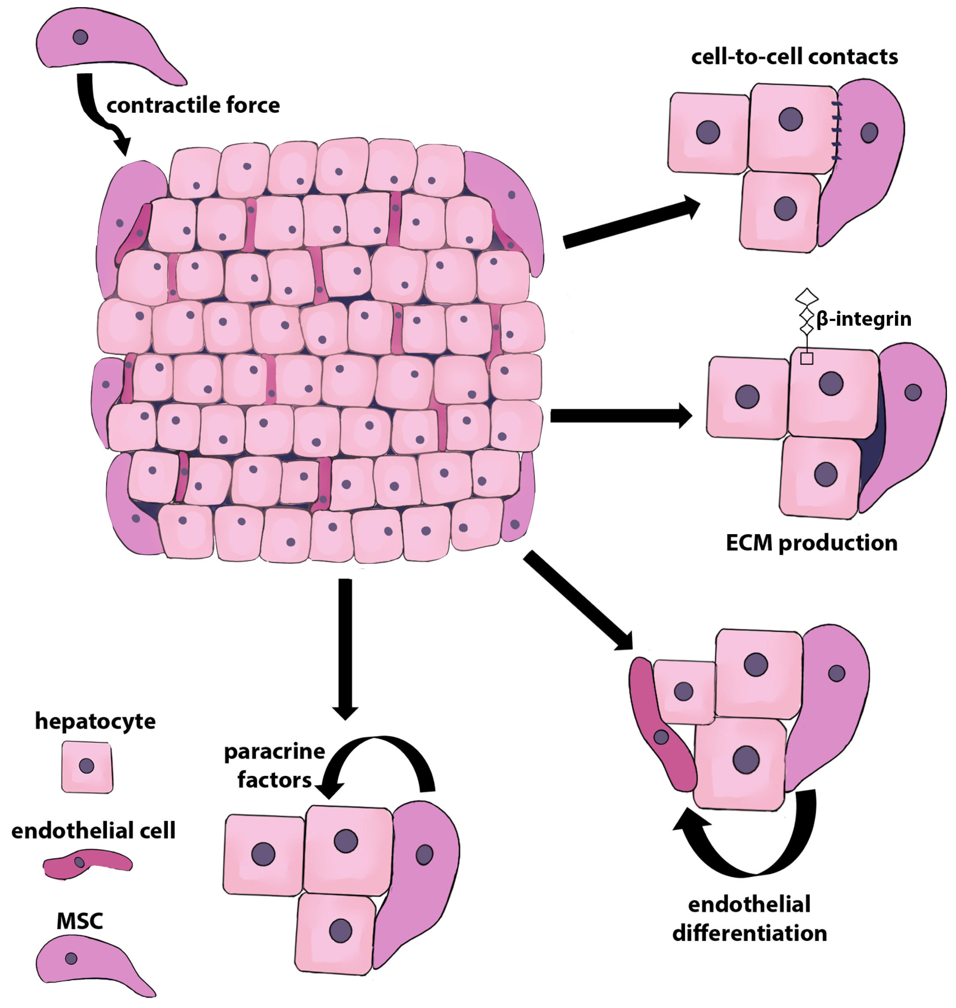
Figure 1. The role of MSCs in the formation of liver organoids. MSCs provide the contractile force for the formation of the heterotypic hepatic organoids and enhance the viability of hepatocytes either through direct contact between hepatocytes and MSCs or via paracrine factors contributing to the proliferation and maintenance of the functional activity of hepatocytes. MSCs produce ECM proteins, facilitating the adhesion of hepatocytes, and enhance the expression of β-integrin by hepatocytes, contributing to the transition of hepatocytes to the S phase of the cell cycle. Within the organoids, MSCs are able to differentiate into endotheliocytes, which form structures similar to a capillary network.
Ijms 24 15212 g001
Figure 2. Reciprocal effects of hepatocytes and MSCs in direct co-cultures. MSCs produce hepatotrophic factors, including HGF, IL-6, and PAI-1, while their expression, in turn, is enhanced by hepatocytes. Hepatotrophic factors enhance the survival and proliferation of hepatocytes and improve their functional activity, as witnessed by heightened albumin production and ureagenesis. MSCs secrete TGF-β, which, through autocrine regulation, stimulates the production of ECM proteins, thereby facilitating hepatocyte adhesion and promoting their proliferation. On the other hand, TNF-α secreted by primary hepatocytes can induce their own apoptosis also via autocrine mechanisms. MSCs significantly reduce the expression of TNF-α by hepatocytes, thereby protecting hepatocytes from cell death. HGF—hepatocyte growth factor; IL-6—interleukin 6; PAI-1—plasminogen activator inhibitor-1; TNF-α—tumor necrosis factor α; TGF-β—trans-forming growth factor β; ECM—extracellular matrix.
Ijms 24 15212 g002
Figure 3. Mechanisms of the anti-apoptotic action of MSCs in the hepatocyte–MSC co-cultures. MSCs inhibit stress-induced apoptosis and cell cycle arrest in hepatocytes through the secretion of the hepatotrophic factors IL-6, HGF, and PGE2, as well as through the production of the miRNA-loaded exosomes. In stressed hepatocytes, MSCs downregulate the expression of the proapoptotic proteins Bax, cleaved caspase 3, and BID and upregulate the expression of the anti-apoptotic protein Bcl-2. MSC-produced PGE2 activates two interconnected signaling pathways involved in liver regeneration. YAP activation is mediated through p-CREB and leads to the suppression of PTEN by miR-29a-3p and subsequent activation of mTOR signaling, ultimately leading to the inhibition of apoptosis and an increase in hepatocyte proliferation. MSCs reduce the miR143 level in hepatocytes subjected to oxidative stress and, accordingly, increase the levels of HK2 and ADRB1. This results in decreased oxidative phosphorylation and a switch to glycolysis, ultimately leading to hepatocyte proliferation and protection from apoptosis. MSCs attenuate damage caused by oxidative stress in hepatocytes by inhibiting miR-486-5p, upregulating PIM1, and blocking TGF-β/Smad signaling. MSCs activate PTEN/AKT signaling pathway in damaged hepatocytes by upregulating miR-20a-5p, thus enhancing their proliferation. MSC-derived exosomal miR-124 promoted liver regeneration by enhancing hepatocyte proliferation through the downregulation of Foxg1. IL-6—interleukin-6; HGF—hepatocyte growth factor; PGE2—prostaglandin E2; YAP—Yes-associated protein; p-CREB—p-cAMP-responsive element binding protein; PTEN—phosphatase and tensin homolog; mTOR—mammalian target of rapamycin; HK2—hexokinase 2; ADRB1—beta-1-adrenergic receptor; PIM1—proviral integration site for Moloney murine leukemia virus kinase 1; TGF-β—transforming growth factor-β.
Ijms 24 15212 g003
Figure 4. The PRL-1-dependent mechanism of hepatoprotective action of MSCs. Normal hepatocytes and MSCs both express PRL-1. After treatment of primary rat hepatocytes with lithocholic acid, the level of PRL-1 expression decreased. Co-cultivation with MSCs resulted in the restoration of PRL-1 expression in hepatocytes. MSC homing was regulated by RhoA-mediated ROCK1 signaling. PRL-1 produced by hepatocytes acted as a chemokine for MSCs due to increased MMP-2 expression in MSCs. On the other side, PRL-1 produced by MSCs increased anaerobic mitochondrial metabolism in damaged hepatocytes, decreasing cytoplasmic lactate and increasing mitochondrial lactate, which ultimately led to increased ATP synthesis and hepatocyte repair. PRL-1—phosphatase of regenerating liver; MMP-2—matrix metalloproteinase 2; ATP—adenosine triphosphate; RhoA—Ras homolog family member A; ROCK1—Rho-associated coiled-coil-containing protein kinase 1.
Ijms 24 15212 g004
Figure 5. Hepatocyte protection from ferroptosis mediated by MSC or MSC derivatives. MSC-derived exosomes loaded with miR-204-5p inhibit ferroptosis by reducing Fe2+ intracellular overload and ROS and MDA levels and by upregulating the expression of GPX4, a protective enzyme that prevents ferroptosis. Also, MSC-derived exosomes induce the stabilization of SLC7A11 via increased expression of CD44 and OTUB1. Thus, MSC may prevent hepatocyte ferroptosis by activation of the antioxidant system.
Ijms 24 15212 g005
Figure 6. Hepatocyte protection from pyroptosis by MSC or MSC derivatives. MSCs reduce NLRP3-caspase 1 inflammasomes in stressed hepatocytes via the expression of the IL-10 anti-inflammatory cytokine. Also, MSC-derived exosomes reduce the expression of the markers of pyroptosis NLRP3, GSDMD, caspase 1, IL-1β, and IL-18 and the suppression of hepatocyte pyroptosis, probably due to the reduction in inflammation. NLRP3—NLR family pyrin domain containing 3; IL-10—interleukin-10; GSDMD—gasdermin D; IL-1β—interleukin-1β; IL-18—interleukin-18.
Ijms 24 15212 g006
Figure 7. Reciprocal mitochondrial transfer between stressed hepatocytes and MSCs. MSCs prevent hepatocyte cell death by donating their mitochondria to hepatocytes. This process promotes the recovery of lipid utilization, averts lipid accumulation, stimulates the biogenesis of mitochondria, and increases ATP production.
Ijms 24 15212 g007
Table 1. Bioactive molecules and EVs protecting hepatocytes from apoptosis produced by MSCs.
MSC Tissue SourceCulture Setup and/or
Bioactive Agents
HepatocytesAnti-Apoptotic Effects and References
BM-MSCsExosomesPrimary rat hepatocytes treated with D-galactosamine and lipopolysaccharideIncrease in autophagosome numbers;
increase in the expression levels of the autophagy-related proteins LC3II and Beclin-1 and the anti-apoptotic protein Bcl-2;
decrease in proapoptotic proteins Bax and cleaved caspase 3 [92]
hESC-derived HuES9.E1 MSCsExosomesTAMH, an immortalized mouse hepatocyte cell line derived from transgenic MT42 male mice overexpressing TGF-α;
THLE-2, an immortalized primary human hepatocyte cell line that expresses phenotypic characteristics of normal adult liver epithelial cells;
HuH-7, a well-differentiated human hepatocarcinoma cell line;
APAP or H2O2 treatment
Transition from the G0 to G1 phase of the cell cycle;
upregulation of TNF-α, IL-6, iNOS, COX-2, and MIP-2;
restoration of NF-κB and STAT3 signaling;
inhibition of caspase 3;
upregulation of anti-apoptotic protein Bcl-xL [93]
Rat BM-MSCsIndirect co-culture or BM-MSC-CMRat hepatocytesIncrease in albumin secretion and urea production [99]
Human orbital fat-derived stem cellsDirect or indirect co-culture
IL-6
Rat hepatocytes treated with serum from an acute liver failure patientIncrease in cell viability;
increase in albumin secretion and urea production [100]
Human umbilical cord MSCsIndirect co-culture
HGF, EGF, and IL-6
Human LO2 cell line treated with acetaminophenProtection from APAP-induced necrosis;
increase in cell viability [110]
Mouse BM-MSCsMSC-CM;
prostaglandin E2
LPS-treated mouse hepatocyte cell line AML12Apoptosis inhibition;
increase in cell proliferation;
YAP activation, suppression of PTEN due to miR-29a-3p, and activation of mTOR signaling [91]
Disclaimer/Publisher’s Note: The statements, opinions and data contained in all publications are solely those of the individual author(s) and contributor(s) and not of MDPI and/or the editor(s). MDPI and/or the editor(s) disclaim responsibility for any injury to people or property resulting from any ideas, methods, instructions or products referred to in the content.