Article

The Clinical and Genetic Characteristics of Streptococcus agalactiae Meningitis in Neonates

1
Division of Pediatric Neonatology, Department of Pediatrics, Chang Gung Memorial Hospital, Taoyuan 333, Taiwan
2
School of Medicine, College of Medicine, Chang Gung University, Taoyuan 333, Taiwan
3
Department of Laboratory Medicine, Linkou Chang Gung Memorial Hospital, Taoyuan 333, Taiwan
4
Department of Medical Biotechnology and Laboratory Science, Chang Gung University, Taoyuan 333, Taiwan
5
Division of Neonatology and Pediatric Hematology-Oncology, Department of Pediatrics, Chang Gung Memorial Hospital, Yunlin 638, Taiwan
*
Author to whom correspondence should be addressed.
Int. J. Mol. Sci. 2023, 24(20), 15387; https://doi.org/10.3390/ijms242015387
Submission received: 7 September 2023 / Revised: 10 October 2023 / Accepted: 16 October 2023 / Published: 20 October 2023
(This article belongs to the Section Molecular Microbiology)

Abstract

:
Streptococcus agalactiae (Group B Streptococcus, GBS) is an important pathogen of bacterial meningitis in neonates. We aimed to investigate the clinical and genetic characteristics of neonatal GBS meningitis. All neonates with GBS meningitis at a tertiary level medical center in Taiwan between 2003 and 2020 were analyzed. Capsule serotyping, multilocus sequence typing, antimicrobial resistance, and whole-genome sequencing (WGS) were performed on the GBS isolates. We identified 48 neonates with GBS meningitis and 140 neonates with GBS sepsis. Neonates with GBS meningitis had significantly more severe clinical symptoms; thirty-seven neonates (77.8%) had neurological complications; seven (14.6%) neonates died; and 17 (41.5%) survivors had neurological sequelae at discharge. The most common serotypes that caused meningitis in neonates were type III (68.8%), Ia (20.8%), and Ib (8.3%). Sequence type (ST) is highly correlated with serotypes, and ST17/III GBS accounted for more than half of GBS meningitis cases (56.3%, n = 27), followed by ST19/Ia, ST23/Ia, and ST12/Ib. All GBS isolates were sensitive to ampicillin, but a high resistance rates of 72.3% and 70.7% to erythromycin and clindamycin, respectively, were noted in the cohort. The virulence and pilus genes varied greatly between different GBS serotypes. WGS analyses showed that the presence of PezT; BspC; and ICESag37 was likely associated with the occurrence of meningitis and was documented in 60.4%, 77.1%, and 52.1% of the GBS isolates that caused neonatal meningitis. We concluded that GBS meningitis can cause serious morbidity in neonates. Further experimental models are warranted to investigate the clinical and genetic relevance of GBS meningitis. Specific GBS strains that likely cause meningitis requires further investigation and clinical attention.

1. Introduction

Streptococcus agalactiae (Group B Streptococcus, GBS) is one of the most common pathogens causing neonatal sepsis and meningitis [1,2,3]. GBS sepsis in neonates can be categorized as early-onset disease (EOD, disease onset before the 7th day of life) and late-onset disease (LOD, disease occurring after the 8th day of life), where both are associated with a high mortality rate of 8–14% and a high risk of neurological complications, especially in neonates with GBS meningitis [4,5,6]. Although the policies of intrapartum antibiotic prophylaxis and routine maternal screening have decreased the incidence of GBS EOD, GBS LOD remains an important threat that causes mortality in the neonatal intensive care unit (NICU) [6,7,8]. Recently, researchers have focused on the molecular epidemiology, predominant type III GBS sepsis in neonates, and genetic characteristics of specific GBS isolates that are associated with GBS sepsis and meningitis [9,10,11,12,13].
GBS meningitis is associated with a high mortality rate and long-term neurological disability in neonates [6,10,14,15]. The high rates of neuropsychiatric and developmental sequelae among GBS meningitis survivors highlight the importance of understanding the pathophysiology and genetic mechanisms, which may help clinicians to develop new therapeutic and/or neuroprotective strategies for the developing brain [15,16,17]. The serotype III GBS hypervirulent clonal complex 17 (CC17) isolates have become a predominant cause of meningitis and GBS LOD in recent years, which may be related to their higher penetration ability to cross the blood–brain barrier (BBB) [18,19,20]. However, the relevant molecular and genetic characteristics of specific GBS strains that are likely to cause meningitis remain unidentified. In this study, we aimed to investigate the clinical and genetic characteristics of GBS isolates that cause neonatal meningitis.

2. Results

2.1. Clinical Characteristics of Neonates with GBS Meningitis

During the study period, a total of 48 neonates with documented GBS meningitis and 140 cases of neonatal GBS sepsis without meningitis were identified and enrolled for analyses. The median (interquartile range [IQR]) gestational age and birth body weight (BBW) of the neonates with GBS meningitis were 38.0 (37.0–39.0) weeks and 2900 (2651.3–3251.3) g, respectively. The majority of cases were term-born neonates, and female patients outnumbered male patients. The patients’ demographics, clinical manifestations, and laboratory results are summarized in Table 1. Neonates with GBS meningitis were not significantly different from those with GBS sepsis without meningitis in patients’ characteristics, but they had significantly more severe symptoms, including a greater likelihood of having respiratory distress, severe sepsis, metabolic acidosis, and coagulopathy (Table 1). Of note, 25% of cases with GBS meningitis were EOD, and 27.3% of the patients with GBS EOD had meningitis.
All cases of invasive GBS diseases were treated with ampicillin. However, neonates with GBS meningitis were treated for a significantly longer duration than those with GBS sepsis (median 22.5 days vs. 12.5 days, p < 0.001). We found that neonates with GBS meningitis had a significantly higher percentage of neurological complications (n = 37, 77.8%) and the survivors were more likely to have neurological sequelae (n = 17, 41.5%) at discharge than the neonates with GBS sepsis (both p < 0.001) (Table 2). The mortality rate of the neonates with GBS meningitis was 14.6% (a total of seven neonates died), which was also higher than that of the neonates with GBS sepsis (Table 2).

2.2. Molecular Characteristics and Antimicrobial Resistance of GBS Isolates That Caused Meningitis

The molecular epidemiology and genetic characteristics of the GBS isolates that caused meningitis in our cohort are summarized in Table 3. Type III GBS accounted for 68.8% (n = 33) of all meningitis cases, followed by serotype Ia (20.8%, n = 10), Ib (8.3%, n = 4), and type II (2.1%, n = 1). Sequence type (ST) is highly correlated with serotypes, and ST17/III GBS accounted for more than half of GBS meningitis cases (56.3%, n = 27), followed by ST19/Ia (10.4%, n = 5) and ST23/Ia or Ib (10.4%, n = 5). Of note, the CC17 GBS isolates (n = 27, 56.3%) and CC12 (n = 6, 12.5%) most commonly caused neonatal meningitis in this study.
All the GBS isolates of neonates with meningitis and those from neonatal sepsis were susceptible to penicillin, ampicillin, vancomycin, and cefotaxime. High resistance rates of 72.3% and 70.7% to erythromycin and clindamycin, respectively, were noted in all GBS isolates. The antibiotic resistance profiles between the GBS isolates of neonatal meningitis and those of neonatal sepsis without meningitis were not significantly different, but a specifically high antibiotic resistance rate was noted in type III (77.1–82.2%), type Ib (100%), and type V (85.7%) GBS isolates. Due to the high correlation between the serotype and sequence type, the antibiotic resistance rate to erythromycin and clindamycin was also high in the CC12 (100%) and CC17 (89.3%) GBS isolates. Additionally, most of the GBS isolates (94.9%) with resistance to erythromycin were also resistant to clindamycin.

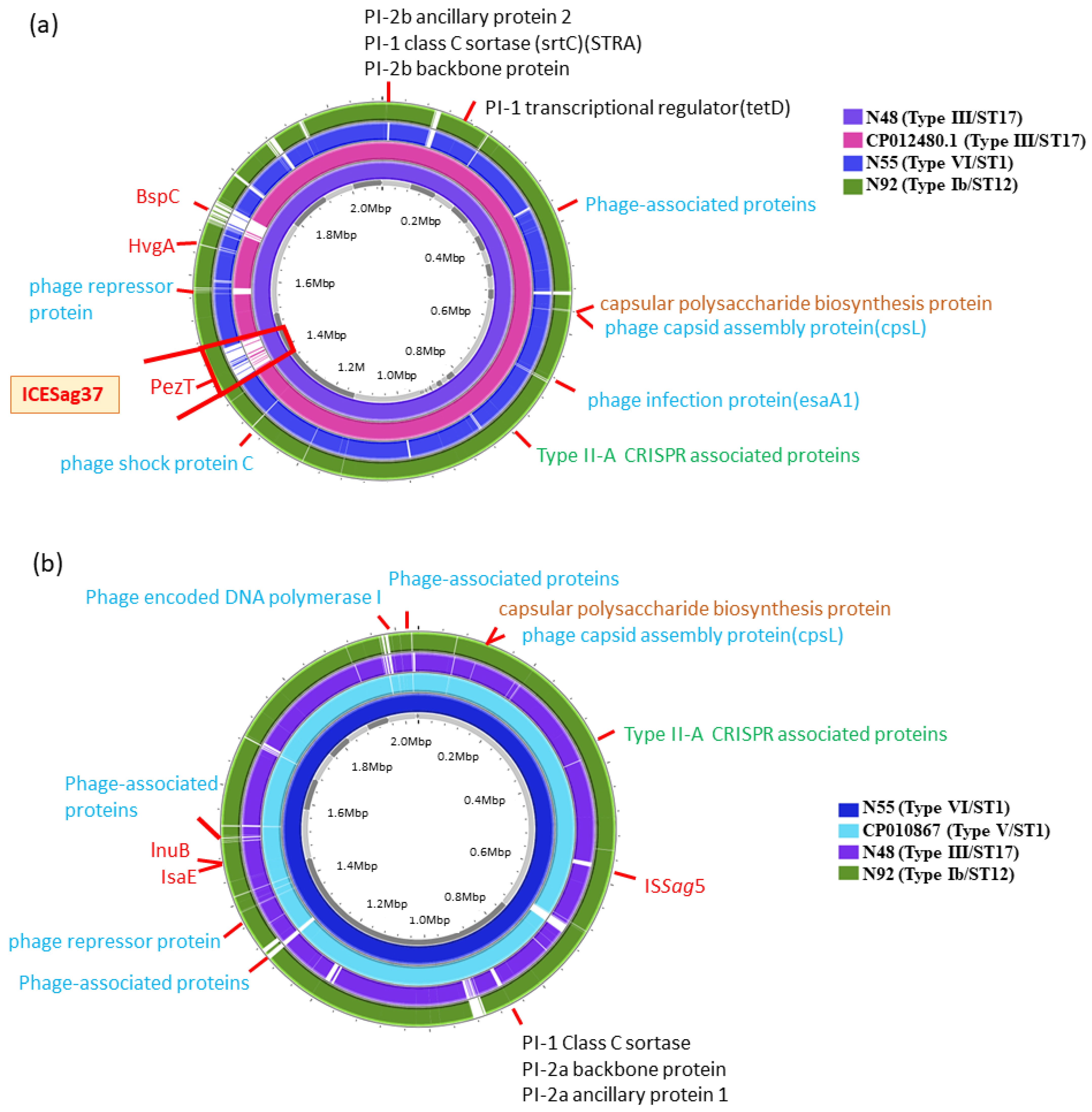
2.3. WGS for GBS Isolates That Caused Neonatal Meningitis

WGS was performed using three type III/ST17 GBS strains, one type Ib/ST12, and three type VI/ST-1 GBS strains, representing GBS meningitis and GBS sepsis without meningitis, respectively. Strain CP012480.1, which is also a type III/ST17 GBS isolate from neonates with invasive GBS diseases, was used as the reference strain. One V/ST1 GBS reference strain (CP010867) from NCBI was also used as the reference strain for comparison. Comparative genome analyses were performed for all GBS isolates and the two reference strains to track the possible genomics of mobile elements and insertion sequences (IS). All genes related to the component systems CovS/R, antibiotic resistance, pilus formation, capsular serotype, and virulence were investigated.
Concerning the genes of component systems CovS/R, most antimicrobial resistance genes and most virulence genes were not significantly different between the GBS strains of neonatal meningitis and those of neonatal sepsis without meningitis (Figure 1). However, the presence of BspC, ICESag37, and PezT was noted in GBS isolates of neonatal meningitis, both in type Ib/CC12 and III/CC17 GBS, but not in the type VI/CC1 GBS strain. The BspC, PezT, and HvgA genes, as well as the pili, are previously reported to be associated with enhanced virulence and pathogenic mechanisms, which lead to life-threatening illness [15,19]. Additionally, the PezT, ICESag37, and BspC genes were not found in the reference CP012480.1 strain (also type III/ST strain from neonatal sepsis). Therefore, we highly suspected that the BspC, PezT, and ICESag37 genes were associated with the occurrence of meningitis in the neonates examined in this study.
PCR was performed for all type Ib and type III GBS isolates to verify the results and confirm the presence of multiple genes in all clinical GBS isolates. The primers used for all targeted genes, including BspC, ICESag37, PezT, and HvgA are summarized in Table 4. The genetic characteristics of all GBS isolates are listed in Table 3. We found the presence of PezT, BspC, and ICESag37 in 60.4%, 77.1%, and 52.1% of all GBS isolates that caused neonatal meningitis in our cohort. Additionally, HvgA was present in 80% of the type III/CC17 GBS isolates of neonatal meningitis.

2.4. Discussion

Compared to numerous studies that have investigated the molecular epidemiology of neonatal GBS invasive diseases and colonization in pregnant women, relatively fewer studies have focused on GBS meningitis [4,21,22]. We found that GBS meningitis is associated with more severe clinical manifestations and worse long-term neurological outcomes in neonates. Most of the GBS isolates that caused neonatal meningitis in our cohort belong to the type III/CC17 and type Ib/CC12 strains, which is compatible with previous studies [4,6,21,22,23,24]. WGS was used to investigate the genetic differences between the GBS isolates of neonatal meningitis and those that caused neonatal sepsis in our cohort, and we found the presence of BspC, HvgA, and PezT genes, as well as ICESag37, to be potentially associated with the occurrence of GBS meningitis. We highlight the requirement of further in vivo and in vitro studies to document the roles and underlying mechanisms of these genes, which can be the future bases of preventive and therapeutic strategies.
In our institute, we have applied the policies of routine GBS screening for pregnant women and intrapartum antibiotic prophylaxis (IAP) since 2004. A change in policy was associated with the predominance of serotype III/ST-17 GBS isolates since 2010 and the increasing prevalence of antibiotic-resistant GBS isolates [12,13,20,25]. More infection control strategies have been carried out in Taiwan in recent decades, which has ultimately led to decreased cases of GBS sepsis and meningitis in neonates since 2018 [6,26,27,28]. However, in neonates with GBS infections, the chance of progression to severe sepsis and meningitis remains unchanged and the mechanisms are not yet fully understood. Therefore, we sought to investigate the genetic characteristics of GBS isolates associated with the occurrence of meningitis.
Various virulence genes and mechanisms have been documented to be associated with the occurrence of GBS meningitis in neonates, such as the enhanced penetration of the BBB caused by HvgA genes, pili, and Srr1/Srr2 genes [15,22,29,30,31]. In addition to the well-known type III GBS specific HvgA genes, we found BspC and PezT genes in most GBS isolates of neonatal meningitis. The BspC gene has been found to interact with host vimentin to promote bacterial adherence to the brain endothelium and inflammation in the in vivo and in vitro models of meningitis, which also leads to the enhanced penetration of the BBB and the occurrence of meningitis [32]. Therefore, targeting the BspC–vimentin interaction is a potential therapeutic strategy to decrease inflammation during GBS meningitis [33]. Although not all GBS isolates that caused meningitis carried both BspC and PezT genes, we suspected that the occurrence of GBS meningitis is associated with both host factors and the virulence of highly pathogenic GBS strains [23,34,35].
Pili are known to be associated with enhanced cell adhesion of GBS isolates, which contributes to transcytosis of the endothelium and increased penetration of the BBB. Therefore, the specific pilus profiles are reported to be associated with sepsis and meningitis [30,31]. Previous studies have concluded that specific pilus profiles in some GBS strains or phylogenetic lineages are associated with a higher risk of meningitis [31,36,37]. While the presence of PI-1 and PI-2b genes is noted in most of the type III/CC17 GBS isolates [36,37], our data showed that the presence of ICESag37, carrying multiple virulence genes and replacing the PI-1, is correlated with the clonal expansion of antibiotic-resistant type III/CC17 GBS strains, which may be associated with the occurrence of meningitis [19].
The PezT gene was initially found in Streptococcus pneumonia and is part of the epsilon/zeta system (the PezA/T system), which is a special toxin/antitoxin system that protects the bacterium itself [38,39]. PezT can activate the release of specific toxins to attack host cells or competing microorganisms, which has been considered a pathogenic and invasive mechanism [38,39]. The PezT affects the cell wall integrity via phosphorylase activities and increases the GBS invasiveness after epithelial lysis, which may contribute to the penetration of the BBB [38,39]. In contrast to a previous study that documented that the PezA/T system is unique in the CC12 GBS strain and contributes to higher virulence [40], we detected the PezT gene in 60.4% of all GBS isolates of neonatal meningitis. The PezA/T system is associated with a higher severity of illness in both the CC12 GBS strain and some non-CC12 GBS strains and deserves further study to investigate its involvement in neonatal meningitis.
An obviously decreased GBS sepsis-attributable mortality rate has been reported in recent decades, from more than 20% in the 1980s to approximately 10.7% in recent studies [3,4,5,41,42]. Improved critical care in neonates and the early implementation of prophylactic antibiotics may account for the reduced mortality rate [21,24]. However, a high rate of neurological complications and long-term neurological sequelae in survivors of GBS sepsis is currently the major concern. In our cohort, GBS EOD was related to a higher percentage of meningitis (27.3%) than other studies, which reported that most GBS meningitis cases are LOD. Additionally, GBS EOD was associated with a significantly higher rate of severe sepsis and a higher mortality rate than GBS LOD. Therefore, further studies for the early identification of specific strains, risk factors, or genetic mechanisms that cause GBS meningitis are urgently needed in the future.
Some limitations in this study should be addressed. All the invasive GBS isolates in this study were from a single center in Taiwan over an extensive period of time. During the prolonged timeframe, the therapeutic and IAP policies may have been changed. Therefore, a multicenter study is needed to investigate the epidemiological and genetic trends of GBS strains that cause neonatal meningitis across different regions and time periods. We applied the PacBioTM SMRT (Pacific Biosciences, Menlo Park, CA, USA) and MiSeqTM (Illumina, San Diego, CA, USA) sequencing technologies for WGS, which have the advantages of easy analysis, few errors, relatively lower cost per sample, and no specific limitations [43,44,45]. However, a gene expression analysis was not performed in this study, and the real mechanisms by which important genes contribute to the occurrence of meningitis were not investigated. Additionally, some early-mortality cases and some invasive GBS strains more than 10 years ago were inevitably lost in this study, which, however, may not have significantly altered the study results.
In conclusion, GBS meningitis in neonates is associated with a higher severity of illness and a high risk of neurological sequelae. GBS meningitis is most likely caused by type III/CC17 and type Ib/CC12 GBS isolates. Genetically, the HvgA, BspC, PezT, and ICESag37 genes, which carries many specific virulence profiles and multiple antibiotic resistance genes, are potentially associated with the enhanced penetrative ability of GBS isolates to cross the BBB and the occurrence of meningitis. Therefore, further in vitro and in vivo models to investigate the roles of these genes on the occurrence of GBS meningitis and the relevance of clinical features are highly suggested. Given such genetic specificity and clinical importance, a multicenter study with more cases of GBS meningitis and GBS isolates is warranted to investigate the molecular mechanisms involved, and the continuous monitoring of neonates with GBS sepsis is critically important in the future.

3. Materials and Methods

3.1. GBS Isolates, Data Collection, and Definition

We conducted a single-center cohort study that enrolled all neonates with GBS meningitis who were hospitalized in the NICUs of Linkou Chang Gung Memorial Hospital (CGMH) between 2003 and 2020. Linkou CGMH is the largest tertiary-level medical center in Taiwan and located in North Taiwan. There are three NICUs with a total capacity of 47 beds equipped with ventilators and 55 beds in special care nurseries in Linkou CGMH. Meningitis was defined based on the standard criteria [46,47] and all cases had positive GBS strains isolated from cerebrospinal fluid (CSF) cultures. We also analyzed neonates with GBS sepsis but without meningitis during the study period, and this study is part of our longitudinal serial studies. All GBS isolates were retrieved from CGMH’s central laboratory and bacterial library. The clinical information of all the patients, including perinatal demographics, clinical manifestations, laboratory data, hospital courses, and outcomes, were retrospectively reviewed and recorded by our research teams. This study was approved by the Institutional Review Board of CGMH (IRB No. 202102291B0), and a waiver for informed consent for anonymous data collection was approved.
We defined severe sepsis, septic shock, and uncomplicated bacteremia based on the definitions of our previous studies and the Centers for Disease Control and Prevention [23,48]. The presence of neurological complications and long-term neurological sequelae in these patients was evaluated based on the definitions used in the previous studies [14,23]. In addition to EOD and LOD, we categorized late LOD (LLOD), when disease onset was after 90 days of life [49]. The severity of illness was evaluated using the Neonatal Therapeutic Intervention Scoring System (NTISS) score [41] at the onset of meningitis, which was defined at the time of the first positive CSF culture.

3.2. Capsular Serotyping, MLST, and Pilus Genes

The capsular serotypes of all GBS isolates were analyzed using the multiplex PCR assay to identify the GBS isolates of types Ia to IX. The DNA isolation method and the PCR assay that amplified and sequenced seven housekeeping genes (adhP, atr, glcK, glnA, pheS, sdhA, and tkt) were based on a standard protocol described in our previous publication [10]. Multilocus sequence typing (MLST) was performed based on the standard procedure described in our previous study [42]. After PCR, the sequence type (ST) was assigned based on the allelic profile of each fragment and determined via the Streptococcus agalactiae MLST database (http://pubmist.org/sagalactiae, accessed on 1 April 2023). All GBS isolates can be clustered into several major clonal complexes (CCs) based on the goeBURST program [25]. Pilus island content was confirmed via standard multiplex PCR method to identify the pilus island (PI) marker, and a multiplex PCR assay was performed to analyze the distribution of GBS PI genes [26]. We checked all the GBS isolates of neonatal meningitis and sepsis using the target genes of alcohol dehydrogenase gbs0054 (adhP) as the housekeeping locus and sag647, sag1406, and san1517 for PI-1, PI-2a, and PI-2b, respectively.

3.3. Antimicrobial Susceptibility Testing

Antimicrobial susceptibility testing was performed for all the GBS isolates using the disc diffusion method as described in previous studies [27]. The double-disk diffusion test was applied to identify inducible clindamycin resistance. All GBS isolates were tested for susceptibility to seven antibiotics, including erythromycin, penicillin, clindamycin, vancomycin, ampicillin, cefotaxime, and teicoplanin, according to the guidelines of Clinical and Laboratory Standards Institute (CLSI) for the disc diffusion method [28].

3.4. Whole Genome Sequencing

Three GBS isolates selected from the III/ST-17 (termed N48, N96, and N5) strains, one type Ib/ST-12 (termed N92), and three type VI/ST-1 (termed N55, N70, and N132) GBS strains were used for whole-genome sequencing (WGS). The type III/ST-17 and type Ib/ST-12 strains were obtained from the cerebrospinal fluid of neonates with meningitis, while the type VI/ST-1 GBS isolates were from the blood culture of neonates with sepsis without meningitis. The GBS isolates selected to perform the WGS were based on previous experiences and that we have found similar WGS results among the same GBS serotypes and sequence types. Additionally, type III/ST-17 and Ib/ST-12 GBS isolates were among the most common GBS strain to cause meningitis. The standard protocol of the lysozyme-sodium dodecyl sulfate-proteinase K method was used to extract DNA. WGS was performed using both PacBioTM SMRT (Pacific Biosciences, Menlo Park, CA, USA) [43] and MiSeqTM (Illumina, San Diego, CA, USA) [44] sequencing technologies. The sequencing library was prepared using a TruSeq DNA LT Sample Prep Kit (Illumina, San Diego, CA, USA) for the Illumina MiSeq system. Genomic libraries were generated using Nextera XT kits (Illumina, San Diego, CA, USA). We used SPAdes (version 3.9.0) to assemble the sequences. All genome sequences were subjected to BLAST analysis using the NCBI genome database to identify possible plasmid sequences. After the de novo-assembled genome was generated, Prokka (v1,12) [50] was used for the genome annotation and identification of rRNA-encoding and tRNA-encoding regions.

3.5. Statistical Analysis

The clinical and genetic characteristics were compared between neonates with GBS meningitis and those with GBS sepsis but without meningitis during the study period. Categorical and continuous variables are expressed as proportions and the median (interquartile, IQR), respectively. Categorical variables were compared using the χ2 test or Fisher’s exact test; odds ratios (ORs) and 95% confidence intervals (CIs) were calculated. Continuous variables were compared using the Mann–Whitney U test and the t-test, depending on the distribution. The trend of the proportions of categorical variables among the subgroups was analyzed via the Cochran–Armitage trend test. The results with p values of <0.05 were considered statistically significant. All statistical analyses were performed using SPSS version 23 (IBM SPSS Statistics).

Author Contributions

Conceptualization, J.-F.H., M.-H.T. and J.-J.L.; Data collection and verification, J.-F.H., S.-M.C., H.-R.H., M.-C.C., W.-J.L. and P.-H.Y.; Formal analysis, J.-F.H., S.-M.C., M.-C.C. and P.-H.Y.; Funding acquisition: J.-F.H., M.-H.T. and J.-J.L.; Investigation, W.-J.L., H.-R.H. and P.-H.Y.; Methodology, J.-F.H., S.-M.C., H.-R.H. and M.-C.C.; Supervision, J.-J.L.; Writing—original draft, J.-F.H. and M.-H.T.; Writing—review and editing, J.-J.L. All authors have read and agreed to the published version of the manuscript.

Funding

This work was supported by the Chang Gung Memorial Hospital (grant no. CMRPG3L1461-3) and The Ministry of Science and Technology in Taiwan (grant no. MOST 111-2314-B-182-045-).

Institutional Review Board Statement

This study was conducted in accordance with the Declaration of Helsinki, and approved by the Institutional Review Board of Chang Gung Memorial Hospital (IRB No. 202102291B0), with a waiver of informed consent because all patient records and information were anonymized and deidentified prior to analysis.

Informed Consent Statement

Patient consent was waived due to the fact that all patient records and information were anonymized and deidentified prior to analysis.

Data Availability Statement

The datasets used/or analyzed during the current study are available from the corresponding author on reasonable request.

Acknowledgments

All authors thank Chiao-Ching Chiang for keeping the database of our NICU, as well as all nursing staff working in our NICUs for keeping extremely detailed patient records, which contributed greatly to the completion of this research.

Conflicts of Interest

The authors declare no conflict of interest.

Abbreviations

CSF: cerebrospinal fluid; CDC: Centers for Disease Control and Prevention; CI: confidence interval; CC: clonal complex; CGMH: Chang Gung Memorial Hospital; EOD: early-onset disease; GBS: Group B Streptococcus; IAP: intrapartum antibiotic prophylaxis; LOD: late-onset disease; MLST: multilocus sequence typing; ST: sequence type; OR: odds ratio; PCR: polymerase chain reaction; TPN: total parenteral nutrition; VAP: ventilator-associated pneumonia.

References

  1. Ouchenir, L.; Renaud, C.; Khan, S.; Bitnun, A.; Boisvert, A.-A.; McDonald, J.; Bowes, J.; Brophy, J.; Barton, M.; Ting, J.; et al. The epidemiology, management, and outcomes of bacterial meningitis in infants. Pediatrics 2017, 140, e20170476. [Google Scholar] [CrossRef]
  2. Ku, L.C.; Boggess, K.A.; Cohen-Wolkowiez, M. Bacterial meningitis in infants. Clin. Perinatol. 2015, 42, 29–45. [Google Scholar] [CrossRef]
  3. Hasbun, R.; Wootton, S.H.; Rosenthal, N.; Balada-Llasat, J.M.; Chung, J.; Duff, S.; Bozzette, S.; Zimmer, L.; Ginocchio, C.C. Epidemiology of meningitis and encephalitis in infants and children in the United States, 2011–2014. Pediatr. Infect. Dis. J. 2019, 38, 37–41. [Google Scholar] [CrossRef]
  4. Romain, A.-S.; Cohen, R.; Plainvert, C.; Joubrel, C.; Béchet, S.; Perret, A.; Tazi, A.; Poyart, C.; Levy, C. Clinical and laboratory features of Group B Streptococcus meningitis in infants and newborns: Study of 848 cases in France, 2001–2014. Clin. Infect. Dis. 2017, 66, 857–864. [Google Scholar] [CrossRef]
  5. Kohli-Lynch, M.; Russell, N.J.; Seale, A.C.; Dangor, Z.; Tann, C.J.; Baker, C.J.; Bartlett, L.; Cutland, C.; Gravett, M.G.; Heath, P.T.; et al. Neurodevelopmental impairment in children after Group B Streptococcus disease worldwide: Systemic review and meta-analysis. Clin. Infect. Dis. 2017, 65, S190–S199. [Google Scholar] [CrossRef]
  6. Nanduri, S.A.; Petit, S.; Smelser, C.; Apostol, M.; Alden, N.B.; Harrison, L.H.; Lynfield, R.; Vagnone, P.S.; Burzlaff, K.; Spina, N.L.; et al. Epidemiology of invasive early-onset and late-onset Group B Streptococcal disease in the United States, 2006 to 2015: Multistate laboratory and population-based surveillance. JAMA Pediatr. 2019, 173, 224–233. [Google Scholar] [CrossRef]
  7. Guan, X.; Mu, X.; Ji, W.; Yuan, C.; He, P.; Zhang, L.; Huang, Y.; Li, J.; Chen, J.; Zhong, H.; et al. Epidemiology of invasive group B streptococcal disease in infants from urban area of South China, 2011–2014. BMC Infect. Dis. 2018, 18, 14. [Google Scholar] [CrossRef]
  8. Horvath-Puho, E.; van Kassel, M.N.; Goncalves, B.P.; de Gier, B.; Procter, S.R.; Paul, P.; van der Ende, A.; Søgaard, K.K.; Hahné, S.J.; Chandna, J.; et al. Mortality, neurodevelopmental impairments, and economic outcomes after invasive group B streptococcal disease in early infancy in Denmark and the Netherlands: A national matched cohort study. Lancet Child Adolesc. Health 2021, 5, 398–407. [Google Scholar] [CrossRef]
  9. Campisi, E.; Rosini, R.; Ji, W.; Guidotti, S.; Rojas-López, M.; Geng, G.; Deng, Q.; Zhong, H.; Wang, W.; Liu, H.; et al. Genomic analysis reveals multi-drug resistant clusters in Group B Streptococcus CC17 hypervirulent isolates causing neonatal invasive disease in southern mainland China. Front. Microbiol. 2016, 7, 1265. [Google Scholar] [CrossRef]
  10. Tsai, M.-H.; Hsu, J.-F.; Lai, M.-Y.; Lin, L.-C.; Chu, S.-M.; Huang, H.-R.; Chiang, M.-C.; Fu, R.-H.; Lu, J.-J. Molecular characteristics and antimicrobial resistance of Group B Streptococcus Strains causing invasive disease in neonates and adults. Front. Microbiol. 2019, 10, 264. [Google Scholar] [CrossRef]
  11. Zurn, K.; Lander, F.; Hufnagel, M.; Monecke, S.; Berner, R. Microaray analysis of Group B Streptococci causing invasive neonatal early and late-onset infection. Pediatr. Infect. Dis. J. 2020, 39, 449–453. [Google Scholar] [CrossRef]
  12. Lohrmann, F.; Berg, A.; Wicker, E.; Imm, A.; Krause, G.; Zürn, K.; Berner, R.; Hufnagel, M.; Lander, F. Prevalence of capsular serotype, pilus island distribution, and antibiotic resistance in pediatric and adult invasive Group B Streptococcus isolates: Data from a nationwide prospective surveillance study in Germany. Pediatr. Infect. Dis. J. 2021, 40, 76–82. [Google Scholar] [CrossRef]
  13. Kadambari, S.; Trotter, C.L.; Heath, P.T.; Goldacre, M.J.; Pollard, A.J.; Goldacre, R. Group B streptococcal disease in England (1998–2017): A population-based observational study. Clin. Infect. Dis. 2021, 72, e791–e798. [Google Scholar] [CrossRef]
  14. Hsu, M.-H.; Hsu, J.-F.; Kuo, H.-C.; Lai, M.-Y.; Chiang, M.-C.; Lin, Y.-J.; Huang, H.-R.; Chu, S.-M.; Tsai, M.-H. Neurological complications in young infants with acute bacterial meningitis. Front. Neurol. 2018, 9, 903. [Google Scholar] [CrossRef]
  15. Tavares, T.; Pinho, L.; Andrade, E.B. Group B Streptococcal neonatal meningitis. Clin. Microbiol. Rev. 2022, 35, e0007921. [Google Scholar] [CrossRef]
  16. de Cambronne, R.D.; Fouet, A.; Picart, A.; Bourrel, A.-S.; Anjou, C.; Bouvier, G.; Candeias, C.; Bouaboud, A.; Costa, L.; Boulay, A.-C.; et al. CC17 Group B Streptococcus exploits integrins for neonatal meningitis development. J. Clin. Investig. 2021, 131, e136737. [Google Scholar] [CrossRef]
  17. Travier, L.; Alonso, M.; Andronico, A.; Hafner, L.; Disson, O.; Lledo, P.-M.; Cauchemez, S.; Lecuit, M. Neonatal susceptibility to meningitis results from the immaturity of epithelial barriers and gut microbiota. Cell Rep. 2021, 35, 109319. [Google Scholar] [CrossRef]
  18. Tazi, A.; Plainvert, C.; Anselem, O.; Ballon, M.; Marcou, V.; Seco, A.; El Alaoui, F.; Joubrel, C.; El Helali, N.; Falloukh, E.; et al. Risk factors for infant colonization by hypervirulent CC17 Group B Streptococcus: Toward the understanding of late-onset disease. Clin. Infect. Dis. 2019, 69, 1740–1748. [Google Scholar] [CrossRef]
  19. Hsu, J.-F.; Tsai, M.-H.; Lin, L.-C.; Chu, S.-M.; Lai, M.-Y.; Huang, H.-R.; Chiang, M.-C.; Yang, P.-H.; Lu, J.-J. Genomic characterization of serotype III/ST17 Group B Streptococcus strains with antimicrobial resistance using whole genome sequencing. Biomedicines 2021, 9, 1477. [Google Scholar] [CrossRef]
  20. Wong, C.H.; Duque, J.R.; Wong, J.S.C.; Chan, C.V.; Lam, C.S.I.; Fu, Y.M.; Cheong, K.N.; Chua, G.T.; Lee, P.P.; Ip, P.; et al. Epidemiology and trends of infective meningitis in neonates and infants less than 3 months old in Hong Kong. Int. J. Infect. Dis. 2021, 111, 288–294. [Google Scholar] [CrossRef]
  21. Dominguez, K.; Lindon, A.K.; Gibbons, J.; Darch, S.E.; Randis, T.M. Group B Streptococcus drives major transcriptomic changes in the colonic epithelium. Infect. Immun. 2023, 91, e0003523. [Google Scholar] [CrossRef]
  22. Chen, N.; Zhang, X.; Zheng, K.; Zhu, L.; Zhang, N.; Liu, L.; Chen, Z.; Liu, G.; He, Q. Increased risk of Group B Streptococcus causing meningitis in infants with mannose-binding lectin deficiency. Clin. Microbiol. Infect. 2019, 25, 384.e1–384.e3. [Google Scholar] [CrossRef] [PubMed]
  23. Lin, C.; Chu, S.-M.; Wang, H.-C.; Yang, P.-H.; Huang, H.-R.; Chiang, M.-C.; Fu, R.-H.; Tsai, M.-H.; Hsu, J.-F. Complicated Streptococcus agalactiae sepsis with/without meningitis in young infants and newborns: The clinical and molecular characteristics and outcomes. Microorganisms 2021, 9, 2094. [Google Scholar] [CrossRef] [PubMed]
  24. Lohrmann, F.; Hufnagel, M.; Kunze, M.; Afshar, B.; Creti, R.; Detcheva, A.; Kozakova, J.; Rodriguez-Granger, J.; Sørensen, U.B.S.; Margarit, I.; et al. Neonatal invasive disease caused by Streptococcus agalactiae in Europe: The DEVANI multi-center study. Infection 2022, 51, 981–991. [Google Scholar] [CrossRef]
  25. Zhou, Y.; Wang, L.-Q.; Yan, Q.; Lee, C.-C.; Hsu, M.-H.; Liao, W.-T.; Zhang, L.; Chiu, C.-H. Genomic Analysis of Group B Streptococcus from Neonatal Sepsis Reveals Clonal CC17 Expansion and Virulence- and Resistance-Associated Traits After Intrapartum Antibiotic Prophylaxis. Clin. Infect. Dis. 2022, 75, 2153–2160. [Google Scholar] [CrossRef] [PubMed]
  26. Khodaei, F.; Najafi, M.; Hasani, A.; Kalantar, E.; Sharifi, E.; Amini, A.; Aghazadeh, M. Pilus–encoding islets in S. agalactiae and its association with antibacterial resistance and serotype distribution. Microb. Pathog. 2018, 116, 189–194. [Google Scholar] [CrossRef]
  27. Matuschek, E.; Ahman, J.; Webster, C.; Kahlmeter, G. Antimicrobial susceptibility testing of colistin-evolution of seven commercial MIC products against standard broth microdilution for Escherichia coli, Klebsiella pneumonia, Pseudomonas aeruginosa, and Acinetobacter spp. Clin. Microbiol. Infect. 2018, 24, 865–870. [Google Scholar] [CrossRef]
  28. Clinical and Laboratory Standards Institute. Performance Standards for Antimicrobial Susceptibility Testing, Twenty-Fifth Informational Supplement, M100, 31st ed.; SLCI: Wayne, PA, USA, 2021. [Google Scholar]
  29. Hsu, J.-F.; Chen, Y.-N.; Chu, S.-M.; Lee, W.-J.; Huang, H.-R.; Chiang, M.-C.; Yang, P.-H.; Tsai, M.-H.; Lu, J.-J. Clonal complex 12 serotype Ib Streptococcus agalactiae strain causing complicated sepsis in neonates: Clinical features and genetic characteristics. Microbiol. Spectr. 2022, 11, e0377822. [Google Scholar] [CrossRef]
  30. Schürch, A.; Arredondo-Alonso, S.; Willems, R.; Goering, R. Whole genome sequencing options for bacterial strain typing and epidemiologic analysis based on single nucleotide polymorphism versus gene-by-gene–based approaches. Clin. Microbiol. Infect. 2018, 24, 350–354. [Google Scholar] [CrossRef]
  31. Sharma, P.; Lata, H.; Arya, D.K.; Kashyap, A.K.; Kumar, H.; Dua, M.; Ali, A.; Johri, A.K. Role of pilus proteins in adherence and invasion of Streptococcus agalactiae to the lung and cervical epithelial cells. J. Biol. Chem. 2013, 288, 4023–4034. [Google Scholar] [CrossRef]
  32. Deng, L.; Spencer, B.L.; Holmes, J.A.; Mu, R.; Rego, S.; Weston, T.A.; Hu, Y.; Sanches, G.F.; Yoon, S.; Park, N.; et al. The group B Streptococcal surface antigen I/II protein, BspC, interacts with host vimentin to promote adherence to brain endothelium and inflammation during the pathogenesis of meningitis. PLOS Pathog. 2019, 15, e1007848. [Google Scholar] [CrossRef] [PubMed]
  33. Manzer, H.S.; Villarreal, R.I.; Doran, K.S. Targeting the BspC-vimentin interaction to develop anti-virulence therapies during Group B Streptococcal meningitis. PLoS Pathog 2022, 18, e1010397. [Google Scholar] [CrossRef] [PubMed]
  34. McGee, L.; Chochua, S.; Li, Z.; Mathis, S.; Rivers, J.; Metcalf, B.; Ryan, A.; Alden, N.; Farley, M.M.; Harrison, L.H.; et al. Multistate, population-based distributions of candidate vaccine targets, clonal complexes, and resistance features of invasive group B Streptococci within the United States, 2015–2017. Clin. Infect. Dis. 2021, 72, 1004–1013. [Google Scholar] [CrossRef]
  35. Furuta, A.; Brokaw, A.; Manuel, G.; Dacanay, M.; Marcell, L.; Seepersaud, R.; Rajagopal, L.; Waldorf, K.A. Bacterial and host determinants of group B Streptococcal infection of the neonates and infant. Front. Microbiol. 2022, 13, 820365. [Google Scholar] [CrossRef]
  36. Springman, A.C.; Lacher, D.W.; A Waymire, E.; Wengert, S.L.; Singh, P.; Zadoks, R.N.; Dele Davies, H.; Manning, S.D. Pilus distribution among lineages of Group B Streptococcus: An evolutionary and clinical perspective. BMC Microbiol. 2014, 14, 159. [Google Scholar] [CrossRef]
  37. Lu, B.; Wu, J.; Chen, X.; Gao, C.; Yang, J.; Li, Y.; Wang, J.; Zeng, J.; Fang, Y.; Wang, D.; et al. Microbiological and clinical characteristics of Group B Streptococcus isolates causing materno-neonatal infections: High prevalence of CC17/PI-1 and PI-2b sublineage in neonatal infection. J. Med. Microbiol. 2018, 67, 1551–1559. [Google Scholar] [CrossRef]
  38. Mutschler, H.; Reinstein, J.; Meinhart, A. Assembly dynamics and stability of the pneumococcal epsilon zeta antitoxin toxin (PezAT) system from Streptococcus pneumonia. J. Biol. Chem. 2010, 285, 21797–21806. [Google Scholar] [CrossRef]
  39. Chan, W.T.; Espinosa, M. The Streptococcus pneumonia pezAT toxin-antitoxin system reduces ß-Lactam resistance and genetic competence. Front. Microbiol. 2016, 7, 1322. [Google Scholar] [CrossRef]
  40. Liu, J.; Chen, F.; Guan, H.; Yu, J.; Yu, J.; Zhao, J.; Liu, Y.; Shen, L. Emerging fatal Ib/CC12 hypervirulent multiresistant Streptococcus agalactiae in young infants with bloodstream infection in China. Front. Microbiol. 2021, 12, 767803. [Google Scholar] [CrossRef] [PubMed]
  41. Dorling, J.S.; Field, D.J.; Manktelow, B. Neonatal disease severity scoring systems. Arch. Dis. Child Fetal. Neonatal. Ed. 2005, 90, F11–F16. [Google Scholar] [CrossRef] [PubMed]
  42. Tien, N.; Ho, C.-M.; Lin, H.-J.; Shih, M.-C.; Ho, M.-W.; Lin, H.-C.; Lin, H.-S.; Chang, C.-C.; Lu, J.-J. Multilocus sequencing typing of invasive Group B Streptococcus in central area of Taiwan. J. Microbiol. Immunol. Infect. 2011, 44, 430–434. [Google Scholar] [CrossRef] [PubMed]
  43. Metcalf, B.J.; Chochua, S.; Gertz, R.E., Jr.; Hawkins, P.A.; Ricaldi, J.; Li, Z.; Walker, H.; Tran, T.; Rivers, J.; Mathis, S.; et al. Short-read whole genome sequencing for determination of antimicrobial resistance mechanisms and capsular serotypes of current invasive Streptococcus agalactiae recovered in the USA. Clin. Microbiol. Infect. 2017, 23, 574.e7–574.e14. [Google Scholar] [CrossRef] [PubMed]
  44. Shelburne, S.A.; Sahasrabhojane, P.; Saldana, M.; Yao, H.; Su, X.; Horstmann, N.; Thompson, E.; Flores, A.R. Streptococcus mitis strains causing severe clinical disease in cancer patients. Emerg. Infect. Dis. 2014, 20, 762–771. [Google Scholar] [CrossRef] [PubMed]
  45. Marine, R.L.; Magana, L.C.; Castro, C.J.; Zhao, K.; Montmayeur, A.M.; Schmidt, A.; Diez-Valcarce, M.; Ng, T.F.F.; Vinjé, J.; Burns, C.C. Comparsion of Illumina MiSeq and the Ion Torrent PGM and S5 platforms for whole-genome sequencing of picornaviruses and caliciviruses. J. Vriol. Methods 2020, 280, 113865. [Google Scholar] [CrossRef]
  46. Erickson, T.A.; Munoz, F.M.; Troisi, C.L.; Nolan, M.S.; Hasbun, R.; Brown, E.L.; Murray, K.O. The epidemiology of meningitis in infants under 90 days of age in a large pediatric hospital. Microorganisms 2021, 9, 526. [Google Scholar] [CrossRef]
  47. Dutta, S.; Sachdeva, N.; Pal, A.; Ray, P. Cerebrospinal fluid and plasma procalcitonin for the diagnosis of neonatal bacterial meningitis. J. Paediatr. Child Heal. 2022, 58, 1425–1430. [Google Scholar] [CrossRef]
  48. Obiero, C.W.; Mturi, N.; Mwarumba, S.; Ngari, M.; Newton, C.; van Hensbroek, M.B.; Berkley, J.A. Clinical features to distinguish meningitis among young infants at a rural Kenyan hospital. Arch. Dis. Child. 2020, 106, 130–136. [Google Scholar] [CrossRef]
  49. Nakwa, F.L.M.; Lala, S.G.M.; Madhi, S.A.M.; Dangor, Z.M. Neurodevelopmental impairment at 1 year of age in infants with previous invasive group B Streptococcal sepsis and meningitis. Pediatr. Infect. Dis. J. 2020, 39, 794–798. [Google Scholar] [CrossRef]
  50. Seemann, T. Prokka: Rapid prokaryotic genome annotation. Bioinformatics 2014, 30, 2068–2069. [Google Scholar] [CrossRef]
Figure 1. Whole-genome sequence analysis of one type III/ST17 GBS strain (N48), one Ib/ST12 GBS strain (N92), one VI/ST1 GBS strain (N55), and two reference strains, CP012480.1 (type III/ST17) and CP010867 (type V/ST1). The genome scales in mega base pairs of these two reference strains are given in the inner circle of (a,b), respectively. The N48 and N92 are from neonates with GBS meningitis, and N55 represents neonatal sepsis without meningitis. TBLASTN comparisons of the genomes of the reference GBS strains are shown in different colors and compared with the complete genomes of three clinical GBS isolates. Of note, the HspC, PezT, and ICESag37 genes are present in N48 and N92, but absent in N55. The HvgA gene is type III/ST-17 GBS-specific and noted in N48 only. Additionally, several genes encoding several phage-associated proteins, PI-1- and PI-2a-associated proteins, and type I CRISPR-associated proteins are present in type Ib/ST12 GBS strains only.
Ijms 24 15387 g001
Table 1. Patient demographics and clinical features of neonates with Group B Streptococcus (GBS) meningitis and those with GBS sepsis but without meningitis from Chang Gung Memorial Hospital (CGMH), 2003–2020.
Neonates with GBS
Meningitis
(Total n = 48)
Neonates with GBS
Sepsis without
Meningitis (n = 140)
p Values
Gestational age, (week)38.0 (37.0–39.0)38.0 (36.0–39.8)0.956
Birth body weight, (g)2900.0 (2651.3–3251.3)2885.0 (2412–3236.3)0.481
Gender, (male/female, n/%)19 (39.6)/29 (60.4)62 (44.3)/78 (55.7)0.615
Birth via NSD/Cesarean section, n (%)32 (66.7)/16 (33.3)98 (70.0)/42 (30.0)0.718
5 min Apgar score < 7, n (%)0 (0)12 (8.6)0.036
Premature rupture of membrane, n (%)7 (14.6)28 (20.0)0.521
Onset of GBS bacteremia (day), median (IQR)19.5 (7.3–33.8)30.0 (11.0–54.8)0.229
 Early-onset sepsis (≤7 days), n (%)12 (25.0)32 (22.9)0.826
 Late-onset sepsis (8–90 days), n (%)34 (70.8)99 (70.7)
 Very-late onset sepsis (>90 days), n (%)2 (4.2)9 (6.4)
Clinical features *, n (%)
 Fever (≥38.3 °C)43 (89.6)110 (78.6)0.131
 Apnea, bradycardia, and/or cyanosis27 (56.3)45 (32.1)0.005
 Ventilator requirement 0.001
  Room air21 (43.8)95 (67.9)
  Nasal canula3 (6.3)5 (3.6)
  Non-invasive ventilator (N-CPAP and N-IMV)3 (6.3)15 (10.7)
  Intubation20 (41.7)17 (12.1)
  High-frequency oscillatory ventilator1 (2.1)8 (5.7)
 Abdominal distension and/or vomiting27 (56.3)44 (31.4)0.003
Hypoglycemia5 (10.4)17 (12.1)1.000
Hypotension12 (25.0)22 (15.7)0.191
 Severe sepsis22 (45.8)39 (27.9)0.031
 Disseminated intravascular coagulopathy8 (16.7)6 (4.3)0.009
 Requirement of blood transfusion **28 (58.3)60 (42.9)0.068
Laboratory data at onset of GBS bacteremia, n (%)
 Leukocytosis (WBC > 20,000/L)25 (52.1)85 (60.7)0.313
 Leukopenia (WBC < 4000/L)17 (35.4)24 (17.1)0.014
 Shift to left in WBC (immature > 20%)10 (20.8)13 (9.3)0.043
 Anemia (hemoglobin level < 11.5 g/dL)27 (56.3)70 (50.0)0.505
 Thrombocytopenia (platelet < 150,000/μL)11 (22.9)19 (13.6)0.169
 Metabolic acidosis12 (25.0)15 (10.7)0.029
 Coagulopathy13 (27.1)17 (12.1)0.022
 C-reactive protein (mg/dL), median (IQR)123.8 (45.8–187.9)21.8 (8.3–57.7)<0.001
* At onset of bacterial bacteremia. ** Including leukocyte poor red blood cell and/or platelet transfusion. All data are expressed as numbers (%) or medians (IQR). IQR: interquartile range; WBC: white blood cell count; N-CPAP: nasal continuous positive airway pressure; N-IMV: non-invasive mechanical ventilation.
Table 2. Neurological complications in neonates with Group B streptococcal (GBS) meningitis and sepsis without meningitis in CGMH, 2003–2020.
Neurological Complications, Sequelae
and Death
Neonates with GBS Meningitis (n = 48)Neonates with GBS Sepsis without Meningitis (n = 140)
Any neurological complications37 (77.8)8 (5.7)
 Seizure22 (45.8)5 (3.6)
 Subdural effusion16 (33.3)1 (0.7)
 Increased intracranial pressure12 (25.0)7 (5.0)
 Ventriculomegaly17 (35.4)0 (0)
 Hydrocephalus6 (12.5)1 (0.7)
 Encephalomalacia6 (12.5)0 (0)
 Subependymal hemorrhage5 (10.4)2 (1.4)
 Intraventricular hemorrhage4 (8.3)4 (2.9)
 Ventriculitis4 (8.3)0 (0)
 Periventricular leukomalacia1 (2.1)1 (0.7)
 Infarction5 (10.4)0 (0)
 Subdural empyema or abscess2 (4.2)0 (0)
 Brain atrophy1 (2.1)0 (0)
Discharge with neurological sequelae17 (41.5)4 (2.9)
Final in-hospital mortality7 (14.6)12 (8.6)
All data are expressed as numbers (%).
Table 3. The genetic characteristics, sequence types, clonal complexes, and serotypes of all Streptococcus agalactiae (GBS) isolates causing neonatal meningitis and sepsis in CGMH, 2003–2020.
GBS Isolates of Neonatal Meningitis
(Total n = 48)
GBS Isolates of Neonatal Sepsis without
Meningitis (Total n = 140)
SerotypesType III GBS strains (n = 33)Non-Type III GBS strains (n = 15)Serotype III GBS strains (n = 92)Non-Type III GBS strains (n = 48)
Sequence typesST17 (27), ST19 (5), ST438 (1)ST1 (1), ST12 (4), ST23 (5), ST24 (3), ST268 (2)ST17 (85), ST19 (5), ST335 (1), ST890 (1)ST1 (14), ST10 (1), ST12 (10), ST23 (11), ST24 (6), ST144 (1), ST452 (1), ST890 (4)
Clonal ComplexCC17 (27), CC19 (5), CC438 (1)CC1 (1), CC12 (6), CC23 (5), CC24 (3)CC17 (85), CC19 (6), CC890 (1)CC1 (14), CC12 (11), CC23 (12), CC24 (6), CC144 (1), CC890 (4)
Genetic characteristics
 Pilus genes
  PI-1 + PI-2a4 (12.1)5 (33.3)6 (6.5)24 (50.0)
  PI-1 + PI-2b8 (24.2)0 (0)9 (9.8)1 (2.1)
  PI-2a only0 (0)10 (66.7)1 (1.1)21 (43.8)
  PI-2b only21 (63.6)0 (0)76 (82.6)2 (4.2)
 Virulence genes
  PezT20 (60.6)9 (60.0)76 (82.6)21 (43.8)
  HvgA27 (81.8)0 (0)85 (92.4)0 (0)
  BspC28 (84.8)9 (60.0)79 (85.9)26 (54.2)
 ICESag3720 (60.6)5 (33.3)76 (82.6)12 (25.0)
 ISSag58 (24.2)2 (13.3)10 (10.9)13 (27.1)
Antibiotic resistance genes
  lsa(E)5 (15.2)7 (46.7)22 (23.9)14 (29.2)
  lun(B)5 (15.2)7 (46.7)22 (23.9)14 (29.2)
Antibiotic resistance patterns [No. (%) of resistant GBS isolates]
 Ery (R) + Clin (R)23 (69.7)6 (40.0)79 (85.9)21 (43.8)
 Ery (S) + Clin (S)8 (24.2)8 (53.3)9 (9.8)26 (54.2)
 Ery (R) + Clin (S)2 (6.1)0 (0)3 (3.3)1 (2.1)
 Ery (S) + Clin (R)0 (0)1 (6.7)1 (1.1)0 (0)
Ery: erythromycin; Clin: clindamycin; R: resistant; S: susceptible. All GBS isolates are susceptible to vancomycin, teicoplanin, ampicillin, penicillin, and cefotaxime.
Table 4. All primers used for targeted genes in PCR and the relative positions of these genes inside or around the integrative and conjugative element, ICESag37, pilus genes, and various virulence genes.
GeneSequence (5′-> 3′ Y)Product Size (bp)
HvgAF: ATGTTTACGAAAAAGTTAAACCAG204
R: CCAAGTTTCCGCTAGTATTAACCG
BspCF: ATATTTTGAGGGCAAGATCGC376
R: AGGTCCAGCTTCAAATCCTTC
ICESag37 headF: ACATAGCCCCGTCAGTATG816
R: ATCACGTGGAGTGGTAGTG
ICESag37 tailF: GCAACGTGGTGAATTGATAGGG1011
R: AAAACTGCACGATCAAACTCCG
lea(E)F: TGTCAAATGGTGAGCAAACG495
R: TGTAAAACGGCTTCCTGATG
Inu(B)F: ACCAAAGGAGAAGGTGACCAA584
R: ACCTTATCTAATCGAGCAGTGGT
PezTF: ATACGAAAATTTACCTTGTCGC926
R: TAAATCCTCGCAATTCTAACCC
PI-1F: CAAGATTGACCGGGTGGAGA325
R: ATGGGCAGTTAGAACGGCAT
PI-2aF: CGGGGTGCAAGTCAATAAGG264
R: GGAGCAGGGCATTTAGAAGGT
PI-2bF: CTCTGCTACCACCAAAGCGT665
R: GTGGGGGTAGGCTTAATGGC
Disclaimer/Publisher’s Note: The statements, opinions and data contained in all publications are solely those of the individual author(s) and contributor(s) and not of MDPI and/or the editor(s). MDPI and/or the editor(s) disclaim responsibility for any injury to people or property resulting from any ideas, methods, instructions or products referred to in the content.

