Review

Photobiomodulation of Neurogenesis through the Enhancement of Stem Cell and Neural Progenitor Differentiation in the Central and Peripheral Nervous Systems

1
Beckman Laser Institute Korea, Dankook University, Cheonan 31116, Republic of Korea
2
Department of Otolaryngology-Head &Neck Surgery, College of Medicine, Dankook University, Cheonan 31116, Republic of Korea
*
Author to whom correspondence should be addressed.
Int. J. Mol. Sci. 2023, 24(20), 15427; https://doi.org/10.3390/ijms242015427
Submission received: 15 September 2023 / Revised: 6 October 2023 / Accepted: 20 October 2023 / Published: 21 October 2023
(This article belongs to the Special Issue Recent Advances in Photobiomodulation Therapy (PBMT))

Abstract

:
Photobiomodulation (PBM) is the regulation of biological processes using light energy from sources such as lasers or light-emitting diodes. Components of the nervous system, such as the brain and peripheral nerves, are important candidate PBM targets due to the lack of therapeutic modalities for the complete cure of neurological diseases. PBM can be applied either to regenerate damaged organs or to prevent or reduce damage caused by disease. Although recent findings have suggested that neural cells can be regenerated, which contradicts our previous understanding, neural structures are still thought to have weaker regenerative capacity than other systems. Therefore, enhancing the regenerative capacity of the nervous system would aid the future development of therapeutics for neural degeneration. PBM has been shown to enhance cell differentiation from stem or progenitor cells to near-target or target cells. In this review, we have reviewed research on the effects of PBM on neurogenesis in the central nervous system (e.g., animal brains) and the peripheral nervous system (e.g., peripheral sensory neural structures) and sought its potential as a therapeutic tool for intractable neural degenerative disorders.

1. Introduction

Neurological diseases such as Alzheimer’s disease, Parkinson’s disease, stroke, multiple sclerosis, epilepsy, migraine, myasthenia gravis, Huntington’s disease, amyotrophic lateral sclerosis (ALS), and peripheral neuropathy affect the nervous system, which includes the brain, spinal cord, and nerves throughout the body [1,2,3,4,5,6,7]. These conditions vary widely in terms of symptoms and severity, and they can be caused by a variety of factors including genetics, infections, immune system disorders, trauma, and environmental factors [8,9,10,11].
The two main nervous systems are the central nervous system (CNS) and peripheral nervous system (PNS), which have distinct anatomical locations and functions within the body [12]. The CNS includes the brain and the spinal cord; it is the central processing unit of the nervous system and the main region where the integration and coordination of sensory and motor information occurs [13]. The PNS includes all nerves and ganglia (collections of nerve cell bodies) that lie outside the CNS; it connects the CNS to the rest of the body, including muscles, organs, and sensory receptors. Thus, the PNS transmits sensory information, including touch, temperature, pain, and other sensory stimuli, from the body’s periphery (e.g., skin, muscles, and organs) to the CNS, which then interprets the signals and formulates appropriate responses [14,15,16].
Neurological diseases are progressive, i.e., they worsen over time. Although many studies have contributed to the development of therapeutic methods to treat such diseases, their precise mechanisms remain unknown, and there is a lack of specific indicators, such as biomarkers, to aid diagnosis and treatment. By the time neurological symptoms become apparent, serious damage may already have occurred, making it difficult to reverse their effects.
Photobiomodulation (PBM), also known as low-level light therapy, is a non-invasive technique that uses lasers or light-emitting diodes (LEDs) with low-level light in the visible and near-infrared (NIR) spectra to regulate biological processes [17,18]. PBM research is being conducted worldwide and has already been shown to be effective in clinical applications, such as tissue repair or pain relief, and in various medical applications including sports medicine [19,20,21,22,23,24,25]. PBM is thought to enhance cellular metabolism and other processes, eventually leading to increased cell proliferation and differentiation [26]. Nervous system components such as the brain and peripheral nerves are important candidate PBM targets due to the lack of current therapeutic modalities for the complete cure of neurological diseases. There are two approaches to the application of PBM in neurological diseases: the prevention or reduction in damage caused by the disease and the regeneration of damaged organs. Recent findings suggest that neural cells can regenerate, which contradicts our previous understanding of these cells. Neurogenesis is the process by which new neurons are created from neural stem cells and progenitor cells. Although this process was once thought to be limited to early development, it continues to occur in two specific regions of the adult brain: the hippocampus and the olfactory bulb [27,28,29,30]. Nevertheless, neural structures are still thought to have weak regenerative capacity compared to other systems [31,32,33,34]. Therefore, enhancing the regenerative capacity of the nervous system could aid the future development of therapeutics for neural degeneration. PBM has been shown to enhance cell differentiation from stem or progenitor cells to near-target or target cells. An overview of the potential targets for PBM-based neurogenesis is shown in Figure 1. Additional details about the papers depicted in Figure 1 can be found in Table 1 and Table 2.
In this review, we have reviewed research on the effects of PBM on neurogenesis in the CNS (e.g., animal brains) and the PNS (e.g., peripheral sensory neural structures) and explored its potential as a therapeutic tool for neural degenerative disorders whose cures are intractable with current therapeutic approaches.

2. Basic Mechanisms of PBM for Cell Differentiation

PBM uses light in the red or NIR spectra to stimulate cellular processes and promote various biological effects. Therefore, to effectively harness the potential of PBM for influencing and regulating cell differentiation, a methodology to maximize light parameters, chromophores, and mitochondrial activity is required.
Light parameters such as wavelength, power density, energy density, and treatment duration are important factors that determine the effectiveness of PBM. PBM is mainly biostimulated through light absorption by the chromophore cytochrome c (Cytc) oxidase; therefore, the range of wavelengths that can be absorbed by Cytc oxidase limits PBM activation [51]. Thus, the most commonly used wavelengths for PBM activation are in the red (600–700 nm) and NIR (700–1100 nm) regions, as these wavelengths penetrate deeper into tissues and are more effective in stimulating cellular responses [26,52]. The absorption of light energy by chromophores such as Cytc oxidase, a key enzyme involved in cellular respiration, triggers a series of biochemical reactions and cell signaling pathways that play important roles in cell differentiation. In particular, mitochondria are directly affected by the action of Cytc oxidase.
The stimulation of mitochondrial activity is among the primary mechanisms of PBM. Mitochondrial stimulation by absorbed light energy enhances the production of adenosine triphosphate (ATP), which is a major energy source for cellular processes. Increased ATP production boosts cell metabolism and provides energy for cell differentiation [53]. PBM promotes the recovery of damaged nerve cells through mitochondrial fission imbalance [54] and influences various intracellular signaling pathways involved in cell differentiation. It modulates the activity of transcription factors, such as cyclic adenosine monophosphate, mitogen-activated protein kinases, nuclear factor kappa B, and neuroligin-3, which play vital roles in regulating gene expression and determining cell fate [18,55,56,57]. The intracellular stimulation of a specific wavelength by PBM affects gene expression and stem cell differentiation and is involved in cell differentiation. PBM can affect gene expression patterns, leading to the up- or downregulation of specific genes involved in cell differentiation. It also promotes the expression of genes related to differentiation markers and suppresses genes associated with proliferation, thereby favoring differentiation processes. PBM has shown the potential to enhance the differentiation of stem cells into specific neural lineages. For example, PBM activated by NIR wavelengths increased the differentiation of adipose stem cells immortalized by human telomerase enzymes into neurons, and PBM treatment using growth factors such as basic fibroblast growth factor promoted the differentiation of adipose stem cells to neuron cells [58,59].

3. Promising Results

3.1. Effects of PBM in the CNS

3.1.1. Neurogenesis

PBM has been investigated for its potential effects on neurogenesis in the CNS, particularly those involving stem cells and neural progenitor cells. PBM has been shown to stimulate the proliferation of neural stem cells and neural progenitor cells into neurons. PBM strongly inhibits reactive gliosis and promotes inflammatory cytokine production, and it can enhance mitochondrial function to improve the neuronal microenvironment, ultimately affecting nerve regeneration [48]. Furthermore, it influences the expression of genes and signaling pathways involved in neuronal differentiation, thereby facilitating the transformation of progenitor cells into functional neurons.
The hippocampus, a brain region important for learning and memory, has been investigated as a region that may influence the effects of PBM on neurogenesis in the CNS. One study found that PBM increased the survival of neonatal neurons in the hippocampus by activating the AKT/GSK-3β/β-catenin pathway [35,36]. Transcranial PBM appears to improve learning, memory, and neural progenitor cell function after traumatic brain injury in rodents; PBM irradiation for 7 consecutive days after photothrombosis induction in an ischemic stroke rat model reduced the infarct area and increased the expression of the neurogenesis and synaptic markers bromodeoxyuridine (BrdU), Ki67, doublecortin, MAP2, spinophilin, and synaptophysin. Moreover, NIR-PBM can reduce cell death and improve learning and memory performance by increasing the expression of neuroprogenitor cells, doublecortin, and TUJ1 after controlled cortical impact or traumatic brain injury induction in mice [37]. Chronic PBM can promote adult hippocampal neurogenesis by activating latent transforming growth factor β1 (TGFβ1) [38]. The papers on PBM and neurogenesis mentioned in the content can be summarized as in Table 1. PBM also enhances the migration and differentiation of neural stem cells in the subventricular zone within the walls of the lateral ventricle, which is another brain region involved in neurogenesis [60].

3.1.2. Neuroprotective Effects of PBM

PBM has neuroprotective effects that support the survival of newly generated neurons during neurogenesis [61,62]. These effects include increasing the production of anti-apoptotic proteins, enhancing cell survival pathways, and reducing oxidative stress and inflammation, thereby creating a favorable environment for the survival and integration of new neurons [39,40,49]. These effects are induced by the activation of AKT, which suppresses the activity of glycogen synthase kinase 3β (GSK3β) and the activation of the extracellular signal-related kinase/forkhead box protein M1 pathway [63]. The effects of PBM are not limited to stress reduction; they also include other neuronal functions such as homeostasis, which is maintained by microglial cells, as well as nerve fiber growth and enhanced synaptic connections between neurons to promote their maturation and differentiation [41,42,43].

Glial Cells

PBM affects glial cells including microglia, astrocytes, and oligodendrocytes, which support neuronal structures. PBM may promote nerve recovery following ischemic stroke by converting the M1 microglial phenotype into the anti-inflammatory M2 phenotype [48] or reducing the expression of tumor necrosis factor-α, interleukin (IL)-1β, and IL-6, which are involved in pro-inflammatory responses in the M1 or M2 microglial phenotype [50]. A combination of PBM and a static magnetic field exerted neuroprotective effects by reducing GFAP and Iba1 expression, oxidative stress, and apoptosis, following their increase through the injection of Aβ25-35 peptide, which causes Alzheimer’s disease; overall, the treatment appeared to improve memory [44]. PBM also influences astrocyte migration and protects astrocytes by maintaining astrocyte proliferation or differentiation; the increased co-expression of GFAP and BrdU/Ki67 indirectly supported this theory [64]. PBM also attenuated oligodendrocyte dysfunction and prevented adverse neurological consequences in a rat model of early life adversity [65].

Synaptogenesis

PBM has the potential to facilitate synaptogenesis, which is the formation of synapses between neurons. It can enhance the development of dendritic spines, promote axonal growth, and facilitate the establishment of functional connections between newly generated neurons and existing neural circuits [45]. After applying PBM to a temporal lobe epilepsy model, neurodegeneration and cognitive decline were attenuated by increasing the expression of the synapse-related protein neuroligin-3 [57]. PBM has also been suggested to promote functional recovery after stroke by inhibiting the neurotoxic astrocyte marker C3d, enhancing synaptic expression, and regulating endogenous apoptotic pathways [46]. PBM via NIR irradiation alleviated anxiety and depression symptoms, as well as neuronal cell death, in TgF344-AD mice, and it suppressed neuronal degeneration by increasing the expression of MBP, MAP2, and PSD95, which are nerve fiber and synapse markers [47]. The papers on PBM and neuroprotective effects, glial cells, and synaptogenesis mentioned in the content can be summarized as in Table 2.
After the regeneration of the soma, cell-to-cell connections are necessary for neuronal structural function and synaptogenesis; neural network formation is crucial in forming these connections. PBM via long-term transcranial NIR irradiation was found to improve excitatory neurotransmission and oxidative metabolism in young rats and to alter metabolic pathways in aged rats to levels found in young rats through the increased survival and differentiation of neonatal neurons [66]. This excitatory neurotransmission may be augmented by stronger synaptic connections.

3.2. Effects of PBM on Peripheral Sensory Neural Structures

3.2.1. Peripheral Neural Diseases

Peripheral neural diseases, particularly sensory neuronal diseases, have various underlying causes [67,68]. For example, hearing loss may be a symptom of peripheral neuropathy, multiple sclerosis, Charcot–Marie–Tooth disease, or Friedreich’s ataxia [68,69,70,71]. Peripheral neuropathy is among the most common peripheral neural diseases causing hearing loss [72]. Hyposmia, or a decrease in the sense of smell, is a symptom of idiopathic olfactory loss, idiopathic olfactory dysfunction, and Guillain–Barré syndrome [73,74,75]. Guillain–Barré syndrome is an autoimmune disorder that affects the PNS and can involve the peripheral nerves responsible for taste [76]. Ramsay Hunt syndrome can impair the function of multiple sensory organs, causing severe sensorineural hearing loss and sometimes leading to olfactory nerve damage that causes a loss of smell. A characteristic symptom of Ramsay Hunt syndrome is facial paralysis caused by damage to the facial nerve, which can also lead to the loss of taste [77].
Conditions that result in the loss of hearing, smell, and/or taste have a major impact on quality of life. Deafness can result in communication difficulties, which can lead to feelings of isolation and frustration during social interactions. Sensory impairments, such as the loss of smell and taste, and facial nerve disorders can limit normal social functioning by reducing emotional well-being and the quality of interpersonal relationships [78,79]. In addition to affecting quality of life through loss of the senses, peripheral nervous disease eventually leads to CNS degeneration. Therefore, recovery of these senses is an important target of current PBM research. Ongoing trials are applying pluripotent cells, such as stem or progenitor cells, to regenerate sensory neural structures. However, these trials have been unsuccessful to date, showing poor survival and a lack of cell differentiation. Therefore, few published studies have demonstrated stem cell PBM augmentation in anosmia and other sensory disorders.

3.2.2. Hearing Loss, Stem Cells, and PBM

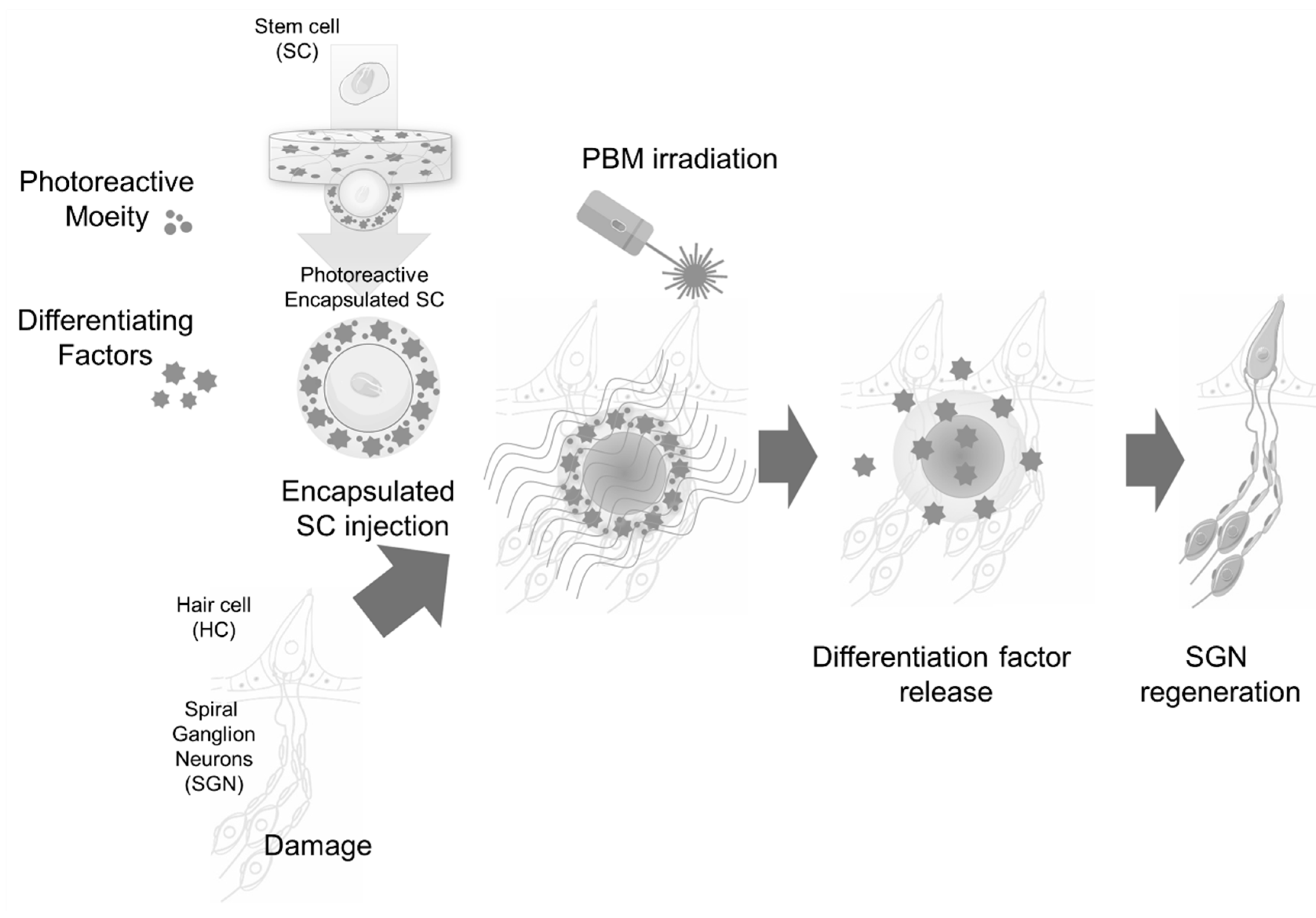
The main cause of hearing loss is damage to cochlear tissue in the inner ear. Normally, hair cells in the cochlea convert external mechanical signals into electrical signals and transmit them to the brain. Hair cell damage disrupts this process, causing hearing loss. Because hair cell damage or loss is irreversible, it is very difficult to treat hearing loss. Therefore, the rescue of hair cells is an important target for therapeutic treatment. Sensorineural hearing loss treatment using stem cells is currently being investigated, with a focus on restoring damaged hair cells. However, due to the structural complexity of the inner ear and the characteristics of the environment within the cochlea, the engraftment and survival rates of stem cells transplanted from outside the body have been insufficient to restore hearing loss. To overcome these limitations, some studies have attempted a PBM-based promotion of cell differentiation for cochlear hair cell regeneration. In an auditory study, otic cell differentiation was enhanced through otic vesicle formation and an increased proliferation of myosin VIIa-positive cells after three-dimensional mESC cultures were irradiated with 630 nm LED light to induce PBM. [80]. Non-invasive irradiation (>800 nm) of the inner ear through the tympanic membrane strengthened otic-like organoids and enhanced the adhesion and differentiation potential of transplanted stem cells within the cochlea [81]. The development of hearing rehabilitation technologies, such as cochlear implants, has allowed patients with profound hearing loss to regain their hearing [82]. However, spiral ganglion neurons, which are auditory neurons that transfer neural signals from the cochlea to the brainstem, must be healthy and functional to attain optimal cochlear implant performance [83]. Considering that these auditory neurons degenerate following hearing deprivation, the performance of cochlear implants tends to decline over time. Thus, the discovery of a technique for regenerating spiral ganglion neurons from stem cells would be highly beneficial to those with hearing loss. Although several studies have proposed methods for the differentiation of auditory neurons from stem cells [84,85,86], optimal methods have not been demonstrated for such auditory neuron differentiation within the cochlea or for stem cell migration to Rosenthal’s canal, where auditory neurons reside. PBM may not be best tool for inducing stem cell differentiation in Rosenthal’s canal because it is located in the medial section of the cochlea. Considering the low penetration rate in this anatomical region, PBM efficiency would be extremely low. However, there are alternatives, such as soluble hydrogel recently developed for delivering light energy to remote anatomical locations [87]. This hydrogel can be modified to encapsulate cell differentiation and homing factors for the neural differentiation of stem cells. Such an approach could allow us to selectively activate stem cells using light energy. A schematic of this methodology is shown in Figure 2.

4. Conclusions and Future Directions

PBM has shown potential to promote neurogenesis in the CNS or PNS by enhancing cell differentiation from stem cells or neural progenitor cells to nearby target cells or target cells. Nevertheless, several limitations and challenges must be considered. As PBM employs non-invasive light, the tissue penetration rate varies depending on the light source utilized, and the results may exhibit biphasic patterns depending on the frequency of irradiations and energy dosage. In other words, an inappropriate optimal dose may either yield no PBM effect on the target or be excessively high, suppressing the intended PBM effect [52,88,89]. Consequently, PBM can be applied to various areas of the body, but to elicit positive treatment outcomes, it is imperative to establish an appropriate protocol tailored to the target tissue. However, because the PBM parameters presented in the papers reported so far are very diverse, the standardization of PBM has not been clearly established to date. Moreover, ongoing research into PBM’s capacity to stimulate neurogenesis in both the central and peripheral nervous systems underscores the evolving nature of this field. Therefore, while the field continues to progress, it is important to consider potential safety and side effects.
Determining the optimal PBM parameters for neurogenesis remains challenging. Parameters such as the wavelength, energy density, treatment duration, and timing of PBM application require further optimization. Different cell types and experimental conditions may require different parameters, making it difficult to establish a standardized protocol. Stem and neural progenitor cell populations in the CNS or peripheral sensory nerves are heterogeneous. Different subtypes of stem or progenitor cells may respond differently to PBM, and their specific requirements for differentiation and neurogenesis may vary. Future advances will depend on understanding this heterogeneity and tailoring PBM approaches accordingly.
Light penetration through tissues is limited, particularly in the CNS. The depth of penetration depends on the light wavelength. Although NIR light can penetrate deeper into tissues than visible light, reaching target cells deep within the CNS may remain a challenge. This limitation could impact the effects of PBM on neurogenesis in deep regions of the CNS, complicating the precise targeting of stem cells or neural progenitor cells for PBM. Ensuring that light reaches the desired cell population within complex neural tissue structures is also difficult. Strategies such as the application of cell-specific markers or targeted delivery methods must be further explored to enhance the specificity of PBM.
The response to PBM can vary among individuals and experimental conditions. Factors such as age, health status, genetic background, and diseases/conditions can influence the cellular response to PBM. To draw reliable conclusions, it is important to standardize experimental conditions and consider variability in cell responses. The long-term effects and safety profile of PBM for neurogenesis require further investigation. PBM has shown promising results in preclinical studies; however, further research is needed to assess its potential adverse effects, determine optimal treatment regimens, and ensure the long-term safety of PBM-based approaches. Thus, although PBM research has demonstrated benefits in preclinical models, transforming these findings into effective clinical applications is a complex process. Clinical studies involving human subjects are necessary to determine the efficacy, safety, and feasibility of PBM for neurogenesis in the CNS or PNS. Addressing these limitations through further research, optimizing PBM parameters, and designing appropriate clinical trials will contribute to a better understanding of the potential of PBM for neurogenesis and its application in regenerative medicine.

Author Contributions

Conceptualization, M.Y.L.; writing—original draft preparation, M.Y.L. and S.-Y.C.; writing—review and editing, M.Y.L. and S.-Y.C.; supervision, M.Y.L.; funding acquisition, M.Y.L. All authors have read and agreed to the published version of the manuscript.

Funding

This work was supported by basic Science Research Program through the National Research Foundation of Korea (NRF) funded by the Ministry of Education [NRF-2020R1A6A1A03043283; NRF-2021R1I1A3047407]; Creative Materials Discovery Program through the National Research Foundation of Korea (NRF) funded by Ministry of Science and ICT [NRF-2019M3D1A1078943]; Bio&Medical Technology Development Program of the National Research Foundation (NRF) funded by the Korean government (MSIT) [RS-2023-00220408].

Institutional Review Board Statement

Not applicable.

Informed Consent Statement

Not applicable.

Data Availability Statement

No new data were created or analyzed in this study. Data sharing is not applicable to this article.

Conflicts of Interest

The authors declare no conflict of interest.

References

  1. Reddy, D.S.; Abeygunaratne, H.N. Experimental and Clinical Biomarkers for Progressive Evaluation of Neuropathology and Therapeutic Interventions for Acute and Chronic Neurological Disorders. Int. J. Mol. Sci. 2022, 23, 11734. [Google Scholar] [CrossRef] [PubMed]
  2. GBD 2016 Stroke Collaborators. Global, regional, and national burden of stroke, 1990–2016: A systematic analysis for the Global Burden of Disease Study 2016. Lancet Neurol. 2019, 18, 439–458. [Google Scholar] [CrossRef] [PubMed]
  3. GBD 2016 Stroke Collaborators. Global, regional, and national burden of multiple sclerosis 1990–2016: A systematic analysis for the Global Burden of Disease Study 2016. Lancet Neurol. 2019, 18, 269–285. [Google Scholar] [CrossRef]
  4. GBD 2016 Stroke Collaborators. Global, regional, and national burden of epilepsy, 1990–2016: A systematic analysis for the Global Burden of Disease Study 2016. Lancet Neurol. 2019, 18, 357–375. [Google Scholar] [CrossRef]
  5. Wang, R.; Nie, X.; Xu, S.; Zhang, M.; Dong, Z.; Yu, S. Interrelated Pathogenesis? Neuronal Intranuclear Inclusion Disease Combining with Hemiplegic Migraine. Headache 2020, 60, 382–395. [Google Scholar] [CrossRef]
  6. Stascheit, F.; Aigner, A.; Mergenthaler, P.; Hotter, B.; Hoffmann, S.; Lehnerer, S.; Meisel, C.; Meisel, A. Serum neurofilament light chain in myasthenia gravis subgroups: An exploratory cohort and case-Control study. Front. Neurol. 2022, 13, 1056322. [Google Scholar] [CrossRef] [PubMed]
  7. Ciurea, A.V.; Mohan, A.G.; Covache-Busuioc, R.A.; Costin, H.P.; Glavan, L.A.; Corlatescu, A.D.; Saceleanu, V.M. Unraveling Molecular and Genetic Insights into Neurodegenerative Diseases: Advances in Understanding Alzheimer’s, Parkinson’s, and Huntington’s Diseases and Amyotrophic Lateral Sclerosis. Int. J. Mol. Sci. 2023, 24, 10809. [Google Scholar] [CrossRef]
  8. Brenner, D.; Ludolph, A.C. Gene-specific treatment of neurological diseases-current state and perspectives. Nervenarzt 2020, 91, 285–286. [Google Scholar] [CrossRef]
  9. Russo, M.V.; McGavern, D.B. Immune Surveillance of the CNS following Infection and Injury. Trends Immunol. 2015, 36, 637–650. [Google Scholar] [CrossRef]
  10. Stephenson, J.; Nutma, E.; van der Valk, P.; Amor, S. Inflammation in CNS neurodegenerative diseases. Immunology 2018, 154, 204–219. [Google Scholar] [CrossRef]
  11. Jorg, S.; Grohme, D.A.; Erzler, M.; Binsfeld, M.; Haghikia, A.; Muller, D.N.; Linker, R.A.; Kleinewietfeld, M. Environmental factors in autoimmune diseases and their role in multiple sclerosis. Cell. Mol. Life Sci. 2016, 73, 4611–4622. [Google Scholar] [CrossRef] [PubMed]
  12. Li, Z.; Jiang, Z.; Lu, L.; Liu, Y. Microfluidic Manipulation for Biomedical Applications in the Central and Peripheral Nervous Systems. Pharmaceutics 2023, 15, 210. [Google Scholar] [CrossRef] [PubMed]
  13. Wulf, M.J.; Tom, V.J. Consequences of spinal cord injury on the sympathetic nervous system. Front. Cell. Neurosci. 2023, 17, 999253. [Google Scholar] [CrossRef] [PubMed]
  14. Lanigan, L.G.; Russell, D.S.; Woolard, K.D.; Pardo, I.D.; Godfrey, V.; Jortner, B.S.; Butt, M.T.; Bolon, B. Comparative Pathology of the Peripheral Nervous System. Vet. Pathol. 2021, 58, 10–33. [Google Scholar] [CrossRef]
  15. Krinke, G.J.; Herrmann, A.; Korner, A.; Landes, C.; Sauner, F. Experience with examination of the spinal cord and peripheral nervous system (PNS) in mice: A brief overview. Exp. Toxicol. Pathol. 2014, 66, 277–280. [Google Scholar] [CrossRef]
  16. Ray, S.; Singhvi, A. Charging Up the Periphery: Glial Ionic Regulation in Sensory Perception. Front. Cell Dev. Biol. 2021, 9, 687732. [Google Scholar] [CrossRef]
  17. Heiskanen, V.; Hamblin, M.R. Correction: Photobiomodulation: Lasers vs. light emitting diodes? Photochem. Photobiol. Sci. 2018, 18, 259. [Google Scholar] [CrossRef]
  18. de Freitas, L.F.; Hamblin, M.R. Proposed Mechanisms of Photobiomodulation or Low-Level Light Therapy. IEEE J. Sel. Top. Quantum Electron. 2016, 22, 348–364. [Google Scholar] [CrossRef]
  19. Derakhshan, R.; Ahmadi, H.; Bayat, M.; Mehboudi, L.; Pourhashemi, E.; Amini, A.; Vatandoust, D.; Aghamiri, S.; Asadi, R.; Sabet, B. The Combined Effects of a Methacrylate Powder Dressing (Altrazeal Powder) and Photobiomodulation Therapy on the Healing of a Severe Diabetic Foot Ulcer in a Diabetic Patient: A Case Report. J. Lasers Med. Sci. 2022, 13, e38. [Google Scholar] [CrossRef]
  20. Santiago, R.; Gomes, S.; Ozsarfati, J.; Zitney, M. Photobiomodulation for modulation of neuropathic pain and improvement of scar tissue. Scars Burn. Heal. 2022, 8, 20595131221134052. [Google Scholar] [CrossRef]
  21. Reis, C.H.B.; Buchaim, D.V.; Ortiz, A.C.; Fideles, S.O.M.; Dias, J.A.; Miglino, M.A.; Teixeira, D.B.; Pereira, E.; da Cunha, M.R.; Buchaim, R.L. Application of Fibrin Associated with Photobiomodulation as a Promising Strategy to Improve Regeneration in Tissue Engineering: A Systematic Review. Polymers 2022, 14, 3150. [Google Scholar] [CrossRef] [PubMed]
  22. Yeh, S.W.; Hong, C.H.; Shih, M.C.; Tam, K.W.; Huang, Y.H.; Kuan, Y.C. Low-Level Laser Therapy for Fibromyalgia: A Systematic Review and Meta-Analysis. Pain. Physician 2019, 22, 241–254. [Google Scholar] [PubMed]
  23. Wickenheisser, V.A.; Zywot, E.M.; Rabjohns, E.M.; Lee, H.H.; Lawrence, D.S.; Tarrant, T.K. Laser Light Therapy in Inflammatory, Musculoskeletal, and Autoimmune Disease. Curr. Allergy Asthma Rep. 2019, 19, 37. [Google Scholar] [CrossRef]
  24. Gonzalez-Munoz, A.; Cuevas-Cervera, M.; Perez-Montilla, J.J.; Aguilar-Nunez, D.; Hamed-Hamed, D.; Aguilar-Garcia, M.; Pruimboom, L.; Navarro-Ledesma, S. Efficacy of Photobiomodulation Therapy in the Treatment of Pain and Inflammation: A Literature Review. Healthcare 2023, 11, 938. [Google Scholar] [CrossRef] [PubMed]
  25. Ailioaie, L.M.; Litscher, G. Photobiomodulation and Sports: Results of a Narrative Review. Life 2021, 11, 1339. [Google Scholar] [CrossRef] [PubMed]
  26. Da Silva, D.; Crous, A.; Abrahamse, H. Photobiomodulation: An Effective Approach to Enhance Proliferation and Differentiation of Adipose-Derived Stem Cells into Osteoblasts. Stem Cells Int. 2021, 2021, 8843179. [Google Scholar] [CrossRef]
  27. Migaud, M.; Batailler, M.; Segura, S.; Duittoz, A.; Franceschini, I.; Pillon, D. Emerging new sites for adult neurogenesis in the mammalian brain: A comparative study between the hypothalamus and the classical neurogenic zones. Eur. J. Neurosci. 2010, 32, 2042–2052. [Google Scholar] [CrossRef]
  28. Ming, G.L.; Song, H. Adult neurogenesis in the mammalian brain: Significant answers and significant questions. Neuron 2011, 70, 687–702. [Google Scholar] [CrossRef]
  29. Kazanis, I. Neurogenesis in the adult mammalian brain: How much do we need, how much do we have? Curr. Top. Behav. Neurosci. 2013, 15, 3–29. [Google Scholar] [CrossRef]
  30. Lledo, P.M.; Valley, M. Adult Olfactory Bulb Neurogenesis. Cold Spring Harb. Perspect. Biol. 2016, 8. [Google Scholar] [CrossRef]
  31. Liu, J.; Wang, Y.; Fu, W. Axon regeneration impediment: The role of paired immunoglobulin-like receptor B. Neural Regen. Res. 2015, 10, 1338–1342. [Google Scholar] [CrossRef] [PubMed]
  32. Zhou, X.; Cui, H.; Nowicki, M.; Miao, S.; Lee, S.J.; Masood, F.; Harris, B.T.; Zhang, L.G. Three-Dimensional-Bioprinted Dopamine-Based Matrix for Promoting Neural Regeneration. ACS Appl. Mater. Interfaces 2018, 10, 8993–9001. [Google Scholar] [CrossRef] [PubMed]
  33. Grimaudo, M.A.; Krishnakumar, G.S.; Giusto, E.; Furlani, F.; Bassi, G.; Rossi, A.; Molinari, F.; Lista, F.; Montesi, M.; Panseri, S. Bioactive injectable hydrogels for on demand molecule/cell delivery and for tissue regeneration in the central nervous system. Acta Biomater. 2022, 140, 88–101. [Google Scholar] [CrossRef]
  34. Hasanzadeh, E.; Seifalian, A.; Mellati, A.; Saremi, J.; Asadpour, S.; Enderami, S.E.; Nekounam, H.; Mahmoodi, N. Injectable hydrogels in central nervous system: Unique and novel platforms for promoting extracellular matrix remodeling and tissue engineering. Mater. Today Bio 2023, 20, 100614. [Google Scholar] [CrossRef] [PubMed]
  35. Guo, S.; Wang, R.; Hu, J.; Sun, L.; Zhao, X.; Zhao, Y.; Han, D.; Hu, S. Photobiomodulation Promotes Hippocampal CA1 NSC Differentiation Toward Neurons and Facilitates Cognitive Function Recovery Involving NLRP3 Inflammasome Mitigation Following Global Cerebral Ischemia. Front. Cell. Neurosci. 2021, 15, 731855. [Google Scholar] [CrossRef]
  36. Liao, Z.; Zhou, X.; Li, S.; Jiang, W.; Li, T.; Wang, N.; Xiao, N. Activation of the AKT/GSK-3beta/beta-catenin pathway via photobiomodulation therapy promotes neural stem cell proliferation in neonatal rat models of hypoxic-ischemic brain damage. Ann. Transl. Med. 2022, 10, 55. [Google Scholar] [CrossRef]
  37. Xuan, W.; Vatansever, F.; Huang, L.; Hamblin, M.R. Transcranial low-level laser therapy enhances learning, memory, and neuroprogenitor cells after traumatic brain injury in mice. J. Biomed. Opt. 2014, 19, 108003. [Google Scholar] [CrossRef]
  38. Wu, X.; Shen, Q.; Zhang, Z.; Zhang, D.; Gu, Y.; Xing, D. Photoactivation of TGFbeta/SMAD signaling pathway ameliorates adult hippocampal neurogenesis in Alzheimer’s disease model. Stem Cell Res. Ther. 2021, 12, 345. [Google Scholar] [CrossRef]
  39. Yang, L.; Dong, Y.; Wu, C.; Youngblood, H.; Li, Y.; Zong, X.; Li, L.; Xu, T.; Zhang, Q. Effects of prenatal photobiomodulation treatment on neonatal hypoxic ischemia in rat offspring. Theranostics 2021, 11, 1269–1294. [Google Scholar] [CrossRef]
  40. Salehpour, F.; Farajdokht, F.; Cassano, P.; Sadigh-Eteghad, S.; Erfani, M.; Hamblin, M.R.; Salimi, M.M.; Karimi, P.; Rasta, S.H.; Mahmoudi, J. Near-infrared photobiomodulation combined with coenzyme Q10 for depression in a mouse model of restraint stress: Reduction in oxidative stress, neuroinflammation, and apoptosis. Brain Res. Bull. 2019, 144, 213–222. [Google Scholar] [CrossRef]
  41. Yang, L.; Wu, C.; Parker, E.; Li, Y.; Dong, Y.; Tucker, L.; Brann, D.W.; Lin, H.W.; Zhang, Q. Non-invasive photobiomodulation treatment in an Alzheimer Disease-like transgenic rat model. Theranostics 2022, 12, 2205–2231. [Google Scholar] [CrossRef] [PubMed]
  42. Meynaghizadeh-Zargar, R.; Sadigh-Eteghad, S.; Mohaddes, G.; Salehpour, F.; Rasta, S.H. Effects of transcranial photobiomodulation and methylene blue on biochemical and behavioral profiles in mice stress model. Lasers Med. Sci. 2020, 35, 573–584. [Google Scholar] [CrossRef] [PubMed]
  43. Tsai, C.M.; Chang, S.F.; Li, C.C.; Chang, H. Transcranial photobiomodulation (808 nm) attenuates pentylenetetrazole-induced seizures by suppressing hippocampal neuroinflammation, astrogliosis, and microgliosis in peripubertal rats. Neurophotonics 2022, 9, 015006. [Google Scholar] [CrossRef]
  44. Blivet, G.; Meunier, J.; Roman, F.J.; Touchon, J. Neuroprotective effect of a new photobiomodulation technique against Abeta(25–35) peptide-induced toxicity in mice: Novel hypothesis for therapeutic approach of Alzheimer’s disease suggested. Alzheimer’s Dement. 2018, 4, 54–63. [Google Scholar] [CrossRef]
  45. Hong, N.; Kang, G.W.; Park, J.O.; Chung, P.S.; Lee, M.Y.; Ahn, J.C. Photobiomodulation regulates adult neurogenesis in the hippocampus in a status epilepticus animal model. Sci. Rep. 2022, 12, 15246. [Google Scholar] [CrossRef] [PubMed]
  46. Feng, Y.; Yang, L.; Ma, X.; Huang, Z.; Zong, X.; Citadin, C.T.; Lin, H.W.; Zhang, Q. Photobiomodulation treatment inhibits neurotoxic astrocytic polarization and protects neurons in in vitro and in vivo stroke models. Neurochem. Int. 2023, 162, 105464. [Google Scholar] [CrossRef]
  47. Yang, L.; Wu, C.; Tucker, L.; Dong, Y.; Li, Y.; Xu, P.; Zhang, Q. Photobiomodulation Therapy Attenuates Anxious-Depressive-Like Behavior in the TgF344 Rat Model. J. Alzheimer’s Dis. 2021, 83, 1415–1429. [Google Scholar] [CrossRef]
  48. Yang, L.; Tucker, D.; Dong, Y.; Wu, C.; Lu, Y.; Li, Y.; Zhang, J.; Liu, T.C.; Zhang, Q. Photobiomodulation therapy promotes neurogenesis by improving post-stroke local microenvironment and stimulating neuroprogenitor cells. Exp. Neurol. 2018, 299, 86–96. [Google Scholar] [CrossRef]
  49. Wang, R.; Dong, Y.; Lu, Y.; Zhang, W.; Brann, D.W.; Zhang, Q. Photobiomodulation for Global Cerebral Ischemia: Targeting Mitochondrial Dynamics and Functions. Mol. Neurobiol. 2019, 56, 1852–1869. [Google Scholar] [CrossRef]
  50. Vogel, D.D.S.; Ortiz-Villatoro, N.N.; Araujo, N.S.; Marques, M.J.G.; Aimbire, F.; Scorza, F.A.; Scorza, C.A.; Albertini, R. Transcranial low-level laser therapy in an in vivo model of stroke: Relevance to the brain infarct, microglia activation and neuroinflammation. J. Biophotonics 2021, 14, e202000500. [Google Scholar] [CrossRef]
  51. Zein, R.; Selting, W.; Hamblin, M.R. Review of light parameters and photobiomodulation efficacy: Dive into complexity. J. Biomed. Opt. 2018, 23, 120901. [Google Scholar] [CrossRef] [PubMed]
  52. Hamblin, M.R. Mechanisms and applications of the anti-inflammatory effects of photobiomodulation. AIMS Biophys. 2017, 4, 337–361. [Google Scholar] [CrossRef] [PubMed]
  53. Hamblin, M.R. Mechanisms and Mitochondrial Redox Signaling in Photobiomodulation. Photochem. Photobiol. 2018, 94, 199–212. [Google Scholar] [CrossRef]
  54. Li, X.; Wang, X.K.; Zhu, Z.J.; Liang, Z.W.; Li, P.H.; Ma, Y.G.; Ding, T.; Li, K.; Zuo, X.S.; Ju, C.; et al. Photobiomodulation provides neuroprotection through regulating mitochondrial fission imbalance in the subacute phase of spinal cord injury. Neural Regen. Res. 2023, 18, 2005–2010. [Google Scholar] [CrossRef] [PubMed]
  55. Yang, L.; Youngblood, H.; Wu, C.; Zhang, Q. Mitochondria as a target for neuroprotection: Role of methylene blue and photobiomodulation. Transl. Neurodegener. 2020, 9, 19. [Google Scholar] [CrossRef] [PubMed]
  56. Mo, S.; Kim, E.Y.; Kwon, Y.S.; Lee, M.Y.; Ahn, J.C. NF-kappaB-mediated anti-inflammatory effects of an organic light-emitting diode (OLED) device in lipopolysaccharide (LPS)-induced in vitro and in vivo inflammation models. Front. Immunol. 2022, 13, 1050908. [Google Scholar] [CrossRef]
  57. Hong, N.; Kim, H.J.; Kang, K.; Park, J.O.; Mun, S.; Kim, H.G.; Kang, B.H.; Chung, P.S.; Lee, M.Y.; Ahn, J.C. Photobiomodulation improves the synapses and cognitive function and ameliorates epileptic seizure by inhibiting downregulation of Nlgn3. Cell Biosci. 2023, 13, 8. [Google Scholar] [CrossRef]
  58. George, S.; Hamblin, M.R.; Abrahamse, H. Photobiomodulation-Induced Differentiation of Immortalized Adipose Stem Cells to Neuronal Cells. Lasers Surg. Med. 2020, 52, 1032–1040. [Google Scholar] [CrossRef]
  59. George, S.; Hamblin, M.R.; Abrahamse, H. Neuronal differentiation potential of primary and immortalized adipose stem cells by photobiomodulation. J. Photochem. Photobiol. 2022, 230, 112445. [Google Scholar] [CrossRef]
  60. Vaz, A.; Ribeiro, I.; Pinto, L. Frontiers in Neurogenesis. Cells 2022, 11, 3567. [Google Scholar] [CrossRef]
  61. Hamblin, M.R.; Salehpour, F. Photobiomodulation of the Brain: Shining Light on Alzheimer’s and Other Neuropathological Diseases. J. Alzheimer’s Dis. 2021, 83, 1395–1397. [Google Scholar] [CrossRef]
  62. Su, M.; Nizamutdinov, D.; Liu, H.; Huang, J.H. Recent Mechanisms of Neurodegeneration and Photobiomodulation in the Context of Alzheimer’s Disease. Int. J. Mol. Sci. 2023, 24, 9272. [Google Scholar] [CrossRef]
  63. Hamblin, M.R. Photobiomodulation for Alzheimer’s Disease: Has the Light Dawned? Photonics 2019, 6, 77. [Google Scholar] [CrossRef]
  64. Yoon, S.R.; Hong, N.; Lee, M.Y.; Ahn, J.C. Photobiomodulation with a 660-Nanometer Light-Emitting Diode Promotes Cell Proliferation in Astrocyte Culture. Cells 2021, 10, 1664. [Google Scholar] [CrossRef] [PubMed]
  65. Huang, Z.; Zhang, Y.; Ma, X.; Feng, Y.; Zong, X.; Jordan, J.D.; Zhang, Q. Photobiomodulation attenuates oligodendrocyte dysfunction and prevents adverse neurological consequences in a rat model of early life adversity. Theranostics 2023, 13, 913–930. [Google Scholar] [CrossRef] [PubMed]
  66. Dos Santos Cardoso, F.; Dos Santos, J.C.C.; Gonzalez-Lima, F.; Araujo, B.H.S.; Lopes-Martins, R.A.B.; Gomes da Silva, S. Effects of Chronic Photobiomodulation with Transcranial Near-Infrared Laser on Brain Metabolomics of Young and Aged Rats. Mol. Neurobiol. 2021, 58, 2256–2268. [Google Scholar] [CrossRef]
  67. Barohn, R.J.; Amato, A.A. Pattern-recognition approach to neuropathy and neuronopathy. Neurol. Clin. 2013, 31, 343–361. [Google Scholar] [CrossRef]
  68. Chung, T.; Prasad, K.; Lloyd, T.E. Peripheral neuropathy: Clinical and electrophysiological considerations. Neuroimaging Clin. N. Am. 2014, 24, 49–65. [Google Scholar] [CrossRef] [PubMed]
  69. Nadol, J.B., Jr.; Hedley-Whyte, E.T.; Amr, S.S.; O’Malley, J.T.; Kamakura, T. Histopathology of the Inner Ear in Charcot-Marie-Tooth Syndrome Caused by a Missense Variant (p.Thr65Ala) in the MPZ Gene. Audiol. Neuro-Otol. 2018, 23, 326–334. [Google Scholar] [CrossRef]
  70. Eggermann, K.; Gess, B.; Hausler, M.; Weis, J.; Hahn, A.; Kurth, I. Hereditary Neuropathies. Dtsch. Arztebl. Int. 2018, 115, 91–97. [Google Scholar] [CrossRef]
  71. Creigh, P.D.; Mountain, J.; Sowden, J.E.; Eichinger, K.; Ravina, B.; Larkindale, J.; Herrmann, D.N. Measuring peripheral nerve involvement in Friedreich’s ataxia. Ann. Clin. Transl. Neurol. 2019, 6, 1718–1727. [Google Scholar] [CrossRef] [PubMed]
  72. Lerat, J.; Magdelaine, C.; Roux, A.F.; Darnaud, L.; Beauvais-Dzugan, H.; Naud, S.; Richard, L.; Derouault, P.; Ghorab, K.; Magy, L.; et al. Hearing loss in inherited peripheral neuropathies: Molecular diagnosis by NGS in a French series. Mol. Genet. Genom. Med. 2019, 7, e839. [Google Scholar] [CrossRef] [PubMed]
  73. Mahmut, M.K.; Musch, M.; Han, P.; Abolmaali, N.; Hummel, T. The effect of olfactory training on olfactory bulb volumes in patients with idiopathic olfactory loss. Rhinology 2020, 58, 410–412. [Google Scholar] [CrossRef] [PubMed]
  74. Hummel, T.; Power Guerra, N.; Gunder, N.; Hahner, A.; Menzel, S. Olfactory Function and Olfactory Disorders. Laryngo-Rhino-Otol. 2023, 102, S67–S92. [Google Scholar] [CrossRef]
  75. Jha, N.K.; Ojha, S.; Jha, S.K.; Dureja, H.; Singh, S.K.; Shukla, S.D.; Chellappan, D.K.; Gupta, G.; Bhardwaj, S.; Kumar, N.; et al. Evidence of Coronavirus (CoV) Pathogenesis and Emerging Pathogen SARS-CoV-2 in the Nervous System: A Review on Neurological Impairments and Manifestations. J. Mol. Neurosci. 2021, 71, 2192–2209. [Google Scholar] [CrossRef]
  76. Soria, E.D.; Candaras, M.M.; Truax, B.T. Impairment of taste in the Guillain-Barre syndrome. Clin. Neurol. Neurosurg. 1990, 92, 75–79. [Google Scholar] [CrossRef]
  77. Sweeney, C.J.; Gilden, D.H. Ramsay Hunt syndrome. J. Neurol. Neurosurg. Psychiatry 2001, 71, 149–154. [Google Scholar] [CrossRef]
  78. Hildebrand, M.S.; Newton, S.S.; Gubbels, S.P.; Sheffield, A.M.; Kochhar, A.; de Silva, M.G.; Dahl, H.M.; Rose, S.D.; Behlke, M.A.; Smith, R.J. Advances in Molecular and Cellular Therapies for Hearing Loss. Mol. Ther. J. Am. Soc. Gene Ther. 2008, 16, 224–236. [Google Scholar] [CrossRef]
  79. Mainland, J.D.; Barlow, L.A.; Munger, S.D.; Millar, S.E.; Vergara, M.N.; Jiang, P.; Schwob, J.E.; Goldstein, B.J.; Boye, S.E.; Martens, J.R.; et al. Identifying Treatments for Taste and Smell Disorders: Gaps and Opportunities. Chem. Senses 2020, 45, 493–502. [Google Scholar] [CrossRef]
  80. Chang, S.Y.; Carpena, N.T.; Mun, S.; Jung, J.Y.; Chung, P.S.; Shim, H.; Han, K.; Ahn, J.C.; Lee, M.Y. Enhanced Inner-Ear Organoid Formation from Mouse Embryonic Stem Cells by Photobiomodulation. Mol. Ther. Methods Clin. Dev. 2020, 17, 556–567. [Google Scholar] [CrossRef]
  81. Chang, S.Y.; Lee, M.Y. Photobiomodulation with a wavelength > 800 nm induces morphological changes in stem cells within otic organoids and scala media of the cochlea. Lasers Med. Sci. 2021, 36, 1917–1925. [Google Scholar] [CrossRef] [PubMed]
  82. Litovsky, R.Y.; Moua, K.; Godar, S.; Kan, A.; Misurelli, S.M.; Lee, D.J. Restoration of spatial hearing in adult cochlear implant users with single-sided deafness. Hear. Res. 2019, 372, 69–79. [Google Scholar] [CrossRef]
  83. Zhang, L.; Chen, S.; Sun, Y. Mechanism and Prevention of Spiral Ganglion Neuron Degeneration in the Cochlea. Front. Cell. Neurosci. 2021, 15, 814891. [Google Scholar] [CrossRef] [PubMed]
  84. Lang, H.; Nishimoto, E.; Xing, Y.; Brown, L.N.; Noble, K.V.; Barth, J.L.; LaRue, A.C.; Ando, K.; Schulte, B.A. Contributions of Mouse and Human Hematopoietic Cells to Remodeling of the Adult Auditory Nerve After Neuron Loss. Mol. Ther. J. Am. Soc. Gene Ther. 2016, 24, 2000–2011. [Google Scholar] [CrossRef]
  85. Matsuoka, A.J.; Morrissey, Z.D.; Zhang, C.; Homma, K.; Belmadani, A.; Miller, C.A.; Chadly, D.M.; Kobayashi, S.; Edelbrock, A.N.; Tanaka-Matakatsu, M.; et al. Directed Differentiation of Human Embryonic Stem Cells Toward Placode-Derived Spiral Ganglion-Like Sensory Neurons. Stem Cells Transl. Med. 2017, 6, 923–936. [Google Scholar] [CrossRef]
  86. Schomann, T.; Mezzanotte, L.; De Groot, J.; Rivolta, M.N.; Hendriks, S.H.; Frijns, J.H.M.; Huisman, M.A. Neuronal differentiation of hair-follicle-bulge-derived stem cells co-cultured with mouse cochlear modiolus explants. PLoS ONE 2017, 12, e0187183. [Google Scholar] [CrossRef] [PubMed]
  87. Peng, K.; Zheng, L.; Zhou, T.; Zhang, C.; Li, H. Light manipulation for fabrication of hydrogels and their biological applications. Acta Biomater. 2022, 137, 20–43. [Google Scholar] [CrossRef]
  88. Xuan, W.; Huang, L.; Hamblin, M.R. Repeated transcranial low-level laser therapy for traumatic brain injury in mice: Biphasic dose response and long-term treatment outcome. J. Biophotonics 2016, 9, 1263–1272. [Google Scholar] [CrossRef]
  89. Cassano, P.; Caldieraro, M.A.; Norton, R.; Mischoulon, D.; Trinh, N.-H.; Nyer, M.; Dording, C.; Hamblin, M.R.; Campbell, B.; Iosifescu, D.V. Reported Side Effects, Weight and Blood Pressure, After Repeated Sessions of Transcranial Photobiomodulation. Photobiomodulation Photomed. Laser Surg. 2019, 37, 651–656. [Google Scholar] [CrossRef]
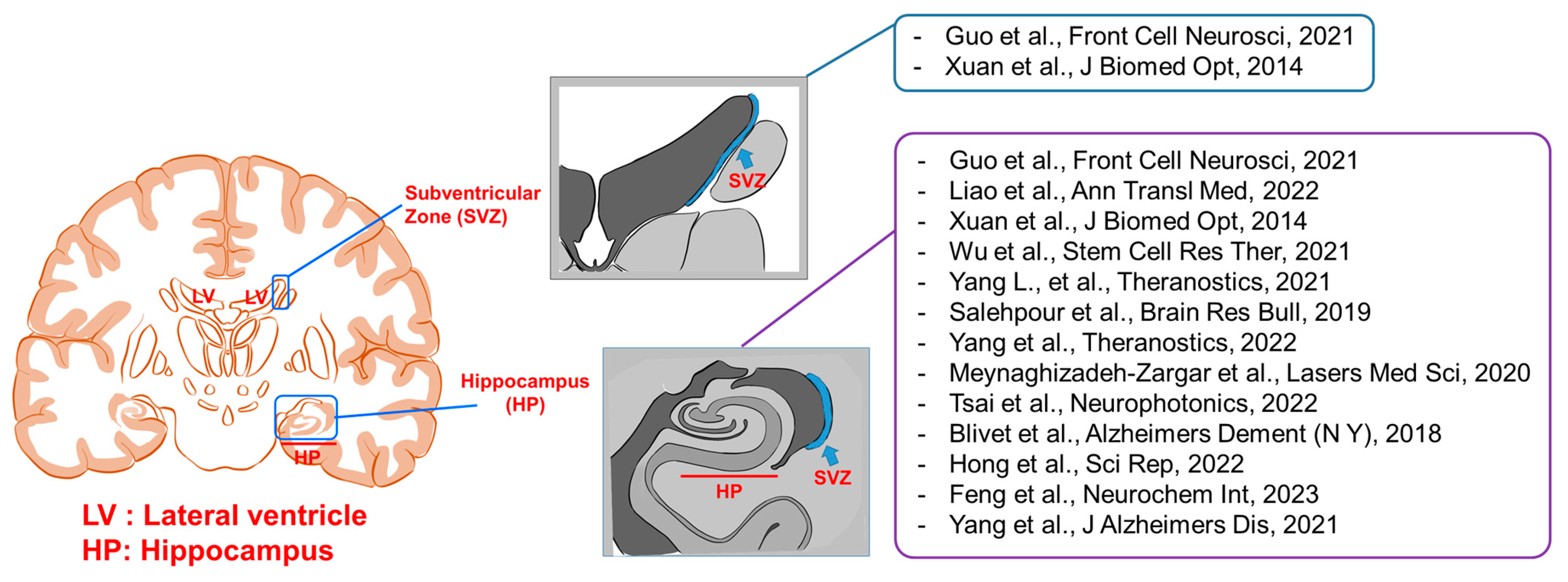
Figure 1. Anatomical targets for neurogenesis through photobiomodulation [35,36,37,38,39,40,41,42,43,44,45,46,47].
Ijms 24 15427 g001
Figure 2. Schematic diagram of cochlear delivery of stem cells encapsulated in photoactive hydrogels with homing and differentiation factors.
Ijms 24 15427 g002
Table 1. Summary of publications on photobiomodulation and neurogenesis.
ReferenceYearLight SourceWavelength (nm)EnergyLocationAnimal ModelTargetMechanismEfficiency
[48]2018Diode laser808120 s × 350 mW/cm2/day
Total: 294 J for 7 days
ScalpPT stroke ratNeuroprogenitor cells in the peri-infarct cortical region
  • Increased NeuN, MAP2, spinophilin, synaptophysin, BrdU, Ki67, and DCX
  • Increased mitochondrial Cytc oxidase activity and ATP levels
  • Decreased GPAP and Iba-1
  • Decreased TNF-α, IL-6, and IL-18; increased IL-4 and IL-10
  • Decreased CD32, 86, and iNOS; increased ARG1, TGFβ1, and CD206
  • Improved cortical neurogenesis
  • Enhanced mitochondrial function
  • Suppresses reactive gliosis
  • Inhibited pro-inflammatory cytokines
[35]2021Diode laser808120 s × 20 mW/cm2
Total: 7.2 J/cm2 for 3 days
Cortical surfaceGlobal cerebral ischemia ratHippocampal astrocyte and microglia cells
  • Increased BrdU and DCX-NeuN
  • Decreased GFAP, NLRP3, cleaved IL-1β, and Iba1
  • Improved cognitive function
  • Elevated endogenous neural stem cell proliferation
  • Protected reactive astrocytes and microglia cells at 58 days
[36]2022Helium–neon laser660600 s × 5 mW/cm2
Total: 42 J/cm2 for 14 days
Scalp and skullHI brain damage ratHippocampal neural stem cells
  • Increased Edu and Nestin
  • Increased p-GSK3β, β-catenin, cyclin D1, PI3K, and p-AKT,
  • Enhanced spatial learning and memory
  • Promoted hippocampal neural stem cell proliferation
[37]2014Laser810720 s × 25 mW/cm2
Total: 54 J/cm2 for 3 days
TranscranialCCI–TBI injury mouseHippocampal and SVZ neurons
  • Increased BrdU, DCX, and TUJ1
  • Downregulated caspase 3
  • Enhanced learning and memory
  • Increased neurogenesis in hippocampus and subventricular zone
[38]2021Semiconductor laser635600 s × 0.1 mW/cm2
Total: 60 J/cm2 for 1 month
Scalp and skullAmyloid precursor protein/presenilin 1 transgenic mouseHippocampal neural stem cells
  • Increased DCX, Nestin, and TUJ1; decreased GFAP
  • Activated TGFβ1
  • Increased Smad2/3 with Smad4; decreased Smad1/5/9 with Smad4
  • Enhanced spatial learning and memory
  • Improved adult hippocampal neurogenesis
ATP, adenosine triphosphate; BrdU, bromodeoxyuridine; CCI, controlled cortical impact; Cytc, cytochrome c; DCX, doublecortin; HI, hypoxic–ischemic; p-GSK3β, glycogen synthase kinase 3β; IL, interleukin; PT, photothrombotic; SVZ, subventricular zone; TBI, traumatic brain injury; TGFβ1, transforming growth factor β1.
Table 2. Summary of publications on photobiomodulation and neuroprotective effects, glial cells, and synaptogenesis.
ReferenceYearLight SourceWavelength (nm)EnergyAnatomical LocationAnimal ModelTarget CellsMechanismEfficiency
[49]2019Diode laser80836 s × 8 mW/cm2
Total: 4 J/cm2 for 14 days
ScalpGlobal cerebral ischemia ratHippocampal CA1 pyramidal neuron cells
  • Increased NeuN, Tom20, Opa1, Mfn1, MMP, and FD
  • Downregulated Bax/Bcl2, Cytc, Apaf1-casp-9, caspase 3, Annexin V, Drip1 GTPase, and Mdivi-1
  • Enhanced spatial learning and memory
  • Protected hippocampal CA1 neurons
  • Inhibited mitochondrial fragmentation and promoted mitochondrial function
[39]2021Diode laser808120 s × 8 mW/cm2
Total: 2.88 J/cm2/week for 3 weeks
Abdomen of pregnant rat from gestation day 1 to 21Neonatal HI ratMyeloid cells and hippocampal astrocytes
  • Increased synaptophysin, spinophilin, MAP2, MBP, NeuN, ATP, mfn2, MitoRed, iNOS, CD32, and S100
  • Decreased F-Jade C, caspase 3, caspase 9, Iba-1, GFAP, and C3d
  • Promoted synaptogenesis
  • Increased memory and mitochondrial function
  • Reduced inflammation and oxidative stress
[40]2019GaAlAs laser8105 s × 6.66 W/cm2
Total: 166.5 J/cm2 for 5 days
Transcranial
Scalp
Sub-chronic restraint stress mousePrefrontal cortex and hippocampal neuron cells
  • Increased TAC, GSH, GPx, and SOD
  • Decreased NF-kB, p38, and JNK
  • Downregulated BAX/Bcl2, Cytc, and caspase-3 and −9
  • Ameliorated depressive-like behaviors
  • Suppressed neuroinflammation, astrogliosis, and microgliosis
[41]2022Laser808120 s × 350 mW/cm2
Total: 126 J/cm2/week for 16 months
ScalpTransgenic TgF344-AD ratMicroglial cells and astrocytes in cortex and hippocampus
  • Increased MBP, MAP2, NeuN, PSD, PSD95, Spin, and SYP
  • Increased bouton vesicles and ATP
  • Increased NeuN, Ki67, BrdU, and DCX
  • Improved typical AD pathology
  • Recruited microglia surrounding amyloid plaques
[42]2020GaAlAs diode laser8101.7 s × 4.75 W/cm2
Total: 5 J/cm2/week for 4 weeks
TranscranialUnpredictable chronic mild stress mouseHippocampal cells
  • Decreased NO, ROS and SOD activity, and serum cortisol
  • Increased TAC and glutathione peroxidase
  • Improved anxiety-like behavior
  • Promoted spatial learning and memory
  • Reduced inflammation and oxidative stress
[43]2022GaAlAs diode laser808100 s × 1.333 W/cm2
Total: 133.3 J/cm2
TranscranialPentylenetetrazole-induced SE ratHippocampal microglial cells and astrocytes
  • Decreased NSE and GFAP
  • Decreased Iba-1 in CA3
  • Increased MT-CO1 in CA3
  • Suppressed neuroinflammation, astrogliosis, and microgliosis
[50]2021Diode laser780120 s × 0.083 W/cm2
Total: 30 J/cm2/week for 60 days
TranscranialIschemic stroke ratBrain tissue cells in the peri-lesional region
  • Increased GFAP; decreased Iba
  • Decreased TNF-α, IL-1β, and IL6
  • Increased astroglial activity
  • Attenuated neuroinflammation
[44]2018RGn500
(LED + laser) with static magnetic field
625
850
LED600 s × 28 mW/cm2
Total: 117.6 J/cm2 for 7 days
Transcranial and abdomen25-35 peptide toxicity mouseHippocampal and frontal cortex cells
  • Decreased lipid peroxidation
  • Decreased GFAP, Iba1, TNF-α, IL-1β, and IL-6
  • Downregulated BAX/Bcl2
  • Decreased Aβ1-42 and pTau
  • Enhanced memory restoration
  • Inhibited oxidative stress and intrinsic apoptosis
  • Alleviated AD phenotype
850Laser
[45]2022Diode laser830750 s × 30 mW/cm2
Total: 180 J/cm2
ScalpSE mouseMaturing granular cells in the hilus
  • Increased NeuN, Ki67, BrdU, and DCX
  • Increased cell proliferation and migration
  • Increased CA1 pyramidal neurons
[46]2023Diode laser808120 s × 350 mW/cm2
Total: 294 J/cm2 for 7 days
SkullPT stroke ratNeuronal dendrites and cortex astrocytes
  • Increased MAP2, synaptophsin, and S100A10
  • Inhibited GFAP and C3d
  • Decreased caspase 9 and BAX
  • Increased Bcl-xL
  • Inhibited neurotoxic astrocytic polarization
  • Preserved synaptic integrity
[47]2021Diode laser808120 s × 350 mW/cm2
Total: 126 J/cm2/week for 8 months
ScalpTransgenic TgF344-AD disease rat
  • Increased MAP2, PSD95, Cytc oxidase activity, ATP, IL-4, and IL-10
  • Decreased caspase 3-, 9, F-jade C, Iba-1, NF-κB, TNF-α, and IL-1β
  • Increased SOD2 activity; decreased MDA, 4-HNE, and p.H2A.X
  • Improved spatial memory
  • Attenuated anxiety- and depression-like behaviors
  • Inhibited neuroinflammation and oxidative stress
25–35, amyloid β25–35; AD, Alzheimer’s disease; ATP, adenosine triphosphate; BrdU, bromodeoxyuridine; Cytc, cytochrome c; DCX, doublecortin; GaAlAs, gallium aluminum arsenide; HI, hypoxic–ischemic; IL, interleukin; LED, light-emitting diode; NF-κB, nuclear factor kappa B; PT, photothrombotic; SE, status epilepticus; TNF-α, tumor necrosis factor-α.
Disclaimer/Publisher’s Note: The statements, opinions and data contained in all publications are solely those of the individual author(s) and contributor(s) and not of MDPI and/or the editor(s). MDPI and/or the editor(s) disclaim responsibility for any injury to people or property resulting from any ideas, methods, instructions or products referred to in the content.