Article

Sperm Capacitation and Kinematics in Phodopus Hamsters

by
Ana Sanchez-Rodriguez
,
Ingrid I. D. Idrovo
,
Juan Antonio Rielo
and
Eduardo R. S. Roldan
*
Department of Biodiversity and Evolutionary Biology, Museo Nacional de Ciencias Naturales (CSIC), Calle Jose Gutierrez Abascal 2, 28006 Madrid, Spain
*
Author to whom correspondence should be addressed.
Int. J. Mol. Sci. 2023, 24(22), 16093; https://doi.org/10.3390/ijms242216093
Submission received: 23 September 2023 / Revised: 28 October 2023 / Accepted: 6 November 2023 / Published: 8 November 2023
(This article belongs to the Special Issue Molecular Mechanisms of Sperm Activation)

Abstract

:
This study was designed to analyze changes in the spermatozoa of three species of Phodopus hamsters incubated under different conditions. Cauda epididymal sperm were incubated for 4 h in modified Tyrode’s medium containing albumin, lactate, pyruvate, and Hepes (mTALP-H), in the same medium with the addition of bicarbonate (mTALP-BH), or with bicarbonate and 20 ng/mL of progesterone (mTALP-BH+P4). Media with bicarbonate are believed to promote capacitation in rodent species. Sperm motility, viability, capacitation patterns, and kinematics were assessed at different times. Capacitation in live cells was quantified after staining with Hoechst 33258 and chlortetracycline. Patterns believed to correspond to non-capacitated cells (F pattern), capacitated, acrosome-intact cells (B pattern), and acrosome-reacted cells (AR pattern) were recognized. Kinematics were examined via computer-assisted sperm analysis (CASA). The results showed a decrease in total motility in all three species in different media, with a sharp decrease in progressive motility in bicarbonate-containing media (without or with progesterone), suggesting hyperactivated motion. However, none of the other signs of hyperactivation described in rodents (i.e., decrease in STR or LIN, together with an increase in ALH) were observed. F pattern cells diminished with time in all media and were generally lower in P. roborovskii and higher in P. campbelli. B pattern cells increased in mTALP-BH media in all species. Progesterone did not enhance the percentage of B pattern cells. Finally, AR pattern cells increased in all species incubated in different media, showing the highest percentage in P. roborovskii and the lowest in P. campbelli. Comparisons between media revealed that there were higher percentages of F pattern cells and lower percentages of B pattern cells over time in medium without bicarbonate (mTALP-H) in comparison to media containing bicarbonate (mTALP-BH; mTALP-BH+P4). Overall, changes consistent with the acquisition of capacitation and development of hyperactivated motility were found; however, further studies are required to better characterize media necessary to support the pathways involved in these processes in Phodopus species.

1. Introduction

Sperm “capacitation” was originally defined as the set of processes that spermatozoa undergo in the female tract and that are needed for fertilization [1]. It was first uncovered in rats [2] and in rabbits [2,3] by showing that sperm cells did not enter the egg until several hours after the injection in the rat’s periovarian sac or into the rabbit’s oviduct. Many studies have been conducted to understand the process of capacitation. Currently, the term capacitation is generally used in a more restrictive way, as many authors reserve this term for the processes occurring before acrosomal exocytosis (the so-called “acrosome reaction”) [4,5,6,7]; the latter involves the fusion of the outer acrosomal membrane with the plasma membrane and is needed for the sperm to penetrate the oocyte [8]. Other authors still argue that capacitation includes all the events occurring in the female tract [9,10], which would, by definition, include acrosomal exocytosis. When spermatozoa undergo capacitation, they change their pattern of movement, loosing progressivity and increasing flagellar amplitude and vigor. This type of motion is called “hyperactivation”, and it was first described in golden hamster spermatozoa [11].
The processes occurring in sperm leading to fertilization can be explored by performing in vitro studies under specific conditions in different incubation media [12]. Capacitation is supported by molecules such as albumin, a cholesterol acceptor that changes membrane fluidity, and bicarbonate, an anion that induces protein phosphorylation by stimulating adenylyl cyclase and increasing cAMP [13]. Energy substrates, such as glucose, lactate, and pyruvate, may also be needed for capacitation [14], and differences exist between species regarding the substrates required and the metabolic pathways used for energy production [15]. Extracellular calcium is also essential. Its entry into the cell during capacitation [16] relates to the modifications of the sperm membrane as well as phosphorylation in tyrosine and an increase in the pH of the acrosomal lumen [17]. Continued calcium entry produces pore formation between the outer acrosomal and plasma membranes, which accelerates acrosomal exocytosis [8]. Other biological molecules are also important in the capacitation of diverse species, including catecholamines [18,19,20], hypotaurine/taurine [21], penicillamine [22,23,24,25], a combination of penicillamine, hypotaurine, and epinephrine [26,27], melatonin [28], serotonin [29], or 5-hydroxytriptamine [30].
Progesterone is an important regulator of capacitation [31,32,33], it can trigger the acrosome reaction [34,35,36], and it changes the pattern of motility, enhancing hyperactivation at different concentrations [37,38]. Progesterone induces a dose-dependent influx of calcium before and after capacitation [39,40], and activates several intracellular signaling mechanisms [34,41]. The effects of progesterone may be mediated by three signaling pathways [39]: increase in intracellular calcium and efflux of chloride and sodium, activation of phospholipases, and phosphorylation of sperm proteins. Progesterone seems to have a non-genomic effect on spermatozoa, acting on atypical progesterone receptors present in the sperm head and the midpiece [42]. Noguchi et al. [33] proposed a regulatory mechanism in golden hamster sperm through which progesterone would induce the acrosome reaction at high concentrations by stimulating the influx of calcium and activation of phospholipase Cδ (PLCδ). At low concentrations, progesterone would activate phospholipase C (PLC), enhancing tyrosine phosphorylation and hyperactivation.
Several species of hamsters have served as models for sperm studies. Hamster spermatozoa have been described as high performers, since they show high sperm swimming velocities, with high levels of sperm ATP, probably due to intense sperm competition [27,43,44]. The golden hamster (Mesocricetus auratus) has been used in studies of fertilization biology [45,46,47]. Progress using this model species has helped our understanding of mechanisms of mammalian fertilization [45,48,49,50]. Chinese hamsters (Cricetulus griseus) have been used to examine acrosomal status in the female tract [51] and sperm–egg interactions [52,53]. Capacitation studies in this species are scarce, as optimal conditions for sperm incubation have not been obtained [27,51]. Another hamster model used in reproductive biology is the Siberian hamster (Phodopus sungorus), particularly because its reproductive physiology can be altered by varying the photoperiod [54]. Studies have examined in vitro and in vivo fertilization [55,56], with limited success in the improvement of in vitro fertilization rates [55].
To the best of our knowledge, there are very few studies on sperm performance in species of the genus Phodopus, and their sperm capacitation has never been addressed. In a previous study, we analyzed sperm traits in different hamster species [27]. We found higher motility, acrosome integrity, and ATP production in Phodopus hamsters in comparison to golden and Chinese hamsters during incubations under conditions that do not appear to support capacitation [27]. The objective of the present work was to analyze the effect of different media on changes in spermatozoa that could be compatible with capacitation and changes in kinematics related to hyperactivation in three species of Phodopus, i.e., P. campbelli, P. roborovskii, and P. sungorus.

2. Results

2.1. Sperm Parameters

Spermatozoa were incubated in three different media that were based on a modified Tyrode’s solution with albumin, lactate, and pyruvate (see Section 4 for media composition). One medium did not contain bicarbonate but had Hepes (mTALP-H). The other two media had bicarbonate (mTALP-BH), and one of them also had 20 ng/mL of progesterone (mTALP-BH+P4). All media contained D-penicillamine, hypotaurine, and epinephrine (PHE). Spermatozoa were incubated in each medium for up to 4 h, under air for the first medium, and under 5% CO2/air for the other two media. At different times (0 h, 2 h, 3 h, and 4 h), aliquots of sperm suspension were obtained to assess sperm parameters.
Analysis of sperm viability (% of live cells) in mTALP-H revealed no significant changes over time in P. campbelli and P. roborovskii, whereas there was a significant decrease at 4 h in P. sungorus (Figure 1A). In mTALP-BH, either with or without progesterone, viability was constant over time in all species, except in P. roborovskii, which showed a significant decrease in viability after 3 h (p < 0.01) and 4 h (p < 0.001), when progesterone was present (Figure 1B,C).
The comparison between spermatozoa of the different species in different media only showed differences between P. roborovskii (p < 0.05) and P. sungorus (p < 0.01) after 3 h and 4 h of incubation, when progesterone was present.
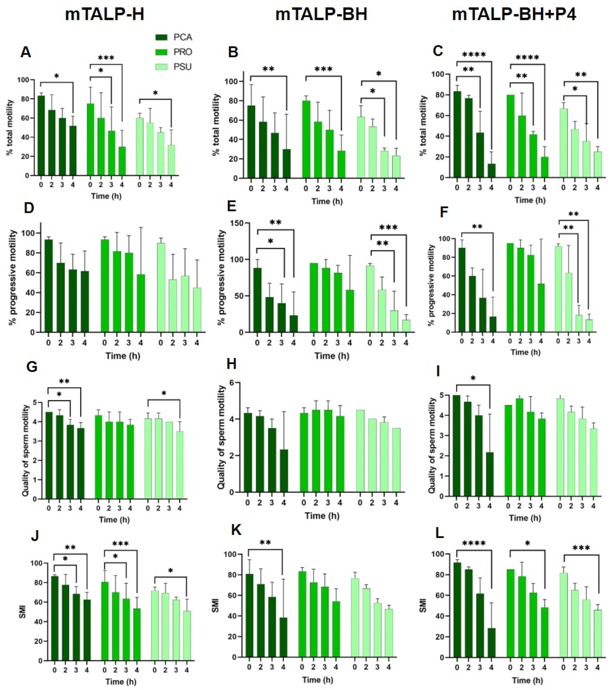
Total motility in sperm incubated in mTALP-H decreased towards the end of incubation in the three Phodopus species (Figure 2A), whereas the proportion of progressive sperm remained similar over time (Figure 2D). The quality of motility decreased in P. campbelli and P. sungorus at the end of incubation (Figure 2G). The sperm motility index (SMI), which is calculated based on the percentage of motility and quality (see Section 4), decreased over time in all three species (Figure 2J). Inter-species comparisons revealed no differences in total and progressive motilities, quality, or SMI at each time point assessed.
In mTALP-BH, total motility decreased at 4 h in all species (Figure 2B). Progressive motility decreased in P. campbelli and P. sungorus, whereas it remained constant in P. roborovskii (Figure 2E). The quality of motility remained constant (Figure 2H) and the SMI only significantly decreased in P. campbelli (p < 0.01) (Figure 2K). The comparison of progressive motility and quality of motility between species only showed significant differences (p < 0.05) at 3 h of incubation (between P. roborovskii and P. sungorus) and at 4 h (between P. campbelli and P. roborovskii). The SMI of capacitated sperm was not different between species throughout time.
The addition of 20 ng/mL of progesterone (mTALP-BH+P4) resulted in a decrease in total motility at 3 h and 4 h of incubation in all species (Figure 2C). The proportion of progressive sperm was maintained in P. roborovskii, but it sharply decreased (p < 0.01) at 3 h in P. sungorus or 4 h in P. roborovskii (Figure 2F). The quality of motility was similar over time in all species, except after 4 h in P. campbelli, when it significantly decreased (p < 0.05) (Figure 2I). The SMI was reduced after 4 h in all three species (Figure 2L).
The comparison between species revealed significantly higher total motility (p < 0.05) after 2 h of incubation in P. campbelli when compared to P. sungorus, as well as higher progressive motility after 3 h in P. roborovskii in relation to P. sungorus. The SMI was significantly higher after 2 h of incubation in P. campbelli than in P. sungorus.
Within species, there were differences in motility among sperm incubated in different media. The sperm of P. campbelli had lower values of total and progressive motility after incubation for 4 h in mTALP-BH (although marginally not significant) and in mTALP-BH+P4 (p < 0.05). In the other two species, there were no statistically significant differences between media at different time points.

2.2. Changes in CTC Staining Patterns

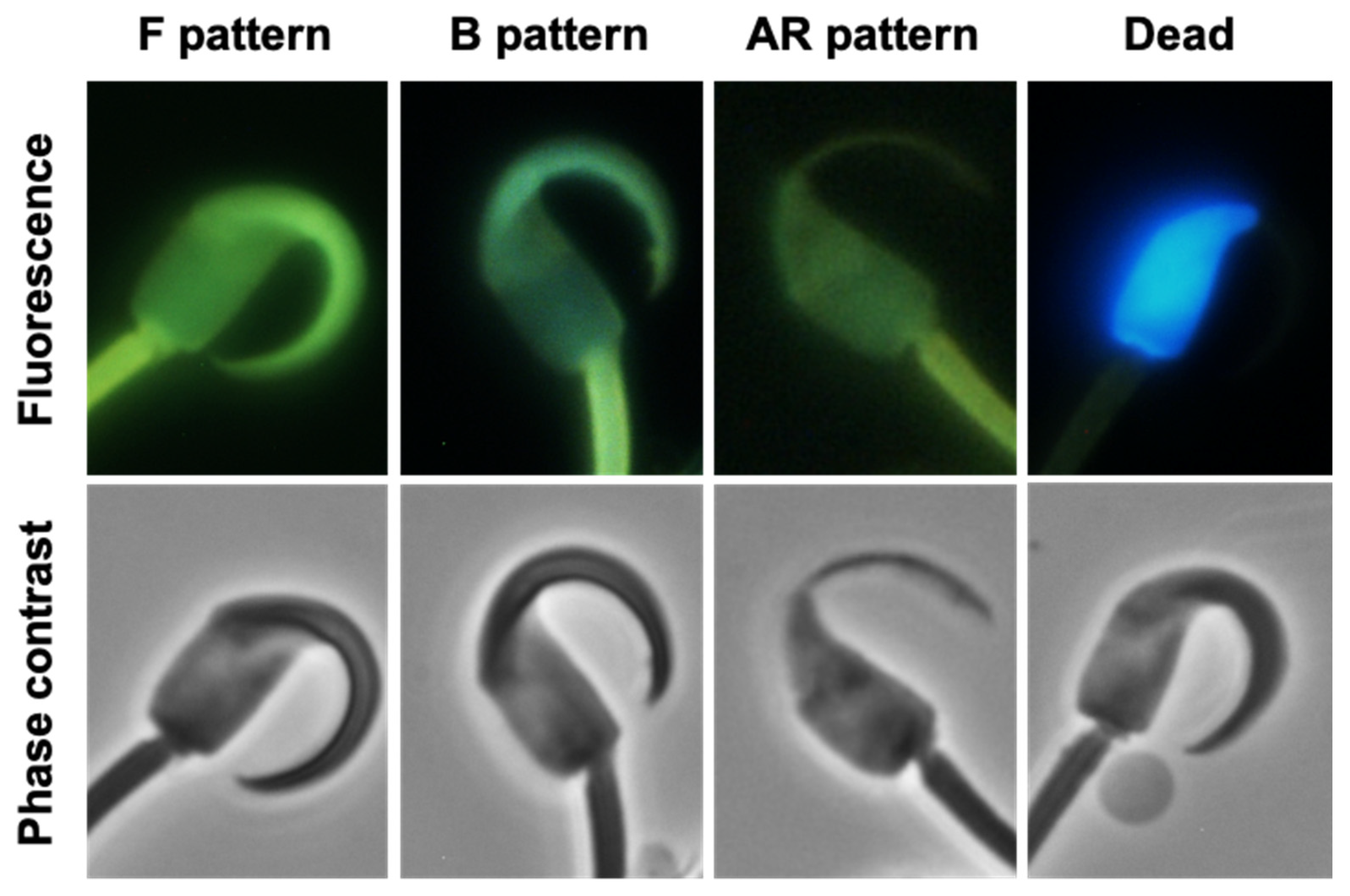
Changes in sperm staining patterns that are presumed to reflect capacitation status were evaluated after staining cells with Hoechst 33258 and chlortetracycline (CTC) (Figure 3). Live cells (i.e., those excluding Hoechst 33258) showing a uniform, bright CTC staining of the head were regarded as non-capacitated (F pattern). Live cells with acrosomes, but unstained in the post-acrosomal region, were considered to be capacitated (B pattern). Live cells with no acrosomes and pale staining were considered to have undergone the acrosome reaction (AR pattern).
The incubation in medium without bicarbonate (mTALP-H) appeared to result in a time-dependent decrease in F pattern cells in the three species, which was significant after 4 h in P. roborovskii. Comparisons between species revealed that P. campbelli had a significantly higher percentage of F pattern cells than P. roborovskii at the end of incubation (Figure 4A). The proportion of cells exhibiting a B pattern was similar over time in the three species (Figure 4D). Similarly, there were no significant increases in AR patterns over time, with the exception of P. roborovskii, which showed a rise after 3 h and 4 h of incubation, mirroring the observation that F pattern cells decreased in this species at the end of incubation. This rise resulted in significantly higher values of AR cells in P. roborovskii in comparison to values recorded in P. campbelli and P. sungorus (Figure 4G).
In the medium with bicarbonate (TALP-BH), only P. roborovskii showed a significant decrease (p < 0.05) of F pattern cells after 3 h and 4 h of incubation. The other two species also exhibited a tendency towards a decrease in values, but, despite the differences between species, they did not reach significance (Figure 4B). Regarding B pattern cells, a significantly higher value was seen in P. sungorus than in P. roborovskii after 2 h of incubation (p < 0.05; Figure 4E). P. roborovskii showed a significant increase (p < 0.05) in AR pattern cells after 4 h of incubation, which was higher than that observed in the other two species from 2 h onwards (Figure 4H).
The addition of progesterone to the medium with bicarbonate (mTALP-BH+P4) led to a decrease in the proportion of cells exhibiting a F pattern; the differences observed between species did not reach significance (Figure 4C). The proportion of B pattern cells tended to increase in the three species after 2 h of incubation; however, there was considerable variation between samples and species, so statistical differences were not evident (Figure 4F). Finally, the AR pattern did not change over time in P. campbelli, but it increased during incubation in the other two species and was statistically significant in P. roborovskii; it was also significantly higher than values in the other species (Figure 4I).
Comparisons were also made between the three media used for the incubation of spermatozoa (Figure S1 and Table S1). In P. campbelli, there were higher percentages of F-pattern cells and lower percentages of B pattern cells over time in the medium without bicarbonate (mTALP-H) in comparison to media with bicarbonate (mTALP-BH; mTALP-BH+P4). No differences were found in cells exhibiting an AR pattern between media. In P. roborovskii, similar trends were observed, but their differences did not reach statistical significance. Finally, in P. sungorus, the same trends were found, with percentages of F pattern cells being higher in the medium without bicarbonate (although they were marginally no significant), and percentages of B pattern cells also being lower in this medium (in this case, with a significant difference). No differences were found among media in the percentage of cells with an AR pattern for each of the three species.
Spontaneous acrosome reactions were also assessed after staining cells with eosin–nigrosin/Giemsa stain. This staining method distinguishes live cells from dead cells and allows for the identification of acrosomal status. The results showed increases in the proportion of live cells lacking an acrosome over time in the three media with no major differences between media (Figure 5). In contrast to the results with Hoechst 33258 and CTC, P. sungorus was the species with higher values of acrosomal loss in live cells at the end of incubations (p < 0.01).

2.3. Sperm Kinematics

Velocity and trajectory parameters were assessed using a computer-assisted sperm analysis (CASA) system at different times of incubation (0 h, 2 h, 3 h, and 4 h) in different media (mTALP-H, mTALP-BH, or mTALP-BH+P4) in the three Phodopus species.

2.3.1. Velocity Parameters

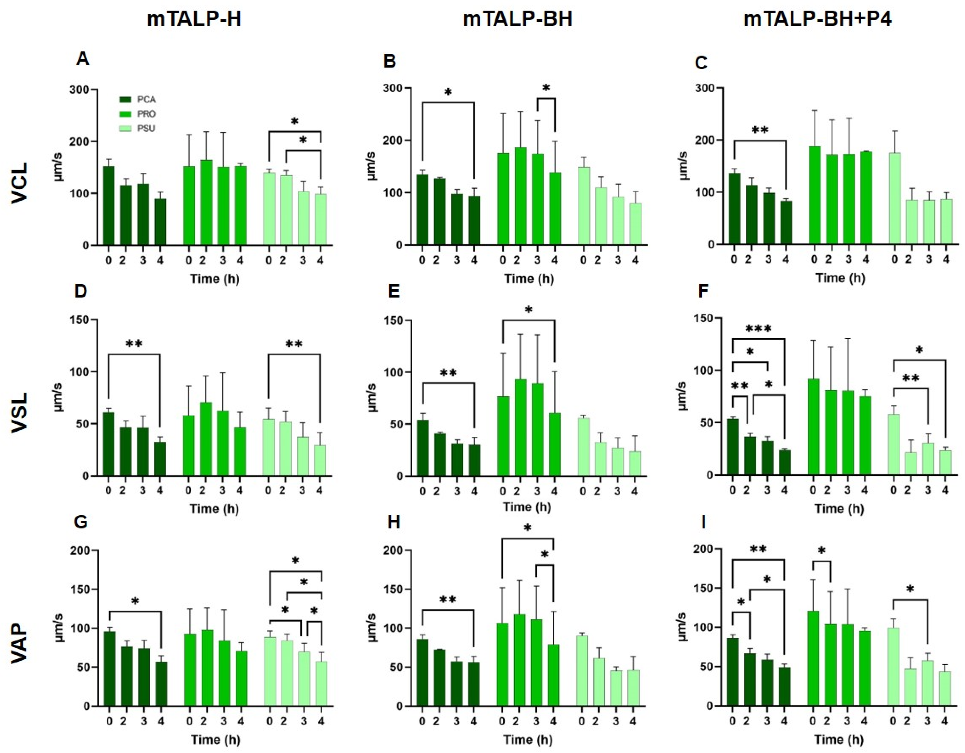
In mTALP-H, P. campbelli and P. sungorus exhibited a significant decrease in velocity parameters towards the end of incubation. P. roborovskii showed no significant differences over time (Figure 6A,D,G). When comparing velocities between species, significant differences were observed in VCL between P. roborovskii and P. sungorus at times of 0 h, 3 h, and 4 h, and between P. campbelli and P. roborovskii from 3 h onwards (p < 0.05). VSL was significantly higher in P. roborovskii compared to the other two species at 3 h (p < 0.05), whereas VAP was not different between species.
The incubation in mTALP-BH resulted in significant changes in velocity parameters. P. campbelli experienced a significant decrease (p < 0.05) after 4 h of incubation in all parameters, whereas P. roborovskii underwent a significant decline after 4 h in VSL and VAP (p < 0.05). In P. sungorus, these parameters were reduced but not significantly (Figure 6B,E,H). In general terms, P. roborovskii showed significantly higher velocities than the other two species.
When sperm were incubated with progesterone in mTALP-BH (mTALP-BH+P4) for up to 4 h, their velocities experienced a reduction in P. campbelli (VCL, VSL, and VAP) and in P. sungorus (VSL and VAP) (Figure 6C,F,I). P. roborovskii presented higher VCL, VSL, and VAP values than the other two species.
For each species, there were no overall differences in velocity parameters between the media.

2.3.2. Trajectory Parameters

In mTALP-H, a significant decline was observed in STR in P. campbelli and in LIN in P. sungorus after 4 h of incubation (Figure 7). ALH significantly decreased after 2 h of incubation in P. campbelli. Interspecies comparisons yielded no significant differences.
In mTALP-BH, the trajectory parameters (Figure 7) of P. campbelli sperm showed significant decreases in LIN after 4 h of incubation (p < 0.05) and in WOB after 2 h of incubation (p < 0.05). P. roborovskii exhibited significant decreases in STR and LIN after 2 h of incubation (p < 0.01). The trajectory parameters of P. sungorus sperm remained unchanged over time. There were no statistical differences between species in trajectories except for WOB, which was higher in P. roborovskii at 2 h.
When progesterone was added to mTALP-BH (mTALP-BH+P4), P. campbelli sperm experienced a reduction in STR and LIN from 2 h onwards, and in ALH after 4 h (p < 0.05) (Figure 7). On the other hand, the trajectory values of P. roborovskii and P. sungorus remained constant over time, except for LIN in P. sungorus, which decreased at 4 h (p < 0.05).
When comparing the three species, STR, LIN, ALH, and BCF values were higher at nearly every time point in P. roborovskii in relation to the other two species.
Within species, there were some significant differences between media. In P. campbelli sperm, there were lower values of STR and LIN after 4 h when progesterone was present in comparison to mTALP-H. WOB was lower at 2 h in this species when incubated in mTALP-BH, with or without progesterone, in comparison to values in mTALP-H. P. roborovskii only showed differences between media in WOB; its value in mTALP-BH was higher than in mTALP-H at 3 h and than in mTALP-BH+P4 at 2 h. There were no differences between media in P. sungorus.

3. Discussion

In the present study, we analyzed changes in the spermatozoa of three species of Phodopus hamsters incubated in three different media. To the best of our knowledge, this has not been examined before in these species. The spermatozoa were incubated in modified Tyrode’s medium with albumin, lactate, pyruvate, and Hepes (mTALP-H) under air, in mTALP-H with bicarbonate under 5% CO2/air (m-TALP-BH), or in mTALP-BH with the addition of 20 ng/mL of progesterone (m-TALP-BH+P4), also under 5% CO2/air. All media contained D-penicillamine, hypotaurine, and epinephrine (PHE). It has been previously found that PHE is required to maintain motility in golden hamster spermatozoa [27,57] but that it does not improve (nor decreased) motility in Phodopus species [27]. Since the requirements for Phodopus sperm capacitation were unknown before this study, we opted to include PHE in incubation media in case they were a requirement for the acquisition of capacitation. We also explored whether progesterone had an effect on changes compatible with capacitation and hyperactivation. Progesterone is present in the oviductal fluid of the golden hamster at a concentration of 44.04–175.06 ng/mL [58]. In this species, 20 ng/mL of progesterone has been found to stimulate capacitation and hyperactivation [32,33], probably activating a tyrosine phosphorylation pathway [33].
mTALP-H medium is normally regarded as a non-capacitating medium in other species [44]. However, it was found that when Phodopus sperm were incubated in mTALP-H, there was a decrease in the percentage of cells exhibiting an F pattern, characteristic of non-capacitated cells. There were also decreases in the proportion of cells showing an F pattern when sperm were incubated with bicarbonate (mTALP-BH) or with bicarbonate plus 20 ng/mL of progesterone (mTALP-BH+P4), but, for the latter two media, these decreases were proportionally and significantly more pronounced. Generally, the values were lowest in P. roborovskii and highest in P. campbelli.
The proportion of B pattern cells (regarded as capacitated sperm) remained similar among species (~20%) over time in mTALP-H. On the other hand, the proportion of cells exhibiting a B pattern experienced a significant increase when sperm were incubated in mTALP-BH, both in the absence or presence of progesterone. These results suggest that sperm from the three Phodopus species may be experiencing capacitation under these conditions, in agreement with what has been found before in other species when the spermatozoa have been incubated in bicarbonate-, calcium-, and albumin-containing media (e.g., mouse species [44]). In contrast to what was observed earlier in the golden hamster [32,33], or in other species (see Section 1), progesterone, at the concentration used here, did not appear to enhance the proportion of B pattern cells. This does not seem to agree with previous studies on P. sungorus [55], which revealed an increase in the rate of in vitro fertilization in a Tyrode’s medium with follicular fluid (which is known to contain a high concentration of progesterone). This discrepancy between our results and previous work may be due to different requirements for progesterone or to differences in progesterone-stimulated pathways.
The proportion of Phodopus sperm showing an AR pattern (suggesting acrosomal exocytosis) increased with time in all media (mTALP-H, mTALP-BH, and mTALP-BH+P4). The increase in AR patterns in mTALP-H was unexpected, and it could suggest loss of the acrosome due to damage or the occurrence of acrosomal exocytosis in this medium. In media with bicarbonate, there were also increases in the proportion of AR pattern cells. In these media, changes occurred in parallel to increases in the proportions of cells with a B pattern. The proportion of cells showing an AR pattern varied between species, being lowest in P. campbelli (~20%) and highest in P. roborovskii (~60%). Earlier studies in the golden hamster [59] showed that ~55% of sperm were motile and acrosome-reacted after 4 h in mTALP capacitation medium [60].
No additional increase in acrosomal loss was observed in the medium containing progesterone, and this could be due, as mentioned above, to differences with other species. Progesterone is reported to have different effects depending on its concentration. For instance, in boars, 10–100 nM of progesterone increases sperm viability, whereas in humans, 50 nM stimulates progressive motility [42]. Low concentrations of progesterone (20 ng/mL) and long capacitation times (5 h) were necessary for golden hamster sperm [32], but such concentrations were not sufficient to trigger an acrosome reaction in the golden hamster. Differences between Phodopus sperm and golden or Chinese hamster sperm have been previously reported [27].
Sperm motion and kinematics in Phodopus sperm under conditions that were presumed to be non-capacitating were previously reported [27]. In the present study, sperm of Phodopus species followed the same pattern of decreased motility when incubated in mTALP-H. Conversely, in mTALP-BH, with or without progesterone, there was a decline in progressive motility, which was sharper in P. campbelli and P. sungorus than in P. roborovskii, and which may suggest the development of hyperactivated motion. Hyperactivation is characterized by an asymmetric movement, a decrease in progressivity, an increase in flagellar amplitude, and a higher sperm vigor [11].
Sperm velocities, assessed with a CASA system, decreased over time in mTALP-BH in the three species. Similarly, several sperm trajectory parameters declined in the three species under these conditions. However, the expected decline in STR or LIN and concomitant increase in ALH was not evident, suggesting that hyperactivation patterns in these species may be different from the well-established patterns observed in other rodent species [44]. When comparisons between Phodopus species were made, P. roborovskii presented higher VCL, VSL, and VAP values than the other two species. Likewise, STR, LIN, ALH, and BCF values were higher at nearly every time point in P. roborovskii in relation to the other two species. In comparison to the golden hamster [27], the three Phodopus species exhibited higher values of velocity and trajectory parameters.
Overall, the present study showed changes in Phodopus sperm incubated in media without or with bicarbonate, and without or with progesterone, that were consistent with the occurrence of capacitation and spontaneous acrosome reactions. Progesterone did not appear to have a marked effect on sperm function in Phodopus. The media used here were based on those used in previous studies in the golden hamster. The latter requires motility factors to survive and undergo capacitation, but they do not appear to be needed for Phodopus sperm. Therefore, additional studies will be required to further characterize capacitation and hyperactivation in Phodopus species. Such studies will help to understand differences in sperm function in these species with extreme phenotype and high-performing sperm cells.

4. Material and Methods

4.1. Animals

Adult males of three hamster species (Phodopus campbelli, P. roborovskii, and P. sungorus) were held in our animal facilities in individual cages and with free access to food and water. They were maintained under controlled conditions of light (14 h of light/10 h of darkness) and temperature (22–24 °C). All animal handling followed the Spanish Animal Protection Regulation RD53/2013 and the European Union Regulation 2010/63 and had the approval of CSIC’s ethics committee and the Comunidad de Madrid (28079-47-A).

4.2. Animal Phenotype, Sperm Collection, and Incubation

Three animals of each species of Phodopus were sacrificed via cervical dislocation. Sperm were collected from the cauda epididymis by swim out, i.e., placing the cauda in the medium and puncturing them with the aid of a pair of scissors and allowing sperm cells to swim out for 10 min at 37 °C. The medium used was a Hepes-buffered modified Tyrode’s medium with albumin, lactate, and pyruvate (mTALP-H), and with D-penicillamine, hypotaurine, and epinephrine. It had the following composition: 120.89 mM NaCl, 2.68 mM KCl, 0.49 mM MgCl2·6H2O, 0.36 mM NaH2PO4·2H2O, 20 mM Hepes, 1.80 mM CaCl2, 5.56 mM glucose,1 mM sodium pyruvate, 10 mM sodium lactate, 4 mg/mL bovine serum albumin, 20 µM D-penicillamine, 100 µM hypotaurine, and 1 µM epinephrine, and had a pH = 7.4 [27].
The medium with sperm was transferred to a pre-warmed plastic tube, and the concentration of sperm was estimated using a Neubauer chamber. The samples were diluted to a final concentration of 20 × 106 sperm/mL in three different media. We used mTALP-H, and a modification of this medium with 15 mM of NaHCO3, adjusting NaCl to 116.89 mM to maintain the osmolality (mTALP-BH). Progesterone (20 ng/mL; P0130, Sigma, Madrid, Spain) was added to the latter medium to promote sperm hyperactivation (mTALP-BH+P4). Sperm suspensions were incubated at 37 °C for 4 h under air (mTALP-H) or under 5% CO2/air (mTALP-BH and mTALP-BH+P4).

4.3. Sperm Motility and Viability Assessment

Total and progressive motilities and quality of motility (from 1 to 5, with 1 being barely moving sperm and 5 being very vigorously moving sperm) were assessed under phase contrast at the time of dilution (time 0) and after 2 h, 3 h, and 4 h of incubation in each medium. The sperm motility index (SMI) was calculated using the equation: [((Quality of motility × 20) + Total motility)/2].
Sperm viability was measured at 0 h, 2 h, 3 h, and 4 h in each individual and each medium, examining cells after staining with eosin–nigrosin/Giemsa, as previously described [61]. Briefly, a sperm suspension was mixed 1:3 with a solution of eosin (E6003, Sigma) and nigrosin (N4754, Sigma) on a pre-warmed slide. After 30 s, smears were performed and dried at 37 °C on a warm plate. For Giemsa staining, smears were fixed with 4% formaldehyde (211328, Panreac, Madrid, Spain) for 10 min, washed, and stained for 60 min in Giemsa (109203, Merck, Madrid, Spain). The slides were mounted with DePeX (360292F, BDH, Madrid, Spain). For viability assessment, sperm with a pale post-acrosomal region were regarded as viable and those with a strong pink coloration as non-viable cells. Acrosomes were distinguished as stained with Giemsa.

4.4. Capacitation Analysis

Sperm aliquots were fixed 1:1 with 2% glutaraldehyde (v/v) (360802F, BDH) in 0.165 M of sodium cacodylate/HCl (301183V, BDH) at 0 h, 2 h, 3 h, and 4 h of incubation. The samples were then stained with Hoechst 33258 (B2883, Sigma) and chlortetracycline (CTC) (C4881, Sigma) to estimate the viability and the functional state (capacitation and acrosome integrity) of spermatozoa, as previously described [44]. Hoechst 33258 stains the DNA of plasma membrane-damaged cells, emitting blue fluorescence, whereas CTC stains the sites that bind calcium, staining cells with green fluorescence and showing patterns related to the cell’s capacitation and acrosomal status, namely (a) “F pattern”: non-capacitated cells (the entire sperm head is stained green with a uniform intensity); (b) “B pattern”: capacitated cells (the acrosomal region is stained green, whereas the post-acrosomal region is unstained or slightly stained); and (c) “AR pattern”: acrosome loss has occurred (sperm head slightly or not stained). Fluorescence staining was detected using a Nikon (Tokyo, Japan) UV-2A 330 nm filter for Hoechst 33258 and a Nikon BV-2A 405 nm filter for CTC. Fifty cells per sample were counted, and the percentage of each pattern was calculated. Acrosome status was also assessed in the eosin–nigrosin/Giemsa preparations.

4.5. Sperm Kinematics

Sperm parameters of velocity and trajectory were analyzed with a computer-assisted sperm analysis (CASA) system (Sperm Class Analyzer, SCA v.6.2, Microptic, Barcelona, Spain). Aliquots of 5 µL of a sperm suspension were diluted in 20 µL of the corresponding medium to achieve a final concentration of 4 × 10⁶ sperm/mL. Each diluted sample was loaded in a microscopy chamber with a depth of 20 μm (Leja, Nieuw-Vennep, The Netherlands), and a set of 6 videos were recorded using a phase contrast microscope, with pseudo-negative phase, connected to a digital video camera (Basler A312fc, Vision Technologies, Glen Burnie, MD, USA), as in previous studies [27].
The swimming parameters registered were the following: VCL (curvilinear velocity, μm/s), VSL (straight-line velocity, μm/s), VAP (average path velocity, μm/s), STR (STR = VSL/VAP), LIN (linearity, LIN = VSL/VCL), WOB (wobble, WOB = VAP/VCL), ALH (amplitude of lateral head displacement, μm), and BCF (beat-cross frequency, Hz).

4.6. Statistical Analysis

Analyses were carried out using GraphPad Prism 9 software (Dotmatics, Boston, MA, USA). Data were transformed before analyses (log10 for all variables, with the exception of percentages which were arcsine transformed). For the study of total and progressive motility, quality of motility, SMI, and sperm viability, two-way ANOVAs with Šídák’s multiple comparisons post-hoc tests were executed for each medium analyzed (mTALP-H, mTALP-BH, and mTALP-BH+P4), with species (P. campbelli, P. roborovskii, and P. sungorus) and time of incubation (0 h, 2 h, 3 h, and 4 h) as fixed effects, including their interaction in the statistical model. Capacitation patterns were assessed using a two-way ANOVA with Tukey’s post-hoc test, considering species and time as fixed effects. Finally, differences in sperm kinetics between species at the same time point were analyzed using a two-way ANOVA with Tukey’s post-hoc test, whereas the differences between times in the same species were examined using a one-way ANOVA. All variables are shown as means ± SD, and significance is marked with asterisks depending on the level of significance (* p < 0.05, ** p < 0.01, *** p < 0.001, and **** p < 0.0001).

Supplementary Materials

The supporting information can be downloaded at: https://www.mdpi.com/article/10.3390/ijms242216093/s1.

Author Contributions

Conceptualization, E.R.S.R. and A.S.-R.; methodology, E.R.S.R., A.S.-R. and J.A.R.; formal analysis, A.S.-R. and E.R.S.R.; experimentation/investigation A.S.-R., I.I.D.I. and J.A.R.; data curation, A.S.-R., I.I.D.I. and E.R.S.R.; writing—original draft preparation, A.S.-R. and E.R.S.R.; writing—review and editing, E.R.S.R. and A.S.-R.; funding acquisition, E.R.S.R. All authors have read and agreed to the published version of the manuscript.

Funding

This work was supported by the Spanish Ministry of Science and Innovation (projects CGL 2016-80577-P and PID 2019-108649GB-I00). A.S.-R. was funded by a “Juan de la Cierva” postdoctoral fellowship (FJC2019-041736-I) from the Ministry of Science and Innovation, co-funded by the European Social Fund. I.I.D.I. was supported by a grant from the Comunidad de Madrid (PEJ-2019-TL/AMB-15136).

Institutional Review Board Statement

The animal study protocol was approved by the Institutional Animal Experimentation Review Board of the Museo Nacional de Ciencias Naturales (CSIC) and the Comunidad de Madrid (28079-47-A).

Informed Consent Statement

Not applicable.

Data Availability Statement

The raw data supporting the conclusions of this article will be made available by the authors, without undue reservation.

Acknowledgments

We thank Esperanza Navarro for supervising animal facilities and for animal care.

Conflicts of Interest

The authors declare no conflict of interest. The funders had no role in the design of the study; in the collection, analyses, or interpretation of data; in the writing of the manuscript, or in the decision to publish the results.

References

  1. Austin, C.R. The capacitation of the mammalian sperm. Nature 1952, 170, 326. [Google Scholar] [CrossRef] [PubMed]
  2. Austin, C.R. Observations on the penetration of the sperm into the mammalian egg. Aust. J. Biol. Sci. 1951, 4, 581–596. [Google Scholar] [CrossRef]
  3. Chang, M.C. Fertilizing capacity of spermatozoa deposited into the Fallopian tubes. Nature 1951, 168, 697–698. [Google Scholar] [CrossRef] [PubMed]
  4. Bedford, J.M. Sperm capacitation and fertilization in mammals. Biol. Reprod. 1970, 2, 128–158. [Google Scholar] [CrossRef]
  5. Bavister, B.D. Capacitation of golden hamster spermatozoa during incubation in culture medium. J. Reprod. Fert. 1973, 35, 161–163. [Google Scholar] [CrossRef] [PubMed]
  6. Austin, C.R. Membrane fusion events in fertilization. J. Reprod. Fert. 1975, 44, 155–166. [Google Scholar] [CrossRef] [PubMed]
  7. Yanagimachi, R. Mechanisms of fertilization in mammals. In Fertilization and Embryonic Development In Vitro; Mastroianni, L., Biggers, J.D., Eds.; Springer: Boston, MA, USA, 1981; pp. 81–182. [Google Scholar] [CrossRef]
  8. Buffone, M.G.; Rodriguez-Miranda, E.; Storey, B.T.; Gerton, G.L. Acrosomal exocytosis of mouse sperm progresses in a consistent direction in response to zona pellucida. J. Cell. Physiol. 2009, 220, 611–620. [Google Scholar] [CrossRef]
  9. Chang, M.C. The meaning of sperm capacitation. A historical perspective. J. Androl. 1984, 5, 45–50. [Google Scholar] [CrossRef]
  10. Gervasi, M.G.; Visconti, P.E. Chang’s meaning of capacitation: A molecular perspective. Mol. Reprod. Dev. 2016, 83, 860–874. [Google Scholar] [CrossRef]
  11. Yanagimachi, R. The movement of golden hamster spermatozoa before and after capacitation. J. Reprod. Fertil. 1970, 23, 193–196. [Google Scholar] [CrossRef]
  12. Puga Molina, L.C.; Luque, G.M.; Balestrini, P.A.; Marín-Briggiler, C.I.; Romarowski, A.; Buffone, M.G. Molecular basis of human sperm capacitation. Front. Cell Dev. Biol. 2018, 6, 72. [Google Scholar] [CrossRef] [PubMed]
  13. Fujinoki, M. Regulation and disruption of hamster sperm hyperactivation by progesterone, 17ß-estradiol and diethylstilbestrol. Reprod. Med. Biol. 2014, 13, 143–152. [Google Scholar] [CrossRef] [PubMed]
  14. Fraser, L.R.; Herod, J.E. Expression of capacitation-dependent changes in chlortetracycline fluorescence patterns in mouse spermatozoa requires a suitable glycolysable substrate. J. Reprod. Fert. 1990, 88, 611–621. [Google Scholar] [CrossRef] [PubMed]
  15. Roldan, E.R.S. Sperm competition and the evolution of sperm form and function in mammals. Reprod. Dom. Anim. 2019, 54 (Suppl. S4), 14–21. [Google Scholar] [CrossRef] [PubMed]
  16. Singh, J.P.; Babcock, D.F.; Lardy, H.A. Increased calcium-ion influx is a component of capacitation of spermatozoa. Biochem. J. 1978, 172, 549–556. [Google Scholar] [CrossRef] [PubMed]
  17. Nakanishi, T.; Ikawa, M.; Yamada, S.; Toshimori, K.; Okabe, M. Alkalinization of acrosome measured by GFP as a pH indicator and its relation to sperm capacitation. Dev. Biol. 2001, 237, 222–231. [Google Scholar] [CrossRef] [PubMed]
  18. Bavister, B.D.; Chen, A.F.; Fu, P.C. Catecholamine requirement for hamster sperm motility in vitro. J. Reprod. Fertil. 1979, 56, 507–513. [Google Scholar] [CrossRef] [PubMed]
  19. Meizel, S.; Working, P.K. Further evidence suggesting the hormonal stimulation of hamster sperm acrosome reactions by catecholamines in vitro. Biol. Reprod. 1980, 22, 211–216. [Google Scholar] [CrossRef]
  20. Cornett, L.E.; Meizel, S. Stimulation of in vitro activation and the acrosome reaction of hamster spermatozoa by catecholamines. Proc. Natl. Acad. Sci. USA 1978, 75, 4954–4958. [Google Scholar] [CrossRef]
  21. Meizel, S.; Lui, C.W.; Working, P.K.; Mrsny, R.J. Taurine and hypotaurine: Their effects on motility, capacitation and the acrosome reaction of hamster sperm in vitro and their presence in sperm and reproductive tract fluids of several mammals. Dev. Growth Differ. 1980, 22, 483–494. [Google Scholar] [CrossRef]
  22. Brogan, P.T.; Beitsma, M.; Henning, H.; Gadella, B.M.; Stout, T.A. Liquid storage of equine semen: Assessing the effect of D-penicillamine on longevity of ejaculated and epididymal stallion sperm. Anim. Reprod. Sci. 2015, 159, 155–162. [Google Scholar] [CrossRef] [PubMed]
  23. Leahy, T.; Rickard, J.P.; Aitken, R.J.; de Graaf, S.P. Penicillamine prevents ram sperm agglutination in media that support capacitation. Reproduction 2016, 151, 167–177. [Google Scholar] [CrossRef] [PubMed]
  24. Uribe, P.; Merino, J.; Bravo, A.; Zambrano, F.; Schulz, M.; Villegas, J.V.; Sanchez, R. Antioxidant effects of penicillamine against in vitro-induced oxidative stress in human spermatozoa. Andrologia 2020, 52, e13553. [Google Scholar] [CrossRef] [PubMed]
  25. Ruiz-Diaz, S.; Oseguera-Lopez, I.; De La Cuesta-Diaz, D.; Garcia-Lopez, B.; Serres, C.; Sanchez-Calabuig, M.J.; Gutierrez-Adan, A.; Perez-Cerezales, S. The presence of D-penicillamine during the in vitro capacitation of stallion spermatozoa prolongs hyperactive-like motility and allows for sperm selection by thermotaxis. Animals 2020, 10, 1467. [Google Scholar] [CrossRef] [PubMed]
  26. Kang, S.S.; Koyama, K.; Huang, W.; Yang, Y.; Yanagawa, Y.; Takahashi, Y.; Nagano, M. Addition of D-penicillamine, hypotaurine, and epinephrine (PHE) mixture to IVF medium maintains motility and longevity of bovine sperm and enhances stable production of blastocysts in vitro. J. Reprod. Dev. 2015, 61, 99–105. [Google Scholar] [CrossRef] [PubMed]
  27. Tourmente, M.; Sanchez-Rodriguez, A.; Roldan, E.R.S. Effect of motility factors D-penicillamine, hypotaurine and epinephrine on the performance of spermatozoa from five hamster species. Biology 2022, 11, 526. [Google Scholar] [CrossRef] [PubMed]
  28. Fujinoki, M. Melatonin-enhanced hyperactivation of hamster sperm. Reproduction 2008, 136, 533–541. [Google Scholar] [CrossRef] [PubMed]
  29. Fujinoki, M. Serotonin-enhanced hyperactivation of hamster sperm. Reproduction 2011, 142, 255–266. [Google Scholar] [CrossRef]
  30. Sugiyama, Y.; Fujinoki, M.; Shibahara, H. Effects of 5-hydroxytryptamine on spermatozoal hyperactivation and in vitro fertilization in mice. J. Reprod. Dev. 2019, 65, 541–550. [Google Scholar] [CrossRef]
  31. Baldi, E.; Casano, R.; Falsetti, C.; Krausz, C.; Maggi, M.; Forti, G. Intracellular calcium accumulation and responsiveness to progesterone in capacitating human spermatozoa. J. Androl. 1991, 12, 323–330. [Google Scholar] [CrossRef]
  32. Libersky, E.A.; Boatman, D.E. Effects of progesterone on in vitro sperm capacitation and egg penetration in the golden hamster. Biol. Reprod. 1995, 53, 483–487. [Google Scholar] [CrossRef] [PubMed]
  33. Noguchi, T.; Fujinoki, M.; Kitazawa, M.; Inaba, N. Regulation of hyperactivation of hamster spermatozoa by progesterone. Reprod. Med. Biol. 2008, 7, 63–74. [Google Scholar] [CrossRef] [PubMed]
  34. Roldan, E.R.S.; Murase, T.; Shi, Q.X. Exocytosis in spermatozoa in response to progesterone and zona pellucida. Science 1994, 266, 1578–1581. [Google Scholar] [CrossRef] [PubMed]
  35. Shi, Q.X.; Roldan, E.R.S. Bicarbonate/CO2 is not required for zona pellucida- or progesterone-induced acrosomal exocytosis of mouse spermatozoa but is essential for capacitation. Biol. Reprod. 1995, 52, 540–546. [Google Scholar] [CrossRef] [PubMed]
  36. Llanos, M.N.; Anaabalon, M.C. Studies related to progesterone-induced hamster sperm acrosome reaction. Mol. Reprod. Dev. 1996, 45, 313–319. [Google Scholar] [CrossRef]
  37. Uhler, M.L.; Leung, A.; Chan, S.Y.; Wang, C. Direct effects of progesterone and antiprogesterone on human sperm hyperactivated motility and acrosome reaction. Fertil. Steril. 1992, 58, 1191–1198. [Google Scholar] [CrossRef] [PubMed]
  38. Sueldo, C.E.; Oehninger, S.; Subias, E.; Mahony, M.; Alexander, N.J.; Burkman, L.J.; Acosta, A.A. Effect of progesterone on human zona pellucida sperm binding and oocyte penetrating capacity. Fertil. Steril. 1993, 60, 137–140. [Google Scholar] [CrossRef] [PubMed]
  39. Baldi, E.; Luconi, M.; Bonaccorsi, L.; Forti, G. Nongenomic effects of progesterone on spermatozoa: Mechanisms of signal transduction and clinical implications. Front Biosci 1998, 3, D1051–D1059. [Google Scholar] [CrossRef] [PubMed]
  40. Harper, C.V.; Barrat, C.L.R.; Publicover, S.J.; Kirkman-Brown, J.C. Kinetics of the progesterone-induced acrosome reaction and its relation to intracellular calcium responses in individual human spermatozoa. Biol. Reprod. 2006, 75, 933–939. [Google Scholar] [CrossRef]
  41. Murase, T.; Roldan, E.R.S. Progesterone and the zone pellucida activate different transducing pathways in the sequence of events leading to diacylglycerol generation during mouse sperm. Biochem. J. 1996, 320, 1017–1023. [Google Scholar] [CrossRef]
  42. Mahé, C.; Zlotkowska, A.M.; Reynaud, K.; Tsikis, G.; Mermillod, P.; Druart, X.; Schoen, J.; Saint-Dizier, M. Sperm migration, selection, survival, and fertilizing ability in the mammalian oviduct. Biol. Reprod. 2021, 105, 317–331. [Google Scholar] [CrossRef] [PubMed]
  43. Tourmente, M.; Villar-Moya, P.; Varea-Sánchez, M.; Luque-Larena, J.J.; Rial, E.; Roldan, E.R.S. Performance of rodent spermatozoa over time is enhanced by increased ATP concentrations: The role of sperm competition. Biol. Reprod. 2015, 93, 64. [Google Scholar] [CrossRef] [PubMed]
  44. Sansegundo, E.; Tourmente, M.; Roldan, E.R.S. Energy metabolism and hyperactivation of spermatozoa from three mouse species under capacitating conditions. Cells 2022, 11, 220. [Google Scholar] [CrossRef] [PubMed]
  45. Hirose, M.; Ogura, A. The golden (Syrian) hamster as a model for the study of reproductive biology: Past, present, and future. Reprod. Med. Biol. 2019, 18, 34–39. [Google Scholar] [CrossRef] [PubMed]
  46. Yanagimachi, R.; Chang, M.C. Fertilization of hamster eggs in vitro. Nature 1963, 200, 281–282. [Google Scholar] [CrossRef] [PubMed]
  47. Yanagimachi, R.; Chang, M.C. In vitro fertilization of golden hamster ova. J. Exp. Zool. 1964, 156, 361–375. [Google Scholar] [CrossRef] [PubMed]
  48. Fujinoki, M.; Kawamura, T.; Toda, T.; Ohtake, H.; Ishimoda-Takagi, T.; Shimizu, N.; Yamaoka, S.; Okuno, M. Identification of 36-kDa flagellar phosphoproteins associated with hamster sperm motility. Biomed. Res. 2001, 133, 361–369. [Google Scholar] [CrossRef] [PubMed]
  49. Fujinoki, M.; Ishimoda-Takagi, T.; Ohtake, H. Serine/threonine phosphorylation associated with hamster sperm hyperactivation. Reprod. Med. Biol. 2004, 3, 223–230. [Google Scholar] [CrossRef]
  50. Fujinoki, M.; Suzuki, T.; Takayama, T.; Shibahara, H.; Ohtake, H. Profiling of proteins phosphorylated or dephosphorylated during hyperactivation via activation on hamster spermatozoa. Reprod. Med. Biol. 2006, 5, 123–135. [Google Scholar] [CrossRef]
  51. Tateno, H.; Tamura-Nakano, M.; Kusakabe, H.; Hirohashi, N.; Kawano, N.; Yanagimachi, R. Sperm acrosome status before and during fertilization in the Chinese hamster (Cricetulus griseus), and observation of oviductal vesicles and globules. Mol. Reprod. Dev. 2021, 88, 793–804. [Google Scholar] [CrossRef]
  52. Yanagimachi, R.; Kamiguchi, Y.; Sugawara, S.; Mikamo, K. Gametes and fertilization in the Chinese hamster. Gamete Res. 1983, 8, 97–117. [Google Scholar] [CrossRef]
  53. Roldan, E.R.S.; Yanagimachi, R. Cross-fertilization between Syrian and Chinese hamsters. J. Exp. Zool. 1989, 250, 321–328. [Google Scholar] [CrossRef] [PubMed]
  54. Hoffman, K. The influence of photoperiod and melatonin on testis size, body weight, and pelage colour in the Djungarian hamster (Phodopus sungorus). J. Comp. Physiol. 1973, 85, 267–282. [Google Scholar] [CrossRef]
  55. Anand, S.; Losee-Olson, S.; Turek, F.W.; Horton, T.H. Differential regulation of luteinizing hormone and follicle-stimulating hormone in male Siberian hamsters by exposure to females and photoperiod. Endocrinology 2002, 143, 2178–2188. [Google Scholar] [CrossRef] [PubMed]
  56. Parkening, T.A. In vitro fertilization of Siberian hamster oocytes. J. Exp. Zool. 1990, 254, 305–312. [Google Scholar] [CrossRef] [PubMed]
  57. Niklowitz, P.; Lerchl, A.; Nieschlag, E. In vitro fertilizing capacity of sperm from FSH-treated photoinhibited Djungarian hamsters (Phodopus sungorus). J. Endocrinol. 1997, 154, 475–481. [Google Scholar] [CrossRef]
  58. Bavister, B.D. A consistently successful procedure for in vitro fertilization of golden hamster eggs. Gamete Res. 1989, 23, 139–158. [Google Scholar] [CrossRef] [PubMed]
  59. Libersky, E.A.; Boatman, D.E. Progesterone concentration in serum, follicular fluid, and oviductal fluid of the golden hamster during the periovulatory period. Biol. Reprod. 1995, 53, 477–482. [Google Scholar] [CrossRef]
  60. Yanagimachi, R. Requirement of extracellular calcium ions for various stages of fertilization and fertilization-related phenomena in the hamster. Gamete Res. 1982, 9, 323–344. [Google Scholar] [CrossRef]
  61. Gomez Montoto, L.; Varea-Sánchez, M.; Tourmente, M.; Martín-Coello, J.; Luque-Larena, J.J.; Gomendio, M.; Roldan, E.R.S. Sperm competition differentially affects swimming velocity and size of spermatozoa from closely related muroid rodents: Head first. Reproduction 2011, 142, 819–830. [Google Scholar] [CrossRef]
Figure 1. Sperm viability of three hamster species incubated in different media for 4 h. (A) Incubation in mTALP-H. (B) Incubation in mTALP-BH. (C) Incubation in mTALP-BH with 20 ng/mL of progesterone (mTALP-BH+P4). Abbreviations: PCA: P. campbelli; PRO: P. roborovskii; and PSU: P. sungorus. Asterisks indicate significant differences between times in a given species: * p < 0.05; ** p < 0.01; and **** p < 0.0001. Results are shown as mean ± SD.
Ijms 24 16093 g001
Figure 2. Sperm motility parameters of three hamster species incubated in different media for 4 h. (AC) Total motility. (DF) Progressive motility. (GI) Quality of motility. (JL) Sperm motility index (SMI; see Section 4). (A,D,G,J) Incubation in mTALP-H. (B,E,H,K) Incubation in mTALP-BH. (C,F,I,L) Incubation in mTALP-BH with progesterone (20 ng/mL) (mTALP-BH+P4). Abbreviations: PCA: P. campbelli; PRO: P. roborovskii; and PSU: P. sungorus. Asterisks indicate significant differences between species: * p < 0.05; ** p < 0.01; *** p < 0.001; and **** p < 0.0001. Results are shown as mean ± SD.
Ijms 24 16093 g002
Figure 3. Chlortetracycline (CTC) and Hoechst 33258 staining patterns of spermatozoa from Phodopus species examined under fluorescence (upper panels) or phase contrast (lower panels) microscopy. The images are of Phodopus campbelli sperm, and the three species showed similar staining patterns. From left to right, the images correspond to pattern “F” live non-capacitated sperm showing a uniform bright staining of the head with CTC; pattern “B” live capacitated sperm showing the acrosomal area stained, contrasting with the lesser stained post-acrosomal region; pattern “AR” live sperm with no acrosomes and pale staining with CTC; and dead sperm showing a uniform staining with Hoechst 33258.
Ijms 24 16093 g003
Figure 4. Analysis of CTC staining patterns in three hamster species incubated in different media for 4 h. (AC) F pattern, presumably corresponding to live non-capacitated sperm. (DF) B pattern, presumably corresponding to live capacitated sperm. (GI) AR pattern, live sperm with no acrosome. (A,D,G) Incubation in mTALP-H. (B,E,H) Incubation in mTALP-BH. (C,F,I) Incubation in mTALP-BH with progesterone (20 ng/mL) (mTALP-BH+P4). Abbreviations: PCA: P. campbelli; PRO: P. roborovskii; and PSU: P. sungorus. Different letters at the same time point indicate significant differences between species (p < 0.05). Results are shown as mean ± SD.
Ijms 24 16093 g004
Figure 5. Spontaneous acrosome reactions (live sperm without acrosome) assessed using eosin–nigrosin/Giemsa in three hamster species incubated in different media for 4 h. (A) Incubation in mTALP-H medium; (B) incubation in mTALP-BH medium; and (C) incubation in mTALP-BH with progesterone (20 ng/mL) (mTALP-BH+P4). Abbreviations: PCA: P. campbelli; PRO: P. roborovskii; and PSU: P. sungorus. Asterisks indicate significant differences between times: * p < 0.05, ** p < 0.01, and *** p < 0.001. Results are shown as mean ± SD.
Ijms 24 16093 g005
Figure 6. Velocity parameters of sperm kinematics in three hamster species incubated in different media for 4 h. (A,D,G) Incubation in mTALP-H. (B,E,H) Incubation in mTALP-BH. (C,F,I) Incubation in mTALP-BH with progesterone (20 ng/mL) (mTALP-BH+P4). Abbreviations: PCA: P. campbelli; PRO: P. roborovskii; and PSU: P. sungorus. Asterisks indicate significant differences between times in a given species: * p < 0.05; ** p < 0.01; and *** p < 0.001. Results are shown as mean ± SD.
Ijms 24 16093 g006
Figure 7. Trajectory parameters of sperm kinematics in three hamster species incubated in different media for 4 h. (A,D,G,J,M) Incubation in mTALP-H. (B,E,H,K,N) Incubation in mTALP-BH. (C,F,I,L,O) Incubation in mTALP-BH with progesterone (20 ng/mL) (mTALP-BH+P4). Abbreviations: PCA: P. campbelli; PRO: P. roborovskii; and PSU: P. sungorus. Asterisks indicate significant differences between times in a given species: * p < 0.05; ** p < 0.01; and *** p < 0.001. Results are shown as mean ± SD.
Ijms 24 16093 g007
Disclaimer/Publisher’s Note: The statements, opinions and data contained in all publications are solely those of the individual author(s) and contributor(s) and not of MDPI and/or the editor(s). MDPI and/or the editor(s) disclaim responsibility for any injury to people or property resulting from any ideas, methods, instructions or products referred to in the content.