
Identification of a Pentasaccharide Lead Compound with High Affinity to the SARS-CoV-2 Spike Protein via In Silico Screening


Abstract
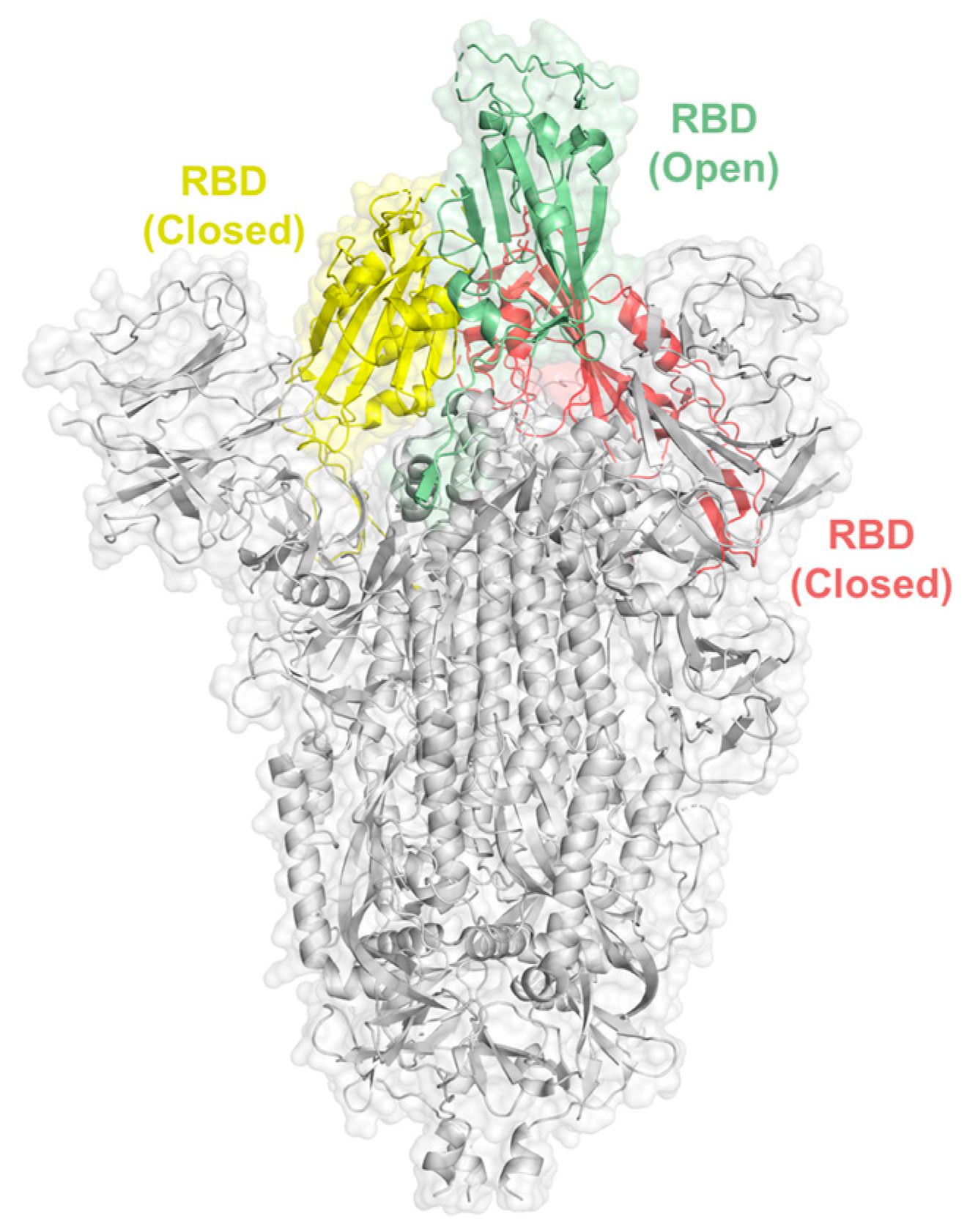
:1. Introduction
2. Results
2.1. Identification of the RBD-Binding Pentasaccharide
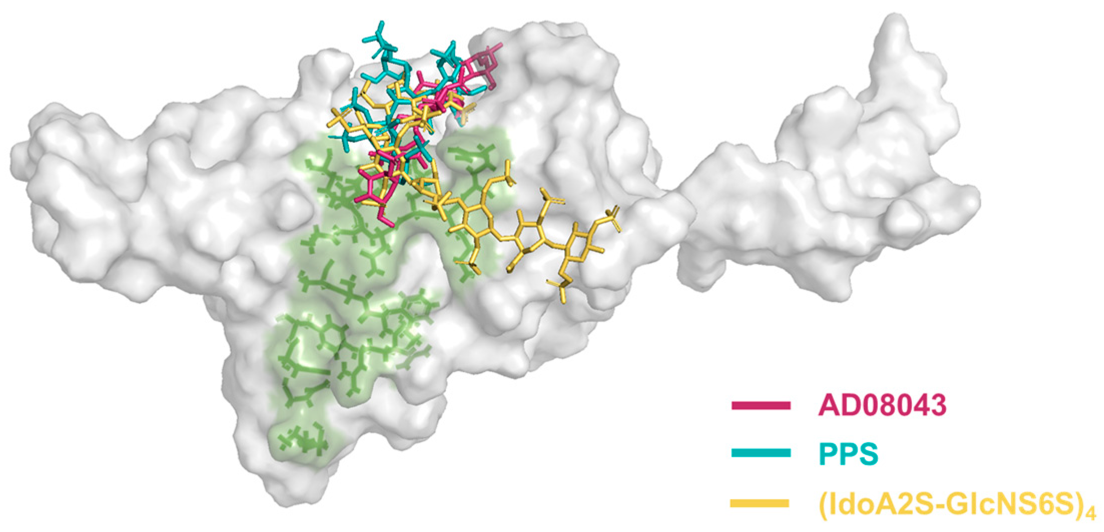
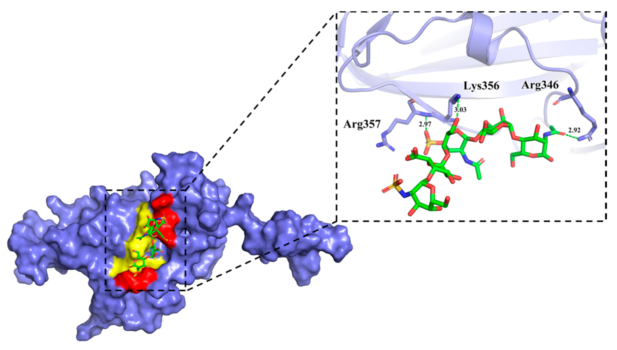
2.2. Analysis of the Molecular Interactions via Molecular Modelling
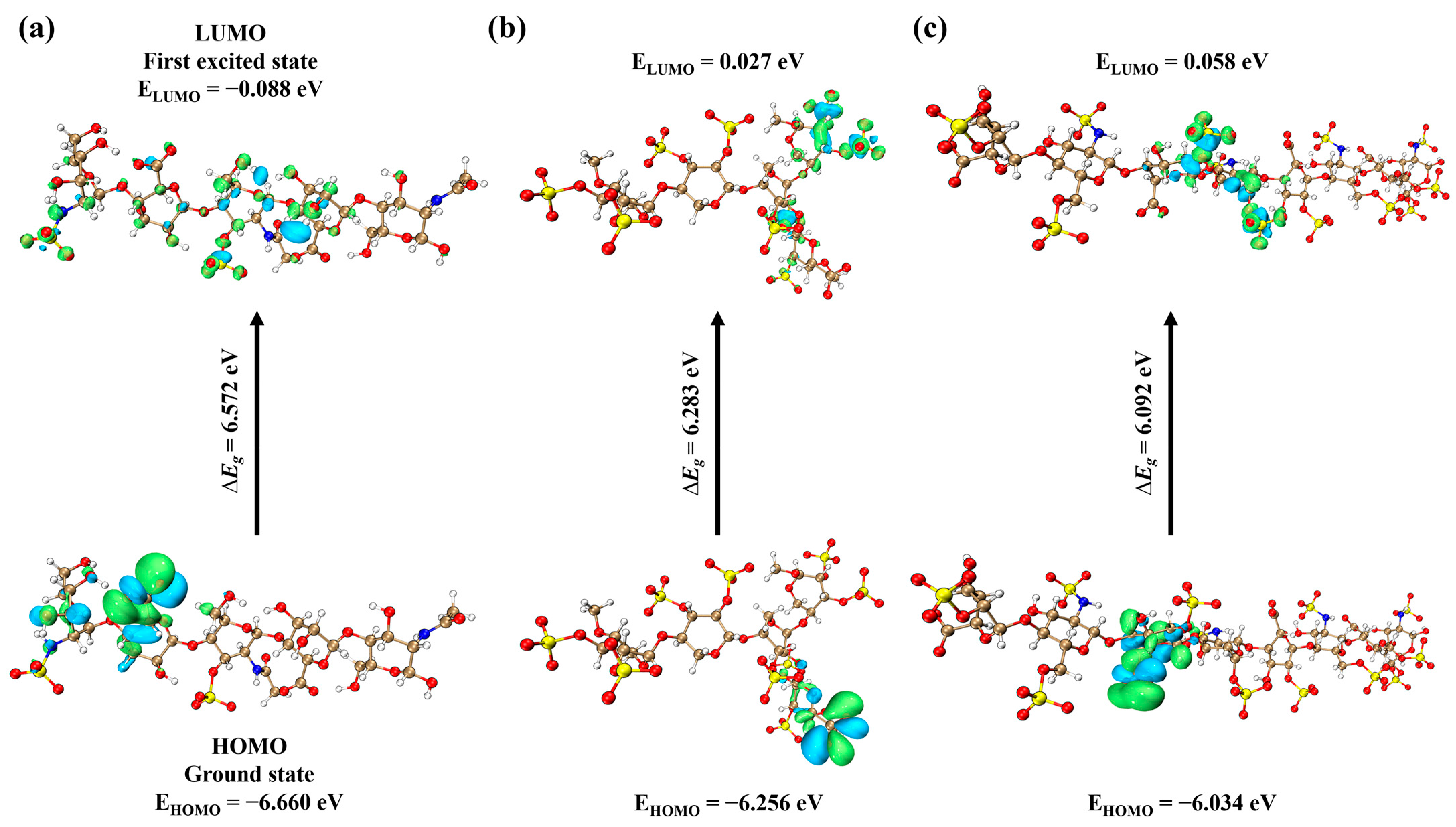
2.3. Frontier Molecular Orbital Analysis of the Ligands
2.4. Evaluation of the HS-Analogues for Druggability via ADMET Studies
3. Discussion
4. Materials and Methods
4.1. Preparation of the Pentasaccharide Library
4.2. Virtual Screening
4.3. Molecular Docking
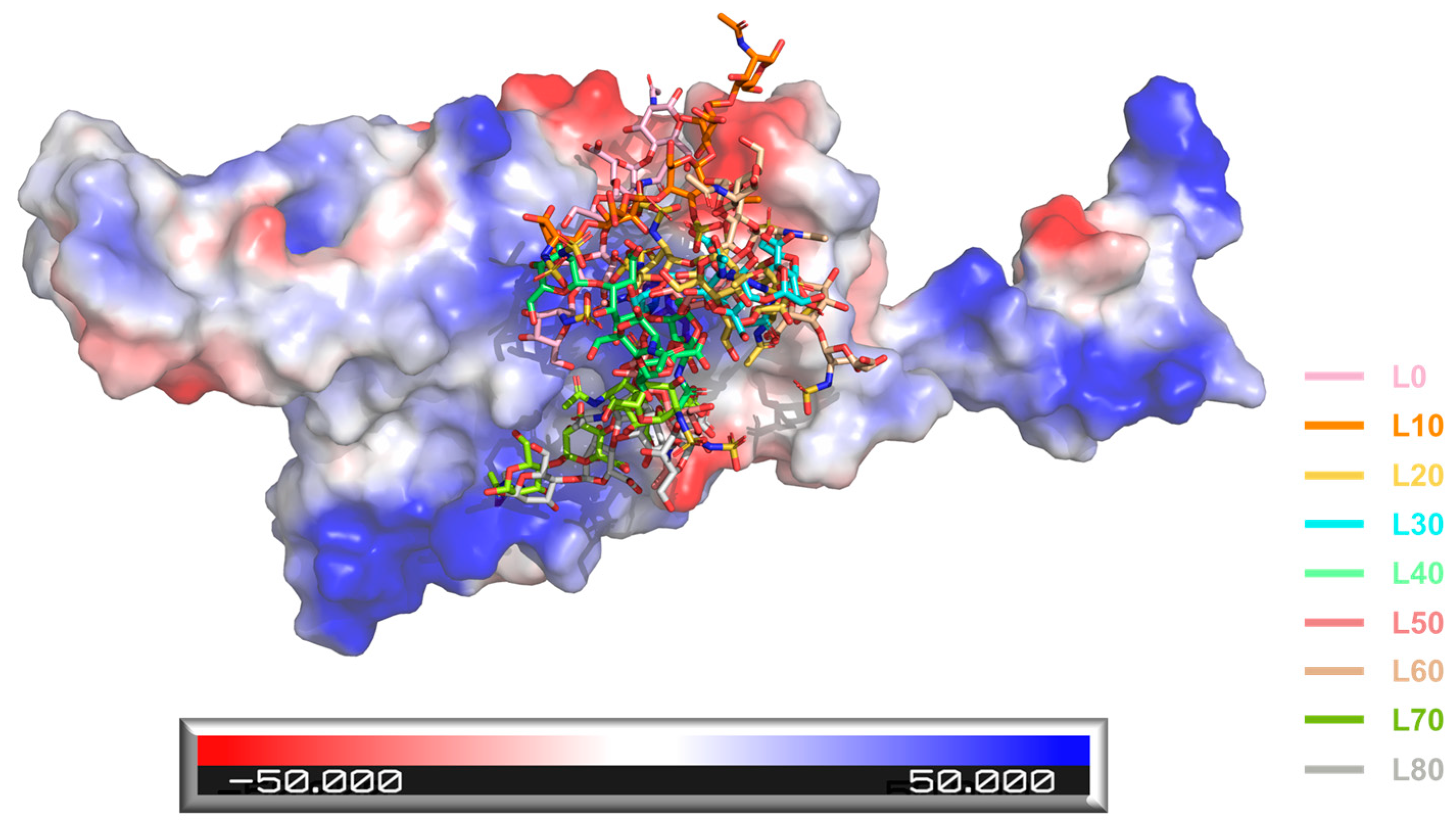
4.4. Molecular Dynamics
4.5. Visualization of Protein–Ligand Complex
4.6. Quantum Chemical Analysis
4.7. ADMET Study
5. Conclusions
Supplementary Materials
Author Contributions
Funding
Institutional Review Board Statement
Informed Consent Statement
Data Availability Statement
Acknowledgments
Conflicts of Interest
References
- Jin, Y.; Yang, H.; Ji, W.; Wu, W.; Chen, S.; Zhang, W.; Duan, G. Virology, Epidemiology, Pathogenesis, and Control of COVID-19. Viruses 2020, 12, 372. [Google Scholar] [CrossRef] [PubMed]
- Ghai, R.R.; Carpenter, A.; Liew, A.Y.; Martin, K.B.; Herring, M.K.; Gerber, S.I.; Hall, A.J.; Sleeman, J.M.; VonDobschuetz, S.; Behravesh, C.B. Animal Reservoirs and Hosts for Emerging Alphacoronaviruses and Betacoronaviruses. Emerg. Infect. Dis. 2021, 27, 1015–1022. [Google Scholar] [CrossRef] [PubMed]
- Wrapp, D.; Wang, N.; Corbett, K.S.; Goldsmith, J.A.; Hsieh, C.L.; Abiona, O.; Graham, B.S.; McLellan, J.S. Cryo-EM structure of the 2019-nCoV spike in the prefusion conformation. Science 2020, 367, 1260–1263. [Google Scholar] [CrossRef]
- Clausen, T.M.; Sandoval, D.R.; Spliid, C.B.; Pihl, J.; Perrett, H.R.; Painter, C.D.; Narayanan, A.; Majowicz, S.A.; Kwong, E.M.; McVicar, R.N.; et al. SARS-CoV-2 Infection Depends on Cellular Heparan Sulfate and ACE2. Cell 2020, 183, 1043–1057.e15. [Google Scholar] [CrossRef] [PubMed]
- Kearns, F.L.; Sandoval, D.R.; Casalino, L.; Clausen, T.M.; Rosenfeld, M.A.; Spliid, C.B.; Amaro, R.E.; Esko, J.D. Spike-heparan sulfate interactions in SARS-CoV-2 infection. Curr. Opin. Struct. Biol. 2022, 76, 102439. [Google Scholar] [CrossRef]
- Liu, L.; Chopra, P.; Li, X.; Bouwman, K.M.; Tompkins, S.M.; Wolfert, M.A.; de Vries, R.P.; Boons, G.J. Heparan Sulfate Proteoglycans as Attachment Factor for SARS-CoV-2. ACS Cent. Sci. 2021, 7, 1009–1018. [Google Scholar] [CrossRef]
- Huang, Y.; Yang, C.; Xu, X.F.; Xu, W.; Liu, S.W. Structural and functional properties of SARS-CoV-2 spike protein: Potential antivirus drug development for COVID-19. Acta Pharmacol. Sin. 2020, 41, 1141–1149. [Google Scholar] [CrossRef]
- Hoffmann, M.; Arora, P.; Gross, R.; Seidel, A.; Hornich, B.F.; Hahn, A.S.; Kruger, N.; Graichen, L.; Hofmann-Winkler, H.; Kempf, A.; et al. SARS-CoV-2 variants B.1.351 and P.1 escape from neutralizing antibodies. Cell 2021, 184, 2384–2393.e12. [Google Scholar] [CrossRef]
- Motozono, C.; Toyoda, M.; Zahradnik, J.; Saito, A.; Nasser, H.; Tan, T.S.; Ngare, I.; Kimura, I.; Uriu, K.; Kosugi, Y.; et al. SARS-CoV-2 spike L452R variant evades cellular immunity and increases infectivity. Cell Host Microbe 2021, 29, 1124–1136.e11. [Google Scholar] [CrossRef]
- Ozono, S.; Zhang, Y.; Ode, H.; Sano, K.; Tan, T.S.; Imai, K.; Miyoshi, K.; Kishigami, S.; Ueno, T.; Iwatani, Y.; et al. SARS-CoV-2 D614G spike mutation increases entry efficiency with enhanced ACE2-binding affinity. Nat. Commun. 2021, 12, 848. [Google Scholar] [CrossRef]
- Hacisuleyman, E.; Hale, C.; Saito, Y.; Blachere, N.E.; Bergh, M.; Conlon, E.G.; Schaefer-Babajew, D.J.; DaSilva, J.; Muecksch, F.; Gaebler, C.; et al. Vaccine Breakthrough Infections with SARS-CoV-2 Variants. N. Engl. J. Med. 2021, 384, 2212–2218. [Google Scholar] [CrossRef] [PubMed]
- Hoffmann, M.; Snyder, N.L.; Hartmann, L. Polymers Inspired by Heparin and Heparan Sulfate for Viral Targeting. Macromolecules 2022, 55, 7957–7973. [Google Scholar] [CrossRef] [PubMed]
- Guimond, S.E.; Mycroft-West, C.J.; Gandhi, N.S.; Tree, J.A.; Le, T.T.; Spalluto, C.M.; Humbert, M.V.; Buttigieg, K.R.; Coombes, N.; Elmore, M.J.; et al. Synthetic Heparan Sulfate Mimetic Pixatimod (PG545) Potently Inhibits SARS-CoV-2 by Disrupting the Spike-ACE2 Interaction. ACS Cent. Sci. 2022, 8, 527–545. [Google Scholar] [CrossRef] [PubMed]
- Mycroft-West, C.J.; Su, D.; Pagani, I.; Rudd, T.R.; Elli, S.; Gandhi, N.S.; Guimond, S.E.; Miller, G.J.; Meneghetti, M.C.Z.; Nader, H.B.; et al. Heparin Inhibits Cellular Invasion by SARS-CoV-2: Structural Dependence of the Interaction of the Spike S1 Receptor-Binding Domain with Heparin. Thromb. Haemost. 2020, 120, 1700–1715. [Google Scholar] [CrossRef] [PubMed]
- Ke, Z.; Oton, J.; Qu, K.; Cortese, M.; Zila, V.; McKeane, L.; Nakane, T.; Zivanov, J.; Neufeldt, C.J.; Cerikan, B.; et al. Structures and distributions of SARS-CoV-2 spike proteins on intact virions. Nature 2020, 588, 498–502. [Google Scholar] [CrossRef]
- Kjellen, L.; Lindahl, U. Specificity of glycosaminoglycan-protein interactions. Curr. Opin. Struct. Biol. 2018, 50, 101–108. [Google Scholar] [CrossRef]
- Patel, V.N.; Lombaert, I.M.; Cowherd, S.N.; Shworak, N.W.; Xu, Y.; Liu, J.; Hoffman, M.P. Hs3st3-modified heparan sulfate controls KIT+ progenitor expansion by regulating 3-O-sulfotransferases. Dev. Cell 2014, 29, 662–673. [Google Scholar] [CrossRef]
- Xia, G.; Chen, J.; Tiwari, V.; Ju, W.; Li, J.P.; Malmstrom, A.; Shukla, D.; Liu, J. Heparan sulfate 3-O-sulfotransferase isoform 5 generates both an antithrombin-binding site and an entry receptor for herpes simplex virus, type 1. J. Biol. Chem. 2002, 277, 37912–37919. [Google Scholar] [CrossRef]
- Xu, C.; Wang, Y.; Liu, C.; Zhang, C.; Han, W.; Hong, X.; Wang, Y.; Hong, Q.; Wang, S.; Zhao, Q.; et al. Conformational dynamics of SARS-CoV-2 trimeric spike glycoprotein in complex with receptor ACE2 revealed by cryo-EM. Sci. Adv. 2021, 7, eabe5575. [Google Scholar] [CrossRef]
- Ennemoser, M.; Rieger, J.; Muttenthaler, E.; Gerlza, T.; Zatloukal, K.; Kungl, A.J. Enoxaparin and Pentosan Polysulfate Bind to the SARS-CoV-2 Spike Protein and Human ACE2 Receptor, Inhibiting Vero Cell Infection. Biomedicines 2021, 10, 49. [Google Scholar] [CrossRef]
- Bertini, S.; Alekseeva, A.; Elli, S.; Pagani, I.; Zanzoni, S.; Eisele, G.; Krishnan, R.; Maag, K.P.; Reiter, C.; Lenhart, D.; et al. Pentosan Polysulfate Inhibits Attachment and Infection by SARS-CoV-2 In Vitro: Insights into Structural Requirements for Binding. Thromb. Haemost. 2022, 122, 984–997. [Google Scholar] [CrossRef] [PubMed]
- Al Sheikh Ali, A.; Khan, D.; Naqvi, A.; Al-Blewi, F.F.; Rezki, N.; Aouad, M.R.; Hagar, M. Design, Synthesis, Molecular Modeling, Anticancer Studies, and Density Functional Theory Calculations of 4-(1,2,4-Triazol-3-ylsulfanylmethyl)-1,2,3-triazole Derivatives. ACS Omega 2021, 6, 301–316. [Google Scholar] [CrossRef] [PubMed]
- Seyfinejad, B.; Ozkan, S.A.; Jouyban, A. Recent advances in the determination of unbound concentration and plasma protein binding of drugs: Analytical methods. Talanta 2021, 225, 122052. [Google Scholar] [CrossRef] [PubMed]
- Hughes, J.D.; Blagg, J.; Price, D.A.; Bailey, S.; Decrescenzo, G.A.; Devraj, R.V.; Ellsworth, E.; Fobian, Y.M.; Gibbs, M.E.; Gilles, R.W.; et al. Physiochemical drug properties associated with in vivo toxicological outcomes. Bioorg. Med. Chem. Lett. 2008, 18, 4872–4875. [Google Scholar] [CrossRef]
- Tandon, R.; Sharp, J.S.; Zhang, F.; Pomin, V.H.; Ashpole, N.M.; Mitra, D.; McCandless, M.G.; Jin, W.; Liu, H.; Sharma, P.; et al. Effective Inhibition of SARS-CoV-2 Entry by Heparin and Enoxaparin Derivatives. J. Virol. 2021, 95, e01987-20. [Google Scholar] [CrossRef] [PubMed]
- Li, B.; Zhang, T.; Li, J.P.; Yu, M.J. Antiviral Disaccharide Lead Compounds against SARS-CoV-2 through Computer-Aided High-Throughput Screen. Chembiochem 2022, 23, e202200461. [Google Scholar] [CrossRef] [PubMed]
- BIOVIA. Discovery Studio Modeling Environment; Version 4.5; Dassault Systèmes: San Diego, CA, USA, 2015. [Google Scholar]
- Arcon, J.P.; Modenutti, C.P.; Avendano, D.; Lopez, E.D.; Defelipe, L.A.; Ambrosio, F.A.; Turjanski, A.G.; Forli, S.; Marti, M.A. AutoDock Bias: Improving binding mode prediction and virtual screening using known protein-ligand interactions. Bioinformatics 2019, 35, 3836–3838. [Google Scholar] [CrossRef]
- Halgren, T. New method for fast and accurate binding-site identification and analysis. Chem. Biol. Drug Des. 2007, 69, 146–148. [Google Scholar] [CrossRef]
- Friesner, R.A.; Murphy, R.B.; Repasky, M.P.; Frye, L.L.; Greenwood, J.R.; Halgren, T.A.; Sanschagrin, P.C.; Mainz, D.T. Extra precision glide: Docking and scoring incorporating a model of hydrophobic enclosure for protein-ligand complexes. J. Med. Chem. 2006, 49, 6177–6196. [Google Scholar] [CrossRef]
- Halgren, T.A.; Murphy, R.B.; Friesner, R.A.; Beard, H.S.; Frye, L.L.; Pollard, W.T.; Banks, J.L. Glide: A new approach for rapid, accurate docking and scoring. 2. Enrichment factors in database screening. J. Med. Chem. 2004, 47, 1750–1759. [Google Scholar] [CrossRef]
- Trott, O.; Olson, A.J. AutoDock Vina: Improving the speed and accuracy of docking with a new scoring function, efficient optimization, and multithreading. J. Comput. Chem. 2010, 31, 455–461. [Google Scholar] [CrossRef] [PubMed]
- Morris, G.M.; Goodsell, D.S.; Halliday, R.S.; Huey, R.; Hart, W.E.; Belew, R.K.; Olson, A.J. Automated docking using a Lamarckian genetic algorithm and an empirical binding free energy function. J. Comput. Chem. 1998, 19, 1639–1662. [Google Scholar] [CrossRef]
- Krieger, E.; Koraimann, G.; Vriend, G. Increasing the precision of comparative models with YASARA NOVA—A self-parameterizing force field. Proteins 2002, 47, 393–402. [Google Scholar] [CrossRef] [PubMed]
- Duan, Y.; Wu, C.; Chowdhury, S.; Lee, M.C.; Xiong, G.; Zhang, W.; Yang, R.; Cieplak, P.; Luo, R.; Lee, T.; et al. A point-charge force field for molecular mechanics simulations of proteins based on condensed-phase quantum mechanical calculations. J. Comput. Chem. 2003, 24, 1999–2012. [Google Scholar] [CrossRef]
- Kumari, R.; Kumar, R.; Lynn, A. g_mmpbsa—A GROMACS Tool for High-Throughput MM-PBSA Calculations. J. Chem. Inf. Model. 2014, 54, 1951–1962. [Google Scholar] [CrossRef] [PubMed]
- Frisch, M.J.; Trucks, G.W.; Schlegel, H.B.; Scuseria, G.E.; Robb, M.A.; Cheeseman, J.R.; Scalmani, G.; Barone, V.; Petersson, G.A.; Nakatsuji, H.; et al. Gaussian 09 Rev. C.01; Gaussian, Inc.: Wallingford, CT, USA, 2016. [Google Scholar]
- Lu, T.; Chen, F. Multiwfn: A multifunctional wavefunction analyzer. J. Comput. Chem. 2012, 33, 580–592. [Google Scholar] [CrossRef]
- Humphrey, W.; Dalke, A.; Schulten, K. VMD: Visual molecular dynamics. J. Mol. Graph. 1996, 14, 33–38. [Google Scholar] [CrossRef]
- Becke, A.D. Density-functional thermochemistry. III. The role of exact exchange. J. Chem. Phys. 1993, 98, 5648–5652. [Google Scholar] [CrossRef]
- Lee, C.; Yang, W.; Parr, R.G. Development of the Colle-Salvetti correlation-energy formula into a functional of the electron density. Phys. Rev. B 1988, 37, 785–789. [Google Scholar] [CrossRef]
- Sundaraganesan, N.; Ilakiamani, S.; Dominic Joshua, B. FT-Raman and FT-IR spectra, ab initio and density functional studies of 2-amino-4,5-difluorobenzoic acid. Spectrochim. Acta A Mol. Biomol. Spectrosc. 2007, 67, 287–297. [Google Scholar] [CrossRef]
- Izadyar, M.; Khavani, M.; Housaindokht, M.R. Sensing Ability of Hybrid Cyclic Nanopeptides Based on Thiourea Cryptands for Different Ions, A Joint DFT-D3/MD Study. J. Phys. Chem. A 2017, 121, 244–255. [Google Scholar] [CrossRef] [PubMed]
- Jasmine, N.J.; Muthiah, P.T.; Arunagiri, C.; Subashini, A. Vibrational spectra (experimental and theoretical), molecular structure, natural bond orbital, HOMO-LUMO energy, Mulliken charge and thermodynamic analysis of N’-hydroxy-pyrimidine-2-carboximidamide by DFT approach. Spectrochim. Acta A Mol. Biomol. Spectrosc. 2015, 144, 215–225. [Google Scholar] [CrossRef] [PubMed]
- Xiong, G.; Wu, Z.; Yi, J.; Fu, L.; Yang, Z.; Hsieh, C.; Yin, M.; Zeng, X.; Wu, C.; Lu, A.; et al. ADMETlab 2.0: An integrated online platform for accurate and comprehensive predictions of ADMET properties. Nucleic Acids Res. 2021, 49, W5–W14. [Google Scholar] [CrossRef] [PubMed]
- Meyer, B.; Thunberg, L.; Lindahl, U.; Larm, O.; Leder, I.G. The antithrombin-binding sequence of heparin studied by n.m.r. spectroscopy. Carbohydr. Res. 1981, 88, C1–C4. [Google Scholar] [CrossRef]
- Petitou, M.; Herault, J.P.; Bernat, A.; Driguez, P.A.; Duchaussoy, P.; Lormeau, J.C.; Herbert, J.M. Synthesis of thrombin-inhibiting heparin mimetics without side effects. Nature 1999, 398, 417–422. [Google Scholar] [CrossRef]
- Zhang, Y.; Wang, Y.; Zhou, Z.; Wang, P.; Xi, X.; Hu, S.; Xu, R.; Du, G.; Li, J.; Chen, J.; et al. Synthesis of bioengineered heparin by recombinant yeast Pichia pastoris. Green Chem. 2022, 24, 3180–3192. [Google Scholar] [CrossRef]


| Ligand Name | Binding Free Energies (kcal/mol) | Dissociation Constant (μM) |
|---|---|---|
| AD08043 | −7.149 | 5.751 |
| (IdoA2S-GlcNS6S)4 | −6.797 | 10.417 |
| PPS | −6.247 | 26.357 |
| Ligand Name | Hydrogen Bond Receptor Atom—Ligand Atom (Bond Length) | Hydrophobic Contact |
|---|---|---|
| AD08043 | (1) Arg355:NH1—O15 (3.18 Å); (2) Arg355:N—O20 (3.06 Å); (3) Arg466:NH1—O17 (3.14 Å); (4) Arg466:NH2—O16 (3.11 Å); (5) Arg466:NH2—O16 (3.18 Å); (6) Tyr396:OH—O28 (2.94 Å); (7) Tyr396:OH—O30 (3.14 Å); (8) Leu517:N—O4 (3.11 Å). | (1) Trp353; (2) Asn354; (3) Pro426; (4) Asp428; (5) Phe429; (6) Phe464; (7) Phe515; (8) Glu516; (9) His519. |
| (IdoA2S-GlcNS6S)4 | (1) Asn334:ND2—O7 (2.94 Å); (2) Asn334:ND2—O12 (3.12 Å); (3) Arg355:N—O73 (3.11 Å); (4) Arg357:N—O62 (3.19 Å); (5) Arg357:NE—O42 (2.95 Å); (6) Arg357:NE—O53 (3.06 Å); (7) Arg357:NH1—O57 (3.00 Å); (8) Arg357:NH1—O89 (2.96 Å); (9) Arg357:NH2—O42 (3.24 Å); (10) Asn360:O—O9 (2.99 Å); (11) Tyr396:OH—2O12 (2.94 Å); (12) Arg466:NH1—O92 (3.14 Å); (13) Arg466:NH2—N76 (3.18 Å). | (1) Pro337; (2) Trp353; (3) Asn354; (4) Ser359; (5) Pro426; (6) Pro463; (7) Phe464; (8) Glu465; (9) Ser514; (10) Phe515; (11) Glu516. |
| PPS | (1) Arg355:NH1—O14 (2.93 Å); (2) Arg355:NH1—O33 (3.14 Å); (3) Arg357:NH1—O5 (3.03 Å); (4) Arg357:NH2—O55 (3.35 Å); (5) Arg466:NH1—O17 (3.28 Å); (6) Arg466:NH2—O23 (3.03 Å). | (1) Tyr396; (2) Pro426; (3) Asp428; (4) Lys462; (5) Pro463; (6) Phe464; (7) Glu465; (8) Ser514; (9) Phe515; (10) Leu517; (11) His519. |
| Receptor Name | Hydrogen Bond Receptor Atom—Ligand Atom (Bond Length) | Hydrophobic Contact |
|---|---|---|
| B.1.1.7 (alpha) variant RBD | (1) Arg355:NH1—O10 (3.18 Å); (2) Arg355:NH2—O9 (3.33 Å); (3) Arg355:NH2—O10 (3.23 Å); (4) Lys462:NZ—O (3.20 Å); (5) Arg466:NH2—O16 (2.84 Å); (6) Arg466:NH2—O25 (2.95 Å). | (1) Tyr396; (2) Pro426; (3) Asp428; (4) Phe429; (5) Thr430; (6) Pro463; (7) Phe464; (8) Ser514; (9) Glu516. |
| B.1.351 (beta) variant RBD | (1) Arg355:NH1—O30 (3.22 Å); (2) Thr430:N—O7 (3.01 Å); (3) Arg466:NE—O20 (3.28 Å); (4) Arg466:N—O26 (3.06 Å); (5) Ile468:N—O21 (2.80 Å); (6) Phe515:O—N (2.91 Å). | (1) Asp428; (2) Phe429; (3) Phe464; (4) Glu465; (5) Asp467; (6) Glu516; (7) Leu517. |
| P.1 (gamma) variant RBD | (1) Asn448:ND2—O22 (2.73 Å); (2) Arg509:NH2—O26 (2.96 Å). | (1) Asn343; (2) Ala344; (3) Thr345; (4) Arg346; (5) Ser373; (6) Trp436; (7) Asn437; (8) Asn440; (9) Leu441; (10) Asp442; (11) Asn450; (12) Tyr451. |
| B.1.617.2 (delta) variant RBD | (1) Arg346:NH2—O5 (2.80 Å); (2) Arg452:NH2—O29 (2.89 Å); (3) Thr470:OG1—O13 (3.13 Å); (4) Thr470:OG1—O15 (2.96 Å); (5) Thr470:O—O20 (2.74 Å); (6) Gly482:O—N1 (3.26 Å). | (1) Ala348; (2) Tyr351; (3) Ala352; (4) Asn354; (5) Tyr451; (6) Ile468; (7) Glu471; (8) Ile472; (9) Phe490. |
| B.1.1.529 (omicron BA.1) variant RBD | (1) Asn343:O—O8 (2.98 Å); (2) Thr345:N—O9 (3.16 Å); (3) Arg346:N—O9 (2.81 Å); (4) Ser349:OG—O20 (3.06 Å); (5) Asn354:ND2—O (2.96 Å); (6) Asn450:ND2—O25 (3.01 Å). | (1) Glu340; (2) Ala344; (3) Phe347; (4) Ala348; (5) Tyr351; (6) Ala352; (7) Tyr449; (8) Leu452. |
| Time Point | Hydrogen Bond Receptor Atom—Ligand Atom (Bond Length) | Hydrophobic Contact |
|---|---|---|
| 0 ns | (1) Arg355:NH1—O10 (3.07 Å); (2) Arg355:O—O20 (2.65 Å); (3) Arg466:NH1—O16 (2.77 Å); (4) Arg466:NH2—O16 (2.83 Å); (5) Phe515:O—O4 (2.66 Å). | (1) Trp353; (2) Tyr396; (3) Asp428; (4) Phe464; (5) Ser514; (6) Glu516; (7) Leu517; (8) His519. |
| 10 ns | (1) Asn394:ND2—O32 (3.07 Å); (2) His519:ND1—O10 (3.01 Å). | / |
| 20 ns | (1) Arg357:NH1—O16 (2.60 Å); (2) Arg357:NH2—O15 (2.81 Å); (3) Arg357:NH2—O16 (2.88 Å); (4) Asn360:ND2—O31 (2.91 Å); (5) Tyr396:OH—O22 (2.73 Å). | / |
| 30 ns | (1) Arg357:NH1—O16 (2.67 Å); (2) Arg357:NH2—O15 (2.76 Å); (3) Asn360:ND2—O29 (2.80 Å); (4) Tyr396:OH—O25 (3.02 Å). | / |
| 40 ns | / | / |
| 50 ns | (1) Tyr396:OH—O30 (2.80 Å). | (1) Arg355; (2) Arg357. |
| 60 ns | (1) Arg357:NH1—O17 (2.81 Å); (2) Arg357:NH2—O15 (2.96 Å); (3) Arg357:NH2—O17 (3.28 Å); (4) Asn360:ND2—O15 (3.23 Å); (5) Asn394:ND2—O29 (2.70 Å). | (1) Ace332; (2) Thr393. |
| 70 ns | (1) Asn354:OD1—O4 (3.10 Å); (2) Arg357:N—O29 (3.04 Å). | (1) Arg346; (2) Phe347; (3) Ala348; (4) Arg355; (5) Lys356. |
| 80 ns | (1) Arg346:NH2—O7 (2.92 Å);(2) Lys356:NZ—O13 (3.03 Å); (3) Arg357:N—O29 (2.97 Å). | (1) Phe347; (2) Ala348. |
| Ligand Name | HOMO (eV) | LUMO (eV) | ∆Eg = ELUMO − EHOMO (eV) |
|---|---|---|---|
| AD08043 | −6.660 | −0.088 | 6.572 |
| PPS | −6.256 | 0.027 | 6.283 |
| (IdoA2S-GlcNS6S)4 | −6.034 | 0.058 | 6.092 |
| Parameters | AD08043 | PPS | (IdoA2S-GlcNS6S)4 |
|---|---|---|---|
| No. of H bond acceptors | 36 | 50 | 81 |
| No. of H bond donors | 18 | 10 | 30 |
| Topological Polar Surface area, TPSA ([Å]2) | 568.400 | 720.460 | 1264.350 |
| Lipophilicity, log P | −6.291 | −8.807 | −15.092 |
| Water Solubility, log S | 1.948 | 5.717 | 8.771 |
| Pfizer Rule | Accepted | Accepted | Accepted |
| Pgp inhibitor | No | No | No |
| Plasma Protein Binding (PPB) | 3.180% | 61.866% | 52.737% |
| Volume Distribution (VD) (L/kg) | 0.140 | −0.324 | −0.926 |
| Blood–Brain Barrier (BBB) Penetration | No | No | No |
| CYP1A2 inhibitor | No | No | No |
| CYP2C19 inhibitor | No | No | No |
| CYP2C9 inhibitor | No | No | No |
| CYP2D6 inhibitor | No | No | No |
| CYP3A4 inhibitor | No | No | No |
| Human hepatotoxicity (H-HT) | Negative | Positive | Positive |
| Rat Oral Acute Toxicity | Low toxicity | High toxicity | Low toxicity |
| Skin Sensitization | Negative | Positive | Negative |
| Carcinogenicity | Negative | Positive | Negative |
| Respiratory Toxicity | Negative | Positive | Positive |
| Drug-likeness | Yes | No | No |
Disclaimer/Publisher’s Note: The statements, opinions and data contained in all publications are solely those of the individual author(s) and contributor(s) and not of MDPI and/or the editor(s). MDPI and/or the editor(s) disclaim responsibility for any injury to people or property resulting from any ideas, methods, instructions or products referred to in the content. |
© 2023 by the authors. Licensee MDPI, Basel, Switzerland. This article is an open access article distributed under the terms and conditions of the Creative Commons Attribution (CC BY) license (https://creativecommons.org/licenses/by/4.0/).
Share and Cite
Li, B.; Zhang, T.; Cao, H.; Ferro, V.; Li, J.; Yu, M. Identification of a Pentasaccharide Lead Compound with High Affinity to the SARS-CoV-2 Spike Protein via In Silico Screening. Int. J. Mol. Sci. 2023, 24, 16115. https://doi.org/10.3390/ijms242216115
Li B, Zhang T, Cao H, Ferro V, Li J, Yu M. Identification of a Pentasaccharide Lead Compound with High Affinity to the SARS-CoV-2 Spike Protein via In Silico Screening. International Journal of Molecular Sciences. 2023; 24(22):16115. https://doi.org/10.3390/ijms242216115
Chicago/Turabian StyleLi, Binjie, Tianji Zhang, Hui Cao, Vito Ferro, Jinping Li, and Mingjia Yu. 2023. "Identification of a Pentasaccharide Lead Compound with High Affinity to the SARS-CoV-2 Spike Protein via In Silico Screening" International Journal of Molecular Sciences 24, no. 22: 16115. https://doi.org/10.3390/ijms242216115