Article

Bisphenol A Effects on Neurons’ Neurochemical Character in the Urinary Bladder Intramural Ganglia of Domestic Pigs

1
Department of Clinical Diagnostics, Faculty of Veterinary Medicine, University of Warmia and Mazury in Olsztyn, Oczapowskiego 14, 10-957 Olsztyn, Poland
2
Agri Plus sp. Z o.o., Marcelinska Street 92, 60-324 Pozan, Poland
3
Department of Clinical Physiology, Faculty of Veterinary Medicine, University of Warmia and Mazury in Olsztyn, Oczapowskiego 13, 10-957 Olsztyn, Poland
*
Author to whom correspondence should be addressed.
Int. J. Mol. Sci. 2023, 24(23), 16792; https://doi.org/10.3390/ijms242316792
Submission received: 25 October 2023 / Revised: 14 November 2023 / Accepted: 25 November 2023 / Published: 27 November 2023
(This article belongs to the Special Issue The Plasticity of the Innervation of the Urogenital System)

Abstract

:
Bisphenol A (BPA), a substance globally used to produce plastics, is part of many everyday items, including bottles, food containers, electronic elements, and others. It may penetrate the environment and living organisms, negatively affecting, among others, the nervous, immune, endocrine, and cardiovascular systems. Knowledge of the impact of BPA on the urinary bladder is extremely scarce. This study investigated the influence of two doses of BPA (0.05 mg/kg body weight (b.w.)/day and 0.5 mg/kg b.w./day) given orally for 28 days on the neurons situated in the ganglia located in the urinary bladder trigone using the typical double immunofluorescence method. In the study, an increase in the percentage of neurons containing substance P (SP), galanin (GAL), a neuronal isoform of nitric oxide synthase (nNOS—used as the marker of nitrergic neurons), and/or cocaine- and amphetamine-regulated transcript (CART) peptide was noted after BPA administration. The severity of these changes depended on the dose of BPA and the type of neuronal factors studied. The most visible changes were noted in the cases of SP- and/or GAL-positive neurons after administering a higher dose of BPA. The results have shown that oral exposure to BPA, lasting even for a short time, affects the intramural neurons in the urinary bladder wall, and changes in the neurochemical characterisation of these neurons may be the first signs of BPA-induced pathological processes in this organ.

1. Introduction

Bisphenol A (BPA) is an organic substance synthesized for the first time in the late 19th century by the condensation of acetone and two molecules of phenols [1]. Due to its properties, such as its high thermal stability, plasticity, and relatively cheap synthesis, BPA is commonly used in the plastic industry to produce polycarbonates and epoxy and vinyl ester resins [2]. BPA is a component of many everyday objects, including, among others, bottles, clothes, electronic elements, house equipment, furniture, and office supplies. This compound is also present in thermal paper, paints, and dental fillings [2,3].
It has been found that BPA may be washed out from plastics and penetrate the environment [4]. Up to now, the presence of this compound has been observed in surface water and tap water, air, plants, house dust, and soil around the world [2,4]. Moreover, BPA penetrates human and animal organisms through the digestive tract, respiratory system, and skin [1]. The main route of exposure to BPA is through the digestive system. Therefore, items containing BPA that come into contact with food and drinking water, such as bottles, dishes, cutlery, and containers, are especially dangerous because BPA may penetrate directly into food and then into the digestive tract [1,2].
Previous studies confirm that BPA shows strong endocrine-disrupting properties [1]. It has been shown that BPA negatively affects various internal organs and systems, including, among others, the nervous, reproductive, and endocrine systems, immune cells, digestive tract, and heart [1,2,5,6,7,8,9]. Moreover, some studies have described correlations between the degree of exposure to BPA and the occurrence frequency of diabetes, obesity, hypertension, and even neurodegenerative diseases [1,2,10,11].
However, the negative influences of BPA on various internal systems are relatively unknown. One such system is the excretory system. Previous studies on this topic have mainly focused on changes in the kidneys and have found that BPA causes developmental disorders and renal failure, accompanied by oliguria and albuminuria [6]. It has also been shown that exposure to BPA results in inflammatory processes in the kidneys, tubular injury, and renal fibrosis [12]. In turn, epidemiological studies on humans have found correlations between exposure to BPA and the glomerular filtration rate, urine albumin-to-creatinine ratio, and the risk of chronic kidney disease [13,14].
Information regarding the effects of BPA on the urinary bladder is even more limited. It is known that exposure to BPA may result in inflammatory processes and neoplasms in this organ [15]. Moreover, BPA may contribute to disturbances in urinary voiding, but the exact mechanisms are unclear [16]. It is possible that BPA can cause urinary voiding dysfunction by affecting the nervous structures that supply this organ. This is especially likely as BPA has been shown to negatively impact neuronal cells in other parts of the nervous system, leading to neurodegenerative changes, synaptogenesis disorders, disturbances in calcium metabolism, and disruptions in the production of neuronal active factors [17,18,19,20,21].
Urinary bladder innervation consists of intrinsic and extrinsic innervation. The former comprises the postsynaptic parasympathetic neuronal cells grouped into the ganglia in the urinary bladder wall and supplying the mucosal and muscular layers [22]. Such ganglia have been described in the urinary bladder of humans, guinea pigs, and domestic pigs, and the largest number of them have been found in the urinary bladder trigone—the triangular-shaped region at the base of the bladder limitedly defined by the two ureteral orifices and the internal urethral meatus [23]. The urinary bladder is also innervated by the nerves of external origin, which are projections of the parasympathetic neurons located in the parasympathetic pelvic ganglia, sympathetic neurons located in sympathetic trunks, hypogastric plexuses, inferior mesenteric ganglia, as well as sensory neurons placed in the dorsal root ganglia [22,24,25,26,27].
Previous studies have shown that neurons located in the urinary bladder intramural ganglia are diverse in terms of their neurochemical characterisation and may contain a wide range of active neuronal factors [28,29,30,31]. Moreover, it has been found that these neurons take part in pathological processes within the urinary bladder and may change their neurochemical profile under the impact of various pathological and toxic factors [32,33,34]. However, it should be underlined that knowledge about the impacts of BPA on the urinary bladder intramural neurons is extremely scarce and limited to one study, in which BPA-induced changes in the number of neurons containing vasoactive intestinal polypeptide were found [34].
Considering the above, the present investigation aimed to evaluate (for the first time) the influence of two various doses of BPA on neurons located in the urinary bladder intramural ganglia and containing neuronal factors, such as substance P (SP), galanin (GAL), a neuronal isoform of nitric oxide synthase (nNOS—used as a marker of nitrergic neurons), and the cocaine- and amphetamine-regulated transcript (CART) peptide, which are known to play important roles in the regulation of the urinary bladder functions [29,30,35,36,37]. The obtained results will allow for a better understanding of the harmful effects of BPA on the functioning of the bladder. Moreover, due to the considerable similarity between the organisation of the intramural ganglia of the urinary bladder in humans and domestic pigs [38,39], the present study may be the first step in understanding the processes connected with BPA’s impact on the human urinary bladder.

2. Results

During the present study, neurons immunoreactive to all active substances included in the study were observed in the ganglia within the urinary bladder wall both in physiological conditions and after BPA administration (Table 1). In the control pigs, the largest group of neurons were cells containing nNOS, the percentage of which was 43.72 ± 1.20% of all PGP 9.5-positive cells. Neurons immunoreactive to CART were slightly less numerous. Their percentage amounted to 38.65 ± 0.30%. In turn, populations of neurons immunoreactive to GAL and/or SP were much less numerous. The percentage was 26.16 ± 0.74% for GAL–positive cells and 20.70 ± 0.82% for neurons containing SP.
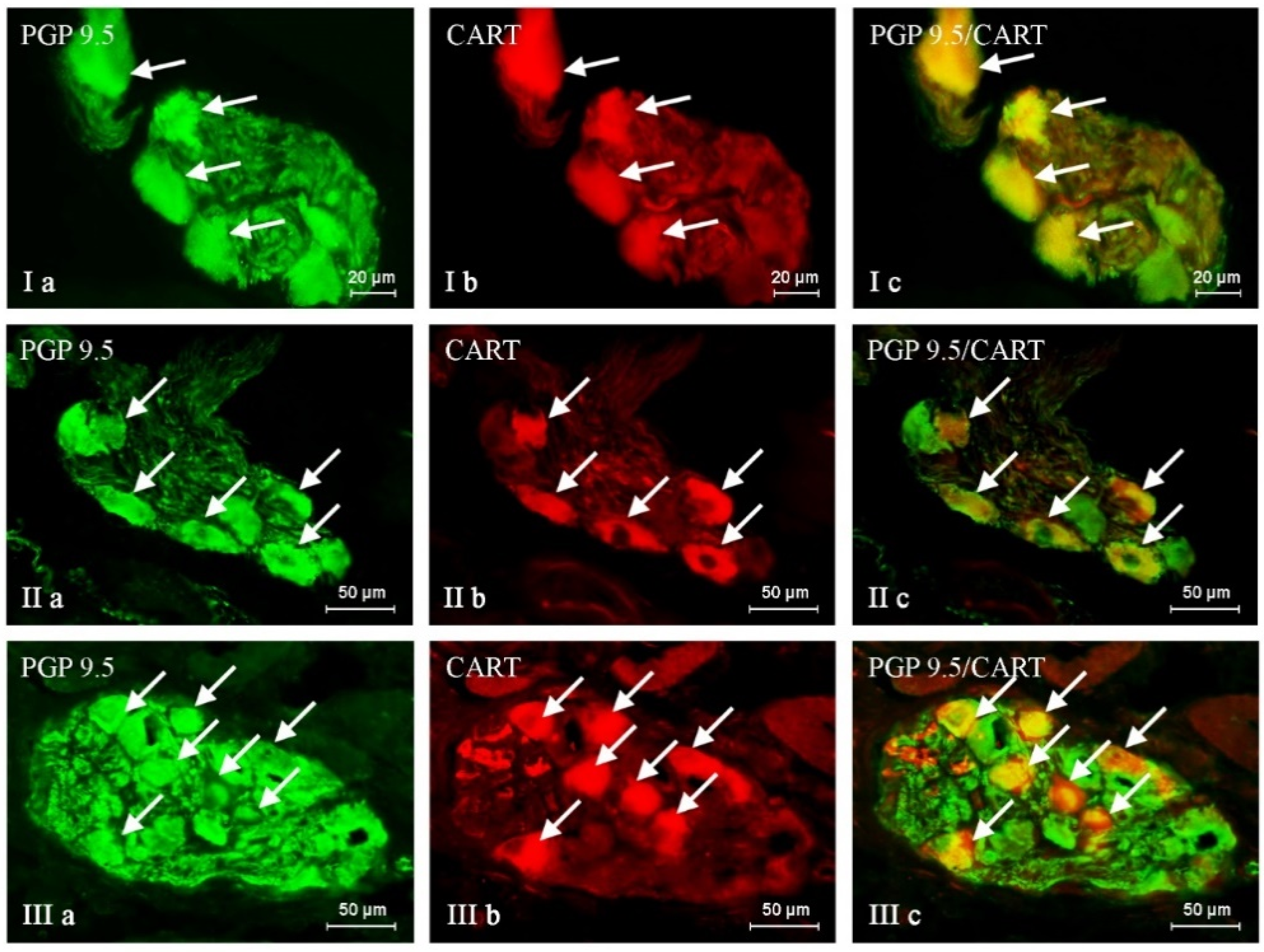
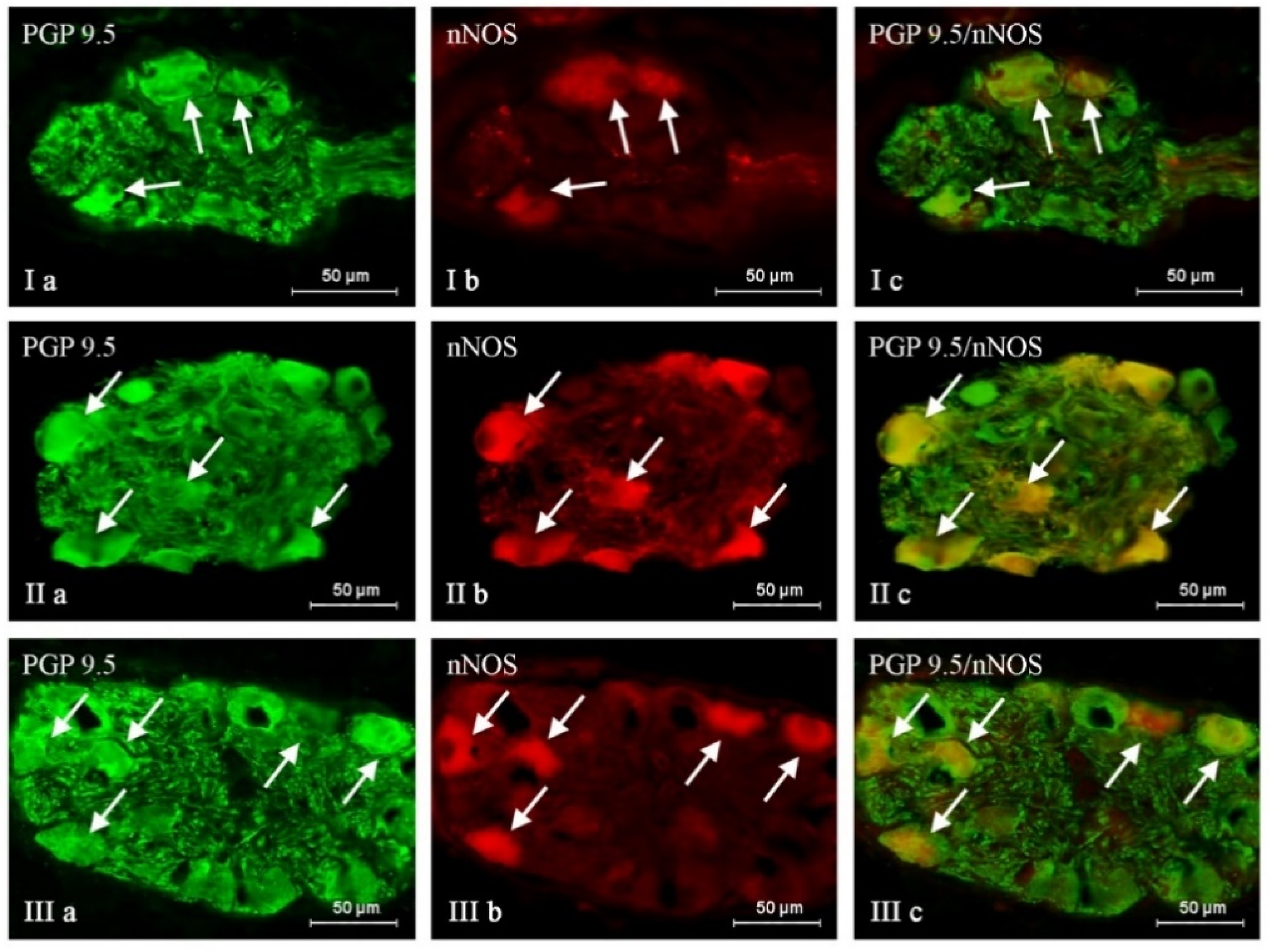
The administration of BPA generally caused an increase in the percentage of neuronal cells containing all active substances studied, and the severity of the changes depended on the type of neuronal factor and the BPA dose (Table 1, Figure 1, Figure 2 and Figure 3). After the administration of a lower BPA dose, the most visible changes were noted in GAL-positive neurons (Figure 1). Their percentage in the BPA 1 group accounted for 35.29 ± 1.16% of all PGP 9.5-positive cells (an increase of about 9 percentage points (p.p.) compared to the control group) (Table 1 and Table 2). The second most visible changes after the administration of the lower dose of BPA were found in the population of neurons containing CART (Figure 2). Their percentage increased by about 6 p.p. and achieved 44.95 ± 0.57% (Table 1 and Table 2). In the BPA 1 group, similar to the control animals, the most numerous populations were neurons containing nNOS, whose percentage was 47.49 ± 0.30% (Table 1, Figure 3). However, the impact of the lower doses of BPA on this population was relatively small because the increase in the percentage of nNOS-positive cells in the BPA 1 group compared to the control pigs accounted for 4 p.p (Table 2). Even less-pronounced changes were observed in the populations of SP-immunoreactive neurons, whose percentage was 23.86 ± 0.40% (an increase of about 3 p.p. compared to the control group) (Table 2, Figure 4).
Higher doses of BPA caused more pronounced changes in the size of the neuronal populations studied. The most visible increase in percentage was noted in the cases of neurons containing SP and/or GAL (Table 1 and Table 2, Figure 1 and Figure 4). The percentage of SP-positive neurons in the BPA 2 group amounted to 34.83 ± 0.64% of all PGP 9.5-immunoreactive cells (an increase of about 14 p.p. compared to the control group) (Table 1 and Table 2). Similar changes (an increase of about 13 p.p.) were observed in neurons containing GAL, the percentage of which in the BPA 2 group amounted to 39.57 ± 0.48% (Table 1 and Table 2). The visible changes in animals in the BPA 2 group also concerned neuronal cells containing CART (Figure 2). The percentage of such neurons amounted to 49.05 ± 1.70%, and compared to the control group, it was higher by 11 p.p (Table 1 and Table 2). The population of nNOS-immunoreactive neuronal cells was the most numerous in the BPA 2 group, but the percentage of such neurons was the least susceptible to changes under the influence of BPA (Figure 3). In particular, the percentage of nNOS-positive neurons was 49.60 ± 1.08% and was only 6 p.p. higher than the value observed in the control animals (Table 1 and Table 2). The summarization of the changes in p.p. noted in the present study is presented in Table 2.

3. Discussion

The results obtained in this study have shown the presence of all studied substances in the neurons located in the intramural ganglia, which is in agreement with previous observations describing these substances in the intramural urinary bladder neurons in various mammal species, including humans [30,31,34,35,36,37,40,41,42,43,44,45]. Conversely, differences concerning the sizes of neuronal populations that are immunoreactive to particular neuronal factors between the present study and previous studies conducted on domestic pigs are clearly visible. In particular, this concerns neurons containing nNOS and CART, the number of which observed in previous studies was significantly smaller than that noted in the present investigation [35,36,40,44]. These discrepancies may result from environmental factors affecting the animals or some differences in methodology, but could also result from interracial differences (in previous studies, the pigs of the Large White Polish breed were investigated) [30,31,40,42,43]. However, the results obtained in the present study are similar to those noted in observations conducted on the human urinary bladder [28], which, together with other studies on the similarities in organisation between human and porcine nervous systems [40], confirms that the domestic pig seems to be a good animal model for studies concerning processes occurring in human neuronal cells.
The presence of the neuronal factors investigated in a relatively high number of neurons strongly suggests the important roles of these substances in regulating urinary bladder functions. This is also reflected in previous studies. It is known that nitric oxide (nitrergic neurons containing nNOS were the most numerous population of neuronal cells studied in the present experiment) affects the activity of muscles located in the urinary bladder wall [36] and regulates intramural blood flow through a relaxant effect on the blood vessels [41]. Moreover, nitric oxide takes part in the conduction of sensory stimuli from the urinary bladder, as evidenced by the presence of nNOS in neurons supplying this organ [40]. In turn, GAL inhibits bladder emptying, probably by activating mechanisms related to opioid substances [37], and SP activates the urinary bladder muscles, increasing the frequency of urination via a simultaneous reduction of its volume [45]. SP and GAL are responsible for relaying sensory information from the urinary bladder to the central nervous system. The role of CART in regulating urinary bladder activity is not well understood, but it is believed to play a role in the growth and maturation of cells within the bladder during development. There is a high likelihood that CART also affects the muscles of the urinary bladder [35,44].
In light of previous studies, it is also known that substances included in this investigation take part in the pathological reactions within the urinary bladder caused by inflammatory processes and toxic factors [40,41,42,43,44]. The current study also confirmed this. However, due to the multidirectional influence of BPA on living organisms and the various reasons for changes in the neurochemical characterisation of neurons, the exact reasons for the obtained results are difficult to explain at the current research stage. In particular, changes noted in the present study may result not only from the increase in the synthesis of neuronal factors on a transcriptional or translational level, or the BPA-induced changes in mRNA or DNA known from previous studies [46,47], but also from the inhibition of the transport of neuronal factors from the cell body to the nerve endings.
The observed changes may result from the direct impact of BPA on the nervous system. Previous studies have reported that BPA shows neurodegenerative properties, appearing as a result of damage to the development of synapses and neurites [48], disturbances in calcium ions’ transport through neuronal cell membranes [49], and mitochondrial failure [50]. Therefore, the changes in the neurochemical characterisation of neurons noted in the present study may be the organism’s response to the neurodegenerative actions of BPA aimed at the neuroprotective processes and maintenance of homeostasis in the nervous system. It is likely that all neuronal factors included in the present study have been described as substances with neuroprotective and antidegenerative properties, both in the central and peripheral nervous system [51,52,53,54,55,56].
The next reason for the observed changes may be connected to the relatively well-known pro-inflammatory activity of BPA and its influence on the immune system [50]. This influence may result in fluctuations in the levels of factors modulating immune cell activity, and it should be underlined that all the neuronal substances studied in the present experiment show such properties. SP is known as the most well-known factor modulating the activity of immune system cells. It stimulates the secretion of pro-inflammatory substances, such as IL-1, IL -6, and TNF-α [57], simultaneously increasing phagocytic activity and histamine release [58,59]. Studies conducted on the urinary bladder have shown that inflammatory processes significantly increase the concentration of SP within this organ, and this phenomenon is accompanied by an increase in the levels of pro-inflammatory cytokines, mast cell degranulation, and lymphocytic infiltration [45]. GAL and nitric oxide are also involved in modulating immune cell activity. GAL, among others, causes an increase in the production of proinflammatory cytokines (mainly IFN-γ) [60] and decreases the synthesis of TNF α [61]. In turn, nitric oxide is one of the most important immunomodulatory factors affecting many processes within various immune cells [62]. Moreover, CART, whose immune system function is unclear, has immunomodulatory activity but, in contrast to SP, causes a reduction in the synthesis and release of pro-inflammatory factors with a simultaneous increase in the activity of anti-inflammatory cytokines [52].
Interestingly, BPA administration caused similar changes (an increase in number) in populations of neurons containing these factors, which have an opposite effect. The reason for such a phenomenon is unclear, but a similar situation has been noted under the impact of BPA within intestinal innervation [63,64]. It should be underlined that the BPA doses used in the present study were relatively low, and they did not cause visible inflammatory symptoms. Admittedly it cannot be excluded that the fluctuations in the levels of neuronal factors noted in the present study may be the first signs of a subclinical inflammatory state, but it is important to stress that this thesis is only a guess and its confirmation is not possible without further comprehensive molecular and/or biochemical research
The third reason for the observed changes may be connected with the impact of BPA on muscular contraction. Although the impacts of BPA on the urinary bladder muscles has not yet been studied, it is known that this substance inhibits the amplitude and frequency of contractions of muscles located in the wall of the intestine and uterus of various mammal species [65,66,67,68,69]. Therefore, similar effects of BPA on the urinary bladder are likely. The mechanisms of such relaxant activities of BPA are not clear. Previous studies suggest various pathways involved in the BPA-induced inhibition of muscular contraction, including the oxytocin- and prostaglandin-related pathway [70], norepinephrine-dependent conduction [71], and/or the nitric oxide-dependent pathway [66]. In light of the present study, where the number of nNOS–positive cells increased after BPA administration with a simultaneous increase in the number of neurons containing the other neuronal substances, which are known to be co-transmitters and/or neuromodulators in nitrergic neurons [72], the possibility of the occurrence of the nitric oxide-dependent pathway in the urinary bladder seems probable.
This study provides evidence that BPA could impact the nerve supply of the urinary bladder. However, it remains unclear whether the amounts of BPA that humans and animals are exposed to in their daily lives could lead to similar changes in nerve supply. In light of this, the European Union’s lower dose of BPA was used in this study (0.05 mg/kg b.w./day), which for many years was considered a tolerable daily intake (TDI), i.e., a safe dose that should not cause any changes in the human body [73]. The TDI for BPA was reduced from 5 to 4 µg/kg b.w./day in 2015, and a proposal for a further reduction exists [74]. Simultaneously, the dose of 0.05 mg/kg b.w./day is still a TDI in some countries outside the European Union [75], although it is not completely neutral for living organisms, which has been confirmed by previous studies [20,34,63,64] and the present investigation. It should be pointed out that the BPA doses used in the present study are higher than the doses to which living organisms are usually exposed in “everyday life” [2]. However, it is known that humans in some situations may be exposed to higher doses of BPA, which is connected with natural environment pollution, particular jobs, or other environmental factors [2,3]. For example, persons with numerous old-type dental fillings can be exposed to BPA in doses as high as 30 mg/day [76]. Moreover, it should be underlined that during the present study, BPA was administered relatively quickly. Considering the widespread exposure of humans and animals to BPA [77], it cannot be excluded that long exposure to even lower doses of this substance would result in changes in neuronal neurochemical profiles.
The present study (for the first time) has shown that BPA affects the neurochemical character of various classes of neurons in the intramural ganglia in the porcine urinary bladder. The observed changes depend on the BPA dose and type of neuronal factor studied. The changes in the populations of various neurochemical classes of neurons confirm the multidirectional impact of BPA on the nervous system. The influence of BPA on the neurons in the urinary bladder wall is visible under the impact of relatively low doses of this substance given for a short period (28 days). The observed changes may result from the neurodegenerative, inflammatory, and/or muscle relaxant properties of BPA. However, elucidating the exact mechanisms of the BPA-induced fluctuations in the neurochemical profile of the nervous structures innervating the urinary bladder, and the effects of such fluctuations on the functioning of this organ requires further comprehensive research.

4. Materials and Methods

The investigation was conducted on 15 female pigs of the Piétrain × Duroc breed. The animals were eight weeks old and weighed about 18–20 kg. During the experiment, the animals were kept in standard conditions in the animal house of the Faculty of Veterinary Medicine at the University of Warmia and Mazury in Olsztyn (Poland). The pigs were held in playpens (five animals in each) and fed twice a day with commercial feedstuff suitable for their animal age and species. Moreover, the animals had constant access to water from mechanical drinkers. All activities within the framework of the experiment were approved by the Local Ethics Committee Responsible for the Experimental Animals in Olsztyn (Poland) (decision numbers 28/2013 of 22 May 2013 and 65/2013/DLZ of 27 November 2013).
After transport, the pigs were randomly divided into three groups—five animals in each group and subjected to a five-day adaptation period. Each group was placed in a separate playpen. The groups of animals included in the experiment were as follows: (1) the control (C) group—in which the animals received orally empty gelatine capsules; (2) BPA 1 group—in which the pigs received capsules with BPA (>99%, catalogue no: 239658-250G, Sigma-Aldrich, Poznan, Poland) at a dose of 0.05 mg/kg body weight (b.w.)./day; (3) BPA 2 group—in which the pigs received capsules with BPA at a dose of 0.5 mg/kg b.w./day. In all groups, the capsules were administered in the same manner before the morning feeding. The animals were weighed every week to establish the exact dose of BPA. All animals were euthanised after 28 days of BPA administration. To this aim, the pigs were premedicated with Stresnil (Janssen, Beerse, Belgium, 75 μL/kg of b.w., given intramuscularly) and after 20 min, they were euthanised with an overdose of sodium thiopental (Thiopental, Sandoz, Kundl, Austria, given intravenously through the marginal ear vein)
Immediately after the death of the animals, the urinary bladder trigones were collected. Tissues were fixed in 4% buffered paraformaldehyde (pH 7.4) for one hour and rinsed in a phosphate buffer for three days with daily buffer exchange. Fragments of the urinary bladder were then put into 18% phosphate-buffered sucrose and stored at 4 °C for at least three weeks. After this period, tissues were frozen at −22 °C, cut into 14 μm thick sections with a cryostat (HM 525, Microm International, Dreieich, Germany), mounted on microscopic slides, and stored at −18 °C until further study.
The fragments of the urinary bladder were studied using the double immunofluorescence technique according to the method previously described by Makowska et al. in 2021 [34]. In brief, the study was performed according to the following scheme (all stages were performed at room temperature): (1) After their removal from the freezer, the slides with urinary bladder trigone fragments were dried for one hour; (2) The tissues were incubated with “blocking” solution (10% normal goat serum, 0.1% bovine serum albumin, 0.01% NaN3, 0.25% Triton x-100, and 0.05% thimerosal in phosphate-buffered saline—PBS) to eliminate non-specific labelling; (3) Urinary bladder fragments were incubated overnight in a humidity chamber with the mixture of two primary antibodies, one of which was directed against the pan-neuronal marker protein gene product 9.5 (PGP 9.5) and the other against one of the following substances: SP, GAL, nNOS, or CART. The specifications of the antibodies are presented in Table 3; (4) The tissues were incubated with species-specific secondary antibodies conjugated with fluorochromes (Table 3) for 1 h for visibility of the complexes “antigen-primary antibody”; (5) The urinary bladder fragments were covered with buffered glycerol and cover slides. After each step of labelling, the slides with the urinary bladder fragments were rinsed in PBS for 30 min with a PBS exchange every 10 min. In order to check the specificity of the labelling, typical tests of antibodies were conducted. These tests included the pre-absorption of the antisera with an appropriate antigen, omission, and the replacement of primary antisera by non-immune sera. All of the above tests yielded negative results which eliminated the nonspecific staining.
The labelled urinary bladder fragments were viewed under an Olympus BX51 microscope (Olympus, Tokyo, Japan) equipped with epi-fluorescence and the appropriate filter sets. To determine the percentage of neuronal cells containing particular neuronal factors, at least 500 cells immunoreactive to PGP 9.5 were evaluated for each substance included in the study. Only double-labelled neurons with a visible nucleus were considered, and the number of PGP 9.5-positive cells included in the study was considered to be 100%. To rule out the double counting of the same cells, the urinary bladder sections subjected to evaluation were located at least 50 μm apart from each other. The obtained results were pooled and presented as mean ± Standard Error of the Mean (SEM).
Our statistical analysis was completed via an Anova test (Statistica 13, StatSoft, Inc., Cracow, Poland), and differences were considered statistically significant at p ≤ 0.05.

Author Contributions

Conceptualization, K.M. and S.G.; Data curation, K.M. and S.G.; Formal analysis, K.M.; Investigation, K.M., P.L. and S.G.; Methodology, K.M. and P.L.; Supervision, S.G.; Visualization, K.M.; Writing—original draft, K.M. and S.G.; Writing—review & editing, P.L. and S.G. All authors have read and agreed to the published version of the manuscript.

Funding

This project was financially supported by statutory grant of Department of Clinical Diagnostics, No. 15.610.019-110.

Institutional Review Board Statement

This study was performed in line with the principles of the Declaration of Helsinki. Approval was granted by the Local Ethics Committee Responsible for the Experimental Animals in Olsztyn (Poland) (decision numbers 28/2013 of 22 May 2013 and 65/2013/DLZ of 27 November 2013).

Informed Consent Statement

Not applicable.

Data Availability Statement

The datasets generated and/or analyzed during the current study are available from the corresponding author upon reasonable request.

Conflicts of Interest

The authors declare no conflict of interest.

References

  1. Rogala, D.; Kulik-Kupka, K.; Spychała, A.; Śnieżek, E.; Janicka, A.; Moskalenko, O. Bisfenol A—niebezpieczny związek ukryty w tworzywach sztucznych. Probl. Hig. Epidemiol. 2016, 97, 213–219. [Google Scholar]
  2. Vandenberg, L.N.; Hauser, R.; Marcus, M.; Olea, N.; Welshons, W.V. Human exposure to bisphenol A (BPA). Reprod. Toxicol. 2007, 24, 139–177. [Google Scholar] [CrossRef] [PubMed]
  3. Suzuki, K.; Ishikawa, K.; Sugiyama, K.; Furuta, H.; Nishimura, F. Content and release of bisphenol A from polycarbonate dental products. Dent. Mater. J. 2000, 19, 389–395. [Google Scholar] [CrossRef] [PubMed]
  4. Czarczyńska-Goślińska, B.; Zgoła-Grześkowiak, A.; Jeszka-Skowron, M.; Frankowski, R.; Grześkowiak, T. Detection of bisphenol A, cumylphenol and parabens in surface waters of Greater Poland Voivodeship. J. Environ. Manag. 2017, 15, 50–60. [Google Scholar] [CrossRef] [PubMed]
  5. Aloisi, A.M.; Della Seta, D.; Rendo, C.; Ceccarelli, I.; Scaramuzzino, A.; Farabollini, F. Exposure to the estrogenic pollutant bisphenol A affects pain behavior induced by subcutaneous formalin injection in male and female rats. Brain Res. 2002, 937, 1–7. [Google Scholar] [CrossRef] [PubMed]
  6. Bosch, R.J.; Quiroga, B.; Muñoz-Moreno, C.; Olea-Herrero, N.; Arenas, M.I.; González-Santander, M.; Reventún, P.; Zaragoza, C.; de Arriba, G.; Saura, M. Bisphenol A: An environmental factor implicated in renal vascular damage. Nefrologia 2016, 36, 5–9. [Google Scholar] [CrossRef] [PubMed]
  7. Braniste, V.; Audebert, M.; Zalko, D.; Houdeau, E. Bisphenol A in the Gut: Another Break in the Wall? In Multi-System Endocrine Disruption, Research and Perspectives in Endocrine Interactions; Bourguignon, J.-P., Ed.; Springer: Berlin/Heidelberg, Germany, 2011; pp. 127–144. [Google Scholar]
  8. MacLusky, N.J.; Hajszan, T.; Leranth, C. The environmental estrogen bisphenol A inhibits estradiol-induced hippocampal synaptogenesis. Environ. Health Perspect. 2005, 113, 675–679. [Google Scholar] [CrossRef]
  9. Sakazaki, H.; Ueno, H.; Nakamuro, K. Estrogen receptor alpha in mouse splenic lymphocytes: Possible involvement in immunity. Toxicol. Lett. 2002, 133, 221–229. [Google Scholar] [CrossRef]
  10. Nakamura, K.; Itoh, K.; Dai, H.; Han, L.; Wang, X.; Kato, S.; Sugimoto, T.; Fushiki, S. Prenatal and lactational exposure to low-doses of bisphenol A alters adult mice behavior. Brain Dev. 2011, 34, 57–63. [Google Scholar] [CrossRef]
  11. Viberg, H.; Fredriksson, A.; Buratovic, S.; Eriksson, P. Dose-dependent behavioral disturbances after a single neonatal bisphenol A dose. Toxicology 2011, 290, 187–194. [Google Scholar] [CrossRef]
  12. Priego, A.R.; Parra, E.G.; Mas, S.; Morgado-Pascual, J.L.; Ruiz-Ortega, M.; Rayego-Mateos, S. Bisphenol A Modulates Autophagy and Exacerbates Chronic Kidney Damage in Mice. Int. J. Mol. Sci. 2021, 22, 7189. [Google Scholar] [CrossRef] [PubMed]
  13. Kataria, A.; Trasande, L.; Trachtman, H. The effects of environmental chemicals on renal function. Nat. Rev. Nephrol. 2015, 11, 610–625. [Google Scholar] [CrossRef]
  14. Moreno-Gómez-Toledano, R.; Arenas, M.I.; Vélez-Vélez, E.; Coll, E.; Quiroga, B.; Bover, J.; Bosch, R.J. Bisphenol a Exposure and Kidney Diseases: Systematic Review, Meta-Analysis, and NHANES 03-16 Study. Biomolecules 2021, 11, 1046. [Google Scholar] [CrossRef] [PubMed]
  15. Tam, N.N.; Zhang, X.; Xiao, H.; Song, D.; Levin, L.; Meller, J.; Ho, S.M. Increased susceptibility of estrogen-induced bladder outlet obstruction in a novel mouse model. Lab. Investig. 2015, 95, 546–560. [Google Scholar] [CrossRef] [PubMed]
  16. Bosch-Panadero, E.; Mas, S.; Civantos, E.; Abaigar, P.; Camarero, V.; Ruiz-Priego, A.; Ortiz, A.; Egido, J.; González-Parra, E. Bisphenol A is an exogenous toxin that promotes mitochondrial injury and death in tubular cells. Environ. Toxicol. 2017, 33, 325–332. [Google Scholar] [CrossRef] [PubMed]
  17. Yokosuka, M.; Ohtani-Kaneko, R.; Yamashita, K.; Muraoka, D.; Kuroda, Y.; Watanabe, C. Estrogen and environmental estrogenic chemicals exert developmental effects on rat hypothalamic neurons and glias. Toxicol. Vitr. 2008, 22, 1–9. [Google Scholar] [CrossRef] [PubMed]
  18. Hajszan, T.; Leranth, C. Bisphenol A interferes with synaptic remodeling. Front. Neuroendocrinol. 2010, 31, 519–530. [Google Scholar] [CrossRef]
  19. Seki, S.; Aoki, M.; Hosokawa, T.; Saito, T.; Masuma, R.; Komori, M.; Kurasaki, M. Bisphenol-A suppresses neurite extension due to inhibition of phosphorylation of mitogenactivated protein kinase in PC12 cells. Chem. Biol. Interact. 2011, 194, 23–30. [Google Scholar] [CrossRef]
  20. Makowska, K.; Szymańska, K.; Palus, K.; Gonkowski, S.; Całka, J. Influence of bisfenol A on chemical coding of the nerve fibers of the cardiac apex in the domestic pig. Med. Wet. 2017, 73, 572–578. [Google Scholar] [CrossRef]
  21. Viberg, H.; Lee, I. A single exposure to bisphenol A alters the levels of important neuroproteins in adult male and female mice. Neurotoxicology 2012, 33, 1390–1395. [Google Scholar] [CrossRef]
  22. Yoshiyama, M.; de Groat, W.C. The role of vasoactive intestinal polypeptide and pituitary adenylate cyclase-activating polypeptide in the neural pathways controlling the lower urinary tract. J. Mol. Neurosci. 2008, 36, 227–2240. [Google Scholar] [CrossRef] [PubMed]
  23. Pirker, M.E.; Montedonico, S.; Rolle, U.; Austvoll, H.; Puri, P. Regional differences in nitrergic neuronal density in the developing porcine urinary bladder. Pediatr. Surg. Int. 2005, 21, 161–168. [Google Scholar] [CrossRef] [PubMed]
  24. Mazur, U.; Lepiarczyk, E.; Janikiewicz, P.; Bossowska, A. Somatostatin immunoreactivity within the urinary bladder nerve fibers and paracervical ganglion urinary bladder projecting neurons in the female pig. J. Chem. Neuroanat. 2021, 117, 102007. [Google Scholar] [CrossRef]
  25. Janikiewicz, P.; Wasilewska, B.; Mazur, U.; Franke-Radowiecka, A.; Majewski, M.; Bossowska, A. The Influence of an Adrenergic Antagonist Guanethidine (GUA) on the Distribution Pattern and Chemical Coding of Dorsal Root Ganglia (DRG) Neurons Supplying the Porcine Urinary Bladder. Int. J. Mol. Sci. 2021, 22, 13399. [Google Scholar] [CrossRef] [PubMed]
  26. Smith-Anttila, C.J.A.; Morrison, V.; Keast, J.R. Spatiotemporal mapping of sensory and motor innervation of the embryonic and postnatal mouse urinary bladder. Dev. Biol. 2021, 476, 18–32. [Google Scholar] [CrossRef] [PubMed]
  27. Pidsudko, Z. Immunohistochemical characteristics and distribution of sensory dorsal root Ganglia neurons supplying the urinary bladder in the male pig. J. Mol. Neurosci. 2014, 52, 71–81. [Google Scholar] [CrossRef] [PubMed]
  28. Dixon, J.S.; Gilpin, S.A.; Gilpin, C.J.; Gosling, J.A. Intramural ganglia of the human urinary bladder. Br. J. Urol. 1983, 55, 195–198. [Google Scholar] [CrossRef] [PubMed]
  29. Dixon, J.S.; Jen, P.Y.; Gosling, J.A. A double-label immunohistochemical study of intramural ganglia from the human male urinary bladder neck. J. Anat. 1997, 190, 125–134. [Google Scholar] [CrossRef]
  30. Pidsudko, Z. Distribution and chemical coding of neurons in intramural ganglia of the porcine urinary bladder trigone. Folia Histochem. Cytobiol. 2004, 42, 3–11. [Google Scholar]
  31. Pidsudko, Z. Immunohistochemical characteristics and distribution of neurons in the intramural ganglia supplying the urinary bladder in the male pig. Pol. J. Vet. Sci. 2013, 16, 629–638. [Google Scholar] [CrossRef]
  32. Zhou, Y.; Ling, E.A. Nitric oxide synthase—Its distribution and alteration in the intramural ganglia of the urinary bladder in normal and urethra-obstructed guinea pigs. Ann. Acad. Med. Singap. 1999, 28, 49–61. [Google Scholar]
  33. Arrighi, S.; Bosi, G.; Cremonesi, F.; Domeneghini, C. Immunohistochemical study of the pre- and postnatal innervation of the dog lower urinary tract: Morphological aspects at the basis of the consolidation of the micturition reflex. Vet. Res. Commun. 2008, 32, 291–304. [Google Scholar] [CrossRef] [PubMed]
  34. Makowska, K.; Lech, P.; Majewski, M.; Rychlik, A.; Gonkowski, S. Bisphenol A affects vipergic nervous structures in the porcine urinary bladder trigone. Sci. Rep. 2021, 11, 12147. [Google Scholar] [CrossRef] [PubMed]
  35. Zvarova, K.; Vizzard, M.A. Distribution and fate of cocaine- and amphetamine-regulated transcript peptide (CARTp)-expressing cells in rat urinary bladder: A developmental study. J. Comp. Neurol. 2005, 489, 501–517. [Google Scholar] [CrossRef] [PubMed]
  36. Yanai, Y.; Hashitani, H.; Hayase, M.; Sasaki, S.; Suzuki, H.; Kohri, K. Role of nitric oxide/cyclic GMP pathway in regulating spontaneous excitations in detrusor smooth muscle of the guinea-pig bladder. Neurourol. Urodyn. 2008, 27, 446–453. [Google Scholar] [CrossRef]
  37. Honda, M.; Yoshimura, N.; Inoue, S.; Hikita, K.; Muraoka, K.; Saito, M.; Chancellor, M.B.; Takenaka, A. Inhibitory role of the spinal galanin system in the control of micturition. Urology 2013, 82, 1188.e9–1188.e13. [Google Scholar] [CrossRef]
  38. Verma, N.; Rettenmeier, A.W.; Schmitz-Spanke, S. Recent advances in the use of Sus scrofa (pig) as a model system for proteomic studies. Proteomics 2011, 11, 776–793. [Google Scholar] [CrossRef]
  39. Lepiarczyk, E.; Majewski, M.; Bossowska, A. The influence of intravesical administration of resiniferatoxin (RTX) on the chemical coding of sympathetic chain ganglia (SChG) neurons supplying the porcine urinary bladder. Histochem. Cell Biol. 2015, 144, 479–489. [Google Scholar] [CrossRef]
  40. Bossowska, A.; Majewski, M. Tetrodotoxin induced changes in the chemical coding of dorsal root ganglion neurons supplying the porcine urinary bladder. Pol. J. Vet. Sci. 2012, 15, 355–363. [Google Scholar] [CrossRef]
  41. Vesela, R.; Aronsson, P.; Andersson, M.; Wsol, V.; Tobin, G. The potential of non-adrenergic, non-cholinergic targets in the treatment of interstitial cystitis/painful bladder syndrome. J. Physiol. Pharmacol. 2012, 63, 209–216. [Google Scholar]
  42. Lepiarczyk, E.; Bossowska, A.; Kaleczyc, J.; Skowrońska, A.; Majewska, M.; Majewski, M. The Influence of Resiniferatoxin (RTX) and Tetrodotoxin (TTX) on the Distribution, Relative Frequency, and Chemical Coding of Noradrenergic and Cholinergic Nerve Fibers Supplying the Porcine Urinary Bladder Wall. Toxins 2017, 9, 310. [Google Scholar] [CrossRef] [PubMed]
  43. Lepiarczyk, E.; Dudek, A.; Kaleczyc, J.; Majewski, M.; Markiewicz, W.; Radziszewski, P.; Bossowska, A. The influence of resiniferatoxin on the chemical coding of caudal mesenteric ganglion neurons supplying the urinary bladder in the pig. J. Physiol. Pharmacol. 2016, 67, 625–632. [Google Scholar] [PubMed]
  44. Zacharko-Siembida, A.; Arciszewski, M. Cocaine- and amphetamine-regulated transcript-like immunoreactivity (CART-LI) in intramural ganglia of porcine urinary bladder trigone. Med. Wet. 2014, 70, 594–598. [Google Scholar]
  45. Steinhoff, M.S.; von Mentzer, B.; Geppetti, P.; Pothoulakis, C.; Bunnett, N.W. Tachykinins and their receptors: Contributions to physiological control and the mechanisms of disease. Physiol. Rev. 2014, 94, 265–301. [Google Scholar] [CrossRef] [PubMed]
  46. Huang, W.; Zhao, C.; Zhong, H.; Zhang, S.; Xia, Y.; Cai, Z. Bisphenol S induced epigenetic and transcriptional changes in human breast cancer cell line MCF-7. Environ. Pollut. 2019, 246, 697–703. [Google Scholar] [CrossRef] [PubMed]
  47. Imanishi, S.; Manabe, N.; Nishizawa, H.; Morita, M.; Sugimoto, M.; Iwahori, M.; Miyamoto, H. Effects of oral exposure of bisphenol A on mRNA expression of nuclear receptors in murine placentae assessed by DNA microarray. J. Reprod. Dev. 2003, 49, 329–336. [Google Scholar] [CrossRef] [PubMed]
  48. Welch, C.; Mulligan, K. Does Bisphenol A Confer Risk of Neurodevelopmental Disorders? What We Have Learned from Developmental Neurotoxicity Studies in Animal Models. Int. J. Mol. Sci. 2022, 23, 2894. [Google Scholar] [CrossRef] [PubMed]
  49. Soriano, S.; Ripoll, C.; Alonso-Magdalena, P.; Fuentes, E.; Quesada, I.; Nadal, A.; Martinez-Pinna, J. Effects of Bisphenol A on ion channels: Experimental evidence and molecular mechanisms. Steroids 2016, 111, 12–20. [Google Scholar] [CrossRef]
  50. Wang, K.; Zhao, Z.; Ji, W. Bisphenol A induces apoptosis, oxidative stress and inflammatory response in colon and liver of mice in a mitochondria-dependent manner. Biomed. Pharmacother. 2019, 117, 109182. [Google Scholar] [CrossRef]
  51. Vasina, V.; Barbara, G.; Talamonti, L.; Stanghellini, V.; Corinaldesi, R.; Tonini, M.; De Ponti, F.; De Giorgio, R. Enteric neuroplasticity evoked by inflammation. Auton. Neurosci. 2006, 126–127, 264–272. [Google Scholar] [CrossRef]
  52. Chang, L.; Chen, Y.; Li, J.; Liu, Z.; Wang, Z.; Chen, J.; Cao, W.; Xu, Y. Cocaine-and amphetamine-regulated transcript modulates peripheral immunity and protects against brain injury in experimental stroke. Brain Behav. Immun. 2011, 25, 260–269. [Google Scholar] [CrossRef] [PubMed]
  53. Fang, P.; Yu, M.; Wan, D.; Zhang, L.; Han, L.; Shen, Z.; Shi, M.; Zhu, Y.; Zhang, Z.; Bo, P. Regulatory effects of galanin system on development of several age-related chronic diseases. Exp. Gerontol. 2017, 95, 88–97. [Google Scholar] [CrossRef]
  54. Furness, J.B. The enteric nervous system and neurogastroenterology. Nat. Rev. Gastroenterol. Hepatol. 2012, 9, 286–294. [Google Scholar] [CrossRef] [PubMed]
  55. Gonkowski, S. Vasoactive Intestinal Polypeptide in the Carotid Body-A History of Forty Years of Research. A Mini Review. Int. J. Mol. Sci. 2020, 21, 4692. [Google Scholar] [CrossRef] [PubMed]
  56. Gonkowski, S.; Gajęcka, M.; Makowska, K. Mycotoxins and the Enteric Nervous System. Toxins 2020, 12, 461. [Google Scholar] [CrossRef] [PubMed]
  57. Shimizu, Y.; Matsuyama, H.; Shiina, T.; Takewaki, T.; Furness, J.B. Tachykinins and their functions in the gastrointestinal tract. Cell. Mol. Life Sci. 2008, 65, 295–311. [Google Scholar] [CrossRef]
  58. Adamus, M. Wpływ Substancji P na komórki krwi [Substance P as a regulatory peptide of hematopoiesis and blood cell functions]. Postep. Hig. Med. Dosw. (Online) 2009, 63, 106–113. (In Polish) [Google Scholar]
  59. Suvas, S. Role of substance P neuropeptide in inflammation, wound healing, and tissue homeostasis. J. Immunol. 2017, 199, 1543–1552. [Google Scholar] [CrossRef]
  60. Koller, A.; Bianchini, R.; Schlager, S.; Münz, C.; Kofler, B.; Wiesmayr, S. The neuropeptide galanin modulates natural killer cell function. Neuropeptides 2017, 64, 109–115. [Google Scholar] [CrossRef]
  61. Su, Y.; Ganea, D.; Peng, X.; Jonakait, G.M. Galanin down-regulates microglial tumor necrosis factor-alpha production by a post-transcriptional mechanism. J. Neuroimmunol. 2003, 134, 52–60. [Google Scholar] [CrossRef]
  62. Palmieri, E.M.; McGinity, C.; Wink, D.A.; McVicar, D.W. Nitric Oxide in Macrophage Immunometabolism: Hiding in Plain Sight. Metabolites 2020, 10, 429. [Google Scholar] [CrossRef] [PubMed]
  63. Szymanska, K.; Calka, J.; Gonkowski, S. Nitric oxide as an active substance in the enteric neurons of the porcine digestive tract in physiological conditions and under intoxication with bisphenol A (BPA). Nitric Oxide 2018, 80, 1–11. [Google Scholar] [CrossRef] [PubMed]
  64. Szymanska, K.; Makowska, K.; Gonkowski, S. The Influence of High and Low Doses of Bisphenol A (BPA) on the Enteric Nervous System of the Porcine Ileum. Int. J. Mol. Sci. 2018, 19, 917. [Google Scholar] [CrossRef]
  65. Dixit, D.; Singh, S.K.; Tiwari, A.K.; Mandal, M.B. Effects of chronic ingestion of Bisphenol A on gut contractility in rats. J. Physiol. Pharm. Pharmacol. 2017, 7, 1109–1115. [Google Scholar] [CrossRef]
  66. Gupta, H.; Deshpande, S.B. Bisphenol A decreases the spontaneous contractions of rat uterus in vitro through a nitrergic mechanism. J. Basic Clin. Physiol. Pharmacol. 2018, 29, 593–598. [Google Scholar] [CrossRef] [PubMed]
  67. Sarkar, K.; Tarafder, P.; Nath, P.P.; Mondal, M.; Paul, G. Bisphenol A inhibits the motor function of duodenal smooth muscle in rat. GSTF J. Adv. Med. Res. 2018, 1, 34–38. [Google Scholar] [CrossRef]
  68. Kabakçi, R.; Macun, H.C.; Polat, İ.M.; Yıldırım, E. Inhibitory effect of Bisphenol A on in vitro feline uterine contractions. Anim. Reprod. Sci. 2019, 205, 27–33. [Google Scholar] [CrossRef]
  69. Zygmuntowicz, A.; Markiewicz, W.; Grabowski, T.; Jaroszewski, J. Effects of Bisphenol A and Bisphenol F on Porcine Uterus Contractility. J. Vet. Res. 2022, 66, 257–265. [Google Scholar] [CrossRef]
  70. An, B.S.; Ahn, H.J.; Kang, H.S.; Jung, E.M.; Yang, H.; Hong, E.J.; Jeung, E.B. Effects of estrogen and estrogenic compounds, 4-tert-octylphenol, and bisphenol A on the uterine contraction and contraction-associated proteins in rats. Mol. Cell. Endocrinol. 2013, 375, 27–34. [Google Scholar] [CrossRef]
  71. Sarkar, K.; Tarafder, P.; Paul, G. Bisphenol A inhibits duodenal movement ex vivo of rat through nitric oxide-mediated soluble guanylyl cyclase and α-adrenergic signaling pathways. J. Appl. Toxicol. 2016, 36, 131–139. [Google Scholar] [CrossRef]
  72. Van Geldre, L.A.; Lefebvre, R.A. Interaction of NO and VIP in gastrointestinal smooth muscle relaxation. Curr. Pharm. Des. 2004, 10, 2483–2497. [Google Scholar] [CrossRef] [PubMed]
  73. Opinion of the Scientific Panel on food additives, flavorings, processing aids and materials in contact with food on a request from the Commission to 2,2-bis(4-hydroxyphenyl) propane (Bisphenol A). EFSA J. 2006, 428, 1–75.
  74. Bisphenol A: EFSA Draft Opinion Proposes Lowering the Tolerable Daily Intake. 2021. Available online: https://www.efsa.europa.eu/en/news/bisphenol-efsa-draft-opinion-proposes-lowering-tolerable-daily-intake (accessed on 28 March 2022).
  75. Almeida, S.; Raposo, A.; Almeida-Gonzales, M.; Carrascosa, C. Bisphenol A: Food exposure and impact on human health. Compr. Rev. Food Sci. Food Saf. 2018, 17, 1503–1517. [Google Scholar] [CrossRef]
  76. Fleisch, A.F.; Sheffield, P.E.; Chinn, C.; Edelstein, B.L.; Landrigan, P.J. Bisphenol A and related compounds in dental materials. Pediatrics 2010, 126, 760–768. [Google Scholar] [CrossRef]
  77. Staples, C.A.; Dorn, P.B.; Klecka, G.M.; O’Block, S.T.; Harris, L.R. A review of the environmental fate, effects, and exposures of bisphenol A. Chemosphere 1998, 36, 2149–2173. [Google Scholar] [CrossRef]
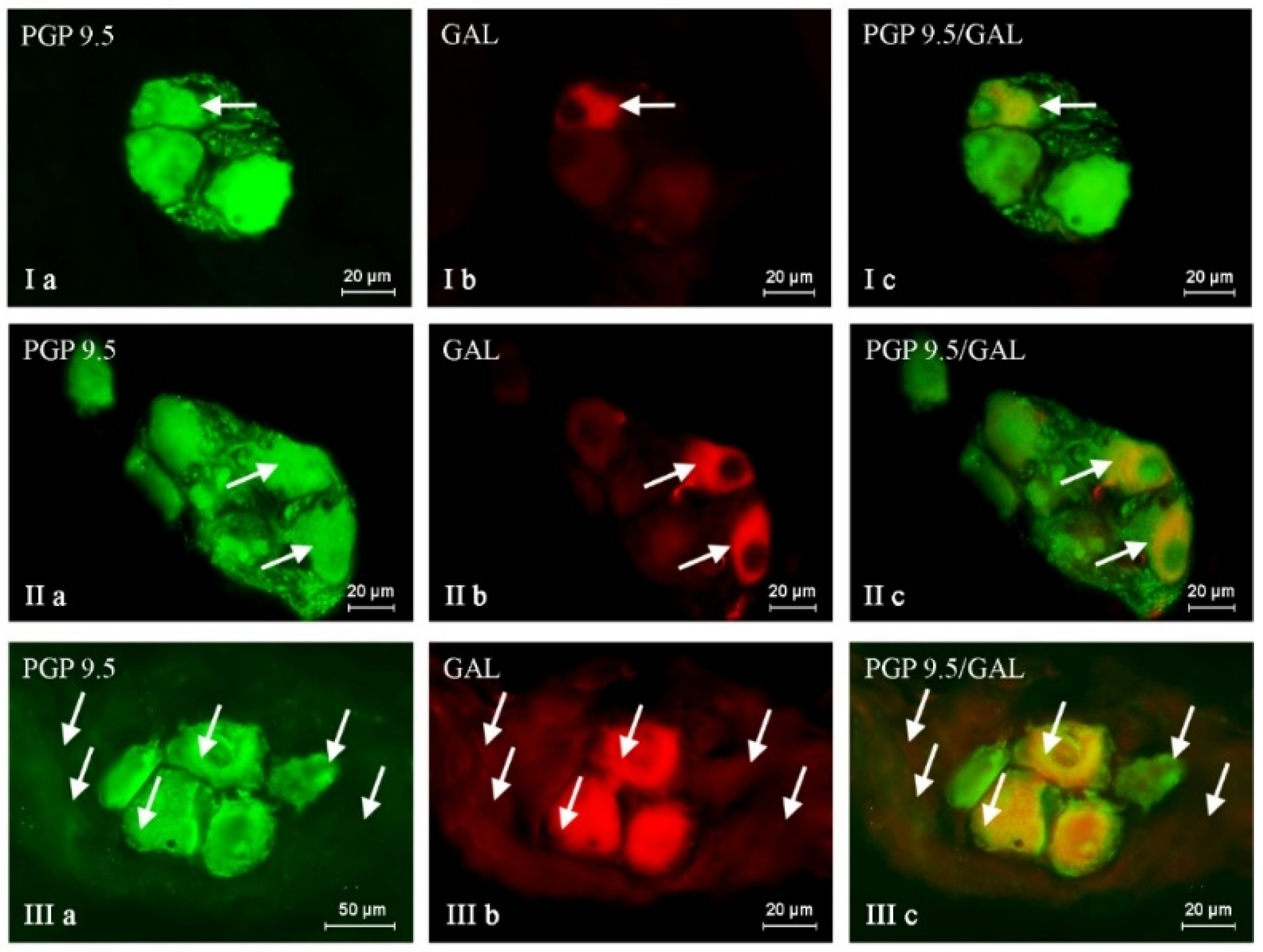
Figure 1. Nerve cells immunoreactive to protein gene product (PGP 9.5; used here as a pan-neuronal marker) (a) and galanin (GAL) (b) in the intrinsic ganglia of the porcine urinary bladder trigone wall under the physiological condition (I), and shown after administration of low (II) and high (III) doses of BPA. GAL-positive neurons are marked with arrows. The right column (c) was created by overlapping pictures (a) and (b).
Ijms 24 16792 g001
Figure 2. Nerve cells immunoreactive to protein gene product (PGP 9.5; used here as a pan-neuronal marker) (a) and cocaine- and amphetamine-regulated transcript (CART) peptide (b) in the intrinsic ganglia of the porcine urinary bladder trigone wall under the physiological condition (I), and after administration of low (II) and high (III) doses of BPA. CART-positive neurons are marked with arrows. The right column (c) was created by overlapping pictures (a) and (b).
Ijms 24 16792 g002
Figure 3. Nerve cells immunoreactive to protein gene product (PGP 9.5; used here as a pan-neuronal marker) (a) and neuronal isoform of nitric oxide synthase (nNOS—used as the marker of nitrergic neurons) (b) in the intrinsic ganglia of the porcine urinary bladder trigone wall under the physiological condition (I), and after administration of low (II) and high (III) doses of BPA. nNOS-positive neurons are marked with arrows. The right column (c) was created by overlapping pictures (a) and (b).
Ijms 24 16792 g003
Figure 4. Nerve cells immunoreactive to protein gene product (PGP 9.5; used here as a pan-neuronal marker) (a) and substance P (SP) (b) in the intrinsic ganglia of the porcine urinary bladder trigone wall under the physiological condition (I), and after administration of low (II) and high (III) doses of BPA. SP-positive neurons are marked with arrows. The right column (c) was created by overlapping pictures (a,b).
Ijms 24 16792 g004
Table 1. The percentage (%) of neurons immunoreactive to pan-neuronal marker protein gene product 9.5 (PGP 9.5) which was simultaneously immunoreactive to substance P (SP), galanin (GAL), neuronal isoform of nitric oxide synthase (nNOS), and/or cocaine- and amphetamine-regulated transcript (CART) peptide in the wall of urinary bladder trigone in the control group (C), the experimental group 1 (BPA 1), and experimental group 2 (BPA 2). Data expressed as mean% ± SEM. The results were considered statistically significant at p < 0.05 and are marked with *.
PGP/SPPGP/GALPGP/nNOSPGP/CART
C20.70 ± 0.8226.16 ± 0.7443.72 ± 1.2038.65 ± 0.30
BPA 123.86 ± 0.40 *35.29 ± 1.16 *47.49 ± 0.30 *44.95 ± 0.57 *
BPA 234.83 ± 0.64 *39.57 ± 0.48 *49.60 ± 1.08 *49.05 ± 1.70 *
Table 2. The increase of the percentage of neurons containing pan-neuronal marker protein gene product 9.5 (PGP 9.5) that were simultaneously immunoreactive to substance P (SP), galanin (GAL), neuronal isoform of nitric oxide synthase, and/or cocaine- and amphetamine-regulated transcript (CART) peptide in the wall of urinary bladder trigone given in the percentage points (p.p.) in both experimental groups (BPA 1 and BPA 2) in comparison to the control group (in control animals, level 0 was assumed).
PGP/SPPGP/GALPGP/nNOSPGP/CART
BPA 1+3.16+9.13+3.77+6.3
BPA 2+14.13+13.41+5.88+10.4
Table 3. List of antisera and reagents used in immunohistochemical investigations.
Primary Antibodies
AntigenCodeSpeciesWorking DilutionSupplier
PGP 9,57863-2004Mouse1:1000Biogenesis Ltd., Poole, UK
SP8450-0505Rat1:1000Bio-Rad (AbD Serotec), Kidlington, UK
CART1-003-61Rabbit1:8000Phoenix Pharmaceuticals, INC, Belmont, CA, USA
GALT-5036Guinea Pig1:2000Peninsula, New York, NY, USA
nNOSAB5380Rabbit1:6000MercMillipore, DEU, İzmir, Turkey
Secondary antibodies
ReagentsWorking dilutionSupplier
Alexa fluor 488 donkey anti-mouse IgG1:1000Invitrogen, Carlsbad, CA, USA
Alexa fluor 546 donkey anti-rabbit IgG1:1000Invitrogen
Alexa fluor 546 donkey anti-rat IgG1:1000Invitrogen
Alexa fluor 546 donkey anti-guinea pig IgG1:1000Invitrogen
Disclaimer/Publisher’s Note: The statements, opinions and data contained in all publications are solely those of the individual author(s) and contributor(s) and not of MDPI and/or the editor(s). MDPI and/or the editor(s) disclaim responsibility for any injury to people or property resulting from any ideas, methods, instructions or products referred to in the content.