Review

Aquaporin-5 Dynamic Regulation

by
Claudia D’Agostino
1,
Dorian Parisis
1,2,
Clara Chivasso
1,
Maryam Hajiabbas
1,
Muhammad Shahnawaz Soyfoo
2 and
Christine Delporte
1,*
1
Laboratory of Pathophysiological and Nutritional Biochemistry, Université Libre de Bruxelles, 1070 Brussels, Belgium
2
Rheumatology Department, CUB Hôpital Erasme, Hôpital Universitaire de Bruxelles (H.U.B), Université Libre de Bruxelles (ULB), Route de Lennik 808, 1070 Brussels, Belgium
*
Author to whom correspondence should be addressed.
Int. J. Mol. Sci. 2023, 24(3), 1889; https://doi.org/10.3390/ijms24031889
Submission received: 5 December 2022 / Revised: 9 January 2023 / Accepted: 10 January 2023 / Published: 18 January 2023
(This article belongs to the Special Issue Aquaporins: Dynamic Role and Regulations)

Abstract

:
Aquaporin-5 (AQP5), belonging to the aquaporins (AQPs) family of transmembrane water channels, facilitates osmotically driven water flux across biological membranes and the movement of hydrogen peroxide and CO2. Various mechanisms have been shown to dynamically regulate AQP5 expression, trafficking, and function. Besides fulfilling its primary water permeability function, AQP5 has been shown to regulate downstream effectors playing roles in various cellular processes. This review provides a comprehensive overview of the current knowledge of the upstream and downstream effectors of AQP5 to gain an in-depth understanding of the physiological and pathophysiological processes involving AQP5.

Graphical Abstract

1. Introduction

Aquaporin-5 (AQP5) belongs to the family of transmembrane aquaporins (AQPs) [1,2] and, more specifically, to the classical subfamily (including AQP0, AQP2, AQP4, AQP5, AQP6, and AQP8) characterized by high sequence homology between species and selective water permeability [3]. Indeed, the expression of rat and human Aqp5 (rAqp5 and hAqp5) in Xenopus oocytes has revealed its selective permeability to water [4,5]. In addition, hAQP5 is permeable to small molecules, including hydrogen peroxide [6] and CO2, but not NH3 [7,8], suggesting an important role in cell survival and adaptation to oxidative conditions.
The common structural features of AQPs, including AQP5, consist of six transmembrane domains (TM 1 to 6), five connecting loops (CL A to E) with loops B and E from opposite membrane sides folding back to form shorter pseudo-transmembrane helices within opposite layers of the membrane bilayer, and intracellular N- and C- termini [9,10,11]. Four monomers, each forming a water-permeable pore, associate in a tetramer presenting an additional central pore that was proposed to be permeable to gas or hydrophobic molecules [12]. Two asparagine-proline-alanine (NPA) motifs, located within the ends of the short pseudo-transmembrane helices formed by loops B and E, and an aromatic-arginine (ar/R) region, constituting the narrowest part of the pore, have been suggested to prevent the movement of protons along with water molecules by a Grotthuss-type mechanism. The solute specificity of AQPs relies on the variable composition of the ar/R region, except for a highly conserved arginine [9,10].
Within the human body, Aqp5 is expressed in various secretory tissues (such as salivary glands, pancreas, lacrimal glands, mammary glands, thyroid, adrenals, pituitary, skin), internal tissues (such as lungs, kidney, liver, spleen, esophagus, stomach, intestine, colon, bladder, adipose tissue), reproductive tissues (such as testes, ovaries, prostate, uterus), muscle tissues (such as heart, skeletal muscle, arteries), nervous tissues (such as brain, cortex, cerebellum, spinal cord, tibial nerve), immune tissues (such as lymph nodes), and ocular tissues (cornea, lens, retina) [13,14,15,16,17]. AQP5 plays an important physiological role in the production of saliva, sweat, tears, and pulmonary secretions [18,19,20,21] and optical properties of the lens [22,23,24].
AQP5, like other AQPs, encounters dynamic regulation consisting of transcriptional and translational modulation, post-translational modifications, and trafficking to the plasma membrane. Therefore, such dynamic regulation plays a key role in physiological and pathophysiological processes.
In this review, we provide an in-depth overview of the current understanding of the AQP5 dynamic regulation.

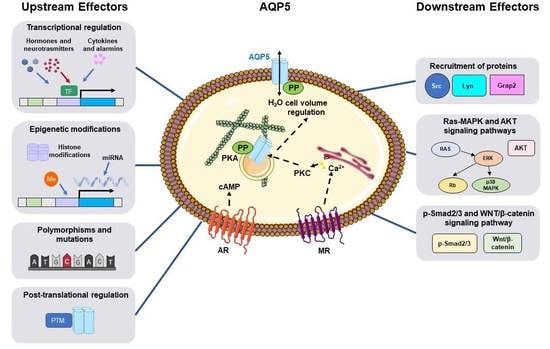
2. Functional Impact of Single Nucleotide Polymorphism and Mutation of Aqp5 Gene

Single nucleotide polymorphisms and mutations of the Aqp5 gene have been associated with a perturbation in mRNA expression, protein trafficking, and water permeability.
The CT/TT genotype of Aqp5 rs1964676 polymorphism has been associated with strong Aqp5 expression and used as a prognostic marker of survival in patients with early breast cancer [25].
The sequencing of the Aqp5 gene promoter region of 50 healthy Caucasian blood donors has identified a common single nucleotide polymorphism characterized by the substitution of C for A at position -1364 (-1364A/C) [26]. This genetic modification was associated with increased binding of transcription factors to the promoter but with reduced cAMP-induced Aqp5 gene transcription [26]. It was also associated with a significant reduction in Aqp5 mRNA and AQP5 protein levels in the human heart and erythrocyte membranes, respectively, suppression of the renin-angiotensin-aldosterone system, and reduced blood pressure increase in response to a high salt diet [26]. In addition, the Aqp5-1364A/C polymorphism has been associated with increased methylation of the promoter and decreased expression in neutrophil granulocytes from human septic patients [27]. Furthermore, strong associations have been found between this Aqp5 polymorphism and increased 30-day survival in severe sepsis [28], reduced susceptibility for major adverse kidney events in sepsis [29], attenuated pulmonary inflammation and higher 30-day survival in acute respiratory distress syndrome [30], decreased prevalence and better resolution of acute kidney injury in patients suffering from pneumonia evoked acute respiratory distress syndrome [31], increased risk of cytomegalovirus infection after kidney transplantation [32], and less severe peritumoral brain edema in patients suffering from meningioma [33].
Other studies have identified some Aqp5 mutations (p.Trp35Ser, p.Ala38Glu, p.Ile45Ser, p.Asn123Asp, p.Asn123Tyr, p.Ile177Phe, and p.Arg188Cys) causing autosomal-dominant diffuse nonepidermolytic palmoplantar keratoderma despite retaining normal expression and the ability to traffic to the cell membrane [34,35,36]. Protein modeling has suggested these mutations may provide AQP5 a gain-of-function impacting its water permeability [34]. Indeed, the substitution of amino acids lining the extracellular end of the water channel (Ile45, Ile177, and Arg188) in a highly-conserved position in extracellular loop A (Trp35) or located on the extracellular surface of AQP5 (Ala38, Asn123) may have a direct or indirect impact on water conductance [34,37]. Furthermore, molecular dynamics on mutated AQP5 (p.Arg188Cys) revealed a remarkable increase in the pore radius inside the selectivity region, facilitating the passage of water molecules and higher water permeability [38]. While it remains unclear how defective epidermal-water-barrier function associated with mutant AQP5 leads to palmoplantar hyperkeratosis, the gain of function of mutated AQP5 may accelerate keratinocyte water uptake and explain the swelling of the epidermal stratum corneum after water exposure.
A heterozygous missense mutation of Aqp5 (p.Leu51Pro) induced autosomal dominant congenital cataracts leading to blindness in infants [39]. Transfection of the mutated Aqp5 in human lens epithelial cells decreased AQP5 expression and increased vimentin expression through the downregulation of miR-124-3p.1 [39]. Aqp5 knockout mice showed that a reduction in lens transparency resulted from increased vimentin expression concomitant to miR-124-3p.1 downregulation and this phenomenon was reverted using miR-124-3p.1 agomir [39]. Altogether, the data suggested AQP5 is involved in lens transparency by upregulating miR-124-3p.1 that targeted vimentin mRNA. Furthermore, it was shown that the permeability of AQP5 to water and carbon dioxide depended on the presence of Leu51 (its side chain facing the tetramer’s centra pore). Indeed, Leu51Arg mutation resulted in a permeability loss to water and carbon dioxide and converted the protein into an anion channel [40]. Further studies are necessary to assess whether the missense mutation of Aqp5 causing autosomal dominant congenital cataract (p.Leu51Pro) would behave similarly to the Leu51Arg mutation.
A naturally occurring rAqp5 point mutation (p.G308A) caused a reduction in saliva secretion due to impaired AQP5 trafficking and increased lysosomal degradation concomitant to possible protein conformational modification without affecting the water permeability of the channel [41]. In addition, the higher activation energy of water transport of the mutated AQP5 may prevent the transient swelling of the acini, delay the agonist-induced shrinkage, and decrease the initial transient secretion of saliva [42]. By comparing data from Aqp5-G103D mutant and wild-type rats, AQP5 was proposed to be responsible for the rapid agonist-induced salivary gland acinar cell shrinkage and to contribute not only to the initial phase of secretion but also to the maintenance of the necessary acinar cell volume during the steady secretion of saliva [42]. It remains to be assessed if a similar mutation occurs in humans and is associated with phenotypic alteration of saliva gland function.

3. Transcriptional Regulation of Aqp5 Expression

Transcriptional regulation of Aqp5 expression is modulated by hormonal, neuronal, and inflammatory stimuli, binding of transcription factors to gene promoter and epigenetic modifications.

3.1. Hormonal, Neuronal, and Inflammatory Stimuli

3.1.1. Steroid Hormones

Steroid hormones have been shown to modulate Aqp5 expression. Indeed, progesterone increased Aqp5 mRNA expression in porcine endometrial luminal epithelial cells during the follicular phase of the estrous cycle, while estradiol had the inverse effect during the early luteal phase of the estrous cycle [43]. In mouse uterus, the Aqp5 mRNA level was increased by estradiol but not by progesterone, suggesting abnormal Aqp5 expression during pregnancy may participate in disrupting normal myometrial activity and reducing implantation events [44,45]. In mouse mammary gland cells, estrogen increases Aqp5 mRNA levels, while prolactin and dexamethasone add the opposite effects [46].
The testosterone-induced upregulation of Aqp5 mRNA levels in ovariectomized rats may account for the reduced uterine fluid volume measured in response to the hormone [47]. The oxytocin-induced Aqp5 expression in the myometrium of late-pregnant rat uterus may participate in events leading to parturition in the rat [48]. In addition, the sudden increase of blood Aqp5 levels on the last day of pregnancy may serve as an indicative marker of delivery initiation [48].
In porcine uterine tissue, Aqp5 mRNA levels were increased by estradiol and oxytocin but decreased by progesterone [49]. In addition, growth hormone decreased Aqp5 mRNA expression in granulosa cells from medium swine ovarian follicles, while prolactin induced an opposite effect in similar cells from large follicles. In addition, both growth hormone and prolactin increased Aqp5 mRNA levels in theca cells from large ovarian follicles. When granulosa and theca cells from medium and large follicles were co-cultured, Aqp5 mRNA levels were increased by the luteinizing hormone, prolactin, and growth hormone in granulosa cells from large follicles, by luteinizing hormone in theca cells from medium and large follicles and by prolactin in theca cells from medium follicles [50]. It was proposed that AQP5 may play important roles in maintaining water homeostasis of the ovarian cells of the pig involved in folliculogenesis, ovulation, and corpus luteum formation and maintenance.
In human endometrial stromal cells, the estradiol-induced increase in Aqp5 expression consequent to the activation of the PI3K/AKT pathway enhanced cell invasion and proliferation and formation of ectopic implants in vivo [51]. Overall, these data suggest the inhibition of Aqp5 expression and AQP5 function may provide new therapeutic options for treating endometriosis.
The presence of an estrogen-response element (ERE), identified within the hAqp5 gene using luciferase reporter assay and electrophoretic mobility shift assay (EMSA), accounts for the estradiol-induced transcriptional regulation of Aqp5 [51].
Corticosteroids upregulated Aqp5 mRNA and AQP5 protein expression in adenocarcinomic human alveolar basal epithelial cells (A549) and may therefore improve pulmonary diseases with airway hypersecretion though this mechanism [52].

3.1.2. Thyroid and Parathyroid Hormones

Thyroid hormones likely modulated AQP5 expression in the lungs, as hypothyroidism had a lowering effect while hyperthyroidism had an opposite effect [53]. These data suggest circulating thyroid hormone levels may affect lung fluid balance through several mechanisms, including the regulation of AQP5 expression and paracellular permeability. A study carried out in parathyroid hormone/parathyroid hormone-related peptide type I receptor null mice suggested parathyroid hormone may promote Aqp5 mRNA expression in lungs [54]. As the receptors to the thyroid and parathyroid hormones belong to the family of G protein-coupled receptors linked to Gαs and Gαp, respectively, it is likely that cAMP/PKA/CREB and intracellular calcium pathways contributed to the regulation of Aqp5 transcription.

3.1.3. Neurotransmitters

In a mouse model of primary focal hyperhidrosis, increased expression of acetylcholine and cholinergic receptor nicotinic alpha 1 subunit led to increased Aqp5 mRNA expression [55]. Furthermore, cisatracurium, an inhibitor of acetylcholine and cholinergic receptor nicotinic alpha 1 subunit, decreased Aqp5 mRNA and AQP5 protein expression [56]. These data suggested that increased acetylcholine levels may account for increased Aqp5 mRNA and AQP5 protein expression leading to primary focal hyperhidrosis in patients [57]. Methacholine, a muscarinic agonist, decreased AQP5 protein expression in human nasal epithelial cells through a mechanism involving the cAMP Response Element-binding protein (CREB) inactivation and NFkB activation, suggesting muscarinic receptor stimulation may regulate Aqp5 transcription though such signaling pathway [58].
β-adrenergic receptor agonists increased Aqp5 transcription in salivary glands and lungs in vivo [59] and murine lung epithelial cell line (MLE-12) in vitro [60], likely via cAMP-dependent and CREB phosphorylation mechanisms.
Gamma-amino butyric acid may be involved in the regulation of AQP5 expression. Indeed, gabapentin, a structural analog of gamma-amino butyric acid, increased the AQP5 expression in corneal cells and lacrimal glands through stimulation of the autonomic nervous system and direct activation of intracellular signaling cascades, including the PKA/CREB pathway [61]. These mechanisms could contribute to restoring the tear film in dry eye disease, thus reducing friction on the ocular surface and consequent ocular pain.
Histamine downregulated Aqp5 mRNA levels in human nasal epithelial cells by activating NFkB [62] and inhibiting CREB phosphorylation [63].

3.1.4. Inflammatory Stimuli

Overexpression of activin, a receptor type 1, a receptor of TGFβ, led to increased Aqp5 mRNA and AQP5 protein levels in patients with primary focal hyperhidrosis [64], suggesting that TGFβ may regulate Aqp5 transcription. In A549 cells (alveolar type II cells) [65], primary culture of rat alveolar epithelial cells and rat lung epithelial-T-antigen negative cells (RLE-6TN; alveolar type II cells) [66], TGFβ1-induced epithelial-mesenchymal transition (EMT) was accompanied by a downregulation of AQP5 protein expression. However, the effect of TGFβ1 on Aqp5 transcription remains to be assessed. Further studies are necessary to determine if SMAD transcription factors, involved in the TGFβ1 signaling pathway, may regulate Aqp5 transcription.
TNFα decreased Aqp5 mRNA and AQP5 protein expression in immortalized human salivary gland acinar cells (NS-SV-AC) [67], MLE-12 cells [68], and human bronchial epithelial cells (BEAS-2B) [69], and AQP5 protein level in conditional transgenic TNFα mice [70]. Lipoxins, endogenous lipids that mediate the resolution of inflammation, reversed the effect of TNFα by promoting Aqp5 expression in the lung in a mouse model of acute pancreatitis-induced acute lung injury [71]. The regulation of Aqp5 transcription by TNFα may likely involve NFkB.
IFNα upregulated both Aqp5 mRNA and AQP5 protein expression in the primary culture of human parotid gland acinar cells [72]. Interleukin-13 (IL-13), a central regulator of Th2-dominated respiratory disorders such as asthma, abolished the increase in Aqp5 mRNA level during airway epithelial cell differentiation [73]. In addition, reduced Aqp5 mRNA and AQP5 protein levels were detected in lungs from an interleukin-13-induced mouse model of asthma [74]. Additional experiments need to be performed to assess the possible role of STAT transcription factors, activated upon IFNα and IL13 stimulation, in Aqp5 transcription.
High-mobility group box 1 (HMGB1), a highly conserved nuclear protein of the alarmin family acting as a chromatin-binding factor, promotes access to transcriptional protein assemblies on specific DNA targets [75,76]. HMGB1 has been involved in Sjögren’s syndrome pathogenesis [77]. Moreover, HMGB1 decreased Aqp5 mRNA levels in a mouse model of Sjögren’s syndrome through the activation of the toll-like receptor 4 (TLR4)/NFkB pathway [78]. In a mouse model of Sjögren’s syndrome, HMGB1 inhibition restored saliva and tear secretion [78,79], suggesting HMGB1 inhibition may represent a new therapeutic strategy for treating the disease.

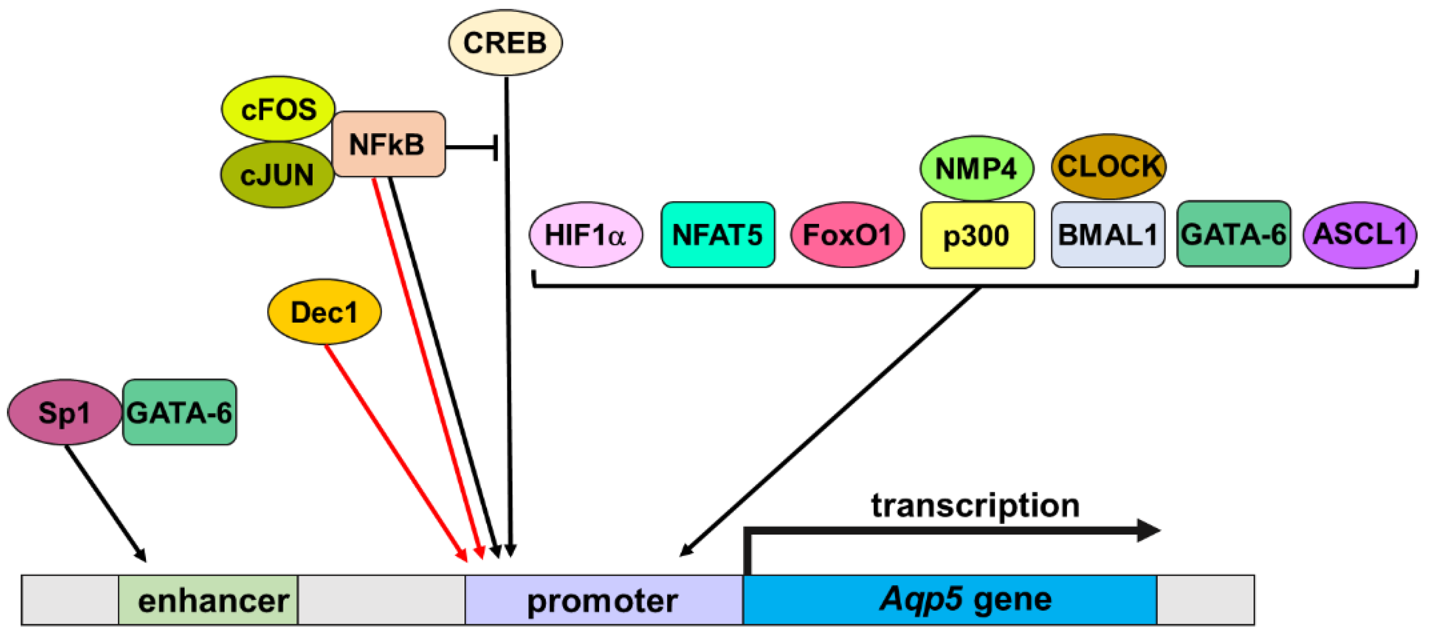
3.2. Transcription Factors

Several transcription factors have been identified as regulators of Aqp5 transcription during physiological and pathological processes (Figure 1).

3.2.1. CREB

The cAMP-induced Aqp5 transcription detected in several cell types [59,60,80,81] was likely linked to the presence of putative cAMP response element binding sites within the Aqp5 gene [59]. Indeed, several studies have suggested a role for p-CREB in the cAMP-induced Aqp5 transcription [62,82,83].

3.2.2. NFkB

Nuclear factor kappa light chain enhancer of activated B cells (NFkB) played a role in either stimulation or inhibition of Aqp5 transcription. Indeed, NFkB was involved in the hyperosmolar-induced increase in Aqp5 mRNA levels in retinal pigmented epithelial cells [84]. By contrast, NFkB was suggested to inhibit the p-CREB-induced Aqp5 transcription [62,83]. Upon lipopolysaccharide stimulation, NFkB was directly involved in the decreased Aqp5 mRNA levels, and the effect was potentiated by interaction with p-c-Jun/c-Fos [85]. NFkB was also involved in the inhibition of Aqp5 transcription induced by tumor necrosis factor α in MLE-12 cells [68].

3.2.3. HIF1α

Hypoxia-inducible factor-1 (HIF1α) has been shown to transactivate Aqp5 gene promoter in various cell types [86,87,88]. In mouse lung epithelial (MLE-15) cells, hyperosmolar induction of Aqp5 mRNA expression involved the activation of ERK and mitogen-activated extracellular regulated kinase (MEK) [89]. These signaling pathways, which lead to the activation of hypoxia-inducible factor-1α (HIF1α) [90], may be involved in the hyperosmolar-induced activation of HIF1α and its binding to HIF1α binding sites to the proximal Aqp5 gene promoter to induce transcription [86]. Decreased HIF1α activity during intervertebral disc degeneration suppressed Aqp5 mRNA and protein expression, compromising their ability to respond to extracellular osmolarity changes [86]. However, hypoxia failed to promote Aqp5 mRNA expression in intervertebral discs [86]. HIF1α may also increase Aqp5 transcription in salivary glands from heat-acclimated rats [91].

3.2.4. NFAT5

Nuclear factor of activated T cells 5 (NFAT5) upregulated Aqp5 mRNA levels in chronic lymphocytic leukemia B cells (MEC-1 cells) and B cells isolated from patients with chronic lymphocytic leukemia [92], lung adenocarcinoma cells [93]. Furthermore, NFAT5 was involved in the hyperosmolar induction of Aqp5 mRNA expression in human retinal pigmented epithelial cells [84], human corneal epithelial cells [94], and mouse intervertebral discs [95]. The transcriptional regulation of Aqp5 by NFAT5 likely plays a role in cell volume regulation upon osmolar stress.

3.2.5. FoxO1

Forkhead box O1 (FoxO1) stimulated the Aqp5 gene promoter in rat salivary gland cells, and injection of FoxO1 inhibitor into mice reduced Aqp5 expression and decreased saliva secretion [96]. Simultaneous downregulation of FoxO1 and Aqp5 mRNA levels in patients with Sjögren’s syndrome may contribute to hyposalivation [96]. In astrocytes and microglia cells, FoxO1 was involved in the lipopolysaccharide-induced Aqp5 expression [97].

3.2.6. NMP4 and p300

Nuclear matrix protein 4 (NMP4), also named Zinc Finger Protein 384 (ZNF384), bound to the Aqp5 gene promoter and induced its transactivation when overexpressed in immortalized human embryonic kidney (HEK293) cells [98]. Interestingly, upon mechanical stimulation of osteoblasts cells, the triggered signals upstream (ERK, Akt, and GSK-3β activation) and downstream (cyclin D1) of β-catenin, as well as the cytoskeleton reorganization and formation of focal adhesions between cells and extracellular substrate were enhanced upon NMP4 deficiency [99]. Therefore, NMP4 may play an inhibitory regulatory role in the response of cells to mechanical stimulation. In addition, NMP4 was shown to interact directly or indirectly with focal adhesion-associated protein p130cas (CAS) [100,101], which can bind to focal adhesion-associated tyrosine kinase (FAK). Besides, AQP5 interacted with β-catenin and other junctional proteins, which displayed lower expression at the junction upon AQP5 overexpression [102]. Besides, AQP5 was not recruited to the junctions upon mechanical stimulation [102]. It was shown that co-activator p300/β-catenin interactions were necessary for p300 transcriptional activation of Aqp5 gene in lung alveolar epithelial cell type I [103]. Further studies will be necessary to determine if another coactivator of β-catenin, such as CREB and other components of the basal transcription machinery, is also involved in such transcriptional control. Further studies will be required to precise the role of the transcriptional regulation of Aqp5 by NMP4 and p300 in the context of cell adhesion.

3.2.7. BMAL1 and CLOCK

A relationship was established between clock genes and Aqp5 expression in rat whole salivary glands as well as isolated acinar and ductal cells. Human, rat, and mouse Aqp5 gene promoter analysis revealed the presence of an enhancer box (E-box) binding sequence [104,105] that acts as a protein-binding site and is necessary for the binding of Basic Helix-Loop-Helix ARNT Like 1 (BMAL1)-circadian locomotor output cycles kaput protein (CLOCK) heterodimer to the promoter region [105]. In addition, Aqp5 mRNA expression had a rhythmic pattern similar to the circadian rhythmic expression of clock genes [106]. Therefore, Aqp5 gene may be a clock-controlled gene and a target for the transcription factors BMAL1-CLOCK heterodimer in salivary glands. Further studies will be valuable to determine if such regulation may play a role in the physiological nocturnal salivary water secretion.

3.2.8. GATA-6

During embryonic lung development in the mouse, GATA Binding Protein 6 (GATA-6) has been shown to transactivate the Aqp5 gene promoter and to increase the Aqp5 mRNA expression in alveolar epithelial type 1 cells [107]. In addition, Sp1 bound to an enhancer and interacted with GATA-6 to promote epigenetic modification of Aqp5 gene [108,109] (see Section 3.3.2).

3.2.9. Dec1 and Achaete-Scute Family BHLH Transcription Factor 1

The differentiated embryo chondrocyte-expressed gene 1 (Dec1) decreased Aqp5 mRNA expression in salivary glands from aged mice [110].
Achaete-Scute Family BHLH Transcription Factor 1 (ASCL1) increased Aqp5 expression in mice infected with Helicobacter pylori [111].

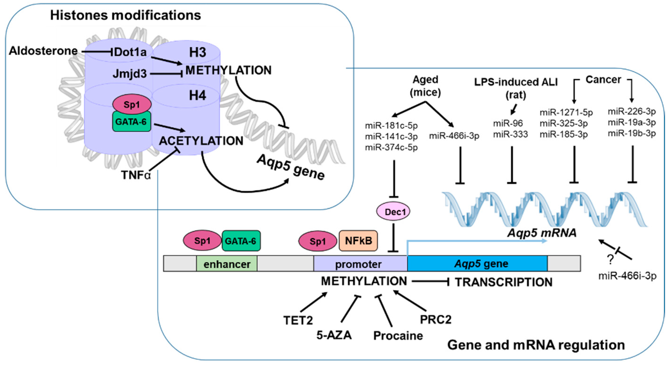
3.3. Epigenetic Modifications

Some epigenetic modifications have been implicated in the regulation of Aqp5 transcription, including DNA methylation, histone modifications, and micro-RNAs (miRNAs) (Figure 2).

3.3.1. DNA Methylation

Considering the high expression of Aqp5 in several cancers (such as breast and lung), it was hypothesized that gene promoter methylation status might account for such observation. Overall, studies suggest Aqp5 gene promoter methylation may be cell and tissue specific.
While Aqp5 mRNA expression was independent of its promoter methylation status in non-tumorigenic mouse mammary gland cells EpH4 [112], studies using the most recent technologies to detect gene promoter methylation using genomic DNA from tumor cells arising from cancer patients showed divergent outcomes. Age-related DNA methylation of the Aqp5 gene promoter was inversely correlated with gene expression. In addition, Aqp5 gene promoter methylation varies according to the type of tumor, with lower methylation and higher gene expression in basal tumors as compared to luminal tumors [113].
In human pancreatic adenocarcinoma, hypomethylation of the Aqp5 gene promoter accounted for its high expression associated with infiltrating immune cells, suggesting Aqp5 may represent a novel prognostic biomarker and a new immune-associated therapeutic target for patients suffering from such cancer [114].
Recent studies in neutrophil granulocytes from human septic patients revealed that the C-allele of the Aqp5-1364A/C polymorphism was associated with an increase in gene promoter methylation and decreased expression [27]. The current lack of therapeutic options to modulate single gene promoter methylation limits the therapeutic approach. Nevertheless, unspecific therapeutic intervention, such as the use of a demethylation agent (5-azacytidine (5-AZA)), has been shown to reduce endotoxemia-induced mouse inflammatory lung injury [115]. The methylation status of the Aqp5 promoter cytosine site (nt-937) was linked to both NFkB binding and Aqp5 expression and may be prognostically relevant in sepsis as higher methylation is associated with sepsis non-survivors [116].
High methylation of a putative CpG island of the Aqp5 gene promoter was associated with low Aqp5 expression in NIH-3T3 mouse fibroblasts and freshly isolated rat alveolar epithelial cells [104]. Conversely, hypomethylation of the Aqp5 gene promoter was associated with high Aqp5 expression in mouse MLE-12 lung epithelial cells [104]. In addition, 5-AZA treatment induced Aqp5 expression in NIH-3T3 cells, and in vitro methylation of the Aqp5 gene promoter inhibited the transcription of a reporter gene in mouse lung epithelial (MLE-12) cells [104]. Therefore, DNA methylation may play a role in the cell-specific expression of Aqp5 [104]. Finally, chromatin immunoprecipitation assays revealed that endogenous Sp1 bound to a hypomethylated, but not highly methylated, Aqp5 gene promoter. Besides, in an immortalized normal human salivary gland ductal cell (NS-SV-DC) line lacking the expression of Aqp5, 5-AZA induced Aqp5 expression that led to functional water permeability [117]. In addition, untreated NS-SV-DC cells presented hypermethylation within the consensus Sp1-binding sites located within the Aqp5 gene promoter, whereas 5-AZA-treated NS-SV-AC cells presented demethylation at CGs located within or outside consensus Sp1 binding sites that cooperatively functioned to induce Aqp5 expression [117]. In response to retinoic acid, Sp1 was also shown to transactivate mAqp5 gene promoter in MLE-12 cells, leading to increased plasma membrane water permeability [118]. In murine aging model C57BL/6CrSlc mice characterized by xerostomia, 5-AZA treatment restored salivary gland function, consequently to increased Aqp5 expression resulting from Aqp5 gene promoter methylation [119]. These data confirm a direct link between the Aqp5 gene promoter methylation status and its expression and further suggest that Sp1 binding to the hypo-methylated promoter likely plays a role in cell type-specific expression of the gene. Furthermore, procaine, an anticancer agent specifically inhibiting DNA methyltransferase (DNMT) 1 activity [120], stimulates Aqp5 mRNA expression in NS-SV-DC cells by inducing CpG island demethylation at Sp1-2 and Sp1-3 sites of the hAqp5 gene promoter [121].
Polycomb Repressive Complex 2 (PRC2), involved in transcription repression, can affect the epigenetic landscape through several mechanisms, including the recruitment of DNA methyltransferases. PRC2 was shown to promote the hypermethylation Aqp5 gene promoter in follicular lymphoma compared to benign follicular hyperplasia [122]. These data suggest the methylation status of the Aqp5 gene promoter may be involved in lymphoma cell biology.
During embryonic salivary gland development, decreased expression of ten-eleven translocation enzyme 2 (TET2; involved in DNA demethylation through the oxidization of 5-methylcytosine (5mC) to 5-hydroxymethylcytosine (5hmC)) was observed concomitantly with increased 5hmC levels and Aqp5 expression at embryonic day 15 (E15) [123]. Therefore, the downregulation of TET2 expression may be a critical epigenetic event that contributes to salivary gland function.

3.3.2. Histone Modifications

In NS-SV-AC cells, TNFα decreased Aqp5 mRNA expression, consequently in an epigenetic mechanism involving the suppression of histone H4 acetylation [67].
In mice, GATA-6 stimulated Aqp5 transcription not directly through DNA binding of GATA-6 but indirectly through interactions with Sp1 that binds to an enhancer that does not contain GATA-6 binding sites [108]. In addition, GATA6/Sp1 interaction interfered with histone deacetylase 3 (HDAC3)/Sp1 binding and recruited transcriptional co-activator/histone acetyltransferase p300, leading to histone acetylation [109]. All these data suggest the GATA-6/SP1 interaction plays a role in the epigenetic modification of the Aqp5 gene promoter during alveolar epithelial cell differentiation.
Jumonji domain containing-3 (Jmjd3), a key histone demethylase involved in the regulation of gene expression, is involved in the Aqp5 gene regulation and plays a role in lung development and function [124]. Indeed, changes in Aqp5 gene expression were associated with locus-specific methylation alterations of histone 3 (H3K27 and H3K4) [124].
Histone H3K79 methyltransferase Dot1a repressed the mouse Aqp5 gene promoter by binding to the promoter region comprised between −389 and −139 [125]. In the kidney, AQP5 acts as a negative regulator of aquaporin-2 (AQP2) trafficking, thereby impairing renal water reabsorption and inducing polyuria [125]. As the gene encoding Dot1a is downregulated by aldosterone, Dot1a may link excessive aldosterone to polyuria by decreasing AQP2 [126] and increasing Aqp5 expression. Further experiments are required to uncover the physiological role of Aqp5 gene regulation by Dot1a in other tissues.

3.3.3. miRNA

In aged mice, an upregulation of the transcription factor Dec1 (differentiated embryo chondrocyte-expressed gene 1) decreased Aqp5 mRNA expression [110]. The modified expression of Dec1 and Aqp5 mRNA levels may result from the downregulation of miR-181c-5p, miR-141-3p, and miR-374c-5p that can bind to Dec1 mRNA and the upregulation of miR-466i-3p that may bind to Aqp5 mRNA [110].
In lipopolysaccharide-induced rat lung damage of disseminated intravascular coagulation, miR-96 and miR-330 targeted Aqp5 mRNA [127], and concomitantly decreased Aqp5 expression was linked to pulmonary edema [128]. These data suggested that in patients with disseminated intravascular coagulation characterized among others by lung damage often consisting in pulmonary edema, miRNA may offer a new therapeutic intervention.
In hepatitis B- virus (HBV)-induced hepatocarcinoma, miR-1271-5p has been shown to target Aqp5 mRNA, and its overexpression resulted in decreased Aqp5 expression to block the cancer progression by reducing cell viability, migration, and invasion [129]. In similar cancers, miR-325-3p also targeted Aqp5 mRNA, and its overexpression inhibited cell proliferation and induced cell apoptosis.
In colorectal cancer and colorectal cancer resistant to 5-fluorouracil (a chemotherapy drug), the upregulated Aqp5 mRNA expression was due to low expression of miR-185-3p that targets Aqp5 mRNA [130]. Furthermore, miR-185-3p mimic enhanced the chemosensitivity of colorectal cancer cells via an effect on the Aqp5 mRNA [130], suggesting such a therapeutic approach may be beneficial to treat 5-fluorouracil-resistant colorectal cancers.
In human breast cancer MDA-MB-231 cells, exosome-mediated delivery of miR-1226-3p, miR-19a-3p, and miR-19b-3p targeted Aqp5 mRNA and impeded cell migration [131].
The overexpression of miR-551b-5p in human umbilical vein endothelial cells (HUVECs) promoted the expression of Aqp5 and cell permeability [132]. As this is unlikely due to a direct effect of the miRNA on the Aqp5 mRNA, further studies are required to address the underlying mechanisms.
Considering several miRNAs can target the Aqp5 gene, they may be useful for the treatment of various cancers, providing the development of suitable delivery methods and miRNA mimetics.

4. Post-Translational Regulation of AQP5

Post-translational modifications have been shown to regulate the cellular function of AQPs. Phosphorylation, lipidation, glycosylation, and ubiquitination have been shown to regulate AQP5.

4.1. Phosphorylation

Multiple putative protein kinase G (PKG) phosphorylation sites are present within AQP5. However, it remains to be assessed whether PKG can phosphorylate AQP5 and account for its acetylcholine-induced trafficking involving nitric oxide / cyclic 3′,5′ monophosphate (cGMP) signaling pathway causing intracellular calcium concentration increase in rat parotid acinar cells [133]. PKC may also be involved in hypoosmotic-induced AQP5 trafficking [134].
Two putative protein kinase A (PKA) phosphorylations sites containing Ser 156 and Thr259 may be involved in cAMP-induced AQP5 trafficking (see Section 5.2 for more details).

4.2. Lipidation

Mass spectrometry analysis conducted on bovine and human lenses has revealed the palmitoylation of AQP5 on C6 located in the N-terminus end, a region highly conserved among many species (including human, bovine, mouse, rat, and pig) [135]. Furthermore, AQP5 palmitoylation seems to occur in a narrow region of the inner lens cortex but not in the lens epithelium, outer cortex, or nucleus [135]. Considering protein palmitoylation promotes lipid raft association [136], this modification may account for the presence of AQP5 in lipid raft from the bovine lens [137] and rat parotid gland [138]. However, additional experiments are necessary to determine if this post-translational modification occurs in other cell types and promotes AQP5 trafficking.

4.3. Glycosylation

AQP5, which contains two putative N-glycosylation sites (Asn124, Asn125), has been suggested to be glycosylated in human cornea (based on the presence of two immunoreactive bands detected by Western blot analysis) [139] but not in human lens fibers [140]. However, further studies are necessary to confirm such post-translational modification before investigating its possible effect on AQP5.

4.4. Ubiquitination

In submandibular glands from non-obese diabetic (NOD) mice with severe combined immunodeficiency disease (SCID) lacking the gene encoding the transcription factor E2F1 (NOD/SCID.E2F1−/−), AQP5 was ubiquitinated, with an increment of 24kDa in molecular weight consistent with three molecules of ubiquitin [141]. Ubiquitination of AQP5 may account for its degradation and subsequently decreased expression observed in submandibular glands from NOD/SCID.E2F1−/− mice [141]. Further studies are warranted to assess if this mechanism may be responsible for hyposalivation.
In lung epithelial cells, AQP5 protein levels were also regulated in part by ubiquitination and proteasomal degradation, and drug repurposing was able to identify a compound capable of increasing AQP5 protein levels by reducing AQP5 ubiquitination and proteasomal degradation [142].

5. AQP5 Trafficking

Protein trafficking is an important cellular process that governs membrane protein function. Based on the high sequence homology between AQP5 and AQP2, it has been suggested that AQP5 trafficking may be regulated very similarly to AQP2 trafficking. The numerous studies on AQP2 trafficking (involved in kidney water reabsorption) [143,144] have set a precedent for the in-depth understanding of the trafficking of other AQPs, including AQP5. Several mechanisms have been involved in triggering and directing the vesicular trafficking of AQP5 to the cell plasma membrane: signal transduction involving intracellular calcium or cyclic 3,5 adenosine mono phosphate (cAMP) increase, the interaction between AQP5 and protein partners and cytoskeleton, as well as other mechanisms which remain to be further characterized (Figure 3).

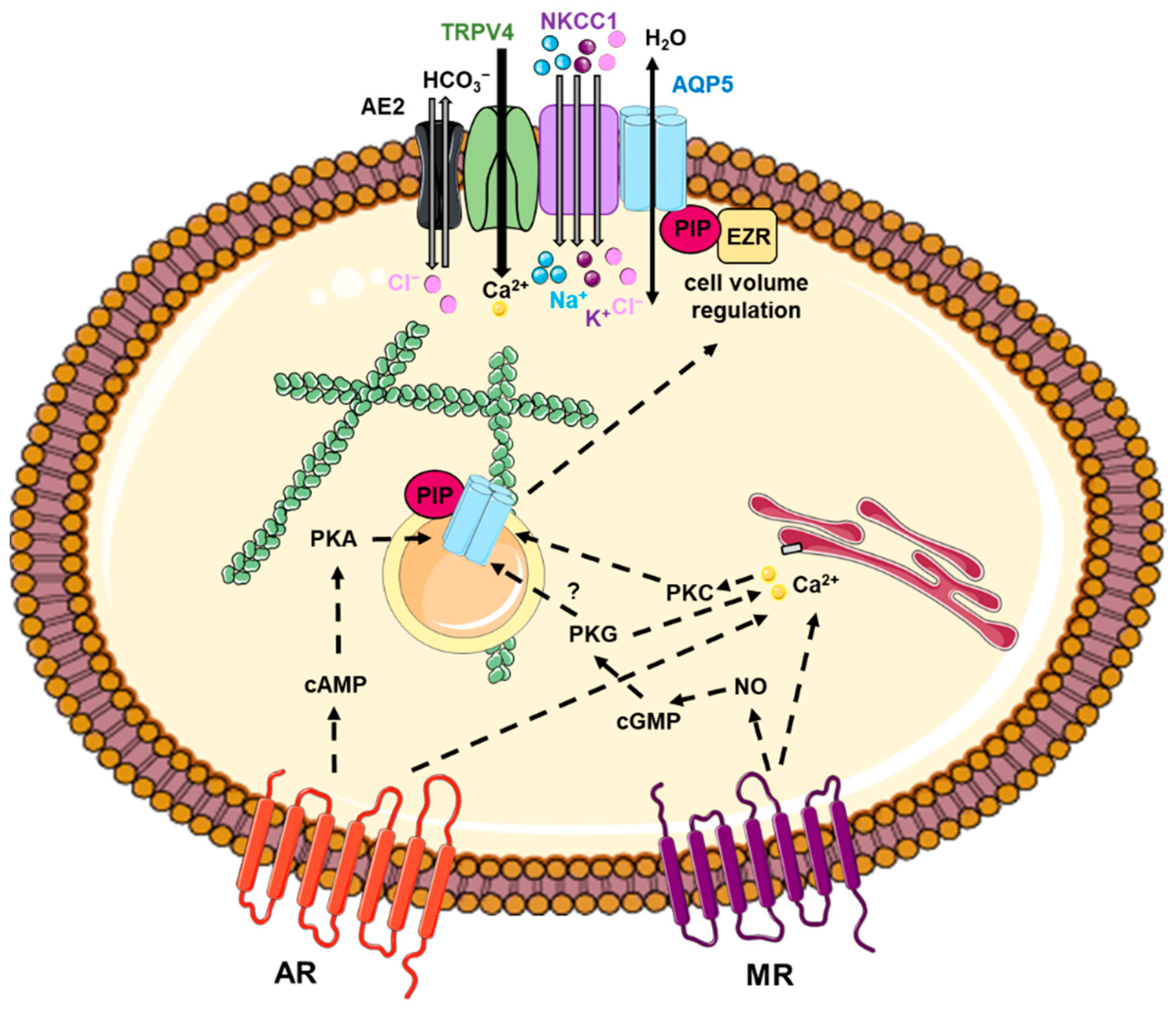
5.1. Involvement of Intracellular Calcium Increase

Intracellular calcium increase has been involved in AQP5 shuttling to the plasma membrane upon activation of muscarinic M3 and α1 adrenergic receptors in rat parotid salivary glands [138,145,146,147,148,149]. The nitric oxide / cGMP signaling pathway causing intracellular calcium concentration increase has also been involved in acetylcholine-induced AQP5 trafficking in rat parotid acinar cells [133]. However, it remains to be determined whether PKG phosphorylates AQP5 and, in such case, at which position(s), considering the presence of multiple potential PKG phosphorylation sites.
Histamine-induced AQP5 trafficking in Aqp5-transfected cells through a calcium signaling pathway [150]. In addition, hypoosmotic stress triggered AQP5 trafficking in transfected cells through a putative PKC-dependent mechanism independent of PKA and Ser156 phosphorylation [134].
Although acute regulation of AQP5 may also take place in the plasma membrane, the plasma membrane diffusion of AQP5 was not dynamically regulated through transient calcium increase and cholesterol depletion [151].

5.2. Involvement of cAMP Increase

cAMP-PKA pathway has been implicated in AQP5 trafficking in various cell types. Indeed, AQP5 trafficked to the plasma membrane upon activation of β adrenergic receptors in rat and mouse parotid glands [145,152] and upon activation of vasoactive intestinal peptide receptors in Brunner’s glands of rat duodenum [153,154].
In MLE-12 cells expressing m AQP5 endogenously, AQP5 trafficking induced by cAMP analog was blocked by PKA inhibitor H89 [60,155]. Moreover, cAMP stimulation had a biphasic effect on AQP5 trafficking in MLE-12 cells. Indeed, short-term exposure (minutes) induced AQP5 internalization through a mechanism involving PKA and lysosome-dependent degradation [155]. By contrast, long-term exposure (hours) induced AQP5 translocation to the plasma membrane through a mechanism involving PKA and AQP5 phosphorylation [155]. However, upon long-term exposure to cAMP analog (30 min to 6 h), AQP5 was internalized in Aqp5-transfected MDCK cells and the primary culture of mouse corneal epithelial cells [156].
In Madin-Darby canine kidney-II (MDCK-II) cells [157] and BEAS-2B human bronchial epithelial cells [158] transfected with an Aqp5 construct, analogs of cAMP or β-adrenergic agonist-induced AQP5 trafficking to the plasma membrane that was inhibited using a PKA inhibitor (H-89).
It was suggested that two consensus PKA phosphorylation sites present within AQP5 might play a role in cAMP-induced trafficking: one at amino acids 152–156 (with Ser 156) within loop D and one at amino acids 224–262 (with Thr259) at the C-terminus. Divergent data exist concerning the role of Ser156 in AQP5 trafficking. Indeed, Ser156 was phosphorylated upon cAMP stimulation and involved in AQP5 trafficking in transfected BEAS-2B human bronchial epithelial cells [158]. In addition, AQP5 was phosphorylated on Ser156 in tumor cells [159]. Ectopic Aqp5 expression promoted cell proliferation upon cAMP-induced Ser156 phosphorylation triggering AQP5 trafficking, suggesting a direct role for AQP5 in carcinogenesis [159]. While Ser156 was also shown not to be involved in AQP5 trafficking in transfected human embryonic kidney 293 (HEK-293) cells, it has been implicated in AQP5 membrane targeting [134]. Considering PKA inhibition increased the membrane abundance of wild-type AQP5 and AQP5 Ser156Ala and Ser156Glu mutants, an additional PKA-dependent mechanism independent of Ser156 phosphorylation was suggested to take part in AQP5 trafficking [134]. This mechanism may involve the phosphorylation of an AQP5-interacting protein partner or alternative PKA phosphorylation sites. By contrast, PKA inhibition increased the trafficking of rat AQP5 (rAQP5) expressed in MDCK-II cells, and various GFP-tagged rAQP5 mutants (Ser152Ala, Thr155Val, Ser156Ala within PKA consensus site) showed lower translocation than GFP-tagged wild-type rAQP5, suggesting dephosphorylation of PKA consensus promoted AQP5 trafficking [160]. Besides, several studies have ruled out the involvement of Thr259 phosphorylation in AQP5 trafficking as mutation of Thr259 to Ala259 in rAQP5 and mAQP5 had no effect on the cAMP-induced protein translocation [157,161]. Altogether, these data support the biphasic regulation of AQP5 trafficking involving distinct short- and long-term regulations [155]. Interestingly, Ser156 phosphorylation does not induce structural changes in AQP5 [134], in contrast to the initial hypothesis formulated based on the high-resolution X-ray crystal structure of the protein [3]. Although two consensus phosphorylation sites (Ser156 and Thr259) are present in cytoplasmic loop D and C-terminus of AQP5, the correlation between their phosphorylation and trafficking signals has not been fully determined [134,161]. It has been proposed that at least three independent mechanisms may account for Aqp5 trafficking: Ser156 phosphorylation, protein kinase A activation, and extracellular osmolarity [134].
Using rAQP5/rAQP1 and rAQP5/rAQP8 chimera, the C-terminus end of rAQP5 has been shown to play a role in its trafficking to the plasma membrane [162,163]. These data have been confirmed using hAQP5 mutant lacking the C-terminal domain (Leu225 to Arg265) [164]. Moreover, additional hAQP5 mutants revealed the role of the 10 last C-terminal amino acids (Arg256 to Arg 265) and Leu262 in AQP5 trafficking [164].
A naturally occurring point mutation (G308A, Gly103 > Asp103 located within the third transmembrane domain) in the rAqp5 gene displayed normal water permeability but reduced trafficking to the plasma membrane and possible lysosomal degradation, both accounting for decreased apical acinar localization in salivary glands and reduced saliva flow [41]. Further studies are required to further decipher the molecular mechanisms accounting for the defective trafficking of the mutated AQP5.
Acute regulation of AQP5 may occur in the plasma membrane as the plasma membrane diffusion of AQP5 was dynamically regulated by physiological stimuli acting through cAMP, PKA, and T259 phosphorylation [151]. However, additional studies are necessary to precise the underlying mechanisms and assess the contribution of AQP5 plasma membrane diffusion in the regulation of water flow during glandular secretions.

5.3. Interactions between AQP5 and Protein Partners

Some AQP5-interacting protein partners have been shown to coordinate and regulate its trafficking and function in mammalian secretory cells [10,102,165], including Na-K-Cl cotransporter 1 (NKCC1), anion exchanger 2 (AE2) [166], Transient Receptor Potential Cation Channel Subfamily V Member 4 (TRPV4) [167], Mucin 5AC (MUC5AC) [168], prolactin-inducible protein (PIP) [169,170], ezrin [171], and junctional proteins [102].
Direct or indirect interactions between NKCC1, AE2, TRPV4, and AQP5 (that may regulate cell volume) [166,167] and between junctional proteins and AQP5 (that may regulate cell-cell adhesion) [102] are unlikely involved in AQP5 trafficking.
The formation of a complex between AQP5 and MUC5AC may ensure the maintenance of normal eye properties and adequate hydration of mucus gel [168].
The interactions between PIP and AQP5 C-terminus [169,170] and between ezrin Four-point-one, Ezrin, Radixin, Moesin (FERM) domain, and AQP5 C-terminus [171] may be involved in AQP5 trafficking. Indeed, PIP-AQP5 interaction was necessary for proper AQP5 localization in lacrimal glands, and loss of the interaction resulted in abnormal AQP5 localization in a mouse model of Sjögren’s syndrome [169]. Furthermore, decreased PIP and Ezrin expression in salivary gland acinar cells from patients suffering from Sjögren’s Syndrome resulted in abnormal AQP5 localization [170,171]. In addition, ezrin regulated AQP5 trafficking in MLE-12 cells [172]. Altogether, these data suggest that PIP and ezrin may regulate AQP5 trafficking by acting as adaptors between AQP5 and the actin cytoskeleton.

5.4. Involvement of Cytoskeleton

Interaction between AQP5-containing vesicles and cytoskeleton has been involved in AQP5 trafficking triggered by intracellular calcium increase. Indeed, inhibitors of microtubules (such as colchicine and vinblastine) and actin filaments (such as cytochalasin B) prevented totally or partially, respectively, calcium-induced AQP5 trafficking [173]. Further studies will be valuable in assessing the role of cytoskeleton dynamics on AQP5 shuttling.

5.5. Involvement of Other Mechanisms

In bovine and other mammalian lenses, AQP5 trafficking occurs during the maturation of lens fibers [23]. Indeed, AQP5 is located within the cytoplasm of lens epithelial cells and young differentiating lens fiber, while its localization shifted to lens fiber cell plasma membranes upon cell maturation [174]. Furthermore, cytoplasmic AQP5 trafficked to the plasma membrane of the lens fiber cells via lysosome secretion [174], considered an unconventional protein secretion pathway [175,176]. In addition, both mechanical and pharmacological reduction in lens tension switched AQP5 labeling from the membrane to cytoplasm, suggesting AQP5 functionality can be dynamically regulated to modify lens water efflux [177]. Direct AQP5-protein interactions involved in these processes remain to be identified.
AQP5 translocated to the apical membrane of mouse mammary gland ductal epithelial cells immediately before parturition and upon progesterone stimulation, suggesting it may regulate milk osmolarity [46]. However, the involved mechanism remains to be further deciphered.
AQP5 was degraded by autophagy in submandibular glands from diabetic mice via a PI3K/Akt/mTOR signaling pathway [178] and from rats with chorda tympani parasympathetic denervation [179]. In the latter, AQP5 autophagy was reversed using an M3 muscarinic receptor agonist, cevimeline [180]. Autophagic degradation of AQP5 in liver cancer cells subjected to heat shock exerted anticancer effects [181]. Upon isoproterenol injection in mice, the AQP5 protein level in the parotid gland increased, then returned to its basal level through a proteolytic degradation involving the calpain system [152].
The Runt-related transcription factor (Runx) /core binding factor β (Cbfb) signaling pathway, involved in salivary gland development, regulated AQP5 trafficking in the acinar cells to allow proper saliva flow [182]. However, the precise mechanism remains to be assessed.

6. Downstream Effectors of AQP5

AQP5 has been shown to activate a wide variety of downstream effectors accounting for cellular functions distinct from water and carbon dioxide permeabilities, including cell migration, invasion, and EMT involved in cancer genesis (Figure 4). AQP5 has been shown to be ectopically expressed or overexpressed in various cancers [183,184], such as breast [185,186] and lung cancer [185,187,188], in which AQP5 expression levels correlated with poor prognosis [186,187]. In other cancers, including prostate [189,190], non-small cell lung [191], colon [192], and cervical [193] cancers, overexpression of AQP5 correlated with metastases to lymph nodes.

6.1. Ras-MAPK Signaling Pathway

AQP5 has been shown to activate the Ras-MAPK signaling pathway in some cancers, and several mechanisms have been proposed to account for this activation.
Indeed, AQP5 phosphorylation on Ser156 (located in the second intracellular loop) by PKA promotes the binding of adaptor molecules (see Section 6.5), which trigger the activation of Ras/Raf-1/mitogen activated protein -kinase 1 and 2 (MEK1/2)/extracellular receptor kinase (ERK1/2) and cyclin-dependent kinases (CDK) that phosphorylated retinoblastoma protein (Rb). The latter induced the expression of genes involved in cell proliferation, migration, and invasion that may drive EMT [192,194,195,196].
The activation of the Rac family of small GTPase 1 (Rac1; another GTPase belonging to the Ras superfamily of small GTP-binding proteins) by AQP5 [185] may also activate the Ras-MAPK signaling pathway [197]. Additional experiments are warranted to confirm these findings and the molecular mechanism involved.
The activation of epidermal growth factor receptor (EGFR) by AQP5 can stimulate the Ras/MAPK signal transduction directly or indirectly (via the activation of phosphoinositide 3-kinase (PI3K)) to lead to cell proliferation and migration of lung cancer cells [188] and human glioma cell lines [198], and keratinocyte chemoattractant expression [199]. In human colorectal cancer cell lines, AQP5 was involved in vascular endothelial growth factor (VEGF) A expression, secretion, and concomitant angiogenesis [200], but the involvement of the Ras-MAPK signaling pathway remains to be assessed.

6.2. Wingless/Integrated (WNT)/β-Catenin Signaling Pathway

The activation of the Wingless/Integrated (WNT)/β-catenin signaling pathway may account for the involvement of AQP5 in EMT. In human colorectal cancer cell lines, Aqp5 silencing altered EMT by reducing the expression of mesenchymal cell markers (N-cadherin, Vimentin, and Snail) and increasing the expression of epithelial cell marker (E-cadherin) [201,202]. The alteration of EMT-related markers mediated by Aqp5 silencing was reversed by the upregulation of β-catenin [201]. In colorectal cancer, AQP5 expression activated the WNT/β-catenin, predicted poor clinical outcome [203], increased chemoresistance to 5-fluorouracil, and promoted tumor growth [204]. In such cancer, Aqp5 silencing inhibited the WNT/β-catenin signaling, increased the chemosensitivity to chemotherapy-inducing apoptosis, and suppressed tumor growth, suggesting AQP5 could be a useful therapeutic target for such cancer [201,204].
Following Helicobacter pylori infection in mice inducing gastritis, transcription factor Achaete-Scute Family BHLH Transcription Factor 1 (ASCL1) increased AQP5 expression, which was responsible for the activation of the WNT/β-catenin signaling pathway [111].

6.3. p-Smad2/3 Pathway

In human colorectal cancer cell lines, AQP5 induced the activation of phosphorylated-mothers against decapentaplegic homolog 2/3 (p-Smad2/3), which resulted in EMT [202]. These data suggested that p-Smad2/3 may form a complex with others against decapentaplegic homolog 4 (Smad4) and translocate to the nucleus to regulate target gene transcription leading to the stimulation of EMT [205].
Further experimentation is necessary to determine if the recruitment of proteins by AQP5 is involved in the activation of the p-Smad2/3 pathway.

6.4. p-Akt Pathway

AQP5 overexpression induced the phosphorylation of AKT, resulting in mouse cornea epithelial cell proliferation promoting tumorigenesis and apoptosis [206]. However, the involved mechanisms remain to be analyzed. AKT activation by AQP5 also stimulates NFkB, which can regulate the transcription of target genes, including that of AQP5 [199]. In hepatocellular carcinoma, AQP5 promoted cell invasion and tumor metastasis in vitro and in vivo and induced EMT involving NFkB activation [207].

6.5. Recruitment of Proteins

The recruitment of proteins can regulate many cellular processes, including gene expression, cell adhesion and migration, and cell proliferation. AQP5 has been shown to recruit some proteins through the presence of protein binding motifs. Indeed, a homologous RGN motif present within the AQP5 extracellular connecting loop C (CL3) [208] has been shown to be involved in fibronectin binding [209] and thereby may play a role in cell migration. In addition, the binding of phosphorylated extracellular loop D fragment of AQP5 (amino acids 88–182) to the SH3 domains of Src protooncogene (Src), Lyn protooncogene (Lyn) and GRB2 Related Adaptor Protein 2 (Grap2) [187] has been shown to play a role in cell migration, invasion and EMT [210,211,212], possibly by activation RAS or RAC1 and activating the MAPK pathway (see Section 6.1). AQP5 is likely recruiting the proteins via a diproline peptide sequence (RTSPVGSP; amino acids 154–161) bearing sequence similarity to the SH3 binding consensus site [187]. Both phosphorylation of AQP5 on Ser156, located within the SH3 consensus binding site, and Asn 185, located within an NPA motif, have been suggested to be critical for the AQP5-induced cell migration and invasion [187]. Interestingly, the Src family of kinases, including Src and Lyn, have been involved in cell volume regulation by modulating the activity of ions transports, some of which are identified as AQP5-interacting protein partners (i.e., TRPV4; NKCC1, and NHE) [213]. Therefore, additional experiments will be valuable to assess the role of Src and Lyn on the cell volume regulation involving AQP5.
AQP5 has also been involved in the regulation of cytoskeleton dynamics through the activation of the Ras-MAPK signaling pathway inducing actin network reorganization (see Section 6.1). Moreover, in epithelial cells, AQP5 C-terminus seemed to efficiently promote microtubule stability assembly that may regulate paracellular water permeability [214,215]. Moreover, Aqp5 knockout mice displayed decreased expression of tight junctions proteins forming the tight junction strands (including claudins and occludin) and involved in scaffolding (including zonula occludens-1 (ZO-1)) which may account for decreased paracellular water permeability [215]. Concomitant to Aqp5 knockout, the loss of interaction between AQP5 and junctional protein [102] may also participate to decreased paracellular water permeability.
Overall, further experiments are necessary to further decipher the molecular mechanism linking the recruitment of proteins by AQP5 to EMT, regulation of the cytoskeleton involved in cell migration and adhesion, and paracellular permeability.

7. Conclusions

Several mechanisms account for the dynamic upstream regulation of AQP5. Indeed, single nucleotide polymorphisms, mutations, and transcriptional control of Aqp5 gene have been shown to impact the expression, trafficking, and/or function of the protein. Moreover, trafficking and localization of AQP5 can also be regulated by post-translational modifications, intracellular signaling pathways (calcium and cAMP-PKA), protein interacting partners, cytoskeleton interaction, and other mechanisms. Furthermore, the dynamic regulation of these upstream events will likely impact not only the AQP5 permeability to water and carbon dioxide but also other cellular pathways resulting from the activation of downstream effectors of AQP5. Currently, these downstream effectors include the Ras-MAPK signaling pathway, WNT/β-catenin signaling pathway, p-Smad2/3 pathway, and recruitment of proteins involved in a variety of biological effects. Overall, a deeper understanding of these finely tuned upstream and downstream mechanisms will improve the current comprehension of physiological and pathophysiological processes involving AQP5.

Author Contributions

Conceptualization, C.D. (Christine Delporte); writing—original draft preparation, writing—review and editing, C.D. (Claudia D’Agostino), D.P., C.C., M.H., M.S.S. and C.D. (Christine Delporte); visualization, C.D. (Claudia D’Agostino), D.P., C.C., M.H., M.S.S. and C.D. (Christine Delporte); supervision, C.D. (Christine Delporte); project administration, C.D. (Christine Delporte); funding acquisition, C.D. (Christine Delporte). All authors have read and agreed to the published version of the manuscript.

Funding

This work was supported by the Université Libre de Bruxelles, the Fonds de la Recherche Scientifique—FNRS (Fund for Scientific Research—PINT-BILAT-P-R.P006.19 (C.D.); Foundation Jaumotte-Demoulin (C.D.); Fund Octave Dupont from the Royal Academy of Belgium (C.D.); Fonds Erasme pour la recherche médicale (D.P.). D.P. is a Research Fellow of the Fonds de la Recherche Scientifique—FNRS.

Institutional Review Board Statement

Not applicable.

Informed Consent Statement

Not applicable.

Data Availability Statement

Not applicable.

Conflicts of Interest

The authors declare no conflict of interest.

References

  1. Agre, P. Aquaporin Water Channels (Nobel Lecture). Angew. Chem. Int. Ed. Engl. 2004, 43, 4278–4290. [Google Scholar] [CrossRef]
  2. Laloux, T.; Junqueira, B.; Maistriaux, L.C.; Ahmed, J.; Jurkiewicz, A.; Chaumont, F. Plant and Mammal Aquaporins: Same but Different. Int. J. Mol. Sci. 2018, 19, 521. [Google Scholar] [CrossRef] [PubMed] [Green Version]
  3. Horsefield, R.; Nordén, K.; Fellert, M.; Backmark, A.; Törnroth-Horsefield, S.; Terwisscha van Scheltinga, A.C.; Kvassman, J.; Kjellbom, P.; Johanson, U.; Neutze, R. High-Resolution X-ray Structure of Human Aquaporin 5. Proc. Natl. Acad. Sci. USA 2008, 105, 13327–13332. [Google Scholar] [CrossRef] [PubMed] [Green Version]
  4. Lee, M.D.; Bhakta, K.Y.; Raina, S.; Yonescu, R.; Griffin, C.A.; Copeland, N.G.; Gilbert, D.J.; Jenkins, N.A.; Preston, G.M.; Agre, P. The Human Aquaporin-5 Gene. Molecular Characterization and Chromosomal Localization. J. Biol. Chem. 1996, 271, 8599–8604. [Google Scholar] [CrossRef] [PubMed] [Green Version]
  5. Raina, S.; Preston, G.M.; Guggino, W.B.; Agre, P. Molecular Cloning and Characterization of an Aquaporin CDNA from Salivary, Lacrimal, and Respiratory Tissues. J. Biol. Chem. 1995, 270, 1908–1912. [Google Scholar] [CrossRef] [PubMed] [Green Version]
  6. Rodrigues, C.; Pimpão, C.; Mósca, A.F.; Coxixo, A.S.; Lopes, D.; da Silva, I.V.; Pedersen, P.A.; Antunes, F.; Soveral, G. Human Aquaporin-5 Facilitates Hydrogen Peroxide Permeation Affecting Adaption to Oxidative Stress and Cancer Cell Migration. Cancers 2019, 11, 932. [Google Scholar] [CrossRef] [Green Version]
  7. Geyer, R.R.; Musa-Aziz, R.; Qin, X.; Boron, W.F. Relative CO(2)/NH(3) Selectivities of Mammalian Aquaporins 0-9. Am. J. Physiol. Cell Physiol. 2013, 304, C985–C994. [Google Scholar] [CrossRef] [Green Version]
  8. Alishahi, M.; Kamali, R. A Novel Molecular Dynamics Study of CO2 Permeation through Aquaporin-5. Eur. Phys. J. E 2019, 42, 151. [Google Scholar] [CrossRef]
  9. Kourghi, M.; Pei, J.V.; De Ieso, M.L.; Nourmohammadi, S.; Chow, P.H.; Yool, A.J. Fundamental Structural and Functional Properties of Aquaporin Ion Channels Found across the Kingdoms of Life. Clin. Exp. Pharmacol. Physiol. 2018, 45, 401–409. [Google Scholar] [CrossRef] [Green Version]
  10. Törnroth-Horsefield, S.; Chivasso, C.; Strandberg, H.; D’Agostino, C.; O’Neale, C.V.T.; Schey, K.L.; Delporte, C. Insight into the Mammalian Aquaporin Interactome. Int. J. Mol. Sci. 2022, 23, 9615. [Google Scholar] [CrossRef]
  11. Henderson, S.W.; Nourmohammadi, S.; Ramesh, S.A.; Yool, A.J. Aquaporin Ion Conductance Properties Defined by Membrane Environment, Protein Structure, and Cell Physiology. Biophys. Rev. 2022, 14, 181–198. [Google Scholar] [CrossRef] [PubMed]
  12. Kitchen, P.; Conner, M.T.; Bill, R.M.; Conner, A.C. Structural Determinants of Oligomerization of the Aquaporin-4 Channel. J. Biol. Chem. 2016, 291, 6858–6871. [Google Scholar] [CrossRef] [PubMed] [Green Version]
  13. Takata, K.; Matsuzaki, T.; Tajika, Y. Aquaporins: Water Channel Proteins of the Cell Membrane. Prog. Histochem. Cytochem. 2004, 39, 1–83. [Google Scholar] [CrossRef]
  14. Agre, P.; King, L.S.; Yasui, M.; Guggino, W.B.; Ottersen, O.P.; Fujiyoshi, Y.; Engel, A.; Nielsen, S. Aquaporin Water Channels--from Atomic Structure to Clinical Medicine. J. Physiol. (Lond.) 2002, 542, 3–16. [Google Scholar] [CrossRef] [PubMed]
  15. Verkman, A.S.; Mitra, A.K. Structure and Function of Aquaporin Water Channels. Am. J. Physiol. Renal. Physiol. 2000, 278, F13–F28. [Google Scholar] [CrossRef] [PubMed] [Green Version]
  16. Delporte, C. Aquaporins and Gland Secretion. Adv. Exp. Med. Biol. 2017, 969, 63–79. [Google Scholar] [CrossRef]
  17. Delporte, C.; Soyfoo, M. Aquaporins: Unexpected Actors in Autoimmune Diseases. Autoimmun. Rev. 2022, 21, 103131. [Google Scholar] [CrossRef]
  18. D’Agostino, C.; Elkashty, O.A.; Chivasso, C.; Perret, J.; Tran, S.D.; Delporte, C. Insight into Salivary Gland Aquaporins. Cells 2020, 9, 1547. [Google Scholar] [CrossRef]
  19. Mobasheri, A.; Barrett-Jolley, R. Aquaporin Water Channels in the Mammary Gland: From Physiology to Pathophysiology and Neoplasia. J. Mammary Gland. Biol. Neoplasia 2014, 19, 91–102. [Google Scholar] [CrossRef] [Green Version]
  20. Yadav, E.; Yadav, N.; Hus, A.; Yadav, J.S. Aquaporins in Lung Health and Disease: Emerging Roles, Regulation, and Clinical Implications. Respir. Med. 2020, 174, 106193. [Google Scholar] [CrossRef]
  21. Song, Y.; Verkman, A.S. Aquaporin-5 Dependent Fluid Secretion in Airway Submucosal Glands. J. Biol. Chem. 2001, 276, 41288–41292. [Google Scholar] [CrossRef] [Green Version]
  22. Grey, A.C.; Walker, K.L.; Petrova, R.S.; Han, J.; Wilmarth, P.A.; David, L.L.; Donaldson, P.J.; Schey, K.L. Verification and Spatial Localization of Aquaporin-5 in the Ocular Lens. Exp. Eye Res. 2013, 108, 94–102. [Google Scholar] [CrossRef] [PubMed] [Green Version]
  23. Petrova, R.S.; Schey, K.L.; Donaldson, P.J.; Grey, A.C. Spatial Distributions of AQP5 and AQP0 in Embryonic and Postnatal Mouse Lens Development. Exp. Eye Res. 2015, 132, 124–135. [Google Scholar] [CrossRef] [PubMed] [Green Version]
  24. Schey, K.L.; Petrova, R.S.; Gletten, R.B.; Donaldson, P.J. The Role of Aquaporins in Ocular Lens Homeostasis. Int. J. Mol. Sci. 2017, 18, 2693. [Google Scholar] [CrossRef] [PubMed] [Green Version]
  25. Lee, S.J.; Kang, B.W.; Kim, J.G.; Jung, J.H.; Lee, J.; Kim, W.W.; Park, H.Y.; Jeong, J.-H.; Jeong, J.Y.; Park, J.-Y.; et al. AQP5 Variants Affect Tumoral Expression of AQP5 and Survival in Patients with Early Breast Cancer. Oncology 2017, 92, 153–160. [Google Scholar] [CrossRef] [PubMed]
  26. Adamzik, M.; Frey, U.H.; Bitzer, K.; Jakob, H.; Baba, H.A.; Schmieder, R.E.; Schneider, M.P.; Heusch, G.; Peters, J.; Siffert, W. A Novel-1364A/C Aquaporin 5 Gene Promoter Polymorphism Influences the Responses to Salt Loading of the Renin-Angiotensin-Aldosterone System and of Blood Pressure in Young Healthy Men. Basic Res. Cardiol. 2008, 103, 598–610. [Google Scholar] [CrossRef]
  27. Rump, K.; Spellenberg, T.; von Busch, A.; Wolf, A.; Ziehe, D.; Thon, P.; Rahmel, T.; Adamzik, M.; Koos, B.; Unterberg, M. AQP5-1364A/C Polymorphism Affects AQP5 Promoter Methylation. Int. J. Mol. Sci. 2022, 23, 11813. [Google Scholar] [CrossRef]
  28. Adamzik, M.; Frey, U.H.; Möhlenkamp, S.; Scherag, A.; Waydhas, C.; Marggraf, G.; Dammann, M.; Steinmann, J.; Siffert, W.; Peters, J. Aquaporin 5 Gene Promoter--1364A/C Polymorphism Associated with 30-Day Survival in Severe Sepsis. Anesthesiology 2011, 114, 912–917. [Google Scholar] [CrossRef] [Green Version]
  29. Bergmann, L.; Nowak, H.; Siffert, W.; Peters, J.; Adamzik, M.; Koos, B.; Rahmel, T. Major Adverse Kidney Events Are Associated with the Aquaporin 5 -1364A/C Promoter Polymorphism in Sepsis: A Prospective Validation Study. Cells 2020, 9, 904. [Google Scholar] [CrossRef] [Green Version]
  30. Rahmel, T.; Rump, K.; Peters, J.; Adamzik, M. Aquaporin 5 -1364A/C Promoter Polymorphism Is Associated with Pulmonary Inflammation and Survival in Acute Respiratory Distress Syndrome. Anesthesiology 2019, 130, 404–413. [Google Scholar] [CrossRef]
  31. Rahmel, T.; Nowak, H.; Rump, K.; Siffert, W.; Peters, J.; Adamzik, M. The Aquaporin 5 -1364A/C Promoter Polymorphism Impacts on Resolution of Acute Kidney Injury in Pneumonia Evoked ARDS. PLoS ONE 2018, 13, e0208582. [Google Scholar] [CrossRef] [Green Version]
  32. Rahmel, T.; Nowak, H.; Rump, K.; Koos, B.; Schenker, P.; Viebahn, R.; Adamzik, M.; Bergmann, L. The Aquaporin 5 -1364A/C Promoter Polymorphism Is Associated with Cytomegalovirus Infection Risk in Kidney Transplant Recipients. Front. Immunol. 2019, 10, 2871. [Google Scholar] [CrossRef] [PubMed] [Green Version]
  33. Lambertz, N.; Hindy, N.E.; Adler, C.; Rump, K.; Adamzik, M.; Keyvani, K.; Bankfalvi, A.; Siffert, W.; Erol Sandalcioglu, I.; Bachmann, H.S. Expression of Aquaporin 5 and the AQP5 Polymorphism A(-1364)C in Association with Peritumoral Brain Edema in Meningioma Patients. J. Neurooncol. 2013, 112, 297–305. [Google Scholar] [CrossRef] [PubMed]
  34. Blaydon, D.C.; Lind, L.K.; Plagnol, V.; Linton, K.J.; Smith, F.J.D.; Wilson, N.J.; McLean, W.H.I.; Munro, C.S.; South, A.P.; Leigh, I.M.; et al. Mutations in AQP5, Encoding a Water-Channel Protein, Cause Autosomal-Dominant Diffuse Nonepidermolytic Palmoplantar Keratoderma. Am. J. Hum. Genet. 2013, 93, 330–335. [Google Scholar] [CrossRef] [Green Version]
  35. Krøigård, A.B.; Hetland, L.E.; Clemmensen, O.; Blaydon, D.C.; Hertz, J.M.; Bygum, A. The First Danish Family Reported with an AQP5 Mutation Presenting Diffuse Non-Epidermolytic Palmoplantar Keratoderma of Bothnian Type, Hyperhidrosis and Frequent Corynebacterium Infections: A Case Report. BMC Dermatol. 2016, 16, 7. [Google Scholar] [CrossRef] [Green Version]
  36. Cao, X.; Yin, J.; Wang, H.; Zhao, J.; Zhang, J.; Dai, L.; Zhang, J.; Jiang, H.; Lin, Z.; Yang, Y. Mutation in AQP5, Encoding Aquaporin 5, Causes Palmoplantar Keratoderma Bothnia Type. J. Investig. Dermatol. 2014, 134, 284–287. [Google Scholar] [CrossRef] [Green Version]
  37. Wada, Y.; Kusakabe, M.; Nagai, M.; Imai, Y.; Yamanishi, K. Japanese Case of Bothnian-Type Palmoplantar Keratoderma with a Novel Missense Mutation of p.Trp35Ser in Extracellular Loop A of Aquaporin-5. J. Dermatol. 2019, 46, e104–e106. [Google Scholar] [CrossRef] [PubMed]
  38. Hadidi, H.; Kamali, R. Molecular Dynamics Study of Water Transport through AQP5-R188C Mutant Causing Palmoplantar Keratoderma (PPK) Using the Gating Mechanism Concept. Biophys. Chem. 2021, 277, 106655. [Google Scholar] [CrossRef] [PubMed]
  39. Tang, S.; Di, G.; Hu, S.; Liu, Y.; Dai, Y.; Chen, P. AQP5 Regulates Vimentin Expression via MiR-124–3p.1 to Protect Lens Transparency. Exp. Eye Res. 2021, 205, 108485. [Google Scholar] [CrossRef]
  40. Qin, X.; Boron, W.F. Mutation of a Single Amino Acid Converts the Human Water Channel Aquaporin 5 into an Anion Channel. Am. J. Physiol.-Cell Physiol. 2013, 305, C663–C672. [Google Scholar] [CrossRef]
  41. Karabasil, M.R.; Hasegawa, T.; Azlina, A.; Purwanti, N.; Yao, C.; Akamatsu, T.; Tomioka, S.; Hosoi, K. Effects of Naturally Occurring G103D Point Mutation of AQP5 on Its Water Permeability, Trafficking and Cellular Localization in the Submandibular Gland of Rats. Biol. Cell 2011, 103, 69–86. [Google Scholar] [CrossRef]
  42. Satoh, K.; Seo, Y.; Matsuo, S.; Karabasil, M.R.; Matsuki-Fukushima, M.; Nakahari, T.; Hosoi, K. Roles of AQP5/AQP5-G103D in Carbamylcholine-Induced Volume Decrease and in Reduction of the Activation Energy for Water Transport by Rat Parotid Acinar Cells. Pflugers Arch.-Eur. J. Physiol. 2012, 464, 375–389. [Google Scholar] [CrossRef] [PubMed]
  43. Tanski, D.; Skowronska, A.; Tanska, M.; Lepiarczyk, E.; Skowronski, M.T. The In Vitro Effect of Steroid Hormones, Arachidonic Acid, and Kinases Inhibitors on Aquaporin 1, 2, 5, and 7 Gene Expression in the Porcine Uterine Luminal Epithelial Cells during the Estrous Cycle. Cells 2021, 10, 832. [Google Scholar] [CrossRef]
  44. de Oliveira, V.; Schaefer, J.; Abu-Rafea, B.; Vilos, G.A.; Vilos, A.G.; Bhattacharya, M.; Radovick, S.; Babwah, A.V. Uterine Aquaporin Expression Is Dynamically Regulated by Estradiol and Progesterone and Ovarian Stimulation Disrupts Embryo Implantation without Affecting Luminal Closure. Mol. Hum. Reprod. 2020, 26, 154–166. [Google Scholar] [CrossRef] [PubMed]
  45. Zhang, Y.; Chen, Q.; Zhang, H.; Wang, Q.; Li, R.; Jin, Y.; Wang, H.; Ma, T.; Qiao, J.; Duan, E. Aquaporin-Dependent Excessive Intrauterine Fluid Accumulation Is a Major Contributor in Hyper-Estrogen Induced Aberrant Embryo Implantation. Cell Res. 2015, 25, 139–142. [Google Scholar] [CrossRef] [PubMed] [Green Version]
  46. Kaihoko, Y.; Tsugami, Y.; Suzuki, N.; Suzuki, T.; Nishimura, T.; Kobayashi, K. Distinct Expression Patterns of Aquaporin 3 and 5 in Ductal and Alveolar Epithelial Cells in Mouse Mammary Glands before and after Parturition. Cell Tissue Res. 2020, 380, 513–526. [Google Scholar] [CrossRef]
  47. Salleh, N.; Mokhtar, H.M.; Kassim, N.M.; Giribabu, N. Testosterone Induces Increase in Aquaporin (AQP)-1, 5, and 7 Expressions in the Uteri of Ovariectomized Rats. J. Membr. Biol. 2015, 248, 1097–1105. [Google Scholar] [CrossRef]
  48. Ducza, E.; Seres, A.B.; Hajagos-Tóth, J.; Falkay, G.; Gáspár, R. Oxytocin Regulates the Expression of Aquaporin 5 in the Late-Pregnant Rat Uterus. Mol. Reprod. Dev. 2014, 81, 524–530. [Google Scholar] [CrossRef] [Green Version]
  49. Skowronska, A.; Mlotkowska, P.; Okrasa, S.; Nielsen, S.; Skowronski, M.T. Modulatory Effects of Steroid Hormones, Oxytocin, Arachidonic Acid, Forskolin and Cyclic AMP on the Expression of Aquaporin 1 and Aquaporin 5 in the Porcine Uterus during Placentation. J. Physiol. Pharmacol. 2016, 67, 311–319. [Google Scholar]
  50. Skowronski, M.T.; Mlotkowska, P.; Tanski, D.; Lepiarczyk, E.; Kempisty, B.; Jaskiewicz, L.; Pareek, C.S.; Skowronska, A. Pituitary Hormones (FSH, LH, PRL, and GH) Differentially Regulate AQP5 Expression in Porcine Ovarian Follicular Cells. Int. J. Mol. Sci. 2019, 20, 4914. [Google Scholar] [CrossRef] [Green Version]
  51. Jiang, X.X.; Fei, X.W.; Zhao, L.; Ye, X.L.; Xin, L.B.; Qu, Y.; Xu, K.H.; Wu, R.J.; Lin, J. Aquaporin 5 Plays a Role in Estrogen-Induced Ectopic Implantation of Endometrial Stromal Cells in Endometriosis. PLoS ONE 2015, 10, e0145290. [Google Scholar] [CrossRef] [PubMed]
  52. Ben, Y.; Chen, J.; Zhu, R.; Gao, L.; Bai, C. Upregulation of AQP3 and AQP5 Induced by Dexamethasone and Ambroxol in A549 Cells. Respir. Physiol. Neurobiol. 2008, 161, 111–118. [Google Scholar] [CrossRef] [PubMed]
  53. Pajouhi, N.; Owji, M.; Naghibalhossaini, F.; Omrani, G.H.R.; Varedi, M. Modulation by Thyroid Hormone of Myosin Light Chain Phosphorylation and Aquaporin 5 Protein Expression in Intact Lung. J. Physiol. Biochem. 2015, 71, 99–106. [Google Scholar] [CrossRef]
  54. Ramirez, M.I.; Chung, U.I.; Williams, M.C. Aquaporin-5 Expression, but Not Other Peripheral Lung Marker Genes, Is Reduced in PTH/PTHrP Receptor Null Mutant Fetal Mice. Am. J. Respir. Cell Mol. Biol. 2000, 22, 367–372. [Google Scholar] [CrossRef] [PubMed] [Green Version]
  55. Lin, J.-B.; Kang, M.-Q.; Huang, L.-P.; Zhuo, Y.; Li, X.; Lai, F.-C. CHRNA1 Promotes the Pathogenesis of Primary Focal Hyperhidrosis. Mol. Cell. Neurosci. 2021, 111, 103598. [Google Scholar] [CrossRef] [PubMed]
  56. Lin, J.-B.; Lin, N.-L.; Li, X.; Kang, M.-Q. Antagonist of Chrna1 Prevents the Pathogenesis of Primary Focal Hyperhidrosis. Ann. Clin. Transl. Neurol. 2022, 9, 786–794. [Google Scholar] [CrossRef] [PubMed]
  57. Du, Q.; Lin, M.; Yang, J.-H.; Chen, J.-F.; Tu, Y.-R. Overexpression of AQP5 Was Detected in Axillary Sweat Glands of Primary Focal Hyperhidrosis Patients. Dermatology 2016, 232, 150–155. [Google Scholar] [CrossRef]
  58. Chang, Y.-L.; Jian, K.R.; Lin, C.-S.; Wang, H.-W.; Liu, S.-C. Dexamethasone Attenuates Methacholine-Mediated Aquaporin 5 Downregulation in Human Nasal Epithelial Cells via Suppression of NF-ΚB Activation. Int. Forum Allergy Rhinol. 2018, 8, 64–71. [Google Scholar] [CrossRef]
  59. Zhou, B.; Ann, D.K.; Flodby, P.; Minoo, P.; Liebler, J.M.; Crandall, E.D.; Borok, Z. Rat Aquaporin-5 4.3-Kb 5’-Flanking Region Differentially Regulates Expression in Salivary Gland and Lung in Vivo. Am. J. Physiol. Cell Physiol. 2008, 295, C111–C120. [Google Scholar] [CrossRef] [Green Version]
  60. Yang, F.; Kawedia, J.D.; Menon, A.G. Cyclic AMP Regulates Aquaporin 5 Expression at Both Transcriptional and Post-Transcriptional Levels through a Protein Kinase A Pathway. J. Biol. Chem. 2003, 278, 32173–32180. [Google Scholar] [CrossRef] [Green Version]
  61. Cammalleri, M.; Amato, R.; Olivieri, M.; Pezzino, S.; Bagnoli, P.; Dal Monte, M.; Rusciano, D. Effects of Topical Gabapentin on Ocular Pain and Tear Secretion. Front. Pharmacol. 2021, 12, 671238. [Google Scholar] [CrossRef] [PubMed]
  62. Chang, Y.-L.; Lin, C.-S.; Wang, H.-W.; Jian, K.R.; Liu, S.-C. Chlorpheniramine Attenuates Histamine-Mediated Aquaporin 5 Downregulation in Human Nasal Epithelial Cells via Suppression of NF-ΚB Activation. Int. J. Med. Sci. 2017, 14, 1268–1275. [Google Scholar] [CrossRef] [PubMed] [Green Version]
  63. Wang, W.; Wang, X.; Ma, L.; Zhang, R. Histamine Downregulates Aquaporin 5 in Human Nasal Epithelial Cells. Am. J. Rhinol. Allergy 2015, 29, 188–192. [Google Scholar] [CrossRef] [PubMed]
  64. Lin, J.-B.; Chen, J.-F.; Lai, F.-C.; Li, X.; Xie, J.-B.; Tu, Y.-R.; Kang, M.-Q. Involvement of Activin a Receptor Type 1 (ACVR1) in the Pathogenesis of Primary Focal Hyperhidrosis. Biochem. Biophys. Res. Commun. 2020, 528, 299–304. [Google Scholar] [CrossRef] [PubMed]
  65. Chen, H.; Zhou, X.; Shi, Y.; Yang, J. Roles of P38 MAPK and JNK in TGF-Β1-Induced Human Alveolar Epithelial to Mesenchymal Transition. Arch. Med. Res. 2013, 44, 93–98. [Google Scholar] [CrossRef]
  66. Willis, B.C.; Liebler, J.M.; Luby-Phelps, K.; Nicholson, A.G.; Crandall, E.D.; du Bois, R.M.; Borok, Z. Induction of Epithelial-Mesenchymal Transition in Alveolar Epithelial Cells by Transforming Growth Factor-Beta1: Potential Role in Idiopathic Pulmonary Fibrosis. Am. J. Pathol. 2005, 166, 1321–1332. [Google Scholar] [CrossRef]
  67. Yamamura, Y.; Motegi, K.; Kani, K.; Takano, H.; Momota, Y.; Aota, K.; Yamanoi, T.; Azuma, M. TNF-α Inhibits Aquaporin 5 Expression in Human Salivary Gland Acinar Cells via Suppression of Histone H4 Acetylation. J. Cell. Mol. Med. 2012, 16, 1766–1775. [Google Scholar] [CrossRef]
  68. Towne, J.E.; Krane, C.M.; Bachurski, C.J.; Menon, A.G. Tumor Necrosis Factor-Alpha Inhibits Aquaporin 5 Expression in Mouse Lung Epithelial Cells. J. Biol. Chem. 2001, 276, 18657–18664. [Google Scholar] [CrossRef] [PubMed] [Green Version]
  69. Mezzasoma, L.; Cagini, L.; Antognelli, C.; Puma, F.; Pacifico, E.; Talesa, V.N. TNF-α Regulates Natriuretic Peptides and Aquaporins in Human Bronchial Epithelial Cells BEAS-2B. Mediat. Inflamm. 2013, 2013, 159349. [Google Scholar] [CrossRef] [Green Version]
  70. Limaye, A.; Hall, B.E.; Zhang, L.; Cho, A.; Prochazkova, M.; Zheng, C.; Walker, M.; Adewusi, F.; Burbelo, P.D.; Sun, Z.J.; et al. Targeted TNF-α Overexpression Drives Salivary Gland Inflammation. J. Dent. Res. 2019, 98, 713–719. [Google Scholar] [CrossRef]
  71. Shi, Z.; Ye, W.; Zhang, J.; Zhang, F.; Yu, D.; Yu, H.; Chen, B.; Zhou, M.; Sun, H. LipoxinA4 Attenuates Acute Pancreatitis-Associated Acute Lung Injury by Regulating AQP-5 and MMP-9 Expression, Anti-Apoptosis and PKC/SSeCKS-Mediated F-Actin Activation. Mol. Immunol. 2018, 103, 78–88. [Google Scholar] [CrossRef] [PubMed]
  72. Smith, J.K.; Siddiqui, A.A.; Modica, L.A.; Dykes, R.; Simmons, C.; Schmidt, J.; Krishnaswamy, G.A.; Berk, S.L. Interferon-Alpha Upregulates Gene Expression of Aquaporin-5 in Human Parotid Glands. J. Interferon Cytokine Res. 1999, 19, 929–935. [Google Scholar] [CrossRef] [PubMed]
  73. Skowron-zwarg, M.; Boland, S.; Caruso, N.; Coraux, C.; Marano, F.; Tournier, F. Interleukin-13 Interferes with CFTR and AQP5 Expression and Localization during Human Airway Epithelial Cell Differentiation. Exp. Cell Res. 2007, 313, 2695–2702. [Google Scholar] [CrossRef]
  74. Krane, C.M.; Deng, B.; Mutyam, V.; McDonald, C.A.; Pazdziorko, S.; Mason, L.; Goldman, S.; Kasaian, M.; Chaudhary, D.; Williams, C.; et al. Altered Regulation of Aquaporin Gene Expression in Allergen and IL-13-Induced Mouse Models of Asthma. Cytokine 2009, 46, 111–118. [Google Scholar] [CrossRef] [Green Version]
  75. Lotze, M.T.; Tracey, K.J. High-Mobility Group Box 1 Protein (HMGB1): Nuclear Weapon in the Immune Arsenal. Nat. Rev. Immunol. 2005, 5, 331–342. [Google Scholar] [CrossRef]
  76. Müller, S.; Scaffidi, P.; Degryse, B.; Bonaldi, T.; Ronfani, L.; Agresti, A.; Beltrame, M.; Bianchi, M.E. New EMBO Members’ Review: The Double Life of HMGB1 Chromatin Protein: Architectural Factor and Extracellular Signal. EMBO J. 2001, 20, 4337–4340. [Google Scholar] [CrossRef]
  77. Sarrand, J.; Baglione, L.; Parisis, D.; Soyfoo, M. The Involvement of Alarmins in the Pathogenesis of Sjögren’s Syndrome. Int. J. Mol. Sci. 2022, 23, 5671. [Google Scholar] [CrossRef]
  78. Wang, D.; Zhou, M.; Wang, Y.; Sun, S. Suppression of High-Mobility Group Box 1 Ameliorates Xerostomia in a Sjögren Syndrome-Triggered Mouse Model. Can. J. Physiol. Pharmacol. 2020, 98, 351–356. [Google Scholar] [CrossRef]
  79. Kim, K.H.; Kim, D.H.; Jeong, H.J.; Ryu, J.S.; Kim, Y.J.; Oh, J.Y.; Kim, M.K.; Wee, W.R. Effects of Subconjunctival Administration of Anti-High Mobility Group Box 1 on Dry Eye in a Mouse Model of Sjögren’s Syndrome. PLoS ONE 2017, 12, e0183678. [Google Scholar] [CrossRef] [Green Version]
  80. Wu, Z.Y.; Yao, Y.; Hu, R.; Dai, F.F.; Zhang, H.; Mao, Z.F. Cyclic Adenosine Monophosphate-Protein Kinase A Signal Pathway May Be Involved in Pulmonary Aquaporin-5 Expression in Ischemia/Reperfusion Rats Following Deep Hypothermia Cardiac Arrest. Genet. Mol. Res. 2016, 15, 251. [Google Scholar] [CrossRef]
  81. Susa, T.; Sawai, N.; Aoki, T.; Iizuka-Kogo, A.; Kogo, H.; Negishi, A.; Yokoo, S.; Takata, K.; Matsuzaki, T. Effects of Repeated Administration of Pilocarpine and Isoproterenol on Aquaporin-5 Expression in Rat Salivary Glands. Acta Histochem. Cytochem. 2013, 46, 187–197. [Google Scholar] [CrossRef]
  82. Wang, W.; Zheng, M. Role of CAMP-PKA/CREB Pathway in Regulation of AQP 5 Production in Rat Nasal Epithelium. Rhinology 2011, 49, 464–469. [Google Scholar] [CrossRef]
  83. Wang, W.; Zheng, M. Nuclear Factor Kappa B Pathway Down-Regulates Aquaporin 5 in the Nasal Mucosa of Rats with Allergic Rhinitis. Eur. Arch. Otorhinolaryngol. 2011, 268, 73–81. [Google Scholar] [CrossRef]
  84. Hollborn, M.; Vogler, S.; Reichenbach, A.; Wiedemann, P.; Bringmann, A.; Kohen, L. Regulation of the Hyperosmotic Induction of Aquaporin 5 and VEGF in Retinal Pigment Epithelial Cells: Involvement of NFAT5. Mol. Vis. 2015, 21, 360–377. [Google Scholar] [PubMed]
  85. Yao, C.; Purwanti, N.; Karabasil, M.R.; Azlina, A.; Javkhlan, P.; Hasegawa, T.; Akamatsu, T.; Hosoi, T.; Ozawa, K.; Hosoi, K. Potential Down-Regulation of Salivary Gland AQP5 by LPS via Cross-Coupling of NF-KappaB and p-c-Jun/c-Fos. Am. J. Pathol. 2010, 177, 724–734. [Google Scholar] [CrossRef] [Green Version]
  86. Johnson, Z.I.; Gogate, S.S.; Day, R.; Binch, A.; Markova, D.Z.; Chiverton, N.; Cole, A.; Conner, M.; Shapiro, I.M.; Le Maitre, C.L.; et al. Aquaporin 1 and 5 Expression Decreases during Human Intervertebral Disc Degeneration: Novel HIF-1-Mediated Regulation of Aquaporins in NP Cells. Oncotarget 2015, 6, 11945–11958. [Google Scholar] [CrossRef] [Green Version]
  87. Zhou, B.; Ann, D.K.; Li, X.; Kim, K.-J.; Lin, H.; Minoo, P.; Crandall, E.D.; Borok, Z. Hypertonic Induction of Aquaporin-5: Novel Role of Hypoxia-Inducible Factor-1α. Am. J. Physiol.-Cell Physiol. 2007, 292, C1280–C1290. [Google Scholar] [CrossRef]
  88. Kawedia, J.D.; Yang, F.; Sartor, M.A.; Gozal, D.; Czyzyk-Krzeska, M.; Menon, A.G. Hypoxia and Hypoxia Mimetics Decrease Aquaporin 5 (AQP5) Expression through Both Hypoxia Inducible Factor-1α and Proteasome-Mediated Pathways. PLoS ONE 2013, 8, e57541. [Google Scholar] [CrossRef] [Green Version]
  89. Hoffert, J.D.; Leitch, V.; Agre, P.; King, L.S. Hypertonic Induction of Aquaporin-5 Expression through an ERK-Dependent Pathway. J. Biol. Chem. 2000, 275, 9070–9077. [Google Scholar] [CrossRef] [Green Version]
  90. Yang, X.-M.; Wang, Y.-S.; Zhang, J.; Li, Y.; Xu, J.-F.; Zhu, J.; Zhao, W.; Chu, D.-K.; Wiedemann, P. Role of PI3K/Akt and MEK/ERK in Mediating Hypoxia-Induced Expression of HIF-1alpha and VEGF in Laser-Induced Rat Choroidal Neovascularization. Investig. Ophthalmol. Vis. Sci. 2009, 50, 1873–1879. [Google Scholar] [CrossRef]
  91. Sugimoto, N.; Matsuzaki, K.; Ishibashi, H.; Tanaka, M.; Sawaki, T.; Fujita, Y.; Kawanami, T.; Masaki, Y.; Okazaki, T.; Sekine, J.; et al. Upregulation of Aquaporin Expression in the Salivary Glands of Heat-Acclimated Rats. Sci. Rep. 2013, 3, 1763. [Google Scholar] [CrossRef] [PubMed]
  92. Chen, B.L.; Li, Y.; Xu, S.; Nie, Y.; Zhang, J. NFAT5 Regulated by STUB1, Facilitates Malignant Cell Survival and P38 MAPK Activation by Upregulating AQP5 in Chronic Lymphocytic Leukemia. Biochem. Genet. 2021, 59, 870–883. [Google Scholar] [CrossRef] [PubMed]
  93. Guo, K.; Jin, F. NFAT5 Promotes Proliferation and Migration of Lung Adenocarcinoma Cells in Part through Regulating AQP5 Expression. Biochem. Biophys. Res. Commun. 2015, 465, 644–649. [Google Scholar] [CrossRef]
  94. Ren, Y.; Lu, H.; Reinach, P.S.; Zheng, Q.; Li, J.; Tan, Q.; Zhu, H.; Chen, W. Hyperosmolarity-Induced AQP5 Upregulation Promotes Inflammation and Cell Death via JNK1/2 Activation in Human Corneal Epithelial Cells. Sci. Rep. 2017, 7, 4727. [Google Scholar] [CrossRef] [Green Version]
  95. Snuggs, J.W.; Tessier, S.; Bunning, R.A.B.; Shapiro, I.M.; Risbud, M.V.; Le Maitre, C.L. TonEBP Regulates the Hyperosmotic Expression of Aquaporin 1 and 5 in the Intervertebral Disc. Sci. Rep. 2021, 11, 3164. [Google Scholar] [CrossRef]
  96. Lee, S.M.; Lee, S.W.; Kang, M.; Choi, J.K.; Park, K.; Byun, J.S.; Kim, D.Y. FoxO1 as a Regulator of Aquaporin 5 Expression in the Salivary Gland. J. Dent. Res. 2021, 100, 1281–1288. [Google Scholar] [CrossRef]
  97. Yu, Y.; Wang, M.; Yu, X.; Yan, Y.; Yu, B.; Zhang, D. Targeting Forkhead Box O1-Aquaporin 5 Axis Mitigates Neuropathic Pain in a CCI Rat Model through Inhibiting Astrocytic and Microglial Activation. Bioengineered 2022, 13, 8567–8580. [Google Scholar] [CrossRef]
  98. Rump, K.; Siffert, W.; Peters, J.; Adamzik, M. The Transcription Factor NMP4 Binds to the AQP5 Promoter and Is a Novel Transcriptional Regulator of the AQP5 Gene. DNA Cell Biol. 2016, 35, 322–327. [Google Scholar] [CrossRef]
  99. Yang, Z.; Bidwell, J.P.; Young, S.R.; Gerard-O’Riley, R.; Wang, H.; Pavalko, F.M. Nmp4/CIZ Inhibits Mechanically-Induced β-Catenin Signaling Activity in Osteoblasts. J. Cell. Physiol. 2010, 223, 435–441. [Google Scholar] [CrossRef] [Green Version]
  100. Nakamoto, T.; Yamagata, T.; Sakai, R.; Ogawa, S.; Honda, H.; Ueno, H.; Hirano, N.; Yazaki, Y.; Hirai, H. CIZ, a Zinc Finger Protein That Interacts with P130(Cas) and Activates the Expression of Matrix Metalloproteinases. Mol. Cell. Biol. 2000, 20, 1649–1658. [Google Scholar] [CrossRef] [Green Version]
  101. Janssen, H.; Marynen, P. Interaction Partners for Human ZNF384/CIZ/NMP4--Zyxin as a Mediator for P130CAS Signaling? Exp. Cell Res. 2006, 312, 1194–1204. [Google Scholar] [CrossRef] [PubMed]
  102. Login, F.H.; Palmfeldt, J.; Cheah, J.S.; Yamada, S.; Nejsum, L.N. Aquaporin-5 Regulation of Cell-Cell Adhesion Proteins: An Elusive “Tail” Story. Am. J. Physiol. Cell Physiol. 2021, 320, C282–C292. [Google Scholar] [CrossRef]
  103. Rieger, M.E.; Zhou, B.; Solomon, N.; Sunohara, M.; Li, C.; Nguyen, C.; Liu, Y.; Pan, J.; Minoo, P.; Crandall, E.D.; et al. P300/β-Catenin Interactions Regulate Adult Progenitor Cell Differentiation Downstream of WNT5a/Protein Kinase C (PKC). J. Biol. Chem. 2016, 291, 6569–6582. [Google Scholar] [CrossRef] [Green Version]
  104. Nomura, J.; Hisatsune, A.; Miyata, T.; Isohama, Y. The Role of CpG Methylation in Cell Type-Specific Expression of the Aquaporin-5 Gene. Biochem. Biophys. Res. Commun. 2007, 353, 1017–1022. [Google Scholar] [CrossRef]
  105. Zheng, L.; Seon, Y.; McHugh, J.; Papagerakis, S. Clock Genes Show Circadian Rhythms in Salivary Glands. J. Dent. Res. 2012, 91, 783–788. [Google Scholar] [CrossRef] [Green Version]
  106. Satou, R.; Sato, M.; Kimura, M.; Ishizuka, Y.; Tazaki, M.; Sugihara, N.; Shibukawa, Y. Temporal Expression Patterns of Clock Genes and Aquaporin 5/Anoctamin 1 in Rat Submandibular Gland Cells. Front Physiol. 2017, 8, 320. [Google Scholar] [CrossRef] [Green Version]
  107. Yang, H.; Lu, M.M.; Zhang, L.; Whitsett, J.A.; Morrisey, E.E. GATA6 Regulates Differentiation of Distal Lung Epithelium. Development 2002, 129, 2233–2246. [Google Scholar] [CrossRef]
  108. Zhou, B.; Francis, T.A.; Yang, H.; Tseng, W.; Zhong, Q.; Frenkel, B.; Morrisey, E.E.; Ann, D.K.; Minoo, P.; Crandall, E.D.; et al. GATA-6 Mediates Transcriptional Activation of Aquaporin-5 through Interactions with Sp1. Am. J. Physiol. Cell Physiol. 2008, 295, C1141–C1150. [Google Scholar] [CrossRef] [Green Version]
  109. Flodby, P.; Li, C.; Liu, Y.; Wang, H.; Rieger, M.E.; Minoo, P.; Crandall, E.D.; Ann, D.K.; Borok, Z.; Zhou, B. Cell-Specific Expression of Aquaporin-5 (Aqp5) in Alveolar Epithelium Is Directed by GATA6/Sp1 via Histone Acetylation. Sci. Rep. 2017, 7, 3473. [Google Scholar] [CrossRef] [Green Version]
  110. Yoshida, K.; Wang, X.; Bhawal, U.K. Dec1 Deficiency Restores the Age-Related Dysfunctions of Submandibular Glands. J. Physiol. Pharmacol. 2021, 72, 571–581. [Google Scholar] [CrossRef]
  111. Zuo, W.; Yang, H.; Li, N.; Ouyang, Y.; Xu, X.; Hong, J. Helicobacter Pylori Infection Activates Wnt/β-Catenin Pathway to Promote the Occurrence of Gastritis by Upregulating ASCL1 and AQP5. Cell Death Discov. 2022, 8, 257. [Google Scholar] [CrossRef] [PubMed]
  112. Arbeithuber, B.; Thuenauer, R.; Gravogl, Y.; Balogi, Z.; Römer, W.; Sonnleitner, A.; Tiemann-Boege, I. Aquaporin 5 Expression in Mouse Mammary Gland Cells Is Not Driven by Promoter Methylation. BioMed Res. Int. 2015, 2015, e460598. [Google Scholar] [CrossRef] [Green Version]
  113. Kiely, M.; Tse, L.A.; Koka, H.; Wang, D.; Lee, P.; Wang, F.; Wu, C.; Tsang, K.H.; Chan, W.-C.; Law, S.H.; et al. Age-Related DNA Methylation in Paired Normal and Tumour Breast Tissue in Chinese Breast Cancer Patients. Epigenetics 2021, 16, 677–691. [Google Scholar] [CrossRef]
  114. Chen, G.; Song, H.; Yang, Z.; Du, T.; Zheng, Y.; Lu, Z.; Zhang, K.; Wei, D. AQP5 Is a Novel Prognostic Biomarker in Pancreatic Adenocarcinoma. Front. Oncol. 2022, 12, 890193. [Google Scholar] [CrossRef]
  115. Thangavel, J.; Malik, A.B.; Elias, H.K.; Rajasingh, S.; Simpson, A.D.; Sundivakkam, P.K.; Vogel, S.M.; Xuan, Y.-T.; Dawn, B.; Rajasingh, J. Combinatorial Therapy with Acetylation and Methylation Modifiers Attenuates Lung Vascular Hyperpermeability in Endotoxemia-Induced Mouse Inflammatory Lung Injury. Am. J. Pathol. 2014, 184, 2237–2249. [Google Scholar] [CrossRef] [Green Version]
  116. Rump, K.; Unterberg, M.; Dahlke, A.; Nowak, H.; Koos, B.; Bergmann, L.; Siffert, W.; Schäfer, S.T.; Peters, J.; Adamzik, M.; et al. DNA Methylation of a NF-ΚB Binding Site in the Aquaporin 5 Promoter Impacts on Mortality in Sepsis. Sci. Rep. 2019, 9, 18511. [Google Scholar] [CrossRef] [Green Version]
  117. Motegi, K.; Azuma, M.; Tamatani, T.; Ashida, Y.; Sato, M. Expression of Aquaporin-5 in and Fluid Secretion from Immortalized Human Salivary Gland Ductal Cells by Treatment with 5-Aza-2’-Deoxycytidine: A Possibility for Improvement of Xerostomia in Patients with Sjögren’s Syndrome. Lab. Investig. 2005, 85, 342–353. [Google Scholar] [CrossRef] [Green Version]
  118. Nomura, J.; Horie, I.; Seto, M.; Nagai, K.; Hisatsune, A.; Miyata, T.; Isohama, Y. All-Trans Retinoic Acid Increases Expression of Aquaporin-5 and Plasma Membrane Water Permeability via Transactivation of Sp1 in Mouse Lung Epithelial Cells. Biochem. Biophys. Res. Commun. 2006, 351, 1048–1053. [Google Scholar] [CrossRef]
  119. Yamamura, Y.; Aota, K.; Yamanoi, T.; Kani, K.; Takano, H.; Momota, Y.; Motegi, K.; Azuma, M. DNA Demethylating Agent Decitabine Increases AQP5 Expression and Restores Salivary Function. J. Dent. Res. 2012, 91, 612–617. [Google Scholar] [CrossRef]
  120. Lee, B.H.; Yegnasubramanian, S.; Lin, X.; Nelson, W.G. Procainamide Is a Specific Inhibitor of DNA Methyltransferase 1. J. Biol. Chem. 2005, 280, 40749–40756. [Google Scholar] [CrossRef] [Green Version]
  121. Wu, F.; Wang, J.; Sun, J.; Shen, L.; Liu, M.; Zhao, E. Procaine Stimulates Aquaporin-5 Expression in Human Salivary Gland Ductal Cells via the Suppression of DNA Methyltransferase-1. Mol. Med. Rep. 2018, 17, 7996–8002. [Google Scholar] [CrossRef]
  122. Bennett, L.B.; Schnabel, J.L.; Kelchen, J.M.; Taylor, K.H.; Guo, J.; Arthur, G.L.; Papageorgio, C.N.; Shi, H.; Caldwell, C.W. DNA Hypermethylation Accompanied by Transcriptional Repression in Follicular Lymphoma. Genes Chromosomes Cancer 2009, 48, 828–841. [Google Scholar] [CrossRef] [Green Version]
  123. Hayashi, T.; Eto, K.; Kadoya, Y. Downregulation of Ten-Eleven Translocation-2 Triggers Epithelial Differentiation during Organogenesis. Differentiation 2022, 125, 45–53. [Google Scholar] [CrossRef]
  124. Li, Q.; Wang, H.Y.; Chepelev, I.; Zhu, Q.; Wei, G.; Zhao, K.; Wang, R.-F. Stage-Dependent and Locus-Specific Role of Histone Demethylase Jumonji D3 (JMJD3) in the Embryonic Stages of Lung Development. PLoS Genet. 2014, 10, e1004524. [Google Scholar] [CrossRef] [Green Version]
  125. Wu, H.; Chen, L.; Zhang, X.; Zhou, Q.; Li, J.-M.; Berger, S.; Borok, Z.; Zhou, B.; Xiao, Z.; Yin, H.; et al. Aqp5 Is a New Transcriptional Target of Dot1a and a Regulator of Aqp2. PLoS ONE 2013, 8, e53342. [Google Scholar] [CrossRef]
  126. Nielsen, J.; Kwon, T.-H.; Praetorius, J.; Frøkiaer, J.; Knepper, M.A.; Nielsen, S. Aldosterone Increases Urine Production and Decreases Apical AQP2 Expression in Rats with Diabetes Insipidus. Am. J. Physiol. Renal. Physiol. 2006, 290, F438–F449. [Google Scholar] [CrossRef] [Green Version]
  127. Zhang, Y.; Chen, M.; Zhang, Y.; Peng, P.; Li, J.; Xin, X. MiR-96 and MiR-330 Overexpressed and Targeted AQP5 in Lipopolysaccharide-Induced Rat Lung Damage of Disseminated Intravascular Coagulation. Blood Coagul. Fibrinolysis 2014, 25, 731–737. [Google Scholar] [CrossRef]
  128. Jin, Y.; Yu, G.; Peng, P.; Zhang, Y.; Xin, X. Down-Regulated Expression of AQP5 on Lung in Rat DIC Model Induced by LPS and Its Effect on the Development of Pulmonary Edema. Pulm. Pharmacol. Ther. 2013, 26, 661–665. [Google Scholar] [CrossRef]
  129. Li, Z.; Ma, L.; Di, L.; Lin, X. MicroRNA-1271-5p Alleviates the Malignant Development of Hepatitis B Virus-mediated Liver Cancer via Binding to AQP5. Mol. Med. Rep. 2021, 23, 386. [Google Scholar] [CrossRef]
  130. Zhou, C.; Kong, W.; Ju, T.; Xie, Q.; Zhai, L. MiR-185-3p Mimic Promotes the Chemosensitivity of CRC Cells via AQP5. Cancer Biol. Ther. 2020, 21, 790–798. [Google Scholar] [CrossRef]
  131. Park, E.-J.; Jung, H.J.; Choi, H.-J.; Jang, H.-J.; Park, H.-J.; Nejsum, L.N.; Kwon, T.-H. Exosomes Co-Expressing AQP5-Targeting MiRNAs and IL-4 Receptor-Binding Peptide Inhibit the Migration of Human Breast Cancer Cells. FASEB J. 2020, 34, 3379–3398. [Google Scholar] [CrossRef]
  132. Yao, Q.; Ke, H.-J.; Yang, Q.; Liao, G.-Y.; Liu, P. Study on the Mechanism of MicroRNA551b-5p in Severe Acute Pancreatitis Capillary Leakage Syndrome. Dis. Markers 2022, 2022, 6373757. [Google Scholar] [CrossRef]
  133. Ishikawa, Y.; Iida, H.; Ishida, H. The Muscarinic Acetylcholine Receptor-Stimulated Increase in Aquaporin-5 Levels in the Apical Plasma Membrane in Rat Parotid Acinar Cells Is Coupled with Activation of Nitric Oxide/CGMP Signal Transduction. Mol. Pharmacol. 2002, 61, 1423–1434. [Google Scholar] [CrossRef] [Green Version]
  134. Kitchen, P.; Öberg, F.; Sjöhamn, J.; Hedfalk, K.; Bill, R.M.; Conner, A.C.; Conner, M.T.; Törnroth-Horsefield, S. Plasma Membrane Abundance of Human Aquaporin 5 Is Dynamically Regulated by Multiple Pathways. PLoS ONE 2015, 10, e0143027. [Google Scholar] [CrossRef]
  135. Wang, Z.; Schey, K.L. Proteomic Analysis of S-Palmitoylated Proteins in Ocular Lens Reveals Palmitoylation of AQP5 and MP20. Investig. Ophthalmol. Vis. Sci. 2018, 59, 5648–5658. [Google Scholar] [CrossRef] [Green Version]
  136. Blaskovic, S.; Blanc, M.; van der Goot, F.G. What Does S-Palmitoylation Do to Membrane Proteins? FEBS J 2013, 280, 2766–2774. [Google Scholar] [CrossRef] [Green Version]
  137. Wang, Z.; Schey, K.L. Proteomic Analysis of Lipid Raft-Like Detergent-Resistant Membranes of Lens Fiber Cells. Investig. Ophthalmol. Vis. Sci. 2015, 56, 8349–8360. [Google Scholar] [CrossRef] [Green Version]
  138. Ishikawa, Y.; Yuan, Z.; Inoue, N.; Skowronski, M.T.; Nakae, Y.; Shono, M.; Cho, G.; Yasui, M.; Agre, P.; Nielsen, S. Identification of AQP5 in Lipid Rafts and Its Translocation to Apical Membranes by Activation of M3 MAChRs in Interlobular Ducts of Rat Parotid Gland. Am. J. Physiol. Cell Physiol. 2005, 289, C1303–C1311. [Google Scholar] [CrossRef]
  139. Garfias, Y.; Navas, A.; Pérez-Cano, H.J.; Quevedo, J.; Villalvazo, L.; Zenteno, J.C. Comparative Expression Analysis of Aquaporin-5 (AQP5) in Keratoconic and Healthy Corneas. Mol. Vis. 2008, 14, 756–761. [Google Scholar]
  140. Barandika, O.; Ezquerra-Inchausti, M.; Anasagasti, A.; Vallejo-Illarramendi, A.; Llarena, I.; Bascaran, L.; Alberdi, T.; De Benedetti, G.; Mendicute, J.; Ruiz-Ederra, J. Increased Aquaporin 1 and 5 Membrane Expression in the Lens Epithelium of Cataract Patients. Biochim. et Biophys. Acta (BBA)-Mol. Basis Dis. 2016, 1862, 2015–2021. [Google Scholar] [CrossRef]
  141. Satoh, K.; Narita, T.; Matsuki-Fukushima, M.; Okabayashi, K.; Ito, T.; Senpuku, H.; Sugiya, H. E2f1-Deficient NOD/SCID Mice Have Dry Mouth Due to a Change of Acinar/Duct Structure and the down-Regulation of AQP5 in the Salivary Gland. Pflug. Arch. 2013, 465, 271–281. [Google Scholar] [CrossRef] [PubMed]
  142. Villandre, J.; White, V.; Lear, T.B.; Chen, Y.; Tuncer, F.; Vaiz, E.; Tuncer, B.; Lockwood, K.; Camarco, D.; Liu, Y.; et al. A Repurposed Drug Screen for Compounds Regulating Aquaporin 5 Stability in Lung Epithelial Cells. Front. Pharmacol. 2022, 13, 828643. [Google Scholar] [CrossRef] [PubMed]
  143. Fenton, R.A.; Murali, S.K.; Moeller, H.B. Advances in Aquaporin-2 Trafficking Mechanisms and Their Implications for Treatment of Water Balance Disorders. Am. J. Physiol. Cell Physiol. 2020, 319, C1–C10. [Google Scholar] [CrossRef] [PubMed]
  144. Centrone, M.; Ranieri, M.; Di Mise, A.; D’Agostino, M.; Venneri, M.; Ferrulli, A.; Valenti, G.; Tamma, G. AQP2 Trafficking in Health and Diseases: An Updated Overview. Int. J. Biochem. Cell Biol. 2022, 149, 106261. [Google Scholar] [CrossRef]
  145. Gresz, V.; Kwon, T.-H.; Gong, H.; Agre, P.; Steward, M.C.; King, L.S.; Nielsen, S. Immunolocalization of AQP-5 in Rat Parotid and Submandibular Salivary Glands after Stimulation or Inhibition of Secretion in Vivo. Am. J. Physiol. Gastrointest. Liver Physiol. 2004, 287, G151–G161. [Google Scholar] [CrossRef] [PubMed]
  146. Ishikawa, Y.; Skowronski, M.T.; Inoue, N.; Ishida, H. Alpha(1)-Adrenoceptor-Induced Trafficking of Aquaporin-5 to the Apical Plasma Membrane of Rat Parotid Cells. Biochem. Biophys. Res. Commun. 1999, 265, 94–100. [Google Scholar] [CrossRef] [PubMed]
  147. Cho, G.; Bragiel, A.M.; Wang, D.; Pieczonka, T.D.; Skowronski, M.T.; Shono, M.; Nielsen, S.; Ishikawa, Y. Activation of Muscarinic Receptors in Rat Parotid Acinar Cells Induces AQP5 Trafficking to Nuclei and Apical Plasma Membrane. Biochim. Biophys. Acta 2015, 1850, 784–793. [Google Scholar] [CrossRef]
  148. Ishikawa, Y.; Eguchi, T.; Skowronski, M.T.; Ishida, H. Acetylcholine Acts on M3 Muscarinic Receptors and Induces the Translocation of Aquaporin5 Water Channel via Cytosolic Ca2+ Elevation in Rat Parotid Glands. Biochem. Biophys. Res. Commun. 1998, 245, 835–840. [Google Scholar] [CrossRef]
  149. Bragiel, A.M.; Wang, D.; Pieczonka, T.D.; Shono, M.; Ishikawa, Y. Mechanisms Underlying Activation of A₁-Adrenergic Receptor-Induced Trafficking of AQP5 in Rat Parotid Acinar Cells under Isotonic or Hypotonic Conditions. Int. J. Mol. Sci. 2016, 17, 1022. [Google Scholar] [CrossRef] [Green Version]
  150. Kim, J.-H.; Park, S.-H.; Moon, Y.W.; Hwang, S.; Kim, D.; Jo, S.-H.; Oh, S.B.; Kim, J.S.; Jahng, J.W.; Lee, J.-H.; et al. Histamine H1 Receptor Induces Cytosolic Calcium Increase and Aquaporin Translocation in Human Salivary Gland Cells. J. Pharmacol. Exp. Ther. 2009, 330, 403–412. [Google Scholar] [CrossRef]
  151. Koffman, J.S.; Arnspang, E.C.; Marlar, S.; Nejsum, L.N. Opposing Effects of CAMP and T259 Phosphorylation on Plasma Membrane Diffusion of the Water Channel Aquaporin-5 in Madin-Darby Canine Kidney Cells. PLoS ONE 2015, 10, e0133324. [Google Scholar] [CrossRef] [PubMed]
  152. Chen, G.; Yao, C.; Hasegawa, T.; Akamatsu, T.; Yoshimura, H.; Hosoi, K. Effects of Isoproterenol on Aquaporin 5 Levels in the Parotid Gland of Mice in Vivo. Am. J. Physiol. Endocrinol. Metab. 2014, 306, E100–E108. [Google Scholar] [CrossRef] [PubMed]
  153. Collaco, A.M.; Jakab, R.L.; Hoekstra, N.E.; Mitchell, K.A.; Brooks, A.; Ameen, N.A. Regulated Traffic of Anion Transporters in Mammalian Brunner’s Glands: A Role for Water and Fluid Transport. Am. J. Physiol. Gastrointest. Liver Physiol. 2013, 305, G258–G275. [Google Scholar] [CrossRef] [PubMed] [Green Version]
  154. Parvin, M.N.; Kurabuchi, S.; Murdiastuti, K.; Yao, C.; Kosugi-Tanaka, C.; Akamatsu, T.; Kanamori, N.; Hosoi, K. Subcellular Redistribution of AQP5 by Vasoactive Intestinal Polypeptide in the Brunner’s Gland of the Rat Duodenum. Am. J. Physiol. Gastrointest. Liver Physiol. 2005, 288, G1283–G1291. [Google Scholar] [CrossRef]
  155. Sidhaye, V.; Hoffert, J.D.; King, L.S. CAMP Has Distinct Acute and Chronic Effects on Aquaporin-5 in Lung Epithelial Cells. J. Biol. Chem. 2005, 280, 3590–3596. [Google Scholar] [CrossRef] [Green Version]
  156. Kumari, S.S.; Varadaraj, M.; Yerramilli, V.S.; Menon, A.G.; Varadaraj, K. Spatial Expression of Aquaporin 5 in Mammalian Cornea and Lens, and Regulation of Its Localization by Phosphokinase A. Mol. Vis. 2012, 18, 957–967. [Google Scholar]
  157. Kosugi-Tanaka, C.; Li, X.; Yao, C.; Akamatsu, T.; Kanamori, N.; Hosoi, K. Protein Kinase A-Regulated Membrane Trafficking of a Green Fluorescent Protein-Aquaporin 5 Chimera in MDCK Cells. Biochim. Biophys. Acta 2006, 1763, 337–344. [Google Scholar] [CrossRef] [Green Version]
  158. Woo, J.; Chae, Y.K.; Jang, S.J.; Kim, M.S.; Baek, J.H.; Park, J.C.; Trink, B.; Ratovitski, E.; Lee, T.; Park, B.; et al. Membrane Trafficking of AQP5 and CAMP Dependent Phosphorylation in Bronchial Epithelium. Biochem. Biophys. Res. Commun. 2008, 366, 321–327. [Google Scholar] [CrossRef]
  159. Woo, J.; Lee, J.; Chae, Y.K.; Kim, M.S.; Baek, J.H.; Park, J.C.; Park, M.J.; Smith, I.M.; Trink, B.; Ratovitski, E.; et al. Overexpression of AQP5, a Putative Oncogene, Promotes Cell Growth and Transformation. Cancer Lett. 2008, 264, 54–62. [Google Scholar] [CrossRef] [Green Version]
  160. Karabasil, M.R.; Hasegawa, T.; Azlina, A.; Purwanti, N.; Purevjav, J.; Yao, C.; Akamatsu, T.; Hosoi, K. Trafficking of GFP-AQP5 Chimeric Proteins Conferred with Unphosphorylated Amino Acids at Their PKA-Target Motif ((152)SRRTS) in MDCK-II Cells. J. Med. Investig. 2009, 56, 55–63. [Google Scholar] [CrossRef] [Green Version]
  161. Hasegawa, T.; Azlina, A.; Javkhlan, P.; Yao, C.; Akamatsu, T.; Hosoi, K. Novel Phosphorylation of Aquaporin-5 at Its Threonine 259 through CAMP Signaling in Salivary Gland Cells. Am. J. Physiol. Cell Physiol. 2011, 301, C667–C678. [Google Scholar] [CrossRef] [PubMed] [Green Version]
  162. Wellner, R.B.; Cotrim, A.P.; Hong, S.; Swaim, W.D.; Baum, B.J. Localization of AQP5/AQP8 Chimeras in MDCK-II Cells: Exchange of the N- and C-Termini. Biochem. Biophys. Res. Commun. 2005, 330, 172–177. [Google Scholar] [CrossRef] [PubMed]
  163. Wellner, R.B.; Hong, S.; Cotrim, A.P.; Swaim, W.D.; Baum, B.J. Modifying the NH2 and COOH Termini of Aquaporin-5: Effects on Localization in Polarized Epithelial Cells. Tissue Eng. 2005, 11, 1449–1458. [Google Scholar] [CrossRef] [PubMed]
  164. Muroi, S.-I.; Isohama, Y. C-Terminal Domain of Aquaporin-5 Is Required to Pass Its Protein Quality Control and Ensure Its Trafficking to Plasma Membrane. Int. J. Mol. Sci. 2021, 22, 13461. [Google Scholar] [CrossRef]
  165. Roche, J.V.; Törnroth-Horsefield, S. Aquaporin Protein-Protein Interactions. Int. J. Mol. Sci. 2017, 18, 2255. [Google Scholar] [CrossRef] [Green Version]
  166. Hwang, S.; Kang, J.Y.; Kim, M.J.; Shin, D.M.; Hong, J.H. Carbonic Anhydrase 12 Mutation Modulates Membrane Stability and Volume Regulation of Aquaporin 5. J. Enzym. Inhib. Med. Chem. 2019, 34, 179–188. [Google Scholar] [CrossRef] [Green Version]
  167. Liu, X.; Bandyopadhyay, B.C.; Bandyopadhyay, B.; Nakamoto, T.; Singh, B.; Liedtke, W.; Melvin, J.E.; Ambudkar, I. A Role for AQP5 in Activation of TRPV4 by Hypotonicity: Concerted Involvement of AQP5 and TRPV4 in Regulation of Cell Volume Recovery. J. Biol. Chem. 2006, 281, 15485–15495. [Google Scholar] [CrossRef] [Green Version]
  168. Bhattacharya, D.; Yu, L.; Wang, M. Expression Patterns of Conjunctival Mucin 5AC and Aquaporin 5 in Response to Acute Dry Eye Stress. PLoS ONE 2017, 12, e0187188. [Google Scholar] [CrossRef] [Green Version]
  169. Ohashi, Y.; Tsuzaka, K.; Takeuchi, T.; Sasaki, Y.; Tsubota, K. Altered Distribution of Aquaporin 5 and Its C-Terminal Binding Protein in the Lacrimal Glands of a Mouse Model for Sjögren’s Syndrome. Curr. Eye Res. 2008, 33, 621–629. [Google Scholar] [CrossRef]
  170. Chivasso, C.; Nesverova, V.; Järvå, M.; Blanchard, A.; Rose, K.L.; Öberg, F.K.; Wang, Z.; Martin, M.; Lhotellerie, F.; Zindy, E.; et al. Unraveling Human AQP5-PIP Molecular Interaction and Effect on AQP5 Salivary Glands Localization in SS Patients. Cells 2021, 10, 2108. [Google Scholar] [CrossRef]
  171. Chivasso, C.; Hagströmer, C.J.; Rose, K.L.; Lhotellerie, F.; Leblanc, L.; Wang, Z.; Moscato, S.; Chevalier, C.; Zindy, E.; Martin, M.; et al. Ezrin Is a Novel Protein Partner of Aquaporin-5 in Human Salivary Glands and Shows Altered Expression and Cellular Localization in Sjögren’s Syndrome. Int. J. Mol. Sci. 2021, 22, 9213. [Google Scholar] [CrossRef] [PubMed]
  172. Muroi, S.-I.; Isohama, Y. Ezrin Regulates Ca2+ Ionophore-Induced Plasma Membrane Translocation of Aquaporin-5. Int. J. Mol. Sci. 2021, 22, 13505. [Google Scholar] [CrossRef] [PubMed]
  173. Tada, J.; Sawa, T.; Yamanaka, N.; Shono, M.; Akamatsu, T.; Tsumura, K.; Parvin, M.N.; Kanamori, N.; Hosoi, K. Involvement of Vesicle-Cytoskeleton Interaction in AQP5 Trafficking in AQP5-Gene-Transfected HSG Cells. Biochem. Biophys. Res. Commun. 1999, 266, 443–447. [Google Scholar] [CrossRef]
  174. Gletten, R.B.; Cantrell, L.S.; Bhattacharya, S.; Schey, K.L. Lens Aquaporin-5 Inserts Into Bovine Fiber Cell Plasma Membranes Via Unconventional Protein Secretion. Investig. Ophthalmol. Vis. Sci. 2022, 63, 5. [Google Scholar] [CrossRef] [PubMed]
  175. Gee, H.Y.; Kim, J.; Lee, M.G. Unconventional Secretion of Transmembrane Proteins. Semin. Cell Dev. Biol. 2018, 83, 59–66. [Google Scholar] [CrossRef] [PubMed]
  176. Buratta, S.; Tancini, B.; Sagini, K.; Delo, F.; Chiaradia, E.; Urbanelli, L.; Emiliani, C. Lysosomal Exocytosis, Exosome Release and Secretory Autophagy: The Autophagic- and Endo-Lysosomal Systems Go Extracellular. Int. J. Mol. Sci. 2020, 21, 2576. [Google Scholar] [CrossRef] [PubMed] [Green Version]
  177. Petrova, R.S.; Bavana, N.; Zhao, R.; Schey, K.L.; Donaldson, P.J. Changes to Zonular Tension Alters the Subcellular Distribution of AQP5 in Regions of Influx and Efflux of Water in the Rat Lens. Investig. Ophthalmol. Vis. Sci. 2020, 61, 36. [Google Scholar] [CrossRef]
  178. Huang, Y.; Shi, X.; Mao, Q.; Zhang, Y.; Cong, X.; Zhang, X.; Zhang, Z.; Wu, L.; Xiang, R.; Yu, G. Aquaporin 5 Is Degraded by Autophagy in Diabetic Submandibular Gland. Sci. China Life Sci. 2018, 61, 1049–1059. [Google Scholar] [CrossRef]
  179. Azlina, A.; Javkhlan, P.; Hiroshima, Y.; Hasegawa, T.; Yao, C.; Akamatsu, T.; Hosoi, K. Roles of Lysosomal Proteolytic Systems in AQP5 Degradation in the Submandibular Gland of Rats Following Chorda Tympani Parasympathetic Denervation. Am. J. Physiol. Gastrointest. Liver Physiol. 2010, 299, G1106–G1117. [Google Scholar] [CrossRef] [Green Version]
  180. Li, X.; Azlina, A.; Karabasil, M.R.; Purwanti, N.; Hasegawa, T.; Yao, C.; Akamatsu, T.; Hosoi, K. Degradation of Submandibular Gland AQP5 by Parasympathetic Denervation of Chorda Tympani and Its Recovery by Cevimeline, an M3 Muscarinic Receptor Agonist. Am. J. Physiol. Gastrointest. Liver Physiol. 2008, 295, G112–G123. [Google Scholar] [CrossRef] [Green Version]
  181. Kudou, M.; Shiozaki, A.; Kosuga, T.; Shimizu, H.; Ichikawa, D.; Konishi, H.; Morimura, R.; Komatsu, S.; Ikoma, H.; Fujiwara, H.; et al. Heat Shock Exerts Anticancer Effects on Liver Cancer via Autophagic Degradation of Aquaporin 5. Int. J. Oncol. 2017, 50, 1857–1867. [Google Scholar] [CrossRef] [PubMed] [Green Version]
  182. Ono Minagi, H.; Sarper, S.E.; Kurosaka, H.; Kuremoto, K.-I.; Taniuchi, I.; Sakai, T.; Yamashiro, T. Runx1 Mediates the Development of the Granular Convoluted Tubules in the Submandibular Glands. PLoS ONE 2017, 12, e0184395. [Google Scholar] [CrossRef] [PubMed] [Green Version]
  183. Wang, L.; Huo, D.; Zhu, H.; Xu, Q.; Gao, C.; Chen, W.; Zhang, Y. Deciphering the Structure, Function, Expression and Regulation of Aquaporin-5 in Cancer Evolution. Oncol. Lett. 2021, 21, 309. [Google Scholar] [CrossRef] [PubMed]
  184. Dajani, S.; Saripalli, A.; Sharma-Walia, N. Water Transport Proteins-Aquaporins (AQPs) in Cancer Biology. Oncotarget 2018, 9, 36392–36405. [Google Scholar] [CrossRef] [PubMed] [Green Version]
  185. Jensen, H.H.; Login, F.H.; Park, J.-Y.; Kwon, T.-H.; Nejsum, L.N. Immunohistochemical Evalulation of Activated Ras and Rac1 as Potential Downstream Effectors of Aquaporin-5 in Breast Cancer in Vivo. Biochem. Biophys. Res. Commun. 2017, 493, 1210–1216. [Google Scholar] [CrossRef] [PubMed]
  186. Jung, H.J.; Park, J.-Y.; Jeon, H.-S.; Kwon, T.-H. Aquaporin-5: A Marker Protein for Proliferation and Migration of Human Breast Cancer Cells. PLoS ONE 2011, 6, e28492. [Google Scholar] [CrossRef] [Green Version]
  187. Chae, Y.K.; Woo, J.; Kim, M.-J.; Kang, S.K.; Kim, M.S.; Lee, J.; Lee, S.K.; Gong, G.; Kim, Y.H.; Soria, J.C.; et al. Expression of Aquaporin 5 (AQP5) Promotes Tumor Invasion in Human Non Small Cell Lung Cancer. PLoS ONE 2008, 3, e2162. [Google Scholar] [CrossRef]
  188. Zhang, Z.; Chen, Z.; Song, Y.; Zhang, P.; Hu, J.; Bai, C. Expression of Aquaporin 5 Increases Proliferation and Metastasis Potential of Lung Cancer. J. Pathol. 2010, 221, 210–220. [Google Scholar] [CrossRef]
  189. Li, J.; Wang, Z.; Chong, T.; Chen, H.; Li, H.; Li, G.; Zhai, X.; Li, Y. Over-Expression of a Poor Prognostic Marker in Prostate Cancer: AQP5 Promotes Cells Growth and Local Invasion. World J. Surg. Oncol. 2014, 12, 284. [Google Scholar] [CrossRef] [Green Version]
  190. Pust, A.; Kylies, D.; Hube-Magg, C.; Kluth, M.; Minner, S.; Koop, C.; Grob, T.; Graefen, M.; Salomon, G.; Tsourlakis, M.C.; et al. Aquaporin 5 Expression Is Frequent in Prostate Cancer and Shows a Dichotomous Correlation with Tumor Phenotype and PSA Recurrence. Hum. Pathol. 2016, 48, 102–110. [Google Scholar] [CrossRef]
  191. Song, T.; Yang, H.; Ho, J.C.M.; Tang, S.C.W.; Sze, S.C.W.; Lao, L.; Wang, Y.; Zhang, K.Y. Expression of Aquaporin 5 in Primary Carcinoma and Lymph Node Metastatic Carcinoma of Non-Small Cell Lung Cancer. Oncol. Lett. 2015, 9, 2799–2804. [Google Scholar] [CrossRef] [PubMed] [Green Version]
  192. Kang, S.K.; Chae, Y.K.; Woo, J.; Kim, M.S.; Park, J.C.; Lee, J.; Soria, J.C.; Jang, S.J.; Sidransky, D.; Moon, C. Role of Human Aquaporin 5 in Colorectal Carcinogenesis. Am. J. Pathol. 2008, 173, 518–525. [Google Scholar] [CrossRef] [PubMed] [Green Version]
  193. Zhang, T.; Zhao, C.; Chen, D.; Zhou, Z. Overexpression of AQP5 in Cervical Cancer: Correlation with Clinicopathological Features and Prognosis. Med. Oncol. 2012, 29, 1998–2004. [Google Scholar] [CrossRef] [PubMed]
  194. Jensen, H.H.; Holst, M.R.; Login, F.H.; Morgen, J.J.; Nejsum, L.N. Ectopic Expression of Aquaporin-5 in Noncancerous Epithelial MDCK Cells Changes Cellular Morphology and Actin Fiber Formation without Inducing Epithelial-to-Mesenchymal Transition. Am. J. Physiol.-Cell Physiol. 2018, 314, C654–C661. [Google Scholar] [CrossRef] [Green Version]
  195. Woo, J.; Lee, J.; Kim, M.S.; Jang, S.J.; Sidransky, D.; Moon, C. The Effect of Aquaporin 5 Overexpression on the Ras Signaling Pathway. Biochem. Biophys. Res. Commun. 2008, 367, 291–298. [Google Scholar] [CrossRef]
  196. Direito, I.; Madeira, A.; Brito, M.A.; Soveral, G. Aquaporin-5: From Structure to Function and Dysfunction in Cancer. Cell. Mol. Life Sci. 2016, 73, 1623–1640. [Google Scholar] [CrossRef] [PubMed]
  197. Hein, A.L.; Post, C.M.; Sheinin, Y.M.; Lakshmanan, I.; Natarajan, A.; Enke, C.A.; Batra, S.K.; Ouellette, M.M.; Yan, Y. RAC1 GTPase Promotes the Survival of Breast Cancer Cells in Response to Hyper-Fractionated Radiation Treatment. Oncogene 2016, 35, 6319–6329. [Google Scholar] [CrossRef] [Green Version]
  198. Yang, J.; Zhang, J.-N.; Chen, W.-L.; Wang, G.-S.; Mao, Q.; Li, S.-Q.; Xiong, W.-H.; Lin, Y.-Y.; Ge, J.-W.; Li, X.-X.; et al. Effects of AQP5 Gene Silencing on Proliferation, Migration and Apoptosis of Human Glioma Cells through Regulating EGFR/ERK/ P38 MAPK Signaling Pathway. Oncotarget 2017, 8, 38444–38455. [Google Scholar] [CrossRef] [Green Version]
  199. Sakamoto, Y.; Hisatsune, A.; Katsuki, H.; Horie, I.; Isohama, Y. Aquaporin 5 Increases Keratinocyte-Derived Chemokine Expression and NF-ΚB Activity through ERK Activation. Biochem. Biophys. Res. Commun. 2014, 448, 355–360. [Google Scholar] [CrossRef]
  200. Wang, W.; Li, Q.; Yang, T.; Li, D.; Ding, F.; Sun, H.; Bai, G. RNA Interference-Mediated Silencing of Aquaporin (AQP)-5 Hinders Angiogenesis of Colorectal Tumor by Suppressing the Production of Vascular Endothelial Growth Factor. Neoplasma 2018, 65, 55–65. [Google Scholar] [CrossRef] [Green Version]
  201. Wang, W.; Li, Q.; Yang, T.; Li, D.; Ding, F.; Sun, H.; Bai, G. Anti-Cancer Effect of Aquaporin 5 Silencing in Colorectal Cancer Cells in Association with Inhibition of Wnt/β-Catenin Pathway. Cytotechnology 2018, 70, 615–624. [Google Scholar] [CrossRef] [PubMed]
  202. Chen, C.; Ma, T.; Zhang, C.; Zhang, H.; Bai, L.; Kong, L.; Luo, J. Down-Regulation of Aquaporin 5-Mediated Epithelial-Mesenchymal Transition and Anti-Metastatic Effect by Natural Product Cairicoside E in Colorectal Cancer. Mol. Carcinog. 2017, 56, 2692–2705. [Google Scholar] [CrossRef] [PubMed]
  203. Shan, T.; Cui, X.; Li, W.; Lin, W.; Li, Y. AQP5: A Novel Biomarker That Predicts Poor Clinical Outcome in Colorectal Cancer. Oncol. Rep. 2014, 32, 1564–1570. [Google Scholar] [CrossRef] [PubMed]
  204. Li, Q.; Yang, T.; Li, D.; Ding, F.; Bai, G.; Wang, W.; Sun, H. Knockdown of Aquaporin-5 Sensitizes Colorectal Cancer Cells to 5-Fluorouracil via Inhibition of the Wnt-β-Catenin Signaling Pathway. Biochem. Cell Biol. 2018, 96, 572–579. [Google Scholar] [CrossRef] [PubMed]
  205. Neuzillet, C.; Tijeras-Raballand, A.; Cohen, R.; Cros, J.; Faivre, S.; Raymond, E.; de Gramont, A. Targeting the TGFβ Pathway for Cancer Therapy. Pharmacol. Ther. 2015, 147, 22–31. [Google Scholar] [CrossRef] [PubMed] [Green Version]
  206. Liu, Y.; Di, G.; Wang, Y.; Chong, D.; Cao, X.; Chen, P. Aquaporin 5 Facilitates Corneal Epithelial Wound Healing and Nerve Regeneration by Reactivating Akt Signaling Pathway. Am. J. Pathol. 2021, 191, 1974–1985. [Google Scholar] [CrossRef]
  207. He, Z.; Dong, W.; Hu, J.; Ren, X. AQP5 Promotes Hepatocellular Carcinoma Metastasis via NF-ΚB-Regulated Epithelial-Mesenchymal Transition. Biochem. Biophys. Res. Commun. 2017, 490, 343–348. [Google Scholar] [CrossRef]
  208. Jensen, H.H.; Login, F.H.; Koffman, J.S.; Kwon, T.-H.; Nejsum, L.N. The Role of Aquaporin-5 in Cancer Cell Migration: A Potential Active Participant. Int. J. Biochem. Cell Biol. 2016, 79, 271–276. [Google Scholar] [CrossRef]
  209. Preciado-Patt, L.; Levartowsky, D.; Prass, M.; Hershkoviz, R.; Lider, O.; Fridkin, M. Inhibition of Cell Adhesion to Glycoproteins of the Extracellular Matrix by Peptides Corresponding to Serum Amyloid A. Toward Understanding the Physiological Role of an Enigmatic Protein. Eur. J. Biochem. 1994, 223, 35–42. [Google Scholar] [CrossRef]
  210. Ortiz, M.A.; Mikhailova, T.; Li, X.; Porter, B.A.; Bah, A.; Kotula, L. Src Family Kinases, Adaptor Proteins and the Actin Cytoskeleton in Epithelial-to-Mesenchymal Transition. Cell Commun. Signal. 2021, 19, 67. [Google Scholar] [CrossRef]
  211. Ingley, E. Functions of the Lyn Tyrosine Kinase in Health and Disease. Cell Commun. Signal. 2012, 10, 21. [Google Scholar] [CrossRef] [Green Version]
  212. Giubellino, A.; Burke, T.R.; Bottaro, D.P. Grb2 Signaling in Cell Motility and Cancer. Expert Opin. Ther. Targets 2008, 12, 1021–1033. [Google Scholar] [CrossRef] [PubMed] [Green Version]
  213. Cohen, D.M. SRC Family Kinases in Cell Volume Regulation. Am. J. Physiol.-Cell Physiol. 2005, 288, C483–C493. [Google Scholar] [CrossRef] [PubMed]
  214. Sidhaye, V.K.; Chau, E.; Srivastava, V.; Sirimalle, S.; Balabhadrapatruni, C.; Aggarwal, N.R.; D’Alessio, F.R.; Robinson, D.N.; King, L.S. A Novel Role for Aquaporin-5 in Enhancing Microtubule Organization and Stability. PLoS ONE 2012, 7, e38717. [Google Scholar] [CrossRef]
  215. Kawedia, J.D.; Nieman, M.L.; Boivin, G.P.; Melvin, J.E.; Kikuchi, K.-I.; Hand, A.R.; Lorenz, J.N.; Menon, A.G. Interaction between Transcellular and Paracellular Water Transport Pathways through Aquaporin 5 and the Tight Junction Complex. Proc. Natl. Acad. Sci. USA 2007, 104, 3621–3626. [Google Scholar] [CrossRef] [PubMed]
Figure 1. Transcription factors regulating Aqp5 transcription. ASCL1: Achaete-Scute Family BHLH Transcription Factor 1; BMAL1: Basic Helix-Loop-Helix ARNT Like 1; CLOCK: circadian locomotor output cycles kaput protein; CREB: PKA/cAMP Response Element-binding protein; Dec1: differentiated embryo chondrocyte expressed gene 1; FoxO1: Forkhead box O1; GATA-6: GATA Binding Protein 6; HIF1α: hypoxia-inducible factor-1; NFAT5: nuclear factor of activated T cells 5; NFkB: Nuclear factor kappa B; NMP4: Nuclear matrix protein 4—also named Zinc Finger Protein 384 (ZNF384). Black and red arrows, respectively, indicate a positive or negative effect on Aqp5 gene transcription. The black bar indicates an inhibitory effect.
Ijms 24 01889 g001
Figure 2. Epigenetic modifications regulating Aqp5 transcription. Synoptic view of known epigenetic regulation of Aqp5 expression across different cell types and medical conditions. 5-AZA: 5-Azacitidine; ALI: Acute Lung Injury; Aqp5: Aquaporin-5; Dec1: Differentiated embryo chondrocyte expressed gene 1; Dot1a: Disruptor of telomeric silencing 1a; GATA-6: GATA-binding factor 6; H3: Histone 3; H4: Histone 4; Jmjd3: Jumonji domain-containing protein-3; LPS: Lipopolysaccharides; miR: MicroRNA; mRNA: Messenger RNA; NFkB: Nuclear factor kappa-light-chain-enhancer of activated B cells; nt-937: Aqp5 promoter methylation site; PRC2: Polycomb Repressive Complex 2; Sp1: Specificity protein 1 transcription factor; TET2: ten-eleven-translocation 2; TNFα: Tumor necrosis factor. The black bar indicates an inhibitory effect.
Ijms 24 01889 g002
Figure 3. Mechanisms involved in AQP5 trafficking. Ach: acetylcholine; AE2: anion exchanger 2; AR: adrenergic receptor; cAMP: cyclic adenosine 3′,5′ monophosphate; cGMP: cyclic guanosine 3′,5′ monophosphate; EZR: ezrin; MR: muscarinic receptor; NKCC1: Na-K-Cl cotransporter 1; NO: nitric oxide; PIP: prolactin-inducible protein; PKA: protein kinase A; PKC: protein kinase C; PKG: protein kinase G; TRPV4: transient Receptor Potential Cation Channel Subfamily V Member 4.
Ijms 24 01889 g003
Figure 4. Downstream effectors of AQP5. AKT: Serine-Threonine Protein Kinase; EGFR: epidermal growth factor receptor; EMT: epithelial-mesenchymal transition; ERK1/2: extracellular receptor kinase; Grap2: GRB2 Related Adaptor Protein 2; Lyn: Lyn protooncogene; p38 MAPK: p38 mitogen-activated protein kinase; RAC1: Rac family of small GTPase 1; Ras: RAS superfamily of small GTP-binding proteins; p-Rb: phosphorylated retinoblastoma protein; p-Smad2/3: phosphorylated-mothers against decapentaplegic homolog 2/3; Src: Src protooncogene; Wnt: Wingless/Integrated.
Ijms 24 01889 g004
Disclaimer/Publisher’s Note: The statements, opinions and data contained in all publications are solely those of the individual author(s) and contributor(s) and not of MDPI and/or the editor(s). MDPI and/or the editor(s) disclaim responsibility for any injury to people or property resulting from any ideas, methods, instructions or products referred to in the content.

