
An Insight into the Impact of Serum Tellurium, Thallium, Osmium and Antimony on the Antioxidant/Redox Status of PCOS Patients: A Comprehensive Study
 , ,
, ,  ,
,
Abstract
:1. Introduction
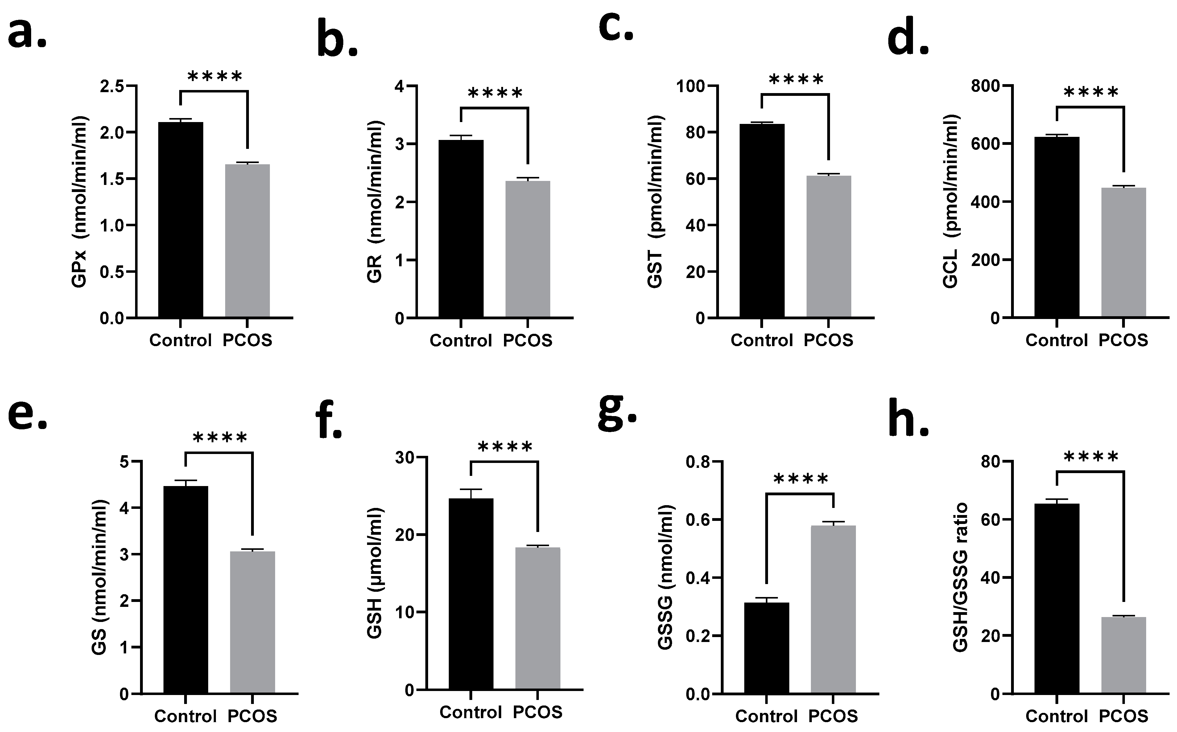
2. Results
3. Discussion
4. Materials and Methods
4.1. Ethical Approval and Study Design
4.2. Collection of Sample and Measurement of Baseline Characteristics
4.3. Serum Te, TI, Os, and Sb Determination
4.4. Assessment of Antioxidant/Redox Status in Serum
4.4.1. Superoxide Dismutase (SOD), Catalase (CAT), Superoxide Anion (SOA), and Hydrogen Peroxide (H2O2)
4.4.2. Lipid Peroxidation (LPO) and Protein Carbonylation (PCC)
4.4.3. Glutathione Status
4.5. Statistical Analysis
5. Conclusions
Author Contributions
Funding
Institutional Review Board Statement
Informed Consent Statement
Data Availability Statement
Acknowledgments
Conflicts of Interest
References
- Tchounwou, P.B.; Yedjou, C.G.; Patlolla, A.K.; Sutton, D.J. Heavy metal toxicity and the environment. Exp. Suppl. 2012, 101, 133–164. [Google Scholar] [CrossRef] [Green Version]
- He, Z.L.; Yang, X.E.; Stoffella, P.J. Trace elements in agroecosystems and impacts on the environment. J. Trace Elem. Med. Biol. 2005, 19, 125–140. [Google Scholar] [CrossRef] [PubMed]
- Mendola, P.; Messer, L.C.; Rappazzo, K. Science linking environmental contaminant exposures with fertility and reproductive health impacts in the adult female. Fertil. Steril. 2008, 89 (Suppl. 2), e81–e94. [Google Scholar] [CrossRef]
- Bloom, M.S.; Louis, G.M.; Sundaram, R.; Kostyniak, P.J.; Jain, J. Associationsbetween blood metals and fecundity among women residing in New York State. Reprod. Toxicol. 2011, 31, 158–163. [Google Scholar] [CrossRef] [PubMed] [Green Version]
- Gong, L.; Yang, Q.; Liu CW, B.; Wang, X.; Zeng, H.L. Assessment of 12 Essential and Toxic Elements in Whole Blood of Pregnant and Non-pregnant Women Living in Wuhan of China. Biol. Trace Elem. Res. 2021, 199, 2121–2130. [Google Scholar] [CrossRef] [PubMed]
- Sengupta, P.; Banerjee, R.; Nath, S.; Das, S.; Banerjee, S. Metals and female reproductive toxicity. Hum. Exp. Toxicol. 2015, 34, 679–697. [Google Scholar] [CrossRef]
- CDC. Third National Report on Human Exposure to Environmental Chemicals 2005. Available online: http://www.jhsph.edu/research/centers-and-institutescenter-for-excellence-in-environmental-health-tracking/Third_Report.pdf (accessed on 12 August 2022).
- Lizneva, D.; Suturina, L.; Walker, W.; Brakta, S.; Gavrilova-Jordan, L.; Azziz, R. Criteria, prevalence, and phenotypes of polycystic ovary syndrome. Fertil. Steril. 2016, 106, 6–15. [Google Scholar] [CrossRef] [PubMed] [Green Version]
- Skrgatic, L.; Baldani, D.P.; Cerne, J.; Ferk, P.; Gersak, K. CAG repeat polymorphism in androgen receptor gene is not directly associated with polycystic ovary syndrome but influences serum testosterone levels. J. Steroid Biochem. Mol. Biol. 2012, 128, 107. [Google Scholar] [CrossRef]
- Coskun, A.; Arikan, T.; Kilinc, M.; Arikan, D.C.; Ekerbiçer, H.C. Plasma selenium levels in Turkish women with polycystic ovary syndrome. Eur. J. Obstetl. Gynecoll. Reprodl. Biol. 2013, 168, 183. [Google Scholar] [CrossRef]
- Abudawood, M.; Tabassum, H.; Alanazi, A.H.; Almusallam, F.; Aljaser, F.; Ali, M.N.; Alenzi, N.D.; Alanazi, S.T.; Alghamdi, M.A.; Altoum, G.H.; et al. Antioxidant Status in Relation to Heavy Metals Induced Oxidative Stress in Patients With Polycystic Ovarian Syndrome (PCOS). Sci. Rep. 2021, 21, 22935. [Google Scholar] [CrossRef]
- Yin, J.; Hong, X.; Ma, J.; Bu, Y.; Liu, R. Serum Trace Elements in Patients With Polycystic Ovary Syndrome: A Systematic Review and Meta-Analysis. Front. Endocrinol. 2020, 11, 572384. [Google Scholar] [CrossRef] [PubMed]
- Zeng, H.L.; Li, H.; Lu, J.; Guan, Q.; Cheng, L. Assessment of 12 Metals and Metalloids in Blood of General Populations Living in Wuhan of China by ICP-MS. Biol. Trace Elem. Res. 2019, 89, 344–353. [Google Scholar] [CrossRef] [PubMed]
- Kirmizi, D.A.; Baser, E.; Turksoy, V.A.; Kara, M.; Yalvac, E.S.; Gocmen, A.Y. Are Heavy Metal Exposure and Trace Element Levels Related to Metabolic and Endocrine Problems in Polycystic Ovary Syndrome? Biol. Trace Elem. Res. 2020, 198, 77–86. [Google Scholar] [CrossRef] [PubMed]
- Yedjou, G.C.; Tchounwou, P.B. In vitro cytotoxic and genotoxic effects of arsenic trioxide on human leukemia cells using the MTT and alkaline single cell gel electrophoresis (comet) assays. Mol. Cell Biochem. 2007, 301, 123. [Google Scholar] [CrossRef] [Green Version]
- Tchounwou, P.B.; Ishaque, A.; Schneider, J. Cytotoxicity and transcriptional activation of stress genes in human liver carcinoma cells (HepG2) exposed to cadmium chloride. Mol. Cell Biochem. 2001, 222, 21. [Google Scholar] [CrossRef]
- Patlolla, A.; Barnes, C.; Yedjou, C.; Velma, V.; Tchounwou, P.B. Oxidative stress, DNA damage and antioxidant enzyme activity induced by hexavalent chromium in Sprague Dawley rats. Environ. Toxicol. 2009, 24, 66. [Google Scholar] [CrossRef] [PubMed] [Green Version]
- Yedjou, G.C.; Tchounwou, P.B. N-acetyl-cysteine affords protection against lead-induced cytotoxicity and oxidative stress in human liver carcinoma (HepG2) cells. Intl. J. Environ. Res. Public Health 2008, 4, 132. [Google Scholar] [CrossRef] [Green Version]
- Kanafchian, M.; Esmaeilzadeh, S.; Mahjoub, S.; Rahsepar, M.; Ghasemi, M. Status of Serum Copper, Magnesium, and Total Antioxidant Capacity in Patients with Polycystic Ovary Syndrome. Biol. Trace Elem. Res. 2020, 193, 111–117. [Google Scholar] [CrossRef]
- Khuwaja, G.; Al-Bratty, M.; Alhazmi, H.A.; Khan, A.; Safhi, M.M.; Ashafaq, M.; Islam, F.; Islam, F.; Taha, M.M. Pharmacological melioration by selenium on the toxicity of tellurium in neuroendocrine centre (pituitary gland) in male wistar rats: A mechanistic approach. Saudi. Pharm. J. 2020, 28, 630–636. [Google Scholar] [CrossRef]
- Liu, J.; Li, N.; Zhang, W.; Wei, X.; Tsang, D.C.W.; Sun, Y.; Luo, X.; Bao, Z.; Zheng, W.; Wang, J.; et al. Thallium con-tamination in farmlands and common vegetables in a pyrite mining city and potentialhealth risks. Environ. Pollut. 2019, 248, 906–915. [Google Scholar] [CrossRef]
- Liu, J.; Luo, X.; Wang, J.; Xiao, T.; Chen, D.; Sheng, G.; Yin, M.; Lippold, H.; Wang, C.; Chen, Y. Thallium con-tamination in arable soils and vegetables around a steel plant-A newly-found sig-nificantsource of Tl pollution in South China. Environ. Pollut. 2017, 224, 445–453. [Google Scholar] [CrossRef] [PubMed]
- Ghaderi, A.; Vahdati-Mashhadian, N.; Oghabian, Z.; Moradi, V.; Afshari, R.; Mehrpour, O. Thallium exists in opioid poisoned patients. DARU J. Pharm. Sci. 2015, 23, 39. [Google Scholar] [CrossRef] [PubMed] [Green Version]
- Peter, A.L.J.; Viraraghavan, T. Thallium: A review of public health and environmental concerns. Environ. Int. 2005, 31, 493–501. [Google Scholar] [CrossRef] [PubMed]
- Rutkowska, A.Z.; Diamanti-Kandarakis, E. Polycystic ovary syndrome and environmental toxins. Fertil. Steril. 2016, 106, 948–958. [Google Scholar] [CrossRef] [PubMed] [Green Version]
- Sundar, S.; Chakravarty, J. Antimony toxicity. Int. J. Environ. Res. Public Health 2010, 7, 4267–4277. [Google Scholar] [CrossRef] [PubMed]
- Rebolledo, J.; Fierens, S.; Versporten, A.; Brits, E.; De Plaen, P.; Van Nieuwenhuyse, A. Human biomonitoring on heavy metals in Ath: Methodological aspects. Arch. Public Health 2011, 69, 10. [Google Scholar] [CrossRef] [PubMed] [Green Version]
- Cunha, R.L.O.R.; Gouvea, I.E.; Juliano, L. A glimpse on biological activities of Tellurium compounds. An. Da Acad. Bras. De Cienc. 2009, 81, 393–407. [Google Scholar] [CrossRef] [Green Version]
- Belzile, N.; Chen, Y.-W. Tellurium in the environment: A critical review focused on natural waters, soils, sediments and airborne particles. Appl. Geochem. 2015, 63, 83–92. [Google Scholar] [CrossRef]
- Burkholz, T.; Jacob, C. Tellurium in Nature. In Encyclopedia of Metalloproteins; Kretsinger, R.H., Uversky, V.N., Permyakov, E.A., Eds.; Springer: New York, NY, USA, 2013. [Google Scholar] [CrossRef]
- Sulaiman, M.A.H.; Al-Farsi, Y.M.; Al-Khaduri, M.M.; Saleh, J.; Waly, M.I. Polycystic ovarian syndrome is linked to increased oxidative stress in Omani women. In.t J. Women’s Health 2018, 10, 763–771. [Google Scholar] [CrossRef]
- Agency for Toxic Substances and Disease Registry. Toxicological Profile for Cadmium; Agency for Toxic Substances and Disease Registry: Atlanta, GA, USA, 2012.
- Cheam, V. Thallium contamination of water in Canada. Water Qual. Res. J. Can. 2001, 36, 851–877. [Google Scholar] [CrossRef]
- Campanella, B.; Onor, M.; D’Ulivo, A.; Giannecchini, R.; D’Orazio, M.; Petrini, R.; Bramanti, E. Human exposure to thallium through tap water: A study from Valdicastello Carducci and Pietrasanta (northern Tuscany, Italy). Sci. Total Environ. 2016, 548–549, 33–42. [Google Scholar] [CrossRef] [PubMed]
- Nriagu, J.O. Thallium. Chem. Eng. News 2003, 81, 153. [Google Scholar] [CrossRef]
- Agency for Toxic Substances and Disease Registry: Toxicological profile for thallium. US Department of Health and Human Services; July 1992. Available online: www.atsdr.cdc.gov/ToxProfiles/tp54.pdf (accessed on 12 August 2022).
- Ma, L.Y.; Liang, C.M.; Yan, S.Q.; Huang, K.; Chen, M.L.; Tao, F.B. Association of thallium exposure during pregnancy with maternal blood pressure and hypertensive disorder complicating pregnancy. Zhonghua Yu Fang Yi Xue Za Zhi 2021, 55, 646–652. (In Chinese) [Google Scholar] [CrossRef] [PubMed]
- Tabassum, H.; Alrashed, M.; Malik, A.; Alanazi, S.T.; Alenzi, N.D.; Ali, M.N.; AlJaser, F.S.; Altoum, G.H.; Hijazy, S.M.; Alfadhli, R.A.; et al. A unique investigation of thallium, tellurium, osmium, and other heavy metals in recurrent pregnancy loss: A novel approach. Int. J. Gynaecol. Obstet. 2022. [Google Scholar] [CrossRef]
- Hanzel, C.E.; Verstraeten, S.V. Thallium induces hydrogen peroxide generation by impairing mitochondrial function. Toxicol. Appl. Pharmacol. 2006, 216, 485–492. [Google Scholar] [CrossRef] [PubMed]
- Komarova, T.; McKeating, D.; Perkins, A.V.; Tinggi, U. Trace Element Analysis in Whole Blood and Plasma for Reference Levels in a Selected Queensland Population, Australia. Int. J. Environ. Res. Public Health 2021, 18, 265. [Google Scholar] [CrossRef] [PubMed]
- Potkonjak, V.; Pavlovich, M. Antimoniosis: A particular form of pneumoconiosis. I. Etiology, clinical and x-ray findings. Int. Arch. Occup. Environ. Health 1983, 51, 199–207. [Google Scholar] [CrossRef]
- Belyaeva, A.P. The effect of antimony on reproduction. Gig. Truda. Prof. Zabol. 1967, 11, 32. [Google Scholar]
- Chen, Y.W.; Yang, C.Y.; Huang, C.F.; Hung, D.Z.; Leung, Y.M.; Liu, S.H. Heavy metals, islet function and diabetes development. Islets 2019, 1, 169–176. [Google Scholar] [CrossRef] [Green Version]
- Friedova, N.; Pelclova, D.; Bures, Z.; Krijt, J.; Kohout, P. Response to the questions related to the article: Osmium absorption after osmium tetroxide skin and eye exposure. Basic Clin. Pharmacol. Toxicol. 2021, 128, 555–556. [Google Scholar] [CrossRef]
- Rotterdam ESHRE/ASRM-Sponsored PCOS Consensus Workshop group. Revised 2003 consensus on diagnostic criteria and long-term health risks related to polycystic ovary syndrome (PCOS). Hum. Reprod. 2004, 19, 41–47. [Google Scholar] [CrossRef] [Green Version]
- de Haan, J.B.; Cristiano, F.; Iannello, R.; Bladier, C.; Kelner, M.J.; Kola, I. Elevation in the ratio of Cu/Zn-superoxide dismutase to glutathione peroxidase activity induces features of cellular senescence and this effect is mediated by hydrogen peroxide. Hum. Mol. Genet. 1996, 5, 283–292. [Google Scholar] [CrossRef] [PubMed]
- Ghneim, H.K.; Al-Sheikh, Y.A.; Alshebly, M.M.; Aboul-Soud, M.A. Superoxide dismutase activity and gene expression levels in Saudi women with recurrent miscarriage. Mol. Med. Rep. 2016, 13, 2606–2612. [Google Scholar] [CrossRef] [PubMed] [Green Version]
- Akiel, M.; Alsughayyir, J.; Basudan, A.M.; Alamri, H.S.; Dera, A.; Barhoumi, T.; Al Subayyil, A.M.; Basmaeil, Y.S.; Aldakheel, F.M.; Alakeel, R.; et al. Physcion Induces Hemolysis and Premature Phosphatidylserine Externalization in Human Erythrocytes. Biol. Pharm. Bull. 2021, 44, 372–378. [Google Scholar] [CrossRef] [PubMed]
- Ghneim, H.K.; Alfhili, M.A.; Alharbi, S.O.; Alhusayni, S.M.; Abudawood, M.; Al-Sheikh, Y.A. Biochemical and molecular assessment of selenium forms for the alleviation of oxidative stress in senescent human fibroblasts. Gen. Physiol. Biophys. 2022, 41, 309–318. [Google Scholar] [CrossRef] [PubMed]
- Ghneim, H.K.; Alfhili, M.A.; Alharbi, S.O.; Alhusayni, S.M.; Abudawood, M.; Aljaser, F.S.; Al-Sheikh, Y.A. Comprehensive investigations of key mitochondrial metabolic changes in senescent human fibroblasts. Korean J. Physiol. Pharmacol. Off. J. Korean Physiol. Soc. Korean Soc. Pharmacol. 2022, 26, 263–275. [Google Scholar] [CrossRef] [PubMed]
- Ghneim, H.K. The effect of Echis coloratus venom on biochemical and molecular markers of the antioxidant capacity in human fibroblasts. Libyan J. Med. 2017, 12, 1304515. [Google Scholar] [CrossRef] [PubMed] [Green Version]
- Reznick, A.Z.; Packer, L. Oxidative damage to proteins: Spectrophotometric method for carbonyl assay. Methods Enzymol. 1994, 233, 357–363. [Google Scholar] [CrossRef]
- Al-Sheikh, Y.A.; Ghneim, H.K.; Alharbi, A.F.; Alshebly, M.M.; Aljaser, F.S.; Aboul-Soud, M.A. Molecular and biochemical investigations of key antioxidant/oxidant molecules in Saudi patients with recurrent miscarriage. Exp. Ther. Med. 2019, 18, 4450–4460. [Google Scholar] [CrossRef] [PubMed]
- Aljaser, F.S.; Ghneim, H.K.; ALshubaily, M.M.; Abudawood, M.; Almajed, F.; Fatima, S.; ALsheikh, Y.A. Glutathione and oxidized nicotinamide adenine dinucleotide redox status in plasma and placental tissue of Saudi patients with intrauterine growth restriction. Saudi Med. J. 2021, 42, 491–498. [Google Scholar] [CrossRef]


| Control | PCOS | p | |
|---|---|---|---|
| Age | 29.16 ± 6.2 | 30.41 ± 6.8 | NS |
| BMI | 25.0 ± 6.08 | 27.23 ± 5.0 | NS |
| Married | 30 | 24 | |
| Thallium (ppb) | 1.41 ± 0.4 | 12.69 ± 1.05 | <0.001 |
| Tellurium (ppb) | 1.32 ± 0.46 | 12.33 ± 1.31 | <0.001 |
| Osmium (ppb) | 1.51 ± 0.45 | 13.0 ± 0.97 | <0.001 |
| Antimony (ppb) | 1.89 ± 0.31 | 2.50 ± 0.23 | <0.001 |
| TAC (µmol/L) | 687.00 ± 11.80 | 640.00 ± 22.0 | <0.001 |
| Te-PCOS r (p) | TI-PCOS r (p) | Sb-PCOS r (p) | Os-PCOS r (p) | |
|---|---|---|---|---|
| TAC-PCOS | −0.132 (0.036) * | −0.210 (0.014) * | −0.142 (0.032) * | −0.194 (0.01) * |
| Te-PCOS | 0.49 (<0.001) ** | 0.53 (<0.0001) *** | 0.480 (<0.001) ** | |
| TI-PCOS | 0.72 (<0.0001) *** | 0.71 (NS) | ||
| Sb-PCOS | 0.53 (<0.001) ** |
Disclaimer/Publisher’s Note: The statements, opinions and data contained in all publications are solely those of the individual author(s) and contributor(s) and not of MDPI and/or the editor(s). MDPI and/or the editor(s) disclaim responsibility for any injury to people or property resulting from any ideas, methods, instructions or products referred to in the content. |
© 2023 by the authors. Licensee MDPI, Basel, Switzerland. This article is an open access article distributed under the terms and conditions of the Creative Commons Attribution (CC BY) license (https://creativecommons.org/licenses/by/4.0/).
Share and Cite
Abudawood, M.; Alnuaim, L.; Tabassum, H.; Ghneim, H.K.; Alfhili, M.A.; Alanazi, S.T.; Alenzi, N.D.; Alsobaie, S. An Insight into the Impact of Serum Tellurium, Thallium, Osmium and Antimony on the Antioxidant/Redox Status of PCOS Patients: A Comprehensive Study. Int. J. Mol. Sci. 2023, 24, 2596. https://doi.org/10.3390/ijms24032596
Abudawood M, Alnuaim L, Tabassum H, Ghneim HK, Alfhili MA, Alanazi ST, Alenzi ND, Alsobaie S. An Insight into the Impact of Serum Tellurium, Thallium, Osmium and Antimony on the Antioxidant/Redox Status of PCOS Patients: A Comprehensive Study. International Journal of Molecular Sciences. 2023; 24(3):2596. https://doi.org/10.3390/ijms24032596
Chicago/Turabian StyleAbudawood, Manal, Lulu Alnuaim, Hajera Tabassum, Hazem K. Ghneim, Mohammad A. Alfhili, Samyah T. Alanazi, Naif D. Alenzi, and Sarah Alsobaie. 2023. "An Insight into the Impact of Serum Tellurium, Thallium, Osmium and Antimony on the Antioxidant/Redox Status of PCOS Patients: A Comprehensive Study" International Journal of Molecular Sciences 24, no. 3: 2596. https://doi.org/10.3390/ijms24032596