Article

Unveiling New Druggable Pockets in Influenza Non-Structural Protein 1: NS1–Host Interactions as Antiviral Targets for Flu

by
Andreia E. S. Cunha
1,†,
Rui J. S. Loureiro
1,*,†,
Carlos J. V. Simões
1,2 and
Rui M. M. Brito
1,2,*
1
Coimbra Chemistry Center—Institute of Molecular Sciences (CQC-IMS), Department of Chemistry, University of Coimbra, 3004-535 Coimbra, Portugal
2
BSIM Therapeutics, Instituto Pedro Nunes, 3030-199 Coimbra, Portugal
*
Authors to whom correspondence should be addressed.
These authors contributed equally to this work.
Int. J. Mol. Sci. 2023, 24(3), 2977; https://doi.org/10.3390/ijms24032977
Submission received: 10 December 2022 / Revised: 19 January 2023 / Accepted: 1 February 2023 / Published: 3 February 2023
(This article belongs to the Special Issue Advanced Antiviral and Antimicrobial Drug Discovery)

Abstract

:
Influenza viruses are responsible for significant morbidity and mortality worldwide in winter seasonal outbreaks and in flu pandemics. Influenza viruses have a high rate of evolution, requiring annual vaccine updates and severely diminishing the effectiveness of the available antivirals. Identifying novel viral targets and developing new effective antivirals is an urgent need. One of the most promising new targets for influenza antiviral therapy is non-structural protein 1 (NS1), a highly conserved protein exclusively expressed in virus-infected cells that mediates essential functions in virus replication and pathogenesis. Interaction of NS1 with the host proteins PI3K and TRIM25 is paramount for NS1’s role in infection and pathogenesis by promoting viral replication through the inhibition of apoptosis and suppressing interferon production, respectively. We, therefore, conducted an analysis of the druggability of this viral protein by performing molecular dynamics simulations on full-length NS1 coupled with ligand pocket detection. We identified several druggable pockets that are partially conserved throughout most of the simulation time. Moreover, we found out that some of these druggable pockets co-localize with the most stable binding regions of the protein–protein interaction (PPI) sites of NS1 with PI3K and TRIM25, which suggests that these NS1 druggable pockets are promising new targets for antiviral development.

1. Introduction

Influenza (flu) is an acute contagious respiratory illness resulting from infection with influenza viruses. Influenza infections represent a serious public health threat worldwide, with winter seasonal outbreaks being responsible for 1 billion cases, 3 to 5 million severe cases, and 290,000 to 650,000 deaths each year [1,2]. Moreover, the influenza virus poses a permanent risk of the emergence of severe flu pandemics, which, as stated by the WHO, are a permanent threat to become a devastating global health event [2]. Besides its serious health impacts, influenza infections also represent a significant economic burden [3], estimated at 11.2 billion dollars yearly in the U.S. [4] and 6 to 14 billion euros yearly in the EU [5].
Influenza’s most common clinical manifestations include fever, cough, sore throat, rhinorrhoea, headache, myalgia, and weakness, and, despite being usually mild in healthy adults, they can be severe and fatal to high-risk groups such as the elderly, children under 59 months and patients with chronic diseases or immunosuppression [6,7,8]. Moreover, healthy adults are also at risk during pandemic outbreaks [9].
Influenza viruses belong to the family Orthomyxoviridae, have a negative sense eight-segment single-stranded RNA genome encoding 11 proteins, and are divided into types A, B, C, and D, of which only A, B, and C infect humans (reviewed in [10]). Influenza viruses A and B are pleomorphic, generally presenting a spherical shape and sizes in the range of 80–120 nm, although cord-like shapes can also occur [11]. Influenza C viruses usually have a filamentous shape with sizes reaching 500 nm [11]. Human influenza A and B viruses are responsible for seasonal flu outbreaks, while influenza C usually causes mild infections. Influenza A is the only type of influenza virus responsible for the emergence of flu pandemics and is the most pathogenic for the human population, being, therefore, the main target of influenza vaccines. Type A influenza viruses are divided into multiple subtypes based on the combination of two proteins on the viral surface: hemagglutinin (HA) and neuraminidase (NA), which have, respectively, 18 and 11 subtypes.
Influenza viruses have a high rate of evolution that arises from two main processes: antigenic drift and antigenic shift (reviewed in [10,12]). Antigenic drift is the gradual accumulation of changes in the viral genome over time that originates a virus of the same subtype but is unrecognizable by the host immune system and results from the lack of proofreading ability in RNA viruses. Antigenic shift or reassortment occurs in influenza A viruses (IAV) and is characterized by the mixture and rearrangement of RNA segments from different IAV subtypes infecting the same cell, originating a new subtype for which most of the population does not have preexisting immunity. This process leads to new pandemics when the newly generated subtype is successful at human-to-human transmission [13]. The diversity of virus strains and subtypes arising from these two processes significantly reduces vaccine effectiveness and leads to the yearly update of vaccines accordingly to the strains circulating each flu season [14].
Besides vaccination, the other main strategy to counteract influenza infections is the use of antiviral drugs. However, the high rate of mutation of the virus has led to increasing levels of drug resistance and therapeutic inefficacy [11,15]. Indeed, there is currently only one class of drugs (i.e., neuraminidase inhibitors) being used in the clinic [8,11], which makes the development of new antivirals against novel viral biological targets an urgent need.
One of the most promising new targets for influenza antiviral therapy is the non-structural protein 1 (NS1), a highly conserved protein exclusively expressed in virus-infected cells that mediates essential functions in virus replication and pathogenesis [10,16]. NS1 role in influenza infection results from two main processes: promotion of viral replication and evasion of the host immune system [16]. Several studies [17,18] have shown that influenza viruses without NS1 suffer a decrease in their replication and dissemination, which suggests that the development of new therapies targeting this viral protein could improve the efficacy and outcomes of influenza antiviral treatment.
The NS1 structure comprises an RNA-binding domain (RBD), responsible for binding the viral RNA, connected through a variable length flexible linker to the effector domain (ED), responsible for interacting with multiple host proteins, and a disordered C-terminal tail [10]. NS1 has a high structural plasticity, which allows it to establish multiple functionally relevant protein–protein interactions (PPI) with host cell proteins, mainly through the ED domain, such as with CPSF30 [17], TRIM25 [19] and PI3K [20].
NS1 interaction with PI3K is of paramount importance in the early stages of infection. PI3K is a lipid kinase involved in the PI3K/Akt signaling pathway, which regulates key cellular processes such as cell survival, apoptosis, proliferation, and differentiation and exerts pro-survival and antiapoptotic effects [21,22]. The activation of PI3K induced by NS1 binding leads to the inhibition of apoptosis in influenza virus-infected cells, thus promoting viral replication [23,24,25]. Additionally, this interaction could also have other cellular effects dependent on the PI3K spatial distribution and on the activation of distinct downstream pathways [26,27].
NS1 interaction with TRIM25 also plays an essential role in influenza infection and pathogenesis. TRIM25 is a member of the TRIM protein family of RING-type E3 ligases that play crucial roles in the regulation of the innate immune signaling pathways responsible for the induction of the type I interferon (IFN) response and for the production of inflammatory cytokines [19,28,29]. Specifically, upon the interaction of the viral RNA sensing RIG-I protein with viral RNA, TRIM25 poly-ubiquitinates the second CARD domain of RIG-I, leading to the activation of the downstream effector mitochondrial antiviral signaling (MAVS) protein and to the subsequent production of interferon α/β [30,31]. The binding of NS1 to the coiled-coil domain of TRIM25 prevents the correct positioning of the PRYSPY domain of TRIM25 required for substrate ubiquitination, hence suppressing the RIG-I/MAVS signaling pathway and interferon production [19]. Additionally, it was recently reported that NS1 also inhibits the TRIM25-mediated ubiquitination of another viral sensing protein, DDX3X, leading to the suppression of IFN production [32], raising the possibility of other effects of the NS1-TRIM25 binding besides the blockage of RIG-I ubiquitination.
Considering the crucial role of the NS1 interactions with PI3K and TRIM25 on influenza infection and pathogenesis and the fact that the interaction surfaces of NS1 with both proteins overlap, in the current work, we explored the potential of these two PPIs to be targets of new antiviral agents.
With the help of in silico methodologies and advances in hardware and computer performance, rational drug discovery has advanced at a breakneck pace. While still in its infancy in comparison to traditional drug discovery methodologies, computer-aided drug discovery (CADD) has aided in the delivery of several success stories [33,34,35,36] that have instilled confidence in its continued use in the pharmaceutical sector. Before entering detailed experimental assays, CADD can save time and money by identifying probable hits for a therapeutic target [37].
A thorough effort on target validation and characterization is required before beginning a drug discovery effort on a new biological target, such as a protein [38]. Because proteins are dynamic and can undergo multiple conformational changes, one important aspect of structure-based ligand design is to assess the protein conformation dynamics of the biological target. Not only major conformational changes but also minor conformational adjustments involving residue side chain movements can impact the complementarity between the ligand and the protein’s binding site. Because protein flexibility influences the variety of possible target conformational states for ligand binding, MD simulations can help in drug design by providing information on the target’s dynamic character [39].
Despite significant progress in the experimental and computational approaches used in the drug discovery process over the years, a remarkable overall failure rate of about 96% has been recorded in various drug discovery projects due to the “undruggability” of various identified protein targets among other challenges [40,41,42]. The target “druggability” (the ability of a biological target to be modulated through binding to a drug-like molecule) is one important feature to be evaluated at the early stages of the drug discovery process.
The goal of the present study was to evaluate the druggability of NS1 by considering the structure’s intrinsic flexibility. Using the three-dimensional structure of full-length NS1, we investigated the structure’s stability and flexibility using multiple molecular dynamics (MD) simulations. Additionally, we looked at the structure’s ability to generate potential binding pockets for small molecules, specifically in the regions of PI3K and TRIM25 binding interfaces. We also calculated the druggability potential of the conserved pockets, or their ability to bind drug-like ligands, as determined by Fpocket [43]. This allowed us to identify promising druggable pockets and to confirm the potential of the NS1’s effector domain (ED) as a drug target.

2. Results and Discussion

2.1. NS1 Structure’s Dynamics

NS1 is preferentially a homodimer with 230 residues per polypeptide chain organized in two structural domains: the RNA-binding domain (RBD) and the effector domain (ED) (Figure 1). The RBD is formed by residues 1–73 and in solution tends to dimerize. Each RBD chain is linked to the effector domain (residues 81–230) by a flexible linker region. As a result of the linker length and structural flexibility, the orientation of the two effector domains relative to each other and to the RBD may vary significantly [44,45].
Although it is well known that NS1 has high conformational plasticity with concerns to the spatial relation between the RBD (RNA binding) and ED (effector) structural domains, our goal in the present work is to access the potential presence of stable and/or transient pockets at the surface of the protein that could be exploited as binding sites for small molecule modulators of NS1 activity. With this objective in mind, we carried out a series of relatively short (100 ns) molecular dynamics (MD) simulations of full-length NS1 and two molecular complexes of NS1: (i) ED-NS1 with the iSH2 domain of the human p85β subunit of PI3K; and (ii) NS1 with the coiled-coil (CC) domain of human TRIM25 (for more details, see Section 3).
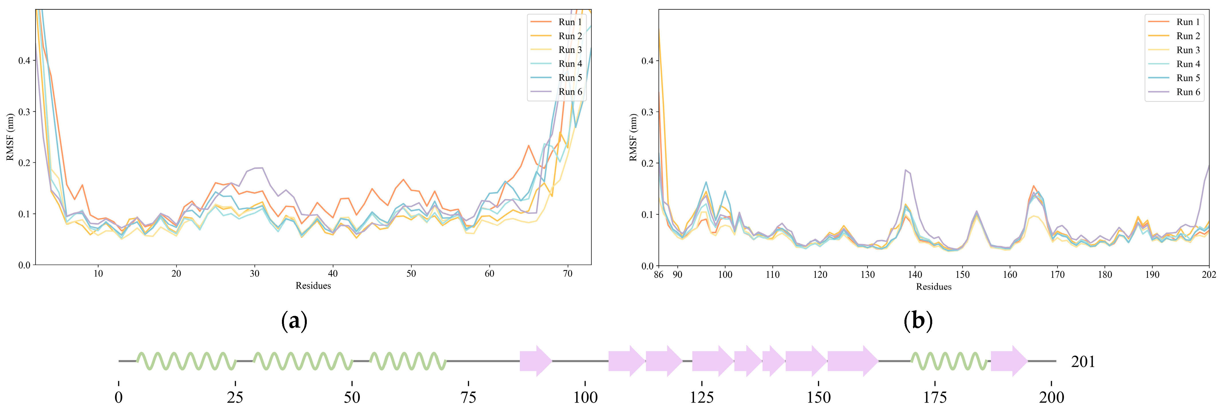
To evaluate the backbone stability and conformational fluctuations of NS1’s structural domains, six replicas of 100 ns molecular dynamics (MD) simulations of the full-length (FL) NS1 were run under the physiologically relevant conditions of 310 K and pH 7.0. The root mean square fluctuation (RMSF) of backbone Cα atoms for all the simulations was computed and plotted (Figure 2), showing that both domains of NS1 are very stable during the molecular dynamics simulations, and even more so the effector domain. The NS1 regions of higher conformational variability are the N-terminal region (RBD) and the linker region. In the case of RBD, the region with higher flexibility occurs in an inter-helical loop between residues 24 and 31, while for the ED, the regions with higher flexibility are those in the vicinity of residues 138, 153, and 165, which are all loop regions. It is thus clear that, if considered individually, each one of the two globular domains of NS1 does not suffer major conformational changes.

2.2. Pocket Identification and Analysis

To assess the potential of NS1 as a drug target for new influenza antivirals, we evaluated the formation of stable or transient druggable pockets on the surface of NS1 along the MD trajectories using the Fpocket algorithm, as described in Section 3.4. A druggability score was assigned to each pocket identified on the NS1 surface (Figure 3). The druggability score ranges between 0 and 1, with 1 meaning that drug-like small molecules can bind to the identified pocket with a high probability and 0 meaning that the pocket is non-druggable or unlikely to interact with a drug-like small molecule.
As can be seen in Figure 3, there is a small number of druggable pockets per conformation sampled (each vertical line in the graphs of Figure 3), and these pockets fluctuate between druggable and non-druggable states in consecutive protein conformations sampled at one-nanosecond intervals. We selected the pockets with a druggability score above 0.5, the minimum consensus cutoff for a pocket to be considered druggable, to analyze the residues comprising the pockets and the associated molecular descriptors.
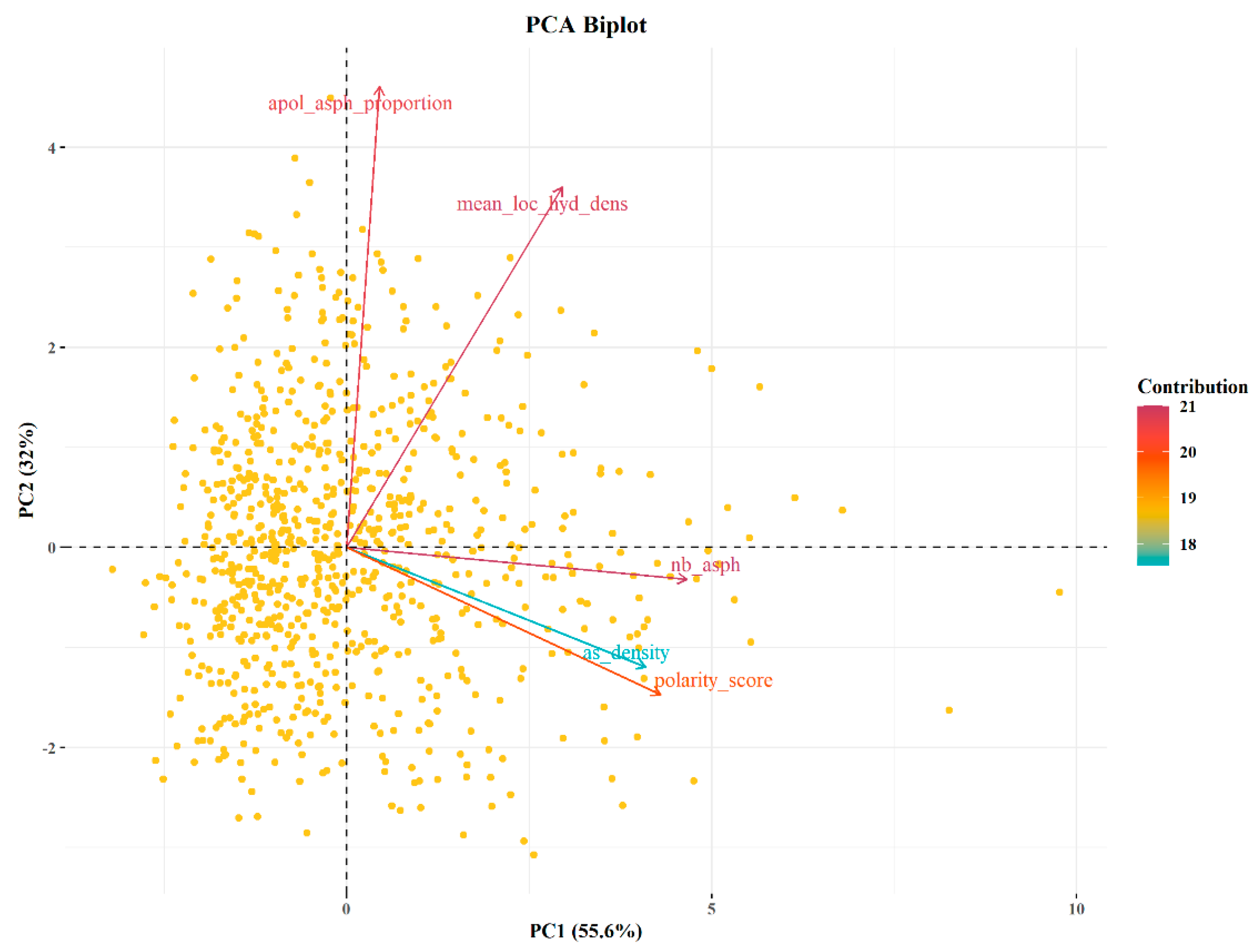
Fpocket calculates the druggability score of a pocket using five different descriptors: (i) the number of alpha spheres fitting the pocket, (ii) the density of the cavity, (iii) the polarity score, (iv) the mean local hydrophobic density, and (v) the proportion of apolar alpha spheres fitting the pocket. Using principal component analysis (PCA), we examined the relationship between the druggability score and the five descriptors. The descriptors were projected onto the first plane of a PCA, which captures 87.64% of the variability (Figure 4). The PCA of the five descriptors reveals that the number of alpha spheres fitting the pocket and the mean local hydrophobic density contribute the most to the druggability score. The number of alpha spheres is related to the size of the cavity, and the mean hydrophobic density identifies the hydrophobic character of the pocket.
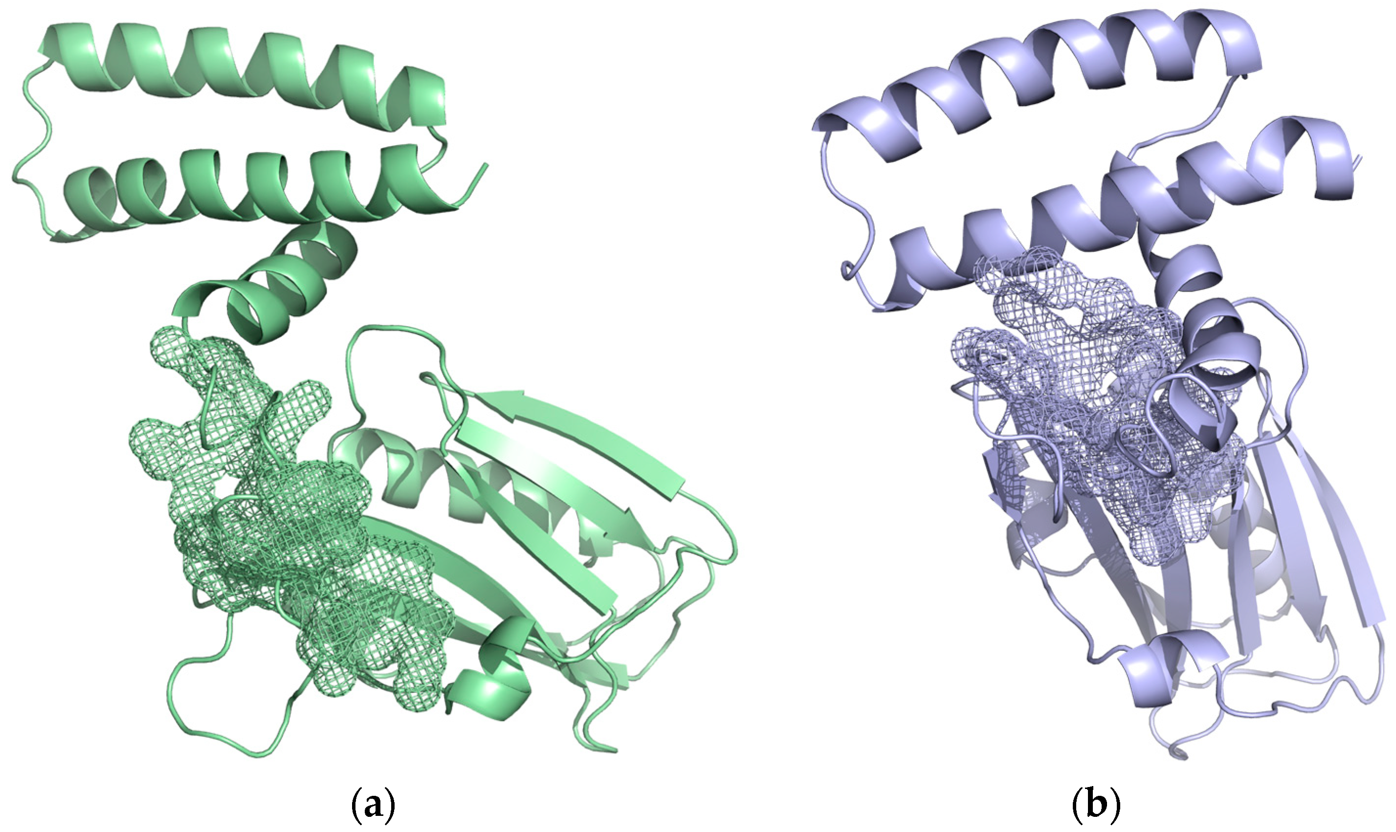
For a more in-depth analysis of the pockets, we analyzed the residues belonging to the pockets with the highest druggability score (Figure 5). The residues belonging to the pocket identified in Figure 5a are R88, Y89, L95, M98, S135, V136, I137, E142, T143, and I145, and the residues belonging to the pocket identified in Figure 5b are A86, S87, S135, E142, T143, L144, I145, and P162.
The residues in the interface of the ED-NS1 with PI3K described in the literature are S87, R88, Y89, T91, M93, L95, E96, E97, M98, S99, R100, D101, W102, C116, R118, N133, F134, S135, E142, T143, L144, I145, L146, R148, E159, S161, P162, L163, P164, and S165 [24,26,46,47,48]. Most of the residues identified in the pockets are located in the interaction surfaces with PI3K and TRIM25 or in their proximity.

2.3. Characterization of Interactions between Influenza NS1 and Human PI3K or TRIM25

We conducted classical MD simulations of the complex between the ED domain of NS1 from the 1918 pandemic H1N1 influenza A virus and the iSH2 domain of the p85β subunit of human PI3K (PDB ID: 6OX7) and analyzed its structural stability by computing the root mean square deviation (RMSD) for both the complex and the interfacial region as well as the root mean square fluctuation (RMSF) of the interfacial region for all frames of the trajectories. The same procedure was applied for the complex between NS1 from the A/Puerto Rico/8/1934 strain of H1N1 influenza A virus and the coiled-coil (CC) domain of human TRIM25.
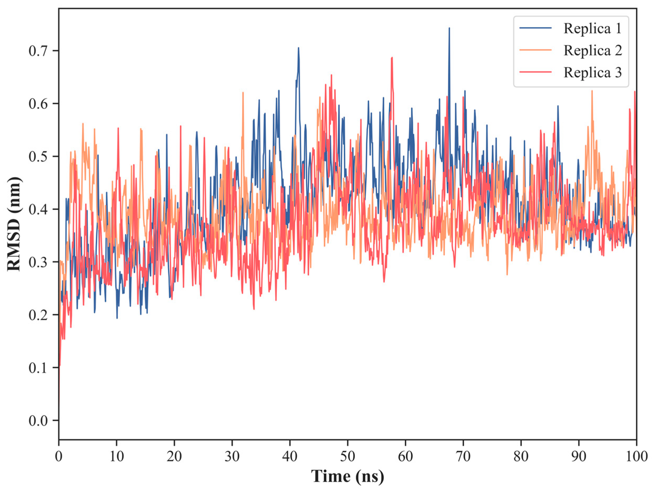
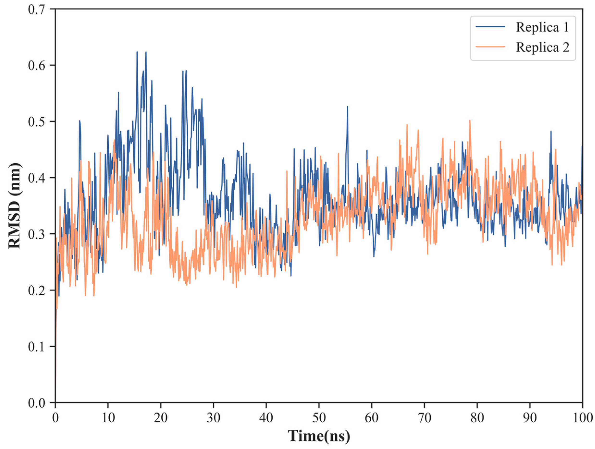
The Cα-RMSD for the complex between ED-NS1 and PI3K (Figure 6) shows a very stable structure, presenting low and constant RMSD values along the three trajectories. Indeed, the average RMSD values are 4.1 ± 0.9 Å in replica 1, 3.9 ± 0.6 Å in replica 2, and 3.7 ± 0.9 Å in replica 3.
A similar trend is observed in the complex between NS1 and TRIM25, with the average RMSD over the Cα atoms ranging from 3.3 ± 0.6 Å in replica 2 to 3.6 ± 0.7 Å in replica 1 (Figure 7).
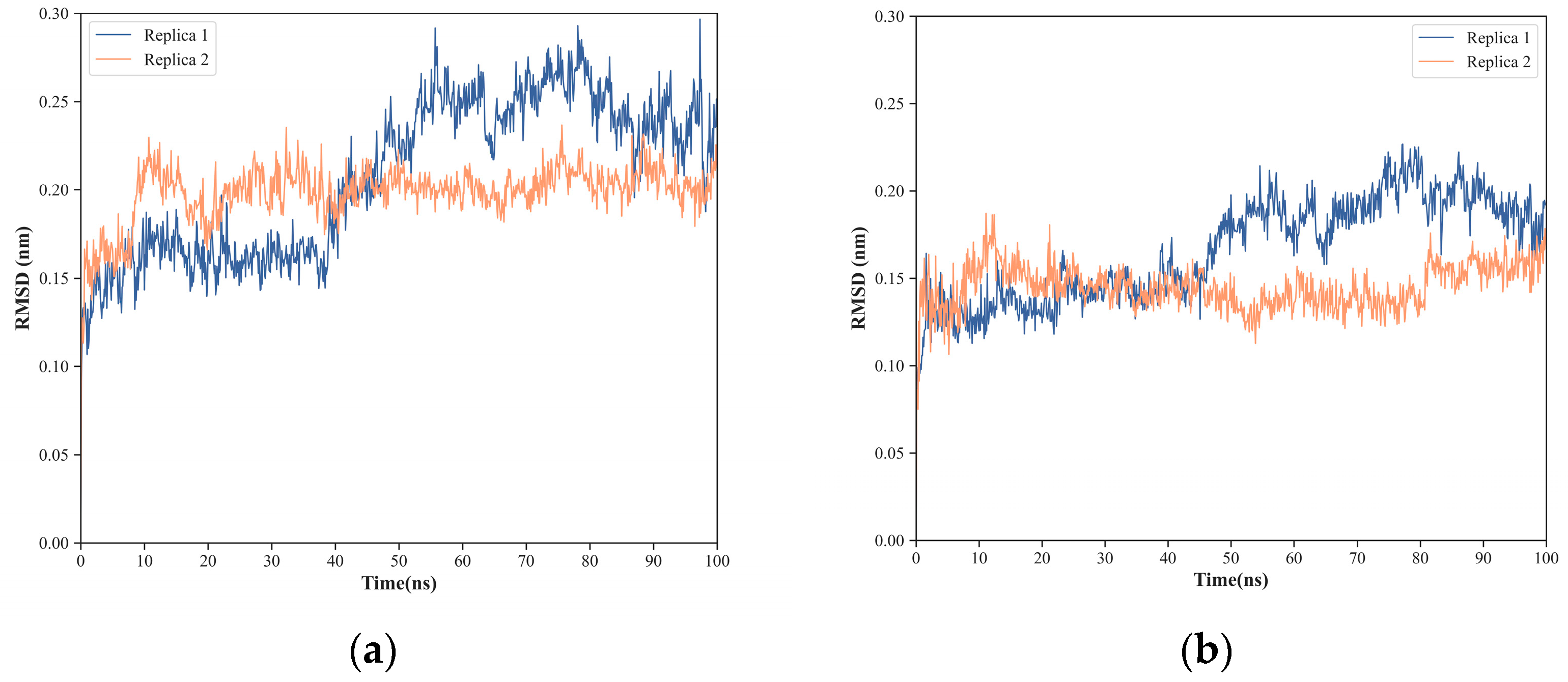
To study in more detail the protein–protein interfacial region for each of the complexes, we defined a set of residues that fulfill the criteria described in Section 3.3 and correspond to 30 NS1 residues and 20 PI3K residues in the case of the NS1-PI3K complex (Table 1) and to 22 NS1 residues and 24 TRIM25 residues in the case of the NS1-TRIM25 complex (Table 2). These are essential regions both to determine the stability of the protein–protein interactions and from a drug design perspective. In both cases, the low RMSD values indicate globally high stability of the interfaces (Figure 8a,b and Figure 9a,b). Nevertheless, a relatively small increase in RMSD (of about 1 Å) is observed for the NS1 and TRIM25 interfacial residues between the 40 and 50 ns of simulation, which denotes that the interface still has some local flexibility despite its overall stability. Indeed, the average RMSD of the interface of both NS1 and PI3K varies from 2.0 ± 0.2 Å in replica 1 to 2.6 ± 0.3 Å in replica 2, while the average RMSD of the interaction surface of NS1 varies between 1.8 ± 0.3 Å in replica 1 and 2.3 ± 0.2 Å in replica 2. Similarly, the average RMSD of the interface of both NS1 and TRIM25 varies from 2.0 ± 0.2 Å in replica 2 to 2.1 ± 0.4 Å in replica 1, while the average RMSD of the interaction surface of NS1 varies between 1.4 ± 0.1 Å in replica 2 and 1.7 ± 0.3 Å in replica 1.
Furthermore, the RMSF values of the interfacial residues of NS1 in the NS1-PI3K and NS1-TRIM25 complexes show very low flexibility, with average values ranging from 1.0 ± 0.5 Å in replica 1 to 1.1 ± 0.5 Å in replica 3 for the NS1-PI3K complex (Figure 10a) and from 0.8 ± 0.4 Å in replica 2 to 0.9 ± 0.5 Å in replica 1 for the NS1-TRIM25 complex (Figure 10b). Additionally, most of the NS1 interfacial residues on the two complexes present RMSF values lower than 1.0 Å, which further indicates the high stability of the interaction surfaces.
The analysis of conformational fluctuations in the interfacial regions of the complexes NS1/PI3K and NS1/TRIM25 showed very stable interfaces over the course of the MD simulations. The stability of the interactions between influenza’s NS1 protein and the human PI3K and TRIM25 proteins, along with their recognized biological significance in the mechanisms of infection and pathogenesis, suggest that they may be suitable drug targets for new influenza antivirals if they co-localize with druggable pockets.

2.4. Binding of NS1 to PI3K and TRIM25 Is Mostly Driven by the Same Six Hotspot Residues

To further explore the type and most prevalent molecular interactions involved in influenza’s NS1 binding to its molecular partners PI3K and TRIM25, we conducted a fine-grained description, over the simulation time, of the preservation of the interfacial pairwise residue–residue contacts and identified the residues that participate to a larger extent on highly conserved residue–residue contacts. This analysis is essential to identify the main inter-residue contacts at the molecular complexes interfaces and the essential residues in complex formation and stabilization (i.e., interaction hotspots).
The analysis of the preservation of the pairwise residue–residue contacts at the interfaces was performed with the MDCons standalone tool [49] and, considering the similarity of the trajectories for each complex and for the sake of simplicity, was restricted to one trajectory per complex. The criterion used to define a residue–residue interfacial contact considers that two interfacial residues are in contact if any of the atoms of one residue is within 5 Å of any atom of the second residue in the interaction partner (the criteria used in CAPRI [50]). In the case of the NS1-PI3K complex, the analysis indicated that 19 inter-residue contacts are completely conserved during the 100 ns of simulation, and the other 29 contacts have more than 70% conservation (Table 3 and Figure 11a), which corresponds to a proportion of 67.74% of the total number of inter-residue contacts in all frames, underlining the stability of the interface. The highly conserved interfacial contacts cluster mainly in the region 87–101, followed by smaller clusters in regions 133–135, 142–148, and 161–163.
Concerning the NS1-TRIM25 complex, we observed that 33 inter-residue contacts are completely conserved during the 100 ns of simulation, and 53 have more than 70% conservation (Figure 11b,c and Table 4 and Table 5), corresponding to proportions of 59.02% and 62.50% of the total number of inter-residue contacts involving the chains I and N of TRIM25 in all frames, respectively, underlining the stability of the interface. The highly conserved interfacial contacts cluster mainly in the region 87–101, followed by smaller clusters in regions 133–135, 145–148, and 161–164.
The number of highly conserved interfacial contacts (i.e., contacts with >70% residence time) each residue participates in was computed (Table 6 and Table 7), determining which residues are more often involved in highly conserved interface contacts and thus likely essential in complex formation and stabilization. This analysis indicates that 66% and 96% of the interfacial residues of NS1 participate in highly conserved interfacial contacts with PI3K and TRIM25, respectively, and pointed out Tyr 89, Leu 95, Met 98, Ser 99, Asp 101 (Glu 101 on the NS1-TRIM25 complex), and Ile 145 as key residues in the stabilization of the two complexes, hence called interaction hotspots. Tyr 89 is a highly conserved residue in human influenza A virus [20,51] and has been recognized in several experimental studies as an essential residue in the interaction between NS1 and PI3K by establishing a particularly strong hydrogen bond with Asp 575 of PI3K [20,46,51,52,53]. This hydrogen bond is buried in the hydrophobic cluster of the binding interface and is preserved in 76.22% of our MD simulation, a much higher value than any other interfacial hydrogen bond (Table 8). However, residues S87, E96, S99, D101, and P162 also participate in relatively stable hydrogen bonds (Table 8). These results are in very good agreement with recent experimental data based on alanine scanning mutagenesis and biolayer interferometry, which showed that besides Tyr 89, residues Leu 95, Met 98, Ser 99, Asp 101, and Ile 145 are essential in NS1 binding to PI3K [53].
The goal of this study was to assess the druggable potential of NS1’s effector domain. By relying only on rigid crystallographic structures, one may miss important conformation fluctuations that are sampled over time, including ones that can be targeted by small drug-like compounds. Pockets at the surface of a protein can transiently form as a result of local flexibility, and it is important to evaluate their physicochemical and geometric properties to predict their druggability and their capacity to accommodate a drug-like molecule. At the same time, we wanted to evaluate the stability and flexibility of the interface between NS1’s effector domain and host proteins, such as PI3K and TRIM25, in order to identify the critical intermolecular contacts and residues (interaction hotspots) establishing those fundamental molecular interactions in influenza infection.
From the work presented here on pocket identification at the surface of NS1’s effector domain, their druggability, and identification of the main residues mediating the interactions between NS1 and two of its molecular patterns (PI3K and TRIM25), we concluded on the existence of six interaction hotspot residues (Tyr 89, Leu 95, Met 98, Ser 99, Asp 101 or Glu 101, and Ile 145), which are all located in or near druggable pockets, denoting a high likelihood that a drug-like molecule could bind to these pockets and disrupt these essential PPIs. This suggests that these druggable pockets may be suitable drug targets for the development of new effective antivirals against influenza infections.

3. Materials and Methods

3.1. NS1 Three-Dimensional Protein Structures

We used the X-ray crystal structure of full-length H6N6 NS1 (PDB ID: 4OPH) as a starting point for molecular dynamics (MD) simulations. The recombinant protein used in the crystal structure determination was expressed in E.coli, and its amino acid sequence corresponds to the avian influenza A H6N6 virus (A/blue-winged teal/MN/993/1980) [45]. Additionally, the recombinant protein harbors the R38A-K41A double substitution designed to prevent aggregation of the full-length protein [45].
We also conducted classical molecular dynamics (MD) simulations of two NS1 molecular complexes starting from the following crystallographic structures: (i) the complex between the ED domain of NS1 from influenza A H1N1 virus (A/Brevig Mission/1/1918) and the iSH2 domain of the human p85β subunit of PI3K (PDB ID: 6OX7) [20] (Figure 12a); and (ii) the complex between NS1 from influenza A H1N1 virus (A/Puerto Rico/8/1934) and the coiled-coil (CC) domain of human TRIM25 (PDB ID: 5NT2) [19] (Figure 12b) to evaluate the stability of the complexes and particularly of their protein–protein interfaces.

3.2. Molecular Dynamics (MD) Protocol and Analysis

For the MD simulations of full-length NS1, we started by reversing the R38A-K41A double substitution present in the X-ray 3D structure to its wild-type sequence R38-K41 using the Rotamers tool and the Dunbrack library in CHIMERA (https://www.cgl.ucsf.edu/chimera/; version 1.16, University of California, San Francisco, CA, USA) [54,55]. Then, CHIMERA was used to assign amino acid residue protonation states at pH 7 and to build missing side chains, ensuring the absence of any atom clashes. MD simulations were carried out with Gromacs v. 2016.4 [56], using the Amber99SB-ILDN [57] force field under periodic boundary conditions and with an explicit water box of TIP3P water molecules (14 Å thick layer of water). Non-bonded interactions were truncated at a cutoff distance of 10 Å for the electrostatic twin-range cutoff and the Van der Waals cutoff. The particle mesh Ewald (PME) method [58] was used for the evaluation of long-range electrostatic interactions. The energy of the system was minimized over a maximum of 50,000 cycles using the steepest descent algorithm. Each production MD simulation was preceded by a 200 ps NVT (constant number of particles, volume, and temperature) equilibration followed by a 200 ps NPT (constant number of particles, pressure, and temperature) equilibration, during which harmonic restraints were imposed on the atomic positions of the protein. The pressure was treated with the Parrinello–Rahman algorithm [59] (1 bar with a coupling constant of 2.0 and isothermal compressibility of 4.5 × 10⁻⁵ bar−1). The temperature was treated with the v-rescale algorithm [60] (310 K with a coupling constant of 0.1), with solute and solvent being separately coupled to the temperature bath. The LINCS algorithm was applied to all bond lengths to constrain them, which allowed the use of an integration time step of 1 fs. Six independent MD simulation replicas of 100 ns were performed in rhombic dodecahedral boxes with periodic boundary conditions and solvation with 52,852 TIP3P water molecules to ensure that each periodic image of a protein only interacts with itself and not to the other periodic images. MD simulations were visualized using Visual Molecular Dynamics 1.9.4 (https://www.ks.uiuc.edu/Research/vmd/; version 1.9.4, University of Illinois, Urbana, IL, USA) [61] and PyMOL (http://www.pymol.org; version 2.5, Schrödinger, New York, NY, USA) [62]. Using GROMACS tools, several properties were calculated along the simulations to validate their quality and stability, including root mean square deviation (RMSD), root mean square fluctuation (RMSF), and gyration radius.

3.3. Molecular Dynamics Simulations of the NS1-PI3K and NS1-TRIM25 Complexes

To study the stability and to identify the main interactions mediating some of NS1’s critical protein–protein interactions (PPIs), we conducted classical molecular dynamics (MD) simulations using as starting points the following crystallographic structures: (i) the complex between the ED domain of NS1 from influenza A H1N1 virus (A/Brevig Mission/1/1918) and the iSH2 domain of the human p85β subunit of PI3K (PDB ID: 6OX7) [20], and (ii) the complex between NS1 from influenza A H1N1 virus (A/Puerto Rico/8/1934) and the coiled-coil (CC) domain of human TRIM25 (PDB ID: 5NT2). In the case of the NS1-TRIM25 complex, comprising one NS1 dimer and two TRIM25-CC dimers, we reduced the system to simulate only one NS1 monomer (chain D) complexed to one TRIM25-CC dimer (chains I and N), considering the large size of the initial structure and that the two interfaces between NS1 and TRIM25 are identical.
The simulations were performed with the GROMACS v.2016.4 software suite [63,64] using the Amber ff99SB-ILDN force field [57]. For the NS1-PI3K complex, three replicas of 100 ns each were performed in rhombic dodecahedral boxes with periodic boundary conditions and solvation with 74,942 TIP3P water molecules (i.e., 25 Å-thick layer of water) to ensure that each protein only interacts with its complex partner in one direction. The same settings were applied to the NS1-TRIM25 complex except for the number of replicas (i.e., two) and the number of solvent molecules (i.e., 209,064 correspondents to a 12 Å-thick layer of water). The leap-frog integrator algorithm was used with a 2 fs time step. The non-bonded interactions were treated with a 10 Å twin-range cutoff and updating the neighbor list every 40 fs. The long-range electrostatic interactions were treated using the particle mesh Ewald (PME) method [58] with an interpolation order of 4 and a grid spacing of 0.16 nm. The temperature was treated with the v-rescale algorithm [60] (300 K with a coupling constant of 0.1), with solute and solvent being separately coupled to the temperature bath. The pressure was treated with the Parrinello–Rahman algorithm [59] (1 bar with a coupling constant of 2.0 and isothermal compressibility of 4.5 × 10⁻⁵ bar−1). The LINCS algorithm was used to constrain all bonds in the NS1-PI3K complex and only those involving hydrogen atoms in the NS1-TRIM25 complex. MD trajectory analysis was conducted using built-in GROMACS tools as well the MDCons standalone tool (version 2.0 of January 2015, KAUST—University of Napoli “Parthenope”, Naples, Italy) [49] (calculation of interfacial contacts conservation during the simulation), the VMD HBonds Plugin (https://www.ks.uiuc.edu/Research/vmd/plugins/hbonds/; version 1.9.4a53 of June 29, 2021, University of Illinois, Urbana, IL, USA, with a donor-acceptor distance cutoff of 3.2 Å and the default value for the angle cutoff) (calculation of hydrogen bonds conservation during the simulation) and others developed in-house (available at https://github.com/RuiJoaoLoureiro2019/PPI-MD-Analysis). Structure and trajectory visualization was performed with PyMol (http://www.pymol.org; version 0.98 of January 2005, Schrödinger, New York, NY, USA).
The definition of protein–protein interface used in the analysis of the simulations was based on the fulfillment of at least one of two criteria for a given residue: (i) any of the residue’s constituent atoms are within 5 Å of any atom in the interaction partner (the criteria used in the Critical Assessment of Predicted Interactions (CAPRI) [50] and in the MDCons tool); and (ii) difference in area ≥1 Å2 of the residue’s solvent accessible surface between the bound and unbound states (criteria used in the PyMol script to identify interfacial residues InterfaceResidues.py).

3.4. Druggable Pockets Identification and Analysis

To evaluate the formation of pockets on the surface of NS1, protein conformations along the MD trajectories were sampled at one-nanosecond intervals. We estimated NS1 surface pockets using Fpocket (https://github.com/Discngine/fpocket; version 3.0, Discngine, Paris, France), a geometry-based method that investigates all cavities of a protein independently of any ligand information [43].
In the present study, we excluded the smallest pockets, characterized by a volume smaller than 500 Å3, which is the minimum size appropriate to bind drug-like molecules [65]. Pockets were described using molecular descriptors, which include (i) physicochemical properties such as atom composition, residue composition, hydrophobicity, polarity, and charge, and (ii) geometrical properties such as the number of residues and atoms per pocket, volume, and sphericity.
Fpocket provides a prediction of pocket druggability. Pockets were defined as druggable if their Fpocket druggability score exceeded 0.5. To visualize the space sampled by the pockets, we performed a principal component analysis (PCA) based on the selected physicochemical and geometric descriptors used to calculate druggability using the Factoextra package in R software.

4. Conclusions

Building on recent literature and our group’s previous work suggesting that NS1 may be a promising drug target for influenza infections, we carried out a druggability analysis of the influenza virus’ NS1 protein, particularly of its effector domain (ED). We explicitly considered the intrinsic flexibility of the protein by performing MD simulations coupled with predictions of druggable pockets. This analysis allowed us to identify several druggable pockets on the surface of the ED that is at least partially conserved throughout most of the simulation time. Concomitantly, we carried out an analysis of the stability of the binding region of NS1 to PI3K and TRIM25 by molecular dynamics that identified the most stable regions in the interfaces of these biologically relevant protein–protein complexes. We concluded that some of the pockets that were partially conserved throughout most of the simulation time co-localize with the most stable regions and essential residues of the binding interfaces between NS1 and the host proteins PI3K and TRIM25, which indicates the potential use of these druggable pockets as targets for new antiviral development.
Therefore, we plan to use these druggable pockets as starting points for the design of a fragment-based drug discovery workflow with the objective of developing novel molecules with antiviral activity against influenza.

Author Contributions

Conceptualization: R.M.M.B. and C.J.V.S.; methodology: A.E.S.C. and R.J.S.L.; formal analysis: A.E.S.C. and R.J.S.L.; writing—original draft: A.E.S.C. and R.J.S.L.; writing—review and editing: R.M.M.B.; visualization: A.E.S.C. and R.J.S.L. All authors have read and agreed to the published version of the manuscript.

Funding

This work was funded by Fundação para a Ciência e Tecnologia, I.P. (FCT), European Regional Development Fund (ERDF), and COMPETE 2020 through grant PTDC/QUI-OUT/32572/2017 “Leads4FLU—Novel antivirals against Influenza: NS1 target validation and lead discovery”. This work was also funded by National funds through FCT within the project with the references UIDB/00313/2020 and UIDP/0013/2020. Rui João Loureiro acknowledges support from the University of Coimbra through contract IT057-20-9919 and computational resources from FCCN MACC Bob (project CPCA/A0/7316/2020). Andreia Cunha acknowledges support from FCT through doctoral fellowship PD/BD/143131/2018.

Data Availability Statement

The data presented in this study are available in the article.

Conflicts of Interest

The authors declare no conflict of interest. The funders had no role in the design of the study; in the collection, analyses, or interpretation of data; in the writing of the manuscript, or in the decision to publish the results.

References

  1. Iuliano, A.D.; Roguski, K.M.; Chang, H.H.; Muscatello, D.J.; Palekar, R.; Tempia, S.; Cohen, C.; Gran, J.M.; Schanzer, D.; Cowling, B.J.; et al. Estimates of Global Seasonal Influenza-Associated Respiratory Mortality: A Modelling Study. Lancet 2018, 391, 1285–1300. [Google Scholar] [CrossRef]
  2. World Health Organization Global Influenza Strategy 2019–2030; Licence: CC BY-NC-SA 3.0 IGO. Cataloguing-in Publication (CIP) Data; World Health Organization: Geneve, Switzerland, 2019.
  3. Macias, A.E.; McElhaney, J.E.; Chaves, S.S.; Nealon, J.; Nunes, M.C.; Samson, S.I.; Seet, B.T.; Weinke, T.; Yu, H. The Disease Burden of Influenza beyond Respiratory Illness. Vaccine 2021, 39, A6–A14. [Google Scholar] [CrossRef]
  4. Putri, W.C.W.S.; Muscatello, D.J.; Stockwell, M.S.; Newall, A.T. Economic Burden of Seasonal Influenza in the United States. Vaccine 2018, 36, 3960–3966. [Google Scholar] [CrossRef] [PubMed]
  5. Preaud, E.; Durand, L.; Macabeo, B.; Farkas, N.; Sloesen, B.; Palache, A.; Shupo, F.; Samson, S.I. Annual Public Health and Economic Benefits of Seasonal Influenza Vaccination: A European Estimate. BMC Public Health 2014, 14, 813. [Google Scholar] [CrossRef] [PubMed]
  6. Influenza (Seasonal). Available online: https://www.who.int/news-room/fact-sheets/detail/influenza-(seasonal) (accessed on 8 March 2022).
  7. Bisen, P.S.; Raghuvanshi, R. Emerging Epidemics: Management and Control, 1st ed.; Wiley-Blackwell: Hoboken, NJ, USA, 2013; p. 422. [Google Scholar]
  8. Fauci, A.S. Harrison’s Principles of Internal Medicine, 17th ed.; McGraw-Hill Medical: New York, NY, USA, 2008; ISBN 978-0-07-159991-7/978-0-07-147692-8/978-0-07-146633-2/978-0-07-147691-1. [Google Scholar]
  9. CDC Seasonal Flu vs. Pandemic Flu Infographic. Centers for Disease Control and Prevention. 2018. Available online: https://www.cdc.gov/flu/resource-center/freeresources/graphics/seasonal-vs-pandemic-flu-infographic.htm (accessed on 11 October 2021).
  10. Rosário-Ferreira, N.; Preto, A.J.; Melo, R.; Moreira, I.S.; Brito, R.M.M. The Central Role of Non-Structural Protein 1 (NS1) in Influenza Biology and Infection. Int. J. Mol. Sci. 2020, 21, 1511. [Google Scholar] [CrossRef] [PubMed]
  11. Gasparini, R.; Amicizia, D.; Lai, P.L.; Bragazzi, N.L.; Panatto, D. Compounds with Anti-Influenza Activity: Present and Future of Strategies for the Optimal Treatment and Management of Influenza. Part I: Influenza Life-Cycle and Currently Available Drugs. J. Prev. Med. Hyg. 2014, 55, 69–85. [Google Scholar]
  12. Carrat, F.; Flahault, A. Influenza Vaccine: The Challenge of Antigenic Drift. Vaccine 2007, 25, 6852–6862. [Google Scholar] [CrossRef] [PubMed]
  13. Paules, C.; Subbarao, K. Influenza. Lancet 2017, 390, 697–708. [Google Scholar] [CrossRef]
  14. Mubareka, S.; Palese, P. Influenza Virus. In Influenza Vaccines for the Future; Springer Basel: Basel, Switzerland, 2011; pp. 3–26. ISBN 978-3-0346-0278-5, 978-3-0346-0279-2. [Google Scholar]
  15. Hussain, M.; Galvin, H.; Haw, T.Y.; Nutsford, A.; Husain, M. Drug Resistance in Influenza A Virus:The Epidemiology and Management. Infect. Drug Resist. 2017, 10, 121–134. [Google Scholar] [CrossRef]
  16. Engel, D.A. The Influenza Virus NS1 Protein as a Therapeutic Target. Antivir. Res. 2013, 99, 409–416. [Google Scholar] [CrossRef]
  17. García-Sastre, A.; Egorov, A.; Matassov, D.; Brandt, S.; Levy, D.E.; Durbin, J.E.; Palese, P.; Muster, T. Influenza A Virus Lacking the NS1 Gene Replicates in Interferon-Deficient Systems. Virology 1998, 252, 324–330. [Google Scholar] [CrossRef] [Green Version]
  18. Talon, J.; Salvatore, M.; O’Neill, R.E.; Nakaya, Y.; Zheng, H.; Muster, T.; García-Sastre, A.; Palese, P. Influenza A and B Viruses Expressing Altered NS1 Proteins: A Vaccine Approach. Proc. Natl. Acad. Sci. USA 2000, 97, 4309–4314. [Google Scholar] [CrossRef] [PubMed]
  19. Koliopoulos, M.G.; Lethier, M.; van der Veen, A.G.; Haubrich, K.; Hennig, J.; Kowalinski, E.; Stevens, R.V.; Martin, S.R.; Reis e Sousa, C.; Cusack, S.; et al. Molecular Mechanism of Influenza A NS1-Mediated TRIM25 Recognition and Inhibition. Nat. Commun. 2018, 9, 1820. [Google Scholar] [CrossRef] [PubMed]
  20. Cho, J.-H.; Zhao, B.; Shi, J.; Savage, N.; Shen, Q.; Byrnes, J.; Yang, L.; Hwang, W.; Li, P. Molecular Recognition of a Host Protein by NS1 of Pandemic and Seasonal Influenza A Viruses. Proc. Natl. Acad. Sci. USA 2020, 117, 6550–6558. [Google Scholar] [CrossRef]
  21. Chang, F.; Lee, J.T.; Navolanic, P.M.; Steelman, L.S.; Shelton, J.G.; Blalock, W.L.; Franklin, R.A.; McCubrey, J.A. Involvement of PI3K/Akt Pathway in Cell Cycle Progression, Apoptosis, and Neoplastic Transformation: A Target for Cancer Chemotherapy. Leukemia 2003, 17, 590–603. [Google Scholar] [CrossRef]
  22. Liu, P.; Cheng, H.; Roberts, T.M.; Zhao, J.J. Targeting the Phosphoinositide 3-Kinase Pathway in Cancer. Nat. Rev. Drug Discov. 2009, 8, 627–644. [Google Scholar] [CrossRef] [PubMed]
  23. Zhirnov, O.P.; Klenk, H.-D. Control of Apoptosis in Influenza Virus-Infected Cells by up-Regulation of Akt and P53 Signaling. Apoptosis 2007, 12, 1419–1432. [Google Scholar] [CrossRef]
  24. Shin, Y.-K.; Li, Y.; Liu, Q.; Anderson, D.H.; Babiuk, L.A.; Zhou, Y. SH3 Binding Motif 1 in Influenza A Virus NS1 Protein Is Essential for PI3K/Akt Signaling Pathway Activation. J. Virol. 2007, 81, 12730–12739. [Google Scholar] [CrossRef]
  25. Ehrhardt, C.; Wolff, T.; Pleschka, S.; Planz, O.; Beermann, W.; Bode, J.G.; Schmolke, M.; Ludwig, S. Influenza A Virus NS1 Protein Activates the PI3K/Akt Pathway To Mediate Antiapoptotic Signaling Responses. J. Virol. 2007, 81, 3058–3067. [Google Scholar] [CrossRef]
  26. Ayllon, J.; García-Sastre, A.; Hale, B.G. Influenza A Viruses and PI3K: Are There Time, Place and Manner Restrictions? Virulence 2012, 3, 411–414. [Google Scholar] [CrossRef]
  27. Gallacher, M.; Brown, S.G.; Hale, B.G.; Fearns, R.; Olver, R.E.; Randall, R.E.; Wilson, S.M. Cation Currents in Human Airway Epithelial Cells Induced by Infection with Influenza A Virus. J. Physiol. 2009, 587, 3159–3173. [Google Scholar] [CrossRef]
  28. Versteeg, G.A.; Rajsbaum, R.; Sánchez-Aparicio, M.T.; Maestre, A.M.; Valdiviezo, J.; Shi, M.; Inn, K.-S.; Fernandez-Sesma, A.; Jung, J.; García-Sastre, A. The E3-Ligase TRIM Family of Proteins Regulates Signaling Pathways Triggered by Innate Immune Pattern-Recognition Receptors. Immunity 2013, 38, 384–398. [Google Scholar] [CrossRef] [PubMed]
  29. Martín-Vicente, M.; Medrano, L.M.; Resino, S.; García-Sastre, A.; Martínez, I. TRIM25 in the Regulation of the Antiviral Innate Immunity. Front. Immunol. 2017, 8, 1187. [Google Scholar] [CrossRef] [PubMed]
  30. Wu, B.; Peisley, A.; Tetrault, D.; Li, Z.; Egelman, E.H.; Magor, K.E.; Walz, T.; Penczek, P.A.; Hur, S. Molecular Imprinting as a Signal-Activation Mechanism of the Viral RNA Sensor RIG-I. Mol. Cell 2014, 55, 511–523. [Google Scholar] [CrossRef] [PubMed]
  31. Xu, H.; He, X.; Zheng, H.; Huang, L.J.; Hou, F.; Yu, Z.; de la Cruz, M.J.; Borkowski, B.; Zhang, X.; Chen, Z.J.; et al. Structural Basis for the Prion-like MAVS Filaments in Antiviral Innate Immunity. eLife 2014, 3, e01489. [Google Scholar] [CrossRef]
  32. Atkinson, S.C.; Heaton, S.M.; Audsley, M.D.; Kleifeld, O.; Borg, N.A. TRIM25 and DEAD-Box RNA Helicase DDX3X Cooperate to Regulate RIG-I-Mediated Antiviral Immunity. Int. J. Mol. Sci. 2021, 22, 9094. [Google Scholar] [CrossRef]
  33. Prada-Gracia, D.; Huerta-Yépez, S.; Moreno-Vargas, L.M. Application of Computational Methods for Anticancer Drug Discovery, Design, and Optimization. Bol. Med. Hosp. Infant. Mex. 2016, 73, 411–423. [Google Scholar] [CrossRef]
  34. Yasmin, T.; Ali, M.T.; Haque, S.; Hossain, M. Interaction of Quercetin of Onion with Axon Guidance Protein Receptor, NRP-1 Plays Important Role in Cancer Treatment: An In Silico Approach. Interdiscip. Sci. Comput. Life Sci. 2017, 9, 184–191. [Google Scholar] [CrossRef]
  35. Moorthy, N.S.H.N.; Sousa, S.F.; Ramos, M.J.; Fernandes, P.A. Molecular Dynamic Simulations and Structure-Based Pharmacophore Development for Farnesyltransferase Inhibitors Discovery. J. Enzym. Inhib. Med. Chem. 2016, 31, 1428–1442. [Google Scholar] [CrossRef]
  36. Milani, M.; Donalisio, M.; Bonotto, R.M.; Schneider, E.; Arduino, I.; Boni, F.; Lembo, D.; Marcello, A.; Mastrangelo, E. Combined in Silico and in Vitro Approaches Identified the Antipsychotic Drug Lurasidone and the Antiviral Drug Elbasvir as SARS-CoV2 and HCoV-OC43 Inhibitors. Antivir. Res. 2021, 189, 105055. [Google Scholar] [CrossRef]
  37. Macalino, S.J.Y.; Basith, S.; Clavio, N.A.B.; Chang, H.; Kang, S.; Choi, S. Evolution of In Silico Strategies for Protein-Protein Interaction Drug Discovery. Molecules 2018, 23, 1963. [Google Scholar] [CrossRef] [PubMed] [Green Version]
  38. Wehrhan, L.; Hillisch, A.; Mundt, S.; Tersteegen, A.; Meier, K. Druggability Assessment for Selected Serine Proteases in a Pharmaceutical Industry Setting. ChemMedChem 2020, 15, 2010–2018. [Google Scholar] [CrossRef] [PubMed]
  39. Salo-Ahen, O.M.H.; Alanko, I.; Bhadane, R.; Bonvin, A.M.J.J.; Honorato, R.V.; Hossain, S.; Juffer, A.H.; Kabedev, A.; Lahtela-Kakkonen, M.; Larsen, A.S.; et al. Molecular Dynamics Simulations in Drug Discovery and Pharmaceutical Development. Processes 2020, 9, 71. [Google Scholar] [CrossRef]
  40. Scannell, J.W.; Blanckley, A.; Boldon, H.; Warrington, B. Diagnosing the Decline in Pharmaceutical R&D Efficiency. Nat. Rev. Drug Discov. 2012, 11, 191–200. [Google Scholar] [CrossRef]
  41. Hingorani, A.D.; Kuan, V.; Finan, C.; Kruger, F.A.; Gaulton, A.; Chopade, S.; Sofat, R.; MacAllister, R.J.; Overington, J.P.; Hemingway, H.; et al. Improving the Odds of Drug Development Success through Human Genomics: Modelling Study. Sci. Rep. 2019, 9, 18911. [Google Scholar] [CrossRef]
  42. Hay, M.; Thomas, D.W.; Craighead, J.L.; Economides, C.; Rosenthal, J. Clinical Development Success Rates for Investigational Drugs. Nat. Biotechnol. 2014, 32, 40–51. [Google Scholar] [CrossRef]
  43. Le Guilloux, V.; Schmidtke, P.; Tuffery, P. Fpocket: An Open Source Platform for Ligand Pocket Detection. BMC Bioinform. 2009, 10, 168. [Google Scholar] [CrossRef]
  44. Mitra, S.; Kumar, D.; Hu, L.; Sankaran, B.; Moosa, M.M.; Rice, A.P.; Ferreon, J.C.; Ferreon, A.C.M.; Prasad, B.V.V. Influenza A Virus Protein NS1 Exhibits Strain-Independent Conformational Plasticity. J. Virol. 2019, 93, e00917-19. [Google Scholar] [CrossRef]
  45. Carrillo, B.; Choi, J.-M.; Bornholdt, Z.A.; Sankaran, B.; Rice, A.P.; Prasad, B.V.V. The Influenza A Virus Protein NS1 Displays Structural Polymorphism. J. Virol. 2014, 88, 4113–4122. [Google Scholar] [CrossRef]
  46. Fan, S.; Macken, C.A.; Li, C.; Ozawa, M.; Goto, H.; Iswahyudi, N.F.N.; Nidom, C.A.; Chen, H.; Neumann, G.; Kawaoka, Y. Synergistic Effect of the PDZ and P85β-Binding Domains of the NS1 Protein on Virulence of an Avian H5N1 Influenza A Virus. J. Virol. 2013, 87, 4861–4871. [Google Scholar] [CrossRef]
  47. Hale, B.G.; Kerry, P.S.; Jackson, D.; Precious, B.L.; Gray, A.; Killip, M.J.; Randall, R.E.; Russell, R.J. Structural Insights into Phosphoinositide 3-Kinase Activation by the Influenza A Virus NS1 Protein. Proc. Natl. Acad. Sci. USA 2010, 107, 1954–1959. [Google Scholar] [CrossRef] [PubMed] [Green Version]
  48. Li, Y.; Anderson, D.H.; Liu, Q.; Zhou, Y. Mechanism of Influenza A Virus NS1 Protein Interaction with the P85β, but Not the P85α, Subunit of Phosphatidylinositol 3-Kinase (PI3K) and Up-Regulation of PI3K Activity. J. Biol. Chem. 2008, 283, 23397–23409. [Google Scholar] [CrossRef] [PubMed]
  49. Abdel-Azeim, S.; Chermak, E.; Vangone, A.; Oliva, R.; Cavallo, L. MDcons: Intermolecular Contact Maps as a Tool to Analyze the Interface of Protein Complexes from Molecular Dynamics Trajectories. BMC Bioinform. 2014, 15, S1. [Google Scholar] [CrossRef] [PubMed]
  50. Méndez, R.; Leplae, R.; De Maria, L.; Wodak, S.J. Assessment of Blind Predictions of Protein-Protein Interactions: Current Status of Docking Methods. Proteins Struct. Funct. Bioinform. 2003, 52, 51–67. [Google Scholar] [CrossRef] [PubMed]
  51. Hale, B.G.; Jackson, D.; Chen, Y.H.; Lamb, R.A.; Randall, R.E. Influenza A Virus NS1 Protein Binds P85β and Activates Phosphatidylinositol-3-Kinase Signaling. Proc. Natl. Acad. Sci. USA 2006, 103, 14194–14199. [Google Scholar] [CrossRef]
  52. Ylösmäki, L.; Schmotz, C.; Ylösmäki, E.; Saksela, K. Reorganization of the Host Cell Crk(L)–PI3 Kinase Signaling Complex by the Influenza A Virus NS1 Protein. Virology 2015, 484, 146–152. [Google Scholar] [CrossRef] [PubMed]
  53. Dubrow, A.; Kim, I.; Topo, E.; Cho, J.-H. Understanding the Binding Transition State After the Conformational Selection Step: The Second Half of the Molecular Recognition Process Between NS1 of the 1918 Influenza Virus and Host P85β. Front. Mol. Biosci. 2021, 8, 716477. [Google Scholar] [CrossRef]
  54. Pettersen, E.F.; Goddard, T.D.; Huang, C.C.; Couch, G.S.; Greenblatt, D.M.; Meng, E.C.; Ferrin, T.E. UCSF Chimera-A Visualization System for Exploratory Research and Analysis. J. Comput. Chem. 2004, 25, 1605–1612. [Google Scholar] [CrossRef]
  55. Yang, Z.; Lasker, K.; Schneidman-Duhovny, D.; Webb, B.; Huang, C.C.; Pettersen, E.F.; Goddard, T.D.; Meng, E.C.; Sali, A.; Ferrin, T.E. UCSF Chimera, MODELLER, and IMP: An Integrated Modeling System. J. Struct. Biol. 2012, 179, 269–278. [Google Scholar] [CrossRef]
  56. Abraham, M.J.; Murtola, T.; Schulz, R.; Páll, S.; Smith, J.C.; Hess, B.; Lindahl, E. GROMACS: High Performance Molecular Simulations through Multi-Level Parallelism from Laptops to Supercomputers. SoftwareX 2015, 1–2, 19–25. [Google Scholar] [CrossRef]
  57. Lindorff-Larsen, K.; Piana, S.; Palmo, K.; Maragakis, P.; Klepeis, J.L.; Dror, R.O.; Shaw, D.E. Improved Side-Chain Torsion Potentials for the Amber ff99SB Protein Force Field. Proteins Struct. Funct. Bioinform. 2010, 78, 1950–1958. [Google Scholar] [CrossRef] [PubMed] [Green Version]
  58. Essmann, U.; Perera, L.; Berkowitz, M.L.; Darden, T.; Lee, H.; Pedersen, L.G. A Smooth Particle Mesh Ewald Method. J. Chem. Phys. 1995, 103, 8577–8593. [Google Scholar] [CrossRef]
  59. Parrinello, M.; Rahman, A. Polymorphic Transitions in Single Crystals: A New Molecular Dynamics Method. J. Appl. Phys. 1981, 52, 7182–7190. [Google Scholar] [CrossRef]
  60. Bussi, G.; Donadio, D.; Parrinello, M. Canonical Sampling through Velocity Rescaling. J. Chem. Phys. 2007, 126, 14101. [Google Scholar] [CrossRef]
  61. Humphrey, W.; Dalke, A.; Schulten, K. VMD: Visual Molecular Dynamics. J. Mol. Graph. 1996, 14, 33–38. [Google Scholar] [CrossRef] [PubMed]
  62. Yuan, S.; Chan, H.C.S.; Hu, Z. Using PyMOL as a Platform for Computational Drug Design. WIREs Comput. Mol. Sci. 2017, 7, e1298. [Google Scholar] [CrossRef]
  63. Van Der Spoel, D.; Lindahl, E.; Hess, B.; Groenhof, G.; Mark, A.E.; Berendsen, H.J.C. GROMACS: Fast, Flexible, and Free. J. Comput. Chem. 2005, 26, 1701–1718. [Google Scholar] [CrossRef]
  64. Berendsen, H.J.C.; van der Spoel, D.; van Drunen, R. GROMACS: A Message-Passing Parallel Molecular Dynamics Implementation. Comput. Phys. Commun. 1995, 91, 43–56. [Google Scholar] [CrossRef]
  65. Hajduk, P.J.; Huth, J.R.; Tse, C. Predicting Protein Druggability. Drug Discov. Today 2005, 10, 1675–1682. [Google Scholar] [CrossRef]
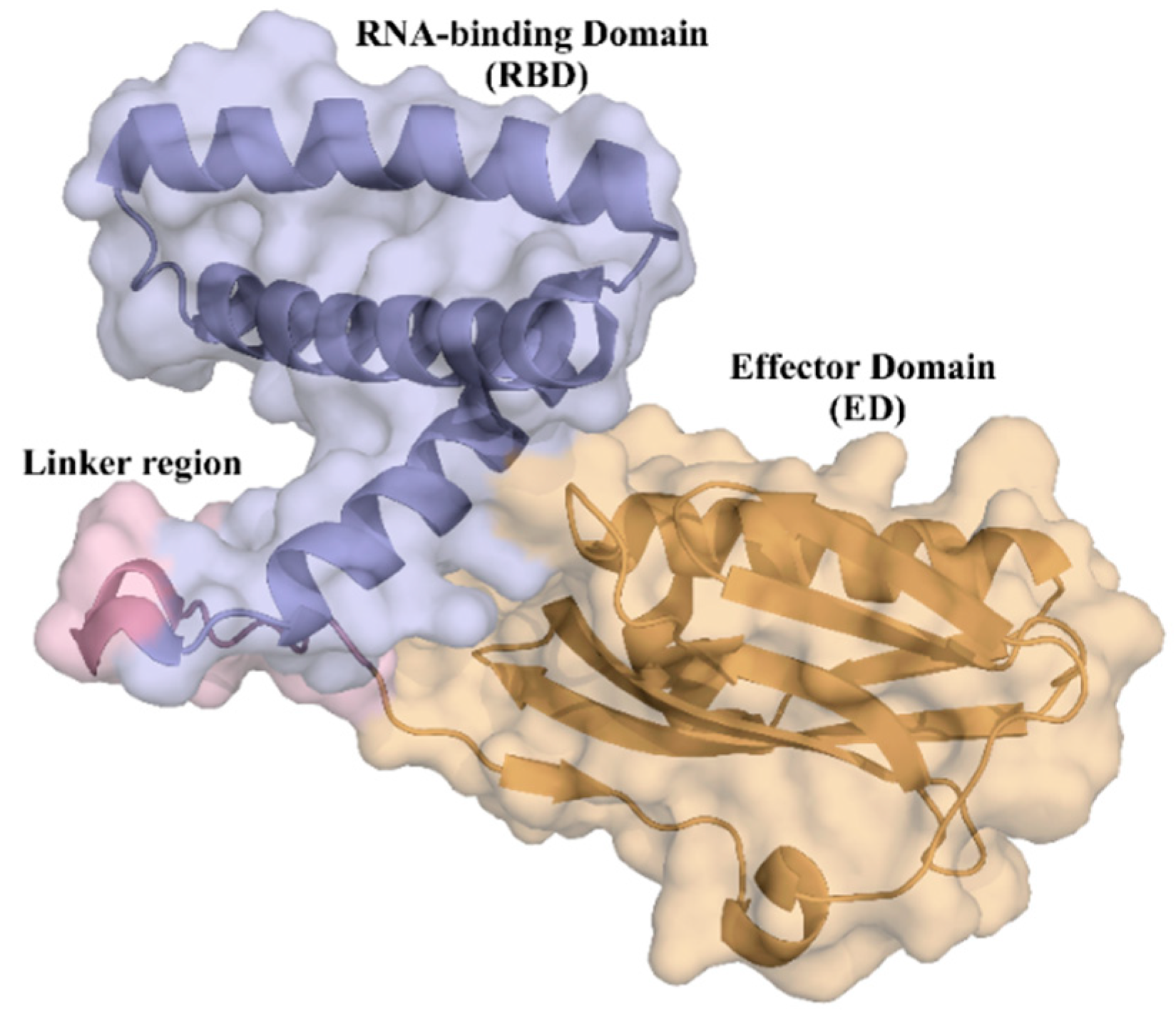
Figure 1. Cartoon depiction of the tridimensional crystal structure of the full-length monomer of NS1 from H6N6 influenza virus with its two structural domains highlighted (Protein Data Bank (PDB) access number 4OPH): the RNA-binding domain (blue) comprising three α-helices; and the effector domain (orange) with α-helices and β-strands. The two domains are connected by a linker region (pink).
Ijms 24 02977 g001
Figure 2. Root mean square fluctuation (RMSF) of NS1 Cα atoms obtained from six independent molecular dynamics simulations. (a) RMSF of RNA-binding domain (RBD). (b) RMSF of effector domain (ED). A diagram representing the secondary structure of the protein along its amino acid sequence is shown at the bottom of the figure (α-helices represented as green coils and β-strands as pink arrows).
Ijms 24 02977 g002
Figure 3. Druggability score of all the pockets identified in full-length NS1 over the sampled conformations obtained for six independent MD simulations.
Ijms 24 02977 g003aIjms 24 02977 g003b
Figure 4. Principal components analysis (PCA) of the physico-chemical properties used to calculate the druggability score with Fpocket.
Ijms 24 02977 g004
Figure 5. Structure of the NS1 subunit highlighting the pockets with the highest druggability score. (a) Snapshot of the 97 ns of run 2 with a pocket in the same color represented in mesh with a druggability score of 0.947. (b) Snapshot of the 99 ns of run 3 with a pocket in the same color represented in mesh with a druggability score of 0.946.
Ijms 24 02977 g005
Figure 6. Cα atoms root mean square deviation (RMSD) calculated over the MD simulation trajectories for the complex between the effector domain (ED) of NS1 and the iSH2 domain of the p85β subunit of human PI3K (PDB ID: 6OX7).
Ijms 24 02977 g006
Figure 7. Cα atoms RMSD calculated over the MD trajectories for the complex between NS1 and the coiled-coil domain of human TRIM25.
Ijms 24 02977 g007
Figure 8. Cα atoms RMSD for the residues in the interface of the complex between NS1 and PI3K, considering the residues from both NS1 and PI3K (a) or just the residues from NS1 (b).
Ijms 24 02977 g008
Figure 9. Cα atoms RMSD for the residues in the interface of the complex between NS1 and TRIM25, considering the residues from both NS1 and TRIM25 (a) or just the residues from NS1 (b).
Ijms 24 02977 g009
Figure 10. RMSF of NS1 interfacial residues. (a) RMSF of the NS1 interfacial residues with PI3K; (b) RMSF of the NS1 interfacial residues with TRIM25.
Ijms 24 02977 g010
Figure 11. Intermolecular contact maps of the complexes between (a) NS1 and PI3K; (b) NS1 and the chain I of TRIM25; and (c) NS1 and the chain N of TRIM25.
Ijms 24 02977 g011
Figure 12. Cartoon representation of (a) crystal structure of the complex between ED-NS1 and p85β subunit of PI3K (PDB ID: 6OX7); (b) crystal structure of the complex between NS1 and the coiled-coil domain of TRIM25 (PDB ID: 5NT2).
Ijms 24 02977 g012
Table 1. Amino acid residues constituting the interfacial region in the NS1-PI3K complex.
NS1 ResiduesPI3K Residues
Ser 87His 450
Arg 88Met 568
Tyr 89Arg 571
Thr 91Lys 572
Met 93Arg 574
Leu 95Asp 575
Glu 96Gln 576
Glu 97Leu 578
Met 98Val 579
Ser 99Trp 580
Arg 100Thr 582
Asp 101Gln 583
Trp 102Lys 584
Cys 116Ala 586
Arg 118Arg 587
Asn 133Gln 588
Phe 134Lys 589
Ser 135Ile 591
Asn 133Asn 592
Phe 134Leu 595
Ser 135
Glu 142
Thr 143
Leu 144
Ile 145
Leu 146
Arg 148
Glu 159
Ser 161
Pro 162
Leu 163
Pro 164
Ser 165
Table 2. Amino acid residues constituting the interfacial region in the NS1-TRIM25 complex.
NS1 ResiduesTRIM25
Ser 87Val 208 (chain N)
Arg 88Ser 211 (chain N)
Tyr 89Gln 212 (chain N)
Leu 90Asn 214 (chain N)
Thr 91Gly 215 (chain N)
Met 93Arg 218 (chain N)
Thr 94Ala 219 (chain N)
Leu 95Asp 221 (chain N)
Glu 96Asp 222 (chain N)
Met 98Val 223 (chain N)
Ser 99Asn 225 (chain N)
Glu 101Arg 226 (chain N)
Arg 118Asp 229 (chain N)
Asn 133Met 232 (chain N)
Ser 135Arg 236 (chain N)
Ile 145Phe 274 (chain I)
Leu 146Ile 277 (chain I)
Arg 148Leu 281 (chain I)
Glu 159Val 322 (chain I)
Ser 161Tyr 323 (chain I)
Pro 162Ile 324 (chain I)
Pro 164Pro 325 (chain I)
Glu 326 (chain I)
Val 327 (chain I)
Table 3. Stable pairwise residue–residue interactions at the interface of the complex between NS1 and PI3K.
NS1 ResiduesPI3K ResiduesResidence Time Ratio
Tyr 89Met 5681.00
Tyr 89Lys 5721.00
Tyr 89Asp 5751.00
Tyr 89Arg 5711.00
Ser 135Lys 5721.00
Ser 99Leu 5781.00
Ser 99Ile 5911.00
Met 98Leu 5781.00
Met 98Asp 5751.00
Leu 146Val 5791.00
Leu 95Leu 5951.00
Leu 95Leu 5781.00
Leu 95His 4501.00
Leu 95Arg 5741.00
Ile 145Val 5791.00
Ile 145Lys 5721.00
Ile 145Gln 5761.00
Ile 145Asp 5751.00
Asn 133Asp 5751.00
Thr 91Arg 5710.99
Asp 101Gln 5880.99
Ser 99Gln 5880.98
Met 98Val 5790.98
Leu 95Asp 5750.98
Arg 148Thr 5820.98
Ser 161Gln 5830.97
Pro 162Gln 5830.97
Arg 100Gln 5880.97
Glu 142Lys 5720.96
Thr 143Lys 5720.95
Pro 162Val 5790.95
Tyr 89Arg 5740.94
Asp 101Arg 5870.93
Thr 91Asp 5750.92
Ser 161Val 5790.92
Ser 87Lys 5720.92
Thr 143Gln 5760.90
Ser 99Leu 5950.90
Glu 96Gln 5880.87
Ser 99Thr 5820.86
Arg 88Met 5680.85
Met 98Thr 5820.84
Asp 101Lys 5890.82
Leu 163Gln 5830.78
Tyr 89Gln 5690.74
Leu 95Gln 5880.73
Ser 99Asn 5920.72
Asp 101Ala 5860.72
Table 4. Stable pairwise residue–residue interactions at the interface of the complex between NS1 and TRIM25 chain N.
NS1 ResiduesTRIM25 ResiduesResidence Time Ratio
Tyr 89Asp 2291.00
Tyr 89Asp 2221.00
Tyr 89Asn 2251.00
Tyr 89Arg 2261.00
Thr 94Arg 2261.00
Thr 91Asp 2221.00
Thr 91Arg 2261.00
Ser 161Arg 2181.00
Ser 99Ala 2191.00
Pro 162Arg 2181.00
Met 98Asp 2221.00
Met 98Arg 2261.00
Met 98Arg 2181.00
Met 98Ala 2191.00
Met 93Arg 2261.00
Leu 146Arg 2181.00
Leu 95Val 2231.00
Leu 95Asp 2221.00
Leu 95Arg 2261.00
Ile 145Asp 2221.00
Ile 145Asp 2211.00
Ile 145Asn 2251.00
Ile 145Arg 2181.00
Glu 101Gln 2121.00
Asn 133Asp 2221.00
Asn 133Arg 2261.00
Leu 146Gly 2150.95
Ser 135Asn 2250.94
Arg 88Asp 2290.94
Met 98Gly 2150.93
Arg 118Ser 2110.93
Arg 148Gly 2150.87
Leu 163Arg 2180.84
Ser 99Ala 2160.83
Ser 161Asn 2140.82
Pro 164Arg 2180.81
Arg 88Arg 2360.75
Ser 87Met 2320.74
Ile 145Ala 2190.74
Leu 146Ala 2190.71
Table 5. Stable pairwise residue–residue interactions at the interface of the complex between NS1 and TRIM25 chain I.
NS1 ResiduesTRIM25 ResiduesResidence Time Ratio
Ser 99Leu 2811.00
Ser 99Ile 3241.00
Leu 95Pro 3251.00
Leu 95Phe 2741.00
Leu 95Ile 3241.00
Leu 95Ile 2771.00
Glu 96Ile 3241.00
Leu 95Val 3270.99
Ser 99Lys 2840.97
Leu 95Leu 2810.97
Leu 95Glu 3260.94
Glu 101Lys 2840.83
Glu 101Lys 3200.78
Table 6. NS1 residues participating in the most stable NS1-PI3K interfacial pairwise interactions.
ResiduesNumber of Pairwise Residue–Residue Interactions
Y896
L956
S996
M984
D1014
I1454
T912
T1432
S1612
P1622
S871
R881
E961
R1001
N1331
S1351
E1421
L1461
R1481
L1631
Table 7. NS1 residues participating in the most stable NS1-TRIM25 interfacial pairwise interactions.
ResiduesNumber of Pairwise Residue–Residue Interactions
L9510
M985
S995
I1455
Y894
E1013
L1463
R882
T912
N1332
S1612
S871
M931
T941
E961
R1181
S1351
R1481
P1621
L1631
P1641
Table 8. Hydrogen bonds formed at the interface of the NS1-PI3K complex.
Residue NS1-EDResidue PI3K-p85βResidence Time (%)
P162Q58326.97
Y89D57576.22
N133D5754.7
T91D5750.3
S99Q5881.9
S87K57214.29
D101Q58842.66
D101Q5880.3
D101Q5884.6
R118Q5833.6
D101R58720.78
P164W5800.3
Y89R5710.2
S165K5840.2
S161Q5831.3
S165S4340.1
T143K5720.9
S99N5920.3
S161Q5839.19
E96Q58842.46
S99Q58822.78
R88N5611.4
T91R5710.4
S87Q5690.1
A86Q5690.1
A86K5720.1
W102R5870.1
E159Q5830.3
M98T5820.9
E142K5726.59
S135K5721.5
Y89M5682
M93R5710.1
Disclaimer/Publisher’s Note: The statements, opinions and data contained in all publications are solely those of the individual author(s) and contributor(s) and not of MDPI and/or the editor(s). MDPI and/or the editor(s) disclaim responsibility for any injury to people or property resulting from any ideas, methods, instructions or products referred to in the content.